Author's personal copy Eye Movement Abnormalities in Multiple Sclerosis
15
Author's personal copy Eye Movement Abnormalities in Multiple Sclerosis Sashank Prasad, MD a, *, Steven L. Galetta, MD b Multiple sclerosis (MS) is an inflammatory condition that affects the central nervous system myelin and is capable of causing a host of neurologic deficits Eye move- ment abnormalities are a common manifestation either at the onset or during the course of the disease. 1 In fact, eye movement abnormalities in MS often correlate with over-all disability from the disease. 2 In this article the authors discuss the disturbances of gaze shifting, gaze holding, and ocular alignment that occur in MS, with an emphasis on underlying neuroanatomic principles and pathologic find- ings on examination. Modern neuroimaging has revolutionized the ability to diagnose MS at its earliest stages and to monitor subclinical, silent disease progression. Although MRI has become an indispensible tool in the management of patients with MS, it is not completely sensitive in detecting lesions and therefore does not replace a careful neurologic examination. For example, small lesions in the brainstem may produce overt clinical abnormalities yet fall below the threshold for detection by MRI. This potential dissociation between clinical and radiographic findings further emphasizes the importance of careful clinico-anatomic localization. The proper assessment of subtle visual disturbances often critically impacts rational clinical decisions in the management of patients with MS. INTERNUCLEAR OPHTHALMOPLEGIA Internuclear ophthalmoplegia (INO) refers to disruption of rapid, coordinated, hori- zontal saccades by slowed or limited adduction. 3–6 Conjugate adduction in normal a Division of Neuro-Ophthalmology, Department of Neurology, Brigham and Women’s Hospital, Harvard Medical School, 75 Francis Street, Boston, MA 02115, USA b Division of Neuro-Ophthalmology, Department of Neurology, Hospital of the University of Pennsylvania, 3W Gates Building, 3400 Spruce Street, Philadelphia, PA 19104, USA * Corresponding author. E-mail address: [email protected] KEYWORDS Multiple sclerosis Eye movement abnormalities Neuroanatomy Neuroimaging Neurol Clin 28 (2010) 641–655 doi:10.1016/j.ncl.2010.03.006 neurologic.theclinics.com 0733-8619/10/$ – see front matter ª 2010 Elsevier Inc. All rights reserved.
-
Upload
uni-potsdam -
Category
Documents
-
view
1 -
download
0
Transcript of Author's personal copy Eye Movement Abnormalities in Multiple Sclerosis

Author's personal copy
Eye MovementAbnormalit ies inMultiple Sclerosis
Sashank Prasad, MDa,*, Steven L. Galetta, MDb
Multiple sclerosis (MS) is an inflammatory condition that affects the central nervoussystem myelin and is capable of causing a host of neurologic deficits Eye move-ment abnormalities are a common manifestation either at the onset or during thecourse of the disease.1 In fact, eye movement abnormalities in MS often correlatewith over-all disability from the disease.2 In this article the authors discuss thedisturbances of gaze shifting, gaze holding, and ocular alignment that occur inMS, with an emphasis on underlying neuroanatomic principles and pathologic find-ings on examination.
Modern neuroimaging has revolutionized the ability to diagnose MS at its earlieststages and to monitor subclinical, silent disease progression. Although MRI hasbecome an indispensible tool in the management of patients with MS, it is notcompletely sensitive in detecting lesions and therefore does not replace a carefulneurologic examination. For example, small lesions in the brainstem may produceovert clinical abnormalities yet fall below the threshold for detection by MRI. Thispotential dissociation between clinical and radiographic findings further emphasizesthe importance of careful clinico-anatomic localization. The proper assessment ofsubtle visual disturbances often critically impacts rational clinical decisions in themanagement of patients with MS.
INTERNUCLEAR OPHTHALMOPLEGIA
Internuclear ophthalmoplegia (INO) refers to disruption of rapid, coordinated, hori-zontal saccades by slowed or limited adduction.3–6 Conjugate adduction in normal
a Division of Neuro-Ophthalmology, Department of Neurology, Brigham and Women’sHospital, Harvard Medical School, 75 Francis Street, Boston, MA 02115, USAb Division of Neuro-Ophthalmology, Department of Neurology, Hospital of the University ofPennsylvania, 3W Gates Building, 3400 Spruce Street, Philadelphia, PA 19104, USA* Corresponding author.E-mail address: [email protected]
KEYWORDS
� Multiple sclerosis � Eye movement abnormalities� Neuroanatomy � Neuroimaging
Neurol Clin 28 (2010) 641–655doi:10.1016/j.ncl.2010.03.006 neurologic.theclinics.com0733-8619/10/$ – see front matter ª 2010 Elsevier Inc. All rights reserved.
Author's personal copy
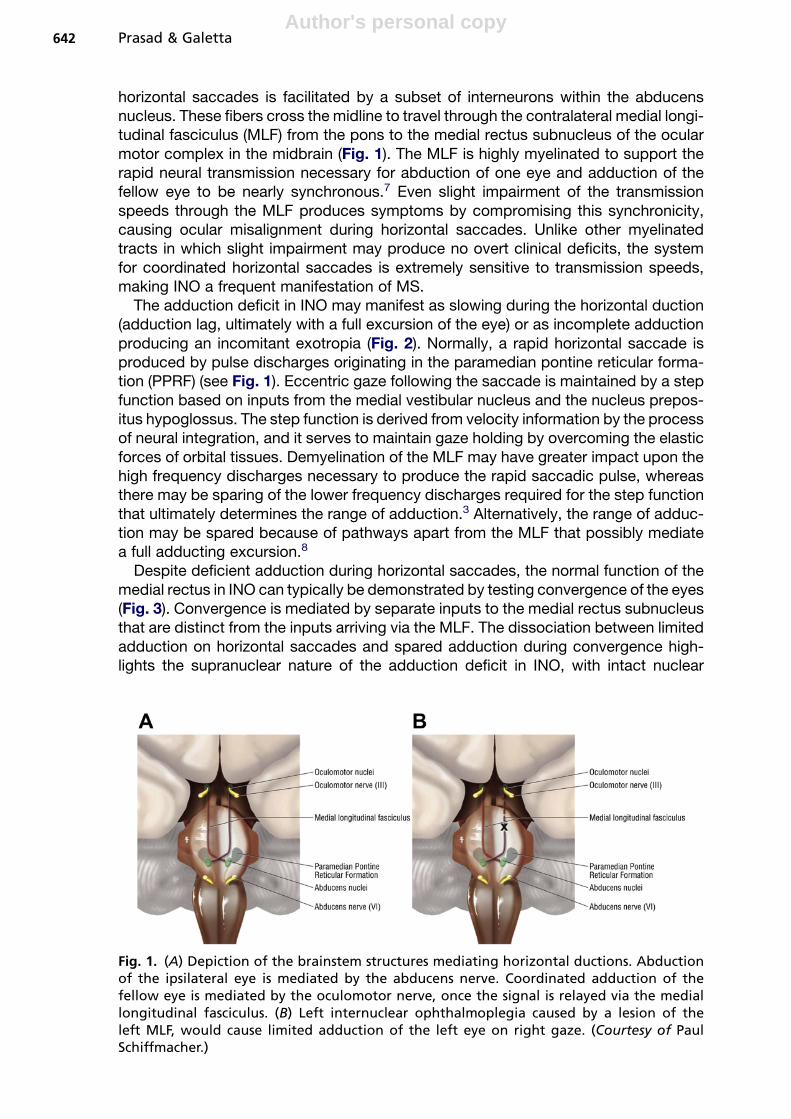
horizontal saccades is facilitated by a subset of interneurons within the abducensnucleus. These fibers cross the midline to travel through the contralateral medial longi-tudinal fasciculus (MLF) from the pons to the medial rectus subnucleus of the ocularmotor complex in the midbrain (Fig. 1). The MLF is highly myelinated to support therapid neural transmission necessary for abduction of one eye and adduction of thefellow eye to be nearly synchronous.7 Even slight impairment of the transmissionspeeds through the MLF produces symptoms by compromising this synchronicity,causing ocular misalignment during horizontal saccades. Unlike other myelinatedtracts in which slight impairment may produce no overt clinical deficits, the systemfor coordinated horizontal saccades is extremely sensitive to transmission speeds,making INO a frequent manifestation of MS.
The adduction deficit in INO may manifest as slowing during the horizontal duction(adduction lag, ultimately with a full excursion of the eye) or as incomplete adductionproducing an incomitant exotropia (Fig. 2). Normally, a rapid horizontal saccade isproduced by pulse discharges originating in the paramedian pontine reticular forma-tion (PPRF) (see Fig. 1). Eccentric gaze following the saccade is maintained by a stepfunction based on inputs from the medial vestibular nucleus and the nucleus prepos-itus hypoglossus. The step function is derived from velocity information by the processof neural integration, and it serves to maintain gaze holding by overcoming the elasticforces of orbital tissues. Demyelination of the MLF may have greater impact upon thehigh frequency discharges necessary to produce the rapid saccadic pulse, whereasthere may be sparing of the lower frequency discharges required for the step functionthat ultimately determines the range of adduction.3 Alternatively, the range of adduc-tion may be spared because of pathways apart from the MLF that possibly mediatea full adducting excursion.8
Despite deficient adduction during horizontal saccades, the normal function of themedial rectus in INO can typically be demonstrated by testing convergence of the eyes(Fig. 3). Convergence is mediated by separate inputs to the medial rectus subnucleusthat are distinct from the inputs arriving via the MLF. The dissociation between limitedadduction on horizontal saccades and spared adduction during convergence high-lights the supranuclear nature of the adduction deficit in INO, with intact nuclear
Fig. 1. (A) Depiction of the brainstem structures mediating horizontal ductions. Abductionof the ipsilateral eye is mediated by the abducens nerve. Coordinated adduction of thefellow eye is mediated by the oculomotor nerve, once the signal is relayed via the mediallongitudinal fasciculus. (B) Left internuclear ophthalmoplegia caused by a lesion of theleft MLF, would cause limited adduction of the left eye on right gaze. (Courtesy of PaulSchiffmacher.)
Prasad & Galetta642
Author's personal copy
and infranuclear components of adduction. In some cases, however, dysfunction ofthe MLF is sufficiently rostral that the medial rectus subnucleus itself is impaired;therefore, impaired convergence will accompany impaired adduction on horizontalsaccades. In this setting, referred to as the anterior INO of Cogan, the distinction isblurred between INO and partial third nerve palsy.9
Patients with unilateral INO typically do not have significant exotropia in primarygaze, likely because of intact convergence tone. In contrast, bilateral MLF lesionsoften cause exotropia (see Fig. 3). This clinical presentation is described as theWEBINO (wall-eyed bilateral INO) syndrome.
The misalignment produced by INO may cause a variety of ophthalmic symptoms,including visual blurring, diplopia, loss of stereopsis, and asthenopia (eye fatigue).10
Normally there is cortical sensory suppression during saccades to eliminate blurfrom retinal slippage, but in INO visual blurring may occur because this mechanismfails to fully suppress inputs from the eye with slowed saccades.3 Visual symptomsare often proportional to the degree of INO, and patients with mild INO may be essen-tially asymptomatic. Because use-related fatigue and Uhtoff’s phenomenon (wors-ening symptoms with elevated body temperature) are common in patients with MS,the symptoms caused by INO may fluctuate over the course of the day.
Many patients with INO have normal horizontal pursuit, optokinetic, and vestibulo-ocular responses.11 These functions may be preserved because they are mediated bylower-frequency neural signals with transmission that is spared despite demyelinationof the MLF, or because they are also mediated by alternate connections between theabducens and oculomotor nuclei.
INO is often associated with a dissociated horizontal nystagmus most prominent inthe abducting eye (Fig. 4). The slow phase of nystagmus is opposite the direction ofattempted gaze, with quick saccades in the direction of attempted gaze. The
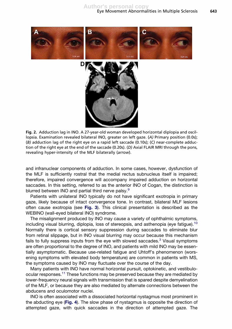
Fig. 2. Adduction lag in INO. A 27-year-old woman developed horizontal diplopia and oscil-lopsia. Examination revealed bilateral INO, greater on left gaze. (A) Primary position (0.0s);(B) adduction lag of the right eye on a rapid left saccade (0.10s); (C) near-complete adduc-tion of the right eye at the end of the saccade (0.20s). (D) Axial FLAIR MRI through the pons,revealing hyper-intensity of the MLF bilaterally (arrow).
Eye Movement Abnormalities in Multiple Sclerosis 643

Author's personal copy
nystagmus has a unique slow phase with an exponentially decaying wave form.3 Withgreater excursions, the amplitude or frequency of the nystagmus may increase.3
Several mechanisms may account for the abducting nystagmus in INO, and theexplanations are not necessarily mutually exclusive. One possibility is that there isa central adaptive response to reduce visual blurring. To attempt to overcome adduc-tion weakness, a compensatory increased saccadic pulse and step could occur (andwould affect both eyes by Hering’s law of dual innervation).3,12 Although the adaptiveresponse may improve the adduction of the paretic eye, it would disturb the abductionof the non-paretic eye in two ways. First, amplification of the pulse would lead tosaccadic hypermetria; second, pulse-step mismatch would lead to slow post-saccadic drift with exponential decay. According to this account, the phenomenonof abducting nystagmus in unilateral INO is expected to be greatest if patients habit-ually fixate with the paretic eye, leading to higher demands for central adaptation. Onthe other hand, if patients fixate with the non-paretic eye, the abducting nystagmusmay not be present.3 In keeping with these predictions, Zee and colleagues13 demon-strated that in some (but not all) patients with INO, prolonged patching (1–5days) of theparetic eye reduces the abducting nystagmus, whereas patching of the non-pareticeye increases it. On the other hand, temporary patching of one eye (or the perfor-mance of horizontal saccades in total darkness) does not mitigate the abducting
Fig. 3. Bilateral INO. A 47-year-old woman presented with horizontal diplopia. Examinationrevealed large-angle exotropia and bilateral INO. (A) Limited adduction of the left eye onright gaze. (B) Limited adduction of the right eye on left gaze. (C) Spared convergence ofthe eyes. (D) Axial FLAIR MRI revealed numerous areas of white matter hyper-intensity(for example, arrow). (E) However, no signal abnormality was detected in the region ofthe MLF in the pons or midbrain. (F) Improvement of INO after 2 months. Improved righteye adduction. (G) Full left gaze.
Prasad & Galetta644

Author's personal copy
nystagmus, suggesting that the nystagmus is generated not by online target-positionerror signals but by a stored, long-term adaptive mechanism.3
A central adaptive mechanism, however, does not fully account for abductingnystagmus, because not all patients demonstrate the predicted changes followingpatching.13 An alternate explanation proposes the disruption of inhibitory fibers thattravel in the MLF and are postulated to cross in the midbrain to arrive at the antagonistmedial rectus of the contralateral eye.14 By this account, impaired inhibition of thesemedial rectus motoneurons reduces the abducting step function in that eye andcauses a slow movement back from the abducted position. A corrective abductingsaccade follows, and the repeating cycle generates abducting nystagmus. This expla-nation, however, would predict hypometric abducting saccades, rather than thehypermetric abducting saccades commonly seen.3
Yet another explanation for abducting nystagmus in INO is that injury to additionalstructures outside the MLF may directly lead to an asymmetric gaze-holding distur-bance, which would manifest with greater severity in the non-paretic eye. By thisaccount, however, the abducting nystagmus would have a typical saw-tooth waveform(in which the slow component has constant velocity directly relating to insufficient gaze-holding mechanisms), rather than the exponentially decaying waveform that is seen.3
In severe INO, the affected eye may demonstrate abduction slowing in addition toadduction slowing. A potential explanation for reduced abduction velocity is thatnormal abduction depends upon appropriate inhibition of the antagonist medial rectusof the same eye, which could be compromised by an MLF lesion.15 An alternativeexplanation, however, is that patients with INO and abduction slowing in fact havemore extensive pontine lesions not limited to the MLF, but also potentially involvingthe abducens nucleus, fascicle, or other structures.6,16
Impaired horizontal saccades are not the only manifestation of an MLF lesion. TheMLF also contains fibers mediating many vertical eye movements (pursuit, vestibular,and otolithic pathways); corresponding impairments of vertical gaze thereforefrequently accompany INO.11,17,18 Impaired vertical pursuit may manifest as ‘‘staircas-ing’’ ductions interrupted by horizontal movements. Patients with bilateral INO mayhave marked impairment of vertical gaze holding, resulting in primary-position orgaze-evoked vertical nystagmus. In contrast to the exponentially decaying slow wave-form of abducting nystagmus, vertical nystagmus in INO has a typical saw-tooth
Fig. 4. Eye movement recordings in a patient with left INO demonstrating abductingnystagmus in the right eye while attempting sustained right gaze. Note the cycles of hyper-metric abduction (black arrow) followed by an exponentially decaying slow waveform backtoward primary position (gray arrow). The left eye demonstrates lower-amplitude, dysconjugateright-beatingnystagmuswithoutover shoots. (AdaptedfromBalohRW,YeeRD,HonrubiaV. Inter-nuclear ophthalmoplegia. I. Saccades and dissociated nystagmus. Arch Neurol 1978a;35:484–9;with permission. Copyright ª 1978 American Medical Association. All rights reserved.
Eye Movement Abnormalities in Multiple Sclerosis 645

Author's personal copy
pattern caused by insufficient gaze-holding mechanisms. Impairment of the utricularpathways within the MLF may additionally lead to vertical misalignment of the eyes,in the form of skew deviation or the full ocular tilt reaction (Fig. 5).
The precise measurement of eye movements, using methods, such as infrared ocu-lography, allows highly accurate detection and quantification of INO. Furthermore,these methods serve as a gold standard by which the accuracy of the bedside exam-ination can be assessed.19 Using this method, Frohman and colleagues found thatsevere INO was accurately detected by virtually all physician observers (regardlessof level of training), but that milder INO was missed by many physicians other thantrained neuro-ophthalmologists.
Various metrics from eye movement recordings have been used to quantify INO.These include the versional dysconjugacy index (VDI), which compares the peakvelocities for abduction in one eye to adduction in the other.20,21 This measure hasthe benefit of cancelling intra- and interindividual variations of absolute saccade veloc-ities (caused by fatigue, for example). The VDI has also been assessed by a Z scoreand histogram analysis, which is a statistical method to better distinguish normaland abnormal results.22 VDI measures use velocity rather than final amplitude becausethe extent of final amplitude in INO is often normal. The first-pass amplitude, on theother hand, evaluates the ratio of abducting and adducting eye position at the timethat the abducting eye has initially completed its saccade.23 Finally, recent studieshave employed a phase-plane analysis, which plots eye velocity directly as a functionof position, removing the effects of temporal variation that arise, for example, fromonset latency.24 Quantified measures of INO may provide a useful way to index theclinical effects of fatigue and Uhtoff’s phenomenon, and ultimately may providea method to objectively assess potential symptomatic treatments.25
Patients with INO frequently have a corresponding abnormality in the pons ormidbrain that is detectable by MRI.26 Frohman and colleagues studied 58 subjectswith MS and INO and found that the sensitivity of proton density imaging, T2-weightedimaging, and fluid-attenuated inversion recovery (FLAIR) imaging was 100%, 88%,and 48%, respectively. It is not clear from this study, however, how the severity ofINO relates to these MRI findings; cases of mild INO may have a higher rate of normalimaging. Furthermore, because patients with MS without INO were not included in thisstudy, the exact specificity of these MRI abnormalities is not known. In some cases,MRI signal abnormality in this region may not have a clinical correlate. Another MRImeasure that has been studied in INO is diffusion tensor imaging (DTI), in which thespatial constraints of water diffusion allow assessment of the integrity of white-mattertracts.27 Fox and colleagues found a modest correlation between INO severity (gradedby VDI) and mean white matter diffusivity in the MLF, showing that DTI measures mayserve as a surrogate marker of brain-tissue integrity.
Fig. 5. A 30-year-old woman with MS developed horizontal, vertical, and torsional diplopia.Examination revealed right INO (with incomitant exotropia greatest in left gaze) and skewdeviation (with comitant right hypertropia in all directions of gaze). A demyelinating lesionof the right MLF accounts for this pattern of misalignment.
Prasad & Galetta646

Author's personal copy
NUCLEAR OR FASCICULAR OCULAR MOTOR PALSY
MS may cause acquired strabismus in the form of nuclear or fascicular palsy of one ofthe three ocular motor nerves.16,28,29 In sixth nerve palsy, a fascicular lesion causesimpaired abduction of the ipsilateral eye with spared adduction of the fellow eye(Fig. 6). A lesion of the sixth nerve nucleus, however, causes ipsiversive gaze palsy,consisting of combined deficits of ipsilateral abduction and contralateral adduction.Rarely, a pontine lesion may affect the sixth nerve nucleus (or pontine paramedianreticular formation) and the ipsilateral MLF. The effect of this lesion is a combined ipsi-versive gaze palsy and an ipsilateral INO, referred to as a ‘‘one-and-a-half’’ syndrome,and the only spared horizontal eye movement is abduction of the contralateral eye onlateral gaze (see Fig. 6).30,31
In third nerve palsy, a fascicular lesion may cause partial deficits of elevation,depression, adduction, or lid elevation in the ipsilateral eye.30 In rare cases, theselesions may be highly selective and cause weakness of a single muscle. Also,a discrete lesion of the third nerve fascicle may mimic superior or inferior divisionalthird nerve palsy (which more often localizes to the anterior cavernous sinus or orbit).32
A nuclear third nerve lesion causes bilateral superior rectus weakness (in addition tothe ipsilateral deficits), because the superior rectus subnucleus issues fibers thattravel through the contralateral nucleus to join the contralateral nerve. In addition,nuclear third nerve palsy often causes bilateral ptosis, because the unpaired centralcaudal nucleus supplies both levator palpebrae muscles.
Normal eyelid position is maintained by inputs to the levator palpebrae. The moto-neurons to both levator palpebrae muscles arise from the unpaired central caudatesubnucleus (CCN) of the ocular motor complex. Lesions affecting these fasciclesmay cause unilateral ptosis, often in addition to other deficits of partial third nerve
Fig. 6. One-and-a-half syndrome from right pontine lesion in a patient with MS. (A) In addi-tion to a complete right gaze palsy (from involvement of the right abducens nucleus), therewas deficient adduction of the right eye on attempted left gaze (from involvement of theright MLF). Left eye abduction is the only spared horizontal eye movement. (B) Axial T2-weighted MRI revealing a right pontine lesion (arrow). (From Frohman TC, Galetta S, FoxR, et al. Pearls & Oy-sters: The medial longitudinal fasciculus in ocular motor physiology.Neurol 2008;70:e57–67; with permission.)
Eye Movement Abnormalities in Multiple Sclerosis 647

Author's personal copy
palsy. Rarely, isolated unilateral or bilateral ptosis may occur.33 The CCN is under thecontrol of the nearby M-group cells, which receive tonic inhibitory inputs from thenucleus of the posterior commissure. Disruption of these inputs in the dorsal midbraincauses eyelid retraction (Collier’s sign). The M-group cells couple the contractions ofthe levator palpebrae muscles to those of the vertically acting eye muscles on thebasis of inputs from the superior colliculi.34 Selective lesions in this location may causeabnormal, dissociated lid and eye movements. Blepharospasm (forceful, involuntarycontractions of the orbicularis oculi) may occur following MS lesions of the brainstem,possibly caused by denervation supersensitivity of the facial nucleus or disinhibition offacial nerve relexes.35 Painful, gaze-evoked blepharoclonus that principally involvesthe orbicularis oculi may also occur in MS, perhaps relating to ephaptic spread ofimpulses. MRI studies of these patients, however, have not revealed a consistentlocalization of lesions.36,37
A lesion of the fourth nerve nucleus or proximal fascicle causes hyperdeviation ofthe contralateral eye. Because of the direction of action of the superior obliquemuscle, the hyperdeviation of a fourth nerve palsy is greatest in contralateral gazeand with ipsilateral head tilt. Rarely, a solitary lesion may cause combined INO andcontralateral fourth nerve palsy, because of the anatomic proximity of the MLF andthe fourth nerve nucleus and fascicle (Fig. 7).38
SKEW DEVIATION
As mentioned earlier, it is possible for skew deviation to accompany INO because theMLF contains utricular pathways maintaining vertical eye position in addition to inter-neurons from the abducens nucleus to the medial rectus subnucleus (see Fig. 5). Incases where there is selective damage of the utricular pathways, however, skew devi-ation will occur in the absence of INO. Imbalance of utricular inputs leads to a cyclo-vertical misalignment of the eyes, typically with a comitant vertical deviation that doesnot follow a pattern characteristic of third or fourth nerve palsy. With a pontine lesion,the ipsilateral eye is lower, and with a midbrain lesion, the ipsilateral eye is higher.Typically, there is relative intorsion of the higher eye (because intorsion of the higher
Fig. 7. A 41-year-old man with combined right INO and left fourth nerve palsy. (A) Axial T2-weighted MRI revealed a lesion in the right dorsal midbrain (arrow), adjacent to the sylvianaqueduct (arrowhead). (B) Schematic diagram of the lesion location and structures involved,including the right fourth nerve nucleus and fascicle before its decussation (1) and the rightMLF (2). Other structures shown are the sylvian aqueduct (3), fourth nerve (4), decussationof the fourth nerve fibers (5), and the inferior colliculi (6). (From Vanooteghem P, Dehaene I,Van ZandyckeM, et al. Combined trochlear nerve palsy and internuclear ophthalmoplegia.Arch Neurol 1992;49:108–9; with permission. Copyright ª 1992 American Medical Associa-tion. All rights reserved.)
Prasad & Galetta648
Author's personal copy
eye exceeds extorsion of the lower eye). In the ocular tilt reaction (OTR), the hyperde-viation is accompanied by head tilt away from the higher eye. In addition to diplopia,many patients with skew deviation or OTR describe tilting of the subjective visualvertical.39
NYSTAGMUS
Gaze holding is mediated by velocity-position neural integrators; for horizontal gaze,these critical structures are in the medulla (the medial vestibular nuclei and the nucleusprepositus hypoglossus) and for vertical gaze, they are in the midbrain (the interstitialnuclei of Cajal).40 The superior vestibular nuclei may also influence vertical gazeholding via connections through the MLF. The neural integrators make projectionsto the cerebellar tonsils (the flocculus and paraflocculus) that function to fine-tunevelocity-position coding and maintain normal gaze holding.
Dysfunction of the neural integrators leads to impaired gaze holding and pathologicnystagmus. One common pattern is gaze-evoked nystagmus, which has a jerk wave-form in which a slow drift back toward primary position is followed by a quick saccadeto re-establish eccentric gaze. More significant gaze-holding impairments lead toprimary position jerk nystagmus. Downbeat nystagmus often results from a cerebellaror cervico-medullary lesion that disrupts projections from the posterior semicircularcanal, resulting in tonic upward deviation of the eyes with fast downward correctivemovements. In contrast, upbeat nystagmus occurs more rarely, following pontome-dullary or pontomesencephalic lesions that disrupt projections from the anterior semi-circular canal. Rebound nystagmus refers to a transient jerk nystagmus that occursupon returning from eccentric gaze to primary position, with the fast phase awayfrom the previous direction of lateral gaze. Acquired periodic alternating nystagmushas a shifting null point, with the direction of nystagmus changing every 90 to 120seconds and an intervening rest period of 5 to 10 seconds.41,42 This condition mayresult from damage to the cerebellar centers that maintain velocity storage mecha-nisms and the stability of the vestibulo-ocular reflex (VOR). An asymmetric, jerkform of see-saw nystagmus, in which there is intorsion and elevation of one eyewith synchronous extorsion and depression of the other eye, may follow midbrainlesions that disrupt inputs to vertical gaze-holding centers.43,44
Another common pattern is pendular nystagmus, which is often the result ofdamage to the interconnections between the brainstem neural integrators and thegaze-holding centers of the cerebellar tonsils.45,46 Demyelination and conductionslowing along these pathways, which is common in MS, sufficiently disrupts theirnormal function and leads to abnormal, spontaneous firing patterns. The onset ofnystagmus may follow the lesion by several months, suggesting that neural deafferen-tation may contribute to the pathophysiology. Combined pendular nystagmus andpalatal tremor often result from a lesion in the Guillain-Mollaret triangle (includingthe dentate nucleus, superior cerebellar peduncle, red nucleus, central tegmentaltract, inferior olive, and finally the inferior cerebellar peduncle).47,48 There is often infe-rior olivary hypertrophy, which may be evident on MRI. Monocular visual loss also maycontribute to acquired dissociated pendular nystagmus.49
Several drugs are available to attempt to dampen nystagmus and may providesymptomatic benefit to patients with MS.50 Clonazepam, baclofen, gabapentin, andmemantine are reasonably well tolerated and are effective in some patients.51 Theaminopyridines have been studied in reducing MS-associated symptoms,52 andmay be specifically helpful in reducing nystagmus. Many forms of pathologicnystagmus are thought to be caused by reduced physiologic inhibition of the
Eye Movement Abnormalities in Multiple Sclerosis 649

Author's personal copy
vestibular nuclei by cerebellar purkinje fibers, and the aminopyridines are potassium-channel blockers that putatively facilitate action potentials in purkinje cells. Both4-aminopyridine53 and 3,4-diaminopyridine54 have shown efficacy in controlled trials,but side effects including nausea, vomiting, and seizures limit the use of thesemedications.
SACCADIC ACCURACY
The accuracy of saccadic excursions is under the control of inputs from the posteriorfastigial nuclei and dorsal vermis in the cerebellum, which calibrate the size of thesaccadic pulse. Dysfunction of these pathways leads to saccadic dysmetria; hyper-metric saccades result from damage to the deep nuclei and hypometric saccadesresult from damage to the vermis alone. Furthermore, dysmetric saccades in onedirection of gaze can occur from unilateral lesions.55 For example, a lesion of the infe-rior cerebellar peduncle (affecting the climbing fibers) may cause contralateral hypo-metric saccades, which occurs because of reduced stimulation of the ipsilateralfastigial nucleus, and consequently reduced stimulation of the contralateral PPRF(Fig. 8). In contrast, a lesion of the Hook bundle region near the superior cerebellarpeduncle will cause contralateral hypermetric saccades (Fig. 9).55 The reason forcontralateral hypermetric saccades is that fibers from the fastigial nucleus to the
Fig. 8. Schematic depiction of the pathways controlling saccadic accuracy. Fibers arisingfrom the inferior olive ascend through the contralateral inferior cerebellar peduncle andsynapse on the dentate nucleus. Then, fibers ascend to traverse the contralateral superiorcerebellar peduncle within the uncinate bundle of Russell and reach the paramedianpontine reticular formation. (Courtesy of Paul Schiffmacher Medical Illustrator, ThomasJefferson University.)
Prasad & Galetta650

Author's personal copy
contralateral PPRF first travel across the midline superiorly to circle the superior cere-bellar peduncle before descending to the PPRF.56
SACCADIC INTRUSIONS
Stable fixation is maintained by pause-cell neurons, which are located in the pontineraphe between the two abducens nuclei. They prevent the occurrence of unwantedsaccadic pulses by tonically inhibiting the saccadic premotor burst neurons in thePPRF and the midbrain. Dysfunction of pause cells leads to extraneous saccadesinterrupting fixation. A square-wave jerk, for example, is a 1- to 5�-movement awayfrom and back to the primary position, with an inter-saccadic latency of 150 to 200milliseconds. Saccadic interruptions with a larger excursion (up to 10–40�) anda shorter inter-saccadic latency (up to 80 milliseconds) are termed macro square-wave jerks. When large saccadic intrusions occur across the midline in a to-and-fropattern, they are termed macro-saccadic oscillations. In ocular flutter, back-to-backhorizontal saccades occur without an inter-saccadic latency. If the saccadic move-ments occur in the horizontal and vertical planes, they are termed opsoclonus.30 Inmicro-saccadic flutter, low-amplitude, back-to-back saccades occur but are gener-ally seen only on ophthalmoscopy or eye movement recordings.57
Fig. 9. Ipsilateral hypometric and contralateral hypermetric saccades following a lesion ofthe Hook bundle region of the superior cerebellar peduncle (SCP) in a patient with MS.(A) Axial FLAIR MRI revealed a lesion in the right SCP. (B) Eye movement recordings demon-strated hypometric saccades to the left (small arrows) and hypermetric saccades to the right(large arrows). (From Frohman EM, Frohman TC, Fleckenstein J, et al. Ocular contrapulsion inmultiple sclerosis: clinical features and pathophysiological mechanisms. J Neurol NeurosurgPsychiatry 2001a;70:688–92; with permission.)
Eye Movement Abnormalities in Multiple Sclerosis 651

Author's personal copy
IMPAIRED SMOOTH PURSUIT AND IMPAIRED SUPPRESSIONOF THE VESTIBULO-OCULAR REFLEX
Smooth pursuit movements function to minimize retinal slippage of a moving foveatedtarget. They are generated by cortical and subcortical areas, including V5/MST, thefrontal eye fields, the dorsolateral pontine nucleus, the cerebellar flocculus and dorsalvermis, the vestibular nuclei, and ultimately the ocular motor nuclei. Lesions to thesepathways are common in MS and often produce low-gain pursuit, in which eye move-ments are disproportionately slower than the moving target.58,59 Compensatorycatch-up saccades are generated to reestablish visual object tracking.
In natural circumstances, head movements often accompany eye movements tomaintain fixation of a moving target. In this situation, the vestibulo-ocular reflexmust be suppressed to maintain fixation. Lesions of the cerebellar flocculus commonlyimpair VOR cancellation, resulting in poor fixation of targets during dynamic head andeye movements. Suppression of the VOR can be assessed by having the subject viewthe thumb on their own outstretched arm while rotating their chair. If VOR cancellationis deficient, the intact VOR causes the eyes to drift opposite the direction of the headmovement, and compensatory catch-up saccades will occur.
SUMMARY
Several eye movement abnormalities occur commonly in MS. The demyelinatinglesions of MS can occur quite selectively within critical brainstem and cerebellar path-ways that mediate coordinated eye movements, gaze holding, and ocular alignment.Delayed neural transmission in these pathways may lead to INO, ocular motor palsy,ocular misalignment, pathologic nystagmus, impaired saccades, saccadic intrusions,or impaired pursuit. Detailed neuro-ophthalmic examination of patients with MS withvisual complaints often yields a diagnosis with highly specific neuroanatomic localiza-tion. In turn, a thorough evaluation may suggest targeted symptomatic therapies andenhance the ability to monitor disease progression.
REFERENCES
1. Frohman EM, Frohman TC, Zee DS, et al. The neuro-ophthalmology of multiplesclerosis. Lancet Neurol 2005;4:111–21.
2. Derwenskus J, Rucker JC, Serra A, et al. Abnormal eye movements predictdisability in MS: two-year follow-up. Ann N Y Acad Sci 2005;1039:521–3.
3. Baloh RW, Yee RD, Honrubia V. Internuclear ophthalmoplegia. I. Saccades anddissociated nystagmus. Arch Neurol 1978;35:484–9.
4. Paton L. Ocular palsies. Br J Ophthalmol 1921;5:250–69.5. Sauvineau C. Un nouveau type de paralyse associee des mouvements horiz-
ments horizontaux des yeux. Bull Soc Ophtalmol Fr 1895;13:524–34.6. Zee DS. Internuclear ophthalmoplegia: pathophysiology and diagnosis. Baillieres
Clin Neurol 1992;1:455–70.7. Weidenheim KM, Epshteyn I, Rashbaum WK, et al. Neuroanatomical localization
of myelin basic protein in the late first and early second trimester human foetalspinal cord and brainstem. J Neurocytol 1993;22:507–16.
8. Kommerell G. Unilateral internuclear ophthalmoplegia. The lack of inhibitoryinvolvement in medial rectus muscle activity. Invest Ophthalmol Vis Sci 1981;21:592–9.
9. Cogan DG. Internuclear ophthalmoplegia, typical and atypical. Arch Ophthalmol1970;84:583–9.
Prasad & Galetta652

Author's personal copy
10. Mills DA, Frohman TC, Davis SL, et al. Break in binocular fusion during headturning in MS patients with INO. Neurology 2008;71:458–60.
11. Baloh RW, Yee RD, Honrubia V. Internuclear ophthalmoplegia. II. Pursuit, optoki-netic nystagmus, and vestibulo-ocular reflex. Arch Neurol 1978;35:490–3.
12. Abel LA, Schmidt D, Dell’Osso LF, et al. Saccadic system plasticity in humans.Ann Neurol 1978;4:313–8.
13. Zee DS, Hain TC, Carl JR. Abduction nystagmus in internuclear ophthalmoplegia.Ann Neurol 1987;21:383–8.
14. Pola J, Robinson DA. An explanation of eye movements seen in internuclear oph-thalmoplegia. Arch Neurol 1976;33:447–52.
15. Feldon SE, Hoyt WF, Stark L. Disordered inhibition in internuclear ophthalmople-gia: analysis of eye movement recordings with computer simulations. Brain 1980;103:113–37.
16. Bronstein AM, Rudge P, Gresty MA, et al. Abnormalities of horizontal gaze.Clinical, oculographic and magnetic resonance imaging findings. II. Gazepalsy and internuclear ophthalmoplegia. J Neurol Neurosurg Psychiatry1990;53:200–7.
17. Evinger LC, Fuchs AF, Baker R. Bilateral lesions of the medial longitudinalfasciculus in monkeys: effects on the horizontal and vertical componentsof voluntary and vestibular induced eye movements. Exp Brain Res 1977;28:1–20.
18. Ranalli PJ, Sharpe JA. Vertical vestibulo-ocular reflex, smooth pursuit and eye-head tracking dysfunction in internuclear ophthalmoplegia. Brain 1988;111(Pt 6):1299–317.
19. Frohman TC, Frohman EM, O’Suilleabhain P, et al. Accuracy of clinical detectionof INO in MS: corroboration with quantitative infrared oculography. Neurology2003;61:848–50.
20. Flipse JP, Straathof CS, Van der Steen J, et al. Binocular saccadic eye move-ments in multiple sclerosis. J Neurol Sci 1997;148:53–65.
21. Ventre J, Vighetto A, Bailly G, et al. Saccade metrics in multiple sclerosis:versional velocity disconjugacy as the best clue? J Neurol Sci 1991;102:144–9.
22. Frohman EM, Frohman TC, O’Suilleabhain P, et al. Quantitative oculographiccharacterisation of internuclear ophthalmoparesis in multiple sclerosis: theversional dysconjugacy index Z score. J Neurol Neurosurg Psychiatry2002;73:51–5.
23. Frohman EM, O’Suilleabhain P, Dewey RB Jr, et al. A new measure of dysconju-gacy in INO: the first-pass amplitude. J Neurol Sci 2003;210:65–71.
24. Serra A, Liao K, Matta M, et al. Diagnosing disconjugate eye movements: phase-plane analysis of horizontal saccades. Neurology 2008;71:1167–75.
25. Davis SL, Frohman TC, Crandall CG, et al. Modeling Uhtoff’s phenomenonin MS patients with internuclear ophthalmoparesis. Neurology 2008;70:1098–106.
26. Frohman EM, Zhang H, Kramer PD, et al. MRI characteristics of the MLF in MSpatients with chronic internuclear ophthalmoparesis. Neurology 2001;57:762–8.
27. Fox RJ, McColl RW, Lee JC, et al. A preliminary validation study of diffusiontensor imaging as a measure of functional brain injury. Arch Neurol 2008;65:1179–84.
28. Bronstein AM, Morris J, Du Boulay G, et al. Abnormalities of horizontal gaze. Clin-ical, oculographic and magnetic resonance imaging findings. I. Abducens palsy.J Neurol Neurosurg Psychiatry 1990;53:194–9.
Eye Movement Abnormalities in Multiple Sclerosis 653

Author's personal copy
29. Moster ML, Savino PJ, Sergott RC, et al. Isolated sixth-nerve palsies in youngeradults. Arch Ophthalmol 1984;102:1328–30.
30. de Seze J, Vukusic S, Viallet-Marcel M, et al. Unusual ocular motor findings inmultiple sclerosis. J Neurol Sci 2006;243:91–5.
31. Frohman TC, Galetta S, Fox R, et al. Pearls & Oy-sters: the medial longitudinalfasciculus in ocular motor physiology. Neurology 2008;70:e57–67.
32. Ksiazek SM, Repka MX, Maguire A, et al. Divisional oculomotor nerve paresiscaused by intrinsic brainstem disease. Ann Neurol 1989;26:714–8.
33. Martin TJ, Corbett JJ, Babikian PV, et al. Bilateral ptosis due to mesencephaliclesions with relative preservation of ocular motility. J Neuroophthalmol 1996;16:258–63.
34. Horn AK, Buttner-Ennever JA. Brainstem circuits controlling lid-eye coordinationin monkey. Prog Brain Res 2008;171:87–95.
35. Jankovic J, Patel SC. Blepharospasm associated with brainstem lesions.Neurology 1983;33:1237–40.
36. Jacome DE. Blepharoclonus in multiple sclerosis. Acta Neurol Scand 2001;104:380–4.
37. Keane JR. Gaze-evoked blepharoclonus. Ann Neurol 1978;3:243–5.38. Vanooteghem P, Dehaene I, Van Zandycke M, et al. Combined trochlear nerve
palsy and internuclear ophthalmoplegia. Arch Neurol 1992;49:108–9.39. Serra A, Derwenskus J, Downey DL, et al. Role of eye movement examination and
subjective visual vertical in clinical evaluation of multiple sclerosis. J Neurol 2003;250:569–75.
40. Bhidayasiri R, Plant GT, Leigh RJ. A hypothetical scheme for the brainstemcontrol of vertical gaze. Neurology 2000;54:1985–93.
41. Keane JR. Periodic alternating nystagmus with downward beating nystagmus. Aclinicoanatomical case study of multiple sclerosis. Arch Neurol 1974;30:399–402.
42. Matsumoto S, Ohyagi Y, Inoue I, et al. Periodic alternating nystagmus in a patientwith MS. Neurology 2001;56:276–7.
43. Halmagyi GM, Aw ST, Dehaene I, et al. Jerk-waveform see-saw nystagmus due tounilateral meso-diencephalic lesion. Brain 1994;117(Pt 4):789–803.
44. Sandramouli S, Benamer HT, Mantle M, et al. See-saw nystagmus as the present-ing sign in multiple sclerosis. J Neuroophthalmol 2005;25:56–7.
45. Lopez LI, Gresty MA, Bronstein AM, et al. Acquired pendular nystagmus: oculo-motor and MRI findings. Acta Otolaryngol Suppl 1995;520(Pt 2):285–7.
46. Averbuch-Heller L, Zivotofsky AZ, Das VE, et al. Investigations of the pathogen-esis of acquired pendular nystagmus. Brain 1995;118(Pt 2):369–78.
47. Guillain G. The syndrome of synchronous and rhythmic palato-pharyngo-laryngo-oculo-diaphragmatic myoclonus. Proc R Soc Med 1938;31:1031–8.
48. Revol A, Vighetto A, Confavreux C, et al. [Oculo-palatal myoclonus and multiplesclerosis]. Rev Neurol (Paris) 1990;146:518–21 [in French].
49. Barton JJ, Cox TA. Acquired pendular nystagmus in multiple sclerosis: clinicalobservations and the role of optic neuropathy. J Neurol Neurosurg Psychiatry1993;56:262–7.
50. Rucker JC. Current treatment of nystagmus. Curr Treat Options Neurol 2005;7:69–77.
51. Shery T, Proudlock FA, Sarvananthan N, et al. The effects of gabapentin andmemantine in acquired and congenital nystagmus: a retrospective study. Br JOphthalmol 2006;90:839–43.
52. Solari A, Uitdehaag B, Giuliani G, et al. Aminopyridines for symptomatic treatmentin multiple sclerosis. Cochrane Database Syst Rev 2003;2:CD001330.
Prasad & Galetta654

Author's personal copy
53. Kalla R, Glasauer S, Schautzer F, et al. 4-aminopyridine improves downbeatnystagmus, smooth pursuit, and VOR gain. Neurology 2004;62:1228–9.
54. Strupp M, Schuler O, Krafczyk S, et al. Treatment of downbeat nystagmus with3,4-diaminopyridine: a placebo-controlled study. Neurology 2003;61(2):165–70.
55. Frohman EM, Frohman TC, Fleckenstein J, et al. Ocular contrapulsion in multiplesclerosis: clinical features and pathophysiological mechanisms. J Neurol Neuro-surg Psychiatry 2001;70:688–92.
56. Solomon D, Galetta SL, Liu GT. Possible mechanisms for horizontal gaze devia-tion and lateropulsion in the lateral medullary syndrome. J Neuroophthalmol1995;15:26–30.
57. Ashe J, Hain TC, Zee DS, et al. Microsaccadic flutter. Brain 1991;114(Pt 1B):461–72.
58. Mastaglia FL, Black JL, Collins DW. Quantitative studies of saccadic and pursuiteye movements in multiple sclerosis. Brain 1979;102:817–34.
59. Sharpe JA, Goldberg HJ, Lo AW, et al. Visual-vestibular interaction in multiplesclerosis. Neurology 1981;31:427–33.
Eye Movement Abnormalities in Multiple Sclerosis 655