Estimating radiating current from digital PCB's with attached wire
Upload
khangminh22Category
view
1download
0
0) P \ KTM I NT Of M' M.TH \ *1! M \N M K \ H I S i I *h !i*;r
Centers for D«s« CoiUtol
June 1, 1985
From: B.R. Miller
Subject: Attached "Arthropod-Borne Virus Information Exchange"
Your copy of the most recent "Arthropod-Borne Virus Information Exchange" is attached.
You will notice that this issue is late. Because of Dr. W. Adrian Chappell's retirement, the editorial office of the "Exchange" has moved to Fort Collins, Colorado, although the printing and distribution are carried out in Atlanta. Thus, through confusion and my own inexperience, time passed.
I know all of us in the arbovirus community wish to thank Dr. Chappell for his work and accomplishments as editor of the "Exchange".
I was amazed to find that there are over 400 names on the mailing list for the "Exchange". Over the last number of years as new names have been added to the mailing list, the number of contributions to the "Exchange" has declined. The reasons for this curious inverse relationship are obscure, nonetheless, the situation needs to be addressed.
The "Info Exchange" can only exist if there is information to exchange. In the past a subscription to the "Exchange" was free. However, in the future, we all must live up to, "in order to receive yemust give".
I know that I need not remind you that the "Info Exchange" exists to publish all kinds of information pertaining to arboviruses. It serves as a unique place to present data that may otherwise never see the light of day in a formal publication, but, nevertheless merits dissemination to interested parties.
As long as sufficient material arrives, I will keep the "Exchange" on its twice yearly publication schedule; otherwise, a single yearly publication will be issued in June. I would certainly entertain ideas from the readership that would serve to motivate us all in sharing our current research with our colleagues in the international arbovirus community.
Please address all communications to the undersigned.
Barry R. Miller, Ph.D.Division of Vector-Borne Viral DiseasesCenter for Infectious DiseaseCenters for Disease ControlPost Office Box 2087Fort Collins, Colorado 80522, U.S.A.
attachment

TABLE OF CONTENTS
Guide for authors........................................................... iAnnouncement: IgM and IgG EIA Serum Reference Controls.................... iiFirst International Seminar on Dengue Hemorrhagic Fever.................... iii
The American Committee on Arthropod-Borne Viruses:Subcommittee on Information Exchange................................. 1Subcommittee on Evaluation of Arthropod-Borne Status........ ........ 92Subcommittee on Interrelationships of Catalogued Arborvirus......... 93Subcommittee for the Collection of Low-Passage Arbovirus Strains.... 94
Arbovirus Reports from:San Juan Laboatories, Dengue Branch, Division of Vector-Borne
Viral Diseases, C1D, CDC, San Juan, Puerto Rico............. . 95Insituto de Medicina Tropical "Pedro Kouri", Ministerio de Salud
Publica, Ciudad de la Habana, Cuba................................ 103Arbovirus Laboratory, Institut Pasteur, Dakar, Senegal............... 113WHO Collaborating Center for Arbovirus Reference and Research,
Institute of Virology, Bratislavia, Czechoslovkia.................. 115Uganda Virus Research Institute, Entebbe, Uganda............. ....... 118Virology Program, State of New Jersey Dept, of Health, Trenton
New Jersey......................................................... 123Institute for Vertebrate Research, Czechoslovak Academy of
Sciences, Brno, Czechoslovakia.................................... 126Virus Laboratory, Faculty of Medicine, Brest Cedex, France.......... 129Dept. Virology, Institute of Tropical Medicine, Antwerp, Belgium
and the Institute of Poliomyelitis and Encephalitides of theUSSR Academy of Medical Sciences, Moscow, USSR.................... 133
Neurovirology Unit, Rayne Institute, St. Thomas' Hospital,London, England........................ .......................... 135
IM P O R T A N T N O T IC E . Thi* exchange is issued lor the sole purpose of timely exchange of information among investigators of arthropodbome
viruses. It contains reports, summaries, observations, and comments submitted voluntarily by qualified agencies and investigators The appear
ance of any information, data, opinions, or views in this exchange does not constitute formal publication. A n y reference to or quotation of any
part of this exchange must be authorized directly by the person or agency which submitted the text.

GUIDE FOR AUTHORS
The Arthropod-borne Virus Information Exchange is issued for the purpose of timely exchange of information among investigators of arthropod-borne viruses. It contains reports, summaries, observations, and comments submitted voluntarily by qualified investigators. The appearance of any information, data, opinions, or views in this publication does not constitute formal publication. Any reference to or quotation of any part of this publication must be authorized directly by the person or agency submitting the article.The editor of the "Information Exchange" cannot authorize references and quotations.
Deadlines for articles to be published are March 1 and September 1.
The following format should be used for all articles submitted:
1. Heading
The heading should be typed with capital letters, including name of laboratory and address. For example:
REPORT FROM THE BIOLOGICAL PRODUCTS PRODUCTION BRANCH, CENTER FOR INFECTIOUS DISEASES, CENTERS FOR DISEASE CONTROL, ATLANTA, GA 30333
2. Body of Report
The text of the report should be as brief as possible to convey the intended message and should make reference to tables and figures included in the report. The text should be single spaced with double spacing between paragraphs.
3. Authors* Names
The names of authors should be in parentheses following the text.
4. Tables and Figures
Tables and figures should be numbered and titled if appropriate. Tables and figures should not be submitted without some description or explanation.
5. Size of Pages
Since there are specific space limitations, the typed material on each page should not exceed 7-1/8" x 9-1/4". The same dimensions apply to tables and figures. If tables and figures are larger than these dimensions, they have to be reduced before being printed.The block shown on this page represents the maximum space available for each page of your report.
Reports should be typed only on one side of each page since they have to be photographed for reproduction. Each page should be numbered. Only the original typed report should be submitted.
i

ANNOUNCEMENT
The Arbovirus Reference Branch, DV3VD, CDC, is gathering a collection of sera to be used as reference controls for IgM and IgG enzyme immunoassays (ElA). When a serum is found to contain either IgM or IgG antibody by EIA, i t is dispensed in 25 pL multiple aliquots, labelled, and stored at -70°C. Later, these w ill be tested for homologous and (limited) heterologous reactivity. I f satisfactory, we w ill make them available, upon request, as EIA controls for state health departments and for the general sc ientific community. Since we have recently begun th is effort, i t w ill be a few months before we are in a position to make these control sera available. Thus far, we have human sera with IgM antibody to viruses eastern equine encephalitis, western equine encephalitis, Sindbis, La Crosse, Colorado tick fever, St. Louis encephalitis and to (3road) Group 3 and sera with IgG antibody to western equine encephalitis and Colorado tick fever viruses.
I f individuals in the sc ientific community have human or horse sera with IgM or IgG antibodies to any other arboviruses, we would appreciate your contacting us i f you would be w illing to share them with us for the aforesaid mentioned control purposes. We consider volumes of 0.5-2.5 ml worthwhile for storage. I f you have smaller volumes but multiple specimens, these may be useful when pooled. For further discussions, please contact:
Charles H. Calisher, Ph.D.Chief, Arbovirus Reference Branch Division of Vector-3orne Viral Diseases, CIO Centers for Disease Control P. 0. 3ox 2037Fort Collins, Colorado 30522-2087 (303) 221-6459
ii

0t wtAir/,
The First International Seminaron
DENGUE H E M O R R H A G IC FEVER
U S Dt.’partment of Health and Human Services Public Health Sim vice
Centric for Disi asr CrmliolCommonwealth of Puerto Rico
As'.iM.int Secrctattat for Envitoi im i. 'Ola l H e a l t h
Pan American Health Oi<|.im/ation Pan Ann:iu .in Samlji y IJurc.in Wmld Health ()ii|ani/,itmn
iii

CONFERENCE FACTS
FIRST INTERNATIONAL SEMINAR ON DENGUE HEMORRHAGIC FEVER IN THE AMERICAS
DUPONT PLAZA HOTEL SAN JUAN, PUERTO RICO
JUNE 14-16, 1985
SPONSORS: The seminar is sponsored by the Puerto Rico Department of Health,the Pan American Health Organization and the Centers for Disease Control.
PROGRAM: The purpose of the seminar is to increase awareness in the medicalcommunities and among health officials in the American region, to the potential threat of epidemic dengue hemorrhagic fever, to acquaint them with current methods of diagnosis and treatment of the disease, and to emphasize the need to implement prevention and control measures in the region. The program presentations will be by invitation only, and will deal with clinical diagnosis and treatment, pathophysiology, pathogehesis, vaccines, laboratory diagnosis, surveillance, prevention and control. Invited speakers are all experts in their field and include Drs. R. Carlson, S. B. Halstead, K. M.Johnson, C. Dotres, S. Nimmanitya, C. Ramfrez Ronda, J. E. Rhode, L. Rosen,P. K. Russell, Sumarmo. The conference chairman is Dr. D. J. Gubler, Chief,Dengue Branch and Director, San Juan Laboratories, Centers for Disease Control. Members of the program and conference committee are R. H. Bermudez, M.D. A. Casta, R. M. de Andino, M.D., G. Kuno, Ph.D., C. Le6n Valiente, M.D., R. J. Mayoral, Atty., R. Miranda Franco, T. P. Monath, M.D., R. J. Novak, Ph.D.,F. Pinheiro, M.D., C. M. Ramirez Ronda, M.D., G. E. Sather, H. Stubbe, M.D.
SCHEDULE: Friday, June 14 Registration 2:00 - 7:00 p.m.Social 7:00 - 9:00 p.m.
Saturday, June 15 Opening Ceremony 8:00 - 9:00 a.ra.Clinical diagnosis &
treatment of DHF 9; 00 a.ra. - 5:00Social 7:00 - 11:00 p.m
Sunday, June 16 Pathogenesis of DHF 8:00 - 11:00 a.mPrevention and control 11:00 a.m. - 4:30Closing Ceremony 5:00 p.m.
REGISTRATION: A fee of $25 U.S. will be charged each participant who registers in advance. After May 15, the registration fee will be $30 U.S. Instructions for advance registration and hotel reservations will be mailed in late February. Checks should be made out to DHF Conference Committee.
CONFERENCE HOTEL: Dupont Plaza - Tel. (809)724-6161
Conference Rates Single Double
U.S.$55 1 5 5
Inquiries regarding the seminar should be addressed to:
Duane J. Gubler, Sc.D.Chief, Dengue Branch and
Director, San Juan Laboratories Centers for Disease Control, CID G. P. 0. Box 4532 San Juan, P!< 00936
iv

AMERICAN COMMITTEE ON ARTHROPOD-BORNE VIRUSES1934 ANNUAL REPORT ON THE CATALOGUE OF ARTHROPOD-BORNE
AND SELECTED VERTEBRATE VIRUSES OF THE WORLD*By
THE SUBCOMMITTEE ON ARTHROPOD-BORNE VIRUS INFORMATION EXCHANGE
SUBCOMMITTEE ON INFORMATION EXCHANGE
I . Objectives:
The objectives of the Catalogue are to register data concerning occurrence and characteristics of newly recognized arthropod-borne viruses and other viruses of vertebrates of demonstrated or potential zoonotic Importance and to disseminate th is Information at quarterly intervals to participatingscientists 1n a ll parts of the world; to collect, reproduce, collate and distribute current Information regarding registered viruses from published materials, laboratory reports and personal communications; and to prepare and distribute an annual summary of data extracted from catalogued virus registrations.
I I . Materials and Methods:
Viruses are registered and Information supplied on a voluntary basis, usually by sc ientists responsible for their Isolation and identification. New registration cards, Information concerning registered viruses and pertinent abstracts of published literature are distributed at quarterly Intervals to participating 1 aboratorles. Abstracts of published artic le s dealing with catalogued viruses are reproduced by special arrangements with the editors of Biological Abstracts, Abstracts on Hygiene and the Tropical Diseases Bulletin.
* The Catalogue 1s supported by the Centers for Disease Control, Atlanta, Georgia.
Note: This report 1s not a publication and should not be used as a reference source 1n published bibliographies.
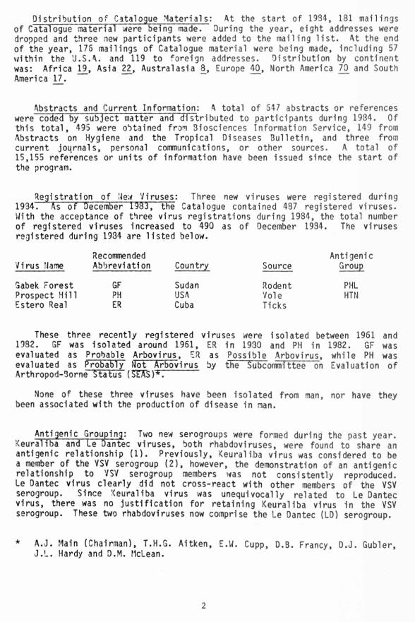
Distribution of Catalogue Materials: At the start of 1934, 181 mailings of Catalogue material were being made. During the year, eight addresses were dropped and three new participants were added to the mailing l is t . At the end of the year, 176 mailings of Catalogue material were being made, Including 57 within the 'J.S.A. and 119 to foreign addresses. Distribution by continent was: Africa 19, Asia 22 Australasia 8, Europe 40, North America 70 and South America 17.
Abstracts and Current Information: A total of 547 abstracts or references were coded by subject matter and distributed to participants during 1984. Of this total, 495 were obtained from 31osciences Information Service, 149 from Abstracts on Hygiene and the Tropical Diseases Bulletin, and three from current joqrnals, personal communications, or other sources. A total of 15,155 references or units of information have been issued since the start of the program.
Registration of New Viruses: Three new viruses were registered during1934. As of December 1983, tRe Catalogue contained 487 registered viruses. With the acceptance of three virus registrations during 1984, the total number of registered viruses increased to 490 as of December 1934. The viruses registered during 1934 are listed below.
Virus NameRecommendedAbbreviation Country Source
AntigenicGroup
Gabek Forest GF Sudan Rodent PHLProspect H ill PH USA Vole HTNEstero Real ER Cuba Ticks
These three recently registered viruses were isolated between 19511932. GF was isolated around 1951, ER in 1930 and PH in 1982. GFevaluated as Probable Arbovirus, ER as Possible Arbovirus, while PH was evaluated as Probably Not Arbovirus by the Subcommittee on Evaluation of Arthropod-3orne Status (SEASY*~.
None of these three viruses have been isolated from man, nor have they been associated with the production of disease in man.
Antigenic Grouping: Two new serogroups were formed during the past year.Keuraliba and Le Dantec viruses, both rhabdoviruses, were found to share an antigenic relationship (1). Previously, Keuraliba virus was considered to be a member of the YSV serogroup (2), however, the demonstration of an antigenic relationship to VSV serogroup members was not consistently reproduced. Le Dantec virus clearly did not cross-react with other members of the VSV serogroup. Since Keuraliba virus was unequivocally related to Le Dantec virus, there was no justification for retaining Keuraliba virus in the VSV serogroup. These two rhabdoviruses now comprise the Le Dantec (LD) serogroup.
* A.J. Main (Chairman), T.H.G. Aitken, E.W. Cupp, D.B. Francy, D.J. Gubler, J.L. Hardy and D.M. McLean.
2

Prospect H ill virus, which was registered in 1984, was shown to be related to the previously registered Hantaan virus. 3oth viruses were found to be related by the indirect immunofluorescent antibody technique although they were quite d istinct (see Prospect H ill virus registration). They now form the Hantaan serogroup.
Barmah Forest virus fina lly was assigned to serogroup A as a new member, albeit an unusual one. "Barmah Forest virus has been characterized in a number of ways including electron microscopy of infected ce lls; physical studies of the virion, it s RNA, and associated proteins; N-terminal sequence analysis of the two envelope glycoproteins; studies of macromolecular species present in infected ce lls; and serological cross-reactions with alphaviruses and bunyaviruses. From these results Barmah Forest virus is clearly an alphavirus since the structure of the v irion, the mode of replication, and the macromolecular species present in infected ce lls are typical of alphaviruses. The N-terminal regions of the two glycoproteins El and E2 show extensive sequence homology (approximately 50%) with those of other alphaviruses. Barmah Forest v irus cross-reacts in hemagglutination inhibition tests, although not in complement fixation tests or in fectiv ity neutralization tests, with other alphaviruses. In some of it s properties Barmah Forest virus is unusual, however. I t cross-reacts in complement fixation and hemagglutination inhibition tests with Umbre virus, a bunyavirus, which o rig ina lly led it to be c lassified as a bunyavirus; the glycosylation pattern of E2 of Barmah Forest virus appears to d iffe r from that of other alphaviruses; and the sedimentation coefficient of the virion appears to be s ligh tly less than that of other alphavlruses."(3) We also have observed that plaque-purified 3armah Forest virus cross-reacts in HI tests, although not in CF or N tests, with other alphavlruses (4). However, we did not cross-test Barmah Forest and Umbrev1ruses.
Recent serologic studies have shown that Ippy virus is related to Lassa virus (5). During attempts to characterize an unidentified virus from rodents and livestock in Zimbabwe, i t was compared in cross-immunofluorescence tests with other viruses Isolated from rodents in Africa, including Ippy and Lassa viruses and other unregistered Lassa-related viruses. The unidentified virus was not found to be related to any of the other viruses with which i t was compared; however i t was discovered that Ippy virus was related to Lassa virus. Antibody to Ippy virus reacted to high t ite r in immunofluorescence tests with Lassa virus and the Lassa-related viruses. In addition, monoclonal antibody to Lassa virus reacted with Ippy virus. The exact relationship of Ippy virus to other arenaviruses of the Tacaribe serogroup remains to be determined (5).
Taxonomic Status of Registered V iruses: Reported changes in the taxonomicc lassification of registered arboviruses are of a provisional nature; and in some instances, new taxonomic placements are based on very s ligh t evidence.
Electron microscopic examination of Gossas virus preparations has shown that this previously unclassified and ungrouped virus possesses rhabdovirus morphology (6). Currently i t is being serologically tested in order to determine 1f i t i s antigenical ly related to any of the established rhabdoviruses.
3

Data obtained for Barmah Forest virus in terms of morphology,morphogenesis, genetic material, replication, and amino acid sequence homology indicates that it is clearly a member of the Alphavirus genus (3,7). The status of Barmah Forest virus also was discussed in thejrevious section on Antigenic Grouping.
A formal proposal has been put forth which would create a fifth genus in the family Bunyaviridae (3). In addition to the Bunyavirus, Nairovirus, Phlebovirus and Uukuvirus genera, the newly proposed taxon would be namecT the Hantavirus genus, and in it ia lly i t would consist of Hantaan and Prospect H ill viruses and other Hantaan virus-related viruses. This proposal has not been acted upon by the International Committee on Taxonon y of Viruses (ICTY).
Previously, the family Filoviridae was proposed as "a taxonomic home" for Marburg and Ebola viruses (9). This proposal was not viewed with favor by the ICTV. I t is planned that, following some alteration, the proposal w ill be resubmitted to the ICTV.
Finally, the ICTV has approved the proposal of the ITCV Togavirus Study Group which recommended the creation of a new family, Flaviviridae, and placement of the Flavivirus genus in th is new taxon (10). Obviously, th is was intended to separate the genus Alphavirus from the genus Flavivirus by removal of the flaviviruses from the family Togaviridae. Viruses of the two genera differ in size, morphology, morphogensis and mode of replication. In addition, the flaviv irus virion usually contains a single major envelope glycoprotein, a smaller membrane protein and the core protein. On the other hand, alphavirus virions contain at least two envelope proteins, one or more which are glycosylated, and a core protein. In the case of alphaviruses, genes for nonstructural proteins are located at the 5 ’ end of the genome; whereas for flavlviruses, genes for structural proteins are located at the 5' end (10).
Synopsis of Information in Catalogue: This synopsis has been compiled primarily to provide a short review of the viruses Included 1n the Catalogue. The following tabulations are designed to draw together groups of viruses showing certain characteristics 1n common, lis t in g viruses according to their known taxonomic status and by their demonstrated serological relationships, and where appropriate, by principal arthropod vector. Isolations from arthropod and animal hosts, continental distribution, involvement 1n human disease, and arbovirus status are indicated. Information 1s also given concerning the recommended level of practice and containment assigned to registered viruses and the basis for assignment to a level. Most of this Information was published previously by the Subcoronittee on Arbovirus Laboratory Safety (SALS)* (11). Three registered viruses listed in tables 5 through 34 have not been rated by SALS. Appendices I and I I , following table 39, w ill provide a description of recommended levels and an explanation of symbols used to define basis. Other tables summarize the taxonomic status of registered viruses; the antigenic groups comprising a given taxon to which registered viruses have been assigned; the numbers of registered viruses
* Composition at time of publication: W.F. Scherer (Chairman, deceased),G.A. Eddy, T.P. Monath, T.E. Walton, and J.M. Richardson (ad hoc member).
Present composition: T.E. Walton (Chairman), J.M. Dalrymple, R. Endrls,J.L. Hardy. T.P. Monath. and J.M. Richardson (ad hoc member).
4

assigned to presently recognized antigenic groups; chronology and areas of isolations of registered viruses; continental distribution by groups; numbers of viruses recovered from naturally infected arthropods and vertebrates; association with human disease; and evaluation of arthropod-borne status of members in various serogroups.
Table 1. Alphabetical and taxonomic l i s ting of registered v iru se s: Table 1 presents an alphabetical l is t in g of the~4§3 viruses registered in the Catalogue as of December 1934. An o ffic ia l or provisional taxonomic c lassification is shown for each registered virus. I f taxonomic status is not indicated, the registered virus is presently unclassified. Also, a recommended abbreviation is given for each virus, which has been formulated according to the guidelines established by the American Committee on Arthropod-3orne Viruses (12). All too often, abbreviations are employed in publications which are of the author's choosing and which do not conform to the recommended abbreviations. Their use is confusing, contrary toestablished guidelines, and erodes a portion of the effort of the Arbovirus Information Exchange program. All arbovirologists who plan to employ abbreviations in print should make every effort to use the recommendedabbreviations.
Antigenic groups to which viruses have been assigned also are shown inth is table. I f no antigenic group is given, the virus is ungrouped andindicates that i t has not been demonstrated to be serologically related to any other known arbovirus.
Table 2. Antigenic groups of registered v iru se s: The o rig ina llydescribed antigenic groups of arboviruses were designated by letters, A, 3, and C; but in present practice, the f ir s t discovered virus of a newly recognized serogroup lends it s name to the antigenic cluster. Before a virus can be assigned to any antigenic group, it must be shown to be serologically related to, but clearly distinguishable from a previously isolated virus.
Table 2 l i s t s the serogroups comprising the various taxa to whichregistered viruses have been assigned. Sixty-two antigenic groups have been designated for viruses registered in the Catalogue. That includes the previously established rabies serogroup. There are several instances inwhichonly a single virus is shown in an antigenic group. That is so because one or more antigenic relatives of that virus have not been registered.
It is also noted that the Tunyavirus genus comprises the old Bunyamwera Supergroup to which several additional serogroups have been added. The most recent additions are the Anopheles 3 and Turlock serogroups. The Bunyamwera Supergroup o rig ina lly was formulated to reflect low-level but reproducibleintergroup relationships usually by complement-fixation and/or hemagglutination-inhibition reactions. In a somewhat analogous situation, the nairoviruses consist of six d istinct serogroups which share low-levelintergroup relationships among themselves. Registered viruses belonging in the Bunyamwera Supergroup constitute s ligh tly more than one-fourth of a ll registered viruses.
Table 3. In it ia l iso lations by decade and country of o r ig in : Table 3l i s t s the in it ia l isolation of specific registered viruses by the decade ofdiscovery and according to the continent or subcontinent and country in which
5

each was f ir s t discovered. Because of the large number of virus names involved, abbreviations are employed. These abbreviations and the associated complete names of the respective viruses may be found in table 1.
Table 4. In it ia l isolation of viruses by continent, country, and chronological period: Sim ilar data were utilized in tables 3 and 4, thoughthey were subjected to sligh tly different analyses and were presented in a different format. Periods or locations which show high numbers of virus isolation undoubtedly reflect the net effect of a number of contributing factors such as the change in emphasis of fie ld programs from a search for viruses causing specific diseases to a systematic search for viruses, new or known, in their natural ecological niche in a given geographical area, refinements 1n isolation and identification techniques, Improved communication between arbovirus 1aboratories, and more rapid dissemination of new Information, as well as the presence in a given area of an arbovirus laboratory with highly active and effective field programs.
Tables 5 through 34 l i s t registered viruses by taxon and, within taxon, by serogroup, with information regarding Isolations from arthropod vectors and vertebrates, and geographic (by continent) distribution based on virus isolation. Data also are presented regarding production of disease in man 1n nature or by laboratory infection, evaluation of arbovirus status, and proved or provisional taxonomic status. These tables now show the biohazard level assigned to each registered virus, and the basis for assignment to a level. Where possible, sets of viruses were grouped additionally according to their actual or suspected principal arthropod vector.
The data presented in these tables clearly illu strate the salient features characteriStic of each set or subset of viruses. Thus, the reader is urged to carefully examine the tables for information that may be of specific interest, or that will provide an overview of the general characteristics of a given group of viruses.
Table 5. Alphaviruses: Alphaviruses clearly are mosquito associated,although a few have been isolated from other arthropods. About one-half of the alphaviruses are associated with birds, while some of them, particularly those of the VEE complex, are associated with rodents.
Eleven alphaviruses have been isolated from man while 12 have been implicated in causing human disease either by infections acquired in nature or in the laboratory. At least seven of these 12 alphaviruses have been responsible for epidemics: chikungunya, eastern equine encephalitis, Mayaro,o'nyong-nyong, Ross River, Venezuelan equine encephalitis, and western equine encephalitis. All of the 12 alphaviruses either are rated as Arbovirus (11 viruses) or Probable Arbovirus (one virus).
3armah Forest virus definitely has been listed with the other alphaviruses in serogroup A. It s redefined characteristics and its status have been discussed in two earlier sections on "Antigenic Grouping" and "Taxonomic Status of Registered Viruses". Although the virus appears to be distantly related to other alphaviruses, it is unusual in some respects. In serology, Barmah Forest virus reacted with other alphaviruses only in the HI test. Almost exclusively, its hemagglutinin was inhibited by antibody to other alphaviruses. Barmah Forest virus did not cross-react in CF and NT.
6

Furthermore, plaque purified 3armah Forest virus s t i l l cross-reacted with Umbre virus in HI and CF tests.
Sindbis virus has been recovered from the organs of insectivorous bats collected in Zimbabwe. Cabassou, chikungunya and VEE viruses represent the other alphaviruses which have been isolated from bats.
Tables 6, 7, and 9 . F Iav lv iru se s: Of the 54 registered flaviv iruses, \1%have been pTacecT fn the mosquito-associated category (table 5), 23£ are considered to be tick-borne (table 7), and 30£ are categorized as not being associated with a proven arthropod vector (table 9). Only West Wile andyellow fever viruses in the mosquito-associated category (table 6) have been isolated from both mosquitoes and ticks.
Twenty-six of the 30 registered flav iv iruses which are mosquito-associated (table 5) are rated as Probable Arbovirus or Arbovirus. The tick-borneflaviv iruses (table 7) contain four registered viruses, Absettarov, Hanzalova, Hypr and Xumlinge, which are very closely related or indistinguishable by conventional serological techniques, though they are said to be clearly differentiated on the basis of c lin ic a l, epidemiological, and ecological markers from 9SSE and other members of the same complex.
Twenty-eight (44X) registered flav iv iruses have been isolated from man, whereas 13 of 30 (50X) mosquito-borne flav iv iruses and nine of 15 (60%) tick-borne flav iv iruses have been implicated in the production of human disease, either through infections acquired in nature or in the laboratory. 3y contrast, only four of 19 (21£) flav iv iruses not associated with a vector have been implicated in the production of human disease. Thus, a total of 31 flaviv iruses have been associated with the production of disease in man.
With the exception of two members (Israe l turkey meningoencephalitis and Koutango viruses), none of the rest of the registered flaviv iruses placed in the "no arthropod vector demonstrated" category are rated above Possib le Arbovirus by SEAS. Seven members are rated as Probably Not or Not Arbovirus. Most of the flaviv iruses listed in table 3 have been isolated from rodents or bats. Israel turkey meningoencephalitis virus has been isolated from domestic turkeys, Cacipacore virus from a wild bird, and \roa virus from a sentinel hamster. Only Dakar bat and Negishi viruses have been isolated from man; that has been the sole source of recovery for Negishi virus.
Tables 9___ through 16. Bunyaviruses, Family 3unyaviridae: Sixteenanti genic ~ sets of viruses plus kaeng khoT virus (SOU) comprise the bunyaviruses. A total of 123 registered viruses have been placed within the Ounyavirus genus.
Table 9. Anopheles A and Anopheles 3 serogroup viru se s: Members of theAnopheles A serogroup have been isolated either from anopheline or both culicine and anopheline mosquitoes. Of the five members of this serogroup, only Tacaiuma virus has been reported to cause a febrile illne ss in man. In addition, th is virus has been isolated from man and from a sentinel monkey. Members of th is serogroup and of the AN3 serogroup appear to be localized. These viruses have been found on only a single continent.
7

Viruses of the \nopheles B serogroup have been isolated only from mosquitoes collected in South America. Neither virus has been associated with infections in man.
Table 10. 3unyamwera serogroup viruses; All members of the Bunyamwera serogroup have been isolated from cull cine or anopheline mosquitoes or both. In addition, Lokern and Main Drain viruses have been isolated from Culicoides insects. Maguari virus has been recovered from livestock, Anhembi, Germiston, Kairi, Macaua and Shokwe viruses from rodents, and Lokern, Main Drain and Tensaw viruses from lagomorphs. Kairi virus also was recovered from a monkey, while Macaua virus was isolated from a bird.
Bunyamwera, Germiston, Ilesha, Shokwe, and Wyeomyia viruses have been Isolated from man. Except for Shokwe virus, those viruses plus Calovo and Tensaw viruses have been shown to be associated with human disease, either through infections acquired in nature or in the laboratory, or both.
Fifteen of the 22 viruses registered in the 3unyarnwera serogroup have been rated as M o v iru s or Probable Arbovirus. None are rated below Possible Arbovlrus.
Members have been found most frequently in North America (eight viruses), South America (eight viruses) and Africa (five viruses). Thus far, only one virus has been recovered in Asia, two in Europe and none in Australasia.
Table 11. Bwamba serogroup and serogroup C v iruses: Both Bwamba andPongola viruses (Bwamba serogroup) are mosquito-associated, and Bwamba virus has been isolated from man. 3wamba virus has been reported to produce a febrile illne ss in man as a result of infections acquired in nature. Thus far, these two viruses have been found in Africa only. Pongola virus has been rated as Arbovirus while Bwamba virus has been rated as Probable Arbovirus.
The Group C viruses have been closely associated with mosquito vectors and small aninals, particularly rodents. Eight group C viruses have been isolated from rodents, and three of these eight additionally have been Isolated from marsupials. Two other viruses have been isolated from marsupials but notrodents. Ten of the twelve viruses have been isolated from man. Only Gumbo Limbo and Vinces viruses have not been isolated from man and, with theexception of those two viruses, all other members have been associated withcases of human febrile illness. In addition, Apeu and Oriboca viruses havebeen reported to infect man as a result of laboratory mishaps. Ten of these viruses have been classified as Arbovirus and two as Probable Arbovirus.
Table 12. California and Capim serogroup viruses; All the California group viruses are associated with mosquito vectors and four members have been recovered from naturally infected rodents. La Crosse, Guaroa, and Tahyna viruses have been isolated from man and, along with California encephalitis and Inkoo viruses, have been associated with disease as a result of infections acquired in nature. In addition, Jamestown Canyon and snowshoe hare viruses recently have been serologically associated with disease in man. Only Inkoo and Tahyna viruses have been isolated on continents other than those of North and South America. On the basis of virus Isolation, the geographic distribution of Tahyna now includes Asia as well as Africa and Europe. Ten of the California group viruses have been rated as Arbovirus, one other as Probable Arbovirus, and the remaining two as Possible Arbovirus.
8

Viruses of the Capim serogroup are associated with mosquito vectors, and four of the members have been isolated from rodents. None of these eight viruses have been associated with disease in man. Capim group members have been recovered only in North and South America. Six of the eight Capim serogroup viruses have been rated as Arbovirus (four viruses) or Probable Arbovirus (two viruses).
Table 13. Gamboa, Guama and Koongol serogroup v iru se s: In addition toGamboa virus, the serogroup contains Pueblo Viejo and San Juan viruses. All virus members have been isolated exclusively from Aedeomyia squamipennis mosquitoes. The viruses appear to have a limited geographic distribution, and they have not been implicated in human infections.
Guama serogroup viruses have been found only in the western hemisphere. Catu and Guama viruses have been isolated from man and have been associated with disease in man as a result of infections acquired in nature. Nine of the 12 Guama group viruses have been rated as Arbovirus or Probable Arbovirus. Viruses of th is serogroup clearly are mosquito-associated and the majority of them appear to be associated with rodents. Ten viruses have been isolated from sentinel animals, primarily mice.
Both Koongol group viruses were isolated in Australia and very l it t le is known about them. These two viruses were rated as Probable Arbovirus.
Table H . Mina tit! an, Q lifantsvle i and Patois serogroup viruses: TheMinatitlan serogroup now contains two registered members. In addition to Minatitlan virus, the group also includes Palestina virus. Several iso lations of Palestina virus have been made from Culex sp. mosquitoes collected in Ecuador, and from sentinel hamsters. Minatit1an virus was isolated from a sentinel hamster exposed near Minatitlan, Mexico. L it t le is known concerning its role in nature.
The Q lifantsvlei group consists of three members, and a ll three were isolated in Africa from mosquitoes. Information on the properties of these viruses has not been readily available.
Viruses of the °atois group now have been isolated in North and South America, and most appear to be associated with mosquito vectors and some with rodent hosts. Babahoyo, Patois, Shark River, and Zegla viruses also were isolated from sentinel hamsters.
None of the viruses from these three serogroups have been isolated from man, nor have they been associated with the production of disease in man.
Table IS. Simbu serogroup v iruses: Essentia lly equal numbers of Simbugroup viruses have been isolated from Culicoides insects and from mosquitoes. None have been recovered from rodents” Eight Simbu serogroup viruses have been isolated from livestock. These include Sabo, Sango, Shamonda and Shunl viruses (N igeria), Douglas and Peaton viruses (Australia), Akabane virus (Japan and Australia) and Sathuperi v irus (India and Africa). In addition, four viruses have been isolated from birds, and Manzanilla virus has been isolated from a monkey. Oropouche and Shuni viruses are the only members that have been isolated from man. Oropouche virus has caused frequent large outbreaks of disease among the human population in Brazil.
9

Simbu group viruses have a wide distribution. Approximately 50£ have been found in Africa or Africa and Asia, while others have been isolated in Asia or Asia and Australasia and North or South America. Only eight of the 21 members of th is serogroup have been rated as Probable Arbovirus or Arbovirus. The remainder have been rated as Possible Arbovi rus.
Table 16. Tete and Turlock serogroups and unassigned (SB'J) viruses: A11 Tete group viruses have been recovered from birds; only two of them (3ahig and Matruh viruses) have been recovered from an arthropod vector (ixodid ticks). None of these viruses have been associated with human infections. Only 3ahig virus 1s rated above Possible Arbovirus.
All viruses of the Turlock serogroup are associated with mosquito vectors. In addition, Turlock and Umbre viruses appear to be associated with birds. Turlock virus has been found in both North and South America. All the other members have been found in a single continent (Africa, Asia, and Europe). Barmah Forest virus has been deleted from the lis t in g of those viruses which belong in the Turlock serogroup.
Only '<aeng Khoi virus remains as a serologically unassigned bunyavirus. Kaeng Khoi virus was isolated from bats, sentinel mice and rats, and cimicid bugs.
Table 17. Phlebotomus fever serogroup viruses: At present, the PHL antigenic group consists of 3$ members, and the entire serogroup comprises the Phlebovirus genus within the family Bunyaviridae. S ic ilia n sandfly fever virus is the type virus for this genus.
The majority of the group members are associated with phlebotomine flie s; only Arumowot, Chagres, Icoaraci, Itaporanga, R ift Valley fever and Zinga viruses have been isolated from mosquitoes. Eight of the phleboviruses have been Isolated from man or have been implicated in the production of disease in man.
Gabek Forest virus was registered and added to the PHL serogroup during 1984 although the virus actually was Isolated 1n 1960. I t has not been recovered from arthropods but 1t has been Isolated from a variety of rodents and a hedgehog collected in various areas of Africa. Gabek Forest virus has been rated as Probable Arbovirus.
R ift Valley fever virus causes serious and extensive disease in domestic animals such as sheep and cattle, and may cause disease in veterinary personnel, fie ld and laboratory workers, as well as herdsmen who handle infected animals. Previous serological studies have indicated that Zinga virus 1s closely related or Identical to R ift Valley fever virus. Consequently Zinga virus has been placed in the Phlebotomus fever serogroup although it may be just another strain of RVF virus. Previously it was listed as an antlgenically ungrouped virus.
Table 13. Tick-borne serogroups other than serogroup B v iruses. Nairovlruses: Members of the six antigenic groups shown in tables 18 and 19constitute the Nairovirus genus in the Bunyaviridae family. CHF-Congo virus was designated the type virus for this genus. Furthermore, reproducible
10

intergroup antigenic relationships have been demonstrated for the six sets of viruses. Only members of the CHF-Congo and NSD serogroups have been associated with the production of disease in man.
Both Congo and Crimean hemorrhagic fever viruses are registered in the Catalogue. I t must be reiterated that the agent of Crimean hemorrhagic fever (CHF) is antigenically indistinguishable from Congo virus. The CHF virus has been implicated in more than two thousand cases of human disease in the USSR. Congo virus also has been associated with the production of disease in man, either as a result of infections acquired in nature or in the laboratory. Thus far, Hazara virus has not been known to be involved in infections of man, and l it t le is known of this antigenic relative of CHF-Congo virus. Allmembers of this serogroup appear to be associated with ixodid ticks although CHF virus was isolated from both ixodid and argasid ticks.
Members of the DGK serogroup have not been isolated from vertebrate hosts, nor from arthropod vectors other than ticks. The majority of the viruses appear to be associated with argasid ticks. These viruses have been found in Africa, Asia and Australasia.
Only Hughes virus of the Hughes serogroup has been isolated from birds. I t has been found in both North and South America while Sol dado virus has been isolated in Africa, Asia and Australasia. A ll Hughes serogroup members have been associated with argasid ticks. A new antigenic member of the Hughes serogroup has been described (13). This virus has been called Puffin Island, and i t has not been registered as of th is moment.
Table 19. Tick-borne serogroups other than serogroup 3 v iruses. Nairoviruses: Nairobi sheep disease virus is an important cause of veterinary disease, while both Dugbe and Ganjam viruses have been isolated repeatedly from ticks taken off of domestic animals. Dugbe and Ganjam viruses have caused febrile illne sse s in man. In the case of NSD virus, one infection in man resulted in a febrile illness, while three others resulted in subclinical serologic conversions. Thus, all three viruses have been isolated from man, and only Dugbe virus has not been associated with infections in man acquired in the laboratory. Pending further c la rifica tion of antigenic relationships, SIRACA considers Ganjam virus to be a variety of NSD virus.
Both Qalyub group viruses were found only in Africa, and both have been isolated from ticks. In addition, Bandia virus has been isolated from rodents.
Except for Avalon virus, members of the Sakhalin antigenic set were isolated only from ixodid ticks. Avalon virus also was recovered from a bird. Sakhalin serogroup viruses are distributed in Asia (PMR,SAK), Australasia (TAG), Europe (CM), and North America (AVA,SAK). Antigenic studies have indicated that Avalon and Paramushir viruses are stra ins of the same virus.
Table 20. Tick-borne serogroups other than serogroup 3 viruses: Atpresent, Uukuniemi serogroup viruses constitute the llukuvirus genui i"n the 3unyaviridae family. Other serogroups listed in that table represent the Hantavirus genus and those provisionally c la ss ified as bunyavirus-1ike.
11

Except for Uukuniemi virus, all members of the llukuniemi serogroup have been isolated only from ticks. Uukuniemi virus also has been recovered from both rodents and birds. Two of the viruses in this serogroup were found in Asia while the other three were discovered in Europe. Hemagglutination-inhibition antibodies to Uukuniemi virus have been detected in the sera of human beings residing in Europe. Grand Arbaud virus has been evaluated as Arbovirus and Uukuniemi as Probable Arbovirus. The rest of the members have been evaluated as Possible Arbovirus.
At present, the Hantavirus genus is only a proposed taxon. I f approved, th is genus w ill eventually contain more than the two registered virus shown in the serogroup. In addition to Hantaan and Prospect H ill virus, other probable members will be the Hantaan-related viruses isolated from rats, and the agent of "Nephropathia Epidemica."
Both Hantaan and Prospect H ill viruses have been isolated from rodents, while Hantaan virus also has been Isolated from man. Hantaan virus 1s the etiologlc agent of hemorrhagic fever with renal syndrome (HFRS) or Korean hemorrhagic fever (KHF), and either is responsible for or Is antigenically closely related to the agent(s) responsible for c lin ica lly sim ilar diseases In the U.S.S.R., Japan, Manchuria, and Eastern and Northern Europe. More than 10,000 cases have occurred 1n Korea alone since the disease was f ir s t recognized in that country in 1951. Prospect H ill virus has not been shown to infect man thus far.
Bhanja virus is the sole registered virus member of the new Bhanja serogroup. Kismayo virus is the unregistered member and previously has been demonstrated to share an antigenic relationship with 3hanja virus. Bhanja virus has been isolated from man and has been implicated 1n a laboratory-acquired human infection.
Two of the Kaisodi group viruses were isolated from ticks collected 1n Asia while the third was isolated in North America. None of these viruses have been found to infect man. Previous Annual Reports have referred to unpublished studies which had suggested that the RNA species and polypeptides of Sllverwater virus resembled those of uukuvlruses. Additional confirming or c larify ing information is s t i l l not available. Kaisodi and Silverwaterviruses had been evaluated as Probable Arbovirus while Lanjan virus had been rated as Possible Arbovirus.
The Upolu serogroup consists of Upolu and Aransas Bay viruses. Both viruses were isolated only from argasid ticks. Neither virus has beenassociated with infections 1n man. One virus has been found in Australia (UPO), and the other in North America (AB).
Table 21. Tick-borne serogroups other than serogroup B v iruses: Thogoto virus has been Isolated from man and has been Involved 1n the production of disease in man. An unregistered antigenic relative of Thogoto virus has been Isolated in S ic ily . Molecular analysis of a Thogoto group virus has Indicated that it s virion RNA species and structural polypeptides resemble those of members of the family Orthomyxoviridae.
12

Nyamanini and the unregistered Midway viruses now constitute the Nyamanini serogroup. Nyamanini virus was isolated from argasid ticks and birds. I t has not been assoicated with the production of disease in man.
Quaranfil virus has been isolated from both man and birds, and has been associated with the production of disease in man as the result of infections acquired in nature. Preliminary molecular studies conducted with Quaranfil virus indicated that th is virus may resemble viruses of the family Orthomyxoviridae. At this point, further verification is required. L itt le is known concerning the behavior of Johnston Atoll virus in nature.
Table 22. Minor antigenic groups of v iru se s: All the viruses listed inth is table are members of minor antigenic groups, and provisionally are c la ssified taxonomically as bunyavirus-1ike members of the Family Bunyaviridae. Most virus members of these minor serogroups have been primarily associated with mosquito vectors.
Bakau group viruses have been recovered only in Asia. Bakau virus has been isolated from both mosquitoes and ticks, and also rodents. Additional information concerning these viruses is not available.
Thus far, all four viruses of the Mapputta group have been found only in Australia. Maprik virus was rated as a Probable Arbovirus while the other three virus members were c la ssified as Possible Arbovirus.
All three Matariya group viruses have been recovered from birds collected in Africa. Nothing is known concerning their possible vector association.
Nyando virus has been isolated from man and from mosquitoes collected in Africa. The Nyando virus infection in man resulted in a febrile illness.
Table 23. Tick-borne serogroups other than serogroup 3 v iru se s: While theviruses TTsted fn table 23 also are tick-borne agents, they differ taxonomical ly from those in tables 13-22 in that they have been c la ssified as orbiviruses in the family Reoviridae. The orbiviruses are relatively resistant to lip id solvents, are inactivated at an acid pH, and possess multiple segments of a double-stranded RNA genome. I t is like ly that members of the genus O rbivirus, and that the crite ria used to define th is genus, w ill be reevaluated in the near future.
Only Colorado tick fever virus of the CTF serogroup and Kemerovo virus of the KEM serogroup have produced disease in man and have been isolated from man.
Members of the Kemerovo group are widely distributed with at least one virus being found in each of the listed continents. Kemerovo virus has been found in both Africa and Asia while Wad Medani virus has been discovered in Africa, Asia and North America. Even though all members of th is serogroup have been isolated from ticks, only three viruses were rated above Possible Arbovirus. All three were rated as Probable Arbovi rus.
Tables 24, 25. Minor antigenic groups of v iruses: Members of these minorantigenic groups have been characterized and taxonomically c la ss ified as orbivi ruses.
13

Several of the viruses in these minor antigenic groups are important in causing disease in large animals. BLU virus causes disease in both wild and domestic ruminants; AHS virus in mules, donkeys and horses; EHD virus in deer and Ibaraki virus in cattle. Both BLU and AHS viruses have a wide geographic di stribution.
Changuinola virus is the only member from these minor antigenic groups which has been isolated from man, and has been reported to produce disease in man. Of the present twelve serogroup members, only Ir itu ia , Jari, and Monte Oourado viruses have not been isolated from an arthropod. All others, including Changuinola virus, appear to be associated with phlebotomine Insects. Registered viruses of the Changuinola serogroup appear to have a limited distribution. Eleven members were recovered only in South America while Changuinola virus was isolated in North America.
Between 1950 and 1980, a total of 178 Changuinola serogroup viruses were isolated in Brazil, Colombia, and Panama. In a recent study, 24 of those viruses were selected as representative specimens and their antigenic, biological, and chemical properties were examined. Twelve of the viruses were distinct by neutralization tests and polyacrylamide gel electrophoresis (PAGE) (14). This study clearly states that "a great many more Changuinola serotypes may exist" (14).
The three viruses of the Corriparta serogroup appear to be associated with mosquitoes. In addition, Corriparta virus was recovered from wild birds. All three viruses are widely separated in their distribution.
Thus far, Ibaraki and EHD viruses have not been associated with any known vector. The EHD virus has been found in Africa and North America, while Ibaraki virus has been recovered only in Asia.
Virus members of the Corriparta, Eubenangee, and Palyam serogroups appear to be primarily mosquito-associated, while members of the Wallal and Warrego serogroup appear to be associated with Culicoides insects. Vector associations appear to be less clear for Eubenangee virus of the EUB serogroup, and for Warrego virus of the WAR serogroup.
Table 26. Minor antigenic groups of viruses: Members of the serogroups listed in th is table and in table 2/ possess a "bullet-shaped" morphology and are c lassified as members of the family Rhabdoviridae. Table 26 contains the Hart Park group viruses, a Xwatta group virus, the newly formed Le Dantec serogroup, an expanded Mossurll group consisting of eight members, and a rabies serogroup consisting of two rabies-related viruses.
All of the Hart Park serogroup members are associated with a mosquito vector and two of the viruses (Hart Park and Flanders) have been isolated from birds. None of these viruses have been associated with disease in man. Thus far, their distribution includes only North and South America.
The Xwatta virus was isolated only once from mosquitoes collected in Surinan. The antigenic relative of Xwatta virus remains unregistered. This unregistered virus was recovered from a bird collected in Brazil.
14

The new Le Oantec serogroup consists of Le Dantec and Keuraliba viruses. Prior to the discovery of an antigenic relationship between these two rhabdoviruses, Keuraliba virus was listed as a member of the VSV serogroup. However, th is relationship was not reproducible and Keuraliba virus was withdrawn from the VSV serogroup when it was demonstrated to be related to Le Oantec virus. Neither virus has been isolated from an arthropod. Le Oantec virus has been isolated from man and Keuraliba virus was isolated from rodents.
Three of the members of the Mossuril serogroup have not been isolated from arthropods. These include Cuiaba, Kern Canyon, and Marco viruses. Kern Canyon virus has been rated as Probably not Arbovirus by SEAS. Previous studies have demonstrated that Kern Canyon virus could be propagated in an Aedes dorsalis cell culture line.
The rabies serogroup consists of kotonkan virus and Lagos bat virus. Kotonkan virus was isolated from Cu liocides spp. collected in Nigeria. I t was rated as Probable Arbovirus by SEAS. Lagos bat virus has been isolated only from bats on several occasions.
Table ?7.__Minor antigenic groups of viru ses: All three viruses of theSawgrass serogroup'were isolated "from ticks collected in North America. All viruses of the Timbo serogroup, including the new Sena Madureira virus, were isolated from lizards, and none of these viruses ever were isolated from arthropods.
Three VSV group viruses have been isolated from phlebotomine flie s, and four others have been recovered from mosquitoes. An additional member, VS-Indiana virus has been isolated from both types of vectors. Piry andVS-Alagoas viruses have not been recovered from arthropods. Of the serogroups listed in this and the preceding table, only members of the VSV serogroup and Le Oantec virus have been shown to infect man. In the VSV serogroup,Chandipura, Piry, VS-Indiana and VS-New Jersey viruses have been isolated from man. These viruses, plus VS-Alagoas virus, have been found to produce disease in man during infections acquired in nature or in the laboratory. Both VS-Indiana and VS-New Jersey viruses readily infect livestock, while Cocal virus has been recovered from a horse and VS-Alagoas virus from a mule.
Table 23^___Minor antigenic groups of v iruses: These antigenic groupsconsist of members which are tax'onomically uncTasiTfied.
Both Boteke group viruses have been isolated in Africa only. Zingilamovirus was recovered from a bird and 3oteke virus was isolated frommosquitoes. Previously published studies have indicated that Zingilamo virus resembles viruses of the family Togaviridae. Pending further information, both viruses of this serogroup w ill be listed as unclassified in this Annual Report.
Malakal and Puchong viruses (Malakal serogroup) have been isolated from mosquitoes only. Malakal virus was recovered from mosquitoes collected in Africa, while Puchong virus was found in Asia.
15

3oth Marburg and Ebola viruses have caused human disease as a result of infections acquired in nature and have been associated withlaboratory-acquired infections. Ebola virus was found to possess a single-stranded RNA which was noninfectious upon extraction. Recent evidence indicates that there might be different serotypes of Ebola virus. Marburg and Ebola viruses have been isolated from man only.
The two viruses of the Tanjong Rabok serogroup have been isolated inMalaysia and neither has been associated with a vector. Tel ok Forest virus was isolated from a wild nonkey and Tanjong Rabok virus from a sentinel monkey.
Table 29. Tacaribe group viruses: Tacaribe group viruses areserologically related to lymphocytic choriomeningitis virus, and they are c lassified taxonomically in the Arenavirus genus. They are primarily rodent viruses, and there is l it t le or no evidence which suggests that they are associated with an arthropod vector in nature. SEAS has judged all members to be Not Arbovirus.
Ippy virus represents a new addition to this serogroup. I t was found to be related to Lassa virus (5). Its antigenic relationship to other members of the Tacaribe serogroup has yet to be determined. Characteristically, Ippy virus has been isolated from Mastomys rodents and from rodents of other species.
Three members of this group have been implicated in the production of severe, often fatal, human disease. These include Junin (Argentinehemorrhagic fever), Machupo (Bolivian hemorrhagic fever), and Lassa (Lassa disease). In addition to causing c lin ica lly frank laboratory-acquired infections, Junin virus also has been reported to cause subclinical laboratory-acquired infections. A subclinical seroconversion to Tacaribe virus has been documented in a laboratory worker handling large quantities of Tacaribe virus. In addition, Pichinde virus has produced subclinical infections in laboratory workers. F inally, the newly registered Flexal virus has produced a febrile illness in a laboratory worker following a laboratory accident. Flexal virus was recovered from rodents trapped in 3razil.
Table 30. Ungrouped mosquito-associated viruses: The viruses in thistable are serologically ungrouped, though they have been clustered together on the basis of their association with a mosquito vector and placed into subsets according to their taxonomic classification. Tataguine virus has been isolated from man, and has been reported to produce disease in man during the course of infections acquired in nature.
Bocas virus was formerly included in the CAL serogroup until i t was demonstrated that it was identical to or closely related to mouse hepatitis virus.
Of the ungrouped orbiviruses associated with mosquito vectors, two viruses have been found in Africa (LEB, OR'J), two in Australasia (JAP, PR) and three in North America (IERI, LLS, LIMA). Llano Seco virus 1s antigenically related to Umatilla virus but its relationship to other established orbivirus groups has not been resolved. Thus it and Umatilla virus have been placed with the ungrouped viruses pending a c larification of their antigenic relationships.
16

Orungo virus has caused human disease as a result of infections acquired in nature; and Lebombo virus, or a closely related virus, has been isolated from human plasma, although it has not been associated with the production of disease in man thus far.
Nodamura virus was isolated from wild-caught mosquitoes in Japan, and it has been demonstrated to produce disease in moths and honey bees. It also has been shown that it replicates in mosquitoes and is experimentally transmitted by mosquitoes. Nodamura virus is now the type species for a previously established genus within the family Nodaviridae. Both the family and the genus Nodayirus were established by ICTV during meetings held at the time of the F ifth International Congress of Virology in 1931.
Cotia virus, a poxvirus, has been reported to produce disease in man. Oubangui virus also is c la ssified provisionally as a poxvirus. However, very l it t le meaningful information is available concerning Oubangui virus.
Table 31. Ungrouped mosquito-associated viruses: These serologically ungrouped viruses have been associated with mosquito vectors, and the majority of them remain taxonomically unclassified. Gomoka and Para viruses have been recovered from sources other than mosquitoes. Two isolates of Gomoka virus were obtained from birds collected in the Central African Republic. Para virus was isoslated from sentinel mice.
Only Aruac, T r in it i, and Termeil viruses were rated above Possi ble Arbovirus. All three were rated as Probable Arbovirus. Most (13/22) of the viruses in this table were recovered in South America, Australasia, and Africa.
Table 32. Ungrouped tick-, Culicoides-, or Phlebotomus-associated v iruses: S lightly less than one-half of the listed viruses are taxonomically unclassified. Except for bovine ephemeral fever, Inhangapi, Ngaingan, Sripur, and Tibrogargan viruses, all other agents listed in table 32 were associated with tick vectors. Inhangapi and Sripur viruses, both c lassified as rhabdoviruses, were associated with phlebotomine flie s. Ngaingan and Tibrogargan viruses were associated with Culicoides insects. Bovine ephemeral fever virus has been isolated from both mosquitoes and Cul icoides insects. Only Issyk-Kul, Tamdy, and Wanowrie viruses in table 32 have been isolated from man. Wanowrie virus has not been associated with human disease either as a result of a laboratory accident or as a result of an infection acquired in nature.
Chobar Gorge virus provisionally has been placed in the Orbivirus genus as a result of information orig ina lly present on the registration card which previously had been overlooked.
Tettnang virus was shown to cross-react in CF tests with mouse hepatitis virus (MHY). Subsequently, three isolates of Tettnang virus were compared to prototype strains of MHV by neutralization tests. The relationship of Tettnang virus to MHV was confirmed; however, the precise relationship of the Tettnang virus isolates to MHV strains remained unclear because of the past passage history of the Tettnang isolates. Further, the question of whether the Tettnang isolates were, in fact, arthropod-borne remains unanswered.
17

Formerly, the Bunyavirldae study group of the ICTV had c lassified Dhori virus as a member of the then newly defined Nairovirus genus. Subsequently, molecular studies indicated that Dhori virus possessed seven virionpolypeptides and seven single-stranded RNA segments which were comparable to those of viruses of the family Orthomyxoviridae.
Issyk-Kul and Keterah viruses have been shown to be closely related or Identical by complement-fixation. Cross-neutralization testing w ill determine whether they are the same virus or antigenic relatives. Pending the results of that testing, these viruses are being listed in the ungrouped category. Issyk-Kul virus has been isolated from the blood of man infected in nature on more than 20 occasions. The infections were c lassified as febrile illnesses.
Estero Real virus, isolated from ticks collected in Cuba, represents a new addition to table 32. I t was found to be antigenlcally ungrouped and has not been taxonomically classified.
Tables 33, 34. Ungrouped viruses, no arthropod vector known: None of the listed viruses have been isolated from an arthropod vector, and only Almpiwar virus was rated higher than Possible Arbovirus. Several of the viruses were rated Probably not Arbovirus or Hot Arbovirus. More than 50% have been Isolated from rodents or birds. Of the viruses listed 1n these two tables, only Bangui virus was isolated from man. In addition, this virus has been associated with the production of human disease as a result of infections acquired in nature.
Twelve of the eighteen viruses listed in tables 33 and 34 have been assigned a provisional taxonomic classification. Recently, Gossas virus was shown to possess rhabdovirus morphology (6). The possib ility that Gossas virus is antigenically related to other rhabdovlruses is being actively investi gated.
Simian hemorrhagic fever virus has produced severe disease in rhesus monkeys imported from India. Other monkey species developed disease following contact with the recently imported sick rhesus monkeys. Simian hemorrhagic fever virus has been classified as Not Arbovirus by SEAS. This virus has been shown to resemble the flavlviruses morphologically and structurally, although an antigenic relationship has not been demonstrated.
A majority of the unclassified viruses shown in table 34 appear to be bird-associated viruses. Four viruses have been recovered from rodents, three from bats, and two others from other vertebrates. Thirteen of these viruses were recovered in Africa and Asia. The remaining five viruses were found in South America.
Table 35 gives continental distribution of viruses 1n different antigenic groups on the basis of virus isolation. Most of the registered viruses are very limited in their distribution. Approximately 36% have been isolated on a single continent only, while 20 or 4.1% have been found on three or more continents. The largest number of viruses have been isolated in South America and Africa.
Table 36 shows the number of viruses, according to antigenic group, which have been isolated from various classes of arthropods. About 49% have been
18

recovered from mosquitoes, 20% from ticks, and 17% from a ll other classes. One hundred and five registered viruses have never been recovered from any arthropod vector. The largest number of viruses which have been isolated from any arthropod, have been recovered from a single class only (351 of 335, 91.2%).
Table 37 presents a sim ilar type of analysis in terms of virus iso lations from various classes of vertebrates. Man and rodents have provided the largest number of virus isolations. Most of the viruses isolated from vertebrates have been recovered from a single class only (195 of 278, 70.1%).
Table 33 l i s t s the viruses in each antigenic group which cause disease in man. Approximately 23% of all registered viruses have been associated with human disease, either as a result of infections acquired in nature or from laboratory accidents, or both. Members of serogroups A and B and those in the Bunyamwera Supergroup constitute 43.5% of a ll registered viruses. These viruses also account for about 54% of the instances in which registered viruses are associated with disease production in man.
An analysis of the SEAS ratings for all registered viruses is presented in table 39, and i t shows that 258 registrations (52.7%) are rated as Possible Arbovirus. Clearly, additional data are required i f we are to have a more precTse rating of the arthropod-borne status of these viruses. Sufficient data are available for about 47% of all registered viruses so that 41% are rated Probable Arbovirus or Arbovirus, while 5% are rated Probably not Arbovirus or Mot Arbovirus.
19

REFERENCES
1. Cropp, C.3. et a l . Personal communication. 1933.
2. Tesh, R.3. et a l . 1933. Antigenic Relationship Among RhabdovirusesInfecting Terrestrial Vertebrates. J. Gen. V irol. 64:169-176.
3. Dalgarno, L. et al. 1934. Characterization of Barmah Forest Virus: AnAlphavirus with Some Unusual Properties. Virology 133:416-426.
4. Karabatsos, N. and Calisher, C.H. 1933. Unpublished data.
5. Swanepoel, R. et al. Personal communication. 1984.
6. Calisher, C.H. and Cropp. C.B. Personal communication. 1984.
7. Bell, J.R. et al. 1984. An Evloutionary Tree Relating Eight Alphaviruses, 3ased on Amino-Terminal Sequences of Their Glycoproteins. Proc. Nat. Acad. Sci. 81:4702-4706.
3. Schmaljohn, C.S. et al. 1935. Antigenic and Genetic Properties Place Viruses Linked to Hemorrhagic Fever with Renal Syndrome into aNewly-Defined Genus of Bunyaviridae. Science. In Press.
9. Kiley, M.P. et a l . 1932. Filoviridae: A Taxonomic Home for Marburg andEbola Viruses? Intervirology 18:24-32.
10. Westaway, E.G., Chairman, ICTV Togavirus Study Group. Personalcommunication. 1984.
11. The Subcommittee on Arbovirus Laboratory Safety of the American Committee on Arthropod-3orne Viruses. 1980. Laboratory safety for arboviruses and certain other viruses of vertebrates. Am. J. Trop. Med. Hyg. 29:1359-1331.
12. American Committee on Arthropod-Borne Viruses. 1959. Arbovirus names. Am. J. Trop. Med. Hyg. 13:731-734.
13. Gould, E.A. et al. 1933. Immunofluorescence studies on the antigenicinterrelationships of the Hughes virus serogroup (Genus Nairovirus) and identification of a new strain. J . Gen. Virol. 64:739-742.
14. Travassos da Rosa, A.P.A. et a l. 1934. Characterization of theChanguinola Serogroup Viruses. (Reoviridae: Orbivirus). Interviroloqy21:38-49.
20

Table 1
ALPHABETICAL AND TAXONOMIC LISTING OF 490 VIRUSES REGISTERED AS OF 31 DEC. 1934 WITH RECOMMENDED ABBREVIATIONS
AND ANTIGENIC GROUPINGS
TAXONOMIC STATUS ANTIGENIC
NAME ABBR. FAMILY GENUS GROUP
ABRAS A3R lunyavirldae Bunyavi rus PAT
A3SETTAR0V ABS Flaviviridae F Iav iv iru s B
ABU HAMMAO AH Bunyaviridae Nai rovi rus DGK
ACADO ACD Reovi ri dae Orbivi rus COR
ACARA ACA 3unyaviridae 3unyavirus CAP
AFRICAN HORSESICKNESS AHS Reovi ri dae Orbivirus AHS
AFRICAN SWINE CEVER ASF Iridoviridae
AGUACATE AG'J Bunyaviridae Phlebovirus PHL
AG'JA PRETA AP Herpesviridae
AINO AINO Bunyaviridae Bunyavirus SIM
AXABANE A'<A Bunyavi ri dae Bunyavirus SIM
ALENQUER ALE Bunyaviridae Phlebovirus PHL
ALFUY ALF F Ia v iv ir i dae F Iav iv iru s B
ALMEIRIM AMR Reoviridae Orbivirus CGL
ALMPIWAR ALM Rhabdovi ri dae
ALTAMIRA ALT Reoviridae Orbivirus CGL
AMAPARI AMA Arenaviri dae Arenavirus TCR
ANANINDEUA ANU Bunyavi ri dae Bunyavirus GMA
ANHANGA ANH Bunyaviri dae Phlebovirus PHL
ANHEMBI AMB Bunyavi ri dae Bunyavi rus BUN
ANOPHELES A ANA Bunyaviridae Bunyavirus ANA
21

TAXONOMIC STATUS ANTIGENIC
NAME AB3R. FAMILY GENUS GROUP
ANOPHELES 3 AN 3 Bunyaviridae Bunyavi rus AN 3
APEU APEU Bunyaviridae Bunyavi rus C
APOI APOI Flavivirldae Flavivi rus B
ARAGUARI ARA
ARANSAS 3AY A3 Bunyaviridae Bunyavirus-l1ke UPO
ARB IA ARB Bunyaviridae Phlebovirus PHL
ARIOE ARI
ARKONAM ARK
AROA AROA Flavivi ridae Flavivirus B
ARUAC ARU Rhabdovlridae
ARUMOWOT AMT Bunyaviridae Phlebovirus PHL
AURA AURA Togaviridae A1phavirus A
AVALON AVA Bunyaviridae Nalrovirus SAK
3A3AH0Y0 BAB Bunyaviridae Bunyavi rus PAT
BAGAZA BAG Flavivi ridae Flavivi rus B
3AHIG BAH Bunyaviridae Bunyavirus TETE
3AKAU 3AK Bunyaviridae Bunyavirus-like BAK
3AKU BAKU Reovlridae Orbivirus KEM
3AN0IA 30 A Bunyaviridae Nairovirus QYB
SANGORAN BGN Rhabdovi ridae MOS
BANGUI BGI Bunyaviridae Bunyavirus-like
BANZI BAN Flavivirldae Flavivirus B
BARMAH FOREST BF Togaviridae A1phavirus A
BARUR BAR Rhabdoviridae MOS
BATA I BAT Bunyaviridae 3unyavirus BUN
22

TAXONOMIC STATUS ANTIGENIC
NAME ABBR. FAMILY GENUS GROUP
B\TAMA 3f 1A Bunyavi ri dae Bunyavirus TETE
3ATKEN b:<n
3AULINE 3AU Reoviridae Orbivi rus KEM
3EBARU BEB Togaviridae Alphavirus A
BELEM 3LM
BELMONT 3EL Bunyaviridae Bunyavirus-1ike
BENEVIDES BVS 3unyavi ri dae Bunyavirus CAP
8ENFICA 3EN Bunyavi ri dae Bunyavirus CAP
3ERTI0GA 3ER 3unyaviridae Bunyavirus GMA
BHANJA 3HA Bunyaviridae 3unyavirus-like 3HA
BIMBO 3B0
3IMITI 31M Bunyavi ri dae Bunyavirus GMA
BIRAO BIR 3unyaviridae Bunyavirus BUN
BLUETONGUE 3LU Reoviridae Orbivirus BLU
30BAYA BOB Bunyaviridae Bunyavirus-1ike
BD3IA 31A Bunyaviridae Bunyavi rus OLI
B0CA5 BOC Coronaviridae Coronavi rus
30RACEIA 30 R Bunyavi ri dae Bunyavi rus ANB
B0TAM3I 30T Bunyavi ri dae 3unyavirus OLI
30TEKE 3TK 3TK
BOUBOUI BOU Flaviviridae F Iav iv iru s B
30VIME EPHEMERAL FEVER 3EF Rhabdovi ridae
3UENAVENTURA BUE Bunyavi ri dae Phlebovirus PHL
3'JJARU 3'JJ 3unyavi ri dae Phlebovirus PHL
23

TAXONOMIC STATUS ANTIGENIC
NAME ABBR. FAMILY GENUS GROUP
3UNYAMWERA BUN Bunyavlridae Bunyavirus BUN
BUNYIP CREEK BC Reoviridae Orbivirus PAL
3'JRG EL ARA3 3EA Bunyaviri dae Bunyavirus-1ike MTY
B'JSHBUSH BSB Bunyaviridae Bunyavirus CAP
B'JSSUQUARA BSQ Flaviviridae Flavivirus B
BUTTONWILLOW 3UT Bunyaviri dae Bunyavirus SIM
BWAMBA BWA Bunyaviridae Bunyavirus BWA
CABASSOU CAB Togaviridae Alphavirus A
CACAO CAC Bunyaviridae Phlebovirus PHL
CACHE VALLEY CV Bunyaviridae Bunyavi rus SUN
CACIPACORE CPC F Ia v iv ir i dae Flavivirus 3
CAIMITO CAI Bunyaviridae Phlebovirus PHL
CALIFORNIA ENC. CE Bunyaviridae Bunyavirus CAL
CALOVO CVO Bunyaviri dae Bunyavirus BUN
CANANEIA CNA 3unyaviridae 3unyavirus GMA
CANOIRU CDU Bunyaviridae Phlebovirus PHL
CANI NOE CAN Reoviridae Orbivi rus CGL
CAPE WRATH CW Reoviridae Orbivirus KEM
CAPIM CAP Bunyaviridae Bunyavirus CAP
CARAPARU CAR Bunyaviri dae Bunyavirus C
CAREY ISLAND Cl Flaviviridae Flavivirus B
CATU CATU Bunyaviri dae Bunyavirus GMA
CHACO CHO Rhabdoviri dae TIM
CHAGRES CHG Bunyaviridae Phlebovirus PHL
CHANOIPURA CHP Rhabdoviridae Vesiculovirus VSV
24

TAXONOMIC STATUS ANTIGENIC
NAME ABBR. FAMILY GENUS GROUP
CHANGUINOLA CGL Reovlridae Orbivirus CGL
CHARLEVILLE CHV Rhabdoviridae MOS
CHEN'JDA CNU Reovlridae Orbivirus KEM
CHIKUNGUNYA CHIK Togaviridae Alphavirus A
CHILI3RE CHI Bunyaviri dae Phlebovirus PHL
CHIM CHIM
CHOBAR GORGE CG Reoviridae Orbivirus
CLO MOR CM Bunyaviridae Nairovirus SAK
COCAL COC Rhabdoviridae Vesiculovirus VSV
COLORADO TICK FEVER CTF Reoviridae Orbivirus CTF
CONGO CON Bunyaviridae Nairovirus CHF-CON
CONNECTICUT CNT Rhabdovi ridae SAW
CORRIPARTA COR Reoviridae Orbi vi rus COR
COT IA COT Poxviridae
COWBONE RIDGE CR FIaviviridae F Iav iv iru s B
CRIMEAN HEM. FEVER CHF Bunyavi ri dae Nairovirus CHF-CON
CSIRO VILLAGE CVG Reoviridae Orbi virus PAL
CUIA3A CUI Rhabdoviridae MOS
O' AGUILAR DAG Reovi ridae Orbivirus PAL
DAKAR BAT DB F la v iv ir i dae FIaviv irus B
DENGUE-1 DEN-1 C1aviviridae F Iav iv iru s B
DENGUE-2 DEN-2 FIaviv iridae F Iav iv iru s B
DENGUE-3 DEN-3 FIaviv iridae F Iav iv iru s B
DENGUE-4 DEN-4 F Iaviv iridae F lav iv irus B
DERA GHAZI KHAN DG'< 3unyaviridae Nairovirus DGK
25

NAME ABBR.
TAXONOMIC STATUS
FAMILY GENUS
ANTIGENICGROUP
DHORI DHO Orthomyxoviridae
DOUGLAS DOU Bunyavi ri dae Bunyavirus SIM
D'JGBE DUG Bunyavi ri dae Nai rovi rus NSD
EAST. EQUINE ENC. EEE Togaviridae Alphavlrus A
EBOLA ZBO MBG
EDGE HILL EH Flavivi ridae Flavivirus B
ENSEADA ENS Bunyavirldae Bunyavi rus-1 ike
ENTEBBE BAT ENT Flavivi ridae Flavivirus B
ESTERO REAL ER
EP. HEM. DIS. EHD Reoviridae 0rb1v1rus EHD
EUBENANGEE EUB Reoviridae Orbiv1rus EUB
EVERGLADES EVE Togaviridae Alphavlrus A
EYACH EYA Reoviridae 0rb1 virus CTF
FLANDERS FLA Rhabdoviridae HP
FLEXAL FLE Arenavi ri dae Arenavirus TCR
FORT MORGAN FM Togaviridae Alphavlrus A
FRIJOLES FRI Bunyavirldae Phlebovlrus PHL
GA3EX FOREST GF Bunyavirldae Phlebovirus PHL
GAMBOA GAM Bunyavi ridae Bunyavirus GAM
GAN GAN G3 Bunyavir1dae 3unyav1rus-l1ke MAP
GANJAM GAN Bunyavirldae Nairovirus NSD
GARBA GAR Bunyaviridae%
Bunyavirus-1ike MTY
GERMISTON GER Bunyavirldae Bunyavirus BUN
GETAH GET Togaviridae Alphavlrus A
GOMOKA GOM
26

TAXONOMIC STATUS ANTIGENIC
NAME ABBR. FAMILY GENUS GROUP
GOROIL GOR 3unyavi ri dae Phlebovirus PHL
GOSSAS GOS Rhabdoviridae
GRAND AR3AU0 GA 3unyavi ri dae Uukuvirus UUK
GRAY LODGE GLO Rhabodoviridae
GREAT ISLAND GI Reoviridae Orbivirus KEM
GUAJARA GJA Bunyaviridae Bunyavirus CAP
GUAM A GMA Bunyaviridae 3unyavirus GMA
GUARATUBA GTB Bunyaviridae Bunyavirus GMA
GUAROA GRO Bunyaviridae Bunyavirus CAL
GUMBO LIMBO GL Bunyaviridae 3unyavirus rw
GURUPI GUR Reoviridae Orbi virus CGL
HANTAAN HTN Bunyaviridae Hantavirus* HTN
HANZALOVA HAN F la v iv ir i dae FIaviv i rus B
HART PARK HP Rhabdoviridae HP
HAZARA HAZ Bunyavi ridae Nairovirus CHF-CON
HIGHLANDS J HJ Togavi ri dae Alphavirus A
HUACHO H'JA Reoviridae Orbi virus KEM
HUGHES HUG Bunyavi ri dae Nai rovi rus HUG
HYPR HYPR F la v iv ir i dae C1avivirus B
IACO IACO 3unyaviridae Bunyavirus BUN
I BARAK I I3A Reoviridae Orbivi rus EHD
ICOARACI ICO Bunyavi ri dae Phlebovirus PHL
IERI IERI Reoviridae Orbivi rus
IFE IFE Reoviridae Orbivirus
ILESHA ILE Bunyaviridae Bunyavi rus BUN
* Proposed genus designation
27

NAME ABBR.
TAXONOMIC STATUS
FAMILY GENUS
ANTIGENICGROUP
ILHEUS ILH Flaviviridae Flavlvirus B
INGWAVUMA ING Bunyavi ri dae Bunyavirus SIM
INHANGAPI INH Rhabdoviridae
ININI INI 3unyaviridae Bunyavirus SIM
INKOO INK Bunyaviridae Bunyavirus CAL
IPPY IPPY Arenavi ri dae Arenavirus TCR
IRITUIA IRI Reovi ri dae Orb1v1rus CGL
ISFAHAN ISF Rhabdoviridae Vesiculovirus VSV
ISRAEL TURKEY MEN. IT Flaviviridae Flavlvirus B
ISSYK-KUL IK
ITAITUBA ITA Bunyaviridae Phlebovirus PHL
ITAPORANGA ITP Bunyaviridae Phlebovirus PHL
ITAQUI ITQ 3unyaviridae 3unyavi rus C
ITIMIRIM ITI Bunyaviridae Bunyavi rus GMA
ITUPIRANSA ITU
JACAREACANGA JAC Reovi ridae Orbivi rus COR
JAMANXI JAM Reoviridae 0rb1 virus CGL
JAMESTOWN CANYON JC Bunyaviridae Bunyavirus CAL
JAPANAUT JAP Reoviridae Orbivirus
JAPANESE ENC. JBE Flaviviridae Flavivlrus B
JARI JARI Reovi ridae Orbivirus CGL
JERRY SLOUGH JS Bunyaviridae Bunyavirus CAL
JOHNSTON ATOLL JA QRF
JOINJAKAKA JOI Rhabdoviridae
JUAN DIAZ JD Bunyaviridae Bunyavirus CAP
28

TAXONOMIC STATUS ANTIGENIC
NAME AB3R. FAMILY GENUS GROUP
J'JGRA JUG FIaviviridae Flavivi rus B
JUNIN JUN Arenavi ri dae Arenavirus TCR
JURONA JUR Rhabdovi ridae Vesiculovi rus v s v
JUTIAPA JUT FIaviviridae Flav iv irus B
KAO AM KAO FIaviviridae -lav i vi rus B
KAENG XHOI :<k Bunyavi ri dae Bunyavi rus SBU
KAIKAL'JR XAI Bunyaviridae Bunyavirus SIM
XAIRI KRI Bunyaviridae Bunyavirus 3UN
KAISOOI KSO 3unyavi ridae Bunyavirus-1ike KSO
KAMESE KAM Rhabdovi ridae MOS
XAMMAVANPZTTAI KMP
KANNAMANGALAM KAN
KAO SHU AN XS Bunyaviridae Nai rovirus OGK
KARIMA3AD KAR Bunyavi ri dae Phlebovirus PHL
KARSH I KSI F Iaviviridae H a v iv iru s B
KASBA KAS Reoviridae Orbivirus PAL
KEMEROVO XEM Reoviridae Orbivi rus KEM
KERN CANYON KC Rhabdoviridae MOS
KETAPANG XET 3unyavi ridae 3unyavirus-l ike BAK
KETERAH KTiR
KEURALIBA XEU Rhabdovi ri dae LD
KEYSTONE KEY 3unyavi ridae Bunyavirus CAL
KHASAN KHA Bunyaviridae Nairovirus CHF-CON
KLAMATH KLA Rhabdoviridae
X0KQ3ERA <OK FI aviviridae F Iav iv iru s B
29

TAXONOMIC STATUS ANTIGENIC
NAME ABBR. FAMILY GENUS GROUP
KOLONGO KOL
KOONGOL KOO 3unyaviridae 3unyavirus KOO
KOTONKAN KOT Rhabdovi ridae Lyssavirus RABIES
KO'JTANGO KOU Flaviviridae FIavivirus B
KOWANYAMA <0W Bunyavi ridae Bunyavirus-1 ike
XUMLINGE KUM Flaviviridae FIavivirus 3
KUNJIN KUN Flaviviridae Flavivi rus B
K'JNUNURRA XNA Rhabdovi ri dae
KWATTA KWA Rhabdoviridae KWA
KYASAN'JR FOR. OIS. <FD Flaviviridae F Iavivirus 3
KYZYLAGACH KYZ Togaviridae A1phavi rus A
LA CROSSE LAC Bunyaviridae Bunyavi rus CAL
LAGOS BAT LB Rhabdoviridae Lyssavi rus RABIES
LA JOYA LJ Rhabdovi ri dae Vesiculovi rus VSV
LANDJIA LJA
LANGAT LGT Flaviviridae FIavivirus 3
LANJAN LJN Bunyaviridae Bunyavirus-1ike KSO
LAS MALOYAS LM Bunyavi ridae Bunyavirus ANA
LASSA LAS Arenaviridae Arenavirus TCR
LATINO LAT Arenaviridae Arenavi rus TCR
LEB0M30 LE3 Reovi ridae Orbi vi rus
LE DANTEC LO Rhabdovi ridae LD
LEDNICE LED 3unyavi ridae Bunyavirus TUR
LIPOVNIK LIP Reoviridae Orbi vi rus KEM
LLANO SECO LLS Reovi ridae Orbivirus *
* Llano Seco virus is related to Umatilla virus. It s relationship to other orbivirus serogroups has not been determined.
30

TAXONOMIC STATUS ANTIGENIC
NAME A3BR. FAMILY GENUS GROUP
LOKERN LOK Bunyaviridae Bunyavi rus BUN
LONE STAR LS Bunyaviri dae Bunyavirus-1 ike
LOUPING ILL LI Flaviviridae F Iav iv iru s B
LUKUNI LUK Bunyaviridae Bunyavi rus ANA
MACAUA MCA Bunyaviri dae Bunyavi rus BUN
MACHUP0 MAC Arenaviridae Arenavi rus TCR
MADRID MAD 3unyaviridae Bunyavirus C
MAGUARI MAG Bunyaviridae 3unyavirus SUN
MAHOGANY HAMMOCK MM Bunyaviridae Bunyavirus GMA
MAIN DRAIN MD Bunyaviri dae Bunyavirus BUN
MALAKAL MAL MAL
MANAWA MWA Bunyaviri dae Uukuvirus UUk
MANZANILLA MAN Bunyaviridae 3unyavi rus SIM
MAPPUTTA MAP Bunyavi ri dae Bunyavirus-1 ike MAP
MAPRIK MPK Bunyavi ri dae Bunyavi rus-1 ike MAP
MAPUERA MPR
MARBURG M3G MSG
MARCO MCO Rhabdoviridae MOS
MARITU3A MTB Bunyaviridae Bunyavirus C
MARRAKAI MAR Reoviridae Orbivirus PAL
MATARIYA MTY Bunyaviridae 3unyavi rus-1 ike MTY
MATRUH MTR Bunyaviri dae Bunyavi rus TETE
MATUCARE MAT
MAYARO MAY Togaviridae Alphavirus A
MELAO MEL Bunyaviridae Bunyavi rus CAL
31

TAXONOMIC STATUS ANTIGENIC
NAME ABBR. FAMILY GENUS GROUP
MERMET MER Bunyavi ri dae Bunyavi rus SIM
MIDDELBURG MID Togaviridae A1phavi rus A
MI NATITLAN MNT Bunyav1r1dae 3unyav1rus MNT
MINNAL MIN
MIRIM MIR 3unyav1r1dae Bunyavirus GMA
MITCHELL RIVER MR Reovlridae Orbivlrus WAR
MODOC MOD Flaviviridae FIavivirus 3
MOJU MOJU Bunyaviridae Bunyavi rus GMA
MOJUI DOS CAMPOS MDC
MONO LAKE ML Reoviridae Orbivi rus KEM
MONT. MYOTIS LEUK. MML Flaviviridae Flavivlrus B
MONTE DOURADO MDO Reoviridae Orbivi rus CGL
MORICHE MOR 3unyavi ri dae Bunyavi rus CAP
MOSQUE IRO MQO Rhabdoviridae HP
MOSSURIL MOS Rhabdoviridae MOS
MOUNT ELGON OAT MEB Rhabdoviridae
M'POKO MPO Bunyaviridae Bunyavirus TUR
MUCAM30 MUC Togaviridae A1phavirus A
MUNGUBA MUN 3unyaviridae Phlebovi rus PHL
MURRAY VALLEY ENC. MVE Flaviviridae C1 avi virus B
MURUTUCU MUR Bunyaviridae Bunyavirus rVs
NAIR03I SHEEP OIS. NSD Bunyaviridae Nai rovi rus NSD
NARANJAL NJL Flaviviridae Flavivirus B
NARIVA NAR Paramyxovi ridae Paramyxovirus
NAVARRO NAV Rhabdoviridae
32

TAXONOMIC STATUS ANTIGENIC
NAME ABBR. FAMILY GENUS GROUP
NO'JMU NDU Togaviridae A1phavirus A
NEGISHI NEG Flavivi rldae F lav iv irus B
NEPUYO NEP 3unyaviridae Bunyavlrus C
NEW MINTO NM Rhabdoviridae SAW
NSAINGAN NGA
NIQUE NIQ 3unyav1r1dae Phlebovirus PHL
NK0L3ISS0N NKO
NODAMURA NOD Nodaviridae Nodavi rus
NOLA NOLA Bunyaviridae Bunyavirus SIM
NORTHWAY NOR Bunyaviridae 3unyavi rus BUN
NTAYA NTA Flavivi ri dae F laviv irus B
NUGGET NUG Reovlridae Orbi virus KEM
NYAMAN INI NYM NYM
NYANDO MDQ Bunyaviridae Bunyavirus-1ike NDO
OKHOTSKIY OKH Reoviridae Orbivirus KEM
OK OLA OKO
OLIFANTSVLEI OLI 3unyaviridae Bunyavi rus OLI
OMSK HEM. FEVER OMSK Flaviviridae F laviv irus B
O'NYONS-NYONG ONN Togaviridae A1phavirus A
0RI30CA ORI Bunyaviridae Bunyavirus C
ORIXIMINA ORX Bunyaviridae Phlebovirus PHL
OROPOUCHE ORO Bunyaviridae Bunyavirus SIM
ORUNGO OR'J Reoviridae Orbivi rus
OSSA OSSA Bunyaviridae Bunyavi rus C
QUANGO OUA
33

NAME ABBR.
TAXONOMIC STATUS
FAMILY GENUS
ANTIGENICGROUP
OUBANGUI OUB Poxviridae
OUREM OUR Reoviridae Orb1v1rus CGL
PACORA PCA Bunyavlridae Bunyav1rus-1ike
PACUI PAC Bunyavlridae Phlebovi rus PHL
PAHAYOKEE PAH Bunyaviridae Bunyavlrus PAT
PALESTINA PLS Bunyavlridae Bunyavirus MNT
PALYAM PAL Reoviridae Orblvirus PAL
PARA PARA
PARAMUSHIR PMR Bunyaviridae Nairovirus SAK
PARANA PAR Arenaviridae Arenavirus TCR
PAROO RIVER PR Reoviridae Orblvirus
PATA PATA Reoviridae Orbivlrus EUB
PATH'JM THAN I PTH Bunyaviridae Nairovirus DGK
PATOIS PAT Bunyavlridae Bunyavirus PAT
PEATON PEA Bunyavlridae Bunyavirus SIM
PHNOM-PENH BAT PPB Flavivirldae Flav1v1rus B
PICHINDE PIC Arenaviridae Arenavirus TCR
PICOLA PI A
PIRY PIRY Rhabdovirldae Vesiculovirus VSV
PIXUNA PIX Togavlridae Alphavlrus A
PLAYAS PLA Bunyavlridae Bunyavirus BUN
PONGOLA PGA Bunyavlridae Bunyavirus BWA
PONTEVES PTV Bunyaviridae Uukuvlrus UUK
POWASSAN POW Flavivirldae Flav1v1rus B
PRETORIA PRE Bunyavlridae Nairovirus DGK
34

TAXONOMIC STATUS
NAME ABBR.
TAXONOMIC
FAMILY
STATUS
GENUS
ANTIGENICGROUP
PROSPECT HILL PH Bunyaviridae Hantavirus* HTN
PUCHONG PUC MAL
PUEBLO VIEJO PV Bunyaviridae Bunyavi rus GAM
PUNTA SALINAS PS Bunyaviridae Nairovi rus HUG
PUNTA TORO PT Bunyaviridae Phlebovi rus PHL
PURUS PUR Reoviridae Orbivirus CGL
QALYUB QYB Bunyaviridae Nairovirus QYB
QUARANFIL QRF QRF
RAZ9AN RAZ Bunyaviridae Bunyavi rus-1ike
RESTAN RES Bunyaviridae Bunyavi rus C
RIFT VALLEY FEVER RVF Bunyaviridae Phlebovirus PHL
RIO BRAVO RB Togaviridae F Iav iv iru s B
RIO GRANDE RG Bunyaviridae Phlebovirus PHL
ROCHAMBEAU RBU
ROCIO ROC F Ia v iv ir i dae F Iav iv iru s B
ROSS RIVER RR Togaviridae A1phavirus A
ROYAL FARM RF Flaviviridae F lav iv irus B
RUSS. SPR. SUM. ENC. RSSE FIaviviridae F Iav iv iru s B
SABO SABO Bunyaviri dae 8unyavi rus SIM
SABOYA SAB Flaviviridae F lav iv irus 3
SAGIYAMA SAG Togaviridae A1phavirus A
SAINT-FLORIS SAF Bunyaviridae Phlebovirus PHL
SAKHALIN SAK Bunyaviridae Nairovirus SAK
SAKPA SPA
SALANGA SGA Poxviridae
* See footnote for Hantaan virus
35

TAXONOMIC STATUS ANTIGENIC
NAME ABBR. FAMILY GENUS GROUP
SALEHABAD SAL Bunyavlridae Phlebovirus PHL
SAL VIEJA SV Flaviviridae Flavivirus B
SAN ANGELO SA Bunyaviridae Bunyavi rus CAL
SANOFLY F. (NAPLES) SFN Bunyavlridae Phlebovirus PHL
SANDFLY F. (SICILIAN) SFS Bunyaviridae Phlebovirus PHL
SANDJIMBA SJA
SANGO SAN Bunyaviridae Bunyavirus SIM
SAN JUAN SJ Bunyaviridae Bunyavirus GAM
SAN PERLITA SP Flaviviridae Flaviv irus B
SANTAREM STM
SANTA ROSA SAR Bunyavlridae Bunyavi rus BUN
SARACA SRA Reoviridae Orbivirus CGL
SATH'JPERI SAT Bunyaviridae Bunyavirus SIM
SAUMAREZ REEF SRE Flaviviridae Flavlvirus B
SAWGRASS SAW Rhabdovi ridae SAW
SEBOKELE SEB
SELETAR SEL Reoviridae Orbivi rus KEM
SEMBALAM SEM
SEMLIXI FOREST SF Togavi ri dae Alphavlrus A
SENA MADUREIRA SM Rhabdoviridae TIM
SEPIK SEP Flaviviridae Flaviv irus B
SERRA DO NAVIO SDN Bunyavlridae Bunyavirus CAL
SHAMONDA SHA Bunyaviridae Bunyavirus SIM
SHARK RIVER SR Bunyaviridae Bunyavirus PAT
SHOKWE SHO Bunyaviridae Bunyavi rus BUN
36

TAXONOMIC STATUS ANTIGENIC
NAME ABBR. FAMILY GENUS GROUP
SHUNI SHU Bunyaviridae Bunyavi rus SIM
SILVERWATER SIL Bunyaviridae Bunyavirus-1ike KSO
SIM3U SIM Bunyavi ri dae Bunyavi rus SIM
SIMIAN HEM. FEVER SHF Flaviviridae
SIM03IS SIN Togavi ridae A1phavi rus A *
SIXGUN CITY SC Reovi ridae Orbi vi rus KEM
SLOVAKIA SLO
SNOWSHOE HARE SSH 3unyavi ri dae 3unyavi rus CAL
SOKULUK SOK Flaviviridae FIavi vi rus B
SOLOAOO SOL 3unyaviridae Nairovirus HUG
SOROROCA SOR Bunyavi ri dae Bunyavirus BUN
SPONDWENI SPO Flaviviridae F Iav iv iru s B
SRIPUR SRI Rhabdoviridae11
ST. LOUIS ENC. SLE Flaviviridae F Iav iv iru s B
STRATFORD STR Flaviviridae F Iav iv iru s B
SUNDAY CANYON SCA 3unyavi ri dae Bunyavi rus-1ike
TACAIUMA TCM Bunyaviridae Bunyavi rus ANA
TACARIBE TCR Arenaviridae Arenavi rus TCR
TAGGERT TAG Bunyaviri dae Nairovirus SAK
TAHYMA TAH Bunyaviridae 3unyavirus CAL
TAMDY TDY Bunyavi ri dae 3unyavirus-like
TAMIAMI TAM Arenavi ridae Arenavirus TCR
TANGA TAN
TANJONG RA30K TR TR
TATAGUINE TAT Bunyaviridae Bunyavirus-1ike
37

TAXONOMIC STATUS ANTIGENIC
NAME ABBR. FAMILY GENUS GROUP
TEHRAN TEH Bunyaviridae Phlebovi rus PHL
TELOK FOREST TF TR
TEMBE TME
TEMBUS'J TMU Flaviviridae Flavivirus B
TENSAW TEN Bunyavi ri dae Bunyavirus BUN
TERMEIL TER
TETE TETE Bunyaviridae Bunyavirus TETE
TETTNANG TET Coronaviridae
THIMIRI THI Bunyaviridae Bunyavirus SIM
THOGOTO THO Orthomyxoviridae THO
THOTTAPALAYAM TPM
TIBROGARGAN TIB Rhabdoviridae
TILLIGERRY TIL Reoviridae Orbivi rus EUB
TIMBO TIM Rhabdovi ri dae TIM
TIMBOTEUA TBT Bunyaviridae Bunyavirus GMA
TINAROO TIN Bunyaviridae Bunyavirus SIM
TLACOTALPAN TLA Bunyaviridae Bunyavirus BUN
TONATE TON Togaviridae Alphavirus A
TOSCANA TOS Bunyaviridae Phlebovi rus PHL
TOURE TOU
TRI3EC TRB Reoviridae Orbi vi rus KEM
TRINITI TNT Togaviridae
TRIVITTATUS TVT Bunyaviridae Bunyavirus CAL
TRU3ANAMAN TRU Bunyaviridae 3unyavirus-like MAP
TSURUSE TSU Bunyaviridae Bunyavi rus TETE
38

TAXONOMIC STATUS ANTIGENIC
NAME ABBR. FAMILY GENUS GROUP
TURLOCK TUR Bunyavi ridae Bunyavi rus TUR
TURUNA TUA Bunyaviridae Phlebovirus PHL
TYULENIY TYU FIaviviridae F lav iv irus B
UGANDA S UGS FIaviviridae F Iav iv iru s B
UMATILLA ’JMA Reovi ridae Orbivirus ★
UM3RE UM3 Bunyavi ri dae Bunyavi rus TUR
UNA UNA Togaviridae Alphavivirus A
UPOLU UPO Bunyaviridae Bunyavirus-1ike UPO
UR’JCURI URU Bunyavi ri dae Phlebovirus PHL
US'JTU USU FIaviviridae F laviv irus B
UTINGA UTI Bunyaviridae Bunyavi rus SIM
UUKUNIEMI UUK Bunyavi ridae Uukuvi rus U'JK
VELLORE VEL Reoviridae Orbi virus PAL
VEN. EQUINE ENC. VEE Togaviridae Alphavirus A
YENKATAPURAM VKT
VINCES VIN Bunyaviridae Bunyavirus C
VIRGIN RIVER VR Bunyavi ri dae Bunyavirus ANA
VS-ALA30AS VSA Rhabdovi ri dae Vesiculovirus VSV
VS-1 NO I ANA ' VSI Rhabdovi ri dae Vesiculovirus VSV
VS-NEW JERSEY VSNJ Rhabdoviridae Vesiculovirus VSV
WAD MEDAN I WM Reoviridae Orbivirus KEM
WALLAL WAL Reoviri dae Orbivirus WAL
WANOWRIE WAN
WARREGO WAR Reoviri dae Orbivirus WAR
WESSELS3R0N WSL FIaviviridae F lav iv irus 3
* See footnote for Llano Seco virus
39

TAXONOMIC STATUS
NAME A8BR.
TAXONOMIC STATUS
FAMILY GENUS
ANTIGENICGROUP
WEST. EQUINE ENC. WEE Togavi ri dae Alphavirus A
WEST NILE WN Flaviv1r1dae Flavivirus 3
WHATAROA WHA Togavi r 1dae A1phavirus A
WITWATERSRAND WIT Bunyavirldae 3unyavirus-like
WONGAL WON Bunyavirldae Bunyavlrus KOO
WONGORR WGR
WYEOMYIA WYO Bunyaviridae Bunyavirus BUN
XIBUREMA XIB Rhabdovlridae
YACAA3A YAC
YAQUINA HEAO YH Reoviridae Orb1 virus KEM
YATA YATA Rhabdoviridae
YELLOW FEVER YF FIaviviridae Flavivirus B
YOGUE YOG
YUG BOGDANOVAC YB Rhabdoviridae Vesiculovirus VSV
ZALIV TERPENIYA ZT Bunyaviridae Uukuvirus UUK
ZEGLA ZEG Bunyaviridae Bunyavirus PAT
ZIKA ZIXA FIaviviridae Flavivirus B
ZING A ZGA Bunyaviridae Phlebovirus PHL
ZINGILAMO ZGO BTK
ZIRQA ZIR Bunyaviridae Nairovirus HUG
40

Table 2. Antigenic Groups of 490 Viruses Registered in Catalogue
Virus Family and Genus Antiqenic Group Abbrevi ati on
No. Registered Viruses in Group Percent
ARENAVIRIDAEArenavi rus Tacari be TCR 11 2.2
BUNYAVIRIDAEBunyavirus 123 25.1(Bunyamwera Anopheles A ANA 5Supergroup) Anopheles B ANB 2
Bunyamwera BUN 22Bwamba BWA 2C C 12Cal iforn i a CAL 13Capim CAP 8Gamboa GAM 3Guama GMA 12Koongol K00 2Minatitlan MNT 2Olifantsvlei OLI 3Patois PAT 6Simbu SIM 21Tete TETE 5Turlock TUR 4Unassi gned SBU 1
Nai rovi rus 23 4.7CHF-Congo CHF-CON 4Dera Ghazi Khan DGK 5Hughes HUG 4Nairobi sheep disease NSD 3Qalyub QYB 2Sakhali n SAK 5
Phlebovirus Phlebotomus fever PHL 35 7.1

Table 2 (Continued)
Virus Family and Genus Antiqenic Group Abbreviation
No. Registered Viruses in Group Percent
BUNYAVIRIDAEUukuvi rus Uukunlemi UUK 5 1.0
Hantavirus* Hantaan HTN 2 0.4
"Bunyavi rus-1 ike" Bakau BAK 2 0.4(Unassigned, probable Bhanja BHA 1 0.2or possible members) Kaisodi KSO 3 0.6
Mapputta MAP 4 0.8Matariya MTY 3 0.6Nyando NDO 1 0.2Upolu UPO 2 0.4Ungrouped 12 2.5
REOVIRIDAEOrbi virus African horsesickness AHS 1 0.2
Bluetongue BLU 1 0.2Changui nola CGL 12 2.5Colorado tick fever CTF 2 0.4Corri parta COR 3 0.6Epizootic hemorrhagic dis. EHD 2 0.4Eubenangee EUB 3 0.6Kemerovo KEM 16 3.3Palyam PAL 7 1.4Wallal WAL 1 0.2Warrego WAR 2 0.4Ungrouped 9 1.8
RHABDOVIRIDAEVesiculovi rus Vesicular stomatitis VSV 10 2.0
Lyssavi rus Rabies 2 0.4
Unassigned or Hart Park HP 3 0.6possible members Kwatta KWA 1 0.2
Le Dantec LD 2 0.4Mossuril MOS 8 1.6Sawgrass SAW 3 0.6T i mbo TIM 3 0.6Ungrouped 14 2.9
Proposed genus designation

Table 2 (Continued)
Virus Family and Genus Antigenic Group Abbreviation
No. Registered Viruses in Group Percent
TOGAVIRIDAEA1phavi rus A A 26 5.3
Possible members Ungrouped 1 0.2
FLAVIVIRIDAEFlavi vi rus B B 64 13.1
Possible members Ungrouped 1 0.2
CORONAVIRIDAE Ungrouped 1 0.2Coronavirus Ungrouped 1 0.2
HERPESVIRIDAE Ungrouped 1 0.2
IRIDOVIRIDAE Ungrouped 1 0.2
NODAVIRIDAENodavi rus Ungrouped 1 0.2
ORTHOMYXOVIRIDAE Thogoto THO 1 0.2Ungrouped 1 0.2
PARAMYXOVIRIDAEParamyxovi rus Ungrouped 1 0.2
POXVIRIDAE Ungrouped 3 0.6
UNCLASSIFIED Boteke BTK 2 0.4Maiakal MAL 2 0.4Marburg MBG 2 0.4Nyamani ni NYM 1 0.2Tanjong Rabok TR 2 0.4Quaranfil QRF 2 0.4Ungrouped 44 9.0
TOTAL 490

Table 3. In it ia l Isolations of Viruses by Decade and Country of Origin
Decade Continent Country Virus
1900-09 Africa S. Africa 3LU1 9 1 0 -T 9 Africa Kenya ASF.NSD1 9 2 0 -2 9 Africa Ni gerla YF
Europe Scotland LIN. America 'J.S.A VSI
1 9 3 0 -3 9 Africa Kenya RVFS. Africa AHSUganda BWA,WN
Asia Japan JEU.S.S.R. RSSE
N. America U.S.A. EEE,SLE,WEES. America Venezuela VEE
1940-49 Africa Uganda BUN,NTA,SF,UG$,21kAAsia Japan NEG
U.S.S.R. OMSKAustralasia Hawaii DEN-1*
Hew Guinea DEN-2*Europe Czechoslovakia HAN
Italy SFN*,SFS*N. America U.S.A. CE.CTF.TVTS. America Brazil ILH
Colombia ANA,ANB,WY01 9 5 0 -3 9 Africa Egypt CNU.QRF.QYB.SIN
Nigeria ILE.L3S. Africa BAN,GER,ING,LEB,MID,MOS,NDU,NYM,
PGA,SIM,SPO,TETE,USU,WIT,WSLSudan WM**Uganda CHIK,C0N,ENT,ND0,0NN,0RU
Asia India ARK,BHA,GAN,KAS,KSO,KFD,MIN,PAL,SAT,VKT,UMB,WAN
Israel ITJapan AKA,APOI,IBA,NOD,SAG,TS'JMalaya BAK,8AT,BEB,GET,KET,LGT,TMU
Australasia Australia MVEPhilippines DEN-3*,DEN-4*
Europe Czechoslovakia HYPR.TAHFinland KUMU.S.S.R. ABS
N. America Canada POWPanama 30C.LJ.PCAU.S.A. CV,EHD,HP,MML,MOD,RB,SA,SSH,TUR,VSNJ
S. America Argentina JUNBrazil APEU,AURA,BSQ,CAP,CAR,CATU.GJA.GMA,
ITQ,MAG,MIR,MOJU,MTB,MUC,MUR,ORI, TCM.UNA
Colombia GRO.NAVTrinidad ARU,BIM,BSB,IERI,KRI,LUK,MAN,MAY,
MEL,NEP,0R0,TCR,TNT
* Isolated in U.S.A. laboratory* * Isolated in Egypt laboratory
44

Table 3 (Continued)
Decade Continent Country Vi rus
Africa Cameroun MK0.0K0Cent. Afr. Rep. BAG,BGN,BIA,BIR,30T,B0U,BTK,MP0,
PATA,YATA,ZGAEgypt ACD,AMT,BAH*,BEA,MTR,MTY,RFKenyaNigeria
THODUG ,K0T,LAS*,SABO,SAN,SHA,SHU
Senegal 3DA,DB,G0S,KEU,K0U,LD,SA8,TAT,T0U,Y0South Africa OLI.SHOSudan GF,HAL§Uganda KA0,KAM,ME3,TAN
Asia Cambodia PP3India BAR, CHP, DHO, KAN, K.MP,SEM,THI, TPM, VElIran KAR*,SAL*,TEH*Japan AINOMalaysia JUG,KTR,LJN,PUC,TRPakistan (West) DGK,HAZ,MWAPersian Gulf ZIRSingapore SELThailand KKU.S.S.R. CHF,KYZ,OKH,SAK,TY'J,ZT
Australasia Australia ALF,ALM,BEr ,8EL,CHV,C0R,DAG,EH,EUB, JAP,J0I,K0K,!<00,K0W,KUN,MAP,MPK,MR, RR, SEP, STR, TRU, ’JPO, WAR, WON
Mew Zealand WHAPacific Island JA*
Europe Czechoslovakia CVO,KEM,LEOtLIP,TRBFinland INK ,U'JKFrance GA.PTVWest Germany M3G
N. America Canada SILGuatemala JUT*Mexico MNT,TLA*Panama AGU,CHG,CHI,CGL,FRI,GAM,JD,LAT,MAD,
MAT,OSSA,PAR,PAT,PT*,ZEGU.S.A. 3'JT,CR,EYE,FLA,GL,HJ ,HUG,JC,JS,KC,
KEY ,KLA,LAC ,L0K ,LS ,MER ,*10 ,MH ,ML ,PAH, SAW,SC,SHF,SR,TAM,TEN,UMA
S. America 3ol ivia MAC**Brazil ACA,AMA, AMB,ANH,ANU,AP,ARA,BEN,BER,
3LM,BOR,BUJ,3VS,CAN,CDU,CHO,COT,GTB, GUR,ICO,INH,IRI,ITP,JUR,MC0,0UR,PAC, PIRY,PIX,SDN,S0R,T3T,TIM,TME,URU,UTI VSA
Colombia 3UE ,PICFrench Guiana CAB
* Isolated in U.S.A. laboratory** Isolated in Panama laboratory § Isolated in Egypt laboratory
45

Table 3 (Continued)
Decade Continent Country Virus
1950-69 S. America Peru HUA*,PS*Surinam KWATrini dad C3C,M0R,NAR,RES,SOL
1 9 7 0 -7 9 ' Africa Cent. Afr. Rep. BBO,B51,BMA,B03,GAR,GOM,GOR,IPPY,KOL,LJA,NOLA,OUA,0UB,SAF,SEB,SGA,SJA,SPA,ZGO
Egypt AH,KS,PTHNigeria IFESeychelles ARI §S. Africa PRE §Zaire EBO
Asia India CG.KAI.SRIIran ISF*Korea HTNMalaysia Cl ,TFU.S.S.R. 3KN,CHIM,IK,KHA,KSI,PMR,RAZ,S0K,TDY
Australasia Australia BC,BF,CVG,DOU,GG,KNA,MAR,NGA,NUG,PEA, PIA,PR,SRE,TAG,TER,TIB,TIL,TIN,WAL, WGR.YAC
Europe Czechoslovakia Sl.0Germany EYA.TETItaly TOSScotland CM,CWU.S.S.R. BAKUYugoslavia YB
N. America Canada AVA,BAU*,GI*Mexico SAR*Panama CAC.CAI.NIQU.S.A. AB,CNT,FM,GLO,LLS,NM,NOR,RG,SCA,SP,
SV,VR,YHS. America Brazil ALE,ALT,CNA,CPC,CUI,ENS,FLE,IACO,ITA,
ITI,ITU,JAC,JAM,MCA,MQC.MPR.MQO,PARA, ROC,SM,STM,TUA
Ecuador abr,bab,n jl,p la ,p l s ,pv ,s j ,vinFrench Guiana INI,RBU,T0NVenezuela AROA
1900-34 Europe Italy ARBN. America Cuba ER-
USA PHS. America Argentina LM
Brazil AMR,JARI,MDO,MUN,ORX,PUR,SRA,XIB
★ Isolated in★ ★ Isolated in§ Isolated inCD Isolated in
U.S.A. laboratory Panama laboratory Egypt laboratory Czechoslovak laboratory
46

Table 4. In it ia l Isolation of 490 Registered Viruses 3y Continent, Country, and Chronological Period
Country 1Before 1930 1940 1950 1960 1970 1980Continent or Area 1930 -39 -49 -59 -69 -79 -84 Total s
AFRICA Cameroon 2 2Cent. Afr. Rep. 11 19 30Egypt 5 7 3 15Kenya 2 1 1 4Nigeria 1 2 7 1 11Senegal 10 10Seychelles 1 1S. Africa 1 1 15 2 1 20Sudan 2 2Uganda 2 5 6 4 17Zaire 1 1
Totals ~T~~ T ~ ” 5“ 23 45 26 n r 113Cambodi a n r ~T~India 12 9 3 24Iran 3 1 4Israel 1 1Japan 1 1 6 1 9Korea 1 1Malaysia 7 5 2 14W. Pakistan 3 3Persian Gulf 1 1Singapore 1 1Thailand 1 1U.S.S.R. (East) 1 1 6 9 17
Totals n r ~T~ 26 31 16 n r 77AUSTRAL- Australia n r 25 21 47ASIA Hawaii 1 1and Johnston Island 1 1
PACIFIC New Guinea 1 1ISLANDS New Zealand 1 1
Philippines 2 2Totals ~T~ “I T ~~T~ 27 21 n r 53
EUROPE! Czechoslovakia 1 T T 5 1 ~ rFinland 1 2 3France 2 2West Germany 1 2 3Ita ly 2 1 i 4Scotland 1 2 3U.S.S.R. (West) 1 1 2Yugoslavia 1 1
Totals 1 n r “ 3~ ~V~ 10 n r i nrm m Canada r 1 n r n rAMERICA Cuba i i
Guatemala 1 iMexico 2 i 3Panama 3 15 3 21'J.S.A. 1 3 3 10 27 13 i 58
Totals T " ~ T ~ 3 14 45 20 2 89
47

Table 4 (Continued)
ContinentCountry or Area
Before1930
1930-39
1940-49
1950-59
1950-69
1970-79
1930-8 4 Totals
SOUTH Argentina 1 1 2AMERICA Solivia 1 1
Brazil 1 13 37 22 3 86Colombi a 3 2 2 7Ecuador 3 8French Guiana 1 3 4Peru 2 2Surinam 1 1Trinidad 13 5 18Venezuela 1 1 2
Totals “ T T " “ T ” ~ 4 ~ 34 49 34 9 131GRAND TOTALS ~ T ~ 10 19 109 209 125 12 490
48

Tab le 5. A l p h a v i r u s e s , F am i ly T o g a v i r id a e
■o
VIRUS
AuraBarmah ForestBebaruCabassouChi kungunyaEastern equine enc.EvergladesFort MorganGetahHighlands JKyzylagachMayaroMidoelburgMucamboNdumuO'nyong-nyongPixunaRoss RiverSagiyamaSemiiki ForestSindbisTonateUnaVen. equine enc. Western equine enc. Uhataroa
ISOLATED FROM ISOLATED INHUMANDISEASE
SALRATI
SNG
COm>CO50
ARTHROPODS VERTEBRATES -1W> ?
v/i -1o-n
Oc
CDr *
CDc r
to<
CDl/» — 4
n *f t 5a
Mosq. Ticks ? f tz r
CD3
c-t3 *
OCL -1
2 * CD r-t-z r
to*3
cd CD to>
CD 3-*i
44to to to CD CL to to f t CD 3 £ fD
3 X o o f t % 3 — -» -1 3 f t
— X3 CL CD o CL T* Ojn*
o o to O
‘-I- to CL 2 . </>nl
rf•
to — ro C+ O
t/i
4 2 S 2 2
4 2 A7 2 2
4 2 S 2 2
4 4 4 4 4 4 3*V IE 21♦ 4 4 4 4 4 4 4 4 4 3* S 204 4 +■ 4 4 4 4 4 4 ♦ + ♦ * 4 4 4 4 2V s 204 4 4 4 4 4 3*V S 20
4 4 4 2 s 204 4 4 4 4 3 A1 204 4 4 ♦ 2 s 204 4 3 IE 224 4 + 4 + + 4 3 S 20
4 3 A1 204 4 4 4 ♦ 4 4 4 4 3 *V S 20
4 4 3 A1 214 4 4 4 4 2 S 20
4 4 4 4 2 S 224 4 4 ♦ 4 4 4 2 S 20
4 3 A1 21+ ♦ + + ♦ + ♦ + 3 A2 204 4 4 4 4 4 + ♦ 4 4 ♦ * 4 2 S 2 0
4 4 4 + 4 + ♦ 3*V IE 214 4 4 + + 2 S 214 4 4 4 4 4 4 4 4 4 4 3*V S 20
4 4 + 4 ♦ 4 4 4 i + 4 + + 2V s 204 4
V =
2 S 20
TAXONOMICSTATUS
Alphavirus
nur a w iu i ~ — ---------requires HEPA filtration of all exhaust air prior to discharge to the outside.
21 = Probable Arbovirus22 ■ Possible Arbovirus23 = Probably not Arbovirus24 = Not Arbovirus
of antibody development; without such vaccination, the next higher containment level is recommended.

Tab le 6. M o sq u i to -B o rn e F l a v i v i r u s e s , Fam i ly F l a v i v i r i d a e
i_nO
VIRUS
ISOLATED FROM ISOLATED IN HUMANDISEASE
SALSRATING
SEAS RATING**TAXONOMIC
STATUS
ARTHROPODS VERTEBRATES
Africa
Asia
Australasia
Europe
North America
South America
Natural Infection
Lab Infection
1 Level
Basis
Mosq. Ticks
Phlebotomlne |
Culicoides
Other
2Of3
| Other Primates)
| Rodents
| Birds| Bats
Marsupials
1 OtherSentinels
Culicine
Anopheline
Ixodid
Aroasid
Alfuy + + + 2 S 20 FlavivirusBagaza + + 2 S 22Banzi + + 4* 4* + 2 S 20 "Bouboui + ♦ ♦ 4* 2 S 22 ••
Bussuquara + ♦ + + + + + 2 S 20 «Dengue-1 + ♦ + + + + + 2 S 20 ••Dengue-2 + 4 ♦ 4- 4* + 4- + 4 2 s 20 "Dengue-3 + 4 + + + + 4 2 s 20 "Dengue-4 + 4 + + + 2 s 20 11Edge Hill ♦ 4 4* 2 s 20 HIlheus + + + + + + + 2 s 20 «
Japanese enc. + + + + + 4- 4* 4- + 3 s 20 ••
Jugra ♦ ♦ 2 s 21 «Kokobera + 4- 2 s 21Kunjin ♦ + + + + + 2 s 20 »
Murray Valley enc. ♦ + 4 ♦ 3 s 20 «
Naranjal ♦ 4- ♦ 3 IE 21 »
Ntaya + 4* 2 s 21 «Rocio ♦ + 4- + + + 4 3* s 20 ••Sepik ♦ 4- 4* 3 IE 21 "
St. Louis enc. + ♦ + + 4* + 4- + ♦ + 4 3 s 20 ••
Spondweni + + + + 4 3 S 20 «
Stratford 4 4* 2 S 22 ••
Tembusu + + + 4- 4- 2 S 21 <•
Uganda S ♦ 4* 4- 4- 2 S 20 ••Usutu 4 4 4- 4- 2 s 22 ••
Wesselsbron 4 ♦ ♦ 4* 4- 4* 4- 4- 4 3*X s 20West Nile ♦ ♦ ♦ ♦ 4 4- 4- 4- 4- + + 4* + 4- 4 3 s 20 ••
Yellow fever + + + 4 + 4- 4- ■4 4 3*V s 20 «
Zika + * + + 4* * 4 4 2 s 20 N
See footnote Table 5 X:Arboviruses restricted by U.S. Department of Agriculture regulations or policy.

Tab le 7. T i c k - B o r n e F l a v i v i r u s e s , F am i ly F l a v i v i r i d a e
VIRUS
ISOLATED FROM ISOLATED IN HUMANDISEASE
SALSRATING
SEAS RATING**
TAXONOMICSTATUS
ARTHROPODS VERTEBRATES
Africa
Asia
Australasia
Europe
North America
South America
Natural Infection
Lab Infection
Level
Basis
Mosq. Ticks
Phlebotomine |
Culicoides
| Other
| Man
| Other Primates]
| Rodents
Birds
| Bats
Marsupials
OtherSentinels
Culicine
| Anophel i ne)
Ixodid
Argasid
Absettarov + ♦ + + ♦ + 4 A4 20 Flaviviru*-Hanzalova + + + + 4 A4 20 «Hypr + + + + + + + + + 4 S 20 "Kadam + + 2 S 21 »Karshi + + 2 s 22 "Kumli nge + + + + + + + + 4 A4 20 »Kyasanur Forest dis. + ♦ + + + + + + + + 4 S 20 ••Langat + + 2 s 20 »Louping ill + + + + + + + + 3*X s 20 "Omsk hem. fever + + ♦ + + ? + + 4 s 20 ••Powassan + + + + ♦ ♦ + 3 s 20 ••Royal Farm + ♦ 2 s 22 »RSSE + + + + + + + + 4 s 20 ••
Saumarez Reef + + + 3 IE 22 ••
Tyuleniy + ♦ + 2 S 21 M* See footnote Table 5
** See footnote Table 5 X:See footnote Table 6

Table
8.
Flavlviruses,
Family Fl
aviviridae
No A
rthropod Vector Demonstrated______
z: < o t- x to
SEAS RATING*
Basis
Level
Lab Infection
Natural Infection
South America
North America
Europe
Australasia
Asia
Africa + ♦
SentinelsOther
Marsupials
BatsBirds
Rodents
Other PrimatesMan
Other
Culicoides
Phlebotomine
Argasid
IxodidAnopheline
Culicine
T3 Ol -O
to **-o» •*- u >> x3 e cO i/> ro 4-t o 1 ^
U •— • a> XD CD Q> IO f- Q. IQ dJ L •o c X5 — Q. C C £ I U <0>p oi 3CL >% O L U O ) *3 *0 U *0 l/l F CO > > > Q_ r—
O DO O U U. L P DT3 C Die O-Q*- C ^a L ( 0 < 0 O ^ C V 1 3 O O O ^ X , «0NJ«0O c < u u u q uj h i_5: z a . (/)
52
See
footnote Table

laoie y. Bunyaviruses, hatm Iy Bunyavindae:Bunyamwera Supergroup, Anopheles A and Anopheles B Serogroup Viruses
UlU>
VIRUS
ISOLATED FROM ISOLATED INHUMANDISEASE
SALSRATING
SEAS RATING**
TAXONOMICSTATUS
ARTHROPODS VERTEBRATES
Africa
Asia
Australasia
Europe
North America
South America
Natural Infection
Lab Infection
Level
Basis
Mosq. Ticks
| Phlebotomine
Culicoides
Other
2Oj3
| Other Primates
| Rodents
Birds1 Bats
Marsupials
OtherSentinels
Culicine
| Anopheline)
Ixodid
Araasid
ANOPHELES A GR.Anopheles A + ♦ 2 S 21 BunyavirusLas Maloyas + ♦ 2 A7 22Lukuni + + + 2 S 22 MTacaiuma + + + + + + + 2 S 21 M
Virgin River + + 2 A7 22
ANOPHELES B GR.Anopheles B ♦ + 2 S 22 BunyavirusBoraceia + ♦ ♦ 2 S 22
** See footnote Table 5

Table
10.
Bunyavlruses,
Family B
unyaviridae:
Bunyamwera Supergroup,
Bunyamwera Se
rogroup
Viruses
TAXONOMIC
STATUS
(/)3k.
«T5>>C3CO
SEAS RATING** O s i r - C ^ O O — 0 <NJ— 0 0 — O O * — C\jC\J*-C\J O t\J *- C\i C\J C\J C\J C\J C\J C\J CSJ C\J (\J C\J CVJ C\J l\J (\J C\J CVJ CVJ CVJ CVJ <VJ CVJ
o</> 2 Basis 1. 1 ,— LlJ i.i LJ LlI UJ Ui
C h-Level cvjcvjcvjcvjcvjcNjcococsjcocvjoocvjcvjcocomcocM cvj no c\j
HUMAN
DISEASE Lab Infection
Natural Infection + + ♦ + ♦
ISOLATED IN
South America ■f ♦ ♦ ♦ + + ♦ ♦
North America + + ♦ ♦ + ♦ + +
Europe + +
Australasia
Asia ♦
Africa ♦ ♦ ♦ ♦ ♦
ISOLATED F
ROM VERTEBRATES
Sentinels ♦ ♦ ♦ ♦ ♦
Other
Marsupials
BatsBirds ♦
Rodents + ♦ ♦ ♦ ♦
Other PrimatesMan
ARTH
ROPODS
Other
Culicoides ♦ ♦
Phlebotomine
Ticks Argasid
Ixodid
Mosq. Anopheline
Culicine
VIRUS
BUNY
AMWERA GR.
Anhembi
Batai
Birao
Bunyamwera
Cache
Valley
Calo
vot
Germ
iston
Iaco
Ilesha
Kairi
Lokern
Macaua
Maguari
Main Drain
Northway
Playas
Santa
Rosa
Shokwe
Sororoca
Tensaw
Tlac
otalpan
Wyeomyia
54
* See
footno
te Table
5 **
See
footnote Table
5 t
May
be strain o
f Batai

Table
11.
Buny
avir
uses
, Fa
mily B
unyaviridae:
Buny
amwe
ra S
upergroup, Bwamba Se
rogr
oup
and
Sero
group
C Viruses
TAXONOMIC
STATUS
Bunyavirus
t/»3V.>t t s r s s s s s s r s s>*c3CO
SEAS RATING** r - OCM CM
O O — O O O O O O O O r — CM CM CM CM CM CM CM CM CM CM CM CM
SALS
RATING Basis CO t/1
LevelCM CM CM CM CM CM CM CM CM CM CM CM CM CM
HOMAN
DISEASE Lab Infection 4* 4-
Natural Infection 4 - 4 - 4- + 4 » 4 * 4 - 4 * 4 - 4
ISOLAT
ED IN
South America 4 - 4 * 4- 4* 4- 4- 4- 4 -4 -
North America 4* 4* 4- 4* 4*
Europe
Australasia
Asia
Africa + +
ISOLATED F
ROM VERT
EBRATES
Sentinels 4 - 4 - ♦ 4- 4- 4* + 4- 4- 4*
Other 4-
Marsupials 4- 4- 4- 4- 4-
Bats 4-
Birds
Rodents 4* 4* 4* 4- 4- 4- 4- 4-
Other Primates
Man + 4 - 4 - 4 ’ 4 ’ 4’ + 4 » 4 > + +
ARTH
ROPODS
Other
Culicoides
Phlebotomine
Ticks Arqasid
Ixodid
Mosq. Anopheline + +
Culicine + 4 » 4 - 4 - 4 - 4 * 4 - 4 - 4 - 4 - 4 ’ 4 -4 -
VIRUS
BWAM
BA GR.
Bwamba
Pongola
GROU
P C
Apeu
Ca
rapa
ru
Gumb
o Limbo
Itaqui
Madr
id
Mari tuba
Muru
tucu
Nepuyo
Orib
oca
Ossa
Restan
Vinces
55
** See
footno
te T
able

Table
I*.
Buny
aviruses,
Family B
unyaviridae:
Bunyamwera S
upergroup, Ca
lifo
rnia
and C
apim
Serog
roup
Viruses
TAXONOMIC
STATUS
Bunyavirus
II M II •1 II •1 •1 II
Bunyavirus
SEAS RATING** O O M O O O O . — O C M O O O c m c m c m c m c m c m c m c m c m c m c m c m c m
r - r - O O O O M M CVJ CVJ CVJ CVJ CVJ C\J CVJ CNJ
C3 BasisU J
CO CO C O C O C O C / D C O C O — 'C O U D C O c o c t < c o o o c o i / i c o
—J —c »—lO <or Level
CM CM CM CM CM CM CM CM CM C**i CM CM CM CVJ CNJ CVJ CVJ CVJ CNJ CNJ CVJ
HUMAN
DISEASE Lab Infection
Natural Infection + + + + + + ♦
South America + + + 4* 4- 4- 4- 4- 4- 4*
North America + + ■*■ + + + + + + 4- 4 - 4 -
ISOLATED
IN Europe + 4-
Australasia
Asia 4-
Africa 4*
31oBLL
oUJ»—<—1ol/>
VERT
EBRATES
Sentinels 4- 4 - 4 - 4- 4- 4- 4* 4- 4- 4- 4- 4* 4-
Other 4>
Marsupials 4*
BatsBirds
Rodents 4 -4 - 4 - 4 - 4* 4* 4 -4 *
Other PrimatesMan 4- 4- +
ARTH
ROPODS
Other 4*
CulicoidesPhlebotomine
Ticks Aroas i d
Ixodid
Mosq. Anopheline ♦ + + ♦
Culicine 4- + + + + + + ♦ ♦ ♦ ♦ 4- 4- 4* 4- 4* «f 4*
VIRUS
CALI
FORN
IA GR.
Cali
forn
ia e
nc.
Guaroa
Inkoo
Jame
stow
n Canyon
Jerr
y Slough
Keystone
La Crosse
Melao
San
Angelo
Serra
do N
avio
Snowsh
oe h
are
Tahyna
Triv
itta
tus
CAPIM
GR.
Acara
Benevides
Benfica
Bushbu
sh
Capi
m Guajara
Juan Diaz
Mori
che
56
See
footno
te T
able

Table
13.
Buny
avir
uses
, Family Bunyaviridae:
Buny
amwe
ra S
upergroup, Gamboa,
Guama
and
Koongol
Sero
group
Viruses
TAXONOMIC
STATUS
Bunyavirus
Bunyavirus
ll II II II •i II li It II
Buny
avirus
SEAS RATING** C\l CNJ CM CM CM CM
r - C M O * — O O r - CM CM O O •—CM CM CM CM CM CM CM CM CM CM CM CM CM CM
C3 on sr Basis S IE 2E
u j u j u j r** «coco — •—11 to l / ) CO < uo on—i •— C ►— uo < or Level CM CM CM CO CM CM CO CO CM CM CM CM CM CM
UJ IT CO < < £1 u jID CO X —
c
Lab Infection
Natural Infection 4* 4-
ISOLAT
ED IN
South America + ♦ 4 - 4 - 4 - 4 - 4 * 4 - 4 - 4 » ♦ + +
North America + + 4*
Europe
Australasia 4- 4-
Asia
Africa
ISOLATED F
ROM VERTEB
RATE
S
Sentinels + + + + ♦ + + 4 - 4 - 4 -
Other
Marsupials 4- + + 4*
Bats 4* 4-
Birds + 4-
Rodents 4- 4* 4 - 4 - 4 - 4 - 4 - 4 -
Other Primates
Man 4* 4-
ARTH
ROPODS
Other
CulicoidesPhlebotomine 4-
Ticks Argasid
Ixodid
Mosq. Anopheline 4- N
Culicine + + + 4- 4* + 4- 4- 4- 4 - 4 - 4 * 4- 4-
VIRUS
GAMB
OA G
R.
Gamboa
Pueblo V
iejo
San
Juan
GUAM
A GR.
Anan
inde
ua
Bertioga
Bimiti
Canane
ia
Catu
Guama
Guar
atub
a It
imirim
Maho
gany
Hammock
Miri
mMo
juTimb
oteu
a
KOONGOL
GR.
Koongol
Wongal
57
See
footno
te T
able

Table
14.
Bunyaviruses,
Family B
unyaviri
dae :
Bunyamwera Supergroup,
Minatitlan,
OHfa
ntsv
lei
and
Patois Se
rogroup
Viruses
TAXONOMIC
STATUS
Bunyavirus
Bunyavirus
Ut3ft.
>»c3
GO
SEAS RATING** CM — C\J <\J
C\J CNJ CM CM CM CM
CM — <M O *— CM CM CM CM <M CM CM
CD (/> Z Basis U J
o o —UJ•—* OO OO < < i/ ) W l i/ )
< C h- •'*2 Level
c\j r o CO CM CM CM CM CM CM CM CM
HUMAN
DISEASE Lab Infection
Natural Infection
ISOLATED IN
South America ♦ ♦ ♦
North America ♦ ♦ ♦ ♦
Europe
Australasia
Asia
Africa
ISOLATED F
ROM VERTEBRATES
SentinelsOther
Marsupials
BatsBirds
Rodents ♦ ♦ ♦
Other PrimatesMan
ARTH
ROPODS
Other
Culicoides
Phlebotomine
Ticks Areasid
Ixodid
Mosq. |
Anopheline ♦
Culicine ♦ ♦ ♦ ♦ ♦ ♦ ♦ ♦
VIRUS
MINA
TITL
AN G
R.
Mina
titl
an
Palestine
OLIFANTSVLEI GR.
Bobia
Botambi
01 ifantsvlei
PATO
IS 6
ft.
Abras
Babahoyo
Paha
yokee
Patois
Shark
River
Zegla
58

Table
lb.
Buny
avir
uses
, Fa
mily B
unyaviridae:
Buny
amwera Supergroup,
Simbu
Sero
grou
p Viruses
TAXONOMIC
STATUS
Bunyavirus
II II II II ll II II ll ll II M
SEAS RATING**<\j ,— O*— o c \jc\jc\jc\jO«— .— (NJ (\J CVJ C\J C\J i C\J CVI CVICVJ<\JCsJC\JC\JC\JC\JC\JC\JCsJC\JC\JC\|C\J<\JCMC\JC\ICVJC\JC\J
o ts) zr Basis
i ■ t UJ I— UJ UJ
—i •—c »-in c
a: Level *roroc\jr,r)CMrocvjc\jc\JC\jrof,r>cvjc\Jc\jc\jCsJCMCsjcof,r>
HUMAN
DISEASE Lab Infection +
Natural Infection + +
South America + + + +
North America + +
ISOLATED IN Europe
Australasia + + + + +
Asia + + + + + +
Africa + -f -f -*• + + + + +
ISOLATED F
ROM VERT
EBRATES
Sentinels +
Other + + + + + + + + + + + +
Marsupials
BatsBirds + + + +
RodentsOther Primates +
Man + +
ARTH
ROPODS
Other
Culicoides + + + + + + + + + + + +
Phlebotomine
Ticks Aroasid
Ixodid
Mosq. Anopheline
Culicine + + + + + + + + + +
VIRUS
Aino
Akab
ane
Butt
onwi
1 low
Douglas
Ingwavuma
Inini
Kaikalur
Manzanil la
Merm
etNo la
Orop
ouch
ePeaton
Sabo
Sango
Sathuperi
Shamonda
Shuni
Simbu
Thimiri
Tina
roo
Utin
ga
59
See
footnote Table
See
footno
te T
able

Table
16.
Bunyaviruses,
Family B
unyavlrldae:
Bunyamwera Supergroup,
Tete and
Turlock
Serogroups and
Unasslgneo Viruses
TAXONOMIC
STATUS
Bunyavirus
m
Bunyavirus
N
Bunyavirus
SEAS RATING** r— CM CM CM CM CM CM CM CM CM *— CM O CM CM CM CM CMCM
o l/> z _1 *—Basis UJuo t/> uo oo < oO l/M/) (/)
< h-Level CM CO CM CM CM CM CM CM CM CM
HUMAN
OISEASE Lab Infection
Natural Infection
ISOLATED IN
South America 4
North America 4
Europe 4> 4> ♦
Australasia
Asia 4> ♦ 4
Africa ♦ ♦ ♦ ♦ 4
ISOLATED F
ROM VERTEBRATES
Sentinels 4 4Other ♦Marsupials
Bats 4*
Birds ♦ ♦ ♦ ♦ ♦ ♦ 4
Rodents
Other PrimatesMan
ARTHROPODS
Other 4-Culicoldes
Phlebotomlne
Ticks Argasid
Ixodid 4 4
Mosq. Anophelin«
Culicine ♦ ♦ ♦ ♦
VIRUS
TETE GR.
Bahig
Batama
Matruh
Tete
Tsuruse
TURLOCK
GR.
Lednlce
N'Po
ko (-Yaba-1)
Turlock
Umbre
•ZDCOun■io oUJ XZz *o—UO c
=5 “
60

Tabl
e 17.
Phie
boviruses, Family Bunyaviridae:
Phlebo
tomu
s Fever
Sero
grou
p Viru
ses
TAXONOMIC
STATUS
Phle
bovi
rus
M M
SEAS RATING** CMCMCSJCNjCVJCMtMCMnJCVJtMCSJCNJPOCViCVJCVJCVJCMCMPJCMCMCSJt'JCVJ
SALS
RATING Basis
i»i i«i i ■ i UJ UJ UJ LaJ— ~ ~ l/> l/> — UO — l/ll/IUOUD
Level
-- -- ^ ~>♦
c\jco<\jcoc\jcoc\jc\jc\jc\jc\j C\jc\j roc\icocsj<\jroc\jco<\j<\jcocsj
HUMAN
DISEASE Lab Infection 4>
Natural Infection ♦ + *
ISOLATED IN
South America + + +-*■ ♦ ♦ + ♦ + ♦ + ♦
North America + + + + + + + + ♦ ♦
Europe ■f
Australasia
Asia 4*
Africa ♦ + ♦ ♦
ISOLATED F
ROM VERTEBRATES
Sentinels + ♦
Other + + ♦
Marsupials 4* 4*
BatsBirds + -f
Rodents + + ♦ ♦ ♦
Other PrimatesMan + + ♦ ♦
ARTH
ROPODS
Other
CulicoidesPhlebotomine + + + + + + + + + > + + + +
Ticks |
Aroasid
I xod i d
C7t/>§
Anopheline 4-
Culicine 4- 4- 4* 4* 4*
VIRUS
Agua
rate
Alen
quer
Anhanga
Arbia
Arun
owot
Buen
aven
tura
Bujaru
Caca
oCaio
ito
Candi ru
Chagres
Chilib
reFrijoles
Gabek
Forest
Gord
i1Icoaraci
Itaituba
Itaporanga
Kari
maba
dMung
uba
Niqu
eOr
ixim
ina
Pacui
Punt
o Toro
Rift.
Valley fever
Rio
Gran
de
. o o o c
I* ♦
61

Table 17. Phleboviruses, Family Bunyaviridae: Phlebotomus Fever Serogroup Viruses
(Continued)
VIRUS
ISOLATED FROM ISOLATED INHUMAN
DISEASESA
RATLSIMG
SEAS RA
TING**
T AXONOMICSTATUS
ARTHROPODS VERTEBRATES>“**-1nCJ
>ft
o>
>cftrt2
5tOj
mc-toT3n>
zo3ft3"
>3rt)-loa;
coocft3*
a*rt>3rt
zatrtC"1a>
3”♦»n>nrt
O3
r~atcr
3—♦>n>ort
o3
r*<a>
03
ft
ftMosq. Ticks
TO3*
ro&ortoa3f*
Oc
non.rt)to
ort3O"I
Oj3
oft:r0)-1“O-1
6s
XIoQ.0)3rtft
03"1CLft
03CWftft
rc(V-1ftCTO
Cj
ft
oft3*fD3
CO
3rt
30)
ft
r>c
n
3rt)
>aoa3"fD
3
XoCL
a .
>
QQJi/>
a.
2 s 22 Phlebovi rusSaint-Floris 2 s 22 »
Salehabad ♦+ 2 s 20 "
SF-Naplcs ♦2 s 20 »
SF-Sicilian 2 A7 22 "
Tehran 2 s 21 »
Toscana ■f 3 IE 22 «
Turuna ♦ 2 s 22 »
Urucuri♦ 3 s 22 ••
Zinga •f
+ *See footnote Table 5 See footnote Table 5
V: See footnote Table 5 X: See footnote Table 6

Table
18.
Nairoviruses,
Family B
unyaviridae:
Tick
-Borne Serogroups Other
Than Se
rogroup
B Viruses
TAXONOMIC
STATUS
Nairovirus
Nairovirus
Nairovirus
SEAS RATING** O O CM CMCM t\J C\J CM <M <M CM CM CMCM CSJ C\J CNJ CM *— CM O vXJ CM CM CM CM
o </> z Basis v© vO UJ<. < oo*— i/1 l/U/1 i/l l/l t/1
h- in < « Level ^ CM ro CM CM CM CM CM CM CM CM CM
3SV3SIONVWIIH
Lab Infection ♦ *f
Natural Infection ♦ ♦
ISOLATED IN
South America ♦ ♦ ♦
North America
Europe
Australasia
Asia ■f ♦ ♦ ♦ ♦ ♦ ♦ ♦
Africa ■f -f- ♦
ISOLATEO F
ROM VERT
EBRATES
SentinelsOther ♦Marsupials
BatsBirds ♦Rodents
Other PrimatesMan
ARTH
ROPODS
Other
Culicoides ♦Phlebotomine
Ticks Argasid ■f + + ♦ + + + +
Ixodid ♦ + -f ♦
Mosq. Anopheline
Culicine
VIRUS
CHF-
CONG
O GR.
Congo
Crimean
hem. fever
Hazara
Khasan
DERA G
HAZI KHAN G
R. !
Abu
Hammad
Dera Ghazi
Khan
Kao
Shuan
Path
um Thani
Pretoria
HUGHES GR.
Hughes
Punta
Salinas
Soldado
Zirga
63
See
footnote Table

O'£•*
Tab le 19. N a i r o v i r u s e s , Fam i ly B u n y a v i r id a e :T ic k - B o r n e Se rog roup s O the r Than Se rog rou p B V i r u s e s
VIRUS
ISOLATED FROM ISOLATED INHUMANDISEASE
SALSRATING
SEAS RATING**
TAXONOMICSTATUS
ARTHROPODS VERTEBRATES
Afri ca
Asia
Australasia
Europe
North America
South America
Natural Infection
Lab Infection
Level
Basis
Mosq. Ticks
Phlebotomine
Cul icoides
Other
3Cu13
1 Other Primates
| Rodents
1 Birds1 Bats
Marsupials
1 OtherSentinels
Culicine
Anophelinej
Ixodid
[ Aroasid
NAIROBI SHEEP DIS.Ougbe •f + ♦ + + + ♦ 3 S 21 NairovirusGanjam + + ■f + + + X 22 "Nairobi sheep dis. + + + + + X 20 "
QALYUB GR.Bandia + ♦ ♦ 2 S 22 NairovirusQalyub + + 2 S 22 11SAKHALIN GR.Avalon + ’ ♦ ♦ 2 S 21 NairovirusClo Mor ♦ ♦ 2 s 22 MParamushir + + 3 IE 22 MSakhalin ♦ + + 2 S 22 ••Taggert +
--- -+ 2 S 22 11
** See footnote Table 5 X:See footnote Table 6

ONLn
Table 2 0 . U u k u v i r u se s , H a n t a v i r u s e s , B u n y a v i r u s - L i k e ,Fam i ly B u n y a v i r id a e : T ic k -B o r n e Se ro g ro u p s
Other Than Se rog rou p B V i r u s e s
concentrations or in animals, then level 4.

Table 21. Family Orthomyxoviridae; Unclassified Viruses:T1ck-Borne Serogroups Other Than Serogroup B Viruses
VIRUS
ISOLATED FROM ISOLATED INHUMANDISEASE
SALSRATING
C/lm>
TAXONOMICSTATUS
ARTHROPODS VERTEBRATES
Asia
Australasia
Europe
North America
South America
Natural Infection
Lab Infection
Level
Basis
s—1
cS**
Mosq. Ticks
Phlebotomine
Cul icoides
| Other
3Cu3
Other Primates
Rodents
BirdsBats
Marsupials
OtherSentinels
nOl
Culicine
| Anophelinej
X0 CL01
[ Aroasld
TH0G0T0 GR.Thogoto • f ♦ ♦ ♦ ♦ ♦ 3 S 21 Orthonyxovi ri da
NYAMANINl GR.Nyanani ni ♦ ♦ ♦ 2 S 21 Unclassified
QUARANFIL GR.Johnston Atol1 ♦ ♦ 2 s 20 UnclasslfiedQuaranfil ♦ ♦
*
■ f 2 s 20
” See footnote Table 5

Tabl
e 22.
Buny
avir
us-L
ike
Viruses, Fa
mily B
unyaviridae:
Mino
r An
tige
nic
Grou
ps o
f Viruses
TAXONOMIC
STATUS
Bunyav
irus
-1ik
e
Bunyavirus-1ike
Bunyavirus-1ike
Bunyavirus-1 ike
SEAS RATING** CM f— C\J CM CM CM *— CM CM CM CM CM CM CM CM CM CM CM CM
SALS
RATING Basis V) V) < l/l UH/l UJCO • c/>
Level CM CM CM CM CM CM CM CO CM CM
HUMAN
DISEASE Lab Infection
Natural Infection 4-
ISOLAT
ED IN
South America
North America
Europe
Australasia *f ♦ + +
Asia 4- 4-
Africa 4* 4* 4- 4-
ISOLATED F
ROM VERTEB
RATE
S
SentinelsOther
Marsupials
BatsBirds 4- 4- 4-
Rodents
Other Primates +
Man 4*
ARTH
ROPODS
Other
Culicoides
Phlebotomine
Ticks Argasid +
Ixodid
Mosq. Anopheline 4-4-4* 4-
Culicine 4* + 4- 4*
VIRUS
BAKA
U GR.
Bakau
Ketapang
MAPP
UTTA
GR.
Gan
Gan
Mapp
utta
Ma
prik
Tr
uban
aman
MATA
RIYA
GR.
Burg el
Arab
Ga
rba
Mata
riya
NYAN
DO GR.
Nyan
do
67
See
foot
note
Table

Table 23. Ortolvlruses, Family Reovlrldae:Tick-Borne Serogroups Other Than Serogroup B Viruses
VIRUS
ISOLATED FROM ISOLATED INHUMANDISEASE
SALRATI
SNG
SEAS RATING**
TAXONOMICSTATUS
ARTHROPODS VERTEBRATES
Africa
>VIOf
Australasia
Europe
North America
South America
Natural Infection
Lab Infection
Level
Basis
Mosq. Ticks
| Phlebotomlne
Cullcoides
| Other
Xo*a
| Other Primates
| Rodents
Birds
CDOff t1/1
MarsupialsOtherSentinels
Cull cine
| Anophellne
Ixodid
Argasld
COLO. TICK FEVER GR.Colorado tick fever + ♦ + + + + + + 2 S 20 OrbivirusEyach ♦ + 2 S 22
KEMEROVO GR.Baku ♦ ♦ 2 s 22 OrblvlrusBauline ' + 2 s 22 "
Cape Wrath + + 2 s 22Chenuda + + 2 s 22Great Island ♦ + 2 s 22 MHuacho + + 2 s 22 M
Kemerovo ♦ + + ♦ + + ♦ 2 s 21 MLipovnik ♦ + 2 s 22 HMono Lake ♦ + 2 s 22Nugget + ♦ 2 s 22Okhotskiy + + 2 s 22Seletar ♦ + 2 s 22Slxgun City ♦ •f 2 s 22 11Tribec + + ♦ + + 2 s 21Wad Medan 1 ♦ ♦ ■f ♦ 2 s 21Yaquina Head ♦ 4* 2 s 22
** See footnote Table 5

Table
24.
Orbiviruses, Fa
mily R
eoviridae:
Mino
r An
tigenic
Groups of Viruses
TAXONOMIC
STATUS
Orbivirus
Orbivirus
Orbivirus
Orbivirus t/>
3U
-r- 3 >
■ OU
o
SEAS RATING** oCM
oCM
C M iM C M ^ -C M C M C M C V JC M C N JC M C M CM CM CM iM CM CM CM CM C\J CM CM CM
CM — CM CM CM CM
•— \MCM CM
SALS
RATING Basis
«/>U J U J U J U J U J U J U J U J U J U J UJ
l/> t / ) •—iU J
l/> —
LevelX CM n n r o o j n r v j n n n n f o n CM CM r o cm r n
HUMAN
DISEASE Lab Infection
Natural Infection ♦
ISOLAT
ED IN
South America
North America ♦ ♦
Europe
Australasia ♦
Asia ♦
Africa ♦ ♦
ISOLATED F
ROM VERTEB
RATE
S
SentinelsOther -f ♦ -
Marsupials
Bats
Birds ♦
Rodents ♦
Other PrimatesMan ♦ —
ARTH
ROPODS
Other
Culicoides ■ t*
Phlebotomine ♦ ♦ ♦ ♦ ♦ «♦» ♦
Ticks Arqasid
Ixodid
Mosq. Anopheline
Culicine +
VIRUS
AFR. HO
RSES
ICKN
ESS
Afr. ho
rses
ickn
ess
oco
Q>u j 3 O O re> c z: oO *-> »— QJ u j 3 - 3 — _ J CO CO CH
ANGU
INOL
A GR.
Alme
irim
A1tami
ra
Caninde
Changuinola
Gurupi
Irituia
Jamanxi
Jari
Monte
Dour
ado
Oure
m Purus
Saraca
CORR
IPAk
TA GR.
Ac ado
Corr
iparta
Jaca
reac
anga
EHD
GA.
Epiz
ooti
c hem.ais.
Ibaraki
69
See
footno
te Table
See
foot
note
Table

Table
25.
Orbiviruses, Family R
eoviridae:
Minor
Antigenic
Groups of Viruses
TAXONOMIC
STATUS
—
Grbivirus
Orbivirus
H
Orbivirus
Orbivirus
SEAS RATING** CVI CM CM CM CM CM r— — CM CM CM CM CM CM CM CM CM CM CM CM CMCM CM CM CM CM
CD on Z Basis UJon on on cn on on co on on on on on< ►— Level cm cm on CM CM CM CM CM CM CM CM CM CM
HllMAN
DISEASE Lab Infection
Natural Infection
ISOLATED IN
South America
North America
Europe
Australasia + + ♦ + ♦ + ♦ ♦
Asia
Africa
ISOLATED F
ROM VERTEB
RATE
S
SentinelsOther + + +
Marsupials
BatsBirds
Rodents
Other Primates
Man
ARTH
ROPODS
Other
Culicoides + ♦ ♦ + ♦ ♦
Phlebotomine
|Ticks Argasid
Ixodid
O’t/>£
Anopheline p—♦ ♦
Culidne
VIRUS
EUBENANGEE GR.
Eubenangee
Pata
Ti 1 ligerry
PAL YAM
GR.
Bunyip C
reek
Csiro
Village
D'Aguilar
Kasba
Marrakai
Palyam
Vellore
QC o_J to<—1 —
WARR
EGG
GR.
Mitchell ftiver
Warrego
70
See
footnote Table

Table
26.
Fami
ly R
habdoviridae;
Lyss
aviruses.
Family Rhabdoviridae:
Mino
r An
tigenic
Groups o
f Vi
ruses
2 < O h- x on
SEAS RATING*
Basis OOOOUQunoOCOOOoQ
Level CsJCMCVJ C S J C V J C O C g C M
UJ ; ZZ OO I < < s: uj on
Lab Infection
— ! Natural InfectionC ISouth America
North America
Europe
Australasia
Asia
Africa
SentinelsOther
Marsupials
Bats
Birds
Rodents
Other PrimatesMan
Other
Culicoides
Phlebotomine
Araasid
Ixodid
AnoDheline
Culicine
■ r— C: 'TJ ro : u oOl T3 OKJ k.
O k r- XI i/l 0 3CT> 3 V. ^ O C U iAU *0 •*- E U U t/>u n r 3 A] 01 <Q o
71
See
footnote Table

Table
27.
Family R
habdovlrldae;
Vesicu
lovi
ruse
s, F
amil
y Rhabdoviridae:
Mino
r An
tigenic
Grou
ps o
f Viruses_______________
Z <o *-X t o
SEAS RATING-
Basis
Level C S j r O C N J C M C N j r O C O C N J C S j r O
U J \ ZZ oo 1< c 2 uj !-~ onc. i
Lab Infection
; Natural Infection
South America
North America
Europe
Australasia
Asia
Africa
SentinelsOther
Marsupials
BatsBirds
RodentsOther Primates
ManOther
Culicoides
Phlebotomine
Araasid
Ixodid
Anopheline
Culicine
§ lU 13•o c E
O <0 0 T3
- - .. ,, T5 ) O*0 *— JC C O r- C Olffl C fo *0 O *"3 >,< —• z -- - U I I I
72
See
footnote Table

'-Ju>
Table 28. Taxonomically Unclassified Viruses: Minor Antigenic Groups of Viruses
VIRUS
ISOLATED FROM ISOLATED IN HllMANDISEASE
SALSRATING
SEAS RATING**
TAXONOMICSTATUS
ARTHROPODS VERTEBRATES - 33
oCL
5 »v>
CL
Cv>r t3CL
CL1/1
Of
mc3O
T3fD
Zo3r t3 *
3
OCL
COocr t3 "
>
3
OCL
ZCLr tC3CL
3- hfDnr t
O3
r ~CLc r
3“ ♦ »fDO
o3
r ~fD<
CD&to
v»Mosq. Ticks "O
3 *
fDc roo3
3CD
Oc
ooCL fD «✓ »
Ort-3 *fD*1
3c-3
of t3 -n3
~ o3
3CLr trr
oCLfD3r t
0 3
3CLl/>
0 3CLr t
3CL3COc■ O
c l ’
or tZTfD3
o ofD3r t
3fD
«/>
nco
3fD
>aooz rfD
3
XoQ .
CL
>3QCjLO
CL
BOTEKE GR. .
Boteke + + 2 s 22 UnclassifiedZingilamo + ♦ 2 s 22 "
MALAKAL GR. ,Malakal + + 2 s 22 UnclassifiedPuchong + + 2 s 22 1
MARBURG GR.Ebola + + + ♦ 4 s 23 UnclassifiedMarburg • f ♦ •f ♦ + 4 s 23 1
TANJONG RABOK GR.Tanjong Rabok + + 2 s 22 UnclassifiedTelok Forest ♦ + 3 IE 22
** See footnote Table 5

Table 29. Arenaviruses, Family Arenaviridae: Tacaribe (LCM) Serogroup Viruses
VIRUS
ISOLATED FROM ISOLATED INHUMANDISEASE
SALRATI
SNG
SEAS RATING**
TAXONOMICSTATUS
ARTHROPODS VERTEBRATES
Africa
Asia
Australasia
Europe
North America
South America
Natural Infection
Lab Infection
Level
Basis
Mosq. Ticks
| PhlebotomineCulicoides
| Other
3cu3
| Other PrimatesRodents
BirdsBats
Marsupials
OtherSentinels
Cul icirje
| Anopheline
XoCL
CL
Argasid
Amapari ♦ ♦ + 2 A5 24 Arenavi rusFlexal ♦ + ♦ 3 S 23Junin ♦ ♦ ♦ ♦ ♦ ♦ ♦ 4 A6 24IppyLassa ♦ •f
• +4- ♦ ♦
24
SS
2224 N
Latino ♦ + 2 A5 24Machupo ■f ♦ ♦ ♦ 4* 4 S 24Parana ♦ ♦ 2 A5 24Pichinde ♦ ♦ ♦ ♦ 4- 2 A5 24Tacaribe ? ? ♦ + 4- 2 A5 24Tamiami ♦ ♦ 2 A5 24
** See footnote Table T

Table 30. Families Bunyaviridae, Coronaviridae, Reoviridae, Nodaviridae, Poxviridae: Antigenically Ungrouped Mosquito-Associated Viruses
cn
VIRUS
ISOLATED FROM ISOLATED IN HUMANDISEASE
SALSRATING
SEAS RATING**
TAXONOMICSTATUS
ARTHROPODS VERTEBRATES
Africa
Asia
Australasia
Europe
North America
South America
Natural Infection
Lab Infection
Level
Basis
Mosq. Ticks
Phlebotomine I
Culicoides
Other
3Of3
I Other Primates!
| Rodents
Birds| Bats
I Marsupials
I OtherSentinels
Culicine
Anophel inef
Ixodid
Argasid
Belmont + + 2 S 22 Bunyavirus-1ikeEnseada ♦ + 3 IE 22Kowanyama + + 2 S 22 ••Pacora ♦ + 2 s 22 NTataguine + + + + + 2 s 21 MWitwatersrand + + + + 2 s 20 "
Bocas + + + + 22 Coronavirus
Ier1 + + 2 s 22 OrbivirusJapanaut + + + 2 s 21Lebombo + + + + 2 s 21 HLlano Seco'*’* + + 3 IE 21 HOrungo + + + + 3 s 21Paroo River + + 3 IE 22 MUmatilla + + + 2 S 20 "
Nodamura + + 3 IE 23 Nodavirus
Cotia + + + + ♦ + 2 S 24 PoxvirusOubangui ♦ + 3 IE 22
** See footnote Table 5tt Although it has been demonstrated that Llano Seco
virus is antigenically related to Umatilla virus, its antigenic relationship to other established orbivirus serogroups is uncertain.

Tabl
e 31
. F
amili
es
Rha
bdov
lrlda
e,
Tog
avlrl
dae;
T
axon
omic
ally
Un
cla
ssifi
ed
V
iruse
s:
An
tige
nic
ally
Ung
roup
ed M
osqu
ito-A
ssoc
iate
d V
iruse
s
TAXO
NOM
ICST
ATUS
0)«oT3k.>TD.Omjtoc T
ogav
lrlda
e
001g-
touc
SEAS RATING** eg cm cm cm eg CM CM CM CM CM CM CM CVJ CVJ CVJ CVJ CVJ CVJ CVJ CVJ CVJ CVJ CVJ —— CVJ C\J CM CVJ C\J CVJ CVJ CVJ CNJ CVJ CVJ CVJ CNJ CM CMNMIM
SALS
RATI
NG
Basis UJ Ui uo —* oo uo • oo UO1.1 ill UJ Ui UJ UJ
i/ ii/ i — m i/) ’-
Level cm cm cm m cm CM
HUMA
NDI
SEAS
E Lab In fec tion
Natural In fec tion
ISO
LATE
D IN
South America ♦ ♦ + ♦ ♦ ♦ ♦
North America ♦
Europe
Austra lasia ■f + + ♦ ♦ ♦
Asia
A frica ♦ + + +
ISO
LATE
D FR
OM VERT
EBRA
TES
Sentinels ♦Other
Marsupials
Bats •
Birds ♦
Rodents
Other PrimatesMan
ARTH
ROPO
DS
Other
Culicoides
Phlebotomine
Tic
ks Argasid
Ixodid
Mos
q. Anopheline ♦ ♦Cull cine ♦ ♦ ♦ ♦ ♦ ♦ ♦
VIRU
S
Aru
ac
Gra
y Lo
dge
Joln
jaka
ka
Kun
unur
ra
X1bu
rem
a Y
ata
Tri
nlt
^
Arko
nam
Gom
oka
Itup
lran
ga
Min
na 1
Nko
lbis
son
Oko
laP
ara
Pi c
ola
Roch
ambe
auTa
nga
Tem
beT
erm
ei1
Ven
kata
pura
mH
ongo
rrYa
caab
a
76
"**
See
foo
tno
te
Tabl
e

Table
32.
Fami
lies
Bunyaviridae, Co
ronaviridae, Iridoviridae,
Orth
omyxoviridae,
Reoviridae,
Rhab
dovi
rida
e;Ta
xono
mica
lly
Uncl
assi
fied
Viruses:
Anti
geni
call
y Ungr
oupe
d Tick-, Cu
lico
ides
-, or P
hleb
otom
us-A
ssociated
Viruses
oP z <
SEAS RATING-
• r— S I *
I/O z—J •— < »-Basis CO I/) •—
i . i i . I 111 i . i U J— U O O O o O — C/O
Level c m ro c m ro X fO n (\J c m p o c o c o c o c m c m c m c o c m
Lab Infection
Natural Infection
South America
North America
Europe
Australasia
Asia -*■ ♦ ♦
Africa
SentinelsOther
Marsupials
BatsBirds
Rodents
Other PrimatesManOther
CulicoidesPhlebotomine
Argasid
Ixodid
Anopheline
Culicine
,5 5_J QC l/> ►—
C C 3 O TJ d L > JC •*- .O C U •—
» —• OO ►—
O 0/TJ JV E
&
O l O —cn.* i. 2
V) (A O U J — ^
3 •- > O 4-» to o C
5 = 3
77
**
See
footnote Table
X:
See
footnote T
able
$ Cuba
a Brazil

Table
33.
Famili
es B
unya
viri
dae,
Herpes
viri
dae,
Reoviridae,
Para
myxo
viMd
ae,
Poxvlrldae,
Rhabdo
vlrl
dae,
Togaviridae:
Anti genically U
ngro
uped
Viruses - No A
rthr
opod
Vec
tor
Known
TAXONOMIC
STATUS
Bunyavirus-1il
Herpesvi rus
Orbivirus
Paranyxovi rus
Poxvirus
Rhab
dovi
rida
eM
Flav
iviridae
SEAS RATING** CM CM CM CM CMCM CMCM OM CMCM -h ro cm ro cm CM CM CM CM CM M
SALS
RATING Basis UJ CO >-• jJ UJ Lxl UI —• l/> CO CO CO </> 1✓>
Level cm co r> ro ro no CM CM CM CM CM CM
HUMAN
DISEASE Lab Infection
Natural Infection 4
ISOLATED IN
South America 4 4 4
North America 4 4
Europe
Australasia 4
Asia tv.
Africa 4 4 4 4 4 4
ISOLATED F
ROM VERTEBRATES
SentinelsOther 4
Marsupials
Bats •f • 4 4 4Birds 4 4-Rodents 4 4 4Other Primates 4Man 4
ARTHROPODS
Other
CulicoidesPhlebotomine
(Ticks Aroasid
Ixodid
cr0£
Anopheli ne
Culicine 0>
VIRUS
Bangui
Bobaya
to4-»
tQ.
l<0>v*-
Nariva
toC7»C•o
3 Almp
iwar
Gossas
Klamath
Moun
t El
gon
bat
Navarro
Simian h
em.
fever
.C►—Qj•*->Oc
§HaOQUCO
«(4
78
See
footnote T
ab!

Table
34.
Taxo
nomically
Uncl
assi
fied
Viruses:
Antige
nica
lly
Ungr
oupe
d Vi
ruses
- No A
rthr
opod
Vec
tor
Known
TAXONOMIC
STATUS
Uncl
assi
fied
M M ■ M M M H M N N
SEAS RATING** <MCNjCSIfS|C\JOslCNjr CNJ<NjCSJ<\J<NiC\i<NJCMCNJC\J C\J CNJ C\J CNJ CNJ CNJ CNJ CNJ C\J CNJ C\J CNJ CNJ CNJ CNJ CNJ CNJ CNJ
c tn zz— i —
Basis i. i ill i. i UJ Ul UJ Ld Ixl
cn c a: Level roromfsjCNJCMfMnrororocNjrocNjcsjcvjcvjCM
3SV3SIUUVWIIH
Lab Infection
Natural Infection
ISOLATED IN
South Airerica 4» 4» ♦ + ♦
North America
Europe
Australasia
Asia 4-4- 4-4-
Africa 4- 4*4* 4- 4- 4- 4» 4-4-
ISOLATED F
ROM VERT
EBRATES
SentinelsOther 4-
Marsupials 4-
Bats 4-4- 4-
Birds ♦ ♦ ♦ ♦ ♦ ♦ 4- 4- 4*
Rodents 4- 4- 4- 4-
Other PrimatesMan
ARTHROPODS
Other
Culicoides
Phlebotomine
Ticks Araas i d
Ixodid
crt/»oAnopheline
Culicine
VIRUS
Aragua
riBe
lem
Bimb
oKa
nmavanpettai
Kann
anan
gala
m Kolo
ngo
Landji a
Mapu
era
flojui dos
Campos
Ouan
goSakpa
Sand
jimba
Sant
arem
Seboke
leSe
mbal
amTh
otta
pala
yam
Toure
Vogue
79
footnote tab

Table 35. Continental Distribution of Grouped and Ungrouped V iru se s ._____Total Aus- North South No. of Conti-
Antigenic in tra l- Eur- Amer- Amer- nents involvedGroup Group Africa Asia asia ope ica ica r 2 T T 5
A T 5 - 6 8 6 2 7 10 18 5 0 2 u 0AHS 1 1 1 0 1 0 0 0 0 1 0 0 0B 54 19 23 13 7 12 10 49 11 3 1 0 03AK 2 0 2 0 0 0 0 2 0 0 0 0 0BHA 1 1 1 0 1 0 0 0 0 1 0 0 0BLU 1 1 1 1 1 1 0 0 0 0 0 1 03TK 2 2 0 0 0 0 0 2 0 0 0 0 0
ANA 5 0 0 0 0 1 4 5 0 0 0 0 0ANB 2 0 0 0 0 0 2 2 0 0 0 0 0BUN 22 5 1 0 2 8 8 20 2 0 0 0 0BWA 2 2 0 0 0 0 0 2 0 0 0 0 0
Q.C 12 0 0 0 0 5 9 10 2 0 0 0 03O CAL 13 1 1 0 2 9 3 11 1 1 0 0 0i-cnCAP 9 0 0 0 0 3 7 5 2 0 0 0 0a>GAM 3 0 0 0 0 1 2 3 0 0 0 0 0Q.GMA 12 0 0 0 0 2 11 11 1 0 0 0 0IS)K00 2 0 0 2 0 0 0 2 0 0 0 0 0i~MNT 2 0 0 0 0 1 1 2 0 0 0 0 0(UOLI 3 3 0 0 0 0 0 3 0 0 0 0 0(OPAT 5 0 0 0 0 4 2 6 0 0 0 0 0c SIM 21 9 5 5 0 2 4 15 5 0 0 0 03COTETE 5 4 1 0 2 0 0 3 2 0 0 0 0
TUR 4 1 1 0 1 1 1 3 1 0 0 0 0SBU 1 0 1 0 0 0 0 1 0 0 0 0 0
CGL 12 0 0 0 0 1 11 12 0 0 0 0 0CTF 2 0 0 0 1 1 0 2 0 0 0 0 0COR 3 1 0 1 0 0 1 3 0 0 0 0 0EHO 2 1 1 0 0 1 0 1 1 0 0 0 0EUB 3 1 0 2 0 0 0 3 0 0 0 0 0HTN 2 0 1 0 0 1 0 2 0 0 0 0 0HP 3 0 0 0 0 2 1 3 0 0 0 0 0XSO 3 0 2 0 0 1 0 3 0 0 0 0 0KEM 15 3 4 1 4 5 1 14 1 1 0 0 0KWA 1 0 0 0 0 0 1 1 0 0 0 0 0LO 2 2 0 0 0 0 0 2 0 0 0 0 0MAL 2 1 1 0 0 0 0 2 0 0 0 0 0MAP 4 0 0 4 0 0 0 4 3 0 0 0 0MBG 2 2 0 0 1 0 0 1 1 0 0 0 0MOS 8 3 1 1 0 1 2 8 0 0 0 0 0MTY 3 3 0 0 0 0 0 3 0 0 0 0 0
CHF-CON 4 2 4 0 2 0 0 2 0 0im 0 0 0i £ DGK 5 2 4 1 0 0 0 3 2 0 0 0 0
HUG 4 1 1 0 1 1 3 2 1 1 0 0 0fO NSD 3 2 1 0 0 0 0 3 0 0 0 0 0
QY3 2 2 0 0 0 0 0 2 0 0 0 0 0SAX 5 0 2 1 1 2 0 4 1 0 0 0 0
N00 1 1 0 0 0 0 0 1 0 0 0 0 0NYM 1 1 0 0 0 0 0 1 0 0 0 0 0PAL 7 0 3 4 0 0 0 7 0 0 0 0 0PHL 35 8 5 0 4 10 14 31 2 2 0 0 0
80

Table 35. (Continued) Continental D istribution of Grouped and Ungrouped Viruses Total Aus- North South No. of Conti -
Anti genic Group
inGroup A fr ica Asia
t ra l -asia
Europe
America
America
entsY ~ T
involved 3' '4' ” T 1
5rT “ 2 I H 1 (T 1 1 5 “ 2 0 0 0 0 0RABIES 2 2 0 0 0 0 0 2 0 0 0 0 0SAW 3 0 0 0 0 3 0 3 0 0 0 0 0TCR 11 2 0 0 0 1 8 11 0 0 0 0 0THO 1 1 0 0 1 0 0 0 1 0 0 0 0TIM 3 0 0 0 0 0 3 3 0 0 0 0 0TR 2 0 2 0 0 0 0 2 0 0 0 0 0UPO 2 0 0 1 0 1 0 2 0 0 0 0 0UUK 5 0 2 0 3 0 0 5 0 0 0 0 0VSV 10 1 2 0 1 3 5 7 3 0 0 0 0WAL 1 0 0 1 0 0 0 1 0 0 0 0 0WAR 2 0 0 2 0 0 0 2 0 0 0 0 0Ungrouped 91 30 19 14 4 12 21 35 2 3 1 0 0Totals T O - ~ m — 51 42 104 " 145 423 48 15 4 1 0
81

Table 36. Humber of Viruses Isolated From Wild Caught Arthropods_____Isolated F'rom:
Phleboto- No. of Class-Antigenic
GroupTotal
1n Group Mosq. Ticksmine C1 ies
Culi- coi des Other
es. Involved — I— 5— 3
r~ ------Z B ^ " 25 1 “I T " ~ T ~ 5 22 2 2AHS 1 0 0 0 1 0 1 0 0B 64 30 17 0 0 2 41 4 03AK 2 2 1 0 0 0 1 1 0BHA 1 0 1 0 0 0 1 0 0BLU 1 0 0 0 1 0 1 0 0BTK, 2 1 0 0 0 0 1 0 0
ANA 5 5 0 0 0 0 5 0 0ANB 2 2 0 0 0 0 2 0 0BUN 22 22 0 0 2 0 20 2 0BWA 2 2 0 0 0 0 2 0 0
CLC 12 12 0 0 0 0 12 0 03O CAL 13 13 0 0 0 1 12 1 0S-01 CAP 8 7 0 0 0 0 7 0 0i-0)GAM 3 3 0 0 0 0 3 0 0CL3 GMA 12 9 0 1 0 0 8 1 0COK00 2 2 0 0 0 0 2 0 0MNT 2 1 0 0 0 0 1 0 0CDOLI 3 3 0 0 0 0 3 0 0
IOPAT 6 6 0 0 0 0 6 0 0C1SIM 21 10 0 0 11 0 11 5 0OGTETE 5 0 2 0 0 0 2 0 0
TUR 4 4 0 0 0 0 4 0 0SBU 1 0 0 0 0 1 1 0 0
CGL 12 1 0 8 0 0 9 0 0CTF 2 0 2 0 0 0 2 0 0COR 3 3 0 0 0 0 3 0 0EHD 2 0 0 0 0 0 0 0 0EUB 3 3 0 0 1 0 2 1 0HTN 2 0 0 0 0 0 0 0 0HP 3 3 0 0 0 0 3 0 0KSO 3 0 3 0 0 0 3 0 0KEM 16 0 16 0 0 0 16 0 0KWA 1 1 0 0 0 0 1 0 0LD 2 0 0 0 0 0 0 0 0MAL 2 2 0 0 0 0 2 0 0MAP 4 4 0 0 0 0 4 0 0MBG 2 0 0 0 0 0 0 0 0MOS 8 3 1 1 0 0 5 0 0MTY 3 0 0 0 0 0 0 0 0
CHF-CON 4 0 4 0 1 0 3 1 0</) 1 <D0G< 5 0 5 0 0 0 5 0 0
HUG 4 0 4 0 0 0 4 0 0£ c«c NSD 3 2 3 0 1 0 1 1 1z > QYB 2 0 2 0 0 0 2 0 0
SA’< 5 0 5 o- 0 0 5 0 0WOO 1 1 0 0 0 0 1 0 0MYM 1 0 1 0 0 0 1 0 0PAL 7 3 0 0 4 0 7 0 0PHI 35 6 0 21 0 0 23 2 0Vs 2 0 2 0 0 0 2 0 0
82

Table 35. (Continued) Number of Viruses Isolated From Wild Caught Arthropods
AntigenicGroup
Total 1n Group Mosq.
Isolated From: Phleboto-
m1ne Cull- Ticks F lie s coides Other
No. of C lasses. Involved — I---- 2---- J
RABIES 2 0 0 0 1 0 1 0 0SAW 3 0 3 0 0 0 3 0 0TCR 11 1 1 0 0 3 3 1 0THO 1 0 1 0 0 0 1 0 0TIM 3 0 0 0 0 0 0 0 0TR 2 0 0 0 0 0 0 0 0UPO 2 0 2 0 0 0 2 0 0UUK 5 0 5 0 0 0 5 0 0VSV 10 5 0 4 0 2 5 3 0WAL 1 0 0 0 1 0 1 0 0WAR 2 1 0 0 2 0 1 1 0Ungrouped 91 42 17 3 3 1 56 5 0Totals 790 — — 99---- 38 30 15 351 31 3
83

Table 38. Number of Viruses Associated with Naturally or ________ Laboratory Acquired Disease in Man _______
AntigenicGroup
Total in Group
InNature
--------CaS:------Infection
EitherNumber
or Both Percen
Group A 25 11 “TT“ 12 45.2Afr. horsesickness 1 0 0 0Group B 54 28 25 31 48.4Bakau 2 0 0 0Bhanja 1 0 1 1 100.0Bluetongue 1 0 0 0Boteke 2 0 0 0
Anopheles A 5 1 0 1 20.0Anopheles 3 2 0 0 0Bunyamwera 22 5 2 6 27.3Bwamba 2 1 0 1 50.0
CL3 C 12 10 2 10 83.3Os_ California 13 7 0 7 53.8cnS- Caplm 8 0 0 0CDa Gamboa 3 0 0 03OOGuama 12 2 0 2 16.7<nKoongol 2 0 0 0<DMinatitlan 2 0 0 0i 01i fantsvlei 3 0 0 0*TJ> Patios 6 0 0 0c3 Simbu 21 2 1 2 9.5GOTete 5 0 0 0
Turlock 4 0 0 0SBU 1 0 0 0
Changuinola 12 1 0 1 8.3Colorado tick fever 2 1 1 1 50.0Corriparta 3 0 0 0Epizoot. hem. dis. 2 0 0 0Eubenangee 3 0 0 0Hantaan 2 1 1 1 50.0Hart Park 3 0 0 0Kaisodi 3 0 0 0Kemerovo 15 1 1 1 6.3Kwatta 1 0 0 0Le Dantec 2 1 0 1 50.0Malakal 2 0 0 0Mapputta 4 0 0 0Marburg 2 2 2 2 100.0Matariya 3 0 0 0Mossuril 8 0 0 0
CHF-Congo 4 2 2 2 50.0V) Dera Ghazi Khan 5 0 0 0
O i/> -j Hughes 4 0 0 0•r- J_ Nairobi sheep dis. 3 3 2 3 100.0Z > Qalyub 2 0 0 0
Sakhalin 5 0 0 0Nyando 1 1 0 1 100.0Nyamanini 1 0 0 0Palyam 7 0 0 0Phlebotomus fever 35 7 1 7 20.0Quaranfi 1 2 1 0 1 50.0Rabies 2 0 0 0
86

Table 33. (Continued) dumber of Viruses Associated with Naturally orLaboratory Acquired Disease in Man
AntigenicGroup
Total in Group
InNature
Lab.Infection
Ei ther Number
or Both Percent
Sawgrass 3 ~ U ~ “13“Tacaribe 11 3 5 6 54.5Tanjong 7abok 2 0 0 0Thogoto 1 1 0 1 100.0T i mbo 3 0 0 0Upolu 2 0 0 0'Jukuniemi 5 0 0 0Vesicular stom. 10 4 3 5 50.0Wallal 1 0 0 0Warrego 2 0 0 0Ungrouped 91 7 0 7 7.7Tota ls---------------------- *93------------------ m ----------------33-------------- TH 2TTT
87

Table 39. Evaluation of Arthropod-Borne Status of 490 Registered Viruses (SEAS)Prob-
Prob- Pos- ably Arbo or Not orAnti - Total ably sible not Not Probably Probablygenic in Arbo- Arbo- Arbo- Arbo- Arbo- Arbo Not ArboGroup Group vi rus virus virus virus virus No. % Nol £
A 26 16 5 5 0 0 21 34.0 0AHS 1 1 0 0 0 0 1 100.0 0B 64 30 10 17 2 5 40 62.5 7 10.9BAK 2 0 1 1 0 0 1 50.0 0BHA 1 0 1 0 0 0 1 100.0 03LU 1 1 0 0 0 0 1 100.0 0BTK 2 0 0 2 0 0 0 0
ANA 5 0 2 3 0 0 2 40.0 0AN3 2 0 0 2 0 0 0 0BUN 22 8 7 7 0 0 15 68.2 03WA 2 1 1 0 0 0 2 100.0 0CL
3 C 12 10 2 0 0 0 12 100.0 0ofc. CAL 13 10 1 2 0 0 11 34.5 0O lu CAP 8 4 2 2 0 0 6 75.0 0<dCL GAM 3 0 0 3 0 0 0 03<S) GMA 12 5 4 3 0 . 0 9 75.0 0<0 KO0 2 0 2 0 0 0 2 100.0 0LQ)MNT 2 0 1 1 0 0 1 50.0 0i OLI 3 0 0 3 0 0 0 0>i PAT 6 1 2 3 0 0 3 50.0 0c3 SIM 21 3 5 13 0 0 8 38.1 0CO
TETE 5 0 1 4 0 0 1 20.0 0TUR 4 1 2 1 0 0 3 75.0 0SBU 1 0 0 1 0 0 0 0
CGL 12 0 1 11 0 0 1 3.3 0CTF 2 1 0 1 0 0 1 50.0 0COR 3 0 1 2 0 0 1 33.3 0EHD 2 0 1 1 0 0 1 50.0 0EUR 3 0 0 3 0 0 0 0HTN 2 0 0 1 1 0 0 1 50.0HP 3 0 1 2 0 0 1 33.3 0KSO 3 0 2 1 0 0 2 66.7 0KEM 16 0 3 13 0 0 3 18.3 0KWA 1 0 0 1 0 0 0 0LD 2 0 0 2 0 0 0 0MAL 2 0 0 2 0 0 0 0MAP 4 0 1 3 0 0 1 25.0 0MBG 2 0 0 0 2 0 0 2 100.0MOS 3 0 0 7 1 0 0 1 12.5MTY 3 0 0 3 0 0 0 0
CHF-CON 4 2 0 2 0 0 2 50.0 0i <5 DGK 5 0 0 5 0 0 0 0
t - 3 HUG 4 1 1 2 0 0 2 50.0 0* r- NSD 3 1 1 1 0 0 2 66.7 0
QYB 2 0 0 2 0 0 0 0SAK 5 0 1 4 0 0 1 20.0 0
88

Table 39 (Continued)
Prob-Prob- Pos- ably Arbo or Not or
Anti- Total ably slble not Not Probably Probablygenic 1n Arbo- Arbo- Arbo- Arbo- Arbo- Arbo Not ArboGroup Group virus virus v1 rus virus virus W. % Wo7~
NDO 1 0 1 0 0 0 1 100.0 0NYM 1 0 1 0 0 0 1 100.0 0PAL 7 0 2 5 0 0 2 28.6 0PHL 35 4 12 19 0 0 16 45.7 0QRF 2 2 0 0 0 0 2 100.0 0RABIES 2 0 1 0 0 1 1 50.0 1 50.0SAW 3 0 0 3 0 0 0 0TCR 11 0 0 1 1 9 0 10 90.9THO 1 0 1 0 0 0 1 100.0 0TIM 3 0 0 3 0 0 0 0TR 2 0 0 2 0 0 0 0UPO 2 0 0 2 0 0 0 0UUK 5 1 1 3 0 0 2 40.0 0VSV 10 3 1 6 0 0 4 40.0 0WAL 1 0 0 1 0 0 0 0WAR 2 0 0 2 0 0 0 0Ungrouped 91 4 10 69 5 3 14 15.4 8 8.8
TOTALS 490 110 92 258 12 18 202 41.2 30 6.1
89

APPENDIX I
Summary Description of Recommended Practice and Containment Levels for Arboviruses and Certain Other Viruses of Vertebrates3 * * (6).
Laboratory Level Practices
PrimaryContainment
SecondaryContainment
1
2
3
4
Standard microbiological practices are requi red.
None. Open bench. None required.
Care required to lim it aerosols and contamination. Limited access.0
Class I or I I 3SCb required for aerosol producing procedures.
Designed to fac ilita te cleaning and disinfection.
All virus materials contained. Special lab gowns required.
Class I or I I BSC or equivalent required for all manipulations of Infectious materials.
Restricted access,d a ir lock fa c ility , controlled unidirectional a ir flow. Exhaust a ir discharged away from building. Work with certain viruses indicated by an * requires HEPA filtra tion of exhaust a ir.
Rigorous containment of all virus manipulations.Change of clothing and shower required.
Class I or I I BSC adequate for work with infectious materials I f all laboratory personnel are immune or Insusceptible. Otherwise, Class I I I 3SC or one- piece positive pressure su its are required.
Fac ility equivalent to separate building. Includes shower fa c ilit ie s , heat-treated biowaste, HEPA filtra tion of a ll exhaust a ir, double-door autoclaves.
3 There are also SALS recommendations concerning vector and vertebrate studies.
b BSC = Biological Safety Cabinets.0 Access limited to persons with knowledge of the biohazard potential. d Access restricted to persons with programmatic or support requirements for
entry.
90

APPENDIX II
Explanation of Symbols Used to Define Basis for Assignment of Viruses to Levels of Practice and Containment (6).
S = Results of SALS surveys and information from the Catalogue.
IE = Insufficient experience with virus; i.e ., experience factor from SALSsurveys was less than 500 in laboratory fa c il it ie s with lowbiocontainment.
A = Additional crite ria 1, 2, 3, 4, etc.
1. Disease in sheep, cattle or horses.
2. Fatal human laboratory infection, 1978, probably aerosol (14). This is recognized to be a unique incident in a long history of work with SFV under minimal biocontainment conditions. However, since the virulence characteristics of the strain responsible in th is case require further study and the prevalence of subclinical infections in laboratories working with SFV remains unknown, the committee recommends Level 3 until further information is available warranting reconsideration at a lower level.
3. Extensive laboratory experience and mild nature of aerosol laboratory infections ju st if ie s Level 2.
4. Placed in Level 4 based on the close antigenic relationship with a known Level 4 agent, Russian spring-summer encephalitis, plus insufficient laooratory experience.
5. Level 2 arenaviruses are not known to cause serious acute disease in man and are not acutely patnogenic for laboratory animals, including primates. Survey experience is sufficient to conclude that laboratory aerosol infection does not occur in the course of routine work with cell cultures and animals not subject to chronic infection. In view of a reported high frequency of laboratory aerosol infection that occurred in workers manipulating high concentrations of Pichinde virus, it is strongly recommended that work with high concentrations of Level 2 arenaviruses be done at Level 3. 6
6. Level assigned to prototype or wild-type virus. A lower level may be recommended for laboratory strains or geographic variants of the virus with well-defined reduced virulence characteristics, as mentioned in the text.
91

REPORT FROM THESUBCOMMITTEE ON EVALUATION OF ARTHROPOD-BORNE STATUS (SEAS)
FOR 1984During 1984, nine newly registered viruses were evaluated by
this subcommittee:
Virus________Serogroup Country____Source_________SEAS RatingProspect Hill HAN USA rodent probably not an arbovirusGabek Forest PHL Sudan rodent probable arbovirus
Estero Real — Cuba tick possible arbovirusKimberley BEF Australia bovine possible arbovirusBerriman BEF Australia bovine possible arbovirusAdelaide River BEF Australia bovine possible arbovirusAntequera ANT Argentina mosquito possible arbovirusBarranqueras ANT Argentina mosquito possible arbovirusResistencia ANT Argentina mosquito possible arbovirus
Only two of these nine registrations had sufficient information to evaluate the arthropod-borne status of the virus. The committee would like to emphasize the importance of vector studies in the descriptions of newly registered viruses.
January 8, 1985 Respectively submitted,A.J. Main, Jr., ChairmanT.H.G. AitkenDr. E.W. CuppD.B. FrancyD.J. GublerJ.L. HardyD.M. McLean
92

REPORT FROM THE SUBCOMMITTEE ON INTERRELATIONSHIPS OF CATALOGUED ARBOVIRUSES (SIRACA)
SIRACA met December 2, 1984 in Baltimore, MD to review available data on the interrelationships of members of the proposed Hantavirus genus in the family Bunyaviridae. Data on serological relationships was presented by SIRACA members and several ad hoc consultants. Based primarily on results of plaque reduction neutralization tests, the following classification was approved for presentation to the scientific community in the Arthropod-Borne Virus Information Exchange.
The proposed genus Hantavirus consists of four types (species): Hantaan, urban rat, Prospect Hill, and Puumala. The urban rat virus is very closely related to Tchoupitoulas, Sapporo rat, and Girard Point viruses which are considered sub-types, varieties or strains of urban rat virus. Other isolates are still being studied. Since urban rat virus is not registered in the Catalogue, a formal name and registration for the original isolate of urban rat virus will be requested.
Comments and suggested changes in the proposed classification are welcome and will be considered by SIRACA.(Robert E. Shope, Chairman SIRACA)
93

AMERICAN COMMITTEE ON ARTHROPOD-BORNE VIRUSES
SUBCOMMITTEE FOR THE COLLECTION OF LOW -PASSAGE ARBOVIRUS STRAINS
The Subcommittee for the Collection of Low-Passage Arbovirus Strains (SCLAS) was established three years ago by the American Committee on Arthropod- Borne Viruses (ACAV) to begin a collection of low-passage strains of arboviruses of public health and veterinary importance. Original or low-passage material is desirable for studies of viral genetics, viral pathogenesis and certain other types of experimental work. However, such strains are often difficult or impossible to obtain from field laboratories. The ACAV felt that there was a need for a collection of low-passage strains of important arboviruses, which would be available at no cost to interested investigators.
At present, about 200 arbovirus strains have been collected, prepared and lyophilized. Most of the virus stocks were made from infected mosquito (C6/36 clone of Aedes albopictus) or Vero cells. These are presently housed at the Yale Arbovirus Research Unit. A list of the available viruses, their origin and passage history is available upon request.
It is hoped that this virus collection will eventually include representative strains of a number of arboviruses from various time periods and geographic regions. SCLAS is seeking the help of interested arbovirologists who are willing to contribute low-passage strains to the collection. Original field material (i.e. mosquito pools or organ suspension) is preferable, but once or twice passaged samples are also acceptable. It is hoped that all readers of the Information Exchange will participate in this program.
Please send all virus samples to: Dr. Robert B. Tesh, Yale ArbovirusResearch Unit, Department of Epidemiology and Public Health, Yale University School of Medicine, 60 College Street, New Haven, Connecticut 06510, U.S.A. For persons submitting viruses from areas outside of the United States, the necessary import permits will be provided upon request. We feel that this program can be a valuable resource for the world arbovirus community, if everyone will participate.
94

REPORT FROM SAN JUAN LABORATORIES, DENGUE BRANCH, DIVISION OF VECTOR-BORNE VIRAL DISEASES, CID, CDC, SAN JUAN, PUERTO RICO
Antigen-Capture ELISA for Identification of Dengue Viruses
We developed a simple antigen-capture enzyme-linked immunosorbent assay (AgC-ELISA) for identifying dengue (DEN) serotypes. Microtiter plates were sensitized with each of four serotype-specific monoclonal antibodies and incubated overnight at 4°C. The hybridomas producing all the monoclonal antibodies used in the experiments were originally isolated by Dr. Mary Kay Gentry of the Walter Reed Army Institute of Research (WRAIR). The DEN 2 (3H5) and DEN 4 (1H10) antibodies were prepared at the Division of Vector-Borne Viral Diseases, CDC, Ft. Collins, Colorado. The DEN 1 (1F1 and 8C2) and DEN 3 (8A1 and 9E1) antibodies were selected and prepared at the WRAIR based on the results of previous characterization by Dr. Donald S. Burke. The sensitized plates were reacted with virus suspensions for 3 hrs, followed by reaction with conjugate (an alkaline phosphatase-conjugated Flavivirus-reactive monoclonal antibody 6B6C-1) for 2 hrs at 36°C. Substrate (p-nitrophenyl phosphate) was added, and color development was read both visually and spectrophotometrically after 6 and 3 hrs, respectively. In each test 4 prototype viruses and normal cell culture fluid were included as positive and negative controls.
DEN strains of known serotype from various parts of the world were grown in mosquito cell cultures. In blind tests, 18 out of 21 DEN 1, 26 out of 29 DEN 2, 21 out of 27 DEN 3, and 22 out of 23 DEN 4 strains were correctly identified either visually or spectrophotometrically. Visual identification was nearly as effective as the spectrophotometric method using an ELISA reader, since most often color developed with only one type of monoclonal antibody. Most of the viruses which could not be identified had virus titers lower than the threshold established with homologous prototype viruses. However, 1 strain of DEN 3 from Puerto Rico could not be identified even though the virus titer slightly exceeded the threshold for the H-87 prototype. Another Puerto Rican dengue 3 was identified, but only with a virus titer of 2 x 10^ MID5 0/IOO Pi, a 1000-fold higher than the prototype threshold. On the other hand, 6 DEN 1, 9 DEN 2, 2 DEN 3, and 6 DEN 4 strains were correctly identified with virus titers lower than the minimum threshold established with the homologous prototypes. The shelf life of the presensitized plates was determined to be 2 and 4 months at 4°C and -15°C, respectively.
(G. Kuno, D. J. Gubler, and N. Santiago de Weil*)
♦University of Puerto Rico, Dept, of Biology
95

Conventional serologic diagnosis of dengue fever depends primarily on the HI and/or CF tests. Although relatively sensitive, these tests do not provide adequate specificity nor a rapid diagnosis. We are evaluating the usefulness of the IgM antibody capture ELISA (MAC-ELISA) and the IgG antibody capture ELISA (GAC-ELISA) tests using the procedures developed by WRAIR. The tests utilize affinity purified goat anti-human IgM or IgG, sucrose acetone extracted dengue mouse brain antigens, peroxidase conjugate prepared with pooled, broadly reactive, high titered human anti-dengue sera and a peroxidase substrate, ABTS or buffered cacodylic acid in H202. Ninety-six well flat bottomed microtiter imraulon II plates are used. The reaction is easy to detect visually, but is read spectrophotometrically with a Dynatech mini reader II at 410 run. The color reaction ranges from colorless (absorbance value or OD of 0.0) in negative specimens to graded levels of blue-green in positive specimens.
Sera from fourteen patients who were considered not dengue based on HI results did not react in the MAC-ELISA test. Samples were collected from onset day thru day 45. One other patient with stable low level HI antibody (1;20 to 1:40) in samples taken on days 10 and 54 reacted with an absorbance value just above the level considered significant (0 .2) when sera were diluted 1:10. This patient probably represents a recent infection.
The occurrence and evolution of IgM antibodies were investigated in 45 serologically and/or virologically confirmed dengue cases. Significant levels of IgM antibody appeared as early as day 1 in some patients for at least one antigen. During the first five days of illness, approximately 50% of patients developed significant levels of IgM antibody. After day 5 the majority of patients reacted at a significant level to at least one dengue serotype. IgM antibodies persisted for as long as 60 days.
The MAC-ELISA antibodies which appear in early acute phase samples may or may not be specific. MAC-ELISA dengue antibodies in convalescent phase sera are generally broadly reactive but the absorbance value at the 1 / 1 0 serum dilution is frequently higher with the homologous antigen as shown by tests done on serum pairs from which a virus was isolated, but this was not consistent nor predictable.
The GAC-ELISA, when used in conjunction with MAC-ELISA, can be used to distinguish between primary and secondary dengue infections. It is generally nonreactive in primary cases, but reacts strongly in secondary cases. The MAC-ELISA and GAC-ELISA titers are generally analogous to those observed with the conventional serologic tests.
Our data suggests that the MAC-ELISA test cannot be used as a substitute for the conventional HI test. It may be useful, however, as a rapid diagnostic tool to provide an etiologic diagnosis in certain instances when only a single sample is available, as such with most fatal cases. A positive reaction has significance but a negative reaction does not rule out dengue.
(G. E. Sather, I. Rios and D. J. Gubler)
Mac-Elisa for the Diagnosis of Dengue Infection
96

Serosurveys were conducted for the second year in Bayam6n, Fajardo, and Caguas, Puerto Rico. These prospective surveys have two basic objectives, one to examine environmental, entomologic and ecologic factors which may influence dengue transmission, and second, data from serosurveys and hospital surveillance for dengue hemorrhagic fever/dengue shock syndrome (DHF/DSS) will be used to test the secondary infection hypothesis of DHF/DSS pathogenesis.
Each year we plan to bleed new cohorts of first grade children attending public schools and rebleed children from the previous cohort with negative dengue serology. Data from the second bleeding provide an estimate of dengue incidence in the community for the preceding year. During 1982, the first year of the project, community population-based surveys were also conducted in all three cities to obtain age-specific antibody prevalence rates. In March-April 1983 we rebled the first cohort of school children and bled a new cohort of first graders.
Blood samples obtained by fingerstick were collected on filter paper discs, air-dried and stored frozen at -20°C until tested by HI. Criteria for seroconversion were a 4-fold rise to at least 1 of 2 dengue antigens tested (Dengue 1 and dengue 4) between the blood samples taken a year apart.
Entomologic data from the three cities will not be discussed here in detail. Briefly, with the exception of a dry period during March-May 1982, indices of larval density were relatively high throughout the year in all three study areas. Thus, Breateau indices (the number of positive containers with Ae. aegypti larvae per 100 houses) were consistently above 20 and as high as 80.
The number of new and secondary infections in each community was estimated in the following manner. Seroconversion or incidence rates were multiplied by the 1980 census figures to give the total number of new infections over the preceding year. The number of secondary infections is estimated by multiplying the number of new infections by the 1982 prevalence of dengue antibody in the respective community. The data are separated by age into two groups, less than 15 years and 15 years or older because in Asia DHF/DSS occurs primarily in the pediatric age group. Ninety-five percent confidence limits for 1982 prevalence of dengue antibody were used in calculating estimated secondary infections. Confidence limits were not used for incidence because these rates probably underestimated the true incidence of dengue in these communities during the 1982 epidemic. The reasons for this will be discussed below.
As was the case in 1982, the prevalence of dengue antibody in first grade children in 1983 was highest in Bayam6n, slightly lower in Fajardo, and substantially lower in Caguas (Table 1). Interestingly, the prevalence of antibody among the 1983 cohort was significantly lower than that of the 1982 cohort in all three communities (X^ test, p <0.0001). This suggests that significant dengue transmission took place in Puerto Rico during 1976, the year before the second cohort was born and agrees with documented epidemic dengue 2 activity in late 1975 and 1976.
Seroepidemiologic Studies of Dengue in Puerto Rico, 1982-1983
97

Data from 1983 agreed with the 1982 observation of higher antibody prevalence in urban schools in Bayam6n and Caguas (Table 2). Fajardo is difficult to categorize in this way and therefore is not included. The difference in prevalence rates between rural and urban areas in each city was highly significant both in 1982 and 1983. It will also be noted in Table 2 that the difference in dengue HI antibody prevalence between Bayam6n and Caguas remains consistent when stratifying by rural and urban areas.
Prevalence data from the 1983 cohort confirm two findings from the 1982 cohort. First, Bayam6n and Fajardo over the last seven years have had greater dengue transmission than Caguas. Second, rural areas have experienced less transmission than urban areas over the same time period. Lower prevalence of dengue antibody in Caguas cannot be explained by a higher proportion of rural area. Neither were there clear differences in Ae. aegypti densities, at least during the past two years. As Caguas is at a slightly higher elevation than Bayam6n and Fajardo, perhaps temperature plays a role. Possible explanations for lower transmission in rural (mostly mountainous) areas include lower human population density, temperature differences, lower vector densities and differences in mosquito species composition. The latter possibility will be investigated in future field studies.
The incidence of dengue infection between March 1982 and March 1983 was relatively low in all three communities, varying from 2.1 to 2.6% (Table 3). Virologic surveillance conducted in 1982 suggests that most dengue 4 transmission occurred in the study areas before the first bleeding. If so, then the true incidence of dengue infection during the 1982 outbreak was probably much higher than the rate of seroconversion we obtained. Prospective serosurveys with paired samples in two other Puerto Rican cities in 1982 showed infection rates of 26 and 35%.
Census data (Table 4) and 1982 antibody prevalence rates (Table 5) were used along with the above incidence rates to calculate estimated numbers of new and secondary dengue infections in the three communities (Table 6).Almost 8,000 dengue infections are estimated to have occurred in the three cities over the one-year study period. An estimated 1300 children less than 15 years of age had secondary infections. Virus isolation data in Puerto Rico during 1982 indicates that virtually all of these were dengue 4 infections.No cases of DHF/DSS conforming to WHO criteria were confirmed in Puerto Rico in 1982, and very few confirmed cases of dengue of any type were reported in Puerto Rico in 1983.
The estimates of number of secondary infections in the three cities depend on several assumptions. One assumption is that the incidence rate for seven year olds can be extrapolated to all age groups. Several pre\ ous studies in Puerto Rico and 1982 data from Salinas and Manatf all indicate chat dengue outbreak infection rates vary little (specially with a new serotype) by age.A second assumption is that the infection rates in Fajardo, Ba^ :'n, and Caguas for 1982-1983 are minimum estimates and thus do not require confidence limits. A justification of this assumption was given above. A third assumption is that the proportion of new secondary infections is equal to the
98

prevalence of antibody in the 1982 bleeding. As dengue 4 was a completely new virus to the region, existing heterologous dengue antibody probably prevented few if any new infections. We thus feel that the estimates for the number of secondary infections are reliable minimum estimates.
Though no cases of DHF/DSS were reported in Puerto Rico in 1982 or 1983, the possibility exists that some may have occurred and were not reported. We maintained close contact with infectious disease specialists and nurse epidemiologists in the major hospitals in the three communities, however, and feel confident that very few cases would have been missed. There was clearly no DHF/DSS outbreak in Puerto Rico and, equally clearly, there were a large number of secondary infections with dengue 4.
This study.then provides epidemiologic data on the secondary infection hypothesis of DHF/DSS in relation to secondary dengue 4 infections. A minimum estimate of such infections in these three cities of Puerto Rico in 1982-1983 is 5400, with 1300 among children less than 15 years of age. Even if 10 cases of DHF/DSS occurred (which we doubt) the rate of DHF/DSS in secondary dengue 4 infections in children would be less than 1 in 100. We do not have data to estimate the proportions of secondarily infected persons with preceding dengue 1, 2, or 3 infections. However, all three of these viruses had circulated within the previous five years in Puerto Rico and caused large epidemics in 1977 and 1978.
(S. H. Waterman, D. J. Gubler, G. E. Sather, R. J. Novak, & G. Kuno)
99

Table 1
Prevalence of Dengue Antibody in First Bayam6n, Caguas, and Fajardo, Puerto Rico,
Grade School Children, March 1982 and March 1983
Prevalence (%)City March 1982 March 1983 Level of Significance*
Bayam6n 65 (836)** 45 (880) p < .0 0 0 1
Fajardo 48 (498) 42 (436) * p < .0 0 0 1
Caguas 43 (988) 33 (838) p < .0 0 0 1
Total 54(2322) 40(2154) p < .0 0 0 1
*X2 test♦♦Percent positive (number tested)
Table 2
Prevalence of Dengue Antibody among First Grade Children in Rural and Urban Schools, Caguas and Bayam6n, Puerto Rico, 1982-1983
Year City Rural Urban Level of Significance*
1982 Bayam6n 38 (149)** 72 (553) p < 1 0 " 7
1983 Bayam6n 20 (133) 48 (595) p < 1 0 " 7
1982 Caguas 16 (196) 56 (626) p < 1 0 " 7
1983 Caguas 12 (155) 47 (500) p < 10~7
♦X2 test♦♦Percent positive (number tested)
100

Table 3
Seroconveraions in Puerto Rican Children Between March 1982 and March 1983
Municipio_____________________ Number_____ (Z)
Caguas 9/346 (2.6)
Bayamdn 4/187 (2.1)
Fajardo 7/307 (2.3)
Total 20/840 (2.4)
♦Only children who were seronegative in 1982, were tested.
Census
City
Table 4
of Caguas, Bayamdn and Fajardo, Puerto Rico,
<15 years ^15 years
by Age, 1980
Total
Caguas 37,230 80,729 117,959
Bayamdn 58,270 137,936 196,206
Fajardo 10,264 21,823 32,087
Totals 105,764 240,488 346,252
101

Table 5
Prevalence Confidence Limits (95Z) of Dengue Antibody by Age in Bayamdn, Fajardo, and Caguas, Puerto Rico, 1982
City1982 Prevalence in X
<15 years* il5 years
Bayamdn 61.8 - 68.2 79.7 - 87.9
Fajardo 43.6 - 52.4 82.1 - 90.1
Caguas 39.9 - 46.1 68.7 - 78.1
*U8ing prevalence estimate from first graders (7 year olds) as estimate for <15-year-age group.
Table 6
Estimated New - and Secondary^ Dengue Infections in Caguas, Bayamdn, and Fajardo, Puerto Rico, 1982-1983
CityEstimated New Dengue Infections Estimated Secondary Infections
<15 years >15 years <15 years .15 years
Caguas 968 2099 386-446 1442-1639
Bayamdn 1224 2897 756-835 2309-2546
Fajardo 236 502 103-124 411-452
Totals 2428 5498 1245-1405 4162-4637
*1980 Census x Incidence rate (1982-1983)
^1980 Census x Incidence rate x 1982 Prevalence rate (95Z Confidence limits)
102

EPIDEMIOLOGICAL ANALYSIS OF CASE FATALITY RATES DURING THE EPIDEMIC OF DENGUE HEMORRHAGIC FEVER. IN CUBA*1931.
Drs. Kourf, G.j GuzmAn, G.Marla,; Bravo, J.Instituto de MccLicina Tropical "Pedro Kourl"
Case fatality rate is indicative in the assessment of an epidemic by measuring patient deaths and also evaluates,
to a certain point, the effectiveness of medical and organizational steps taken to avoid death. During the 1931 epidemic of DHF in Cuba, 344 203 patients were reported: 10, 309 were classed as seriously ill and 153 died. As there was no history of the epidemic, neither in Cuba nor
in the Caribbean region, case fatality rate was to be expected to be high at the onset, decreasing towards the end of the epidemic, considering the different actions - taken in Cuba which included early hospitalization and- therapeutic measures, organization of expert groups to advise health services in the different regions of the - country, and the experience adquired by our physicians in the handling of patients.
Figure 1,clearly demonstrates the case fatality rate curve increases mainly, during August. Three moments of high morbidity are reflected in the curve. Extreme points
where low morbidity might distort the analysis, are excluded.
103

During the epidemic, prevalence of seriously and very —
seriously ill cases was reported daily. Talcing this ' act into account, the term "SEVERITY” is indicative of the prevalence of severe cases in relation to the total of patients reported in the same period. The figure shows - very clearly that severity increases in July and even —
much more in August.
On the other hand, when the clinical charts of fatal cases
were analized, we observed that those patients, died in - spite of all therapeutic measures taken to treat them, we arc convinced that, in Dengue Hemorrhagic Fever, there arc a group of patients in whom homeostasis disorders arc so deep that salvation is practically impossible.
In Cuba, all possible conditions wore created to offer the whole population an effective specialized medical care and to reduce, to a minimum, the effects of the epidemic. For this reason we are able to affirm that the case fatality here analiped (0,45 x 1000 patients) refers to the patients
mentioned above, and that those patients who could were saved. This fact supports the interpretation of the obser
ved phenomenon as being derived from the host-virus interac tion.
104

The increase of viral virulence after successive passages
in an efficient host has been described and may explain - our observations.
Rosen has stated that not all dengue virus strains demons tratc the same virulence and has based his hypothesis on the etiopathology of DIIF, precisely on viral virulence. Based on our findings we support the criteria that viral virulence is a factor to be taken into account when analyzing an epidemic of Dengue Hemorrhagic Fever.
The time between an infective bite and the appearance of a second infective case of dengue can be calculated from the incubation periods of °5-30 days. From figure 1, we assume chat the first passage of the viral strain in hu mans occurred in June, which determined an increase in the frccuency of severe cases (severity) in July. At this point in passage, the increase of case fatality rate was still low. After a second passage in humans, severity had a maximum peak and then case fatality rate experienced an accelerated increase.
These statements, which are based on an epidemiological - observation, might be, in our opinion, of great importance and are supported by the observations of other a uthors -in relation to the possible increase in the virulence of circulating virus in South-East Asia.
105

FIGURE I
MOMS.
SEVERE CASES IOOO PATIENTS
CASE DEATHS________FATALITY |000 p^TIENTS
106

I N F E C T I O N O F A POIKILOT1IERMIC C E L L L I N E W I T H TWO F L A V I V I R U S E S
Morier, L.; M. Soler and M.R. Aleman.Institute) do Mcdicina Tropical "Pedro Kourx”
The poikilothermic XL-2 cell line (M.Pudney, M.G.R.Varma and C.J. Leake, 1973) has been infected with numerous viruses. IJs response has been tested against 46 arboviruses, including 9 Flavivirtis (C.J. Leake, and col. 1977). The infection of t hese cells by alpha- viruses has been successful. In previous studies we have tested XL-2 infection by East Equine Encephalomieli is (EEE) as well as West Equine Encephalomieli*is (WEE) viruses and describe the effec' observed.Nonetheless, infec'ion with Flavivirus has not been achieved with the same effec*. Of the 9 Flaviviruses tested by Leake only * wo had a cytopaJhogenic effec (CPE): Japanese B Encephalitis and West Nile viruses. The latter was the only one to produce plaques.In 1 he present study we attempt to infect the XL-2 cell line with two Flaviviruses: Dengue-2 (D2) and St. Louis Encephalitis (SLE) viruses.Confluent XL-2 monolayers were inoculated with the D2 and SLE viruses and incubated at 28°C. The cells were checked daily for 14 days to test for CPE and at the same time, he method described for plaque formation by De Madrid y Porterfield in 1969 was applied. For both viruses, the results were negative. We then decided to test the infected cells by Indirect Immunofluorescence (Yamagishi and Yoshioka, 1977) at 24 hours and for 14 days post-infec ion against ascitic hyperimmune liquid.
No specific fluorescence was observed in the cells infec ed with D2 virus, corroborating Leake»s findings in an additional technique for viral detection.
In the cells infected with SLE virus, a perinuclear fluorescence was observed by the 4th day post-infection which became more intense by the 5th day.
The SLE virus can no’ only be detected in the XL-2 cell line but it can also be maintained through serial passes in the cell line and always detected by immunofluorescence.
107

D E N G U E A N D I N T E R F E R O N
Drs. Guzman, G. Marfa; Soler, Maritza; Aguilera, A.; Kourf,G. Instituto dc Medicina Tropical "Pedro Kourf"Centro de Investigaciones Bioldgicas
Antiviral activity has been the most important charactcri^ tic of Interferon (INF) since it was discovered in 1957 (1), and has been used, in vivo, in viral infections such
as hepatitis (2) and cytomegalovirus (3)»
In 19S1, an epidemic of DIIF/DSS appeared in Cuba caused by
dengue type 2. Of the 344 203 patients, 10 312 were seriously ill and 158 deaths reported (4)*
In order to avoid serious complications of the disease, a
group of children clinically diagnosed during the epidemic (5) as DHF/DSS cases were treated intramuscularly (IM) with
leukocyte interferon during the first 5 days of the disease.
Of the I65 children treated in Cuba with INF (50 000 u/Kg),* only 44 developed; DHF/DSS Grade II. This is in contrast to the 160 children used as controls (non IFN treated). Of these 32 developed DIIF/DSS grade II/, 8 III-IV and 2 died.
Y/hen INF was used in a group of 43 patients who had previously developed types III and IV, no favourable changes were obser ved during their evolution.
units/kg of weight
108

Although the results of this study were not conclusive, they appear to be indicative of an "in vivo" action of INF on in
fection by dengue viruses when it is used during the first
stages of the disease.
The above mentioned findings led to the development of an "in vitro" study concerning the interaction of INF and dengue viruses. The preliminary results of this study are discussed in this paper.
LLCMK0 cells grown in 24 well plastic plates and kept in - MEM + 2% calve serum were used. These cells were treated 36 hrs, 24 hrs, 12 hrs, and 6 hrs before viral infection with varying dilutions of INF. At the moment of viral inoculation the medium containing interferon was eliminated and 0.05 ml/ wfell of dengue virus type 2 inoculated (19 passages in mouse and 3 passages in C^36 cells), at a pre-determined concentration of 100 PFU according to its titre.
In each experiment, plaque reduction percents in interferon treated cultures were determined.
Table 1 shows the plaque reduction percent in relation to the dilutions of INF used,and the time of infection.
109

As can be observed, when cells were previously treated (12- fo hours), viral replication v/as inhibited. As time of contact with INF increased plaque reduction increased in rela
tion to the dilution of INF• It is of interest that inhibi
tion of viral replication could not be attained ’./hen colls were treated 6 hrs before viral inoculation. In this last- case it was not possible to use higher concentrations of INF because they proved to be toxic for the cells.
The results presented, although preliminary, appear to be indicative of a possible Min vitro’* inhibition of viral re
plication of dengue virus type 2 by INF rf- f when it is - used before infection. This might be related to the above mentioned clinical study (Limonta et. al) where an apparent
decrease ofcomplications of the disease was observed when INF was used during the first stages of the disease.
110

Table 1: Plaque reduction percent (%) in LLCMIC^ cell cultures treated with varying dilutions of- INF between 6 and 36 hrs before infection - by dengue virus type 2.
Dilution of 6 hrs 12 hrs 24 hrs 36 hrs
INF
1/50 0% 89% 9155 91%1/100 0% 78% 93S5 86%
1/200 0% 7<# 8i% 76%
1/400 0% 67% 77% 61%
l/oOO 0% 67% 80% 69%
1/1600 0% .... .19?_____ ..iff,.... 58%
Ill

BIBLIOGRAPHY
1. Isaacs A. j Lindenmann J. (1957)* Virus interference I the Interferon% Proc. Roy SOC (London), 147> 2 58.
?. . Groonborg IIBj Pollard RBj Lotwick L-Ij Gregory P.Bj Robinson W.S.j Merigan TC (197o)* Effoct of human leu lcocyto Intorforon on hepatitis B virus infoctions in patients with chronic activo hepatitis* Now Engl* J* Med. *>95, 517.
3. Dunnick J.K.j Galasso G.J* (1970): Clinical trials with exogenous intefforoo*J.Infect. Dis. 139> 109.
4. Kouri, G.J MAs, P.j GuamAn, Marfaj Solor, Maritzaj Gc> yonochoa, A.j Morior, L.j (1903)* Dengue Hemorrhagic Fever in Cuba, 19S1: Rapid Diagnosis of tho Etiologic Agent. Bull, PAHO 17> 126.
5 . Limonta, M.j Raralrer:, V. j Ldpon, Saura P.j Aguilera,A. PontAn, E. j Barcelona, S.j GonzAloz, A.j Dujarric, R. j Dotroo, C.j Lcgdn, 0.} Solman-IIoussoin, E. (1934)* Uso del Intorforon loucocitario duranto una epidenia do - donguo honorrAgico (virus tino II) on Cuba. Interferon y Biotecnologia 1, jv3, 15*
112

REPORT FROM THE ARBOVIRUS LABORATORY, INSTITUT PASTEUR, DAKAR, SENEGAL.
Comparison of five methods used to isolate yellow fever virus.
During the 1983 yellow fever epidemic in Burkina Faso (formerly Upper Volta), blood samples and liver specimens from patients and pools of mosquitoes were collected.Five methods for the isolation or detection of the virus were used and compared. These methods included intracerebral inoculation of the newborn mouse, inoculation of mosquito cell lines (Aedes pseudoscutel1aris Mos 61 and Aedes aeqypti C20), intrathoracic inoculation of the mosquito Toxorhynchites brevipalpis and an ELISA method for the detection of YF antigen (l).The results are given in table 1. pseudoscutellaris cell line (Mos 61) has the advantage of sensitivity and relatively short - 3 to 6 days - delay when the virus identification was performed using a monoclonal antibody.The detection of YF antigen from blood samples by ELISA with a monoclonal antibody and the IgM capture allowed us to diagnose YF infection in 12 of 17 people tested (2).During this study, one strain of Crimean-Congo haemorrhagic fever virus was also isolated from the blood of a patient who presented with haemorrhagic syndrome and jaundice at first diagnosed as yellow fever. The CCHF virus was isolated by inoculation into suckling mice. The virus was not isolated by inoculation in cell culture or in Toxorhynchites.
(J.F. SALUZZO, T.P. MONATH, M. CORNET, V. DEUBEL and J.P. DIGOUTTE).(To be published in "Bull. Wld, Hlth. Orq").
REFERENCES1. MONATH, T.P. and NYSTROM, R. (1984), Am. J. Trop.
Med. Hyg., 22* 151-157.2. SALUZZO, J.F. et al. Isolation of CCHF and RVF
viruses in Upper Volta, (1984), LANCET, 8387 (i). P. 1179.
113

114
TABLE 1 :
Comparative assay of methods for isolation or detection of yellow fever virus (data previously reported in
Wkly. Epldem. Rec.t 1984, 59., 329-336)
Specimens No of METHODSspecimens Suckling
miceAe. aeqypti cells Mos 61 Toxorhynchites ELISA
Blood 17 3a 2 5 5 3
Liver 13 9 7 11 11 8Mosquitoes (pools) 121 NT NT 26 24 4
Time required
(days)14-23 ^ 7 3-6 13-19 1
Number of virus isolations or detections
NT Not tested

REPORT FROM WHO COLLABORATING CENTER FOR ARBOVIRUS REFERENCE AND RESEARCH, INSTITUTE OF VIROLOGY, BRATISLAVA, CZECHOSLOVAKIA
Variation of four Aedes aegypti strains of mosquitoes in susceptibility to and transmissibility of Tahyfta
virus infection
Aedes aegypti mosquitoes of four different strains were compared in our experiments. The strain signed "London” originated from London School of Hygiene and Tropical Medicine having been kept in laboratories there for decades of years.The strain "Basel" in literature known as "Rockefeller strain" has been reared for years in Swiss Tropical Institute in Basel. The strain "Bangkok" was establisted from Ae. aegypti eggs collected as larvae in May, 1984 in Bangkok, Thailand.The mosquitoes of "Bangkok" strain went through 4 generations in the laboratory until used in our experiments. The strain "Ifakara" descended from the eggs of an engorged Ae. aegypti female collected in May, 1982 in the village Ifakara,Tansania, East Africa. The strain was colonized in Swiss Tropical Institute in Basel. In the time of our experiments did not go through more then 12 generations.
The mosquitoes were fed on 3-to 4-day-old viraemic white mice, inoculated intracerebrally /i.c./ 24 hr prior feeding with 0.01 ml of 10% mouse brain suspension containing 10®*^ mouse i.c. LD^q/O.OI ml of fahyfia virus /strain M^/. After 24 hr, the viraemia in i.c. inoculated mice titered 10^*^ mouse i.c. LD^q/0.01 ml. After beeing fed, selected optimum engorged mosquitoes were kept individualy in carton containers 9 cm in diameter and 10 cm high provided with swabs soaked in 10% sugar solution. Mosquitoes were allowed to lay eggs onto a wet
115

filter paper* In 7 days intervals an 1- to 3-day-old mouse was put into contact with each mosquitoe to allow its feeding and virus transfer. Three subsequent series of virus transmissions
were made.Out of 100 selected on viraemic newborn mice well engorged
Ae, aegypti strain "London" mosquitoes in the first transmission experiment transfered virus to the newborn mouse 50 mosquitoes /transmission rate 50.0%/, in the second experiment 39 out of 82 mosquitoes /transmission rate 47*5%/* In the third transmission experiment transmitted virus 27 out of 71 survived \ mosquitoes /transmission rate 38.0%/. Totaly, at least once
i } transmitted virus 60 out of 100 mosquitoes, which is trans- ' ' mission rate of 60%. Out of these 60 mosquitoes transfered I virus 24 mosquitoes only once, 15 two times and 21 three times.
Out of 35 mosquitoes examined for the presence of virus 26 were positive /infection rate 74.2%/, 6 of them being infective without aparent succesful transmission.
Out of 100 selected Ae. aegypti strain "Basel" mosquitoes in the first transmission experiment transfered virus to the newborn mouse only two mosquitoes /transmission rate 2.0%/, in the second experiment 6 mosquitoes out of 88 /transmission rate 6.8%/, and in the third 2 mosquitoes out of 75 survived /transmission rate 2*7%/. Totaly, the virus transmitted 6 females, one three times and four only once. For the presence of virus 76 mosquitoes were examined, out of which 18 were positive /infection rate 23«7%/«
Out of 50 selected Ae, aegypti strain "Bangkok" mosquitoes, in the first transmission experiment transfered virus 3 mosquitoes /transmission rate 6,0%/, in the second experiment out of 48 mosquitoes transfered 2 /transmission rate 4,2%/,.
and in the third out of 45 transfered virus again 2 mosquitoes
'J
p
116

/transmission rate 4»4%/» Totaly, the virus transmitted 5 mosquitoes, one three times and four one time** In the infectivity test out of 34 examined mosquitoes 6 were positive /infection rate 17.6%/.
Out of 50 selected Ae. aegypti strain "Ifakara" mosquitoes in the first experiment, out of 47 in the second and out of 41 mosquitoes in the third experiment none transfered virus to the newborn mouse. All 20 mosquitoes tested for the infectivity were negative.
/M. Labuda, 0. Kofcuch/
117

REPORT FROM THE UGANDA VIRUS RESEARCH INSTITUTE, ENTEBBE, UGANDA
I Serological survey for HI Antibodies to Yellow Fever, Chikungunya and Dengue-2 in human and cattle sera
A total of 173 human and 69 cattle sera collected between December 1982 and August 1 9 8 3 were examined for HI antibodies to yellow fever, Chikungunya and dengue—2 viruses. In all III tests, 4 to 8 HA units were used and the tests were incubated over night at 4 C. The results given in Table 1 shovj that:-
(i) Many sera from Entebbe area (i.e. UVRI & Tjitebbe General Hosp.) carried antibodies to YF virus; sera from other localities had lower percentages of YF antibodies.
(ii) Antibodies to dengue-2 were detected in low percentages in all localities sampled.
(iii) Antibodies to chikungunya virus were prevalent in Nkokonjeru where 7 strains of this virus were isolated in 1 9 8 2 and also in UVRI. The high titres detected suggest that CHIK virus is active in these areas. Similarly many sera from Luwero refugee camps were positive at high titres suggesting that this virus is active in such camps.
(iv) The low titres of antibodies detected in a few cattle sera suggested that these animals were not infected with any of tested viruses but that the positive sera were due to cross- reactions with other viruses.
II Surveillance for Six Arboviruses in Human Sera from UVRI and NkokonjeruThirty three human sera collected between July and September 1983 from
febrile patients at the Institute Clinic and 35 sera collected during the same period from Nkokonjeru were tested for HI antibodies to six viruses known to infect man. The results given in Table 2 show that yellow fever is prevalent in Entebbe and supported the results reported in Section I above. Because of the low antibody titres detected, we suspect that any positive serum was due to cross reaction with other viruses in the flavirus antigenic group or a response to vaccination against yellow fever.
The finding given in the previous section that CHIX virus is active in both Entebbe and Nkokonjeru is again displayed in Table 2 by the high antibody titres in positive sera. Cther viruses tested were inactive in the sampled patients.
118

Ill Surveillance for KVF in Kan and Cattle in Uganda
Twenty four human blood samples were collected from persons who handle animals and animal products in the Uganda heat Packers abattoir. Similarly 67 blood samples were collected from cattle which had been brought to this abattoir from various areas of Uganda. Thirty three human sera from refugee camps were also tested.
Four of 24 human sera gave low antibody titres to KVF as did 3 sera from Luwero refugee camps. In contrast, four of the cattle sera gave moderately high titres to HVF (Table 3)*
These results show that none of the workers who handle animals and animal products at the Uganda Keat Packers was infected with HVF; antibodies detected in the four workers are thought to be due to cross reactions with other viruses in the phlebotomus antigenic group. This finding suggests that cattle proces ed in the Uganda Meat Packers have not been infected with HVF virus which is transmitted readily from cattle to man during slaughter.
There is therefore a need to continue sampling cattle brought to various abattoirs for the early detection of HVF which exists in Uganda as shown by studies conducted several years ago.
(M. Kalunda, A. Mukuye and D. Kiguli)
119

Table 1: HE Antibodies to yellow fever, dengue-2 and chikungunya viruses in human and cattle sera
PlaceNo.
Species Tested
Yellow Fever Dengue-2 ChikungunyaNo. Pos. at a given
titreNo. Pos. at
titrea given No. Pos. at a given titre
*10' 20 40 Total **10 20 40 Total ef *10 20 40 80;? 160 Total *
Pos. Pos. Pos. Pos. Pos. Pos.Nkokonjeru Human 40 - 1 1 2 5 1 - - 1 3 1 1 - - 9 11 28UVRI h 48 6 2 8 17 3 1 - 4 8 - 1 2 1 1 5 10EBB Hosp. t l 29 3 1 4 14 1 - - 1 3 1 1 - - - 2 7Luwero Camps ” 33 1 1 2 6 - - 1 1 3 1 1 1 6 3 12 36UI.IP+ tf 23 1 - 1 4 1 - - 1 4 - - 1 - - 1 4UKP+ Cattle 69 1 1 2 3 3 — — 3 4 1 1 1 1 4 6
* Reciprocal of serum dilution+ Uganda Keat Packers

Table 2t HI Antibodies to Arboviruses in Human Sera from UVRI and Nkokonjeru
Virus Strain UVRI Clinic Nkokonjeru Hosp.NoTeste d No, Pos. at a given titre 1To.
— Tested No. Pos. at a given titre10 20 40 80*7/160 J&f1 P6s. 10* 20 40 80*160 Total Pos. £ Pos
CHIK E 103 33 l l M OJ 4 12 35 2 1 - - 9 12 34YF PN 33 VJ1 I-* 1 1 1 6 18 35 2 - - - 2 6Dengue-2 TR 1751 33 - - - 0 0 35 - - - - 0 0Banzi SAH 336 33 - - 1 - 1 3 35 1 - 1 - 2 6SFV Ae 42 33 - - - 0 0 35 1 2 - - 3 9Dakar Bat Ar 249 33 1 ----- ---- 1 3 35 - 1 - - - 1 3
* Reciprocal of serum dilution

Table 3s HI Antibodies to HVF virus in Human and Cattle Sera
Place Species No. No. Pos. at a given titre Total Pos. $ P08Tested *
10 20 40 80 160
Luwero Camps Human 33 3 - - - - 3 9.1ii 24 2 2 - - - 4 16.7
U1IP+ Cattle 67 3 2 2 2 9 13.4
* Reciprocal of serum dilution+ Uganda Keat Packers
122

REPORT FROM THE VIROLOGY PROGRAM
STATE OF NEW JERSEY DEPARTMENT OF HEALTH
TRENTON, NEW JERSEY
A rb o v iru s S u r v e i l la n c e In New Jersey, 198*4
During the 198*4 s u r v e i l l a n c e per iod, from May In to November,2718 mosquito poo ls con ta in in g up to 100 mosquitoes each were tested f o r v i r u s e s In day o ld ch ick s . There were 107 mosquito poo ls p o s i t iv e fo r Eastern e n c e p h a l i t i s (EE) and Western e n c e p h a l i t i s (WE) was iso la ted f rom 106.
Table 1 summarizes the c o l le c t io n area t o t a l s , spec ie s o f mosquito and time o f c o l l e c t io n fo r the EE I s o la t e s . A c t i v i t y began w ith the e a r l y August c o l l e c t io n and continued In to November. There were 10*4 i s o la t e s from Cul 1 seta melanura pools a t fourteen (1*4) s i t e s and one each o f Anopheles quad r Im acu la tu s , Culex s a l i n a r i u s and Culex restuans from two s I t e s .
WE mosquito a c t i v i t y I s summarized In Table I I . The June c o l le c t io n s y ie lded th f i r s t I s o la te w ith continued ob se rva t ion o f WE a c t i v i t y In to October. There were n inety-tw o (92) I s o l a t e s from Cu l I seta melanura poo ls at th i r te e n (13) s i t e s , four (*4 ) Cul ex restuans at fou r (*4) s i t e s , two (2) Coqu11 l e t t l d l a perturbans at two (2) s i t e s , fou r (*4 ) Cul ex s a l I n a r i u s from an In d iv id u a l s i t e and one each o f Aedes so l 1 I cl t a n s , Anopheles c r u c i a n s , Anopheles quadrlmaculatus and Culex t e r r l t a n s from fou r (*4) s i t e s .
EE I s o la t e s were a l s o made from August in to November from seventeen (17) horses In f i v e (5) southern coun t ie s and from three (3) pheasant f lo c k s In August, September and October.
There was a s i n g le s e ro lo g ic confirmed ca se o f EE In a s i x (6) year o ld male from Cumberland County. Onset o f I l l n e s s was September 11, 198*4.
Sen t ine l ch icken f lo c k s o f f iv e (5) cocke ra ls were p laced at four (*4 ) s i t e s In May. The f lo c k s were bled b i-w eek ly and S t . Lou is e n c e p h a l i t i s hem ogg lut inat ion In h lb i t l o n t e s t s were conducted. There were no conve rs ion s observed in the 235 sera tested.
(Shahledy Shahled, Bernard T ay lo r and Wayne P I z z u t I )
123

V i r o l o g y ProgramS ta t e o f New Je r se y , Department o f Health T renton , New J e r se y 08625
TABLE I
] 3 Q k

V i r o l o g y ProgramS ta te o f New Je r se y , Department o f Health Trenton, New Je r se y 08625
TABLE I I
AREACOLLECTED
MOSQUITOSPECIES
198UWE MOSQUITO POOL ISOLATES
FOR WEEK ENDING
7/, ^13 20
8, 8 r10
8 / 317 in
8;'31 u z8
10 , % 1026
AREATOTALS
WoodbIne Cs. melanura 10 26
WoodbIne C. s a i l n a r l u s
WoodbIne Cq. perturbans
WoodbIne
DennISv 111 e
D e n n I s v 1 1 le
Green Bank
Green Bank
Green Bank
Bass R i v e r
Bass R i v e r
Horse Case _______Areas
Horse Case fatal
Horse Case
Horse Case
A.quad r Imacu1 atus
Cs. melanura
Cq. per tu rbans
Cs. melanura
res tuans
C. t e r r I tans
Cs. melanura
C. r e s lu an s
Cs. melanura
A. c r u c i a n s
A. so l I I cl tans
C. re s tuans
Weekly T o ta l s 10 15 11 13 13
1L
12
1
2
106

REPORT FROM THE INSTITUTE FOR VERTEBRATE RESEARCH, CZECHOSLOVAK ACADEMY OF SCIENCES, 603 65 BRNO, CZECHOSLOVAKIA
Antigenic relationships of the Bhanja serogroup
Two known Bhanja serogroup (1) viruses (Bhanja virus - BHA; Kismayo virus - KIS) were examined by plaque-reduction neutralization test (PRNT) for serological cross-reactivity. All virus strains used were at the 7th to 16th suckling mouse (SM) brain passage level.
Virus Strain Tick source Country Year Ref.BHA Bg 326 H. punctata Bulgaria 1974 2BHA Bg 335/6 H. sulcata Bulgaria 1974 2BHA ISS.IR.205 H. punctata Italy 1967 3BHA Yu 7 H. punctata Yugoslavia 1974 4BHA G 690 H. intermedia India 1954 5BHA ArD 9540 A. variegatum Senegal 1969 6BHA Hp 9 Hyal. marginatum Somalia 1974 7KIS Rh 91 R. pulchellus Somalia 1974 1KIS Rh 92 R. pulchellus Somalia 1974 1Abbr.j H.=Haemaphysalis Ai=Amblyomma Hyal.=Hyalomma
R.=RhipicephalusHyperimmune sera were prepared in 4-wk-old female ICR mice
by 3 intraperitoneal injections given at weekly intervals; immunizing antigens were infected SM brains (as 10% suspensions in PBS pH 7.2 with 0.75% bovine serum albumin). Control normal mouse serum was prepared by the same schedule, using 10% suspension of normal SM brain. The sera were heat-inactivated (56 C for 30 min), and diluted serially twofold.
PRNT was performed in microplate cultures (8) of Vero cells using a constant virus inoculum (ca. 50 PFU) against varying dilutions of the sera. Antiserum-virus mixtures were incubated at 37 C for 90 min in the wells, and after 4 hours overlayed with 1.5% carboxymethylcellulose. The microcultures were then incubated at 37 C for 8 days and stained with naphthalene black. Serum titres were recorded as the dilution producing 50% plaque inhibition, and expressed as logp reciprocal values. Algorithm for the evaluation of a cross-neutralization matrix has been suggested previously (8).
Cluster analysis of the data (Tab.) resulted in a dendrogram which shows a very low (if any) cross-neutralization between on* ir-ro viruses• Five antigenic clusters or singletons appear
value 8 which means fourfold lower average heterolo- vs. homologous (i.e. 10) ones:
on a ana kj.5 at the log? gous titres
126

Tab. Cross-neutralization of the Bhanja serogroup viruses, expressed as log2 reciprocal PRNT titres of sera
VirusesAntisera
Bg326Bg335/6
ISS.205
Yu7
G690
ArD9540
Hp9
Rh91
Rh92
n.SMB
Bg 326 10 10 10 10 7.5 7.5 7 6 6 4Bg 335/6 10 10 10 9 7 7 6.5 5 5.5 4ISS.IR.205 11 11 11 11 10 9 8.5 5 5.5 4Yu 7 10 10 10 10 9 9 9 5 5 4G 690 7.5 8 8 8.5 11.5 8 9 6 6.5 4ArD 9540 7 6.5 6.5 6.5 7 hi 6 4.5 4.5 4Hp 9 7 6 6 7 5-5 5.5 10 4.5 4.5 4Rh 91 5 5.5 5.5 5 5 5 4.5 11£ 11 4Rh 92 5 5 5 5 5 5 5 11 12 4
Dendrogram of antigenic relationships among the Bhanja serogroup strains. The axis shows log? values of the index of antigenic resemblance (8). d
127

(A) European strains of BHA virus, isolated from H. punctata (or H. sulcata);
(B) ArD 9540 (BHA), isolated in Senegal from A» variegatum:(C) G 690 (BHA - prototype strain), isolated in India from H.
intermedia!(D) Hp 9 (BHA), isolated in Somalia from Hyal. marginatum:(E) Rh 91 and Rh 92 (KIS), isolated in Somalia from R. pulchellus.
While the European strains of BHA virus are antigenically almost identical, it has been possible to differentiate by PRNT the strains isolated elsewhere (Africa, India) and from other tick species.Acknowledgment. The virus strains were kindly supplied by Dr.A . M7 Butenko^(Inst. Poliomyel. Vir. Enc., Moscow), Dr. L. V# D ’Lima (Nat. Inst. Virol., Poona), Dr. V. Punda%(Inst. Prevent. Med., Zagreb), Dr. P. Verani (1st. Super. Sanita, Rome) and Director of the Inst. Pasteur, Dakar.
REFERENCES1. Butenko A. M., Gromashevsky V. L., Lvov D. K., Popov V. F.:
Vop. Virusol• 24: 661-665, 1979.2. Pavlov P., Rosick^ B., Hubdlek Z., Daniel M., BdrdoS V.,
Min£?> J., Juficov6 Z.: Folia parasit. (Prague) 25: 67-73, 1978.3. Verani P., Balducci M., Lopes M. C., Sacca G.: Am. J. trop.
Med. Hyg. 19: 103-105, 1970.4. Vesenjak-Hirjan J., Calisher C. H., Brudnjak Z., Tovornik D.,
Skrtic N., Lazuisk J. S.: Am. J. trop. Med. Hyg. 26: 1003-1008, 1977.
5. Shah K. V., Work T. H.: Ind. J. med. Res. 57: 793-798, 1969.6. Camicas J. L., Deubel V., Heme G., Robin Y.: Rev. Elev. Mdd.
v6t. Pays trop. 34: 257-261, 1981.7. Butenko A. M., Gromashevsky V. L., Lvov D. K., Popov V. F.:
Med. Parazit. (Moscow) 48/3: 37-39, 1979.8. HubAlek Z., Chanas A. C., Johnson S. K., Simpson D. I. H.:
J. gen. Virol. 42: 357-362, 1979.
(Z. Hubdlek, J. Halouzka)
128

REPORT FROM THE VIRUS LABORATORY, FACULTY OF MEDICINE, BP 815, 29279 BREST CEDEX,FRANCE
Meaban virus, a new flavivirus isolated from the seabird tick Ornithodoros (Alectorobius) maritimus in France.
Seven strains of a new flavivirus, for which the name of Meaban virus is proposed, were isolated from the seabird tick Ornithodoros (A.) maritimus collected during 1981 and 1982 in nests of herring gulls (Larus argentatus Pont.) on islands of South Brittany, France. The new virus was compared serologically with 65 other flaviviruses including Tyuleniy virus and was found to be most closely related to, but different from Saumarez Reef virus, an agent previously isolated in Australia and Tasmania (St George eit a^. , 1977) .
MATERIAL AND METHODS
TICK COLLECTIONS0. (A.) maritimus ticks were collected in Southern Brittany, France, in
1981 and 1982, from two different seabird reserves:- Meaban Island, "Golfe du Morbihan" (47° 3IN - 2° 56w) where 147 specimens werecollected in nests of herring gulls (L. argentatus) on July 3, 1981;
- Penfred Island, Glenan Archipelago (48° 15N - 3° 58w) where 59 specimens wereobtained on May 4, 1982, also from herring gulls nests.
ISOLATION PROCEDURES AND VIROLOGICAL STUDIESThey were conducted as previously described (Chastel et_ al ., 1981). Pools of
triturated ticks were inoculated by ic route into 24-48 hour old suckling mice (s.m.) and isolates were identified by physico-chemical, serological and EM methods. Final identification of isolate Brest/Ar/T707, designed as the prototype strain, was achieved at YARU (Dr A.J. Main) by comparing the isolate with 65 flaviviruses using cross CF and HI tests.
RESULTS
Seven strains, apparently identical when compared by cross HI tests were isolated from ticks collected in 1981 and 1982: five from Meaban Island (among them the T707 isolate, prototype strain) and two from Penfred Island. All strains were reisolated from the same material kept at -70°C.
For isolate T707 ("Meaban virus"), hemagglutinin was obtained from infected s.m. brain, with a pH range of 5.8 to 6 . 8 and an optimal titer of 1:4096 at +4°C and pH 6.4. In CF tests, this antigen reacted with group B fluid (1:512) and not with other reference grouping ascitic fluids. In addition, Brest/Ar/T707 sera reacted in HI tests with 5 flaviviruses currently handled in the Brest Virus
129

Laboratory: Wesselsbron, Tick-borne encephalitis (Hypr), West Nile (K99), Dengue type 2 and Yellow fever (17D). In cross NT tests using adult mice and ic route, T707 appeared different from West Nile (a flavivirus previously isolated in France and infecting 0. (A.) maritimus ticks in Azerbaidjan), Tyuleniy, and Sauraarez Reef viruses, but nearest to Saumarez Reef.
Finally, Brest/Ar/T707 was sent to Y.A.R.U. where it was compared by HI and CF tests with all the available flaviviruses and variants in the collection. These tests showed the strain was unique, differing from 65 other flaviviruses (table 1 ).
At the C.D.C., Fort Collins, Colorado, USA, Brest/Ar/T707 was compared by CF, HI and plaque-reduction NT tests with a number of flaviviruses and found clearly distinct from Langat, Negishi, Tyuleniy, Saumarez Reef and Carey Island (Dr N. Karabatsos, pers. com., 1984). In plaque-reduction NT tests complete separation of Tyuleniy, Saumarez Reef and T707 was demonstrated. In CF tests,T707 is also different from CSIRO 122, another new flavivirus from Ixodes uriae in the Macquarie Islands (Kemp et al., Third Arbovirus Symp., Australia, Brisban, Aust., Feb. 1982, pp.152-157).
In EM, morphology and morphogenesis of T707 isolate were typical for flaviviruses, the only particular finding being the frequent accumulation of virions in a single layer between endothelial and pericapillary glial cells of an injured capillary.
COMMENTS
Meaban virus is the third Flavivirus isolated in France, the two others being West Nile virus from Culex modestus mosquitoes and human beings (Hannoun et al., 1964) and tick-borne encephalitis virus from Ixodes ricinus ticks (Hannoun et al ., 1971). It is also the third tick-borne virus isolated from the tick species 0. (A.) maritimus Vermeil and Marguet 1967, after Soldado virus from Europe and Morocco, and an Orbivirus of the Kemerovo serogroup, Chenuda complex from Morocco (Chastel et al., 1981).
At that time, we have no information on the possible pathogenicity of Meaban virus for animals or men. We did not detect any Meaban virus HI antibody in 562 sera collected in human beings living in Brittany. However, other tick- borne flaviviruses associated with seabirds cure either demonstrated human pathogens (West Nile and Russian Spring-Summer encephalitis) or suspected pathogens (Tyuleniy and Saumarez Reef), and precautions for any Laboratory work with Meaban virus are essential.
To be p u b lish e d in d e t a i l s in "A rch ive s o f V iro lo g y " , 1984.[chastel C., Le Lay G. and Quillien M.C. - Virus Laboratory, Brest, France]. [Main A.J. - Yale Arbovirus Research Unit, New Haven, Connecticut, USA].[Guiguen C. and Beaucournu J.C. - Medical Entomology, Rennes, France].
130

Table 1. Complement-fixation and hemagglutination-inhibition tests comparingBrest/Ar/T707 with other Group B viruses (Dr A.J. Main Jr., Y.A.R.U.).
ANTCF
Ht/Ho::
Murray Valley Encephalitis 16/64Tyuleniy (FinV-724) 16/128Sepik 128/>1024CSIKO 122 (Gadget's Gully) <0/64Saumarez Reef 128/256Usutu 128/256Tyuleniy (TAR) 64/256Banzi <8/32Ilheus 64/512Wesselsbron <8/64Apoi <8/128Edge Hill <8/128TBE. RSSE 64/128Tyuleniy (LEIV 6c) 512/>1024Entebbe bat <8/16Israel Turkey Encephalitis 64/256Kadam 16/64Royal Farm 64/256Ntaya <8/32Stratford <8/32Tembusu <8/32West Nile 64/512Langat 16/128Dakar bat <8/64Spondweni <8/64Saboya 32/512Dengue 3 16/512Bussuqura <8/256Uganda S 32/64Dengue 4 32/64Kyasanur Forest Disease 64/256Louping 111 64/512Batu Cave 16/128Powassan <8/64BeAn 3276000 32/512TBE, Hypr 32/1024Kunjin 1 6 / 1 2 8US bat salivary gland 128/1024St. Louis Encephalitis 128/1024
Brest/Ar/T707
IGEN ANTIBODYHI
Ht/HoCF
Ht/HoHI
Ht/Ho
1 0 / 1 6 0 256/4096 640/8192040/1280 256/4096 5120/81920160/>10240 256/4096 320/81920<10/40 256/4096 160/81920
8 0 / 1 6 0 128/4096 160/819201 6 0 / 1 2 8 0 128/4096 640/81920l60/>10240 128/4096 1280/81920<10 / 2 0 128/4096 640/8192010/5120 128/4096 2560/81920
<10/40 128/4096 1280/9192010/160 128/4096 640/8192010/80 128/4096 640/819202 0 / 1 2 8 0 64/4096 80/81920
320/2560 64/4096 160/81920<10/40 64/4096 40960/81920320/640 64/4096 640/8192040/640 64/4096 1280/8192010/5120 64/4096 80/81920
< 1 0 / 1 6 0 64/4096 10240/81920- 64/4096 -
<10/160 64/4096 640/81920l0/>10240 64/4096 640/8192010/640 64/4096 320/81920
< 1 0 / 1 0 64/4096 80/81920<10/40 64/4096 40/81920160/10240 64/4096 10240/81920
10 /8 0 64/4096 80/81920<1 0 / 8 0 64/4096 640/81920< 1 0 /8 0 32/4096 640/81920<10/40 32/4096 320/81920
1 0 / 1 6 0 32/4096 160/8192040/640 32/4096 80/8192010/640 32/4096 1280/81920
<10/40 32/4096 80/8192010/- 32/4096 -
40/640 32/4096 160/8192080/2560 16/4096 2560/8192080/5120 16/4096 320/81920160/2560 16/4096 640/81920
131

Table 1 (continued)
AlfuyCarey Island Jugra Negishi Cowbone Ridge Bukalasa bat Sikulu Dengue 1 Bouboui KokoberaJapanese EncephalitisDengue 2Phnom PenhRocioModocYellow feverMontana Myotis LeucoencTamanaZikaJutiapaYokoseAroaKoutangoKarshiOmsk hemorrhagic fever BagazaPolyvalent group B
16/256 2 0 / 1 6 016/256 40/-32/512 20/640
16/8 10/320<8/64 10/40
32/64 <10/8064/256 160/320<8/32 <10/10<8/32 <10/208/64 <10/20
64/512 8 0 / 1 2 8 032/512 2 0/-8/128 10 / 8 08/128 10/320
64/>1024 <10/-<8/128 <10/8016/512 2 0 / 1 6 016/512 10/320
16/- 2 0/-128/- 40/-32/- 40/10-640
16/4096 160/8192016/4096 -
16/4096 <10/8192016/4096 40/8192016/4096 320/819208/4096 320/819208/4096 10/81920
<8/4096 160/81920<8/4096 -
<8/4096 160/81920<8/4096 320/81920<8/4096 <10/81920<8/4096 320/81920<8/4096 -
<8/4096 40/81920<8/4096 640/81920<8/4096 -
<8/4096 <10/81920<8/4096 320/81920<8/4096 160/81920<8/4096 20/81920<8/4096 <10/81920<8/4096 -
<8/4096 -
:: Heterologous titre/Homologous titre.

REPORT FROM THE DEPARTMENT OF VIROLOGY, INSTITUTE OF TROPICAL MEDICINE, ANTWERP, BELGIUM AND THE INSTITUTE OF POLIOMYELITIS AND ENCEPHALITIDES OF THE USSR ACADEMY OF MEDICAL SCIENCES, MOSCOW, USSR.(van der Groen G, Tkachenko E, Ivanov A)Table 1. Comparison of IFA titer of reference sera against Prospect Hill virus and Nephropathia epidemica HMllnas strain on 3 different Hantaviral antigens in E6 cells.
b bSerum CG38-83 CGI8-20 HNT76-118
c aProspect Hill 64 512 128NE 256 2048 512USSR West 800 800 1600USSR East 400 20 400a: Reciprocal of the highest dilution for which still 10 cells
show characteristic fluorescence, b: Hantavirus isolated in E6 cell culture starting from lungs of
wild life Cl. glareolus captured in the western part of the USSR (Bashkiria area). Slides lot no. CG38-38 9/7/83, CG18-20 25/9/84 and HNT76-118 lot no. 435 25/10/84 were used in the test.
c: Immune sera prepared in monkeys against Prospect Hill (C115) and NE (C119) Hflllnas strain and kindly provided by Dr. D. Goldgaber.
The two Hantaviruses CG38-83 and CG18-20 isolated from Cl. g i areolus in the Western part of the Soviet-union do cross react with the reference sera (kindly supplied by Dr. D. Goldgaber) prepared against Prospect Hill virus, isolated from a Microtus sp. in the USA and Hailnas strain isolated from a Cl. glareolus in Sweden. Both sera do react 8 times better on CG18-20 than on CG38-83 strain.
133

REPORT FROM THE DEPARTMENT OF VIROLOGY, INSTITUTE OF TROPICAL MEDICINE, ANTWERP, BELGIUM, DEPARTMENT OF ZOOLOGY, STATE UNIVERSITY CENTRE ANTWERP, BELGIUM, AND INSTITUTE OF POLIOMYELITIS AND ENCEPHALITIDES OF THE USSR ACADEMY OF MEDICAL SCIENCES, MOSCOW, USSR.Prevalence of Hantavirus antibodies in serum and Hantaviral antigen in the lungs of wild life rodentia, insectivora and carnivora captured in BelgiumFive different rodent species and one insectivora species (see table 1) captured in Belgium, showed evidence for Hantavirus infection. Majority of the sera (1049) as well as lung specimen (485) were collected in the area of Turnhout, Northern part of the country. Apodemus sylvaticus and Clethrionomys glareolus were captured in exact the same area. The Cl. glareolus was the preferential host for the belgian Hantavirus, as was reported previously in Scandinavia and the western part of the USSR. In five percent of the antigen positive Cl. glareolus, no evidence of antibody presence could be found. Analogous results were obtained by rodents captured in the western part of the USSR. This means that serological screening alone does not give the real prevalence of infection among wild life rodents. In ten percent of the animals, Hantaviral antigen was demonstrated in the lungs, using the antigen capturing technique (1). In addition, 91 sera of 6 rodent and 2 insectivore species, as well as 139 cat sera and 521 fox sera were screened on Hantan infected Vero E6 cells by IFA. All were negative when screened at 1:16 dilution.
SERA: number of sera screened; AB+: number of positive sera at 1:16dilution in the indirect immunofluorescent antibody test using E6 cells infected with Hantaan virus strain 76-118; ANTIG: number oflungspecumens; AG+: number of antigen positive lungspecimens positive by antigen ELISA capturing technique; AB + AG: number of animalssimultaneously screened for the presence of antibody and antigen: -- negative for both, ++ positive for both, +- antibody positive and antigen negative, -+ antibody negative and antigen positive.
1. Tkachencko EA, Ivanov AP, Dzagurova TK, Donets MA, Rezaphin G V , Leshchinskaya EV. Immunosorbent assays for diagnosis of haemorrhagic fever with renal syndrome. Lancet 1981; 1: 257-258.(van der Groen G, Beelaert G, Hoofd G, Verhagen R, Delairs H, Tkachenko EA, Ivanov AP)
Table 1. Prevalence of Hantavirus antibodies in serum and Hantaviral antigen in the lungs of wild life rodentia, insectivora and carnivora captured in Belgium.
sera AB+ ANTIG AG+ AB - + ♦ -AB + +
RODENTIAApodemus sylvaticus 1024 1 60 0 44 43 0 1 0Rattus norvegicus 55 1 46 0 37 36 0 1 0Microtus aqrestis 4 0 18 1 4 4 0 0 0Ondatra zibthica 131 0 132 1 128 127 0 0 1Clethrionomys 421 36 436 79 133 96 26 4 7qlareolus6 other species INSECTIVORA
85 0 49 0 35 35 0 0 0Sorex araneus 0 0 45 2 0 0 0 0 02 other species CARNIVORA
0 0 6 0 0 0 0 0 02 species 6 0 6 0 6 6 0 0 0Total 1726 38 798 83 387 347 26 6 8
134

Rego rt from Neu rovi rof ogy Urnt*. Rayne Inst i. tut St._ Ihomasf Hosgitaf^ London SE_1 fEH England..
Effects of fmmunosuggressi on usj ng C clo_leucfne on an SFV BZlZ^i fnfect^on fn mice
Investigations into the mechanism by whicn the aviruient strain P7(74) of Semliki Forest virus (SFV) oroduces CNSdemyel inat ion with associated meningoencepnal omyel it j a still continues. Previous reoorts from this laooratory have shown that the demyelination process is immuno1oyically mediated. Thetechniques used included high dose irradiation (1), cyicooho- sphamide, which suapresses 8 cell activity and to a lesser decree T cells (2) and athymic mice (3 and a).
In this study we have used cycloloucine (CD a drug whicn causes destruction of the thymus anG abolishes T cell responses to investigate further the role of T lymphocytes in a SFV £7(74) infection in mice.
Three experiments were carried out each using three arouos of mice. One group of mice was given 120 no CL/g weight id for three consecutive days. W furtner grouo were inoculated l.b. with 0.1 ml solution containing 4.0 logs of SFV A7<74) avid the final group given the 3 day course of CL 24 hours following the virus inoculation.
The thymus and spleens, as percentage body weight, were drastically reduced in both infected and non-infected CL treated mice, although spleen weights return to normal levels by PID 14. Histologically the thymus and spleen tissues were atrophied and
135

t»Qin«? c e l l s snowed a marked vacuo 1 at ion. C e i l number** were d w
c reated in thymus and spleen. the l a t t e r being a t t r i b u te d to
reduc t ion of c e l l o in the thymus dependent areas of the ttaileim
end a reduct ion In the number of h i s t i o c y t e * .
Us ing the Quchterlony technique, t o t a l lgM l e v e l s in CL.
t rea ted mice were comparable to mice in fected with SFV £1774 only.
T h i s s u g ge s t s that Gi. did not a f fe c t the IgM produc ing & c e l l s .
IgB, however, was not detectab le in the CL t rea ted in fec ted mice
su g ge s t in g that CL, in d e s t ro y in g the thymus abol iel ie 't T ce l
he lp necessary fo r IgB production.
V i r u s t i t res in the blood and u r m * of mice g iven Cl. am'
v i r u s were comparable to mice g iven v i r u s only. T h i s c lea rance
of v i r u s may pp a t t r ib u te d «o the normal IgM product ion^
Measurement of v i r u s t i t res in the b r a in s of CL t reated
in fected mice <Fig 1) were s i g n i f l e a n t l y increased on P in 7 and
PID 10 f a l l i n g to lower, but p e r s i s t e n t l y ra i sed l e v e l s up to anti
i n c lu d in g PID 34. In the immunocompetent mice b r a in v i r u s i s
c lea red by PID 11.
H i s t o l o g i c a l l y the degree of pathology in the b r a in s of mice
g i ven CL fo l l o w in g 8FV P774, i s d r a s t i c a l l y reduced. DemyeJin-
a t i o n i s absent a lthough m e n in g i t i s and p e r i v a s c u l a r c u f f i n g i t
present yet reduced in s e ve r i t y . Our r e s u l t s show at l i g h t m ic ro
scopy leve l that CL causes v a cuo la t lon of the CNG in our mice
s i m i l a r to that seen by Small et al (3) who showed that t h i s wa*i
due to v a cu o la t i o n of the myelin. We are, at present, i n v e s t
i g a t i n g these l e s i o n s at EM leve l . In ad d i t i o n i t i s honed that
radio immunoassay techn iques w i l l help to determine the amount of
CL in the b r a in s of in fected and non in fected mice.
136

In summary, cyci oieuci ne by destroying the thymus abolishes T ceils and indirectly the production of IcG. Demyelination in this model is prevented verifying toe previous evidence which strongly suggests that it is T ceil mediated. however the enact mechanism by whicn this is oreventeo is still not clear.
Amor S, Webb H E.
fitftltDCtl(1) Faraxerley J K. Personal communication.(2) Sucking A J, Jagelman S and Webb H E. Brain lysosomal giycosidase activity in immunosuppressed mice infected with a- virulent Semliki Forest virus. Infect. Immun. (1977) 15, No. 2 386-391.(3) Jagelman S, Suckling A J, Webb H E and Bowen E T W. The pathogenesis of avirulent Semliki Forest virus infections in athymic nude mice. wT. Gen. Virol. (1978). 4 1, 599-607.(4) Farakerley J K, Amor S and Webb H E. Reconstitution of Semliki Forest virus infected mice induces immune mediated pathological changes in the CNS. Clin. Exp. Immunol. (1983) 52, 115- 120.(5) Small B H, Carnegie P R and Anderson R M. Cycloleucine- induced vacuolatiun of myelin is associated with inhibition of orotem metnylation. Neurosci. Lett. (1981). 21 (3), 287-292.
137

SFV
A774
titr
es L
og
ICLD
,o/0.
02 m
lB ra in v i r u s t i t r e s in m ice g iv e n SFV A774 ( - 0- 0- ) and m ice g iv e n SFV A774 and c y c l o l e u c in e ( • )
eU.S. GOVERNMENT PRINTING OFFICE. 198 5 -5WW-<*3& 1 5 562 REGION NO. 4
138
Copyright © 2022 FDOKUMEN