Structural Abnormalities of the AChR Caused by Mutations Underlying Congenital Myasthenic Syndromes
Upload
independentCategory
view
2download
0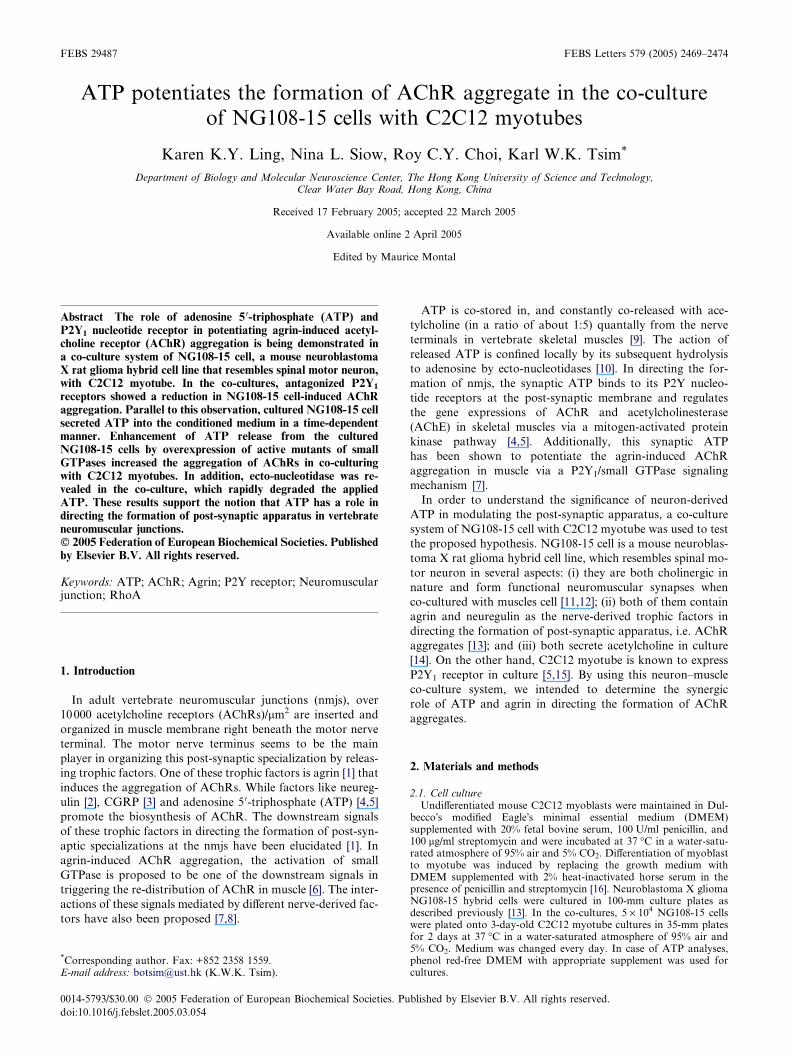
FEBS 29487 FEBS Letters 579 (2005) 2469–2474
ATP potentiates the formation of AChR aggregate in the co-cultureof NG108-15 cells with C2C12 myotubes
Karen K.Y. Ling, Nina L. Siow, Roy C.Y. Choi, Karl W.K. Tsim*
Department of Biology and Molecular Neuroscience Center, The Hong Kong University of Science and Technology,Clear Water Bay Road, Hong Kong, China
Received 17 February 2005; accepted 22 March 2005
Available online 2 April 2005
Edited by Maurice Montal
Abstract The role of adenosine 5 0-triphosphate (ATP) andP2Y1 nucleotide receptor in potentiating agrin-induced acetyl-choline receptor (AChR) aggregation is being demonstrated ina co-culture system of NG108-15 cell, a mouse neuroblastomaX rat glioma hybrid cell line that resembles spinal motor neuron,with C2C12 myotube. In the co-cultures, antagonized P2Y1
receptors showed a reduction in NG108-15 cell-induced AChRaggregation. Parallel to this observation, cultured NG108-15 cellsecreted ATP into the conditioned medium in a time-dependentmanner. Enhancement of ATP release from the culturedNG108-15 cells by overexpression of active mutants of smallGTPases increased the aggregation of AChRs in co-culturingwith C2C12 myotubes. In addition, ecto-nucleotidase was re-vealed in the co-culture, which rapidly degraded the appliedATP. These results support the notion that ATP has a role indirecting the formation of post-synaptic apparatus in vertebrateneuromuscular junctions.� 2005 Federation of European Biochemical Societies. Publishedby Elsevier B.V. All rights reserved.
Keywords: ATP; AChR; Agrin; P2Y receptor; Neuromuscularjunction; RhoA
1. Introduction
In adult vertebrate neuromuscular junctions (nmjs), over
10000 acetylcholine receptors (AChRs)/lm2 are inserted and
organized in muscle membrane right beneath the motor nerve
terminal. The motor nerve terminus seems to be the main
player in organizing this post-synaptic specialization by releas-
ing trophic factors. One of these trophic factors is agrin [1] that
induces the aggregation of AChRs. While factors like neureg-
ulin [2], CGRP [3] and adenosine 5 0-triphosphate (ATP) [4,5]
promote the biosynthesis of AChR. The downstream signals
of these trophic factors in directing the formation of post-syn-
aptic specializations at the nmjs have been elucidated [1]. In
agrin-induced AChR aggregation, the activation of small
GTPase is proposed to be one of the downstream signals in
triggering the re-distribution of AChR in muscle [6]. The inter-
actions of these signals mediated by different nerve-derived fac-
tors have also been proposed [7,8].
*Corresponding author. Fax: +852 2358 1559.E-mail address: [email protected] (K.W.K. Tsim).
0014-5793/$30.00 � 2005 Federation of European Biochemical Societies. Pu
doi:10.1016/j.febslet.2005.03.054
ATP is co-stored in, and constantly co-released with ace-
tylcholine (in a ratio of about 1:5) quantally from the nerve
terminals in vertebrate skeletal muscles [9]. The action of
released ATP is confined locally by its subsequent hydrolysis
to adenosine by ecto-nucleotidases [10]. In directing the for-
mation of nmjs, the synaptic ATP binds to its P2Y nucleo-
tide receptors at the post-synaptic membrane and regulates
the gene expressions of AChR and acetylcholinesterase
(AChE) in skeletal muscles via a mitogen-activated protein
kinase pathway [4,5]. Additionally, this synaptic ATP
has been shown to potentiate the agrin-induced AChR
aggregation in muscle via a P2Y1/small GTPase signaling
mechanism [7].
In order to understand the significance of neuron-derived
ATP in modulating the post-synaptic apparatus, a co-culture
system of NG108-15 cell with C2C12 myotube was used to test
the proposed hypothesis. NG108-15 cell is a mouse neuroblas-
toma X rat glioma hybrid cell line, which resembles spinal mo-
tor neuron in several aspects: (i) they are both cholinergic in
nature and form functional neuromuscular synapses when
co-cultured with muscles cell [11,12]; (ii) both of them contain
agrin and neuregulin as the nerve-derived trophic factors in
directing the formation of post-synaptic apparatus, i.e. AChR
aggregates [13]; and (iii) both secrete acetylcholine in culture
[14]. On the other hand, C2C12 myotube is known to express
P2Y1 receptor in culture [5,15]. By using this neuron–muscle
co-culture system, we intended to determine the synergic
role of ATP and agrin in directing the formation of AChR
aggregates.
2. Materials and methods
2.1. Cell cultureUndifferentiated mouse C2C12 myoblasts were maintained in Dul-
becco�s modified Eagle�s minimal essential medium (DMEM)supplemented with 20% fetal bovine serum, 100 U/ml penicillin, and100 lg/ml streptomycin and were incubated at 37 �C in a water-satu-rated atmosphere of 95% air and 5% CO2. Differentiation of myoblastto myotube was induced by replacing the growth medium withDMEM supplemented with 2% heat-inactivated horse serum in thepresence of penicillin and streptomycin [16]. Neuroblastoma X gliomaNG108-15 hybrid cells were cultured in 100-mm culture plates asdescribed previously [13]. In the co-cultures, 5 · 104 NG108-15 cellswere plated onto 3-day-old C2C12 myotube cultures in 35-mm platesfor 2 days at 37 �C in a water-saturated atmosphere of 95% air and5% CO2. Medium was changed every day. In case of ATP analyses,phenol red-free DMEM with appropriate supplement was used forcultures.
blished by Elsevier B.V. All rights reserved.

2470 K.K.Y. Ling et al. / FEBS Letters 579 (2005) 2469–2474
For some cases, the cultures were pretreated with apyrase (2 U/ml;Sigma) for 1 h to eliminate all free nucleotides, then followed by a gen-tle wash and the drug application in apyrase-free medium [7]. The drugtreatment was applied onto the co-cultures (6 h after plating) for 2 dayswith refreshment of medium and drugs every 24 h. These drugsincluded adenosine 2 0,5 0-diphosphate (A2P5P; specific antagonist forP2Y1 receptor), suramin and pyridoxal-phosphate-6-azophenyl-2 0,4 0-disulphonic acid (tetrasodium salt; PPADS). These P2Y-antagonistswere purchased from Sigma (St. Louis, MO), Botulinum toxin (BoNT;blocks synaptic release of acetylcholine; fromWako Chemicals, Osaka,Japan), and Y27632 (inhibitor of Rho kinase; from Calbiochem, SanDiego, CA).
2.2. cDNA constructs and transfectioncDNAs encoding the wild-type RhoA (RhoAWT; GenBank
AY026068), Rac1 (GenBank AY491395), Cdc42 (GenBankAF205635) were generated by RT-PCR from rat muscle RNA. Theconstitutively active mutants of corresponding GTPases were obtainedby site-directed mutagenesis of the wild-type cDNAs: RhoAV14, aminoacid glycine at position 14 was mutated to valine; Rac1V12 andCdc42V12, amino acid glycine at position 12 was mutated to valine.As for the dominant-negative mutants: RhoAN19, amino acid serineat position 19 was mutated to asparagine; Rac1N17 and Cdc42N17, ami-no acid threonine at position 17 was mutated to asparagine [17]. AllcDNAs were subcloned into pcDNA3, verified therein, and used fortransfection. The transfection of GTPase cDNAs into C2C12 myo-blasts was done by using Lipofectamine Plus (Invitrogen Life Technol-ogies), while the transfection of NG108-15 cells was done by standardcalcium phosphate precipitation [13,16]. The transfection efficiencywas determined by enzymatic staining of control cells that were co-transfected with b-galactosidase cDNA in the same expression vector;it was consistently �30%. After a day of transfection, the myoblastswere induced to form myotube for 3 days. In the co-cultures,NG108-15 cells were added onto C2C12 myotube cultures for 2 days.For the co-culture of transfected NG108-15 cells with C2C12myotubes, the transfected cells were seeded onto myotubes 6 h afterthe transfection.
2.3. Determination of ATP concentrationATP in the conditioned media was assayed by luciferase-luciferin
preparation (Wako Chemicals). Assay premix containing 50 ll ofsample medium in 100 mM phosphate buffer (pH 7.6) and 25 ll lucif-erase-luciferin reagent was subjected to luminescence detection usingFLOUstar Optima microplate reader (BMG Labtechnologies, Offen-burg, Germany). ATP concentration of individual sample was calcu-lated from plot of ATP standards (0–100 nM or 0–100 lM).Determination of ecto-nucleotidase activity was modified from Vol-lmayer et al. [18]. The culture medium were carefully aspirated, andcells were washed twice with phosphate-free physiological saline solu-tion (140 mM NaCl, 5 mM KCl, 1 mM CaCl2, 1 mM MgCl2, 10 mMglucose, and 20 mM HEPES, pH 7.4). ATP of 500 lM (2 ml) in theabove saline buffer was applied onto the cultures (35-mm plate).Hydrolysis of ATP was monitored by assaying the ATP concentrationin the saline buffer in different time intervals. Samples were diluted fivetimes before the ATP assay.
Fig. 1. NG108-15 cell-induced AChR aggregation in the co-culture ismediated by small GTPase. (A) NG108-15 cells were co-cultured withC2C12 myotubes for 2 days, and AChR aggregates were counted.Differentiated NG108-15 cells (dNG108-15) were induced by 1 mMdibutyryl cAMP as described in [13]. Rho kinase inhibitor Y27632(from 5 to 20 lM) was added for 48 h during the course of co-culture.(B) C2C12 myotubes cultured in 35-mm plate were transfected withRhoAWT (2 lg), RhoAN19 (1–4 lg), or pcDNA 3 (2 lg; served as acontrol) were co-cultured with NG108-15 cells to induce AChRaggregation. The neuron-induced AChR aggregation was abolished inmyotubes overexpressing RhoAN19. In both (A) and (B), AChRaggregates were stained and examined as described in Section 2. * inthis and other figures, the difference from the control level (exceptwhere noted) is significant (P < 0.05); ** that difference is highlysignificant (P < 0.001). Values represent the number of AChR aggre-gates per field in a single myotube and in means ± S.E.M., N = 4, eachwith triplicate samples. Bar, 20 lm.
2.4. Other assaysThe AChR aggregates were labeled with 10�8 M tetramethylrhod-
amine-conjugated a-bungarotoxin (TMR-BuTX; Molecular Probes,Eugene, OR) for 1 h at room temperature, washed with PBS, fixedin ethanol and mounted in glycerol [13]. The number of AChR aggre-gates were counted under a 40· objective on Zeiss Axiophot equippedwith phase and fluorescence optics as described in [7]. Western blotanalysis of agrin was performed according to [13] by using anti-agrinantibody AGR-530 (Stressgen Biotechnologies, Victoria, BC, Canada).Protein concentration was determined by using Bio-Rad protein assaykit (Hercules, CA), and by which, the concentration of sample proteinwas compared against the standard curve that was generated from therelative absorbance (595 nm) of BSA standards (0.1–0.6 lg/ml) addedwith dye reagent. Statistical tests were made by the PRIMER program,version 1: differences from basal or control values (as shown in theplots) were classed as significant [\] where P < 0.05 and highly signifi-cant [\\] where P < 0.001.
3. Results
In NG108-15 cell and myotube co-culture, NG108-15 cell is
known to secrete agrin and to cause AChR aggregation on the
surface of myotube [13]. As shown in Fig. 1A, the co-culturing
with NG108-15 cell induced �8-fold increase of AChR aggre-
gate on the surface of C2C12 myotubes. A further increase in
AChR aggregation was revealed when the co-culture was using
differentiated NG108-15 cells. The increase in AChR aggrega-
tion could due to the increased expression of agrin during neu-
ronal differentiation as reported previously [13]. The aggregate
had at least 10 lm in size detectable at the edge and the bottom
of muscle fiber (Fig. 1A). The characteristics of these aggre-
gates were very similar to the previous reports, and which were
K.K.Y. Ling et al. / FEBS Letters 579 (2005) 2469–2474 2471
shown to be induced by NG108-15 cell-released agrin [13]. The
downstream signaling of NG108-15 cell-induced AChR aggre-
gation, in particular the role of small GTPase, has not been
demonstrated in the co-culture. In order to validate this signal-
ing cascade of NG108-15 cell-induced AChR aggregation is in-
deed similar to that of agrin [6,7], Rho kinase inhibitor Y27632
(5–20 lM; effective concentration in blocking agrin-induced
AChR aggregation in C2C12; [6]) was applied onto the neu-
ron–muscle co-culture, and the number of AChR aggregate
was counted. Blocking Rho kinase activity significantly re-
duced the NG108-15 cell-induced AChR aggregation, about
40–80%, in a dose-dependent manner (Fig. 1A). Moreover,
when C2C12 myotube was overexpressed with dominant-neg-
ative RhoA (RhoAN19) and co-cultured with NG108-15 cells
for 2 days, the formation of NG108-15 cell-induced AChR
aggregates was abolished (Fig. 1B). The effect of RhoAN19 in
blocking the formation of AChR aggregate was increased
when the amount of RhoAN19 DNA was increased in the
transfection. In contrast, the overexpression of wild type
RhoA (RhoAWT) did not significantly affect the formation of
AChR aggregate in the neuron–muscle co-culture. These re-
sults support the notion that the signaling mechanism of
NG108-15 cell-induced AChR aggregation in the co-culture
is similar to that of agrin application in myotubes.
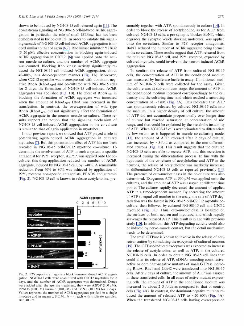
In our previous report, we showed that ATP played a role in
potentiating agrin-induced AChR aggregation in cultured
myotubes [7]. But this potentiation effect of ATP has not been
revealed in NG108-15 cell-C2C12 myotube co-culture. To
determine the involvement of ATP in such a system, a specific
antagonist for P2Y1 receptor, A2P5P, was applied onto the co-
culture; this drug application reduced the number of AChR
aggregate, induced by NG108-15 cell, by �40%. A remarkable
reduction from 60% to 80% was achieved by application of
P2Y1 receptor non-specific antagonists, PPADS and suramin
(Fig. 2). NG108-15 cell is known to release acetylcholine, pre-
Fig. 2. P2Y1-specific antagonists block neuron-induced AChR aggre-gation. NG108-15 cells were co-cultured with C2C12 myotubes for 2days, and the number of AChR aggregates was determined. Drugswere added after the apyrase treatment; they were A2P5P (100 lM),PPADS (100 lM) suramin (100 lM) and BoNT (10 nM) for 2 days.Values represent the number of AChR aggregates per field in a singlemyotube and in means ± S.E.M., N = 4, each with triplicate samples.Bar, 40 lm.
sumably together with ATP, spontaneously in culture [14]. In
order to block the release of acetylcholine, as for ATP, from
cultured NG108-15 cells, a pre-synaptic blocker BoNT, which
degrades the synaptic vesicle docking molecules, was applied
onto the co-culture. Similar to P2Y receptor antagonists,
BoNT reduced the number of AChR aggregate being formed
in the co-culture. These results suggest that ATP, released from
the cultured NG108-15 cell, and P2Y1 receptor, expressed by
cultured myotube, could involve in the neuron-induced AChR
aggregation.
To confirm the release of ATP from cultured NG108-15
cells, the concentration of ATP in the conditioned medium
was measured by luciferase-luciferin assay. Conditioned med-
ium of NG108-15 cells were collected for the assay. Given
the culture was at sub-confluent stage, the amount of ATP in
the conditioned medium increased correspondingly to the cell
density and the culturing time, and which reached a saturating
concentration of �5 nM (Fig. 3A). This indicated that ATP
was spontaneously released by cultured NG108-15 cells into
the medium. In a higher density of cell culture, the amount
of ATP did not accumulate proportionally over longer time
of culture but reached saturation at concentration of nM
range, and that could be explained by the metabolic instability
of ATP. When NG108-15 cells were stimulated to differentiate
by low-serum, as it happened in muscle co-culturing model
[13], the amount of ATP, released after 2 days of culture,
was increased by �5-fold as compared to the non-differenti-
ated neurons (Fig. 3B). This result suggests that the cultured
NG108-15 cells are able to secrete ATP, and this secretion is
increased during the differentiation process. In line with the
hypothesis of the co-release of acetylcholine and ATP in the
neurons, the release of acetylcholine was markedly increased
in differentiated NG108-15 cells as reported previously [14].
The presence of ecto-nucleotidases in the co-culture was also
determined. Exogenous ATP at 500 lM was applied onto the
cultures, and the amount of ATP was assayed at different time
points. The cultures rapidly decreased the amount of applied
ATP in a time-dependent manner. By correcting the amount
of ATP to equal cell number in the assay, the rate of ATP deg-
radation was the fastest in NG108-15 cell-C2C12 myotube co-
culture, then followed by cultured NG108-15 cell and C2C12
myotube (Fig. 3C). Thus, ecto-nucleotidase is localized on
the surfaces of both neuron and myotube, and which rapidly
scavenges the released ATP. This result is in line with previous
study [10]. In addition, this ATP-degrading activity could also
be induced by nerve–muscle contact, but the detail mechanism
needs to be determined.
The small GTPase is known to involve in the release of neu-
rotransmitter by stimulating the exocytosis of cultured neurons
[19]. The GTPase-induced exocytosis was expected to increase
the release of acetylcholine, as well as ATP, in the cultured
NG108-15 cells. In order to obtain NG108-15 cell lines that
could alter its release of ATP, cDNAs encoding constitutive-
active or dominant-negative mutants of small GTPase includ-
ing RhoA, Rac1 and Cdc42 were transfected into NG108-15
cells. After 2 days of culture, the amount of ATP was assayed
in these transfected cells. In all cases of active mutant express-
ing cells, the amount of ATP in the conditioned medium was
increased by about 2–3 folds as compared to that of control
cells (Fig. 4A). In contrast, the dominant-negative mutants re-
duced the amount of released ATP to �20–60% (Fig. 4A).
When the transfected NG108-15 cells having overexpression

Fig. 3. Release and degradation of ATP in the cultures of NG108-15cells and C2C12 cells. (A) Different density of undifferentiatedNG108-15 cells (6.25, 12.5 and 25 · 103) were plated on 12-well plateand cultured with phenol red-free growth medium. Conditionedmedium collected from cultures after 0, 1 and 2 days were subjectedto ATP analyses using luciferase-luciferin preparation. ATP concen-tration, calibrated from a standard curve, was plotted to correlate cellnumber and day of culture. (B) For measuring the ATP released indifferentiated NG108-15 cells, 12.5 · 103 cells were cultured for 1 or 2days in either high-serum medium (undifferentiated) or low-serummedium (differentiated). Conditioned medium were then collectedand subsequently assayed as above. (C) To assay the ecto-nucleotid-ase activity in co-culture, confluent C2C12 myoblasts were stimulatedto form myotubes for 3 days, and then cultured with or withoutNG108-15 cells for another 2 days. Conditioned medium werewashed away twice and replaced with 500 lM ATP in saline buffer,followed by incubation in water-saturated 5% CO2 incubator at37 �C. ATP concentration in bathing solution was collected andassayed as mentioned before. The amount of ATP during the assaywas corrected to equal number of cell density (or cell nucleus inmyotube) in all cases. Values were means ± S.E.M., N = 4, each withtriplicate samples.
Fig. 4. The release of ATP by NG108-15 cell modulates its AChRaggregation activity in the co-culture with C2C12 myotube. (A)NG108-15 cells were transfected with dominant positive/negativemutants of Rho GTPases (RhoAV14/RhoAN19, Rac1V12/Rac1N17 andCdc42V12/Cdc42N17) and control vector. After 2 days of culture, ATPin conditioned medium was collected and assayed as mentioned before.(B) NG108-15 cells were transfected with 2 lg of RhoAN19, RhoAN19,or pcDNA 3 (control) were co-cultured with C2C12 myotubes for 2days to induce AChR aggregation. The neuron-induced AChRaggregation in myotubes was potentiated when co-culturing withneuron over-expressing RhoAV14 and reduced when co-culturing withneuron over-expressing RhoAN19. In addition, apyrase-treated condi-tioned medium (0.5 ml) from these RhoA mutant transfected NG108-15 cells were collected and applied onto cultured C2C12 myotubes (35-mm plate) for testing the bioactivity of agrin. AChR aggregates werestained and examined as in Fig. 1. Inset shows the amount of agrinrecognized by anti-agrin antibody at �200 kDa in a western blot fromthe cell lysates (50 lg protein) of the transfected cells. Values representthe ATP concentration per equal cell density or the number of AChRaggregates per field in a single myotube, and they are inmeans ± S.E.M., N = 4, each with triplicate samples.
2472 K.K.Y. Ling et al. / FEBS Letters 579 (2005) 2469–2474
of either constitutive-active or dominant-negative mutant of
small GTPase (e.g. RhoAV14 and RhoAN19, respectively) were
co-cultured with C2C12 myotubes, the neuron-induced AChR
aggregation was altered. The active mutant (RhoAV14)
expressing NG108-15 cells increased the number of AChR
aggregate. In contrast, the dominant-negative RhoAN19
K.K.Y. Ling et al. / FEBS Letters 579 (2005) 2469–2474 2473
mutant expressing cells reduced the aggregation (Fig. 4B). The
alternation of AChR aggregation under the expressions of dif-
ferent GTPases in the transfected NG108-15 cells did not due
to the change of agrin expression or secretion. The conditioned
medium collected from these RhoA mutant transfected
NG108-15 cells were treated with apyrase to eliminate all free
nucleotides, and which were then applied onto cultured myotu-
bes; they all showed equal activity in aggregating AChR (Fig.
4B). Moreover, the transfected cells expressed equal amount of
agrin as revealed by anti-agrin antibody on their cell lysates
(Fig. 4B, inset). Thus, these results support the hypothesis that
ATP is being released from NG108-15 cells and has a directive
role in forming the post-synaptic apparatus in the neuron–
muscle co-culture.
4. Discussion
The ATP concentration at the nmjs could reach �1 mM
[9,10], which bombards its receptors located on the surface
of post-synaptic membrane. The high concentration of ATP
at the synaptic cleft is maintained whenever the contraction
of muscle is triggered, and therefore the functional role of
ATP is being proposed here. Together with previous studies
from our laboratory, synaptic ATP at the nmj, released mainly
by the motor neuron, plays two distinct roles in directing the
formation of post-synaptic apparatus at vertebrate nmjs: (i)
to stimulate the transcriptional activities of AChR and AChE
genes [4,5]; and (ii) to synergize with agrin in inducing AChR
aggregation [7]. For the first time, the current results in the
neuron–muscle co-culture support the functional role of ATP
in directing AChR aggregation during the nmj formation. Be-
sides the formation of post-synaptic apparatus, several short-
term effects of ATP at the nmjs have also been reported. In
skeletal muscles, ATP potentiated responses to acetylcholine
[20]. In Xenopus embryonic nerve–muscle co-cultures, focal
application of ATP increased spontaneous synaptic currents,
while on myocytes it potentiated the response to acetylcholine
[21]. In addition, ATP also stabilizes the rapidly degraded
AChRs in rat myotubes [22]. Some of aforementioned func-
tions of ATP are being proposed to be mediated by P2Y recep-
tors that localized specifically at the nmjs [20,23].
The biological actions of ATP at the nmjs might be mediated
by different subtypes of P2Y receptors localized on the post-
synaptic membrane. Among different subtypes of P2Y recep-
tors, P2Y1 and P2Y2 receptors are highly expressed in adult
skeletal muscles of avian and rodent, as well as in cultured
C2C12 myotubes [4,15]. These two P2Y receptors share a sim-
ilar downstream signaling mechanism, and the recognition of
these P2Y receptors is mainly depended on their pharmacolog-
ical properties. By using P2Y1-specific antagonist (e.g. A2P5P),
here we have clearly shown that P2Y1 is one of the P2Y recep-
tors to account for the ATP effects in the neuron–muscle
co-culture. However, the possible role of P2Y2 receptor in
mediating the ATP-potentiated effect in this co-culture has
not been determined here. UTP is the strongest agonist for
P2Y2 receptor; however, its concentration at the nmj has not
been determined. The pre-synaptic cholinergic vesicle of motor
neuron is predicted [10] to contain UTP at �10% of their con-
tent of ATP, and thus a significant amount of synaptic UTP
levels is expected at the nmj. Following this prediction, the re-
lease of UTP together with ATP is expected in cultured
NG108-15 cells. In line of this notion, recent analysis on
NG108-15 cell-conditioned medium by HPLC suggested the
low concentration of UTP (Ling et al., unpublished data). In
addition, conversion can also occur of ATP to UTP via ecto-
enzymes and endogenous UDP [24], which might add a further
slower phase to the activation. These different lines of evidence
therefore suggest the possible involvement of P2Y2 receptor in
the aggregating AChRs in our neuron–muscle co-culture.
By acting on its post-synaptic receptors, the synaptic ATP
could be one of the nerve-derived factors that maintain the
nmj specializations. However, we have not yet address the pos-
sible retrograde regulation on the pre-synaptic neurons. Since
mRNA of P2Y2/6 receptor subtypes, but not P2Y1/4, are de-
tected by RT-PCR in NG108-15 cells, while UTP, a specific
agonist of P2Y2 receptor, can activate RhoA in NG108-15
cells (Ling et al., unpublished data), suggesting that synaptic
ATP may act on its receptors on the pre-synaptic neuron
and possibly modulate cytoskeleton rearrangement thereby
receptor distribution in the neuronal counterpart. The treat-
ment of A2P5P in the co-cultures excludes the possibility of
blocking the neuronal P2Y1 receptor that is not present in
NG108-15 cell. Therefore, the response mediated by P2Y1
receptor in the co-culture is in the muscle, not in the neuron.
The functional role of ATP and its receptors in directing the
formation of post-synaptic apparatus might not only restrict to
the nmjs, instead it might extend to neuron–neuron synapses in
the brain. Several lines of evidence support this notion. First,
P2Y1 receptor mRNA and protein are widely expressed in neu-
rons of different regions of the brain [25], while the P2Y2 recep-
tor mRNA has also been shown to occur in many brain
regions [26]. In line with these observations, recent studies
from our laboratory also suggest the expression of P2Y recep-
tors in cultured rat cortical neurons (Tsim et al., unpublished
results). Second, ATP is known to be generally co-released
with neurotransmitter at central and peripheral cholinergic
and bioaminergic synapses [27] and even some GABA-ergic
neuronal synapses [28]. Besides neurons, glial cells have also
been shown to release ATP to modulate the neuronal activity
[29]. Third, the functional role of ATP in neurotransmission
has been proposed, whereas ATP contributes to the regulation
of the NMDA-induced inward currents and facilitates excit-
atory transmission in hippocampal neurons [30]. Lastly,
P2X7 receptor has been reported to be localized at many brain
excitatory pre-synaptic terminals [31], again suggesting the
ATP co-transmission there. Hence, further investigation is
needed as to whether the post-synaptic actions of the P2Y1
and/or P2Y2 receptors now being uncovered at the nmjs have
a wider relevance at such central synapses.
Acknowledgements: This work was supported by grants from the Re-search Grants Council of Hong Kong (HKUST 6283/03M; 6237/04M and AoE/B-15/01 to K.W.K.T.). R.C.Y.C. was supported bythe post-doctoral matching fund from Hong Kong University of Sci-ence and Technology (HKUST). N.L.S. holds a Croucher FoundationScholarship.
References
[1] Sanes, J.R. and Lichtman, J.W. (2001) Induction, assembly,maturation and maintenance of a postsynaptic apparatus. Nat.Rev. Neurosci. 2, 791–805.
2474 K.K.Y. Ling et al. / FEBS Letters 579 (2005) 2469–2474
[2] Fischbach, G.D. and Rosen, K.M. (1997) ARIA: a neuromuscu-lar junction neuregulin. Annu. Rev. Neurosci. 20, 429–458.
[3] Fontaine, B., Klarsfeld, A., Hokfelt, T. and Changeux, J.P. (1986)Calcitonin gene-related peptide, a peptide present in spinal cordmotoneurons, increases the number of acetylcholine receptors inprimary cultures of chick embryo myotubes. Neurosci. Lett. 71,59–65.
[4] Choi, R.C., Man, M.L., Ling, K.K., Ip, N.Y., Simon, J., Barnard,E.A. and Tsim, K.W. (2001) Expression of the P2Y1 nucleotidereceptor in chick muscle: its functional role in the regulation ofacetylcholinesterase and acetylcholine receptor. J. Neurosci. 21,9224–9234.
[5] Choi, R.C., Siow, N.L., Cheng, A.W., Ling, K.K., Tung, E.K.,Simon, J., Barnard, E.A. and Tsim, K.W. (2003) ATP acts viaP2Y1 receptors to stimulate acetylcholinesterase and acetylcholinereceptor expression: transduction and transcription control. J.Neurosci. 23, 4445–4456.
[6] Weston, C., Gordon, C., Teressa, G., Hod, E., Ren, X.D. andPrives, J. (2003) Cooperative regulation by Rac and Rho of agrin-induced acetylcholine receptor clustering in muscle cells. J. Biol.Chem. 278, 6450–6455.
[7] Ling, K.K., Siow, N.L., Choi, R.C., Ting, A.K., Kong, L.W. andTsim, K.W. (2004) ATP potentiates agrin-induced AChR aggre-gation in cultured myotubes: activation of RhoA in P2Y1
nucleotide receptor signaling at vertebrate neuromuscular junc-tions. J. Biol. Chem. 279, 31081–31088.
[8] Trinidad, J.C., Fischbach, G.D. and Cohen, J.B. (2004) TheAgrin/MuSK signaling pathway is spatially segregated from theneuregulin/ErbB receptor signaling pathway at the neuromuscularjunction. J. Neurosci. 20, 8762–8770.
[9] Silinsky, E.M. and Redman, R.S. (1996) Synchronous release ofATP and neurotransmitter within milliseconds of a motor nerveimpulse in the frog. J. Physiol. 492, 815–822.
[10] Zimmermann, H. (1999) Two novel families of ectonucleotidases:molecular structures, catalytic properties and a search forfunction. Trends Pharmacol. Sci. 20, 231–236.
[11] Christian, C.N., Daniels, M.P., Sugiyama, H., Vogel, Z., Jacques,L. and Nelson, P.G. (1978) A factor from neurons increases thenumber of acetylcholine receptor aggregates on cultured musclecells. Proc. Natl. Acad. Sci. USA 75, 4011–4015.
[12] Soreq, H., Miskin, R., Zutra, A. and Littauer, U.Z. (1983)Modulation in the levels and localization of plasminogen activa-tor in differentiating neuroblastoma cells. Dev. Brain Res. 7, 257–269.
[13] Pun, S. and Tsim, K.W. (1997) Antisense agrin cDNA transfec-tion blocks neuroblastoma cell-induced acetylcholine receptoraggregation when co-cultured with myotubes. Mol. Cell Neurosci.10, 87–99.
[14] McGee, R., Simpson, P., Christian, C., Mata, M., Nelson, P. andNirenberg, M. (1978) Regulation of acetylcholine release fromneuroblastoma x glioma hybrid cells. Proc. Natl. Acad. Sci. USA75, 1314–1318.
[15] Tung, E.K., Choi, R.C., Siow, N.L., Jiang, J.X., Ling, K.K.,Simon, J., Barnard, E.A. and Tsim, K.W. (2004) P2Y2 receptoractivation regulates the expression of acetylcholinesterase andacetylcholine receptor genes at vertebrate neuromuscular junc-tions. Mol. Pharmacol. 66, 794–806.
[16] Siow, N.L., Choi, R.C., Cheng, A.W., Jiang, J.X., Wan, D.C.,Zhu, S.Q. and Tsim, K.W. (2002) A cyclic AMP-dependentpathway regulates the expression of acetylcholinesterase duringmyogenic differentiation of C2C12 cells. J. Biol. Chem. 277,36129–36136.
[17] Bishop, A.L. and Hall, A. (2000) Rho GTPases and their effectorproteins. Biochem. J. 348, 241–255.
[18] Vollmayer, P., Koch, M., Braun, N., Heine, P., Servos, J., Israr,E., Kegel, B. and Zimmermann, H. (2001) Multiple ecto-nucle-otidases in PC12 cells: identification and cellular distribution afterheterologous expression. J. Neurochem. 78, 1019–1028.
[19] Doussau, F., Gasman, S., Humeau, Y., Vitiello, F., Popoff, M.,Boquet, P., Bader, M.F. and Poulain, B. (2000) A Rho-relatedGTPase is involved inCa2+-dependent neurotransmitter exocyto-sis. J. Biol. Chem. 275, 7764–7770.
[20] Henning, R.H. (1997) Purinoceptors in neuromuscular transmis-sion. Pharmacol. Ther. 74, 115–128.
[21] Fu, W.M., Chen, Y.H., Lee, K.F. and Liou, J.C. (1997)Regulation of quantal transmitter secretion by ATP and proteinkinases at developing neuromuscular synapses. Eur. J. Neurosci.9, 676–685.
[22] O�Malley, J.P., Moore, C.T. and Salpeter, M.M. (1997) Stabil-ization of acetylcholine receptors by exogenous ATP and itsreversal by cAMP and calcium. J. Cell. Biol. 138, 159–165.
[23] Tsim, K.W., Choi, R.C., Siow, N.L., Ling, K.K., Jiang, J.X.,Tung, E.K, Lee, H.H., Xie, Q.H., Simon, J. and Barnard,E.A. (2003) ATP induces post-synaptic gene expressions invertebrate skeletal neuromuscular junctions. J. Neurocyto. 32,603–617.
[24] Lazarowski, E.R., Homolya, L., Boucher, R.C. and Harden, T.K.(1997) Identification of an ecto-nucleoside diphosphokinase andits contribution to interconversion of P2 receptor agonists. J. Biol.Chem. 272, 20402–20407.
[25] Webb, T.E., Simon, J. and Barnard, E.A. (1998) Regionaldistribution of [35S]2 0-deoxy 50-O-(1-thio) ATP binding sites andthe P2Y1 messenger RNA within the chick brain. Neuroscience84, 825–837.
[26] Moore, D.J., Chambers, J.K., Wahlin, J.P., Tan, K.B., Moore,G.B., Jenkins, O., Emson, P.C. and Murdock, P.R. (2001)Expression pattern of human P2Y receptor subtypes: a quanti-tative reverse transcription-polymerase chain reaction study.Biochim. Biophys. Acta 1521, 107–119.
[27] Zimmermann, H. (1994) Signalling via ATP in the nervoussystem. Trends Neurosci. 17, 420–426.
[28] Jo, Y.-H. and Role, L.W. (2002) Coordinate release of ATP andGABA at in vitro synapses of lateral hypothalamic neurons. J.Neurosci. 22, 4794–4804.
[29] Newman, E.A. (2003) Glial cell inhibition of neurons by release ofATP. J. Neurosci. 23, 1659–1666.
[30] Ortinau, S., Laube, B. and Zimmermann, H. (2003) ATP inhibitsNMDA receptors after heterologous expression and in culturedhippocampal neurons and attenuates NMDA-mediated neuro-toxicity. J. Neurosci. 23, 4996–5003.
[31] Sperlagh, B., Kofalvi, A., Deuchars, J., Atkinson, L., Milligan,C.J., Buckley, N.J. and Vizi, E.S. (2002) Involvement of P2X7
receptors in the regulation of neurotransmitter release in the rathippocampus. J. Neurochem. 81, 1196–1211.
Copyright © 2022 FDOKUMEN