Atmospheric pressure chemical ionization tandem mass spectrometry of carotenoids
10
International Journal of Mass Spectrometry 312 (2012) 163–172 Contents lists available at ScienceDirect International Journal of Mass Spectrometry j our na l ho me page: www.elsevier.com/locate/ijms Atmospheric pressure chemical ionization tandem mass spectrometry of carotenoids Richard B. van Breemen ∗ , Linlin Dong, Natasa D. Pajkovic 1 Department of Medicinal Chemistry and Pharmacognosy, University of Illinois College of Pharmacy, Chicago, IL 60612, United States a r t i c l e i n f o Article history: Received 29 April 2011 Received in revised form 23 July 2011 Accepted 27 July 2011 Available online 4 August 2011 Keywords: Carotenoid Carotene Xanthophyll Lycopene APCI Tandem mass spectrometry a b s t r a c t Carotenoids are natural pigments synthesized by plants and photosynthetic microorganisms, some of which, like -carotene, are precursors of vitamin A, and others such as lutein and lycopene might function in the prevention of age-related macular degeneration and prostate cancer, respectively. Mass spectrom- etry provides high sensitivity and selectivity for the identification and quantitative analysis of carotenoids in biological samples, and previous studies have described how atmospheric pressure chemical ioniza- tion (APCI) offers distinct advantages over electrospray and fast atom bombardment for the analysis of specific carotenoids. Since APCI product ion tandem mass spectra have been reported for only a few carotenoids, a detailed investigation of twelve carotenes and xanthophylls was carried out using both positive ion and negative ion APCI tandem mass spectrometry with collision-induced dissociation. Using protonated molecules as precursor ions in positive ion mode and radical anions in negative ion mode, characteristic fragment ions were identified that may be used to distinguish between carotenoids. © 2011 Elsevier B.V. All rights reserved. 1. Introduction Carotenoids are polyisoprenoid compounds consisting of two groups, hydrocarbon carotenes and oxygenated derivatives called xanthophylls. The extended system of conjugated double bonds of carotenoids produce characteristic UV and visible absorption spec- tra (including yellow, orange and red) with absorption maxima in the range of 400–500 nm [1]. Synthesized by photosynthetic plants and microorganisms, more than 600 carotenoids have been identi- fied [2]. In plants, carotenoids protect plants from photo-oxidative damage and transfer energy to chlorophyll in the process of pho- tosynthesis [3]. In addition to these functions, approximately 50 carotenoids are precursors of vitamin A which is essential for vision, cellular differentiation and embryological development. It has been suggested that carotenoids might function as in vivo antioxidants, prevent age-related macular degeneration [4] and prevent various forms of cancer including prostate cancer [5]. The identification and quantitative analysis of carotenoids can be challenging due to their instability in the presence of light, ∗ Corresponding author at: Department of Medicinal Chemistry and Pharmacog- nosy, University of Illinois College of Pharmacy, 833 S. Wood St., Chicago, IL 60612, United States. Tel.: +1 312 996 9353; fax: +1 312 996 7107. E-mail address: [email protected] (R.B. van Breemen). 1 Current address: Merck & Co., 770 Sumneytown Pike, West Point, PA 19486, United States. heat and oxygen. In biological samples such as human serum and tissue, carotenoids are often present at low concentrations and in the presence of potentially interfering compounds. Since carotenoids are unstable at the high temperatures used for gas chromatography, high-performance liquid chromatography (HPLC) is usually used for their separation, and UV/vis absorbance and/or mass spectrometry is used for the detection and characterization [6]. In particular, the application of liquid chromatography–mass spectrometry (LC–MS) to the analysis of carotenoids offers the advantages of high sensitivity and superior selectivity that are essential for the identification and quantitation of carotenoids. Among the ionization techniques compatible with LC–MS, the most successful for carotenoid studies have been electro- spray [7] and atmospheric pressure chemical ionization (APCI) [8]. Although carotenes and xanthophylls form molecular ions or proto- nated molecules during positive ion electrospray, the hydrocarbon carotenes do not ionize when using negative ion electrospray. However, APCI forms abundant positively or negatively charged molecular ions or protonated and deprotonated molecules of both carotenes and xanthophylls. Although mass spectrometry has been used for carotenoid analysis for many years, there have been few tandem mass spectrometric studies of these compounds. Previously, the most comprehensive tandem mass spectrometric analysis of carotenoids utilized fast atom bombardment (FAB) in studies of 17 carotenoids [9]. Using FAB, molecular ions of both xanthophylls and carotenes were detected in positive mode, but no carotenoid ions were detected during negative ion FAB mass spectrometry. 1387-3806/$ – see front matter © 2011 Elsevier B.V. All rights reserved. doi:10.1016/j.ijms.2011.07.030
-
Upload
independent -
Category
Documents
-
view
0 -
download
0
Transcript of Atmospheric pressure chemical ionization tandem mass spectrometry of carotenoids

Ao
RD
a
ARRAA
KCCXLAT
1
gxcttafi
dtccspf
b
nU
U
1d
International Journal of Mass Spectrometry 312 (2012) 163– 172
Contents lists available at ScienceDirect
International Journal of Mass Spectrometry
j our na l ho me page: www.elsev ier .com/ locate / i jms
tmospheric pressure chemical ionization tandem mass spectrometryf carotenoids
ichard B. van Breemen ∗, Linlin Dong, Natasa D. Pajkovic1
epartment of Medicinal Chemistry and Pharmacognosy, University of Illinois College of Pharmacy, Chicago, IL 60612, United States
r t i c l e i n f o
rticle history:eceived 29 April 2011eceived in revised form 23 July 2011ccepted 27 July 2011vailable online 4 August 2011
a b s t r a c t
Carotenoids are natural pigments synthesized by plants and photosynthetic microorganisms, some ofwhich, like �-carotene, are precursors of vitamin A, and others such as lutein and lycopene might functionin the prevention of age-related macular degeneration and prostate cancer, respectively. Mass spectrom-etry provides high sensitivity and selectivity for the identification and quantitative analysis of carotenoidsin biological samples, and previous studies have described how atmospheric pressure chemical ioniza-tion (APCI) offers distinct advantages over electrospray and fast atom bombardment for the analysis of
eywords:arotenoidaroteneanthophyllycopenePCI
specific carotenoids. Since APCI product ion tandem mass spectra have been reported for only a fewcarotenoids, a detailed investigation of twelve carotenes and xanthophylls was carried out using bothpositive ion and negative ion APCI tandem mass spectrometry with collision-induced dissociation. Usingprotonated molecules as precursor ions in positive ion mode and radical anions in negative ion mode,characteristic fragment ions were identified that may be used to distinguish between carotenoids.
andem mass spectrometry
. Introduction
Carotenoids are polyisoprenoid compounds consisting of tworoups, hydrocarbon carotenes and oxygenated derivatives calledanthophylls. The extended system of conjugated double bonds ofarotenoids produce characteristic UV and visible absorption spec-ra (including yellow, orange and red) with absorption maxima inhe range of 400–500 nm [1]. Synthesized by photosynthetic plantsnd microorganisms, more than 600 carotenoids have been identi-ed [2].
In plants, carotenoids protect plants from photo-oxidativeamage and transfer energy to chlorophyll in the process of pho-osynthesis [3]. In addition to these functions, approximately 50arotenoids are precursors of vitamin A which is essential for vision,ellular differentiation and embryological development. It has beenuggested that carotenoids might function as in vivo antioxidants,revent age-related macular degeneration [4] and prevent various
orms of cancer including prostate cancer [5].The identification and quantitative analysis of carotenoids cane challenging due to their instability in the presence of light,
∗ Corresponding author at: Department of Medicinal Chemistry and Pharmacog-osy, University of Illinois College of Pharmacy, 833 S. Wood St., Chicago, IL 60612,nited States. Tel.: +1 312 996 9353; fax: +1 312 996 7107.
E-mail address: [email protected] (R.B. van Breemen).1 Current address: Merck & Co., 770 Sumneytown Pike, West Point, PA 19486,nited States.
387-3806/$ – see front matter © 2011 Elsevier B.V. All rights reserved.oi:10.1016/j.ijms.2011.07.030
© 2011 Elsevier B.V. All rights reserved.
heat and oxygen. In biological samples such as human serumand tissue, carotenoids are often present at low concentrationsand in the presence of potentially interfering compounds. Sincecarotenoids are unstable at the high temperatures used for gaschromatography, high-performance liquid chromatography (HPLC)is usually used for their separation, and UV/vis absorbance and/ormass spectrometry is used for the detection and characterization[6]. In particular, the application of liquid chromatography–massspectrometry (LC–MS) to the analysis of carotenoids offers theadvantages of high sensitivity and superior selectivity that areessential for the identification and quantitation of carotenoids.
Among the ionization techniques compatible with LC–MS,the most successful for carotenoid studies have been electro-spray [7] and atmospheric pressure chemical ionization (APCI) [8].Although carotenes and xanthophylls form molecular ions or proto-nated molecules during positive ion electrospray, the hydrocarboncarotenes do not ionize when using negative ion electrospray.However, APCI forms abundant positively or negatively chargedmolecular ions or protonated and deprotonated molecules of bothcarotenes and xanthophylls.
Although mass spectrometry has been used for carotenoidanalysis for many years, there have been few tandem massspectrometric studies of these compounds. Previously, the mostcomprehensive tandem mass spectrometric analysis of carotenoids
utilized fast atom bombardment (FAB) in studies of 17 carotenoids[9]. Using FAB, molecular ions of both xanthophylls and caroteneswere detected in positive mode, but no carotenoid ions weredetected during negative ion FAB mass spectrometry.
1 rnal o
Fttcosdossspt
2
2
f�w�U3�dl
64 R.B. van Breemen et al. / International Jou
Since atmospheric pressure ionization techniques have replacedAB for most mass spectrometric applications and since, amonghese ionization techniques, only APCI can form both posi-ively charged and negatively charged molecular ion species ofarotenoids, we carried out tandem mass spectrometric analysisf a series of twelve carotenoids using APCI. Product tandem masspectra of protonated carotenoids obtained using APCI MS–MS pro-uced fragment ions that were similar to the tandem mass spectraf radical cations observed previously using FAB, but there wereome significant differences. Although negative ion tandem masspectra of carotenoids obtained using APCI MS–MS exhibited someimilar fragmentation, unique and complementary fragmentationathways were observed that may be used for carotenoid charac-erization, identification and measurement.
. Materials and methods
.1. Materials
HPLC-grade methanol, acetonitrile and hexane were purchasedrom Fisher Scientific (Fair Lawn, NJ). Lycopene (�,�-carotene),-carotene ((6′R)-�,�-carotene) and �-carotene (�,�-carotene)ere purchased from Sigma–Aldrich (St. Louis, MO). [13C6]--Carotene was a gift from Johan Lugtenburg of Leidenniversity (Leiden, The Netherlands). Astaxanthin ((3S,3′S)-
,3′-dihydroxy-�,�-carotene-4,4′-dione), �-cryptoxanthin ((3R)-,�-carotene-3-ol), zeaxanthin ((3R,3′R,6′R)-�,�-carotene-3,3′-iol), capsanthin ((3R,3′S,5′R)-3,3′-dihydroxy-�,�-caroten-6′-one),utein ((3R,3′R,6′R)-�,�-carotene-3,3′-diol), and helenien ((3R,3′R)-
Hα-Carotene
1
35
17 16
18
97 1311 13’ 9’11’ 7’
18’3’
1’
16’ 17’15’
15
5’
OHβ-Carotene
Lycopen e
53116
17
7 9 11 13 15
201918
13’11’
9’7’
5’ 3’ 1’
17’
16
18’19’20’15’
H
γ-Carotene
C15H31O
HO
H
β-Apo-8’-carotenal
O
O
HelenienHC15H31O
Fig. 1. Carotenoid chem
f Mass Spectrometry 312 (2012) 163– 172
�,�-carotene-3,3′-diol-dipalmitate) were purchased from Chro-maDex (Santa Ana, CA). Echinenone was purchased fromCaroteNature (Lupsingen, Switzerland), and �-apo-8′-carotenal(8′-apo-�-caroten-8′-al) was purchased from Hoffman-La Roche(Nutley, NJ). �-Carotene was isolated from a tomato oleoresin(LycoRed, Beer-Sheva, Israel) using reversed phase HPLC asdescribed previously [9]. Chemical structures of these carotenoidsare shown in Fig. 1.
2.2. Methods
Positive and negative ion APCI mass spectra and product iontandem mass spectra were obtained using a Thermo (San Jose,CA) Quantum triple quadrupole mass spectrometer, and accuratemass measurements of product ions were confirmed using a Waters(Manchester, UK) Q-TOF-2 or QTOF Synapt quadrupole time-of-flight hybrid mass spectrometer. Stock solutions of 100 �M wereprepared under dim light by dissolving 1 mg of each carotene orxanthophyll in hexane or methanol, respectively. Each stock solu-tion was diluted to a concentration of approximately 2 �M usingmethanol prior to positive ion APCI or acetonitrile prior to negativeion APCI. Carotenoid solutions were introduced into the mass spec-trometer by flow-injection or direct infusion at a solvent flow rateof 200–250 �L/min. The ion source parameters were optimized foreach carotenoid using positive ion or negative ion APCI.
For the experiments carried out on the Q-TOF-2 quadrupoletime-of-flight hybrid mass spectrometer, the optimum corona cur-rent was between 3–5 �A during positive ion APCI and 4–6 �Aduring negative ion APCI. The cone voltage was optimized to
O
O
OHO
OAstaxan thin
O
OH
O CapsanthinCapsanthin
Echinenone
’
O β Cryptoxanthin
O
OH
L t i
O β-Cryptoxanthin
OH
Z thi
OH Lutein
O Zeaxanthin
ical structures.

rnal of
3ioancsta
3
3
pppaAdsmsn
R.B. van Breemen et al. / International Jou
0–40 V. Argon was used as the collision gas and the collision-nduced dissociation energies were 15–40 eV. The instrumentalperating parameters for the TSQ Quantum triple quadrupole weres follows: discharge current 5–8 �A for positive and 8–10 �A foregative ion APCI; APCI vaporizer temperature 350–400 ◦C; andapillary temperature 300 ◦C. Positive and negative ion APCI masspectra were recorded followed by the corresponding product ionandem mass spectra of the protonated molecules or the molecularnions, respectively.
. Results
.1. APCI MS
Protonated molecules were detected as the base peaks in theositive ion APCI mass spectra of all of the carotenes and xantho-hylls except for those of lutein and helenien, in which the baseeaks corresponded to the loss of water, [MH−18]+, or palmiticcid, [MH−C16H31COOH]+, respectively. When using positive ionPCI, molecular ions and/or protonated molecules may be observedepending on the solvent composition [8], and the use of protic
olvents such as methanol as in this investigation favored the for-ation of protonated carotenoids. In the negative ion APCI masspectra of all the carotenes and xanthophylls, except that of hele-ien, molecular ions were the base peaks. The base peak of helenien,
100137.1
β-Carote269
H137 177
H
50177.2
147.1 203.2269.2
209.2 321.
H
413400
H
100
137.1
177.2
[13C6]-β-Ca137
* *
272H
0150 20 0 25 0 30 0
bund
ance
0
50121.1
147.1
203.2272.2
215.2326.
419406 *
150 200 250 300
Rel
ativ
e a 150 200 250 300100
50
123.1α-Carot137
H
100 121.1Lyco
0
137.1 177.2147.1
203.2 269.2 321.
413
150 20 0 25 0 30 0
50
137.1
177.2203.2
269.2 321 3
413
Lyco
m/z150 20 0 25 0 30 0
0
321.3
Fig. 2. Positive ion APCI tandem mass spectra of carotenes. (A) �-Caro
Mass Spectrometry 312 (2012) 163– 172 165
which is a palmitoyl ester, corresponded to palmitate. Deproto-nated molecules were also detected in all of the negative ion APCImass spectra, especially those of the carotenes, but were less abun-dant than the molecular ions.
3.2. APCI MS–MS
Since most carotenoids showed little fragmentation duringAPCI mass spectrometry, CID with product ion tandem mass spec-trometry was used to form fragment ions that might be helpfulfor structural characterization. Positive ion APCI tandem massspectra of the carotenes and selected xanthophylls are shown inFigs. 2 and 3, respectively, and negative ion APCI tandem massspectra of carotenes and selected xanthophylls are shown inFigs. 4 and 5, respectively. The tandem mass spectra of all twelvecarotenoids are summarized in Table 1 (positive ion APCI) andTable 2 (negative ion APCI).
3.2.1. ˇ-Carotene and [13C6]-ˇ-caroteneIn the positive ion APCI tandem mass spectrum of �-carotene
(Fig. 2 and Table 1), an ion of low abundance was observed at
m/z 445 corresponding to the elimination of toluene, [MH−92]+,from the protonated molecule. A corresponding ion was detectedat m/z 448 in the tandem mass spectrum of [13C6]-�-carotene.Comparing the tandem mass spectra of �-carotene and [13C6]-537.4[M+H]+ne
H+80
3 347.3 445.4
413.3 457.4
[MH-92]+ [MH-80]+
400.3
H
92
H
543.5[M+H]+
+rotene
H
* *
83H
400.3
350 40 0 45 0 50 0
3460.4353.3
419.3
448.4[MH-83]+[MH-95]+
406.3
*95
350 400 450 500350 400 450 500
537 4[M+H]+
ene
123
413 H +
137177
pene
537.4
3 347.3 413.3 457.4[MH-80]+
123
350 40 0 45 0 50 0
537.4[M+H]+
[MH-80]+
H +137
H
pene H
350 40 0 45 0 50 0
457.4347.3 413.3
A
B
C
D
tene; (B) [13C6]-�-carotene; (C) �-carotene; and (D) lycopene.

166 R.B. van Breemen et al. / International Journal of Mass Spectrometry 312 (2012) 163– 172
100 147.1Astaxan thin OH
O147
HH
H +
0
50
135.1173.1
201.1 597.4285.2 379.3 579.4
[M+H]+[MH-18]+
HOO
H
150 20 0 25 0 30 0 35 0 40 0 45 0 50 0 55 0 60 00
100
50
135.1
119.1553 4177 2[M+H]+
β-Cryptoxanthin H137
135
0
50 553.4177.2145.1
203.2461.4267.2
400.3 [MH-18]+[MH-92]+
[MH-153]+.
535.4473.4
abu
ndan
ce
085045083043003062022081041 420 46 0 500
HO 400
100
H+
Rel
ativ
e
Lutein
HO
OH
430H
+
135
100
50
135.1
119.1 175.214 1
[M+H]+416
551.4
420 46 0 50 0
477.4H
0850450830430030620220810410
145.1 569.4
495.4459.4430.3
[ ]
[MH-139]+.[MH-18-56]+
416.3
416
100135.1
50119.1
569.4
175.2
145.1
[M+H]+
Zeaxanthin135 OH
HO416 H+
0416.3 477.4 551.4
[MH-18]+[MH-153]+. [MH-92]+
m/z085045083043003062022081041001 42 0 46 0 50 0
489.4
A
C
D
B
Fig. 3. Positive ion APCI tandem mass specta of selected xanthophylls. (A) Astaxanthin; (B) �-cryptoxanthin; (C) lutein; and (D) zeaxanthin.
Table 1Positive ion APCI product ion tandem mass spectra with collision-induced dissociation of protonated carotenoids.
Carotenoid Formula [M+H]+ [MH–H2O]+ [MH–C6H8]+ [MH–C7H8]+ Other significant ions
�-Apo-8′-carotenal C30H40O 417.3a (100) 399.3 (4.5) 325.3 (7.9) 95.1 (43.7); 119.1 (58.0); 157.1 (47.8); 293.2 (14.4); 338.3(10.0); 361.3 (5.6); 389.3 (2.1)
Astaxanthin C40H54O4 597.4 (13.2) 579.4 (3.8) 147.1 (100); 173.1 (29.2); 201.1 (17.0); 285.2 (8.6); 379.3 (5.9)Capsanthin C40H56O3 585.4 (100) 567.4 (22.2) 505.4 (3.6) 493.4 (4.9) 109.1 (77.5); 127.1 (63.1); 135.1 (10.2); 219.2 (12.5); 475.4
(4.7); 487.4 (2.2)�-Carotene C40H56 537.4 (34.5) 457.4 (3.2) 123.1 (100);b 137.1 (29.3); 177.2 (28.9); 413.3 (3.8)�-Carotene C40H56 537.4 (100) 457.4 (4.6) 445.4 (5.9) 137.1 (83.0); 177.2 (56.8); 269.2 (19.0); 400.3 (4.4); 413.3 (3.5)[13C10]-�-Carotene 12C30
13C10H56 543.5 (100) 460.4 (10.0) 448.4 (4.9) 137.1 (77.4); 177.2 (61.8); 272.2 (24.6); 406.3 (5.8); 419.3 (6.4)�-Carotene C40H56 537.4 (100) 457.4 (7.9) 119.1 (73.9); 137.1 (45.2); 145.1 (55.9); 177.2 (50.5); 255.2
(20.0); 269.2 (7.3); 399.3 (7.9)�-Cryptoxanthin C40H56O 553.4 (45.3) 535.4 (1.4) 473.4 (4.0) 461.4 (12.4) 119.1 (53.1); 135.1 (100); 177.2 (41.9); 400.3 (6.3)Echinenone C40H54O 551.4 (91.0) 471.4 (8.4) 459.4 (31.3) 133.1 (38.1); 203.1 (100); 255.2 (32.7); 495.4 (1.6); 536.4 (2.7)Helenien C72H116O4 1045.9 (91.8) 953.8 (18.4) 135.1 (100); 175.1(63.2); 201.2 (56.8); 267.2 (41.2); 331.2
(24.5); 411.3 (30.8); 533.4 (67.3); 697.6 (27.0); 789.7 (68.4);817.7 (19.6)
Lutein C40H56O2 569.4 (29.8) 551.4 (2.8) 477.4 (5.9) 119.1 (44.2); 135.1 (100); 175.2 (40.6); 416.3 (5.1); 430.3 (4.9);459.4 (5.3); 495.4 (5.5)
Lycopene C40H56 537.4 (50.6) 457.4 (12.1) 121.1 (100); 137.1 (62.9); 177.2 (53.1); 413.3 (4.9)Zeaxanthin C40H56O2 569.4 (54.1) 551.4 (3.6) 489.4 (3.3) 477.4 (5.7) 119.1 (33.2); 135.1 (100); 175.2 (37.6); 416.3 (6.0), 459.4 (2.6)
a m/z (relative abundance).b Bold-face indicates fragment ions of particular utility for distinguishing isomers.

R.B. van Breemen et al. / International Journal of Mass Spectrometry 312 (2012) 163– 172 167
-.
β-Carotene
H
203H
243269
HH
295100
536.4M-.
H
189 H
295
H
50
444.4243.2203.2
189.2 430.4
[M-92]-.
457.4[M-79]-
295.2269.2[M-106]-.
-.480269203
H H
α-Carotene
0100 15 0 20 0 600300 055052 35 0 50 0450400
100
536.4M-.
abun
danc
e
0
50
444.4203.2189.2 430.4
269.2243.2 480.4[M-56]-.
[M-106]-.
100 150 200 600300250 550350 500450400
[M-92]-.
295.2
Rel
ativ
e a 100 150 200 600300250 550350 500450400
100
50
467.4[M-69]-
[M-69-92]-
Lycopen e
467
-.
0
398.3361.3
375.3
536.4M-.[M-69-69]-.[M-69-106]-
006002051001 30 0 055052 35 0 50 0450400
[M 69 92]
100 γ-Carotene 467269203
467.4
50
M-.
[M-69]-
[M-69-92]-
γ-Carotene 467
-.
[M-69-106]-
269203
H H
m/z
0361.3269.2203.2 375.3 536.4
450 55 0500400350150 20 0 25 0 30 0
M[ ][ ]
100
A
C
D
B
Fig. 4. Negative ion APCI tandem mass specta of carotenes. (A) �-Carotene; (B) �-carotene; (C) lycopene; and (D) �-carotene.
Table 2Negative ion APCI product ion tandem mass spectra with collision-induced dissociation of carotenoid molecular anions.
Carotenoid Formula M− [M−H2O]− [M−C7H8]− [M−C8H10]− Other significant ions
�-Apo-8′-carotenal C30H40O 416.3a (100) 239.2 (10.0); 280.2 (4.3); 359.3 (2.8); 401.3 (4.9)Astaxanthin C40H54O4 596.4 (100) 504.4 (49.6) 490.4 (16.0) 167.0 (54.8); 203.0 (25.3); 233.1 (53.9); 297.2 (26.5);
323.3 (10.8); 337.3 (38.4); 363.3 (22.5); 389.3 (47.7);429.4 (9.6); 581.4 (9.3)
�-Carotene C40H56 536.4 (100) 444.4 (19.0) 430.4 (13.7) 189.2 (13.5); 203.2 (17.7); 243.2 (11.5); 269.2 (12.5);295.2 (6.2); 413.4 (2.2); 457.4 (4.7); 480.4 (15.0)b
�-Carotene C40H56 536.4 (100) 444.4 (19.6) 430.4 (11.4) 189.2 (9.3); 203.2 (13.4); 243.2 (13.6); 269.2 (7.0);295.2 (3.9); 457.4 (5.4)
�-Carotene C40H56 536.4 (5.1) 203.2 (2.3); 269.2 (3.3); 361.3 (8.1); 375.3 (7.4); 467.4(100)
Capsanthin C40H56O3 584.4 (100) 569.4 (1.4) 492.4 (2.5) 478.3 (1.4) 151.1 (11.1); 169.1 (23.8); 209.2 (7.9); 235.2 (30.3);246.2 (25.2); 273.2 (18.6); 299.2 (14.7); 325.2 (15.9);365.2 (8.3); 375.3 (46.1); 391.3 (29.1)
�-Cryptoxanthin C40H56O 552.4 (100) 534.4 (39.4) 203.1 (5.6); 243.1 (5.1); 269.1 (5.1); 519.4 (37.0)Echinenone C40H54O2 550.4 (100) 458.4 (14.4) 444.3 (5.3) 151.1 (5.7); 191.1 (5.8); 217.2 (4.4); 373.3 (8.3); 535.4
(2.6)Helenien C72H116O4 1044.9 (3.1) 255.2 (100)Lutein C40H56O2 568.5 (100) 550.5 (36.6) 137.1 (8.2); 429.4 (2.6); 512.4 (2.7); 535.4 (29.3)Lycopene C40H56 536.4 (8.4) 361.3 (17.6); 375.3 (9.0); 398.3 (19.3); 467.4 (100)Zeaxanthin C40H56O2 568.5 (100) 550.5 (82.6) 137.1 (3.2); 187.0 (5.6); 201.1 (9.4); 267.1 (2.7); 362.1
(4.2); 466.3 (6.0), 535.4 (67.2)
a m/z (relative abundance).b Bold-face indicates fragment ions of particular utility for distinguishing isomers.
168 R.B. van Breemen et al. / International Journal of Mass Spectrometry 312 (2012) 163– 172
167OH
O233
HH
HH
-.
596.4M-.
100 Astaxan thin
HOO
429 363
389
H
H
H
50167.1
233.2
203.1504.4
389.2337.2
297.2 363.2490.4
429.3 581.4[M-15]-
[M-92]-.
[M-106]-.
-.β-Cryptoxanthin
203H243
269
H H
0150 20 0 25 0 30 0 35 0 40 0 45 0 50 0 550 600
M-.100
552.4
[M 18]
HO
abun
danc
e
0
50 534.4
519.4
203.1
[M-18-15]-
[M-18]-.
150 200 250 300 350 400 450 500 550 600
243.1 269.2
-.Lutein
OH512
Rel
ativ
e a 150 200 250 300 350 400 450 500 550 600100
50
568.4
550 4
M-.
[M-18]-.
HO 429
150 20 0 25 0 30 0 35 0 40 0 45 0 50 0 55 0 60 00
550.4
535.4137.1
201.2 429.3 512.4
M-18-15]-
100 568.4 M-.
Zeaxanthin
HO
OH
550H
201 -.
50
550.4
535.4
[M-18]-.
[M-18-15]-
m/z100 15 0 20 0 25 0 30 0 35 0 40 0 45 0 50 0 55 00
201.2187.2 362.3137.1 267.2 466.4
A
C
D
B
ylls. (A
�padobLcaocfwni
tatra�
Fig. 5. Negative ion APCI tandem mass spectra of selected xanthoph
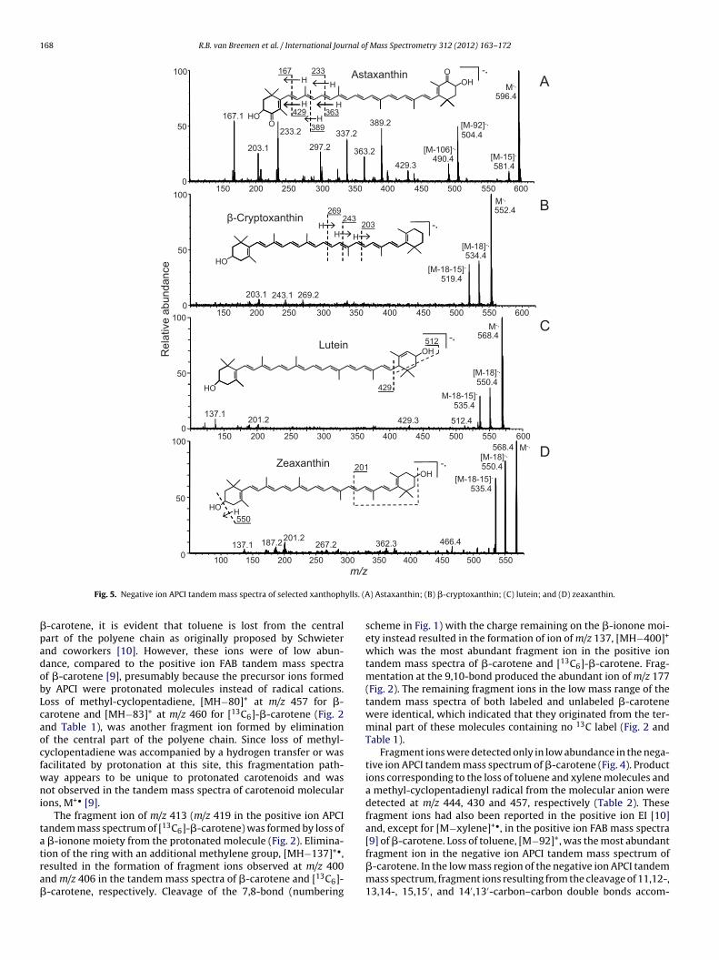
-carotene, it is evident that toluene is lost from the centralart of the polyene chain as originally proposed by Schwieternd coworkers [10]. However, these ions were of low abun-ance, compared to the positive ion FAB tandem mass spectraf �-carotene [9], presumably because the precursor ions formedy APCI were protonated molecules instead of radical cations.oss of methyl-cyclopentadiene, [MH−80]+ at m/z 457 for �-arotene and [MH−83]+ at m/z 460 for [13C6]-�-carotene (Fig. 2nd Table 1), was another fragment ion formed by eliminationf the central part of the polyene chain. Since loss of methyl-yclopentadiene was accompanied by a hydrogen transfer or wasacilitated by protonation at this site, this fragmentation path-ay appears to be unique to protonated carotenoids and wasot observed in the tandem mass spectra of carotenoid molecular
ons, M+• [9].The fragment ion of m/z 413 (m/z 419 in the positive ion APCI
andem mass spectrum of [13C6]-�-carotene) was formed by loss of �-ionone moiety from the protonated molecule (Fig. 2). Elimina-
ion of the ring with an additional methylene group, [MH−137]+•,esulted in the formation of fragment ions observed at m/z 400nd m/z 406 in the tandem mass spectra of �-carotene and [13C6]--carotene, respectively. Cleavage of the 7,8-bond (numbering) Astaxanthin; (B) �-cryptoxanthin; (C) lutein; and (D) zeaxanthin.
scheme in Fig. 1) with the charge remaining on the �-ionone moi-ety instead resulted in the formation of ion of m/z 137, [MH−400]+
which was the most abundant fragment ion in the positive iontandem mass spectra of �-carotene and [13C6]-�-carotene. Frag-mentation at the 9,10-bond produced the abundant ion of m/z 177(Fig. 2). The remaining fragment ions in the low mass range of thetandem mass spectra of both labeled and unlabeled �-carotenewere identical, which indicated that they originated from the ter-minal part of these molecules containing no 13C label (Fig. 2 andTable 1).
Fragment ions were detected only in low abundance in the nega-tive ion APCI tandem mass spectrum of �-carotene (Fig. 4). Productions corresponding to the loss of toluene and xylene molecules anda methyl-cyclopentadienyl radical from the molecular anion weredetected at m/z 444, 430 and 457, respectively (Table 2). Thesefragment ions had also been reported in the positive ion EI [10]and, except for [M−xylene]+•, in the positive ion FAB mass spectra[9] of �-carotene. Loss of toluene, [M−92]+, was the most abundant
fragment ion in the negative ion APCI tandem mass spectrum of�-carotene. In the low mass region of the negative ion APCI tandemmass spectrum, fragment ions resulting from the cleavage of 11,12-,13,14-, 15,15′, and 14′,13′-carbon–carbon double bonds accom-
rnal of
p2icrs
3
oaic1irititmTma[s
(t4Afimis
3
ft1efAiimivmt
lfmpsstat[x
R.B. van Breemen et al. / International Jou
anied by hydrogen transfer to the ion were detected at m/z 203,43, 269, and 295, respectively (Table 2 and Fig. 4). The fragment
on of m/z 189 was probably formed by cleavage of the 10,11-arbon bond without hydrogen transfer. These ions had not beeneported using positive ion FAB MS–MS [9] or positive ion EI masspectrometry [10].
.2.2. ˛-CaroteneA provitamin A carotenoid, �-carotene differs from �-carotene
nly by the position of a double bond in one of the terminal rings,n �-ionone moiety (Fig. 1). Most of the fragment ions observedn the positive ion APCI product ion tandem mass spectrum of �-arotene (Fig. 2) were the same as those for �-carotene (e.g., m/z37, m/z 413 and m/z 457). However, the most abundant fragment
on in the positive ion tandem mass spectrum of �-carotene, cor-esponding to the �-ionone moiety of m/z 123, was not observedn the positive ion APCI mass spectrum of �-carotene. Formation ofhe ion of m/z 123 was facilitated by the position of the double bondn the terminal ring, which helped stabilize the resulting carboca-ion. Since this ion was not observed in the positive ion APCI tandem
ass spectrum of �-carotene, �-carotene, or lycopene (Fig. 2 andable 1), it may be used to distinguish �-carotene from these iso-eric carotenes. The ion of m/z 123 was not reported previously as
n abundant ion in the FAB tandem mass spectrum of �-carotene9] and was not observed using negative ion APCI tandem masspectrometry (Table 2).
The negative ion APCI tandem mass spectrum of �-caroteneFig. 4) was similar to the corresponding negative ion tandem spec-rum of �-carotene and included ions of m/z 189, 203, 243, 269, 295,30, 444, and 457. The main difference between the negative ionPCI tandem mass spectra of �-carotene and �-carotene was the
ragment ion of m/z 480 which was detected only in the negativeon APCI tandem mass spectrum of �-carotene (Table 2). The ion of
/z 480 was formed by retro-Diels-Alder fragmentation of the �-onone moiety and had been observed in the positive ion FAB masspectrum of �-carotene [9].
.2.3. LycopeneAn acyclic carotene without provitamin A activity, lycopene
ragmented during positive ion APCI tandem mass spectrometryo form numerous low mass ions (e.g., m/z 121, m/z 137 and m/z77) that represented cleavages of the polyene chain (Fig. 2). How-ver, no unique fragment ions which could differentiate lycopenerom �-carotene or �-carotene were observed during positive ionPCI tandem mass spectrometry. This was different from positive
on FAB tandem mass spectrometry [9] in which a unique lycopeneon was formed, [M−69]+, corresponding to elimination of a ter-
inal isoprene group from the molecular ion. Loss of the terminalsoprene group is free radical-directed and was therefore an unfa-orable fragmentation pathway for protonated lycopene. Loss ofethyl-cyclopentadiene was observed at m/z 457 but not loss of
oluene at m/z 445 (Table 1).In the negative ion APCI product ion tandem mass spectrum of
ycopene, the base peak was detected at m/z 467 (Fig. 4) and wasormed by the elimination of a terminal isoprene group from the
olecular ion. This fragment ion, [M−69]−, has been reported inrevious studies of lycopene using positive ion FAB tandem masspectrometry [9] and positive ion EI mass spectrometry [10]. Theecond most abundant product ion corresponded to the loss ofwo isoprene groups from the molecular anion and was observed
t m/z 398 (Fig. 4 and Table 2). Other product ions correspondedo elimination of an isoprene group and a molecule of toluene,M−69–92]− at m/z 375, and elimination of an isoprene group plusylene, [M−69–106]−.Mass Spectrometry 312 (2012) 163– 172 169
3.2.4. �-CaroteneA provitamin A carotenoid, �-carotene contains a �-ionone moi-
ety like �-carotene and �-carotene, but the other terminus is acycliclike lycopene (Fig. 1). During positive ion APCI tandem mass spec-trometry, the fragmentation of �-carotene was similar to that oflycopene, �-carotene and �-carotene with abundant ions of lowmass such as m/z 137 and m/z 177 and ions of lower abundance athigher mass such as [M−80]+• of m/z 457 and m/z 269 (Table 1). Nounique fragment ions were detected during positive ion APCI thatdistinguished �-carotene from lycopene, �-carotene or �-carotene.
Like lycopene, the negative ion APCI tandem mass spectrumof �-carotene contained a base peak of m/z 467 corresponding tothe loss of a terminal isoprene group, and ions of m/z 375 andm/z 361 formed by loss of an isoprene group plus toluene or lossof an isoprene group plus xylene (Fig. 4 and Table 1). Since �-carotene contains a �-ionone group, ions of m/z 203 and m/z 269were detected that include this moiety as observed in the nega-tive ion APCI tandem mass spectra of �-carotene and �-carotene(Fig. 4). Therefore, �-carotene can be distinguished from lycopene,�-carotene and �-carotene by the detection of ions that character-ize both the acyclic terminus of lycopene (m/z 467, m/z 375 and m/z361) and the �-ionone moiety of provitamin A compounds (m/z 203and m/z 269).
3.2.5. ˇ-Cryptoxanthin�-Cryptoxanthin is a provitamin A carotenoid that is similar
in structure to �-carotene except for the presence of a hydroxylgroup on one of the two rings Fig. 1). Some fragmentation path-ways during positive ion APCI mass spectrometry were similar tothose of �-carotene (Table 1) and included loss of toluene at m/z 461and elimination of methyl-cyclopentadiene at m/z 473. Eliminationof water from the protonated molecule, which is characteristic ofhydroxylated xanthophylls [9], was observed at m/z 535, and lossof the hydroxylated ring with cleavage at the 7,8 carbon–carbonbond from the protonated molecule was observed at m/z 400. Likepositive ion APCI tandem mass spectra of lutein and zeaxanthin,the base peak of �-cryptoxanthin was detected at m/z 135 and cor-responded to the dehydrated terminal ring with cleavage at the 7,8carbon–carbon bond (Fig. 3).
Two abundant fragment ions were detected in the negative ionAPCI tandem mass spectrum of �-cryptoxanthin, [M−H2O]−• at m/z534 and [M−H2O–CH3]− at m/z 519 (Fig. 5). As in the correspondingtandem mass spectra of the provitamin A carotenoids �-carotene,�-carotene and �-carotene (Fig. 4 and Table 2), several fragmentions of low abundance corresponding to cleavages of the polyenechain were detected at m/z 269 and m/z 203.
3.2.6. ZeaxanthinZeaxanthin is a xanthophyll that is similar in structure to �-
carotene except that it contains a hydroxyl group on each ofthe �-ionone rings (Fig. 1). During positive ion APCI product iontandem mass spectrometry, the protonated molecule of zeaxan-thin showed a characteristic loss of water at m/z 551 but at lowabundance. Like �-carotene, losses of methyl-cyclopentadiene andtoluene were observed in low abundance at m/z 489 and m/z 477,respectively (Fig. 3 and Table 1). A fragment ion corresponding tothe elimination of a hydroxylated terminal ring with cleavage ofthe 7,8 carbon–carbon bond was observed at m/z 416 which issimilar to the ion of m/z 400 in the tandem mass spectrum of �-cryptoxanthin. Like �-cryptoxanthin, abundant fragmentation ofthe polyene chain was observed. The base peak of m/z 135 cor-responded to a dehydrated terminal ring with cleavage at the 7,8
carbon–carbon bond, and the next most abundant fragment ionof m/z 175 was the dehydrated terminal ring like that of m/z 135except that cleavage occurred at the 9,10 carbon–carbon bond(Fig. 3).
1 rnal o
dwcc2pwsi
3
csDaot(mm[toz
ua(fgitmTi
3
dmdmicsmo(
tacc7frwtdah2c
70 R.B. van Breemen et al. / International Jou
Like �-cryptoxanthin, the negative ion APCI product ion tan-em mass spectrum of zeaxanthin was dominated by the loss ofater, [M−18]−• at m/z 550 and loss of water plus a methyl radi-
al, [M−18–15]− at m/z 535 (Fig. 5). Fragmentation of the polyenehain of zeaxanthin produced ions of low abundance at m/z 187,01 and 267 (Table 2). The immediate precursor of these ions wasrobably the dehydrated ion of m/z 550. No unique fragment ionsere observed in the negative ion APCI product ion tandem mass
pectrum of zeaxanthin that could be used to distinguish it fromsomeric lutein.
.2.7. LuteinLutein differs from zeaxanthin only by the position of a
arbon–carbon double bond in one of the rings and is structurallyimilar to �-carotene except that the rings are hydroxylated (Fig. 1).uring positive ion APCI tandem mass spectrometry, lutein formedbundant fragment ions below m/z 300 that were similar to thosef zeaxanthin including a base peak of m/z 135. Like zeaxan-hin, fragment ions were also observed at m/z 551, 477, and 416Fig. 3 and Table 1). However, a unique lutein fragment ion of/z 495 was formed by the loss of water from the protonatedolecule and retro-Diels-Alder fragmentation of the �-ionone ring,
MH−18–56]+. Another unique lutein ion of m/z 430 was formed byhe elimination of the �-ionone ring. Therefore, the fragment ionsf m/z 495 and m/z 430 may be used to distinguish lutein fromeaxanthin (Fig. 3 and Table 1).
The most abundant fragment ions in the negative ion APCI prod-ct ion tandem mass spectrum of lutein were detected at m/z 550nd m/z 535 and corresponded to [M−H2O]−• and [M−H2O–CH3]−
Fig. 5). Although of low abundance (Table 2), an ion of m/z 429 wasormed by elimination of the terminal ring containing the unconju-ated carbon–carbon double bond and was similar to the fragmenton of m/z 428 reported previously for lutein using positive ion FABandem mass spectrometry [9]. A retro-Diels-Alder fragment ion of/z 512 was also observed at low abundance (Fig. 5 and Table 2).
hese fragmentation pathways were not detected in the negativeon APCI MS–MS spectrum of zeaxanthin.
.2.8. CapsanthinThe most abundant fragment ions in the positive ion APCI tan-
em mass spectrum of capsanthin were observed at m/z 127 and/z 109 and corresponded to the five-membered ring and a dehy-rated five-membered ring, respectively (Table 1). As in the tandemass spectra of lutein, zeaxanthin and �-cryptoxanthin (Fig. 3), an
on of m/z 135 was observed that is characteristic of carotenoidsontaining a hydroxylated �-ionone ring. Fragment ions corre-ponding to the loss of water, methyl-cyclopentadiene, toluene,ethyl-cyclopentadiene plus water, and toluene plus water were
bserved at m/z 567, 505, 493, 487, and 475, respectivelyTable 1).
In the negative ion APCI tandem mass spectrum of capsan-hin (Table 2), most fragment ions contained the carbonyl groupnd the five-membered ring due to stabilization of the negativeharge by the carbonyl oxygen. The ion of m/z 169 contained thearbonyl group and was probably formed by the cleavage of the,8-bond accompanied by a hydrogen transfer. A series of abundantragment ions containing the carbonyl group and five-membereding were observed at m/z 391, 365, 325, 299, 273, and 246, andere formed by cleavage of the polyene chain with a hydrogen
ransfer to the neutral leaving group (Table 2). The most abun-ant product ion was detected at m/z 375 and was likely formed
s a result of cleavage of the 9,10-carbon bond accompanied by aydrogen transfer to the leaving group. A complementary ion of m/z09 was observed with the charge on the fragment containing thearbonyl group.f Mass Spectrometry 312 (2012) 163– 172
3.2.9. AstaxanthinThe base peak of the positive ion APCI tandem mass spectrum
of astaxanthin was detected at m/z 147 and corresponded to adehydrated terminal ring with cleavage of the 7,8 carbon–carbonbond (Fig. 3). A similar ion of m/z 201 was formed by fragmenta-tion at the 10,11 carbon–carbon bond with loss of water from thering. Loss of water from the protonated molecule of m/z 597 wasobserved at m/z 579. Loss of toluene was not observed from the pro-tonated molecule, although such fragmentation had been reportedpreviously for astaxanthin molecular ions using FAB tandem massspectrometry [9].
In the negative ion APCI product ion tandem mass spectrum ofastaxanthin, the two most abundant fragment ions were detectedat m/z 167 and m/z 233 and corresponded to cleavage of the 7,8-carbon–carbon bond or cleavage of the 11,12-bond with hydrogentransfer from the leaving group to the ion (Fig. 5). Complementaryions, [M−167]− and [M−233]−, were detected at m/z 429 and m/z363 in the negative ion APCI tandem mass spectrum of astaxan-thin, and similar fragment ions have been reported for the positiveion EI [11] and FAB tandem mass spectra of astaxanthin [9]. Addi-tional polyene fragments ions in this sequence were observed atm/z 389, 323, and 297 (Fig. 5 and Table 2). A fragment ion of m/z504, formed by the loss of toluene from the molecular ion, wasdetected in high relative abundance (50%), and losses of a methylradical, [M−15]−, and xylene, [M−106]−•, were detected at m/z 581and m/z 490, respectively. Another abundant ion of m/z 337 wasdetected that corresponded to loss of toluene from the ion of m/z429, [M−167–92]−.
3.2.10. HelenienHelenien is a dipalmitoyl ester of zeaxanthin (Fig. 1), and
collision-induced dissociation of the protonated molecule pro-duced ions of m/z 789 and m/z 533 corresponding to losses oneor two molecules of palmitic acid, respectively (Table 1). As in theproduct ion tandem mass spectrum of zeaxanthin, the base peakwas observed at m/z 135, and another abundant ion was detectedat m/z 175. Loss of toluene from the protonated molecule of hele-nien resulted in the formation of the ion of m/z 953, and eliminationof toluene from the ion of m/z 789 formed the ion of m/z 697. Dur-ing negative ion APCI tandem mass spectrometry of the palmitoylcarotenoid ester helenien, the molecular ion was of low abundancedue to extensive in-source fragmentation (Table 2). The base peakof m/z 255 in the tandem mass spectrum corresponded to palmitate,and no other abundant fragment ions were observed.
3.2.11. EchinenoneDuring positive ion APCI with CID, the base peak in the MS–MS
spectrum of protonated echinenone (m/z 551) was detected at m/z203 (Table 1) due to fragmentation at the 10,11-carbon bond withthe charge remaining on the ketone moiety. Note that during EI[11], loss of an uncharged radical containing the ketone, [M−203]+,was observed instead and is characteristic of carotenoids contain-ing a keto group conjugated to the polyene chain. Retro-Diels-Alderfragmentation of the ring containing the ketone, [MH−56]+, formedthe product ion of m/z 495, and loss of a methyl radical was detectedat m/z 536. Finally, an abundant fragment ion of m/z 459 wasformed by elimination of toluene, [MH−92]+, and loss of methyl-cyclopentadiene, [MH−80]+, was detected at m/z 471 (Table 1).
During negative ion APCI tandem mass spectrometry, the molec-ular anion of echinenone (m/z 550) eliminated molecules of toluene(m/z 458) and xylene (m/z 444) (Table 2). Loss of a methyl radical
was also observed at m/z 535. Fragmentation of the polyene chainwith the charge on the ketone occurred with a hydrogen transfer toform ions of m/z 151, m/z 191, m/z 217, and m/z 373. The selectiveformation of ions containing the oxygen atom during negative ion
rnal of
Ac
3
(itiIwf
tcmo2mw
4
AsnAmmtatm[u
inAficppbmic(ifao
mwbsuwcwbod
R.B. van Breemen et al. / International Jou
PCI was probably due to the favorable stabilization of the negativeharge by the carbonyl oxygen.
.2.12. ˇ-Apo-8′-carotenalThe fragmentation pattern of protonated �-apo-8′-carotenal
m/z 417) formed during APCI was similar to that of its molecularon observed during FAB tandem mass spectrometry [9] except forhe detection of a fragment ion of m/z 361 corresponding to elim-nation of the terminal aldehyde group, [MH−C3H4O]+ (Table 1).ons of m/z 399 and 389 were detected corresponding to losses of
ater and carbon monoxide, respectively. Elimination of toluenerom the protonated molecule was observed at m/z 325 (Table 1).
�-Apo-8′-carotenal did not fragment extensively during nega-ive ion APCI tandem mass spectrometry (Table 2). A fragment ionorresponding to the loss of a methyl radical, [M−15]−, from theolecular anion of m/z 416 was detected at m/z 401, and cleavage
f the polyene chain produced fragment ions of m/z 280 and m/z39 with the charge remaining on the aldehyde moiety. The frag-ent ion of m/z 359 was formed by cleavage of the polyene chainith elimination of •C3H5O.
. Discussion
Solvent composition influences carotenoid ionization duringPCI [8], and use of the protic solvent methanol as the primaryolvent for positive ion APCI facilitated the formation of proto-ated molecules instead of molecular ions during this investigation.lthough deprotonated carotenoids can be formed in addition toolecular ions when using a solvent system containing methanol,ethyl tert-butyl ether and ammonium acetate [8], the use of ace-
onitrile during negative ion APCI in this study produced molecularnions. Previously, it was reported that positive ion FAB ioniza-ion of carotenoids formed molecular ions but not protonated
olecules, negative ion FAB analysis produced no carotenoid ions12,13], and the use of positive ion EI produced carotenoid molec-lar ions [11].
Molecular anions were usually the base peaks of the negativeon APCI tandem mass spectra, but the corresponding proto-ated molecules were rarely the base peaks of the positive ionPCI tandem mass spectra. A possible explanation for the lack of
ragmentation observed during negative ion APCI with collision-nduced dissociation might be that molecular anions, formed byapture of low energy electrons [8], were less energetic than therotonated molecules formed during positive ion APCI. Anotherossibility might be that due to differences in charge localizationetween the protonated molecules and the molecular anions, theolecular anions were generally more stable. Abundant product
ons corresponding to fragmentation of the polyene chain wereharacteristic of all of the positive ion APCI tandem mass spectraTable 1) but were observed in few of the corresponding negativeon tandem mass spectra (Table 2). Low mass ions corresponding toragmentation of the polyene chain were also reported to be char-cteristic of positive ion EI [11] and FAB tandem mass spectrometryf carotenoids [9].
Elimination of neutral molecules of toluene, [MH−92]+, andethyl-cyclopentadiene, [MH−80]+, from protonated carotenoidsas observed in most of the positive ion APCI tandem mass spectra
ut at low relative abundance. In positive ion FAB tandem masspectra of carotenoids, elimination of toluene from the molec-lar ions was more abundant, and loss of xylene, [M−106]+•,as often observed instead of [M−80]+•. The polyene carotenoid
hain is the origin of the neutral molecules of toluene and xylene
hich are formed through free radical rearrangement as reportedy Schwieter et al. [10], and this mechanism is supported byur studies using 13C-labeled �-carotene (Fig. 2). Free radicalriven fragmentation pathways should be less favorable for pro-
Mass Spectrometry 312 (2012) 163– 172 171
tonated carotenoids than for the corresponding molecular ions.Although losses of toluene or xylene from the molecular anionswere observed in less than half of the negative ion APCI mass spec-tra of carotenoids (Table 2), these product ions, when present, weremore abundant than in the corresponding positive ion tandem massspectra.
Previously, we reported how positive ion FAB mass spectrom-etry may be used to characterize xanthophylls and carotenes andto distinguish between isomeric carotenoids. The ability to obtaincomplementary negative ion and positive ion tandem mass spec-tra during APCI provided some additional structural informationthat was not observed using positive ion FAB mass spectrome-try. For example, xanthophylls containing one or more hydroxylgroups eliminated water during positive ion APCI tandem massspectrometry, probably facilitated by protonation of the hydroxylgroup during ionization, but this fragmentation pathway was notalways observed during negative ion APCI MS–MS or during pos-itive ion FAB, probably since loss of water from a molecularion instead of a protonated molecule would require a hydrogenrearrangement.
Isomeric �-carotene, �-carotene, �-carotene, and lycopene maybe distinguished using negative ion APCI tandem mass spectrom-etry by observing the characteristic �-carotene ion of m/z 480formed by retro-Diels-Alder fragmentation of the �-ionone moiety,the ion of m/z 467 formed by elimination of a terminal isoprenegroup (lycopene and �-carotene), and the ion of m/z 398 formedby elimination of two terminal isoprene groups (lycopene only)(Fig. 4). During positive ion APCI, the base peak of the tandemmass spectrum of �-carotene was detected at m/z 123, which cor-responds to the �-ionone moiety and distinguishes this compoundfrom isomeric �-carotene or lycopene (Fig. 2). Note that the ion ofm/z 123 was not observed using FAB tandem mass spectrometry [9].Finally, lutein and zeaxanthin may be distinguished by the obser-vation of unique ions in the lutein tandem mass spectra includingthe ions of m/z 429 (negative ion APCI) and m/z 430 (positive ionAPCI), formed by loss of the �-ionone moiety, and the retro-Diels-Alder fragment ions of m/z 512 during negative ion APCI (Fig. 5) andm/z 495 (retro-Diels-Alder fragmentation plus loss of water) duringpositive ion APCI (Fig. 3).
5. Conclusions
Systematic studies of carotenoid fragmentation pathways havebeen reported previously using positive ion EI [11] and FAB [9]but not APCI. Due to problems with pyrolysis and interfacing toHPLC systems, APCI has replaced EI for carotenoid studies. APCIand electrospray have replaced FAB for carotenoid studies dueto enhanced sensitivity and ease of interfacing with HPLC [6,8],and APCI is preferred to electrospray for carotenoid LC–MS anal-ysis due to its linearity of response and wider dynamic range[14]. Unlike FAB and electrospray which can produce molecularion species but not negatively charged ions of all carotenoids,APCI can form protonated molecules as well as molecular anions,M−• As shown during this investigation, the advantage of gen-erating positively and negatively charged molecular ion speciesof carotenoids is that each type of precursor ion can producecomplementary structural information, and some of the prod-uct ions formed during negative ion APCI of carotenoids havenot be reported previously. Together these data more completelycharacterize each carotenoid and may be used to differentiate iso-meric compounds. These tandem mass spectra may also guide thedevelopment of LC–MS–MS assays for the selective quantitative
analysis of carotenoids by enabling the identification of precursorion/product ion pairs for selected reaction monitoring as illus-trated in the LC–MS–MS assay developed in this laboratory forlycopene [15].
1 rnal o
A
NCDa
R
[
[
[
[
[
72 R.B. van Breemen et al. / International Jou
cknowledgements
This research was supported by grants R01 CA101052 from theational Cancer Institute and P50 AT00155 provided to the UIC/NIHenter for Botanical Dietary Supplements Research by the Office ofietary Supplements and the National Center for Complementarynd Alternative Medicine.
eferences
[1] G. Britton, Structure and properties of carotenoids in relation to function, FASEBJ. 9 (1995) 1551–1558.
[2] O. Straub, Lists of natural carotenoids, in: H. Pfander (Ed.), Key to Carotenoids,Birkhauser Verlag, Basel, 1987.
[3] B. Demmig-Adams, A.M. Gilmore, W.W. Adams Jr., Carotenoids 3: in vivo func-tion of carotenoids in higher plants, FASEB J. 10 (1996) 403–412.
[4] S. Carpentier, M. Knaus, M. Suh, Associations between lutein, zeaxanthin, andage-related macular degeneration: an overview, Crit. Rev. Food Sci. Nutr. 49
(2009) 313–326.[5] R.B. van Breemen, N. Pajkovic, Multitargeted therapy of cancer by lycopene,Cancer Lett. 269 (2008) 339–351.
[6] R.B. van Breemen, Innovations in carotenoid analysis using liquid chromatog-raphy/mass spectrometry, Anal. Chem. 68 (1996) 299A–304A.
[
f Mass Spectrometry 312 (2012) 163– 172
[7] R.B. van Breemen, Electrospray liquid chromatography–mass spectrometry ofcarotenoids, Anal. Chem. 67 (1995) 2004–2009.
[8] R.B. van Breemen, C.-H. Huang, Y. Tan, L.C. Sander, A.B. Schilling, Liquid chro-matography/mass spectrometry of carotenoids using atmospheric pressurechemical ionization, J. Mass Spectrom. 31 (1996) 975–981.
[9] R.B. van Breemen, H.H. Schmitz, S.J. Schwartz, Fast atom bombardmenttandem mass spectrometry of carotenoids, J. Agric. Food Chem. 43 (1995)384–389.
10] U. Schwieter, H.R. Bolliger, L.H. Chopart-dit-Jean, G. Englert, M. Kofler, A, v.Konig, G. Planta, R. Ruegg, W. Vetter, O. Isler, Synthesen in der Carotinoid-Reihe,Chimia 19 (1965) 294–302.
11] C.R. Enzell, G.W. Francis, S. Liaaen-Jensen, Mass spectrometric studies ofcarotenoids. 2. A survey of fragmentation reactions, Acta Chem. Scand. 23(1969) 727–750.
12] H. Schmitz, R.B. van Breemen, S.J. Schwartz, Fast-atom bombardment andcontinuous-flow fast-atom bombardment mass spectrometry in carotenoidanalysis, Methods Enzymol. 213 (1992) 322–336.
13] R.B. van Breemen, H.H. Schmitz, S.J. Schwartz, Continuous-flow fast atombombardment liquid chromatography/mass spectrometry of carotenoids, Anal.Chem. 65 (1993) 965–969.
14] R.B. van Breemen, Liquid chromatography/mass spectrometry of carotenoids,
Pure Appl. Chem. 69 (1997) 2061–2066.15] L. Fang, N. Pajkovic, Y. Wang, C. Gu, R.B. van Breemen, Quantitativeanalysis of lycopene isomers in human plasma using high-performance liq-uid chromatography–tandem mass spectrometry, Anal. Chem. 75 (2003)812–817.