Supramolecular Organization of the Repetitive Backbone Unit of the Streptococcus pneumoniae Pilus
Upload
independentCategory
view
2download
0
HUMAN NEUROSCIENCEORIGINAL RESEARCH ARTICLE
published: 16 December 2014doi: 10.3389/fnhum.2014.01002
Asynchronous recruitment of low-threshold motor unitsduring repetitive, low-current stimulation of the humantibial nerveJesse C. Dean1, Joanna M. Clair-Auger2, Olle Lagerquist3 and David F. Collins2*1 Division of Physical Therapy, Medical University of South Carolina, Charleston, South Carolina, USA2 Faculty of Physical Education and Recreation and Centre for Neuroscience, University of Alberta, Edmonton, AB, Canada3 Northern Alberta Institute of Technology, Edmonton, AB, Canada
Edited by:Parveen N. S. Bawa, Simon FraserUniversity, Canada
Reviewed by:Kevin Power, Memorial University,CanadaStephan Riek, The University ofQueensland, Australia
*Correspondence:David F. Collins, Faculty of PhysicalEducation and Recreation andCentre for Neuroscience, Universityof Alberta, E-488 Van Vliet Centre,Edmonton, AB, T6G 2H9 Canadae-mail: [email protected]
Motoneurons receive a barrage of inputs from descending and reflex pathways. Muchof our understanding about how these inputs are transformed into motor output inhumans has come from recordings of single motor units during voluntary contractions.This approach, however, is limited because the input is ill-defined. Herein, we quantifythe discharge of soleus motor units in response to well-defined trains of afferent inputdelivered at physiologically-relevant frequencies. Constant frequency stimulation of thetibial nerve (10–100 Hz for 30 s), below threshold for eliciting M-waves or H-reflexes witha single pulse, recruited motor units in 7/9 subjects. All 25 motor units recruited duringstimulation were also recruited during weak (<10% MVC) voluntary contractions. Higherfrequencies recruited more units (n = 3/25 at 10 Hz; n = 25/25 at 100 Hz) at shorterlatencies (19.4 ± 9.4 s at 10 Hz; 4.1 ± 4.0 s at 100 Hz) than lower frequencies. Whena second unit was recruited, the discharge of the already active unit did not change,suggesting that recruitment was not due to increased synaptic drive. After recruitment,mean discharge rate during stimulation at 20 Hz (7.8 Hz) was lower than during 30 Hz(8.6 Hz) and 40 Hz (8.4 Hz) stimulation. Discharge was largely asynchronous from thestimulus pulses with “time-locked” discharge occurring at an H-reflex latency with only a24% probability. Motor units continued to discharge after cessation of the stimulation in89% of trials, although at a lower rate (5.8 Hz) than during the stimulation (7.9 Hz). This worksupports the idea that the afferent volley evoked by repetitive stimulation recruits motorunits through the integration of synaptic drive and intrinsic properties of motoneurons,resulting in “physiological” recruitment which adheres to Henneman’s size principle andresults in relatively low discharge rates and asynchronous firing.
Keywords: motoneuron, electrical stimulation, reflex, sensorimotor integration, motor unit
INTRODUCTIONUnderstanding how motoneurons transform synaptic input intomotor output is fundamental to understanding their role inthe neural control of human movement. During voluntary con-tractions, motoneurons receive synaptic inputs from descendingand reflex pathways. The present thinking is that the currentsthat drive motoneuron discharge come from synaptic inputs,intrinsic properties of the neurons themselves (e.g., persistentinward currents; PICs) and metabotropic mechanisms that reg-ulate the strength of these currents over a wide range. Theseideas about motoneuron discharge (see Heckman and Enoka,2012 for review) are based on recordings from motoneurons inreduced animal models (Schwindt and Crill, 1980; Bennett et al.,1998b; Heckman and Lee, 2001), from motor units in humans(Kiehn and Eken, 1997; Gorassini et al., 1998, 2002a) and fromcomputational models (Elbasiouny et al., 2006; Powers et al.,2012). A limitation of studying how motoneurons transformsynaptic input into motor output during voluntary contractions
in humans is that the temporal characteristics of the synapticinput are inherently ill-defined. One way to circumvent thisproblem is to study motor unit discharge in response to trainsof electrically-evoked afferent impulses. In this way, the temporalcharacteristics of the synaptic drive are relatively well-definedand the relationship between synaptic drive and motor outputcan be quantified (Kudina, 1988; Jones and Bawa, 1995; Bawaand Chalmers, 2008; Binboga et al., 2011). The purpose of thepresent experiments was to characterize the recruitment andongoing discharge of human motoneurons when they receivetrains of afferent impulses over a range of physiologically relevantfrequencies.
Presently, we deliver trains of impulses to human motoneuronsby stimulating afferents in the tibial nerve at different frequenciesand measure the output by recording the discharge of soleusmotor units. The afferent volley evoked by a single suprathresholdpulse delivered to the tibial nerve comprises activity in axons frommuscle spindles, Golgi tendon organs and cutaneous receptors
Frontiers in Human Neuroscience www.frontiersin.org December 2014 | Volume 8 | Article 1002 | 1
Dean et al. Physiological recruitment during repetitive stimulation
(Burke et al., 1983) and traverses mono- and polysynapticpathways to motoneurons (Burke et al., 1984). The first impulsesreach the motor pool in ∼15 ms, with impulses traveling alongslower axons and/or through polysynaptic pathways arriving ∼6–10 ms later (Burke et al., 1984). Although the effects of the afferentvolley on motoneurons can be both excitatory and inhibitory(Burke et al., 1983; Marchand-Pauvert et al., 2002; Binbogaet al., 2011), the net result is a relatively synchronous dischargeof the motor pool known as an H-reflex (Hoffmann, 1918),which is primarily due to inputs from Ia afferents acting throughpredominantly monosynaptic pathways (Pierrot-Deseilligny andMazevet, 2000). During low-intensity repetitive stimulation ofthe tibial nerve, motor unit activity can develop gradually andhas been qualitatively reported to be asynchronous from thestimulation pulses (Lang and Vallbo, 1967; Burke and Schiller,1976; Collins et al., 2001). Similar contractions develop whenvibration is applied over a tendon or muscle belly, a phenomenonknown as the tonic vibration reflex (TVR; De Gail et al., 1966;Hagbarth and Eklund, 1966; Burke and Schiller, 1976). In contrastto during electrical stimulation, however, motor unit activityduring the TVR has been reported to be phase-locked to themechanical stimulus (Burke and Schiller, 1976). More recently, wehave shown that during repetitive stimulation of the tibial nervemotor unit discharge can be both synchronous with each stimuluspulse, as an H-reflex (Klakowicz et al., 2006; Bergquist et al., 2011;Clair et al., 2011), and “asynchronous” from each stimulus pulse(Collins et al., 2001; Bergquist et al., 2011). The current studyrepresents a quantitative analysis of how such trains of afferentinput are transformed into motor output.
We delivered electrical stimulation to the tibial nerve at a lowcurrent, below the threshold at which a single pulse elicited ameasurable soleus M-wave, H-reflex or ankle extensor torque.Stimulating at such a low current minimized the number ofmotor units recruited during repetitive stimulation and thusreduced the chances of more than one motor unit dischargingsimultaneously, at an H-reflex latency, enabling us to more easilyquantify motor unit discharge patterns. Low current stimulationalso avoided participant discomfort and thus the potential of par-ticipants “tensing-up” and/or producing descending commandsthat contribute to motor unit recruitment. Our working hypoth-esis is that the recruitment and ongoing discharge of motor unitsduring repetitive electrical stimulation in humans reflects theintegration of currents that arise from synaptic drive and intrinsicproperties of the neurons themselves, resulting in motor unitdischarge that can be either “time-locked” to each stimulus pulse(i.e., H-reflexes) or “asynchronous” from the stimulus pulses.We predicted that low-current electrical stimulation will recruitmotor units that are recruited during weak voluntary contractions(Sypert and Munson, 1981), consistent with the synaptic source ofrecruitment which follows Henneman’s size principle (Hennemanet al., 1965; Calancie and Bawa, 1984), and that recruitmentwill occur over relatively long time periods (on the order ofseconds), consistent with the amplification of synaptic inputsby PICs in motoneurons (Spielmann et al., 1993; Bennett et al.,1998a; Heckman and Lee, 2001; Gorassini et al., 2002b). We alsopredicted that recruitment will occur with no measurable increasein synaptic drive, as quantified by a modified version of the
paired motor unit technique. The results of the detailed analysesof motor unit discharge during repetitive stimulation describedherein provide insight into how sensory input is transformed intomotor output in humans which may prove to be useful for inves-tigating pathophysiological aspects of sensorimotor integrationafter injury or disease.
METHODSExperiments were conducted on nine healthy adult volunteers(7 male, 2 female) ranging in age from 22 to 44. All subjects pro-vided written informed consent before participation in the study.The experiments were approved by the University of AlbertaHealth Research Ethics Board and were conducted in accordancewith the Declaration of Helsinki. Low-current electrical stimula-tion was delivered over the tibial nerve while soleus motor unitactivity and ankle extensor torque were recorded. Motor unitrecruitment latencies and discharge patterns were compared fora range of stimulation frequencies.
EXPERIMENTAL SETUPTo record isometric ankle extensor torque, subjects were seatedon the chair of a Biodex dynamometer (System 3, Biodex MedicalSystems, Inc, Shirley, NY, USA) with the right hip flexed to 110◦,the right knee flexed to 90◦, the right ankle at 90◦, the right lateralmalleolus aligned with the axis of the dynamometer, and the footstrapped to the Biodex footplate. The subject’s trunk was at anapproximate angle of 20◦ reclined from the vertical. Surface EMGwas collected using self-adhesive electrodes (1” × 1”, DisposableA10043 Gel Electrodes; Vermed Inc. Bells Falls, VT, USA) placedover the soleus.
Subjects completed 1–3 submaximal contractions of the ankleextensors to warm up prior to the collection of maximum iso-metric voluntary contraction (MVC) data. They were instructedto push down as if they were pressing a gas pedal, rapidly increaseforce to a maximum and hold this contraction for 5 s. Followingthe practice trials, each subject completed between 2 and 4 MVCs,separated by one minute of rest, until the MVC torques variedby less than 10% for two successive contractions. The MVC wasquantified as the maximum torque achieved in a single trial dur-ing the time period beginning 1 s after the start of the contraction.
After the MVCs were completed, fine wires (0.002 inch diam-eter, stainless steel; A-M Systems Inc. Carlsborg, WA, USA) wereinserted into the soleus muscle belly using a 23-gauge needle torecord the activity of single motor units. After insertion, subjectsheld a weak voluntary contraction (<10% MVC) while the finewires were slowly retracted until a clear individual motor unitwas detected. All EMG data were band-pass filtered between 30and 5000 Hz. All data were sampled at 10,000 Hz and stored forsubsequent analysis.
ELECTRICAL STIMULATIONSurface electrodes (1” × 1”, Disposable A10043 Gel Electrodes;Vermed Inc. Bells Falls, VT, USA) were placed behind the kneein the popliteal fossa to stimulate the tibial nerve. Rectangularelectrical pulses (1 ms duration) were delivered using a Digitimerconstant current stimulator (DS7A, Welwyn Garden City, Hert-fordshire, England) under computer control. Before each trial, thestimulation current was varied in 0.05 mA increments to find the
Frontiers in Human Neuroscience www.frontiersin.org December 2014 | Volume 8 | Article 1002 | 2
Dean et al. Physiological recruitment during repetitive stimulation
highest current at which single pulses were sub-threshold for bothM-waves and H-reflexes in both the surface and fine wire motorunit EMG and did not produce any detectable ankle extensortorque. Subjects were instructed to remain relaxed during thestimulation, and none reported any discomfort.
Prior to the electrical stimulation trials, subjects performeda voluntary contraction in which they increased ankle extensortorque up to approximately 10% of their MVC, an estimateof the expected maximum torque during the electrical stimu-lation trials, and then decreased the contraction back to rest.Subjects were instructed to increase and decrease the strengthof their contraction as slowly as possible. At the beginning ofeach electrical stimulation trial, subjects received 3 single pulsesof stimulation separated by 5 s to confirm that the stimulationcurrent was sub-threshold. If an M-wave, H-reflex, or torqueresponse was present, the stimulation intensity was reduced andthe trial was restarted. Five seconds after the last single pulse, a30 s stimulation train was delivered at one of seven frequencies:10, 20, 30, 40, 60, 80, or 100 Hz. The order of these frequencieswas randomized for each subject. In many trials soleus EMGactivity and ankle extensor torque remained after the stimula-tion was turned off, in which case subjects were instructed to“relax completely” (>1 s after the end of stimulation), whichhas been shown to terminate any involuntary sustained activ-ity (Collins et al., 2002). After the seven electrically stimulatedtrials, one for each frequency, another voluntary contractionwith slowly increasing and decreasing torque was performed,with torque increased to the maximum torque level producedduring the electrically stimulated trials. The trials for voluntarycontractions were performed to determine the torque level atwhich motor units were first recruited voluntarily and to ensurethat the same motor units were recorded during the differentelectrical stimulation trials. Two minutes of rest separated eachtrial.
Upon completion of these trials, the fine wire electrodes wereslowly retracted while the subjects maintained a weak voluntarycontraction. The electrodes were moved until the original motorunit was no longer detectable and a new motor unit was identi-fied. The procedure described in the paragraph above was thenrepeated. This series of steps was performed until either: (1) thefine wire electrode was pulled out of the muscle belly; (2) fourhours had elapsed from the beginning of the experiment; or (3)the subject expressed discomfort from sitting in the same positionfor the duration of the experiment.
MOTOR UNIT DETECTIONSingle motor unit data were analyzed using Spike 2 software(Cambridge Electronic Design Limited; Cambridge, UK). Indi-vidual motor units were discriminated using the template match-ing function of the software and validated by visual inspection.Discriminating individual motor units during electrical stimula-tion can be complicated by the simultaneous firing of multiplemotor units at M-wave or H-reflex latencies, particularly whenthe stimulation is suprathreshold for M-waves and/or H-reflexes.At the low stimulation currents used for the majority of thetrials in this study, motor unit discharge was often unrelatedto the timing of each stimulation pulse. This, along with the
relatively small number of motor units recruited by the low-current stimulation, allowed easier discrimination of individualmotor units.
In the present experiments, we compared the discharge pat-terns of motor units recruited by a range of stimulation fre-quencies, and ensured that we were analyzing the same motorunit during successive trials using post-hoc template matching.To confirm that the fine wire electrodes did not change locationduring the electrical stimulation trials, we compared the motorunits recruited by a voluntary contraction before these trials to themotor units recruited voluntarily after the electrical stimulationtrials. If the same motor units were not activated, the electricalstimulation data were not used.
DATA ANALYSIS AND STATISTICSWe were interested in the effect of different stimulation frequen-cies on the recruitment and firing patterns of an individual motorunit. Therefore, we performed detailed analysis only on motorunits that were recruited by at least 3 stimulation frequencies;motor units had to be recruited with a stimulation frequency ator below 60 Hz. With this recruitment criterion, we analyzed datacollected from a total of 25 distinct motor units.
To address the predictions derived from our working hypoth-esis, we quantified the following characteristics of motor unitdischarge, which are described in more detail in the subsequentparagraphs: (1) ankle extensor torque at recruitment; (2) numberof recruited motor units and recruitment latency; (3) temporalrelationship between stimulation pulses and the discharge ofsingle motor units; (4) instantaneous discharge rate after electricalstimulation; and (5) discharge rate at the time of recruitment ofadditional motor units. For each of the statistical tests, post-hoctests were performed as appropriate when significance (p < 0.05)was found. Post-hoc Tukey comparisons were performed follow-ing ANOVAs, and post-hoc reduced Chi-square comparisons witha Bonferroni correction were performed following Chi-squaretests. All descriptive statistics are reported as mean ± standarddeviation.
ANKLE EXTENSOR TORQUE AT RECRUITMENTWe predicted that low-current stimulation would recruit lowthreshold motor units. To test this, we calculated the ankle exten-sor torque at the time of motor unit recruitment during bothelectrically stimulated and voluntary contractions, normalized bythe subject’s MVC ankle extensor torque. Motor unit recruitmentwas defined as the beginning of continuous motor unit firing. Insome cases, motor units discharged once or twice, but then wentsilent again before beginning to fire continuously. Therefore, thebeginning of continuous firing was calculated as the first motorunit discharge for which all subsequent interspike intervals wereshorter than 600 ms. We used the criteria of 600 ms to identify“continuous” firing as was used previously by Gorassini et al.(2002b) based on the work of Matthews (1996) who identified300 ms as the longest inter-spike interval for continuously firingsoleus motor units. By using a criteria that was double thisminimum value we were confident that we were not includ-ing in our analyses periods of isolated or sporadic motor unitactivity.
Frontiers in Human Neuroscience www.frontiersin.org December 2014 | Volume 8 | Article 1002 | 3

Dean et al. Physiological recruitment during repetitive stimulation
NUMBER OF MOTOR UNITS RECRUITED AND RECRUITMENT LATENCYBased on our working hypothesis, we expected higher stimulationfrequencies to recruit more motor units at shorter latencies. Todetermine whether stimulation frequency had a significant effecton the number of motor units recruited (out of 25) we performeda Pearson Chi-square analysis. For each trial in which a motor unitwas recruited by electrical stimulation, we calculated the recruit-ment latency as the time between the start of stimulation (i.e.,the first stimulus pulse in the train) and the onset of continuousfiring as described above. A repeated measures one-way ANOVAwas performed to determine whether stimulation frequency hada significant effect on recruitment latency.
TEMPORAL RELATIONSHIP BETWEEN STIMULATION PULSES ANDSUBSEQUENT DISCHARGESTo determine whether motor unit firing was synchronous orasynchronous from the stimulation pulses, we generated post-stimulus time histograms (PSTHs; bin width = 1 ms) of themotor unit discharges following each stimulation pulse for alltrials in which the stimulation rate was 10 or 20 Hz. At higherstimulation frequencies, the interval between stimulation pulseswas too short (≤33.3 ms) to reliably detect motor units firing atan H-reflex latency (typically ∼35 ms). For all trials at 10 and20 Hz, we calculated the mean and standard deviation of thenumber of motor unit discharges for each bin of the PSTH. Wejudged synchronous firing to occur when the number of motorunit discharges at a given latency exceeded the mean plus twotimes the standard deviation. For all trials, there was a singlepeak of synchronous firing that was 1–2 ms in duration andoccurred between 35 and 44 ms after the stimulation pulse. Thesepeaks were defined to be due to motor units firing at an H-reflexlatency. Similar peaks at an M-wave or any other latency werenever seen.
We predicted that motor units would fire at a rate within anarrow range, largely asynchronous from the stimulation pulses.The instantaneous discharge rate was calculated for all motorunits recruited by 10–40 Hz electrical stimulation. At higherstimulation frequencies more stimulation artifacts were presentper unit time and more motor units were recruited, makingindividual motor units more likely to become obscured. Peakdischarge rate was defined as the instantaneous discharge ratevalues during the 5 s period with the highest average value.We performed repeated measures one-way ANOVAs to identifysignificant differences in the onset period and the peak dischargerate during 20, 30, and 40 Hz stimulation. These comparisonswere performed for the nine motor units that were recruited ateach of these stimulation frequencies.
Motor units that fired at an H-reflex latency after a stimulationpulse were assumed to fire as a result of the electrically-evokedafferent input. Therefore, we quantified the time between eachstimulation pulse and the previous motor unit discharge, herebytermed “after firing period”, and calculated the probability of astimulation pulse delivered at this time causing a motor unitto fire at an H-reflex latency. We considered this probability anindicator of motor unit excitability, as a motor unit closer to itsfiring threshold would be more likely to fire in response to theelectrically-evoked afferent volley.
INSTANTANEOUS DISCHARGE RATE AFTER CESSATION OF ELECTRICALSTIMULATIONIf, as hypothesized, motor unit discharge was at least partiallydriven by activation of PICs, we would expect firing to besustained beyond the end of stimulation. Sustained firing wasdeemed to be present if a motor unit continued to fire for at leastone second after the end of electrical stimulation. To determinewhether the electrically-evoked afferent volley influenced motorunit discharge rate, we performed a repeated measures one-wayANOVA to compare the discharge rate during the last second ofstimulation to that during the first second of sustained firing afterstimulation ended. This comparison was performed for the ninemotor units that were recruited at stimulation frequencies of 20,30, and 40 Hz.
DISCHARGE AT THE TIME OF RECRUITMENT OF ADDITIONAL MOTORUNITSFinally, we predicted that motor unit recruitment wouldoccur without an increase in synaptic drive. We tested thisusing data from trials in which multiple motor units wererecruited during the 30 s of electrical stimulation. In a mod-ified version of the “paired motor unit recording” technique(Kiehn and Eken, 1997; Gorassini et al., 1998, 2002a,b), wemonitored the discharge rate of the first recruited motor unit(the “control unit”) at the time when an additional motor unit(the “test unit”) was recruited. This approach relies on theassumption that there is a linear relationship between the synapticdrive received by the control and test motor units. If the dis-charge rate of the control unit was constant, this was taken asan indication that the synaptic drive to the motor pool was alsoapproximately constant. To identify any changes in discharge rate,our surrogate measure of synaptic drive, we performed a repeatedmeasures one-way ANOVA to determine whether the control unitinstantaneous discharge rate changed significantly from the 1 sprior to the recruitment of the test unit to the 1 s period followingthis recruitment. We also tested whether discharge rates wereaffected by the afferent volley produced by the electrical stimu-lation by performing a repeated measures one-way ANOVA com-paring discharge rate during the last second of stimulation andthe first second of sustained firing for both control and test units.These comparisons were performed for the ten instances in whichtwo distinct motor units were recruited by 10, 20, 30, or 40 Hzstimulation.
RESULTSElectrical stimulation was delivered at a current below the thresh-old required for a single pulse to elicit either an M-wave oran H-reflex, or produce any ankle extensor torque (Figure 1A).When this stimulation, which ranged from 2.5–10 mA acrossparticipants, was delivered at a constant frequency (10–100 Hz),soleus motor unit activity and ankle extensor torque developedgradually in seven of the nine subjects and thus analyses wereperformed only on data from those seven subjects. Figure 1 showsdata from a single subject in whom stimulation at 60 Hz (panel B)resulted in the gradual development of torque (∼4% MVC)and the recruitment of 2 distinct motor units whose dischargewas sustained after the stimulation ended. Data from 25 motor
Frontiers in Human Neuroscience www.frontiersin.org December 2014 | Volume 8 | Article 1002 | 4

Dean et al. Physiological recruitment during repetitive stimulation
FIGURE 1 | Data from a single subject showing torque and motorunit activity evoked by low-current electrical stimulation. (A) Thestimulation was delivered at a current at which a single pulse did notelicit any measurable change in ankle extensor torque, surface or
intramuscular motor unit EMG. (B) Delivering this low-currentstimulation for 30 s at 60 Hz (as shown by the solid horizontal line)resulted in the gradual development of motor unit activity and ankleextensor torque.
units that fit our recruitment criterion (see Methods) wereanalyzed.
ANKLE EXTENSOR TORQUE AT RECRUITMENTEach of the motor units recruited during low-current electricalstimulation was also recruited during a voluntary contractionof similar strength. An example of data recorded from a singlesubject is shown in Figure 2. All 25 motor units recruited duringelectrical stimulation were recruited with a relatively weak (<10%MVC) voluntary contraction.
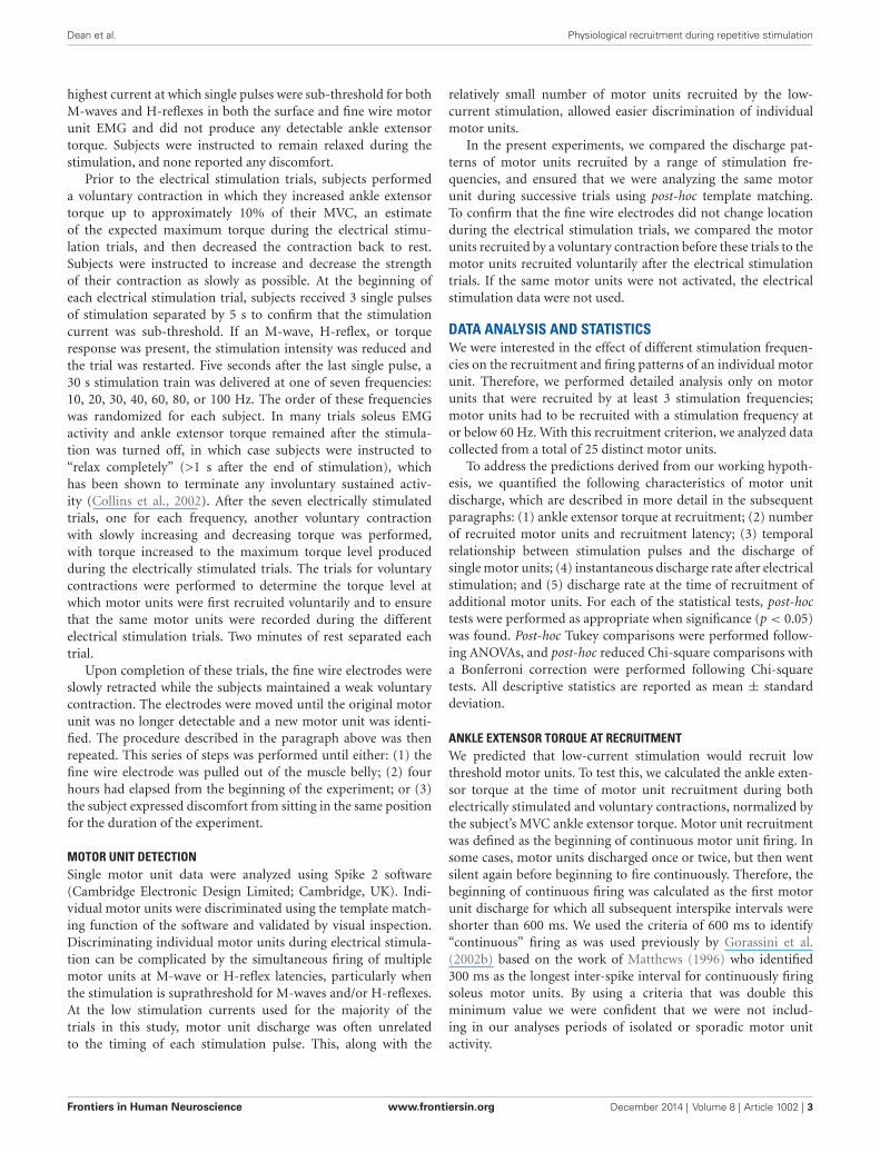
NUMBER OF MOTOR UNITS RECRUITED AND RECRUITMENT LATENCYDuring 30 s of constant frequency low-current stimulation, motorunits did not respond to the first stimulation pulse, but ratherwere silent for a given latency before being recruited (Figure 3A),or were not recruited at all. Higher stimulation frequenciesrecruited significantly more motor units within the 30 s of stimu-lation (Chi-square, p < 0.05; Figure 3B). A motor unit recruitedby a given stimulation frequency was always recruited by eachof the higher stimulation frequencies. For example, the 3 motorunits recruited at 10 Hz stimulation were also recruited at all ofthe other stimulation frequencies.
Recruitment latencies spanned almost the entire course of theelectrical stimulation, ranging from 0.5 to 29.6 s (Figure 3C).Several of the motor units were not recruited for a relativelylong time, as 22 of the motor units had a recruitment latencylonger than 10 s in at least one trial, and 9 of the motor unitshad a recruitment latency longer than 20 s in at least one trial.Higher stimulation frequencies recruited motor units at signifi-cantly shorter latencies than low frequencies (ANOVA, p < 0.05).
FIGURE 2 | All motor units recruited during low-current stimulationwere also recruited during weak voluntary contractions. The torquelevel at which a motor unit first fired was measured for both electricallystimulated and voluntary contractions, as illustrated for a motor unitrecorded from a single subject.
This is illustrated in Figure 3D, in which the 25 motor units aregrouped into populations based on the minimum stimulationfrequency at which they were recruited. For example, the linelabeled 10 Hz illustrates the average recruitment latency acrossstimulation frequencies for the 3 motor units recruited during10 Hz stimulation. The effect of increasing stimulation frequencyon decreasing recruitment latency is evident in each of thesepopulations.
Frontiers in Human Neuroscience www.frontiersin.org December 2014 | Volume 8 | Article 1002 | 5
Dean et al. Physiological recruitment during repetitive stimulation
FIGURE 3 | Number of units recruited and latency of discharge onsetwere dependent on stimulation frequency. (A) The time between thebeginning of stimulation and the onset of motor unit firing is illustrated for asingle motor unit at three stimulation frequencies. The solid horizontal lineshows the duration of the stimulation. (B) Higher stimulation frequenciesrecruited more motor units, with significant differences (∗p < 0.05) betweenthe indicated stimulation frequencies. (C) The change in recruitment latency
with stimulation frequency is illustrated for each of the 25 motor units. Theminimal stimulation frequency required to recruit each motor unit varied.(D) The 25 motor units were grouped by the minimal stimulation frequencyrequired for their recruitment, and the average recruitment latencies werecalculated for each group. Higher stimulation frequencies recruited motorunits at shorter latencies, with significant differences (∗p < 0.05) between theindicated stimulation frequencies.
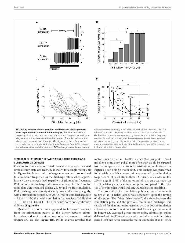
TEMPORAL RELATIONSHIP BETWEEN STIMULATION PULSES ANDSUBSEQUENT DISCHARGESOnce motor units were recruited, their discharge rate increaseduntil a steady-state was reached, as shown for a single motor unitin Figure 4A. Motor unit discharge rate was not proportionalto stimulation frequency, as the discharge rate reached approx-imately the same peak level regardless of stimulation frequency.Peak motor unit discharge rates were compared for the 9 motorunits that were recruited during 20, 30 and 40 Hz stimulation.Peak discharge rate was significantly lower, albeit only slightly,with a stimulation frequency of 20 Hz (motor unit discharge rate= 7.8 ± 1.1 Hz) than with stimulation frequencies of 30 Hz (8.6± 1.1 Hz) or 40 Hz (8.4 ± 1.1 Hz), which were not significantlydifferent (Figure 4B).
Qualitatively, motor units appeared to fire asynchronouslyfrom the stimulation pulses, as the latency between stimu-lus pulses and motor unit action potentials was not constant(Figure 5A, see also Figure 1B). PSTH analysis revealed that
motor units fired at an H-reflex latency (1–2 ms peak ∼35–44ms after a stimulation pulse) more often than would be expectedfrom a completely asynchronous distribution, as illustrated inFigure 5B for a single motor unit. This analysis was performedfor all trials in which a motor unit was recruited by a stimulationfrequency of 10 or 20 Hz. In these 12 trials (n = 9 motor units),24% (range 18–58%) of the motor unit discharges occurred at anH-reflex latency after a stimulation pulse, compared to the ∼2–4% of the time that would indicate true asynchronous firing.
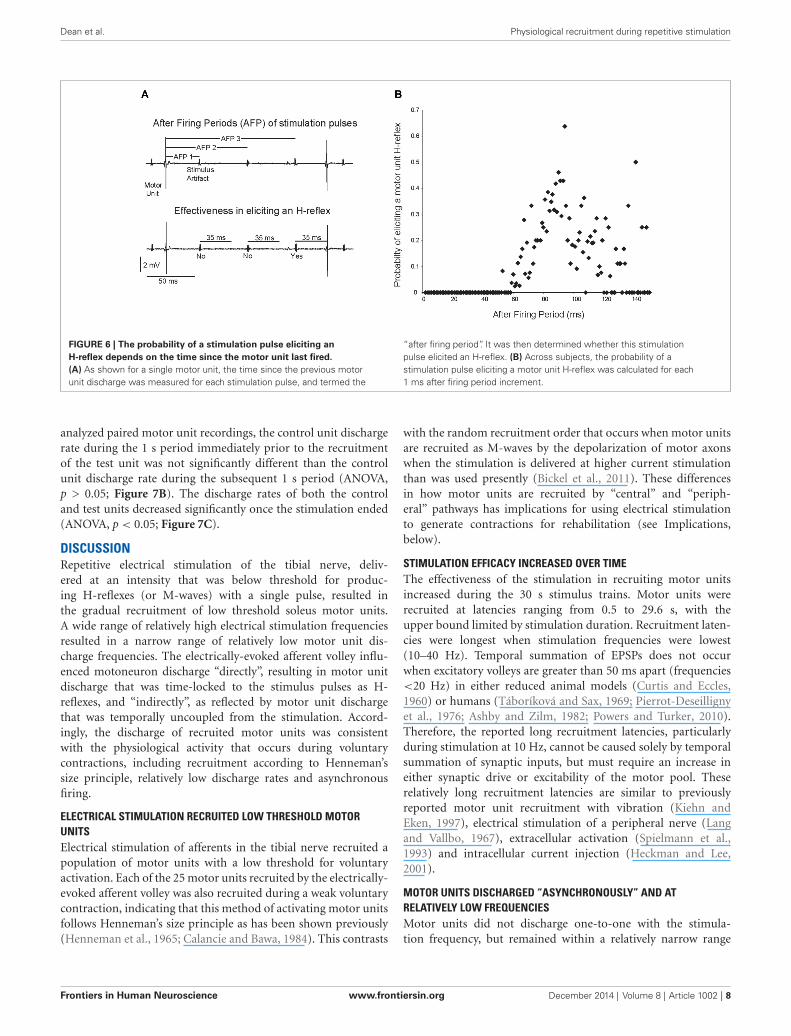
The probability of a stimulation pulse causing a motor unitto fire at an H-reflex latency was dependent upon the timingof the pulse. The “after firing period”, the time between thestimulation pulse and the previous motor unit discharge, wascalculated for all motor units recruited by 10 or 20 Hz stimulation(12 trials, 9 motor units), as illustrated for a single motor unitin Figure 6A. Averaged across motor units, stimulation pulsesdelivered within 50 ms after a motor unit discharge (after firingperiod <50 ms) never caused the motor unit to fire at an H-reflex
Frontiers in Human Neuroscience www.frontiersin.org December 2014 | Volume 8 | Article 1002 | 6

Dean et al. Physiological recruitment during repetitive stimulation
FIGURE 4 | Motor unit discharge rate was not proportional tostimulation frequency. (A) As illustrated for a single motor unit activated bythree stimulation frequencies, there is a gradual increase in discharge rate,which then reaches a steady-state. The solid horizontal line shows theduration of the stimulation. (B) Averaged across subjects, the peak discharge
rate was significantly (∗p < 0.05) lower during 20 Hz stimulation than during30 or 40 Hz stimulation. (C) Averaged across subjects and stimulationfrequency, motor unit discharge rate during the first second of sustainedfiring was significantly (∗p < 0.05) lower than the discharge rate during thelast second of stimulation.
FIGURE 5 | Motor unit activity is only partially asynchronous fromthe stimulation pulses. (A) The latencies between motor unit dischargesand the previous stimulation pulses were quantified for the nine motorunits that were recruited with a stimulation frequency of ≤20 Hz, asillustrated for an excerpt of activity for a single motor unit. (B) Motor units
fired at an H-reflex latency more often than would be expected fromrandom chance, as illustrated for a single motor unit that fired at anH-reflex latency 18% of the time. For all analyzed motor units, thisH-reflex latency peak exceeded the mean plus two standard deviations ofthe number of discharges per bin.
latency (Figure 6B). As the after firing period grew longer, theprobability of the motor unit firing at an H-reflex latency aftera stimulation pulse increased up to a maximum of 0.63 with anafter firing period of 92 ms. Again, this analysis was performed forthe 12 trials in which a motor unit was recruited by a stimulationfrequency of 10 or 20 Hz.
INSTANTANEOUS DISCHARGE RATE AFTER CESSATION OF ELECTRICALSTIMULATIONMotor units often continued to fire after stimulation had ended,with sustained firing for at least one second occurring in 102 of115 trials (89%). For the 9 motor units recruited during 20, 30and 40 Hz stimulation, the discharge rate during the last secondof stimulation (7.9 ± 1.1 Hz) was significantly higher than thedischarge rate during the first second of sustained firing (5.8 ±
1.8 Hz) after stimulation ended (Figure 4C). The duration of the
sustained activity was not quantified, as subjects were instructedto relax completely in order to allow 2 min of rest between trialsand limit experiment duration. Although subjects stated that theywere relaxed and were not voluntarily contracting, instructingthem to “relax completely” terminated the motor unit activity.
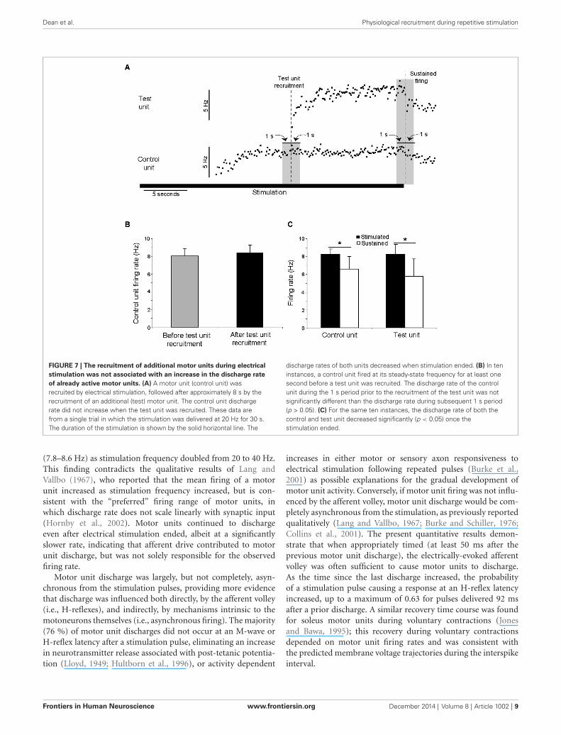
DISCHARGE AT THE TIME OF RECRUITMENT OF ADDITIONAL MOTORUNITSIn 10 trials, two clearly distinguishable motor units were recruitedduring the stimulation, as illustrated in Figure 7A. The dischargerate of the first motor unit recruited (“control unit”) followedthe pattern described above, with an initial increase followedby a steady-state in discharge rate. This discharge rate thenremained constant even as additional motor units (“test units”)were subsequently recruited. In no trials did the discharge rate ofa control unit increase as a test unit was recruited. Across the ten
Frontiers in Human Neuroscience www.frontiersin.org December 2014 | Volume 8 | Article 1002 | 7
Dean et al. Physiological recruitment during repetitive stimulation
FIGURE 6 | The probability of a stimulation pulse eliciting anH-reflex depends on the time since the motor unit last fired.(A) As shown for a single motor unit, the time since the previous motorunit discharge was measured for each stimulation pulse, and termed the
“after firing period”. It was then determined whether this stimulationpulse elicited an H-reflex. (B) Across subjects, the probability of astimulation pulse eliciting a motor unit H-reflex was calculated for each1 ms after firing period increment.
analyzed paired motor unit recordings, the control unit dischargerate during the 1 s period immediately prior to the recruitmentof the test unit was not significantly different than the controlunit discharge rate during the subsequent 1 s period (ANOVA,p > 0.05; Figure 7B). The discharge rates of both the controland test units decreased significantly once the stimulation ended(ANOVA, p < 0.05; Figure 7C).
DISCUSSIONRepetitive electrical stimulation of the tibial nerve, deliv-ered at an intensity that was below threshold for produc-ing H-reflexes (or M-waves) with a single pulse, resulted inthe gradual recruitment of low threshold soleus motor units.A wide range of relatively high electrical stimulation frequenciesresulted in a narrow range of relatively low motor unit dis-charge frequencies. The electrically-evoked afferent volley influ-enced motoneuron discharge “directly”, resulting in motor unitdischarge that was time-locked to the stimulus pulses as H-reflexes, and “indirectly”, as reflected by motor unit dischargethat was temporally uncoupled from the stimulation. Accord-ingly, the discharge of recruited motor units was consistentwith the physiological activity that occurs during voluntarycontractions, including recruitment according to Henneman’ssize principle, relatively low discharge rates and asynchronousfiring.
ELECTRICAL STIMULATION RECRUITED LOW THRESHOLD MOTORUNITSElectrical stimulation of afferents in the tibial nerve recruited apopulation of motor units with a low threshold for voluntaryactivation. Each of the 25 motor units recruited by the electrically-evoked afferent volley was also recruited during a weak voluntarycontraction, indicating that this method of activating motor unitsfollows Henneman’s size principle as has been shown previously(Henneman et al., 1965; Calancie and Bawa, 1984). This contrasts
with the random recruitment order that occurs when motor unitsare recruited as M-waves by the depolarization of motor axonswhen the stimulation is delivered at higher current stimulationthan was used presently (Bickel et al., 2011). These differencesin how motor units are recruited by “central” and “periph-eral” pathways has implications for using electrical stimulationto generate contractions for rehabilitation (see Implications,below).
STIMULATION EFFICACY INCREASED OVER TIMEThe effectiveness of the stimulation in recruiting motor unitsincreased during the 30 s stimulus trains. Motor units wererecruited at latencies ranging from 0.5 to 29.6 s, with theupper bound limited by stimulation duration. Recruitment laten-cies were longest when stimulation frequencies were lowest(10–40 Hz). Temporal summation of EPSPs does not occurwhen excitatory volleys are greater than 50 ms apart (frequencies<20 Hz) in either reduced animal models (Curtis and Eccles,1960) or humans (Táboríková and Sax, 1969; Pierrot-Deseillignyet al., 1976; Ashby and Zilm, 1982; Powers and Turker, 2010).Therefore, the reported long recruitment latencies, particularlyduring stimulation at 10 Hz, cannot be caused solely by temporalsummation of synaptic inputs, but must require an increase ineither synaptic drive or excitability of the motor pool. Theserelatively long recruitment latencies are similar to previouslyreported motor unit recruitment with vibration (Kiehn andEken, 1997), electrical stimulation of a peripheral nerve (Langand Vallbo, 1967), extracellular activation (Spielmann et al.,1993) and intracellular current injection (Heckman and Lee,2001).
MOTOR UNITS DISCHARGED “ASYNCHRONOUSLY” AND ATRELATIVELY LOW FREQUENCIESMotor units did not discharge one-to-one with the stimula-tion frequency, but remained within a relatively narrow range
Frontiers in Human Neuroscience www.frontiersin.org December 2014 | Volume 8 | Article 1002 | 8
Dean et al. Physiological recruitment during repetitive stimulation
FIGURE 7 | The recruitment of additional motor units during electricalstimulation was not associated with an increase in the discharge rateof already active motor units. (A) A motor unit (control unit) wasrecruited by electrical stimulation, followed after approximately 8 s by therecruitment of an additional (test) motor unit. The control unit dischargerate did not increase when the test unit was recruited. These data arefrom a single trial in which the stimulation was delivered at 20 Hz for 30 s.The duration of the stimulation is shown by the solid horizontal line. The
discharge rates of both units decreased when stimulation ended. (B) In teninstances, a control unit fired at its steady-state frequency for at least onesecond before a test unit was recruited. The discharge rate of the controlunit during the 1 s period prior to the recruitment of the test unit was notsignificantly different than the discharge rate during subsequent 1 s period(p > 0.05). (C) For the same ten instances, the discharge rate of both thecontrol and test unit decreased significantly (p < 0.05) once thestimulation ended.
(7.8–8.6 Hz) as stimulation frequency doubled from 20 to 40 Hz.This finding contradicts the qualitative results of Lang andVallbo (1967), who reported that the mean firing of a motorunit increased as stimulation frequency increased, but is con-sistent with the “preferred” firing range of motor units, inwhich discharge rate does not scale linearly with synaptic input(Hornby et al., 2002). Motor units continued to dischargeeven after electrical stimulation ended, albeit at a significantlyslower rate, indicating that afferent drive contributed to motorunit discharge, but was not solely responsible for the observedfiring rate.
Motor unit discharge was largely, but not completely, asyn-chronous from the stimulation pulses, providing more evidencethat discharge was influenced both directly, by the afferent volley(i.e., H-reflexes), and indirectly, by mechanisms intrinsic to themotoneurons themselves (i.e., asynchronous firing). The majority(76 %) of motor unit discharges did not occur at an M-wave orH-reflex latency after a stimulation pulse, eliminating an increasein neurotransmitter release associated with post-tetanic potentia-tion (Lloyd, 1949; Hultborn et al., 1996), or activity dependent
increases in either motor or sensory axon responsiveness toelectrical stimulation following repeated pulses (Burke et al.,2001) as possible explanations for the gradual development ofmotor unit activity. Conversely, if motor unit firing was not influ-enced by the afferent volley, motor unit discharge would be com-pletely asynchronous from the stimulation, as previously reportedqualitatively (Lang and Vallbo, 1967; Burke and Schiller, 1976;Collins et al., 2001). The present quantitative results demon-strate that when appropriately timed (at least 50 ms after theprevious motor unit discharge), the electrically-evoked afferentvolley was often sufficient to cause motor units to discharge.As the time since the last discharge increased, the probabilityof a stimulation pulse causing a response at an H-reflex latencyincreased, up to a maximum of 0.63 for pulses delivered 92 msafter a prior discharge. A similar recovery time course was foundfor soleus motor units during voluntary contractions (Jonesand Bawa, 1995); this recovery during voluntary contractionsdepended on motor unit firing rates and was consistent withthe predicted membrane voltage trajectories during the interspikeinterval.
Frontiers in Human Neuroscience www.frontiersin.org December 2014 | Volume 8 | Article 1002 | 9
Dean et al. Physiological recruitment during repetitive stimulation
POSSIBLE MECHANISMSThe amplification of synaptic input by PICs in spinal neurons(Lee and Heckman, 2000; Heckman and Lee, 2001; Heckman andEnoka, 2012) or the neuromodulatory facilitation of PICs (Perrieret al., 2002) could produce the type of motor unit dischargeseen in the present experiments. Consistent with this idea, thepaired motor unit recordings provide evidence that the gradualrecruitment presently observed was not due to an increase insynaptic drive but rather was due to an increase in excitabilityof the motor pool. This technique has been used previouslyto monitor synaptic drive to the motor pool at a time whennew motor units are being recruited (Kiehn and Eken, 1997;Gorassini et al., 1998, 2002a,b). In the present study, controlmotor units were sensitive to changes in synaptic drive; they firedat approximately 8 Hz, well below the maximal discharge rateof 20 Hz of voluntarily recruited soleus motor units (Bellemareet al., 1983). Additionally, motor unit discharge rates increasedsignificantly when stimulation frequency increased from 20 to 30Hz and discharge rates of both control and test units decreasedsignificantly when stimulation ended. Thus, although “control”units were sensitive to changes in the electrically-evoked afferentvolley, their discharge rate did not increase significantly at thetime of recruitment of “test” units. This result suggests thatrecruitment of the additional motor unit was due to an increase incurrent provided by a post-synaptic mechanism such as activationof PICs in the motoneuron, not a pre-synaptic mechanism suchas increased synaptic drive (Tokuno et al., 2003). We acknowl-edge that the control unit discharge rate may not exactly reflectchanges in synaptic drive (Fuglevand et al., 2006), particularly athigher stimulation frequencies. However, previous paired motorunit results identify motoneurons as the most likely location ofPIC activation (Powers et al., 2008; Vandenberk and Kalmar,2014).
Increases in descending inputs or the magnitude of theelectrically-evoked afferent volley cannot be ruled out ashaving influenced the observed motor unit recruitment. Forexample, ascending input to the brainstem may have promptedthe release of monoamines such as serotonin from the raphenucleus (Alvarez et al., 1998) or norepinephrine from the locuscoeruleus (Lai et al., 1989). To directly address this possibility,the experiments described herein should be repeated in patientswith complete spinal cord injuries, in whom the electricalstimulation of peripheral nerves would not be expected to havesupraspinal effects. While our paired motor unit recordingssuggest that it is unlikely that increased synaptic drive to themotor pool contributed to the observed contractions, we cannotrule out the possibility that increases in the descending inputor the electrically-evoked afferent volley contributed to motorunit recruitment but was too small to significantly increasethe discharge of our control unit. However, similar gradually-developing contractions and sustained firing have been observedin individuals with complete spinal cord injuries (Nickolls et al.,2004), suggesting that descending drive is not the primarycontributor to the motor unit discharge behavior described inthe present experiments. Further, repetitive activation of axonstypically results in an activity-dependent hyperpolarisation, thusincreasing axonal thresholds to electrical stimulation, which
may have reduced and not increased the magnitude of theelectrically-evoked afferent volley over the course of a stimulustrain.
IMPLICATIONSThe present results describe how motoneurons transform afferentfeedback into motor output in individuals with no neurologicalimpairments. While such transformation may be a key contribu-tor to voluntary contractions, afferent feedback during voluntarymovements would be more temporally diffuse than the relativelysynchronous activation of sensory axons that occurs duringelectrical stimulation. The synchronous nature of the electrically-evoked afferent volley is a limitation of the present study as itdecreases the physiological relevance of our findings, however, it isa strength in that it allowed us to characterize how motoneuronsrespond to discrete excitatory volleys which would not bepossible with vibration or voluntary contractions. We suggest thatutilizing this “low-current stimulation” approach on individualswith neurological impairments may provide novel insight intohow sensorimotor transformation is affected by injury or disease.
These findings also have implications for understanding howmotor units are recruited during neuromuscular electrical stim-ulation. Electrical stimulation is used for rehabilitation afteran injury or disease to prevent muscle atrophy, generate func-tional movements, or preserve motor unit types. Electrical stim-ulation, however, recruits motor units in a non-physiologicalorder (Sheffler and Chae, 2007; Bickel et al., 2011) and leavesslow fatigue-resistant motor units, which are the most likelyto develop disuse atrophy, (Burnham et al., 1997), relativelyinactive. In contrast, generating muscle contractions throughafferent feedback recruits motor units synaptically (Hennemanet al., 1965; Bennett et al., 1998a), thereby first activating thefatigue-resistant muscle fibers (Sypert and Munson, 1981) withrelatively weak stimulation. Thus, electrical stimulation deliveredto recruit motor units by the electrically-evoked afferent vol-ley may prove to be beneficial to reduce muscle atrophy andgenerate fatigue-resistant contractions. Such synaptic recruit-ment could also help maintain the biophysical properties ofmotoneurons, which are sensitive to changes in synaptic drive(see Gardiner et al., 2005 for review). In addition, modulatingthe excitability of a motor pool with low-current electrical stim-ulation of peripheral nerves may facilitate residual descendingdrive to augment voluntary commands and generate musclecontractions.
CONCLUSIONSPresently we show that human motoneurons transform a widerange of synaptic input frequencies into a relatively narrow rangeof low output frequencies. Motor unit discharge was stronglyinfluenced by properties intrinsic to the motoneurons them-selves and was not solely driven by processes temporally-coupledwith the synaptic input. This work supports the idea that sen-sory input evoked during electrical stimulation activates PICs inmotoneurons, and describes a method of studying sensorimotorintegration in humans with a more clearly defined input tospinal neurons than those produced by vibration or voluntarycontraction.
Frontiers in Human Neuroscience www.frontiersin.org December 2014 | Volume 8 | Article 1002 | 10
Dean et al. Physiological recruitment during repetitive stimulation
ACKNOWLEDGMENTSThis work was supported by the Alberta Heritage Foundation forMedical Research, Canadian Institutes of Health Research, andNatural Sciences and Engineering Research Council of Canada.The authors thank Dr. KE Jones for helpful comments during thepreparation of this manuscript.
REFERENCESAlvarez, F. J., Pearson, J. C., Harrington, D., Dewey, D., Torbeck, L., and Fyffe,
R. E. (1998). Distribution of 5-hydroxytryptamine-immunoreactive boutonson alpha-motoneurons in the lumbar spinal cord of adult cats. J. Comp. Neu-rol. 393, 69–83. doi: 10.1002/(sici)1096-9861(19980330)393:1<69::aid-cne7>
3.3.co;2-xAshby, P., and Zilm, D. (1982). Characteristics of postsynaptic potentials produced
in single human motoneurons by homonymous group 1 volleys. Exp. Brain Res.47, 41–48. doi: 10.1007/bf00235884
Bawa, P., and Chalmers, G. (2008). Responses of human motoneurons to high-frequency stimulation of Ia afferents. Muscle Nerve 38, 1604–1615. doi: 10.1002/mus.21184
Bellemare, F., Woods, J. J., Johansson, R., and Bigland-Ritchie, B. (1983). Motor-unit discharge rates in maximal voluntary contractions of three human muscles.J. Neurophysiol. 50, 1380–1392.
Bennett, D. J., Hultborn, H., Fedirchuk, B., and Gorassini, M. (1998a). Short-term plasticity in hindlimb motoneurons of decerebrate cats. J. Neurophysiol.80, 2038–2045.
Bennett, D. J., Hultborn, H., Fedirchuk, B., and Gorassini, M. (1998b). Synapticactivation of plateaus in hindlimb motoneurons of decerebrate cats. J. Neuro-physiol. 80, 2023–2037.
Bergquist, A. J., Clair, J. M., and Collins, D. F. (2011). Motor unit recruitment whenneuromuscular electrical stimulation is applied over a nerve trunk comparedwith a muscle belly: triceps surae. J. Appl. Physiol. 110, 627–637. doi: 10.1152/japplphysiol.01103.2010
Bickel, C. S., Gregory, C. M., and Dean, J. C. (2011). Motor unit recruitment duringneuromuscular electrical stimulation: a critical appraisal. Eur. J. Appl. Physiol.111, 2399–2407. doi: 10.1007/s00421-011-2128-4
Binboga, E., Prasartwuth, O., Pehlivan, M., and Türker, K. S. (2011). Responses ofhuman soleus motor units to low-threshold stimulation of the tibial nerve. Exp.Brain Res. 213, 73–86. doi: 10.1007/s00221-011-2779-8
Burke, D., Gandevia, S. C., and McKeon, B. (1983). The afferent volleys responsiblefor spinal proprioceptive reflexes in man. J. Physiol. 339, 535–552.
Burke, D., Gandevia, S. C., and McKeon, B. (1984). Monosynaptic and oligosy-naptic contributions to the human ankle jerk and H-reflex. J. Neurophysiol. 52,435–448.
Burke, D., Kiernan, M. C., and Bostock, H. (2001). Excitability of humanaxons. Clin. Neurophysiol. 112, 1575–1585. doi: 10.1016/S1388-2457(01)00595-8
Burke, D., and Schiller, H. H. (1976). Discharge pattern of single motor units in thetonic vibration reflex of human triceps surae. J. Neurol. Neurosurg. Psychiatry39, 729–741. doi: 10.1136/jnnp.39.8.729
Burnham, R., Martin, T., Stein, R., Bell, G., MacLean, I., and Steadward, R. (1997).Skeletal muscle fibre type transformation following spinal cord injury. SpinalCord 35, 86–91. doi: 10.1038/sj.sc.3100364
Calancie, B., and Bawa, P. (1984). Recruitment order of motor units dur-ing the stretch reflex in man. Brain Res. 292, 176–178. doi: 10.1016/0006-8993(84)90904-1
Clair, J. M., Anderson-Reid, J. M., Graham, C. M., and Collins, D. F. (2011).Postactivation depression and recovery of reflex transmission during repetitiveelectrical stimulation of the human tibial nerve. J. Neurophysiol. 106, 184–192.doi: 10.1152/jn.00932.2010
Collins, D. F., Burke, D., and Gandevia, S. C. (2001). Large involuntary forcesconsistent with plateau-like behavior of human motoneurons. J. Neurosci. 21,4059–4065.
Collins, D. F., Burke, D., and Gandevia, S. C. (2002). Sustained contractionsproduced by plateau-like behaviour in human motoneurones. J. Physiol. 538,289–301. doi: 10.1113/jphysiol.2001.012825
Curtis, D. R., and Eccles, J. C. (1960). Synaptic action during and after repetitivestimulation. J. Physiol. 150, 374–398.
De Gail, P., Lance, J. W., and Neilson, P. D. (1966). Differential effects on tonic andphasic reflex mechanisms produced by vibration of muscles in man. J. Neurol.Neurosurg. Psychiatry 29, 1–11. doi: 10.1136/jnnp.29.1.1
Elbasiouny, S. M., Bennett, D. J., and Mushahwar, V. K. (2006). Simulation ofCa2+ persistent inward currents in spinal motoneurones: mode of activationand integration of synaptic inputs. J. Physiol. 570, 355–374. doi: 10.1113/jphysiol.2005.099119
Fuglevand, A. J., Dutoit, A. P., Johns, R. K., and Keen, D. A. (2006). Evaluation ofplateau-potential-mediated ‘warm up’ in human motor units. J. Physiol. 571,683–693. doi: 10.1113/jphysiol.2005.099705
Gardiner, P., Beaumont, E., and Cormery, B. (2005). Motoneurones “learn” and“forget” physical activity. Can. J. Appl. Physiol. 30, 352–370. doi: 10.1139/h05-127
Gorassini, M., Bennett, D. J., and Yang, J. F. (1998). Self-sustained firing ofhuman motor units. Neurosci. Lett. 247, 13–16. doi: 10.1016/s0304-3940(98)00277-8
Gorassini, M., Yang, J. F., Siu, M., and Bennett, D. J. (2002a). Intrinsic activationof human motoneurons: possible contribution to motor unit excitation. J.Neurophysiol. 87, 1850–1858. doi: 10.1152/jn.00024.2001
Gorassini, M., Yang, J. F., Siu, M., and Bennett, D. J. (2002b). Intrinsic activa-tion of human motoneurons: reduction of motor unit recruitment thresh-olds by repeated contractions. J. Neurophysiol. 87, 1859–1866. doi: 10.1152/jn.00025.2001
Hagbarth, K. E., and Eklund, G. (1966). “Motor effects of vibratory muscle stimuliin man,” in Muscular Afferents and Motor Control. Proceedings of the First NobleSymposium, ed R. Granit (Stockholm: Almqvist and Wiksell), 177–186.
Heckman, C. J., and Enoka, R. M. (2012). Motor unit. Compr. Physiol. 2, 2629–2682.doi: 10.1002/cphy.c100087
Heckman, C. J., and Lee, R. H. (2001). “Advances in measuring active dendriticcurrents in spinal motoneurons in vivo,” in Motor Neurobiology of the SpinalCord, ed C. Timothy Cope (New York: CRC Press), 89–106.
Henneman, E., Somjen, G., and Carpenter, D. O. (1965). Excitability andinhibitibility of motoneurons of different sizes. J. Neurophysiol. 28, 599–620.
Hoffmann, P. (1918). Uber die Beziehungen der Sehnenreflexe zur wilkurlichenBewegung und zum Tonus. Z. Biol. 68, 351–370.
Hornby, T. G., McDonagh, J. C., Reinking, R. M., and Stuart, D. G. (2002).Motoneurons: a preferred firing range across vertebrate species? Muscle Nerve25, 632–648. doi: 10.1002/mus.10105
Hultborn, H., Illert, M., Nielsen, J., Paul, A., Ballegaard, M., and Wiese, H. (1996).On the mechanism of the post-activation depression of the H-reflex in humansubjects. Exp. Brain Res. 108, 450–462. doi: 10.1007/bf00227268
Jones, K. E., and Bawa, P. (1995). Responses of human motoneurons to Ia inputs:effects of background firing rate. Can. J. Physiol. Pharmacol. 73, 1224–1234.doi: 10.1139/y95-174
Kiehn, O., and Eken, T. (1997). Prolonged firing in motor units: evidence of plateaupotentials in human motoneurons? J. Neurophysiol. 78, 3061–3068.
Klakowicz, P. M., Baldwin, E. R., and Collins, D. F. (2006). Contribution of m-wavesand h-reflexes to contractions evoked by tetanic nerve stimulation in humans. J.Neurophysiol. 96, 1293–1302. doi: 10.1152/jn.00765.2005
Kudina, L. P. (1988). Excitability of firing motoneurones tested by Ia afferent volleysin human triceps surae. Electroencephalogr. Clin. Neurophysiol. 69, 576–580.doi: 10.1016/0013-4694(88)90170-8
Lai, Y. Y., Strahlendorf, H. K., Fung, S. J., and Barnes, C. D. (1989). The actionsof two monoamines on spinal motoneurons from stimulation of the locuscoeruleus in the cat. Brain Res. 484, 268–272. doi: 10.1016/0006-8993(89)90369-7
Lang, A. H., and Vallbo, Å. B. (1967). Motoneuron activation by low intensitytetanic stimulation of muscle afferents in man. Exp. Neurol. 18, 383–391. doi: 10.1016/0014-4886(67)90056-8
Lee, R. H., and Heckman, C. J. (2000). Adjustable amplification of synaptic inputin the dendrites of spinal motoneurons in vivo. J. Neurol. Sci. 20, 6734–6740.
Lloyd, D. P. C. (1949). Post-tetanic potentiation of response in monosynaptic reflexpathways of the spinal cord. J. Gen. Physiol. 33, 147–170. doi: 10.1085/jgp.33.2.147
Marchand-Pauvert, V., Nicolas, G., Burke, D., and Pierrot-Deseilligny, E. (2002).Suppression of the H reflex in humans by disynaptic autogenetic inhibitorypathways activated by the test volley. J. Physiol. 542, 963–976. doi: 10.1113/jphysiol.2002.021683
Frontiers in Human Neuroscience www.frontiersin.org December 2014 | Volume 8 | Article 1002 | 11
Dean et al. Physiological recruitment during repetitive stimulation
Matthews, P. B. (1996). Relationship of firing intervals of human motor units to thetrajectory of post-spike after-hyperpolarization and synaptic noise. J. Physiol.492, 597–628.
Nickolls, P., Collins, D. F., Gorman, R. B., Burke, D., and Gandevia, S. C.(2004). Forces consistent with plateau-like behaviour of spinal neuronsevoked in patients with spinal cord injuries. Brain 127, 660–670. doi: 10.1093/brain/awh073
Perrier, J. F., Alaburda, A., and Hounsgaard, J. (2002). Spinal plasticity mediatedby postsynaptic L-type Ca2+ channels. Brain Res. Brain Res. Rev. 40, 223–229.doi: 10.1016/s0165-0173(02)00204-7
Pierrot-Deseilligny, E., Bussel, B., Held, J. P., and Katz, R. (1976). Excitabilityof human motoneurones after discharge in a conditioning reflex. Electroen-cephalogr. Clin. Neurophysiol. 40, 279–287. doi: 10.1016/0013-4694(76)90151-6
Pierrot-Deseilligny, E., and Mazevet, D. (2000). The monosynaptic reflex: a tool toinvestigate motor control in humans. Interest and limits. Neurophysiol. Clin. 30,67–80. doi: 10.1016/s0987-7053(00)00062-9
Powers, R. K., Elbasiouny, S. M., Rymer, W. Z., and Heckman, C. J. (2012). Con-tribution of intrinsic properties and synaptic inputs to motoneuron dischargepatterns: a simulation study. J. Neurophysiol. 107, 808–823. doi: 10.1152/jn.00510.2011
Powers, R. K., Nardelli, P., and Cope, T. C. (2008). Estimation of the contribution ofintrinsic currents to motoneuron firing based on paired motoneuron dischargerecords in the decerebrate cat. J. Neurophysiol. 100, 292–303. doi: 10.1152/jn.90296.2008
Powers, R. K., and Turker, K. S. (2010). Deciphering the contribution of intrinsicand synaptic currents to the effects of transient synaptic inputs on human motorunit discharge. Clin. Neurophysiol. 121, 1643–1654. doi: 10.1016/j.clinph.2009.10.041
Schwindt, P. C., and Crill, W. E. (1980). Properties of a persistent inwardcurrent in normal and TEA-injected motoneurons. J. Neurophysiol. 43,1700–1724.
Sheffler, L. R., and Chae, J. (2007). Neuromuscular electrical stimulation inneurorehabilitation. Muscle Nerve 35, 562–590. doi: 10.1002/mus.20758
Spielmann, J. M., Laouris, Y., Nordstrom, M. A., Robinson, G. A., Reinking, R. M.,and Stuart, D. G. (1993). Adaptation of cat motoneurons to sustained andintermittent extracellular activation. J. Physiol. 464, 75–120.
Sypert, G. W., and Munson, J. B. (1981). Basis of segmental motor control:motoneuron size or motor unit type? Neurosurgery 8, 608–621. doi: 10.1097/00006123-198105000-00020
Táboríková, H., and Sax, D. S. (1969). Conditioning of H-reflexes by a preced-ing subthreshold H-reflex stimulus. Brain 92, 203–212. doi: 10.1093/brain/92.1.203
Tokuno, H. A., Kocsis, J. D., and Waxman, S. G. (2003). Noninactivating,tetrodotoxin-sensitive Na+ conductance in peripheral axons. Muscle Nerve 28,212–217. doi: 10.1002/mus.10421
Vandenberk, M. S., and Kalmar, J. M. (2014). An evaluation of paired motor unitestimates of persistent inward current in human motoneurons. J. Neurophysiol.111, 1877–1884. doi: 10.1152/jn.00469.2013
Conflict of Interest Statement: The authors declare that the research was conductedin the absence of any commercial or financial relationships that could be construedas a potential conflict of interest.
Received: 30 July 2014; accepted: 25 November 2014; published online: 16 December2014.Citation: Dean JC, Clair-Auger JM, Lagerquist O and Collins DF (2014)Asynchronous recruitment of low-threshold motor units during repetitive, low-current stimulation of the human tibial nerve. Front. Hum. Neurosci. 8:1002.doi: 10.3389/fnhum.2014.01002This article was submitted to the journal Frontiers in Human Neuroscience.Copyright © 2014 Dean, Clair-Auger, Lagerquist and Collins. This is an open-accessarticle distributed under the terms of the Creative Commons Attribution License (CCBY). The use, distribution and reproduction in other forums is permitted, providedthe original author(s) or licensor are credited and that the original publication in thisjournal is cited, in accordance with accepted academic practice. No use, distribution orreproduction is permitted which does not comply with these terms.
Frontiers in Human Neuroscience www.frontiersin.org December 2014 | Volume 8 | Article 1002 | 12
Copyright © 2022 FDOKUMEN