Association between haptoglobin and IgM levels and the clinical progression of caseous lymphadenitis...
7
RESEARCH ARTICLE Open Access Association between haptoglobin and IgM levels and the clinical progression of caseous lymphadenitis in sheep Bruno L Bastos 1 , Dan Loureiro 1 , José T Raynal 1 , Maria T Guedes 1 , Vera Lúcia Costa Vale 1 , Lilia F Moura-Costa 1 , José E Guimarães 2 , Vasco Azevedo 3 , Ricardo W Portela 1 and Roberto Meyer 1* Abstract Background: Sheep caseous lymphadenitis (CLA), caused by Corynebacterium pseudotuberculosis (Cp), is associated with direct economic losses and presents significant zoonotic potential. Despite the importance of the disease, a satisfactory vaccine model has not been developed. Thus, this study aimed to investigate the association between haptoglobin (Hp) and IgM levels and the clinical progression of CLA in primarily infected sheep and in sheep immunized with Cp- secreted antigens adjuvanted with Quillaja saponaria saponins. These animals were kept with CLA-positive sheep to simulate natural exposure that occurs in field conditions. During the experiment, the Hp and IgM levels were monitored for 21 days, and the development of internal CLA lesions was investigated through necropsies on day182 post-immunization. Results: Primarily infected sheep in Group 2 (inoculated with 2x10 5 Cp virulent strain) had higher Hp values between the first and ninth days post inoculation (PI) than sheep in Group 1 (control; P < 0.05). Immunized animals in Group 3 had significantly higher Hp values between the third and seventh days PI, compared with the control group (P < 0.01). Binary logistic regression (BLR) analysis of primarily infected sheep indicated an association between Hp concentration and CLA clinical progression: animals with high Hp values had 99.9% less risk of having CLA abscesses than animals with low Hp levels (Odds ratio = 0.001, P < 0.05). Both experimental groups had significantly higher IgM titers than the control group around the ninth and eleventh days PI (P < 0.05). The BLR analysis for immunized sheep indicated an association between IgM levels and clinical progression: sheep with high IgM titers had 100.0% less risk of having CLA abscesses than animals with low IgM levels (Odds ratio = 0.000, P < 0.05). Conclusions: Resistance to C. pseudotuberculosis infection is supported by the early acute phase response, in which up-regulation of Hp and IgM were predictive of a lower risk of CLA lesion development. Because the immunogen used in this study induced a high production of both Hp and IgM, Q. saponaria saponin should be considered a promising candidate in vaccine formulations against sheep CLA. Keywords: Caseous lymphadenitis, Small ruminants, Quillaja saponaria, Acute phase response, Haptoglobin, Immunoglobulin M * Correspondence: [email protected] 1 Laboratório de Imunologia e Biologia Molecular, Departamento de Biointeração, Instituto de Ciências da Saúde, Universidade Federal da Bahia, Av. Reitor Miguel Calmon, S/N - Vale do Canela, Salvador, BA CEP 40140-100, Brazil Full list of author information is available at the end of the article © 2013 Bastos et al.; licensee BioMed Central Ltd. This is an open access article distributed under the terms of the Creative Commons Attribution License (http://creativecommons.org/licenses/by/2.0), which permits unrestricted use, distribution, and reproduction in any medium, provided the original work is properly cited. Bastos et al. BMC Veterinary Research 2013, 9:254 http://www.biomedcentral.com/1746-6148/9/254
-
Upload
independent -
Category
Documents
-
view
3 -
download
0
Transcript of Association between haptoglobin and IgM levels and the clinical progression of caseous lymphadenitis...
Bastos et al. BMC Veterinary Research 2013, 9:254http://www.biomedcentral.com/1746-6148/9/254
RESEARCH ARTICLE Open Access
Association between haptoglobin and IgM levelsand the clinical progression of caseouslymphadenitis in sheepBruno L Bastos1, Dan Loureiro1, José T Raynal1, Maria T Guedes1, Vera Lúcia Costa Vale1, Lilia F Moura-Costa1,José E Guimarães2, Vasco Azevedo3, Ricardo W Portela1 and Roberto Meyer1*
Abstract
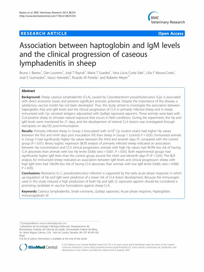
Background: Sheep caseous lymphadenitis (CLA), caused by Corynebacterium pseudotuberculosis (Cp), is associatedwith direct economic losses and presents significant zoonotic potential. Despite the importance of the disease, asatisfactory vaccine model has not been developed. Thus, this study aimed to investigate the association betweenhaptoglobin (Hp) and IgM levels and the clinical progression of CLA in primarily infected sheep and in sheepimmunized with Cp- secreted antigens adjuvanted with Quillaja saponaria saponins. These animals were kept withCLA-positive sheep to simulate natural exposure that occurs in field conditions. During the experiment, the Hp andIgM levels were monitored for 21 days, and the development of internal CLA lesions was investigated throughnecropsies on day182 post-immunization.
Results: Primarily infected sheep in Group 2 (inoculated with 2x10⁵ Cp virulent strain) had higher Hp valuesbetween the first and ninth days post inoculation (PI) than sheep in Group 1 (control; P < 0.05). Immunized animalsin Group 3 had significantly higher Hp values between the third and seventh days PI, compared with the controlgroup (P < 0.01). Binary logistic regression (BLR) analysis of primarily infected sheep indicated an associationbetween Hp concentration and CLA clinical progression: animals with high Hp values had 99.9% less risk of havingCLA abscesses than animals with low Hp levels (Odds ratio = 0.001, P < 0.05). Both experimental groups hadsignificantly higher IgM titers than the control group around the ninth and eleventh days PI (P < 0.05). The BLRanalysis for immunized sheep indicated an association between IgM levels and clinical progression: sheep withhigh IgM titers had 100.0% less risk of having CLA abscesses than animals with low IgM levels (Odds ratio = 0.000,P < 0.05).
Conclusions: Resistance to C. pseudotuberculosis infection is supported by the early acute phase response, in whichup-regulation of Hp and IgM were predictive of a lower risk of CLA lesion development. Because the immunogenused in this study induced a high production of both Hp and IgM, Q. saponaria saponin should be considered apromising candidate in vaccine formulations against sheep CLA.
Keywords: Caseous lymphadenitis, Small ruminants, Quillaja saponaria, Acute phase response, Haptoglobin,Immunoglobulin M
* Correspondence: [email protected]ório de Imunologia e Biologia Molecular, Departamento deBiointeração, Instituto de Ciências da Saúde, Universidade Federal da Bahia,Av. Reitor Miguel Calmon, S/N - Vale do Canela, Salvador, BA CEP 40140-100,BrazilFull list of author information is available at the end of the article
© 2013 Bastos et al.; licensee BioMed Central Ltd. This is an open access article distributed under the terms of the CreativeCommons Attribution License (http://creativecommons.org/licenses/by/2.0), which permits unrestricted use, distribution, andreproduction in any medium, provided the original work is properly cited.

Bastos et al. BMC Veterinary Research 2013, 9:254 Page 2 of 7http://www.biomedcentral.com/1746-6148/9/254
BackgroundCaseous lymphadenitis (CLA) is a chronic infectiousdisease of sheep and goats caused by the gram-positive bac-teria Corynebacterium pseudotuberculosis (Cp). In additionto the direct economic losses that may occur due to leatherdepreciation and weight gain delay [1,2], CLA presentszoonotic potential. Published data strongly indicate thathuman Cp infection represents an important and signifi-cant zoonosis [3].Despite the importance of the disease, a satisfactory
vaccine model for sheep and goats has not been devel-oped. Current knowledge of the immunity induced byCp indicates that resistance to infection involves bothnonspecific and specific host responses. Antibodies helpprotect against infection [4,5], but for full protectionvaccine models must stimulate cellular immunity, suchas the activation of CD8+ cells and IFN-γ production byTh1 cells [6-9]. The role of the innate immune system inCp infection has recently been investigated. It was dem-onstrated that serum concentrations of haptoglobin(Hp), serum amyloid A and α1 acid glycoprotein wereincreased in a CLA experimental model in sheep [10]. Itwas also suggested that sheep that did not developclinical signs of CLA in field conditions, had significantlyhigher Hp levels during the acute phase of the diseasethan sheep that developed superficial abscesses. Al-though the exact role of Hp in defending against infec-tion by Cp was not identified, data suggested that innateimmune mechanisms contributed to the resolution ofinfection or resistance to the development of CLA pyo-granulomas [11].Considering the hypothesis that full protection against
Cp would be achieved by Th1 T cell activation, Quillajasaponaria-derived saponins can be considered as poten-tial candidates in vaccine formulations [12]. These sapo-nins have been used in the formulation of veterinaryvaccines, stimulating the Th1 response and the produc-tion of specific cytolytic T lymphocytes [13]. The aim ofthis study was to investigate the association betweenhaptoglobin and IgM levels and the clinical progressionof CLA (presence or absence of lesions) in primarilyinfected sheep and in sheep immunized with a saponin-adjuvanted immunogen.
MethodsBacterial strains and secreted antigen productionWe used two Cp strains in this study. The T1 strain wasused to produce secreted antigens in a previously opti-mized chemically defined medium (CDM) [14]. Thisantigenic solution (CDM-secreted antigen) was used asimmunogen. A virulent strain of Cp named VD57 wasused in the experimental infection model, as previouslydescribed [15].
Animals and experimental inoculationsFifteen one-year-old Santa Inês ewes were used in theexperiment. Santa Inês is a naturalized breed fromnortheastern Brazil and the experimental animals allcame from the same flock. Animal selection was basedon several criteria, including body condition, helminth-free state as defined by fecal egg count, normochromicmucosa at visual inspection, normal rectal temperatureand absence of abscesses or enlarged lymph nodes onpalpation. No sheep had clinical signs of CLA and allwere seronegative on an indirect ELISA at the beginningof the study.We determined our sample size based on the experi-
mental design described previously to investigate theacute phase protein response in an experimental modelof ovine caseous lymphadenitis [10]. Factors such as thepreliminary (pilot) nature of this study and the require-ment of sacrificing all experimental animals to investi-gate internal lesions in subclinically infected sheep alsomotivated the use of a small sample size, according topreviously published guidelines [16]. Animals weredivided into three groups. Group 1 (n = 3) was inocu-lated with sterile 0.9% saline solution on day zero andkept in an individual pen separated from the flock.Group 2 (n = 6), the experimental infection group, wasinoculated with 1 ml of saline containing 2 × 10⁵ CFU ofthe Cp virulent strain VD57 on day zero. Group 3 (n = 6)was immunized with 1 ml of a solution containing 250 μgof CDM-secreted antigens adjuvanted with 1.5 mg ofQuillaja saponaria saponin (Sigma-Aldrich) on day zero,and received an immunization booster on day 56. Theadjuvant was prepared by dissolving the saponin at aconcentration of 150 mg/ml in sterile 0.9% saline solutionas a stock solution and filtering through a 0.22 μmmembrane. On the immunization days, the saponin stocksolution was added to the secreted antigen to a final con-centration of 1.5 mg/ml [17], and the mixture was stirredfor 30 minutes before administration. All inoculationswere performed subcutaneously in the right flank. Anadditional 12 CLA-positive sheep, presenting externallesions and positive serology by ELISA, were kept withGroups 2 and 3 from day zero (time of inoculation/immunization) until day 182 (last day of observation) tosimulate natural exposure that occurs in field conditions.These animals were kept on pasture during the day,with free access to water, and returned to pens by theend of each day to receive protein and mineralsupplementation.This experiment was conducted under the authority of
Brazilian Law nº11.794, 8 October 2008 (statements onthe use of animals in research experiments), afterapproval by the Secretariat of Animal Health of theMinistry of Agriculture, Livestock and Food Supply.Slaughter was conducted at a slaughterhouse supervised

Bastos et al. BMC Veterinary Research 2013, 9:254 Page 3 of 7http://www.biomedcentral.com/1746-6148/9/254
by the National Meat Inspection Service of the sameMinistry.
Sample collection and analysisBlood samples were collected before inoculation/immu-nization (day zero) and on days 1, 3, 5, 7, 9, 11, 14 and21 post-inoculation (PI). On each occasion, one bloodaliquot was collected using vacuum tubes withoutanticoagulant and allowed to clot at room temperature,for serum collection. These samples were tested todetermine Hp and IgM levels.Serum Hp concentration was estimated based on
hemoglobin-binding capacity in microtitration plates, aspreviously described [11]. A standard curve was devel-oped with standard Hp solutions (Sigma-Aldrich) dilutedfrom 1.050 to 0.008 g/L. Fifty μL of Hp standard orserum sample was added to 50 μL of 0.9% saline solutionin each well. Next, 50 μL of sheep methemoglobin solu-tion (30 mg/dL) was added and plates were incubatedfor 10 minutes at 20°C. A blank (50 μL of 0.9% saline)was run with each serum sample. Following incubation,a guaiacol reagent (150 μL, pH 4.0) and 50 μL of H2O2
solution (0.02 mol/L) were added. After 5 minutes, ab-sorbance at 490 nm was measured using a microplatereader (Bio-Rad, Hercules, CA).The indirect ELISA for detection of Cp-specific IgM
antibodies was performed as previously described [11].ELISA microplates were sensitized with Cp supernatantantigens (1:100 in carbonate-bicarbonate buffer, pH 9.6)and incubated at 4°C for 16 hours. The blocking stepwas made with 5% skim milk in PBS-T and incubatedfor 2 hours at 37°C. Serum samples were pre-treated(v/v 1:1) with Dynabeads Protein G (Invitrogen, Carlsbad,CA), and 100 μL of these samples were added to the platesin duplicate and incubated for 1 hour at 37°C. A rabbitanti-ovine IgM polyclonal antibody conjugated withhorseradish peroxidase (Serotec, Raleigh, NC) was used asdetection antibody (1:20,000 in PBS-T) and incubated for45 minutes at 37°C. Finally, the reaction was developedwith 100 μL/well of a solution containing H2O2 andtetramethylbenzidine (Moss Inc., Pasadena, MD) for15 minutes, and stopped with 4 N H2SO4. Results wereread in an ELISA plate reader (Bio-Rad) at 450 nm.Although blood sampling focused on the acute phase
period (until the 21st day), clinical assessment continuedfor six months, with all animals clinically examinedtwice per month through the inspection of superficiallymph nodes and routine blood sampling. Externalabscesses found during the experiment period weresampled through an incision with sterile surgical equip-ment, followed by drainage of purulent material intosterile flasks for bacteriological assays. After the 182-dayobservation period, animals were necropsied at the BabyBode slaughterhouse (Feira de Santana, BA, Brazil), which
has an official meat inspection system and performspostmortem pathology inspections. All external andinternal lymph nodes, and tissue samples with lesionssuspected to be caused by Cp, were collected andmicrobiologically assayed according to previously de-scribed protocols [15,18].
Statistical analysisData were analyzed using IBM SPSS statistics, version20.0. The distribution of Hp and IgM results was graph-ically expressed through boxplots for each samplingtime. Results were compared between groups, and sig-nificant differences (P < 0.05) were determined using thenonparametric Mann–Whitney test. Based on the sum-mary of records of external abscesses and post-mortemexamination (Table 1), a second series of graphics wasmade using the animals’ data clustered by their CLAclinical status: with CLA lesions (n = 5 in Group 2, n = 3in Group 3) or without CLA lesions (n = 1 in Group 2,n = 3 in Group 3). Binary logistic regression analysis wasused to examine the association between variables inGroups 2 and 3 through the odds ratio (OR) calculation[1,19]. The presence or absence of CLA lesions wastreated as the predicted dichotomous categorical variable(outcome), while Hp and IgM levels during the acutephase were considered as predictor continuous variables.A model construct was done using a forward selectionof the variables. Adequacy of the final models wasassessed by the Hosmer and Lemeshow goodness-of-fittest (P). P values < 0.05 were considered significant.Complementary tables presenting the single haptoglobin(g/l) and IgM (optical density at 490 nm) values for eachsheep during the acute phase period are provided asadditional files.
ResultsThe results of CLA lesion detection are presented inTable 1. Group 1 (control) animals did not develop anylesions. In Groups 2 and 3, there was a notable absenceof clinical signs of infection or inflammatory responseduring the 182 days post-infection, except for one ani-mal (#122) in Group 2, which developed an abscess atthe inoculation site. At slaughter, internal abscesses wererecorded in different sites and organs in five sheep inGroup 2 (animals # 122, 150, 156, 181 and 189) and inthree sheep in Group 3 (animals # 51, 61 and 141). Thenumber of lesions in each animal ranged from one to 18in the lungs, liver and lymph nodes. All purulent contentof lesions had positive Cp isolation and identification. Aparticular tropism for the lungs was detected in theprimarily infected animals (Group 2), but not in theimmunized sheep that presented CLA lesions. Onesheep from Group 2 (#602) was naturally resistant to Cpinfection and did not develop any pyogranulomas or
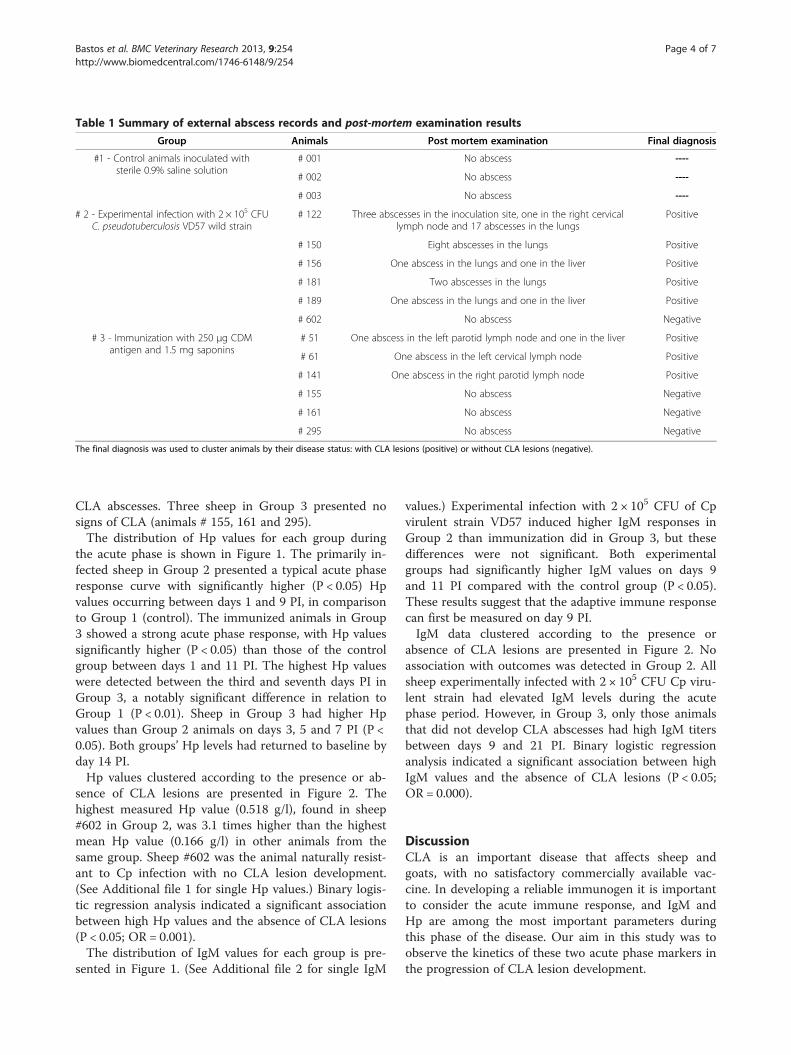
Table 1 Summary of external abscess records and post-mortem examination results
Group Animals Post mortem examination Final diagnosis
#1 - Control animals inoculated withsterile 0.9% saline solution
# 001 No abscess ----
# 002 No abscess ----
# 003 No abscess ----
# 2 - Experimental infection with 2 × 10⁵ CFUC. pseudotuberculosis VD57 wild strain
# 122 Three abscesses in the inoculation site, one in the right cervicallymph node and 17 abscesses in the lungs
Positive
# 150 Eight abscesses in the lungs Positive
# 156 One abscess in the lungs and one in the liver Positive
# 181 Two abscesses in the lungs Positive
# 189 One abscess in the lungs and one in the liver Positive
# 602 No abscess Negative
# 3 - Immunization with 250 μg CDMantigen and 1.5 mg saponins
# 51 One abscess in the left parotid lymph node and one in the liver Positive
# 61 One abscess in the left cervical lymph node Positive
# 141 One abscess in the right parotid lymph node Positive
# 155 No abscess Negative
# 161 No abscess Negative
# 295 No abscess Negative
The final diagnosis was used to cluster animals by their disease status: with CLA lesions (positive) or without CLA lesions (negative).
Bastos et al. BMC Veterinary Research 2013, 9:254 Page 4 of 7http://www.biomedcentral.com/1746-6148/9/254
CLA abscesses. Three sheep in Group 3 presented nosigns of CLA (animals # 155, 161 and 295).The distribution of Hp values for each group during
the acute phase is shown in Figure 1. The primarily in-fected sheep in Group 2 presented a typical acute phaseresponse curve with significantly higher (P < 0.05) Hpvalues occurring between days 1 and 9 PI, in comparisonto Group 1 (control). The immunized animals in Group3 showed a strong acute phase response, with Hp valuessignificantly higher (P < 0.05) than those of the controlgroup between days 1 and 11 PI. The highest Hp valueswere detected between the third and seventh days PI inGroup 3, a notably significant difference in relation toGroup 1 (P < 0.01). Sheep in Group 3 had higher Hpvalues than Group 2 animals on days 3, 5 and 7 PI (P <0.05). Both groups’ Hp levels had returned to baseline byday 14 PI.Hp values clustered according to the presence or ab-
sence of CLA lesions are presented in Figure 2. Thehighest measured Hp value (0.518 g/l), found in sheep#602 in Group 2, was 3.1 times higher than the highestmean Hp value (0.166 g/l) in other animals from thesame group. Sheep #602 was the animal naturally resist-ant to Cp infection with no CLA lesion development.(See Additional file 1 for single Hp values.) Binary logis-tic regression analysis indicated a significant associationbetween high Hp values and the absence of CLA lesions(P < 0.05; OR = 0.001).The distribution of IgM values for each group is pre-
sented in Figure 1. (See Additional file 2 for single IgM
values.) Experimental infection with 2 × 10⁵ CFU of Cpvirulent strain VD57 induced higher IgM responses inGroup 2 than immunization did in Group 3, but thesedifferences were not significant. Both experimentalgroups had significantly higher IgM values on days 9and 11 PI compared with the control group (P < 0.05).These results suggest that the adaptive immune responsecan first be measured on day 9 PI.IgM data clustered according to the presence or
absence of CLA lesions are presented in Figure 2. Noassociation with outcomes was detected in Group 2. Allsheep experimentally infected with 2 × 10⁵ CFU Cp viru-lent strain had elevated IgM levels during the acutephase period. However, in Group 3, only those animalsthat did not develop CLA abscesses had high IgM titersbetween days 9 and 21 PI. Binary logistic regressionanalysis indicated a significant association between highIgM values and the absence of CLA lesions (P < 0.05;OR = 0.000).
DiscussionCLA is an important disease that affects sheep andgoats, with no satisfactory commercially available vac-cine. In developing a reliable immunogen it is importantto consider the acute immune response, and IgM andHp are among the most important parameters duringthis phase of the disease. Our aim in this study was toobserve the kinetics of these two acute phase markers inthe progression of CLA lesion development.

Figure 1 Distribution of haptoglobin and IgM values in sheep during CLA’s acute phase period. Values are expressed as boxplot graphics,in which boxes represent the median value (line within box) and 25th and 75th percentiles (bottom and top of box); whiskers represent the 2.5th
and 97.5th percentiles; and dots represent outliers. Group 1, control group: animals inoculated with 0.09% saline solution; Group 2: inoculationwith 2 × 10⁵ CFU of Corynebacterium pseudotuberculosis (Cp) VD57 wild strain; Group 3: immunization with 250 μg of CDM-secreted antigen plus1.5 mg saponin. Significant differences (Sig.) between groups in Mann–Whitney test are presented at the top of each graphic with superscriptletters: a = Groups 1 and 2, b = Groups 1 and 3, c = Groups 2 and 3.
Bastos et al. BMC Veterinary Research 2013, 9:254 Page 5 of 7http://www.biomedcentral.com/1746-6148/9/254
Three sheep in Group 3 (animals # 51, 61 and 141)developed CLA abscesses, probably as a result of naturalexposure to sheep with clinical CLA and draininglesions. The observed tropism for the lungs was previ-ously described in a study evaluating lesions in miceinoculated with four equine Cp strains, which demon-strated distinct tropism for the liver, spleen, lungs ormesenteric lymph nodes [20]. In this study, because theanimals were naturally exposed to the pathogen, it isvery difficult to define the tropism of the circulatingstrain in the flock.The saponin-adjuvanted CDM-secreted antigen in-
duced a strong acute phase response between days 1 and7 after immunogen inoculation. Haptoglobin is one ofthe most sensitive acute phase markers of inflammatoryand infectious conditions in ruminants. This protein canbe used to assess the innate immune response to vaccin-ation in sheep [21]. The existence of an Hp reaction inovine CLA has been previously reported, occurringapproximately 7 days PI [10]. However, our data showthat this acute phase response can be detected from day1 PI, as shown in Figure 1.
The OR obtained for Group 2 shows that the animalwith high Hp values during the acute phase responsehad 99.9% less risk of developing CLA abscesses thananimals with low Hp levels. Although Group 3 had thehighest Hp levels, no association between Hp levels andCLA lesion development could be detected. We wereunable to detect an association because inoculation withthe saponin-adjuvanted immunogen induced a strongHp reaction in all sheep. It has been suggested that ahigh Hp level during the acute phase of Cp infectionrepresents a good prognosis for the clinical progressionof CLA in sheep [11]. One possible role of Hp againstinfection is related to iron metabolism: high levels ofserum Hp might contribute to iron sequestration, makingthe mineral less available for bacterial growth, as in thecase of Mycobacterium tuberculosis infection [22].The OR calculated for Group 3 indicated that sheep
with high IgM titers during the acute phase period had100.0% less risk of developing CLA abscesses thananimals with low IgM levels. Few studies regarding IgMantibody levels during Cp infection have been publishedto date, but our results agree with studies demonstrating

Figure 2 Haptoglobin and IgM values in Groups 2 and 3 according to CLA status. Values are expressed as mean ± standard error. Sheepwere clustered by the absence (green) and presence (black) of CLA lesions. Group 2: inoculation with 2 × 10⁵ CFU of Cp VD57 wild strain; Group3: immunization with 250 μg of CDM-secreted antigen plus 1.5 mg saponin. The association between variables and the clinical progression(presence or absence of CLA lesions) was examined through binary logistic regression, and odds ratios (OR) are presented in grey boxes. Associationwas considered significant if the P value was less than 0.05. The Hosmer and Lemeshow test P values were 0.221 (Group 2) and 0.803 (Group 3).
Bastos et al. BMC Veterinary Research 2013, 9:254 Page 6 of 7http://www.biomedcentral.com/1746-6148/9/254
the protective effect of the humoral antigen-specific IgMresponse against Nocardia brasiliensis [23,24] and Myco-bacterium tuberculosis [25].Based on the Hp and IgM results of Group 3, it is
possible to infer that the saponin-adjuvanted CDM-secreted antigen induced early defense mechanismsduring the acute phase period. However, it is not plaus-ible to consider Hp and IgM as uniquely responsible forprotection against CLA. Although few vaccine modelsusing saponins as adjuvant have been tested in veterin-ary medicine, prior studies have shown promising resultsin protecting against murine visceral leishmaniasis [26],bovine respiratory syncytial virus [27] and swine foot-and-mouth disease virus [28]. These results indicate thatsaponins may be a good choice for adjuvant in CLAvaccine models.
ConclusionsEstablishing the kinetics of Hp and IgM productionduring the acute phase of infection in our two modelsenabled us to characterize the association of thesevariables with the clinical progression of CLA in sheep.The results indirectly demonstrate that resistance to Cpinfection is supported by the early acute phase response,with up-regulation of Hp and IgM predicting a lowerrisk of developing CLA lesions. Because the immunogenused in this study induced high production of both Hpand IgM, Quillaja saponaria saponin should be consideredas a promising candidate in CLA vaccine formulations.
Understanding the pathways initially activated by Cp anti-gens is crucial to construct an effective vaccine model. Wehighly recommend further studies on the modulation ofimmunity by adjuvants during the acute phase of CLAusing a wider variety of immunological markers.
Additional files
Additional file 1: Complementary table presenting the singlehaptoglobin value (g/l) for each sheep during the acute phase period.
Additional file 2: Complementary table presenting the single IgMvalue (optical density at 490 nm) for each sheep during the acutephase period.
AbbreviationsCp: Corynebacterium pseudotuberculosis; CLA: Caseous lymphadenitis;PI: Post-inoculation; Hp: Haptoglobin; IgM: Immunoglobulin M; BHI: Brainheart infusion; PBS-T: Phosphate-buffered saline with 0.05% Tween 20;BLR: Binary logistic regression; OR: Odds ratio.
Competing interestsAll authors have no competing interests.
Authors’ contributionsBLB undertook sample collection and laboratory experiments, analyzed theresults and drafted the manuscript. DL, JTR and MTG were responsible forlaboratory experiments. JEG and VA contributed to the design of field datacollection. VLCV, LFMC, RWP and RM contributed to the design, writing ofthe manuscript and coordination of the study. All authors have read andapproved the manuscript.

Bastos et al. BMC Veterinary Research 2013, 9:254 Page 7 of 7http://www.biomedcentral.com/1746-6148/9/254
AcknowledgementsThe authors are grateful for financial support from CAPES, CNPq, FAPEMIG,FAPEX, FUNDECE and RENORBIO. V Azevedo and R Meyer are researchfellows at CNPq. BL Bastos is a CNPq PhD fellow.
Author details1Laboratório de Imunologia e Biologia Molecular, Departamento deBiointeração, Instituto de Ciências da Saúde, Universidade Federal da Bahia,Av. Reitor Miguel Calmon, S/N - Vale do Canela, Salvador, BA CEP 40140-100,Brazil. 2Laboratório de Análises Clínicas, Departamento de Patologia eClínicas, Escola de Medicina Veterinária, Universidade Federal da Bahia,Avenida Adhemar de Barros, 500 - Ondina, Salvador, BA CEP 40170-110,Brazil. 3Laboratório de Genética Molecular e Celular, Departamento deBiologia Geral, Instituto de Ciências Biológicas, Universidade Federal deMinas Gerai, Avenida Antonio Carlos, 6627, Pampulha, Belo Horizonte, MG,Brazil.
Received: 26 April 2013 Accepted: 10 December 2013Published: 13 December 2013
References1. Arsenault JO, Girard C, Dubreuil P, Daignault DO, Galarneau JR, Boisclair J,
Simard C, Bélanger D: Prevalence of and carcass condemnation frommaedi-visna, paratuberculosis and caseous lymphadenitis in culledsheep from Quebec, Canada. Prev Vet Med 2003, 59:67–81.
2. Guimarães AS, Carmo FB, Pauletti RB, Seyffert N, Ribeiro D, Lage AP,Heinemann MB, Miyoshi A, Azevedo V, Gouveia AMG: Caseouslymphadenitis: epidemiology, diagnosis and control. The IIOAB Journal2011, 2:33–43.
3. Bastos BL, Dias Portela RW, Dorella FA, Ribeiro D, Seyffert N, Castro TLP,Miyoshi A, Oliveira SC, Meyer R, Azevedo V: Corynebacteriumpseudotuberculosis: immunological responses in animal models andzoonotic potential. J Clin Cell Immunol 2012, S4:005.
4. Zaki MM: Relation between the toxogenicity and pyogenicity ofCorynebacterium ovis in experimentally infects mice. Res Vet Sci 1976,20:197–200.
5. Yozwiak ML, Songer JG: Effect of Corynebacterium pseudotuberculosisphospholipase D on viability and chemotactic responses of ovineneutrophils. Am J Vet Res 1993, 54:392–397.
6. Walker J, Jackson H, Brandon M, Meeusen E: Lymphocyte subpopulationsin pyogranulomas of caseous lymphadenitis. Clin Exp Immunol 1991,86:13–18.
7. Lan DT, Taniguchi S, Makino S, Shirahata T, Nakane A: Role of endogenoustumor necrosis factor alpha and gama interferon in resistance toCorynebacterium pseudotuberculosis infection in mice. Microbiol Immunol1998, 42:863–70.
8. Lan DTB, Makino S, Shirahata T, Yamada M, Nakane A: Tumor necrosisfactor alpha and gama interferon are required for the development ofprotective immunity to secondary Corynebacterium pseudotuberculosisinfection in mice. J Vet Med Sci 1999, 61:1203–1208.
9. Paule BJA, Azevedo V, Regis LF, Carminati R, Bahia CR, Vale VLC,Moura-Costa LF, Freire SM, Nascimento I, Schaer R, Goes AM, Meyer R:Experimental Corynebacterium pseudotuberculosis primary infection ingoats: kinetics of IgG and interferon-gama production, IgG avidity andantigen recognition by Western blotting. Vet Immunol Immunopathol2003, 96:129–139.
10. Eckersall PD, Lawson FP, Bence L, Waterston MM, Lang TL, Donachie W,Fontaine MC: Acute phase protein response in an experimental model ofovine caseous lymphadenitis. BMC Vet Res 2007, 3:35.
11. Bastos BL, Meyer R, Guimarães JE, Ayres MC, Guedes MT, Moura-Costa LF,Burghgrave US, Sena L, Azevedo V, Portela RW: Haptoglobin andfibrinogen concentrations and leukocyte counts in the clinicalinvestigation of caseous lymphadenitis in sheep. Vet Clin Pathol 2011,40:496–503.
12. Sun HX, Xie Y, Ye YP: Advances in saponin-based adjuvants. Vaccine 2009,27:1787–1796.
13. Rajput ZI, Hu S, Xiao C, Arijo AG: Adjuvant effects of saponins on animalimmune responses. J Zhejiang Univ Sci B 2007, 8:153–161.
14. Moura-Costa LF, Paule BJA, Azevedo V, Freire SM, Nascimento I, Schaer R,Regis LF, Vale VLC, Matos DP, Bahia RC, Carminati R, Meyer R: Chemically
defined synthetic medium for Corynebacterium pseudotuberculosisculture. Braz J Health Anim Produc 2002, 3:1–9.
15. Moura-Costa LF, Bahia RC, Carminati R, Vale VLC, Paule BJA, Portela RW,Freire SM, Nascimento I, Schaer R, Barreto LMS, Meyer R: Evaluation of thehumoral and cellular immune response to different antigens ofCorynebacterium pseudotuberculosis in Caninde’ goats and their potentialprotection against caseous lymphadenitis. Vet Immunol Immunopathol2008, 126:131–141.
16. Bacchetti P, Deeks SG, McCune JM: Breaking free of sample size dogma toperform innovative translational research. Sci Transl Med 2011, 3:87. ps24.
17. Patarroyo JH, Portela RW, Castro RD, Couto Pimentel J, Guzman F, PatarroyoME, Vargas MI, Prates AA, Dias Mendes MA: Immunization of cattle withsynthetic peptides derived from the Boophilus microplus gut protein(Bm86). Vet Immunol Immunopathol 2002, 88:163–172.
18. Dercksen DP, Brinkhof JMA, Dekker-Nooren T, Maanen K, Bode CF, Baird G,Kamp EM: A comparison of four serological tests for the diagnosis ofcaseous lymphadenitis in sheep and goats. Vet Microbiol 2000, 75:167–175.
19. Welch RJ, Lawless KM, Litwin CM: Antituberculosis IgG antibodies as amarker of active Mycobacterium tuberculosis disease. Clin. VaccineImmunol 2012, 19:522.
20. Nieto NC, Foley JE, MacLachlan NJ, Yuan T, Spier SJ: Evaluation of hepaticdisease in mice following intradermal inoculation with Corynebacteriumpseudotuberculosis. Am J Vet Res 2009, 70:257–262.
21. Eckersall PD, Lawson FP, Kyle CE, Waterston M, Bence L, Stear MJ, Rhind SM:Maternal undernutrition and the ovine acute phase response tovaccination. BMC Vet Res 2008, 4:1.
22. Rohini K, Srikumar PS, Jyoti S, Mahesh KA: Alteration in the levels ofserum micronutrients in tuberculosis patients. Int J Biol Med Res 2013,4:2958–2961.
23. Salinas-Carmona MC, Pérez-Rivera I: Humoral immunity throughimmunoglobulin M protects mice from an experimentalactinomycetoma infection by Nocardia brasiliensis. Infect Immun 2004,72:5597–604.
24. Gonzalez-Suarez ML, Salinas-Carmona MC, Pérez-Rivera I: IgM but not IgGmonoclonal anti-Nocardia brasiliensis antibodies confer protectionagainst experimental actinomycetoma in BALB/c mice. FEMS ImmunolMed Microbiol 2009, 57:17–24.
25. Shin AR, Lee KS, Lee JS, Kim SY, Song CH, Jung SB, Yang CS, Jo EK, Park JK,Paik TH, Kim HJ: Mycobacterium tuberculosis HBHA protein reacts stronglywith the serum immunoglobulin M of tuberculosis patients. Clin VaccineImmunol 2006, 13:869–875.
26. Sousa CBP, Santos WR, Casas CP, Souza EP, Tinoco LW, Silva BP, Palatnik M,Parente JP: Protective vaccination against murine visceral leishmaniasisusing aldehyde-containing Quillaja saponaria sapogenins. Vaccine 2004,22:2470–2479.
27. Ellis JA, West KH, Waldner C, Rhodes C: Efficacy of a saponin-adjuvantedinactivated respiratory syncytial virus vaccine in calves. Can Vet J 2005,46:155–162.
28. Xiao C, Rajput ZI, Hu S: Improvement of a commercial foot-and-mouthdisease vaccine by supplement of Quil A. Vaccine 2007, 25:4795–4800.
doi:10.1186/1746-6148-9-254Cite this article as: Bastos et al.: Association between haptoglobin andIgM levels and the clinical progression of caseous lymphadenitis insheep. BMC Veterinary Research 2013 9:254.
Submit your next manuscript to BioMed Centraland take full advantage of:
• Convenient online submission
• Thorough peer review
• No space constraints or color figure charges
• Immediate publication on acceptance
• Inclusion in PubMed, CAS, Scopus and Google Scholar
• Research which is freely available for redistribution
Submit your manuscript at www.biomedcentral.com/submit