IL-2 immunotherapy in chronically SIV-infected Rhesus Macaques
Upload
khangminh22Category
view
2download
0
Biomedicines 2022, 10, 655. https://doi.org/10.3390/biomedicines10030655 www.mdpi.com/journal/biomedicines
Review
Assessing the Future of Solid Tumor Immunotherapy
Prajna Guha 1,2,*, Kara R. Heatherton 1,†, Kyle P. O’Connell 1,†, Ian S. Alexander 1,3,4 and Steven C. Katz 1,2,3,5
1 Roger Williams Medical Center, Immuno‐Oncology Institute, Providence, RI 02908, USA;
[email protected] (K.R.H.); [email protected] (K.P.O.);
[email protected] (I.S.A.); [email protected] (S.C.K.) 2 Department of Surgery, Boston University Medical Center, Boston, MA 02118, USA 3 TriSalus™ Life Sciences, Inc., Westminster, CO 80031, USA 4 McCormick School of Engineering, Northwestern University, Evanston, IL 60208, USA 5 Department of Medicine, Roger Williams Medical Center, Providence, RI 02908, USA
* Correspondence: [email protected]; Tel.: +401‐456‐5783; Fax: +401‐456‐2398
† These authors contributed equally to this work.
Abstract: With the advent of cancer immunotherapy, there has been a major improvement in pa‐
tient’s quality of life and survival. The growth of cancer immunotherapy has dramatically changed
our understanding of the basics of cancer biology and has altered the standards of care (surgery,
radiotherapy, and chemotherapy) for patients. Cancer immunotherapy has generated significant
excitement with the success of chimeric antigen receptor (CAR) T cell therapy in particular. Clinical
results using CAR‐T for hematological malignancies have led to the approval of four CD19‐targeted
and one B‐cell maturation antigen (BCMA)‐targeted cell therapy products by the US Food and Drug
Administration (FDA). Also, immune checkpoint inhibitors such as antibodies against Programmed
Cell Death‐1 (PD‐1), Programmed Cell Death Ligand‐1 (PD‐L1), and Cytotoxic T‐Lymphocyte‐As‐
sociated Antigen 4 (CTLA‐4) have shown promising therapeutic outcomes and long‐lasting clinical
effect in several tumor types and patients who are refractory to other treatments. Despite these
promising results, the success of cancer immunotherapy in solid tumors has been limited due to
several barriers, which include immunosuppressive tumor microenvironment (TME), inefficient
trafficking, and heterogeneity of tumor antigens. This is further compounded by the high intra‐
tumoral pressure of solid tumors, which presents an additional challenge to successfully delivering
treatments to solid tumors. In this review, we will outline and propose specific approaches that may
overcome these immunological and physical barriers to improve the outcomes in solid tumor pa‐
tients receiving immunotherapies.
Keywords: adoptive cell therapy; CAR‐NK; CAR‐T; cytokine release syndrome; immune
checkpoint inhibitors; myeloid derived suppressor cells; On‐target‐off‐tumor‐toxicity; single chain
variable fragment (scFv); tumor microenvironment
1. Introduction
Cancer immunotherapy dates back to William B. Coley in 1891 when he began treat‐
ing cancer patients with different combinations of bacteria and their derivatives to pro‐
voke an immune response [1]. While the immune system was not understood or recog‐
nized at that time, Coley and others observed that patient responses to infection were on
occasion associated with tumor regression. Although his work with ”Coley’s Toxins” was
not widely accepted in his time, he continued to treat over 1000 patients during his 40
years as a physician and is now regarded as the “Father of Cancer Immunotherapy” [2].
Cancer immunotherapy is transforming multidisciplinary cancer care and opening
new therapeutic avenues. Current types of immunotherapy that have been explored in‐
clude monoclonal antibodies (mAb), immune checkpoint inhibitors (ICI), oncolytic viral
platforms (OV), cancer vaccines, adoptive cell therapy (ACT), and various combinatorial
Citation: Guha, P.; Heatherton, K.R.;
O’Connell, K.P.; Alexander, I.S.;
Katz, S.C. Assessing the Future of
Solid Tumor Immunotherapy.
Biomedicines 2022, 10, 655.
https://doi.org/10.3390/
biomedicines10030655
Academic Editor: Satoshi Wada
Received: 31 January 2022
Accepted: 9 March 2022
Published: 11 March 2022
Publisher’s Note: MDPI stays neu‐
tral with regard to jurisdictional
claims in published maps and institu‐
tional affiliations.
Copyright: © 2022 by the authors. Li‐
censee MDPI, Basel, Switzerland.
This article is an open access article
distributed under the terms and con‐
ditions of the Creative Commons At‐
tribution (CC BY) license (http://crea‐
tivecommons.org/licenses/by/4.0/).

Biomedicines 2022, 10, 655 2 of 19
approaches [3]. In this review article, we will focus on the current standards of care in
cancer immunotherapy, with a strong focus on the promise and limitations of ACT [4],
which includes tumor‐infiltrating lymphocytes (TIL’s), CAR‐T cells, and CAR‐natural
killer cells (CAR‐NK).
2. Standard of Care Therapies
When a patient is first diagnosed with cancer, various treatments can be offered. The
appropriate management plan for a given patient depends on several factors including
disease type, sites of disease, and patient physiologic status. For years, the standard cancer
treatments included surgery, chemotherapy, radiation, or a combination approach. Surgi‐
cal resection is potentially curative in selected cases, but most advanced solid tumor pa‐
tients are not candidates for this approach [5]. Most patients with advanced solid tumors
require multidisciplinary care including chemotherapy or radiation [6].
A major limitation of cytotoxic chemotherapy drugs is that the drugs generally lack
specificity and attack both normal and tumor cells, causing potentially severe side effects
[7]. Radiation therapy is often used in combination with chemotherapy or surgery because
the use of radiation alone cannot cure most forms of cancer [8]. Typical adverse effects
include dry, itchy, and swelling skin, and overall stiffness and fatigue [9]. Even when
combinatorial treatment based on surgery, chemotherapy, and radiation achieve disease
control, durability or cure are quite rare. Cancer immunotherapy is becoming increasingly
embedded in multidisciplinary cancer care in part due to its ability to provide durable
disease control in a higher proportion of patients under some circumstances. Expanding
the impact of immunotherapy to include more solid tumor types will require further ad‐
vances that overcome critical barriers related to immunosuppression and targeted deliv‐
ery, among others.
3. Basics of Immunotherapy
The immune system is known to play a pivotal role in tumorigenesis, hence the role
of immunotherapies in treating different kinds of tumors has become crucial. In the 1990s,
the first tumor associated antigen was cloned (melanoma associated antigen 1) and im‐
munogenic tumor antigens were discovered, implying that the immune system can rec‐
ognize and clear them [10]. Interferon α2 was approved by the US FDA for adjuvant treat‐
ment of stage IIb/III melanoma in 1995 and IL‐2 was FDA approved for the treatment of
metastatic melanoma and renal cell carcinoma [10]. In the 21st century, several ICIs were
evaluated and approved for the immunotherapy of different types of cancers.
There have been many immunotherapy clinical trials with varying levels of success.
Most of these trials have involved the use of mAbs, checkpoint inhibitors, OVs, cancer
vaccines, and ACT [11,12]. Thus far, immunotherapies combating hematological cancers
and a select group of solid tumors have demonstrated success [13]. New cases of leukemia,
lymphoma, and myeloma (hematological cancers) accounted for 9.9% of new cancer cases
diagnosed in the US in 2020, while solid tumors made up the rest of the 90% of cancer
cases [14]. Immunotherapies, such as CAR‐T cells for treating hematological cancers, are
typically administered intravenously. Once in circulation, CAR‐T cells have easier access
to target cells in the setting of hematologic malignancies as compared to solid tumors.
Although there has been success in hematological cancers, especially targeting CD19,
most of the clinical trials focused on solid tumors have yielded less encouraging results
[15]. There are several obstacles within solid tumor TMEs, such as regulatory T cells
(Tregs), myeloid derived suppressor cells (MDSCs), tumor‐associated macrophages
(TAMs), suppressive cytokines (IL‐10, TGF‐β), hypoxia, and barriers to effective drug de‐
livery, including high intra‐tumoral pressure. Systemic infusion of immunotherapies can
cause on‐target‐off‐tumor toxicities (OTOTT) and poor trafficking of the CAR‐T cells to
the tumor site further compromises the therapeutic index [16,17]. Success with solid tu‐
mor cell therapy will require a greater understanding of the TME, innovative cell engi‐
neering strategies, and appropriate delivery techniques.

Biomedicines 2022, 10, 655 3 of 19
4. Solid Tumor Immunotherapy Barrier Overview
Clinical success with immunotherapy for solid tumors has, in many cases, been very
difficult to achieve. While surgical resection, chemotherapy, and radiation are often the
primary treatment modalities for solid tumors, these methods may fail initially or be un‐
able to provide durable disease control. To achieve greater and more consistent success in
patients with advanced solid tumors, immunotherapy must be enabled to overcome chal‐
lenges posed by solid tumors that are not present in hematological cancers.
4.1. Tumor Microenvironment (TME) General Features and Soluble Mediators
The vasculature in solid tumors is highly abnormal, consisting of capillaries that are
often leaky with markedly impaired perfusion [18,19]. This irregular stroma and vascula‐
ture typically promote hypoxic and acidic conditions, creating a challenging environment
for cell therapy performance [20]. The typical solid tumor TME also limits the penetration
of cell therapy products due to high intra‐tumoral pressure, leaving much of the tumor
mass inaccessible and hence it remains untreated [21,22]. The solid tumor TME also pre‐
sents immunologic challenges due to immunosuppressive cytokines and suppressor cells,
the nature of which may vary by anatomic site and disease histology [23,24]. Immunosup‐
pressive programming within solid tumor TMEs often represents a broad network of cells
and soluble mediators.
MDSCs and Tregs are major components that contribute to the immunosuppressive
TME in solid tumors. MDSCs and Tregs are known to expand in several murine tumor
models and promote T cell dysfunction [25]. Furthermore, the immunologic landscape
and TME differ between different organs and should be accounted for when targeting
these cell types based on the organ of interest. For example, the liver, an inherently im‐
munosuppressive organ, was found to promote a unique suppressive program among
MDSC compared with the lung in models of metastatic disease [26]. The implications of
organ‐specific immunosuppressive programming may be clinically relevant, as different
disease sites within the same patients could require tailored approaches.
Other contributing factors to a suppressive solid tumor TME include chemokines and
cytokines produced by immune cells that activate transcription factors such as AP‐1,
NFκB, and STAT3, which support malignant cell proliferation and survival [27]. NFκB
and STAT3 are highly activated in many types of cancer and control cell survival, prolif‐
eration, and growth, as well as angiogenesis, invasiveness, and chemokine and cytokine
production [28]. Cytokines regulated by NF‐κB and STAT3 can either be tumor‐inducing
(TNF, IL‐23, IL‐6) or tumor‐inhibiting (IFNα, IFNγ, TRAIL) [28]. In colitis associated col‐
orectal cancer and hepatocellular carcinoma, tumor promotion is supported by IL‐6 in a
STAT3‐dependent signaling mechanism [29]. Critical growth factors and cytokines, in‐
cluding IL‐6, IL‐11, IL‐22, HGF, and EGF, in addition to oncogenic tyrosine kinases such
as c‐Met and Src, cause STAT3 dependent activation of tumor growth [29–32]. Not only
do NFκB and STAT3 directly drive tumor cell biology, but they have also been implicated
in the programming of suppressive immune cells, which may in turn drive failure of both
endogenous immunity and immunotherapeutics [4,33,34].
TNF‐α is a known critical player in tumor signaling pathways and immune cell ma‐
nipulation within the TME and is mainly produced by activated macrophages, T lympho‐
cytes, and natural killer (NK) cells. In cancer immunotherapy, TNF‐α acts as a mediator
of anti‐tumor immune responses and several immunotherapies have shown reduced anti‐
tumor activity in the presence of TNF‐α antagonists [35,36]. TNF‐α is also known, how‐
ever, to upregulate exhaustion markers TIM‐3 in CD8+ T cells induced by programmed
cell death‐1 (PD‐1) antibody therapy [37]. TNF‐α also plays an important role in metasta‐
sis by increasing the expression of angiogenic factors such as IL‐8, vascular endothelial
growth factor (VEGF) in endothelial cells of the TME, and basic fibroblast growth factor
(bFGF).

Biomedicines 2022, 10, 655 4 of 19
TGF‐β, a pleiotropic growth factor, under normal physiological conditions maintains
homeostasis by inhibiting the growth of cells and stimulating apoptosis. However, the
role of TGF‐β in carcinogenesis is complex. TGF‐β acts as a pro‐ or anti‐tumorigenic factor
depending on the stage of tumorigenesis. In the initial stages, TGF‐β inhibits tumor
growth due to cell‐cycle blockade in cells undergoing transformation and during the later
stages become pro‐tumorigenic due to resistance developed to the anti‐proliferative activ‐
ity of TGF‐β by tumor cells [38]. TGF‐β is known to recruit Tregs and myeloid cells with
a pro‐tumorigenic polarization such as neutrophils, MDSCs, macrophages, and tolero‐
genic DCs and reduces NK cell and CD8+ T cell function [39].
4.2. T Cell Exhaustion in the TME
Naïve T cells transform to CD8+ effector T cells following antigen stimulation, which
produces cytokines to kill tumor cells. Effector T cells either undergo apoptosis or differ‐
entiate over time into memory T cells. In the solid TME, T cells often express high levels
of inhibitory receptors, lose their ability to produce IL‐2, TNF‐α, IFN‐γ, and granzyme,
and enter a state of exhaustion [40]. Constant exposure to a tumor antigen leads to the
enhanced expression of inhibitory receptors such as PD‐1, cytotoxic T lymphocyte anti‐
gen‐4 (CTLA‐4), T‐cell immunoglobulin domain and mucin domain protein‐3 (TIM‐3),
lymphocyte activation gene‐3 (LAG‐3), T‐cell immunoglobulin and immunoreceptor ty‐
rosine‐based inhibitory motif domain (TIGIT), and band T lymphocyte attenuator (BTLA)
[40,41]. T cell function can be restored by blocking these inhibitory receptors as validated
by clinical successes of PD‐1 and CTLA‐4 antibodies. However, combination therapies
targeting multiple inhibitory molecules might be required for the efficient revival of T cell
function but needs to be optimized since excessive T cell function could lead to increased
cytokine release and autoimmune reactions. Moreover, certain tumors are “cold” and lack
a high degree of neo‐antigen expression and T cell infiltration. In such situations, combi‐
natorial approaches including agents such as toll‐like receptor agonists may enable en‐
hanced checkpoint responsiveness [42].
4.3. Novel Delivery Technologies
Off‐target effects and high intra‐tumoral pressure are significant issues associated
with solid tumor immunotherapy treatment. The goal of novel delivery approaches in
immunotherapy protocols is to enable the targeted and optimal delivery of therapies in
tumors so that there are minimal off‐target effects, optimizing the therapeutic index.
Moreover, innovative delivery solutions may facilitate modulation of organ‐specific im‐
munosuppressive programs. Novel delivery strategies may involve device technologies
or creative drug formulations. For example, liposomal nanoparticles complexed with a
PD‐L1 trap plasmid and cationic protamine to form lipid‐protamine‐DNA (LPD) nano‐
particles have been tested as a method for targeting tumor tissue using aminoethyl anis‐
amide ligands [43]. When mice bearing orthotopic colorectal tumors were injected intra‐
venously with oxaliplatin and tumor targeted LPD, there was synergistic inhibition of tu‐
mor with reduced toxicity compared to mice treated with PD‐L1 antibodies and oxali‐
platin [43]. Also, nanomedicines can be designed to improve drug penetration at tumor
sites [44]. In another study, 100 nm nanoparticles composed of gelatin were coated with
10 nm quantum dots that were released on exposure to matrix metalloproteinases, which
are prevalent in TME [44]. In this study, nanoparticles were intra‐tumorally injected into
fibrosarcoma tumors in a dorsal skin‐fold model, and the quantum dots that were deliv‐
ered on nanoparticles had improved penetration into the tumor as compared to nonreac‐
tive quantum dots alone [44]. Local delivery using implantable scaffolds and intra‐tu‐
moral injections is desirable but is not always feasible for tumors that are not easily acces‐
sible. Regional delivery using specialized approaches designed to optimize therapeutic
delivery through the modulation of pressure and flow have shown promise in pre‐clinical
models and clinical trials with a durable antitumor activity and low toxicity profile in
solid tumors [45–47].
Biomedicines 2022, 10, 655 5 of 19
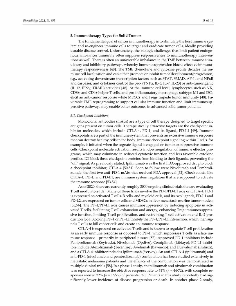
5. Immunotherapy Types for Solid Tumors
The fundamental goal of cancer immunotherapy is to stimulate the host immune sys‐
tem and re‐engineer immune cells to target and eradicate tumor cells, ideally providing
durable disease control. Unfortunately, the biologic challenges that limit patient endoge‐
nous anti‐cancer immunity often suppress responsiveness to immunotherapy interven‐
tions as well. There is often an unfavorable imbalance in the TME between immune stim‐
ulatory and inhibitory pathways, whereby immunosuppression blocks effective immuno‐
therapy responsiveness [48]. The TME chemokine and cytokine profile dictates the im‐
mune cell localization and can either promote or inhibit tumor development/progression,
e.g., activating downstream transcription factors such as STAT, SMAD, AP‐1, and NFκB
and caspases, and cytokines control the pro‐ (TNFα, IL‐6, IL‐7, IL‐23) or anti‐tumorigenic
(IL‐12, IFNγ, TRAIL) activities [48]. At the immune cell level, lymphocytes such as NK,
CD8+, and CD4+ helper T cells, and pro‐inflammatory macrophage subtype M1 and DCs
elicit an anti‐tumor response while MDSCs and Tregs impede tumor immunity [48]. Fa‐
vorable TME reprograming to support cellular immune function and limit immunosup‐
pressive pathways may enable better outcomes in advanced solid tumor patients.
5.1. Checkpoint Inhibitors
Monoclonal antibodies (mAbs) are a type of cell therapy designed to target specific
antigens present on tumor cells. Therapeutically attractive targets are the checkpoint in‐
hibitor molecules, which include CTLA‐4, PD‐1, and its ligand, PD‐L1 [49]. Immune
checkpoints are a part of the immune system that prevents an excessive immune response
that can destroy healthy cells in the body. Immune checkpoint signaling within T cells, for
example, is initiated when the cognate ligand is engaged on tumor or suppressive immune
cells. Checkpoint molecule activation results in downregulation of immune effector pro‐
grams, which may culminate in reduced cytotoxic function and less favorable cytokine
profiles. ICI block these checkpoint proteins from binding to their ligands, preventing the
“off” signal. As previously stated, Ipilimumab was the first FDA‐approved drug to block
a checkpoint inhibitor, CTLA‐4 [50,51]. Soon to follow were Nivolumab and Pembroli‐
zumab, the first two anti–PD‐1 mAbs that received FDA approval [52]. Checkpoints, like
CTLA‐4, PD‐1, and PD‐L1, are immune system regulators that are supposed to activate
the immune response [53,54].
As of 2020, there are currently roughly 3000 ongoing clinical trials that are evaluating
T cell modulators [52]. Many of these trials involve the PD‐1/PD‐L1 axis or CTLA‐4. PD‐1
is expressed on activated T cells, B cells, and myeloid cells, and its two ligands, PD‐L1 and
PD‐L2, are expressed on tumor cells and MDSCs in liver metastasis murine tumor models
[55,56]. The PD‐1/PD‐L1 axis causes immunosuppression by inducing apoptosis in acti‐
vated T cells, facilitating T cell exhaustion and anergy, enhancing Treg immunosuppres‐
sive function, limiting T cell proliferation, and restraining T cell activation and IL‐2 pro‐
duction [55]. Blocking PD‐1 or PD‐L1 inhibits the PD‐1/PD‐L1 interaction, which then sig‐
nals T cells to kill cancer cells and create an immune response.
CTLA‐4 is expressed on activated T cells and is known to regulate T cell proliferation
as an early immune response as opposed to PD‐1, which suppresses T cells as a late im‐
mune response—primarily in peripheral tissues [57]. Approved PD‐1 inhibitors include
Pembrolizumab (Keytruda), Nivolumab (Opdivo), Cemiplimab (Libtayo); PD‐L1 inhibi‐
tors include Atezolizumab (Tecentriq), Avelumab (Bavencio), and Durvalumab (Imfinzi);
and a CTLA‐4 inhibitor includes Ipilimumab (Yervoy). An anti‐CTLA‐4 (ipilimumab) and
anti‐PD‐1 (nivolumab and pembrolizumab) combination has been studied extensively in
metastatic melanoma patients and the efficacy of the combination was demonstrated in
multiple clinical trials [58]. In a phase 1 study, an ipilimumab and nivolumab combination
was reported to increase the objective response rate to 61% (n = 44/72), with complete re‐
sponses seen in 22% (n = 16/72) of patients [59]. Patients in this study reportedly had sig‐
nificantly lower incidence of disease progression or death. In another phase 2 study,

Biomedicines 2022, 10, 655 6 of 19
patients with this combination therapy had an increase in the 2‐year overall survival to
63.8% [60]. In the phase 3 study, patients treated with nivolumab plus ipilimumab, com‐
pared to ipilimumab or nivolumab monotherapy, had a higher objective response rate
(57%, 19%, and 44%, respectively), longer median progression free survival (PFS, 11.5, 2.9,
and 6.9 months, respectively), and lower incidence of disease progression or death [61].
Results of the outcomes after 3‐year and 4‐year follow‐up of patients pointed towards the
superior clinical benefits of combination therapy over monotherapy [62,63]. They have
helped improve clinical patients with different types of cancers, including breast, bladder,
cervical, colon, liver, and lung [64]. Having the ability to impact such a wide range of
cancers is a considerable leap for immunotherapy. Although there are currently no precise
biomarkers to predict the response of ICI, its efficacy, in general, is associated with the
expression of (PD‐1, PD‐L1, and CTLA‐4) and tumor mutation burden [55,65]. Detecting
therapeutic response, prognostic biomarkers, and utilizing a combination of two or more
ICIs can significantly improve their efficacy.
A mechanism to overcome the limited efficacy of PD‐1/PDL‐1 ICIs is to target other
TME‐associated immune checkpoint molecules, such as TIM‐3 and LAG‐3. A phase 2
study conducted on 72 patients treated with LAG‐3 IgG4 mAb (LAG‐525) and an anti‐PD‐
1 (spartalizumab) antibody for advanced solid tumors and hematologic malignancies
showed promising results, especially in neuroendocrine tumors, small cell lung cancer,
and diffuse large B‐cell lymphoma. The anti‐LAG‐3/PD‐1 combination demonstrated a
clinical benefit rate at 24 weeks of 86%, 27%, and 81%, respectively, in the indications
mentioned above (NCT03365791) [66]. There are seven anti‐TIM‐3 monoclonal antibodies
and one anti‐PD‐1 and TIM‐3 bispecific Ab (RO7121661) undergoing clinical develop‐
ment. Sym021 (anti‐PD‐1), sym022 (anti‐LAG‐3), and sym023 (anti‐TIM‐3) were evaluated
as single or combination treatments in phase 1 trials for solid tumors or lymphomas
(NCT03311412, NCT03489369, and NCT03489343) [67]. The monotherapy and combina‐
tion therapy were well tolerated with two partial responses observed in the combination
group. Overall, a multi‐checkpoint inhibition strategy appears rational, given the presence
of redundancy and biologic complexity in most solid organ TMEs.
5.2. Bi‐Specific Antibodies
Bispecific monoclonal antibodies (BsAb) are genetically engineered recombinant an‐
tibodies that can simultaneously target two antigens. To date, two BsAbs, blinatumomab
and emicizumab, have been approved in the US and the EU. Currently, more than 60 BsAb
drugs are in pre‐clinical trials and 30 are in clinical trials. Two‐thirds of the BsAbs focus
on the treatment of cancer by bringing effector T cells closer to cancer cells that express
specific surface antigens (BiTE) [68]. BiTEs only trigger T cell cytotoxicity and cytokine
production when both antigen binding sites are occupied [69] and is known to preferen‐
tially activate memory T cells [70–72]. BiTEs are small in size and rapidly penetrate tumors
and tissues, however they also get quickly cleared by kidneys and continuous dosing is
required [73,74].
The BiTE blinatumomab has demonstrated clinical response at very low doses in pa‐
tients with non‐Hodgkin lymphoma as compared to intact antibodies such as rituximab
(anti‐CD20) [74]. The side effects specific to BiTEs are neurotoxicity and CRS, which was
observed in the first FDA‐approved mAb, blinatumomab [74]. Emicizumab, a humanized
bispecific antibody that binds to both activated coagulation factors IX and X, was ap‐
proved for treatment of acquired hemophilia A, a severe bleeding disorder caused by in‐
hibitory auto‐antibodies against coagulation factor VIII [68]. BsAbs are in development
and a proper understanding of protein engineering and design to create these molecules
with proper delivery strategy is crucial to overcome treatment related adverse events.
BiTEs likely will require the presence of T cell infiltrates within tumors to mediate their
mechanism of action. As such, immunologically “cold” tumors with a negligible lympho‐
cyte presence may pose significant challenges for this class of drug.

Biomedicines 2022, 10, 655 7 of 19
5.3. Oncolytic Viruses (OV)
Cancer cells have impaired antiviral defenses, which makes them vulnerable to OV.
After infection, OVs cause lysis of cancer cells, thereby releasing the antigens and stimu‐
lating the immune response towards the remaining tumor cells. It was observed in the
19th century that some cancer patients would go into a small state of remission, most no‐
tably leukemia patients who contracted influenza [75,76]. OVs work in two different ways:
they can be selected, to target, replicate in, and lyse tumor cells while avoiding healthy
tissue and/or induce an immune response to have the body’s innate immunity do the kill‐
ing [77]. Although there is a push to use genetically engineered OVs, some naturally oc‐
curring viruses, such as Reolysin, a proprietary variant of the non‐pathogenic reovirus
that naturally resides in the digestive or respiratory tract, could be used to combat cancer
as well [78]. T‐Vec (Imlygic), an attenuated herpesvirus encoding granulocyte‐macro‐
phage colony stimulating factor (GM‐CSF), is the only FDA‐approved OV for treating
melanoma patients [79]. GM‐CSF can enhance the inflammatory response by activating
immune cells and is used as an immunostimulant in cancer therapies, as mentioned above.
However, tumor‐derived GM‐CSF, such as in pancreatic cancer and in liver metastases,
cause the activation and expansion of immunosuppressive MDSC, which can suppress
effector T cell functions [26,56,80]. Hence, organ and disease‐specific biology should be
taken into account to better identify patients who will be likely to respond to treatment
and to decipher potential mechanisms of treatment resistance.
5.4. Cancer Vaccines
Dr. William B. Coley’s “toxins” paved the way for the modern study of cancer im‐
munology. Observing the effects of fever on sarcoma patients, Dr. Coley started inoculat‐
ing his patients with Streptococcus pyogenes and Serratia marcescens. Johnston et al.
would later validate Coley’s work and bring cancer vaccines into the mainstream. Cancer
vaccines are categorized as cellular, viral vector, or molecular (DNA, peptide, or RNA)
vaccines. Cellular vaccines are developed from an autologous or allogeneic tumor cell line.
Dendritic cells are used to develop cellular cancer vaccines due to their roles as antigen‐
producing cells. Viral vector vaccines promote tumor‐directed immune responses by de‐
livering antigens via T‐cell priming [81]. Also, mRNA vaccines encode antigens that ex‐
press proteins following internalization, which causes an immune response. These vac‐
cines can deliver high numbers of antigens with a low risk of infection or insertional mu‐
tagenesis. In general, vaccines for solid tumors may often face the challenge of TMEs al‐
ready programmed in a highly immunosuppressive manner, which limits the ability of
patients to develop meaningful anti‐tumor responses to tumor antigens.
5.4.1. Preventative vs. Therapeutic Vaccines
Cancer vaccines that aim to reduce cancer incidence, morbidity, and mortality are
termed preventative or prophylactic. These vaccines have found success in the primary
prevention of cancers, secondary to both HBV (Hepatitis B virus) and HPV (Human Pap‐
illomavirus). In contrast, therapeutic cancer vaccines are utilized to treat an existing dis‐
ease or prevent relapse or metastases [82]. Therapeutic cancer vaccines accomplish this by
producing responses directed against antigens specific to tumors with the goal to activate
the immune system through antigen presentation [83]. A challenge in developing cancer
vaccines is the possibility that the host has already become tolerant to the targeted tumor‐
associated antigens based on suppressive TME programming.

Biomedicines 2022, 10, 655 8 of 19
5.4.2. Clinical Trials
Therapeutic cancer vaccine clinical trials have seen few successes. Only a handful of
vaccines have been approved in the US or EU, while several phase 3 clinical trials have
failed to deliver results leading to discontinuation [83]. The successful products are BCG
(TheraCys & TICE), Sipuleucel‐T (PROVENGE), and T‐Vec (IMLYGIC) [84]. BCG, a vac‐
cine to prevent and treat urothelial carcinoma, became FDA approved in 1990 in the form
of TheraCys. A phase 3 trial treating patients with intravesical BCG vaccine demonstrated
a 5‐year disease‐free survival of 45% [85]. Sipuleucel‐T, an autologous cellular immuno‐
therapy for metastatic castrate‐resistant prostate cancer, was approved in 2010 [84]. GVAX
vaccines are genetically modified tumor cells that can secrete immune stimulatory GM‐
CSF. Several clinical trials testing GVAX vaccines in melanoma, pancreatic cancer, lung
cancer, and prostate cancer have shown limited efficacy [86–89]. Combination therapies,
including ICI that reverse immunosuppression with cancer, may improve the likelihood
of success. Cancer vaccine progress lies in the identification of multiple immunogenic an‐
tigens, generating potent vaccine vectors, and overcoming solid tumor‐mediated immu‐
nosuppression.
5.5. Adoptive Cell Therapy (ACT)
ACT is another form of cell therapy that uses autologous or allogeneic immune cells
to eliminate cancer. The patient’s own (autologous) immune cells can be isolated, bioen‐
gineered for tumor antigen specificity, expanded, and reinfused into the patient, as shown
in Figure 1. The ability to isolate and expand immune cells ex vivo offers the advantages
of being able to select immune cells with high‐avidity recognition and effector function,
expand them to large numbers in the absence of inhibitory factors that exist in vivo, and
the ability to manipulate the host’s TME before the infusion to better support immune cell
function and persistence [90].
Figure 1. Adoptive cell transfer from patient tumor or blood. (1) Production begins with isolation of
peripheral blood mononuclear cells (PBMC) from leukapheresis or tumor is excised and multiple
individual cultures are isolated and (2) plated separately followed by (3) selection and activation of
T cells. (4) T cells then undergo genetic modification for generating CAR‐T cells or tumor cultures
are assayed for specific tumor recognition. (5) Cells are expanded in presence of interleukins and
when desired dose cell numbers are achieved, expanded cells are harvested and dose is formulated.
(6) QC tests are performed to ensure that drug meets release criteria and is then fused into patients
with or without conditioning lymphodepleting chemotherapy (6).
Biomedicines 2022, 10, 655 9 of 19
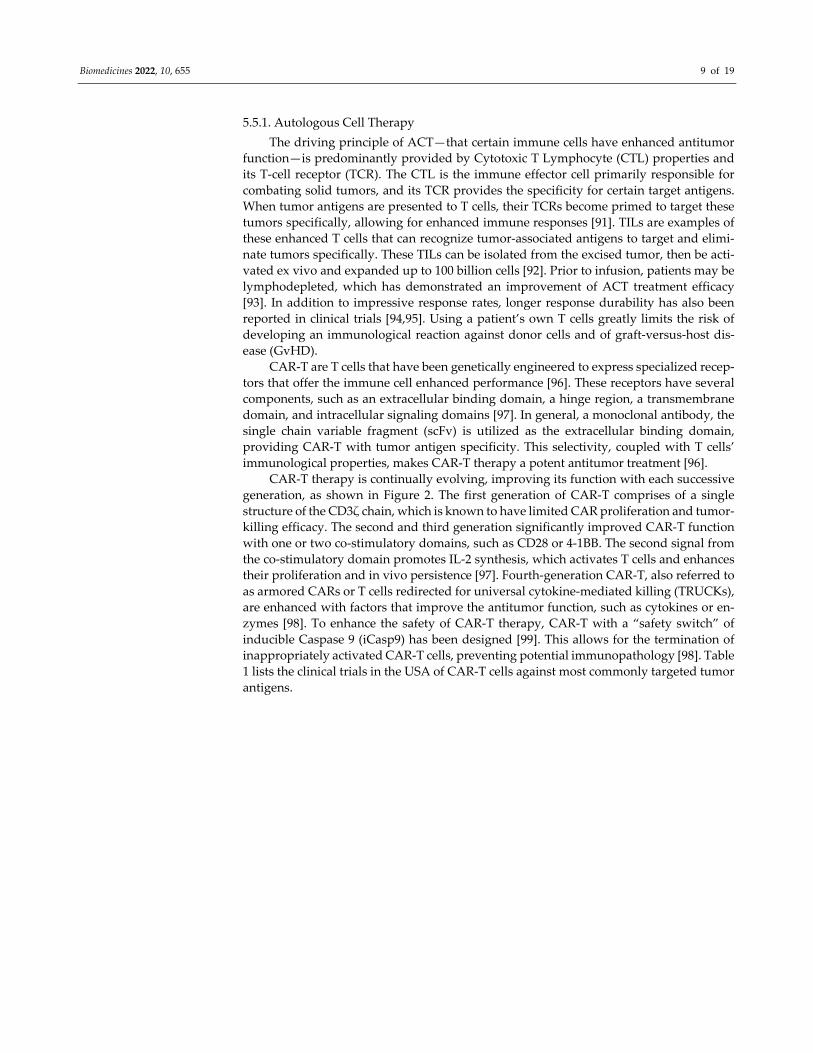
5.5.1. Autologous Cell Therapy
The driving principle of ACT—that certain immune cells have enhanced antitumor
function—is predominantly provided by Cytotoxic T Lymphocyte (CTL) properties and
its T‐cell receptor (TCR). The CTL is the immune effector cell primarily responsible for
combating solid tumors, and its TCR provides the specificity for certain target antigens.
When tumor antigens are presented to T cells, their TCRs become primed to target these
tumors specifically, allowing for enhanced immune responses [91]. TILs are examples of
these enhanced T cells that can recognize tumor‐associated antigens to target and elimi‐
nate tumors specifically. These TILs can be isolated from the excised tumor, then be acti‐
vated ex vivo and expanded up to 100 billion cells [92]. Prior to infusion, patients may be
lymphodepleted, which has demonstrated an improvement of ACT treatment efficacy
[93]. In addition to impressive response rates, longer response durability has also been
reported in clinical trials [94,95]. Using a patient’s own T cells greatly limits the risk of
developing an immunological reaction against donor cells and of graft‐versus‐host dis‐
ease (GvHD).
CAR‐T are T cells that have been genetically engineered to express specialized recep‐
tors that offer the immune cell enhanced performance [96]. These receptors have several
components, such as an extracellular binding domain, a hinge region, a transmembrane
domain, and intracellular signaling domains [97]. In general, a monoclonal antibody, the
single chain variable fragment (scFv) is utilized as the extracellular binding domain,
providing CAR‐T with tumor antigen specificity. This selectivity, coupled with T cells’
immunological properties, makes CAR‐T therapy a potent antitumor treatment [96].
CAR‐T therapy is continually evolving, improving its function with each successive
generation, as shown in Figure 2. The first generation of CAR‐T comprises of a single
structure of the CD3ζ chain, which is known to have limited CAR proliferation and tumor‐
killing efficacy. The second and third generation significantly improved CAR‐T function
with one or two co‐stimulatory domains, such as CD28 or 4‐1BB. The second signal from
the co‐stimulatory domain promotes IL‐2 synthesis, which activates T cells and enhances
their proliferation and in vivo persistence [97]. Fourth‐generation CAR‐T, also referred to
as armored CARs or T cells redirected for universal cytokine‐mediated killing (TRUCKs),
are enhanced with factors that improve the antitumor function, such as cytokines or en‐
zymes [98]. To enhance the safety of CAR‐T therapy, CAR‐T with a “safety switch” of
inducible Caspase 9 (iCasp9) has been designed [99]. This allows for the termination of
inappropriately activated CAR‐T cells, preventing potential immunopathology [98]. Table
1 lists the clinical trials in the USA of CAR‐T cells against most commonly targeted tumor
antigens.

Biomedicines 2022, 10, 655 10 of 19
Figure 2. Four generations of CAR‐T cells. First generation CARs comprise of single chain variable
fragment (scFv) antibody (orange) fused to transmembrane domain (purple) to TCR signaling com‐
ponent of CD3ζ (green) at the cytoplasmic tail. Second generation CARs have a CD28 co‐stimulatory
signaling domain (red) which enhances proliferation and cytotoxicity. Third generation CARs con‐
tain an additional co‐stimulatory domain, 4‐1BB (blue) to the second generation CARs which en‐
hances proliferation, minimizes T cell exhaustion and improves CAR‐T cells persistence. Fourth
generation CARs also called “T cell directed for universal cytokine‐mediated killing” (TRUCKs) are
engineered to secrete cytokines (gray) to attract immune cells (NK and macrophages).
Table 1. Commonly targeted solid tumor antigens in clinical trials in USA.
Antigen Cancer Phase Trial ID #
CD20 Melanoma, Non Hodgkin Lymphoma, Mantle cell Lymphoma Phase 1/2 NCT04160195, NCT04186520
CD171 Neuroblastoma Phase 1 NCT02311621
CEA Lung, colorectal, gastric, breast, pancreatic, peritoneal, liver Phase 1 NCT03682744, NCT01373047,
NCT03818165, NCT02416466,
NCT02850536
Claudin 18.2 Gastric, pancreatic Phase 1 NCT04404595
EGFRIII Glioblastoma, gliosarcoma and brain tumor Phase 1/2 NCT01454596, NCT03283631
EGFR806 Central nervous system tumor, pediatric glioma Phase 1 NCT03638167, NCT03618381
GD2 Glioma Phase 1 NCT04099797
Glypican‐3 Liver Phase 1 NCT02932956, NCT02905188
NCT04377932
HER2 Central nervous system tumor, pediatric glioma, breast Phase 1/2 NCT03500991, NCT03696030
NCT02442297, NCT03740256
NCT04483778, NCT03618381
NCT00924287, NCT04650451
NCT01109095
HLA‐A2 Ependymoma Phase 1 NCT01795313
IL‐13Rα2 Glioblastoma, cutaneous melanoma Phase 1 NCT02208362, NCT04119024
NCT04003649
KK‐LC‐1 Epithelial Phase 1 NCT05035407
Mesothelin Ovarian, cervical, pancreatic, lung, breast, mesothelioma Phase 1/2 NCT01583686, NCT04577326
NCT03054298, NCT02159716
NCT02414269
PSCA Prostate cancer, metastatic pancreatic Phase 1/2 NCT03873805, NCT02744287
TAA‐T
Hematopoietic malignancies, acute myeloid leukemia, MDS,
Hodgkin lymphoma, B cell lymphoma
Phase 1 NCT02203903, NCT03843294
VEGFR2 Metastatic melanoma, renal Phase 1/2 NCT01218867

Biomedicines 2022, 10, 655 11 of 19
5.5.2. Allogeneic Cell Therapy
Allogeneic cell therapy offers several advantages over autologous cell therapy but
also presents different challenges. The “off‐the‐shelf” aspect of allogeneic cell therapy
saves patients valuable time by eliminating the long “vein‐to‐vein” lag encountered with
autologous products. Since cells originate from healthy donors who are not exposed to
chemotherapy, the risk of production failure may be lower. Also, there is consistency be‐
tween the doses as they are generated from the same donor and hence are considered the
same lot. The complex production of doses can be performed in a more controlled way
using standard quality assurance with the option of scaling up the manufacturing process,
thereby contributing to lower production costs. Unfortunately, rejection, GvHD, and poor
persistence are challenges in the allogeneic ACT space. There are several variants of al‐
logeneic ACT products, which we discuss below.
Building on the success of CAR‐T cells, CARs have also been applied to NK cells.
While CAR‐T therapy has proven effective in treating hematological and solid tumors,
CAR‐NK cells have certain advantages in a few areas. Unlike T cells, NK cells are not
bound by HLA restrictions [100]. This property allows CAR‐NK to be used in an alloge‐
neic setting, with the potential of becoming a universal “off‐the‐shelf” cellular therapy
product [101]. It has been demonstrated clinically that CAR‐NK can survive in vivo for
several weeks to months [102]. Also, CAR expression on NK cells allows for more effective
cytotoxicity in solid tumors compared to normal NK‐mediated cytotoxicity, as they inher‐
ently possess tumor cytotoxicity and can be activated independent of CAR mechanisms
via natural cytotoxicity receptors (NCRs)—such as NKp30, NKp44, and NKp46, and
DNAM‐1 co‐stimulatory receptor and NKG2D [103].
NK cells can also be derived from several sources, such as cord blood, induced plu‐
ripotent stem cells (iPSCs), and PBMCs [104]. In an early clinical trial (NCT03383978) NK
cell line, NK‐92 is currently being tested. NK cells generated from PBMCs or cord blood
relies on the use of irradiated feeder cells, such as K562. iPSCs present an alternative
source for generating NK cells without the feeder cells [105]. Also, iPSCs can be genetically
modified prior to NK cell differentiation, thereby enabling the provision of an unlimited
supply of NK cells [106].
Allogeneic γδ T cells play an important role in tissue homeostasis and cancer immu‐
nosurveillance. These cells have been infused into patients after lymphodepleting chem‐
otherapy and were shown to expand in vivo without causing GvHD [107]. γδ T cells are
abundant in tissues and may have an edge over αβ T cells for developing therapeutic
strategies for treating solid tumors [108]. Polyclonal γδ T cells transduced with CD19 CAR
and GD2 CAR have demonstrated anti‐tumor effects in vitro and in in vivo murine mod‐
els [109,110].
Despite the potential benefits of allogeneic cell therapy, there are significant chal‐
lenges that need to be overcome for their successful implementation. Some of the chal‐
lenges include an immunologic mismatch between donor and recipient that may cause
undesirable GvHD. Conversely, if a recipient’s immune system recognizes and reacts to
allogeneic therapy, the dose may be rejected, thereby limiting its therapeutic activity. It is
likely that autologous and allogeneic ACT products will both find a place within the
multi‐disciplinary management of solid tumor patients. The relative value of autologous
and allogeneic therapy in specific clinical settings will be determined by disease biology,
typical patient physiology, and the existing therapeutic landscape.
5.5.3. Combination Therapy or Other Strategies of Regional Delivery
To increase the likelihood of success with immunotherapy in challenging solid tumor
indications, a variety of combinatorial approaches are being explored. Multiple clinical
studies have shown that CAR‐T therapy or chemotherapy alone achieved limited efficacy
to treat solid tumors [111,112]. However, studies have found that many chemotherapeutic
agents, such as cyclophosphamide, doxorubicin, oxaliplatin, fluorouracil, and

Biomedicines 2022, 10, 655 12 of 19
gemcitabine, reduce tumor burden and have immunomodulatory effects that enhance im‐
munotherapy [113]. Radiotherapy has been known to react synergistically with CAR‐T
therapy. Not only does radiotherapy eradicate the tumor, but it can also sensitize tumor
cells to cytotoxic lymphocytes in the murine colon adenocarcinoma model [114], modulate
the TME to make it conducive to CAR‐T infiltration, traffic in the highly angiogenic insu‐
linoma mouse models [115], and improve tumor antigen presentation as observed in tu‐
mors implanted in flanks of mice [116].
CAR‐T treatment can be combined with other immunotherapies as well. Tumors of‐
ten employ their immunosuppressive microenvironments to escape the therapeutic ef‐
fects of CAR‐T therapy [117]. Antibodies that block CTLA‐4, PD‐1, and PD‐L1 can be used
to overcome the immunosuppressive nature of the TME and allow the CAR‐T to maintain
efficacy [118]. Combinations of CAR‐T with chemokines such as CXCR2 or CCR4 have
been found to help CAR‐T trafficking and persistence in solid tumors [118]. It has also
been found that combining CAR‐T therapy with OVs produces synergistic effects. OVs
can assist in the entry of CAR‐T into tumors [119]. CAR‐T has also been shown to better
survive in solid tumors in the presence of OVs [120].
5.5.4. Side Effects
Although CAR cellular therapy has proven highly beneficial in treating tumors, these
results are often accompanied by potential toxic side effects. Overcoming these side effects
is paramount to the successful application of these cell therapies. The most prevalent of
these side effects is CRS, or elevated inflammatory cytokines resulting from therapy‐in‐
duced immune activation. CRS can range from mild to severe, with the latter being po‐
tentially life‐threatening. Symptoms can include high fever, malaise, fatigue, myalgia,
nausea, anorexia, tachycardia/hypotension, capillary leak, cardiac dysfunction, renal im‐
pairment, and hepatic failure disseminated intravascular coagulation [121].
Another potential side effect is OTOTT in CAR cellular therapy, which can be
avoided only in cases where the target antigen is exclusively restricted to the tumor. Un‐
fortunately, many CAR cells target antigens are shared on normal tissue, resulting in
OTOTT [122]. On‐target/off‐tumor recognition is often observed in the gastrointestinal,
hematologic, and pulmonary organ systems [123]. Targeting colon cancer with CAR ther‐
apy using the carcinoembryonic antigen (CEA) has caused severe colitis due to recogni‐
tion of the normal colon tissue [124]. Anaphylaxis is another concern when treating pa‐
tients with CAR cells due to the murine mAb origin of the antigen‐recognition domains
[122]. These domains can be recognized as a foreign protein and elicit an immune response
[125].
6. Summary
The major roadblock for ACT is to translate this treatment modality to solid malig‐
nancies to treat solid tumors effectively and to improve its survival, persistence, and effi‐
cacy. While autologous treatments such as CAR‐T cell therapy have proven effective, HLA
restrictions prevent the drug’s universal application. This also makes the manufacturing
of cellular therapy incredibly expensive and time‐consuming, which is problematic for
some patients with highly proliferative diseases, as it can result in disease progression.
While CAR‐NK cells do not face these same HLA restrictions, this therapy is more novel
and requires further exploration. The development of a universal “off‐the‐shelf” cellular
therapy would revolutionize immunotherapy.
The immunosuppressive TME of the solid tumor remains a major obstacle to over‐
come, with the potential for variation among anatomic sites. Combination therapies with
checkpoint blockade, vaccines, OVs, cell therapies, etc., have shown that these different
methods have the potential to counter immunosuppression more effectively while also
providing direct tumor killing activity in some cases. Further combinations are required
to address the difficulties presented by solid tumors. Novel delivery methods are also
worthy of consideration to optimize the therapeutic concentration in solid tumors and to

Biomedicines 2022, 10, 655 13 of 19
overcome delivery barriers imposed by physical forces within the TME. High‐pressure
delivery of CAR‐T cellular therapy has shown increased penetration and persistence of
cells in tumors [45,46]. Regional delivery of cell therapy may also enhance the therapeutic
index by limiting systemic toxicity, including CRS and neurologic side effects.
7. Conclusions and Perspectives
Although immunotherapy has shown great promise in certain indications, ICIs and
cell therapy have thus far failed to make a major impact on certain solid tumor indications.
CAR‐T cell therapy has completely revolutionized patient care for some hematologic ma‐
lignancies, but has yet to achieve similar levels of success in the realms of solid organ
primary or metastatic malignancies. An unprecedented number of clinical trials of CAR‐
T cells in solid tumors are ongoing with novel strategies creating increased levels of opti‐
mism. Barriers to immunotherapy success in solid tumors such as CAR‐T cell trafficking,
persistence, immunosuppressive TME, and antigen heterogeneity have been identified.
Hence, increased understanding of the critical interactions between tumors and immune
responses is imperative. Combinatorial approaches specifically tailored to both the dis‐
ease and organ biology hold the greatest promise in extending the positive impact of im‐
munotherapy to a greater number of patients in need. Also, the development of specific
biomarkers to delineate the use of the optimal immunotherapy is critical to ensure the best
treatment option for patients. Overall, novel cancer immunotherapies have revolutionized
cancer treatment for patients by enhancing clinical outcomes for patients and results from
ongoing research should also assist in its continued progress in the field.
Author Contributions: Conceptualization, P.G., S.C.K.; writing—original draft preparation, K.R.H.,
K.P.O., I.S.A.; writing—review and editing, P.G., S.C.K. All authors have read and agreed to the
published version of the manuscript.
Funding: This article received no external funding.
Institutional Review Board Statement: Not applicable.
Informed Consent Statement: Not applicable.
Data Availability Statement: Not applicable.
Conflicts of Interest: The authors declare no conflict of interest.
Abbreviations
ACT adoptive cell therapy
ADCs antibody‐drug conjugated
AP‐1 activator protein 1
bFGF basic fibroblast growth factor
BsAb bispecific monoclonal antibodies
BCMA B‐cell maturation antigen
BsAb bispecific antibody
BiTEs bispecific T cell engagers
BTLA band T lymphocyte attenuator
CAR‐NK chimeric antigen receptor NK cells
CAR‐T chimeric antigen receptor T cells
CRS cytokine release syndrome
CTLA‐4 cytotoxic T lymphocyte antigen 4
FDA food and drug administration
GvHD graft‐versus‐host disease
HBV hepatitis B virus
HPV human papillomavirus
HLA human leukocyte antigen
iCasp9 inducible Caspase 9
ICI immune checkpoint inhibitors

Biomedicines 2022, 10, 655 14 of 19
IFN interferon
IL interleukin
iPSC induced pluripotent stem cells
LAG‐3 lymphocyte activation gene 3 protein
LPD lipid‐protamine‐DNA
mAb monoclonal antibody
MDSCs myeloid derived suppressor cells
NF‐κB nuclear factor kappa B
mRNA messenger RNA
OTOTT on‐target‐off‐tumor‐toxicity
OV oncolytic virus
PD‐1 programmed cell death protein
PD‐L1 programmed cell death protein and its ligand
scFv single chain variable fragment
STAT3 signal transducer and activator of transcription 3
TAM tumor associated macrophages
TGF‐β transforming growth factor beta
TIL’s tumor‐infiltrating lymphocytes
TIM‐3 T‐cell immunoglobulin domain and mucin do‐
main protein 3
TME tumor microenvironment
Tregs regulatory T cells
TIGIT T‐cell immunoglobulin and immunoreceptor ty‐
rosine‐based inhibitory motif domain
TNF tumor necrosis factor
TRAIL TNF related apoptosis inducing ligand
VEGF vascular endothelial growth factor
References
1. Kucerova, P.; Cervinkova, M. Spontaneous regression of tumour and the role of microbial infection—Possibilities for cancer
treatment. Anti‐Cancer Drugs 2016, 27, 269–277. https://doi.org/10.1097/cad.0000000000000337.
2. Dobosz, P.; Dzieciątkowski, T. The Intriguing History of Cancer Immunotherapy. Front. Immunol. 2019, 10, 2965.
https://doi.org/10.3389/fimmu.2019.02965.
3. Mellman, I.; Coukos, G.; Dranoff, G. Cancer immunotherapy comes of age. Nature 2011, 480, 480–489. https://doi.org/10.1038/na‐
ture10673.
4. Burga, R.A.; Thorn, M.; Point, G.R.; Guha, P.; Nguyen, C.T.; Licata, L.A.; DeMatteo, R.P.; Ayala, A.; Espat, N.J.; Junghans, R.P.;
et al. Liver myeloid‐derived suppressor cells expand in response to liver metastases in mice and inhibit the anti‐tumor efficacy
of anti‐CEA CAR‐T. Cancer Immunol. Immunother. 2015, 64, 817–829. https://doi.org/10.1007/s00262‐015‐1692‐6.
5. Swanson, G.P.; Rynearson, K.; Symmonds, R. Significance of margins of excision on breast cancer recurrence. Am. J. Clin. Oncol.
2002, 25, 438–441.
6. DeVita, V.T., Jr.; Chu, E. A History of Cancer Chemotherapy. Cancer Res. 2008, 68, 8643–8653. https://doi.org/10.1158/0008‐
5472.can‐07‐6611.
7. De Angelis, C. Side Effects Related to Systemic Cancer Treatment: Are We Changing the Promethean Experience with
Molecularly Targeted Therapies? Curr. Oncol. 2008, 15, 198–199. https://doi.org/10.3747/co.v15i4.362.
8. Baskar, R.; Lee, K.A.; Yeo, R.; Yeoh, K.‐W. Cancer and Radiation Therapy: Current Advances and Future Directions. Int. J. Med.
Sci. 2012, 9, 193–199. https://doi.org/10.7150/ijms.3635.
9. Cheng, H.; Wang, Z.; Fu, L.; Xu, T. Macrophage Polarization in the Development and Progression of Ovarian Cancers: An
Overview. Front. Oncol. 2019, 9, 421. https://doi.org/10.3389/fonc.2019.00421.
10. Jiang, T.; Zhou, C. The past, present and future of immunotherapy against tumor. Transl. Lung Cancer Res. 2015, 4, 253–264.
https://doi.org/10.3978/j.issn.2218‐6751.2015.01.06.
11. Ventola, C.L. Cancer Immunotherapy, Part 1: Current Strategies and Agents. Pharm. Ther. 2017, 42, 375–383.
12. Filin, I.Y.; Solovyeva, V.V.; Kitaeva, K.V.; Rutland, C.S.; Rizvanov, A.A. Current Trends in Cancer Immunotherapy. Biomedicines
2020, 8, 621. https://doi.org/10.3390/biomedicines8120621.
13. Maeda, H.; Khatami, M. Analyses of repeated failures in cancer therapy for solid tumors: Poor tumor‐selective drug delivery,
low therapeutic efficacy and unsustainable costs. Clin. Transl. Med. 2018, 7, 11. https://doi.org/10.1186/s40169‐018‐0185‐6.
14. Siegel, R.L.; Miller, K.D.; Goding Sauer, A.; Fedewa, S.A.; Butterly, L.F.; Anderson, J.C.; Cercek, A.; Smith, R.A.; Jemal, A. Col‐
orectal cancer statistics, 2020. CA Cancer J. Clin. 2020, 70, 7–30.

Biomedicines 2022, 10, 655 15 of 19
15. Hou, B.; Tang, Y.; Li, W.; Zeng, Q.; Chang, D. Efficiency of CAR‐T Therapy for Treatment of Solid Tumor in Clinical Trials: A
Meta‐Analysis. Dis. Markers 2019, 2019, 3425291. https://doi.org/10.1155/2019/3425291.
16. Duffy, A.G.; Greten, T.F. Immunological off‐target effects of standard treatments in gastrointestinal cancers. Ann. Oncol. 2013,
25, 24–32. https://doi.org/10.1093/annonc/mdt349.
17. Giampieri, R.; Del Prete, M.; Prochilo, T.; Puzzoni, M.; Pusceddu, V.; Pani, F.; Maccaroni, E.; Mascia, R.; Baleani, M.G.; Meletani,
T.; et al. Off‐target effects and clinical outcome in metastatic colorectal cancer patients receiving regorafenib: The TRIBUTE
analysis. Sci. Rep. 2017, 7, 45703. https://doi.org/10.1038/srep45703.
18. Brown, J.M.; Giaccia, A.J. The unique physiology of solid tumors: Opportunities (and problems) for cancer therapy. Cancer Res.
1998, 58, 1408–1416.
19. Khawar, I.A.; Kim, J.H.; Kuh, H.‐J. Improving drug delivery to solid tumors: Priming the tumor microenvironment. J. Control
Release 2015, 201, 78–89. https://doi.org/10.1016/j.jconrel.2014.12.018.
20. Vaupel, P.; Thews, O.; Hoeckel, M. Treatment resistance of solid tumors: Role of hypoxia and anemia. Med. Oncol. 2001, 18, 243–
259.
21. Trédan, O.; Galmarini, C.M.; Patel, K.; Tannock, I.F. Drug Resistance and the Solid Tumor Microenvironment. J. Natl. Cancer
Inst. 2007, 99, 1441–1454. https://doi.org/10.1093/jnci/djm135.
22. Chai, L.F.; Prince, E.; Pillarisetty, V.G.; Katz, S.C. Challenges in assessing solid tumor responses to immunotherapy. Cancer Gene
Ther. 2019, 27, 528–538. https://doi.org/10.1038/s41417‐019‐0155‐1.
23. Whiteside, T.L. The tumor microenvironment and its role in promoting tumor growth. Oncogene 2008, 27, 5904–5912.
https://doi.org/10.1038/onc.2008.271.
24. Poggi, A.; Musso, A.; Dapino, I.; Zocchi, M.R. Mechanisms of tumor escape from immune system: Role of mesenchymal stromal
cells. Immunol. Lett. 2014, 159, 55–72. https://doi.org/10.1016/j.imlet.2014.03.001.
25. Lindau, D.; Gielen, P.; Kroesen, M.; Wesseling, P.; Adema, G.J. The immunosuppressive tumour network: Myeloid‐derived
suppressor cells, regulatory T cells and natural killer T cells. Immunology 2012, 138, 105–115. https://doi.org/10.1111/imm.12036.
26. Guha, P.; Gardell, J.; Rabinowitz, B.; Lopes, M.; DaSilva, N.A.; Rowley, D.; Katz, S.C. Monocytic and granulocytic myeloid‐
derived suppressor cell plasticity and differentiation are organ‐specific. Oncogene 2020, 40, 693–704.
https://doi.org/10.1038/s41388‐020‐01559‐7.
27. Grivennikov, S.I.; Greten, F.R.; Karin, M. Immunity, inflammation, and cancer. Cell 2010, 140, 883–899.
https://doi.org/10.1016/j.cell.2010.01.025.
28. Yu, H.; Pardoll, D.; Jove, R. STATs in cancer inflammation and immunity: A leading role for STAT3. Nat. Cancer 2009, 9, 798–
809. https://doi.org/10.1038/nrc2734.
29. Grivennikov, S.; Karin, E.; Terzic, J.; Mucida, D.; Yu, G.Y.; Vallabhapurapu, S.; Scheller, J.; Rose‐John, S.; Cheroutre, H.; Eckmann,
L.; et al. IL‐6 and Stat3 are required for survival of intestinal epithelial cells and development of colitis‐associated cancer. Cancer
Cell 2009, 15, 103–113.
30. Becker, C.; Fantini, M.C.; Schramm, C.; Lehr, H.A.; Wirtz, S.; Becker, A.; Burg, J.; Strand, S.; Kiesslich, R.; Huber, S.; et al. TGF‐
beta suppresses tumor progression in colon cancer by inhibition of IL–6 trans‐signaling. Immunity 2004, 42, P139.
https://doi.org/10.1055/s‐2004‐831593.
31. Bollrath, J.; Phesse, T.; von Burstin, V.A.; Putoczki, T.; Bennecke, M.; Bateman, T.; Nebelsiek, T.; Lundgren‐May, T.; Canli, Ö.;
Schwitalla, S.; et al. gp130‐Mediated Stat3 Activation in Enterocytes Regulates Cell Survival and Cell‐Cycle Progression during
Colitis‐Associated Tumorigenesis. Cancer Cell 2009, 15, 91–102. https://doi.org/10.1016/j.ccr.2009.01.002.
32. Popivanova, B.K.; Kitamura, K.; Wu, Y.; Kondo, T.; Kagaya, T.; Kaneko, S.; Oshima, M.; Fujii, C.; Mukaida, N. Blocking TNF‐α
in mice reduces colorectal carcinogenesis associated with chronic colitis. J. Clin. Investig. 2008, 118, 560–570.
https://doi.org/10.1172/jci32453.
33. Wang, D.J.; Ratnam, N.M.; Byrd, J.C.; Guttridge, D.C. NF‐kappaB functions in tumor initiation by suppressing the surveillance
of both innate and adaptive immune cells. Cell Rep. 2014, 9, 90–103.
34. Wang, Y.; Shen, Y.; Wang, S.; Shen, Q.; Zhou, X. The role of STAT3 in leading the crosstalk between human cancers and the
immune system. Cancer Lett. 2018, 415, 117–128. https://doi.org/10.1016/j.canlet.2017.12.003.
35. Van Horssen, R.; Hagen, T.L.M.T.; Eggermont, A.M.M. TNF‐α in Cancer Treatment: Molecular Insights, Antitumor Effects, and
Clinical Utility. Oncologist 2006, 11, 397–408. https://doi.org/10.1634/theoncologist.11‐4‐397.
36. Zhao, L.; Ching, L.‐M.; Kestell, P.; Baguley, B.C. The antitumour activity of 5,6‐dimethylxanthenone‐4‐acetic acid (DMXAA) in
TNF receptor‐1 knockout mice. Br. J. Cancer 2002, 87, 465–470. https://doi.org/10.1038/sj.bjc.6600479.
37. Bertrand, F.; Montfort, A.; Marcheteau, E.; Imbert, C.; Gilhodes, J.; Filleron, T.; Rochaix, P.; Andrieu‐Abadie, N.; Levade, T.;
Meyer, N.; et al. TNFalpha blockade overcomes resistance to anti‐PD‐1 in experimental melanoma. Nat. Commun. 2017, 8, 2256.
38. Massagué, J. TGFβ in cancer. Cell 2008, 134, 215–230.
39. Berraondo, P.; Sanmamed, M.F.; Ochoa, M.C.; Etxeberria, I.; Aznar, M.A.; Pérez‐Gracia, J.L.; Rodriguez‐Ruiz, M.E.; Ponz‐Sarvise,
M.; Castañón, E.; Melero, I. Cytokines in clinical cancer immunotherapy. Br. J. Cancer 2019, 120, 6–15.
https://doi.org/10.1038/s41416‐018‐0328‐y.
40. Jiang, Y.; Li, Y.; Zhu, B. T‐cell exhaustion in the tumor microenvironment. Cell Death Dis. 2015, 6, e1792.
https://doi.org/10.1038/cddis.2015.162.
41. Zarour, H.M. Reversing T‐cell Dysfunction and Exhaustion in Cancer. Clin. Cancer Res. 2016, 22, 1856–1864.
https://doi.org/10.1158/1078‐0432.ccr‐15‐1849.
Biomedicines 2022, 10, 655 16 of 19
42. Ribas, A.; Medina, T.; Kummar, S.; Amin, A.; Kalbasi, A.; Drabick, J.J.; Barve, M.; Daniels, G.A.; Wong, D.J.; Schmidt, E.V.; et al.
SD‐101 in Combination with Pembrolizumab in Advanced Melanoma: Results of a Phase Ib, Multicenter Study. Cancer Discov.
2018, 8, 1250–1257. https://doi.org/10.1158/2159‐8290.cd‐18‐0280.
43. Song, W.; Shen, L.; Wang, Y.; Liu, Q.; Goodwin, T.J.; Li, J.; Dorosheva, O.; Liu, T.; Liu, R.; Huang, L. Synergistic and low adverse
effect cancer immunotherapy by immunogenic chemotherapy and locally expressed PD‐L1 trap. Nat. Commun. 2018, 9, 2237.
https://doi.org/10.1038/s41467‐018‐04605‐x.
44. Wong, C.; Stylianopoulos, T.; Cui, J.; Martin, J.; Chauhan, V.P.; Jiang, W.; Popović, Z.; Jain, R.K.; Bawendi, M.G.; Fukumura, D.
Multistage nanoparticle delivery system for deep penetration into tumor tissue. Proc. Natl. Acad. Sci. USA 2011, 108, 2426–2431.
https://doi.org/10.1073/pnas.1018382108.
45. Katz, S.C.; Hardaway, J.; Prince, E.; Guha, P.; Cunetta, M.; Moody, A.; Wang, L.J.; Armenio, V.; Espat, N.J.; Junghans, R.P. HITM‐
SIR: Phase Ib trial of intraarterial chimeric antigen receptor T‐cell therapy and selective internal radiation therapy for CEA+
liver metastases. Cancer Gene Ther. 2019, 27, 341–355. https://doi.org/10.1038/s41417‐019‐0104‐z.
46. Katz, S.C.; Moody, A.E.; Guha, P.; Hardaway, J.C.; Prince, E.; Laporte, J.; Stancu, M.; Slansky, J.E.; Jordan, K.R.; Schulick, R.D.;
et al. HITM‐SURE: Hepatic immunotherapy for metastases phase Ib anti‐CEA CAR‐T study utilizing pressure enabled drug
delivery. J. Immunother. Cancer 2020, 8, e001097. https://doi.org/10.1136/jitc‐2020‐001097.
47. Narayanan, J.S.S.; Vicente, D.A.; Ray, P.; Chai, L.F.; Erdem, S.; Carr, M.J.; Capacio, B.A.; Cox, B.F.; Jaroch, D.B.; Katz, S.C.; et al.
Pressure‐enabled delivery of gemcitabine in an orthotopic pancreatic cancer mouse model. Surgery 2020, 168, 448–456.
https://doi.org/10.1016/j.surg.2020.04.059.
48. Labani‐Motlagh, A.; Ashja‐Mahdavi, M.; Loskog, A. The Tumor Microenvironment: A Milieu Hindering and Obstructing An‐
titumor Immune Responses. Front. Immunol. 2020, 11, 940. https://doi.org/10.3389/fimmu.2020.00940.
49. Pento, J.T. Monoclonal Antibodies for the Treatment of Cancer. Anticancer Res. 2017, 37, 5935–5939. https://doi.org/10.21873/an‐
ticanres.12040.
50. Graziani, G.; Tentori, L.; Navarra, P. Ipilimumab: A novel immunostimulatory monoclonal antibody for the treatment of cancer.
Pharmacol. Res. 2012, 65, 9–22. https://doi.org/10.1016/j.phrs.2011.09.002.
51. Lipson, E.J.; Drake, C.G. Ipilimumab: An Anti‐CTLA‐4 Antibody for Metastatic Melanoma. Clin. Cancer Res. 2011, 17, 6958–6962.
https://doi.org/10.1158/1078‐0432.ccr‐11‐1595.
52. Robert, C. A decade of immune‐checkpoint inhibitors in cancer therapy. Nat. Commun. 2020, 11, 3801.
https://doi.org/10.1038/s41467‐020‐17670‐y.
53. Andrews, L.P.; Yano, H.; Vignali, D.A.A. Inhibitory receptors and ligands beyond PD‐1, PD‐L1 and CTLA‐4: Breakthroughs or
backups. Nat. Immunol. 2019, 20, 1425–1434. https://doi.org/10.1038/s41590‐019‐0512‐0.
54. Quezada, S.; Peggs, K.S. Exploiting CTLA‐4, PD‐1 and PD‐L1 to reactivate the host immune response against cancer. Br. J. Cancer
2013, 108, 1560–1565. https://doi.org/10.1038/bjc.2013.117.
55. Meyers, D.E.; Bryan, P.M.; Banerji, S.; Morris, D.G. Targeting the PD‐1/PD‐L1 axis for the treatment of non‐small‐cell lung cancer.
Curr. Oncol. 2018, 25, e324–e334.
56. Thorn, M.; Guha, P.; Cunetta, M.; Espat, N.J.; Miller, G.; Junghans, R.P.; Katz, S.C. Tumor‐associated GM‐CSF overexpression
induces immunoinhibitory molecules via STAT3 in myeloid‐suppressor cells infiltrating liver metastases. Cancer Gene Ther. 2016,
23, 188–198. https://doi.org/10.1038/cgt.2016.19.
57. Buchbinder, E.I.; Desai, A. CTLA‐4 and PD‐1 Pathways: Similarities, Differences, and Implications of Their Inhibition. Am. J.
Clin. Oncol. 2016, 39, 98–106.
58. Rotte, A. Combination of CTLA‐4 and PD‐1 blockers for treatment of cancer. J. Exp. Clin. Cancer Res. 2019, 38, 255.
https://doi.org/10.1186/s13046‐019‐1259‐z.
59. Postow, M.A.; Chesney, J.; Pavlick, A.C.; Robert, C.; Grossmann, K.; McDermott, D.; Linette, G.P.; Meyer, N.; Giguere, J.K.;
Agarwala, S.S.; et al. Nivolumab and Ipilimumab versus Ipilimumab in Untreated Melanoma. N. Engl. J. Med. 2015, 372, 2006–
2017. https://doi.org/10.1056/nejmoa1414428; Erratum in N. Engl. J. Med. 2018, 379, 2185.
60. Hodi, F.S.; Chesney, J.; Pavlick, A.C.; Robert, C.; Grossmann, K.F.; McDermott, D.F.; Linette, G.P.; Meyer, N.; Giguere, J.K.;
Agarwala, S.S.; et al. Combined nivolumab and ipilimumab versus ipilimumab alone in patients with advanced melanoma: 2‐
year overall survival outcomes in a multicentre, randomised, controlled, phase 2 trial. Lancet Oncol. 2016, 17, 1558–1568.
https://doi.org/10.1016/s1470‐2045(16)30366‐7.
61. Larkin, J.; Chiarion‐Sileni, V.; Gonzalez, R.; Grob, J.J.; Cowey, C.L.; Lao, C.D.; Schadendorf, D.; Dummer, R.; Smylie, M.;
Rutkowski, P.; et al. Combined Nivolumab and Ipilimumab or Monotherapy in Untreated Melanoma. N. Engl. J. Med. 2015, 373,
1270–1271.
62. Wolchok, J.D.; Chiarion‐Sileni, V.; Gonzalez, R.; Rutkowski, P.; Grob, J.‐J.; Cowey, C.L.; Lao, C.D.; Wagstaff, J.; Schadendorf, D.;
Ferrucci, P.F.; et al. Overall Survival with Combined Nivolumab and Ipilimumab in Advanced Melanoma. N. Engl. J. Med. 2017,
377, 1345–1356. https://doi.org/10.1056/nejmoa1709684.
63. Hodi, F.S.; Chiarion‐Sileni, V.; Gonzalez, R.; Grob, J.‐J.; Rutkowski, P.; Cowey, C.L.; Lao, C.D.; Schadendorf, D.; Wagstaff, J.;
Dummer, R.; et al. Nivolumab plus ipilimumab or nivolumab alone versus ipilimumab alone in advanced melanoma (Check‐
Mate 067): 4‐year outcomes of a multicentre, randomised, phase 3 trial. Lancet Oncol. 2018, 19, 1480–1492.
https://doi.org/10.1016/s1470‐2045(18)30700‐9.
64. Johnson, D.B.; Chandra, S.; Sosman, J.A. Immune Checkpoint Inhibitor Toxicity in 2018. JAMA 2018, 320, 1702–1703.
https://doi.org/10.1001/jama.2018.13995.

Biomedicines 2022, 10, 655 17 of 19
65. Duffy, M.J.; Crown, J. Biomarkers for Predicting Response to Immunotherapy with Immune Checkpoint Inhibitors in Cancer
Patients. Clin. Chem. 2019, 65, 1228–1238. https://doi.org/10.1373/clinchem.2019.303644.
66. Uboha, N.V.; Milhem, M.M.; Kovacs, C.; Amin, A.; Magley, A.; Das Purkayastha, D.; Piha‐Paul, S.A. Phase II study of spartali‐
zumab (PDR001) and LAG525 in advanced solid tumors and hematologic malignancies.. J. Clin. Oncol. 2019, 37, 2553–2553.
https://doi.org/10.1200/jco.2019.37.15_suppl.2553.
67. Lakhani, N.; Spreafico, A.; Tolcher, A.; Rodon, J.; Janku, F.; Chandana, S.; Oliva, M.; Sharma, M.; Abdul‐Karim, R.; Hansen, U.;
et al. 1019O Phase I studies of Sym021, an anti‐PD‐1 antibody, alone and in combination with Sym022 (anti‐LAG‐3) or Sym023
(anti‐TIM‐3). Ann. Oncol. 2020, 31, S704. https://doi.org/10.1016/j.annonc.2020.08.1139.
68. Labrijn, A.F.; Janmaat, M.L.; Reichert, J.M.; Parren, P.W.H.I. Bispecific antibodies: A mechanistic review of the pipeline. Nat.
Rev. Drug Discov. 2019, 18, 585–608. https://doi.org/10.1038/s41573‐019‐0028‐1.
69. Brischwein, K.; Parr, L.; Pflanz, S.; Volkland, J.; Lumsden, J.; Klinger, M.; Locher, M.; Hammond, S.A.; Kiener, P.; Kufer, P.; et
al. Strictly Target Cell‐dependent Activation of T Cells by Bispecific Single‐chain Antibody Constructs of the BiTE Class. J.
Immunother. 2007, 30, 798–807. https://doi.org/10.1097/cji.0b013e318156750c.
70. Brischwein, K.; Schlereth, B.; Guller, B.; Steiger, C.; Wolf, A.; Lutterbuese, R.; Offner, S.; Locher, M.; Urbig, T.; Raum, T.; et al.
MT110: A novel bispecific single‐chain antibody construct with high efficacy in eradicating established tumors. Mol. Immunol.
2006, 43, 1129–1143. https://doi.org/10.1016/j.molimm.2005.07.034.
71. Lutterbuese, R.; Raum, T.; Kischel, R.; Hoffmann, P.; Mangold, S.; Rattel, B.; Friedrich, M.; Thomas, O.; Lorenczewski, G.; Rau,
D.; et al. T cell‐engaging BiTE antibodies specific for EGFR potently eliminate KRAS‐ and BRAF‐mutated colorectal cancer cells.
Proc. Natl. Acad. Sci. USA 2010, 107, 12605–12610. https://doi.org/10.1073/pnas.1000976107.
72. Torisu‐Itakura, H.; Schoellhammer, H.F.; Sim, M.‐S.; Irie, R.F.; Hausmann, S.; Raum, T.; Baeuerle, P.A.; Morton, D.L. Redirected
Lysis of Human Melanoma Cells by a MCSP/CD3‐bispecific BiTE Antibody That Engages Patient‐derived T Cells. J. Immunother.
2011, 34, 597–605. https://doi.org/10.1097/cji.0b013e3182307fd8.
73. Klinger, M.; Brandl, C.; Zugmaier, G.; Hijazi, Y.; Bargou, R.C.; Topp, M.S.; Gökbuget, N.; Neumann, S.; Goebeler, M.; Viardot,
A.; et al. Immunopharmacologic response of patients with B‐lineage acute lymphoblastic leukemia to continuous infusion of T
cell‐engaging CD19/CD3‐bispecific BiTE antibody blinatumomab. Blood 2012, 119, 6226–6233.
74. Goebeler, M.‐E.; Knop, S.; Viardot, A.; Kufer, P.; Topp, M.S.; Einsele, H.; Noppeney, R.; Hess, G.; Kallert, S.; Mackensen, A.; et
al. Bispecific T‐Cell Engager (BiTE) Antibody Construct Blinatumomab for the Treatment of Patients with Relapsed/Refractory
Non‐Hodgkin Lymphoma: Final Results From a Phase I Study. J. Clin. Oncol. 2016, 34, 1104–1111.
https://doi.org/10.1200/jco.2014.59.1586.
75. Donnelly, O.; Harrington, K.; Melcher, A.; Pandha, H. Live viruses to treat cancer. J. R. Soc. Med. 2013, 106, 310–314.
https://doi.org/10.1177/0141076813494196.
76. Kelly, E.; Russell, S.J. History of Oncolytic Viruses: Genesis to Genetic Engineering. Mol. Ther. 2007, 15, 651–659.
https://doi.org/10.1038/sj.mt.6300108.
77. Zou, Y.; Luo, Y.; Zhang, J.; Xia, N.; Tan, G.; Huang, C. Bibliometric analysis of oncolytic virus research, 2000 to 2018. Medicine
2019, 98, e16817. https://doi.org/10.1097/md.0000000000016817.
78. Fukuhara, H.; Ino, Y.; Todo, T. Oncolytic virus therapy: A new era of cancer treatment at dawn. Off. J. Jpn. Cancer Assoc. 2016,
107, 1373–1379.
79. Bommareddy, P.K.; Patel, A.; Hossain, S.; Kaufman, H.L. Talimogene Laherparepvec (T‐VEC) and Other Oncolytic Viruses for
the Treatment of Melanoma. Am. J. Clin. Dermatol. 2016, 18, 1–15. https://doi.org/10.1007/s40257‐016‐0238‐9.
80. Pylayeva‐Gupta, Y.; Lee, K.E.; Hajdu, C.H.; Miller, G.; Bar‐Sagi, D. Oncogenic Kras‐Induced GM‐CSF Production Promotes the
Development of Pancreatic Neoplasia. Cancer Cell 2012, 21, 836–847. https://doi.org/10.1016/j.ccr.2012.04.024.
81. Jou, J.; Harrington, K.J.; Zocca, M.‐B.; Ehrnrooth, E.; Cohen, E.E. The Changing Landscape of Therapeutic Cancer Vaccines—
Novel Platforms and Neoantigen Identification. Clin. Cancer Res. 2020, 27, 689–703. https://doi.org/10.1158/1078‐0432.ccr‐20‐0245.
82. Lollini, P.L.; Cavallo, F.; Nanni, P.; Forni, G. Vaccines for tumour prevention. Nat. Cancer 2006, 6, 204–216.
https://doi.org/10.1038/nrc1815.
83. Morse, M.A.; Gwin, W.R., III; Mitchell, D.A. Vaccine Therapies for Cancer: Then and Now. Target. Oncol. 2021, 16, 121–152.
84. DeMaria, P.J.; Bilusic, M. Cancer Vaccines. Hematol. Oncol. Clin. N. Am. 2019, 33, 199–214.
85. Lamm, D.L.; Blumenstein, B.A.; Crawford, E.D.; Montie, J.E.; Scardino, P.; Grossman, H.B.; Stanisic, T.H.; Smith, J.A.; Sullivan,
J.; Sarosdy, M.F.; et al. A Randomized Trial of Intravesical Doxorubicin and Immunotherapy with Bacille Calmette–Guérin for
Transitional‐Cell Carcinoma of the Bladder. N. Engl. J. Med. 1991, 325, 1205–1209. https://doi.org/10.1056/nejm199110243251703.
86. Laheru, D.; Lutz, E.; Burke, J.; Biedrzycki, B.; Solt, S.; Onners, B.; Tartakovsky, I.; Nemunaitis, J.; Le, D.; Sugar, E.; et al. Allogeneic
Granulocyte Macrophage Colony‐Stimulating Factor–Secreting Tumor Immunotherapy Alone or in Sequence with
Cyclophosphamide for Metastatic Pancreatic Cancer: A Pilot Study of Safety, Feasibility, and Immune Activation. Clin. Cancer
Res. 2008, 14, 1455–1463. https://doi.org/10.1158/1078‐0432.ccr‐07‐0371.
87. Lipson, E.J.; Sharfman, W.H.; Chen, S.; McMiller, T.L.; Pritchard, T.S.; Salas, J.T.; Sartorius‐Mergenthaler, S.; Freed, I.; Ravi, S.;
Wang, H.; et al. Safety and immunologic correlates of Melanoma GVAX, a GM‐CSF secreting allogeneic melanoma cell vaccine
administered in the adjuvant setting. J. Transl. Med. 2015, 13, 214. https://doi.org/10.1186/s12967‐015‐0572‐3.
88. Salgia, R.; Lynch, T.; Skarin, A.; Lucca, J.; Lynch, C.; Jung, K.; Hodi, F.S.; Jaklitsch, M.; Mentzer, S.; Swanson, S.; et al. Vaccination
With Irradiated Autologous Tumor Cells Engineered to Secrete Granulocyte‐Macrophage Colony‐Stimulating Factor Augments

Biomedicines 2022, 10, 655 18 of 19
Antitumor Immunity in Some Patients With Metastatic Non–Small‐Cell Lung Carcinoma. J. Clin. Oncol. 2003, 21, 624–630.
https://doi.org/10.1200/jco.2003.03.091.
89. Small, E.J.; Sacks, N.; Nemunaitis, J.; Urba, W.J.; Dula, E.; Centeno, A.S.; Nelson, W.G.; Ando, D.; Howard, C.; Borellini, F.; et al.
Granulocyte Macrophage Colony‐Stimulating Factor–Secreting Allogeneic Cellular Immunotherapy for Hormone‐Refractory
Prostate Cancer. Clin. Cancer Res. 2007, 13, 3883–3891. https://doi.org/10.1158/1078‐0432.ccr‐06‐2937.
90. Rosenberg, S.A.; Restifo, N.P.; Yang, J.C.; Morgan, R.A.; Dudley, M.E. Adoptive cell transfer: A clinical path to effective cancer
immunotherapy. Nat. Cancer 2008, 8, 299–308. https://doi.org/10.1038/nrc2355.
91. Dudley, M.E.; Rosenberg, S.A. Adoptive‐cell‐transfer therapy for the treatment of patients with cancer. Nat. Rev. Cancer 2003, 3,
666–675. https://doi.org/10.1038/nrc1167.
92. Itzhaki, O.; Hovav, E.; Ziporen, Y.; Levy, D.; Kubi, A.; Zikich, D.; Hershkovitz, L.; Treves, A.J.; Shalmon, B.; Zippel, D.; et al.
Establishment and Large‐scale Expansion of Minimally cultured “Young” Tumor Infiltrating Lymphocytes for Adoptive Trans‐
fer Therapy. J. Immunother. 2011, 34, 212–220. https://doi.org/10.1097/cji.0b013e318209c94c.
93. Muranski, P.; Boni, A.; Wrzesinski, C.; Citrin, D.; Rosenberg, S.A.; Childs, R.W.; Restifo, N.P. Increased intensity
lymphodepletion and adoptive immunotherapy—how far can we go? Nat. Clin. Pract. Oncol. 2006, 3, 668–681.
https://doi.org/10.1038/ncponc0666.
94. Rosenberg, S.A.; Packard, B.S.; Aebersold, P.M.; Solomon, D.; Topalian, S.L.; Toy, S.T.; Simon, P.; Lotze, M.T.; Yang, J.C.; Seipp,
C.A.; et al. Use of Tumor‐Infiltrating Lymphocytes and Interleukin‐2 in the Immunotherapy of Patients with Metastatic Mela‐
noma: A preliminary report. N. Engl. J. Med. 1988, 319, 1676–1680. https://doi.org/10.1056/nejm198812223192527.
95. Besser, M.J.; Shapira‐Frommer, R.; Itzhaki, O.; Treves, A.J.; Zippel, D.B.; Levy, D.; Kubi, A.; Shoshani, N.; Zikich, D.; Ohayon,
Y.; et al. Adoptive Transfer of Tumor‐Infiltrating Lymphocytes in Patients with Metastatic Melanoma: Intent‐to‐Treat Analysis
and Efficacy after Failure to Prior Immunotherapies. Clin. Cancer Res. 2013, 19, 4792–4800. https://doi.org/10.1158/1078‐0432.ccr‐
13‐0380.
96. Porter, D.L.; Levine, B.L.; Kalos, M.; Bagg, A.; June, C.H. Chimeric Antigen Receptor–Modified T Cells in Chronic Lymphoid
Leukemia. N. Engl. J. Med. 2011, 365, 725–733. https://doi.org/10.1056/nejmoa1103849.
97. Hartmann, J.; Schüßler‐Lenz, M.; Bondanza, A.; Buchholz, C.J. Clinical development of CAR T cells—challenges and opportu‐
nities in translating innovative treatment concepts. EMBO Mol. Med. 2017, 9, 1183–1197.
https://doi.org/10.15252/emmm.201607485.
98. Chmielewski, M.; Abken, H. TRUCKs: The fourth generation of CARs. Exp. Opin. Biol. Ther. 2015, 15, 1145–1154.
https://doi.org/10.1517/14712598.2015.1046430.
99. Gargett, T.; Brown, M.P. The inducible caspase‐9 suicide gene system as a “safety switch” to limit on‐target, off‐tumor toxicities
of chimeric antigen receptor T cells. Front. Pharmacol. 2014, 5, 235.
100. Lorenzo‐Herrero, S.; López‐Soto, A.; Sordo‐Bahamonde, C.; Gonzalez‐Rodriguez, S.; Vitale, M.; Gonzalez, S. NK Cell‐Based
Immunotherapy in Cancer Metastasis. Cancers 2018, 11, 29. https://doi.org/10.3390/cancers11010029.
101. Hermanson, D.L.; Kaufman, D.S. Utilizing Chimeric Antigen Receptors to Direct Natural Killer Cell Activity. Front. Immunol.
2015, 6, 195. https://doi.org/10.3389/fimmu.2015.00195.
102. Miller, J.S.; Soignier, Y.; Panoskaltsis‐Mortari, A.; McNearney, S.A.; Yun, G.H.; Fautsch, S.K.; McKenna, D.; Le, C.; DeFor, T.E.;
Burns, L.J.; et al. Successful adoptive transfer and in vivo expansion of human haploidentical NK cells in patients with cancer.
Blood 2005, 105, 3051–3057. https://doi.org/10.1182/blood‐2004‐07‐2974.
103. Geller, M.A.; Cooley, S.; Judson, P.L.; Ghebre, R.; Carson, L.F.; Argenta, P.A.; Jonson, A.L.; Panoskaltsis‐Mortari, A.; Curtsinger,
J.; McKenna, D.; et al. A phase II study of allogeneic natural killer cell therapy to treat patients with recurrent ovarian and breast
cancer. Cytotherapy 2011, 13, 98–107. https://doi.org/10.3109/14653249.2010.515582.
104. Veluchamy, J.P.; Kok, N.; van der Vliet, H.J.; Verheul, H.M.W.; De Gruijl, T.D.; Spanholtz, J. The Rise of Allogeneic Natural
Killer Cells as a Platform for Cancer Immunotherapy: Recent Innovations and Future Developments. Front. Immunol. 2017, 8,
631. https://doi.org/10.3389/fimmu.2017.00631.
105. Cichocki, F.; Bjordahl, R.; Gaidarova, S.; Mahmood, S.; Abujarour, R.; Wang, H.; Tuininga, K.; Felices, M.; Davis, Z.B.; Bendzick,
L.; et al. iPSC‐derived NK cells maintain high cytotoxicity and enhance in vivo tumor control in concert with T cells and anti‐
PD‐1 therapy. Sci. Transl. Med. 2020, 12, eaaz5618.
106. Li, Y.; Hermanson, D.L.; Moriarity, B.S.; Kaufman, D.S. Human iPSC‐Derived Natural Killer Cells Engineered with Chimeric
Antigen Receptors Enhance Anti‐tumor Activity. Cell Stem Cell 2018, 23, 181–192.e5.
107. Wilhelm, M.; Smetak, M.; Schaefer‐Eckart, K.; Kimmel, B.; Birkmann, J.; Einsele, H.; Kunzmann, V. Successful adoptive transfer
and in vivo expansion of haploidentical gammadelta T cells. J. Transl. Med. 2014, 12, 45.
108. Khairallah, C.; Chu, T.H.; Sheridan, B.S. Tissue Adaptations of Memory and Tissue‐Resident Gamma Delta T Cells. Front. Im‐
munol. 2018, 9, 2636. https://doi.org/10.3389/fimmu.2018.02636.
109. Deniger, D.C.; Switzer, K.; Mi, T.; Maiti, S.; Hurton, L.; Singh, H.; Huls, H.; Olivares, S.; Lee, D.A.; Champlin, R.E.; et al. Bispecific
T‐cells expressing polyclonal repertoire of endogenous gammadelta T‐cell receptors and introduced CD19‐specific chimeric
antigen receptor. Mol. Ther. 2013, 21, 638–647.
110. Capsomidis, A.; Benthall, G.; Van Acker, H.H.; Fisher, J.; Kramer, A.M.; Abeln, Z.; Majani, Y.; Gileadi, T.; Wallace, R.; Gustafsson,
K.; et al. Chimeric Antigen Receptor‐Engineered Human Gamma Delta T Cells: Enhanced Cytotoxicity with Retention of Cross
Presentation. Mol. Ther. 2018, 26, 354–365. https://doi.org/10.1016/j.ymthe.2017.12.001.
Biomedicines 2022, 10, 655 19 of 19
111. Lamers, C.H.; Klaver, Y.; Gratama, J.W.; Sleijfer, S.; Debets, R. Treatment of metastatic renal cell carcinoma (mRCC) with CAIX
CAR‐engineered T‐cells‐a completed study overview. Biochem. Soc. Trans. 2016, 44, 951–959.
112. Lamers, C.H.; Sleijfer, S.; van Steenbergen, S.; van Elzakker, P.; van Krimpen, B.; Groot, C.; Vulto, A.; Bakker, M.D.; Oosterwijk,
E.; Debets, R.; et al. Treatment of Metastatic Renal Cell Carcinoma With CAIX CAR‐engineered T cells: Clinical Evaluation and
Management of On‐target Toxicity. Mol. Ther. 2013, 21, 904–912. https://doi.org/10.1038/mt.2013.17.
113. Vierboom, M.P.; Bos, G.M.; Ooms, M.; Offringa, R.; Melief, C.J. Cyclophosphamide enhances anti‐tumor effect of wild‐type p53‐
specific CTL. Int. J. Cancer 2000, 87, 253–260. https://doi.org/10.1002/1097‐0215(20000715)87:2<253::aid‐ijc17>3.0.co;2‐a.
114. Reits, E.A.; Hodge, J.W.; Herberts, C.A.; Groothuis, T.A.; Chakraborty, M.; Wansley, E.K.; Camphausen, K.; Luiten, R.M.; De
Ru, A.H.; Neijssen, J.; et al. Radiation modulates the peptide repertoire, enhances MHC class I expression, and induces success‐
ful antitumor immunotherapy. J. Exp. Med. 2006, 203, 1259–1271. https://doi.org/10.1084/jem.20052494.
115. Ganss, R.; Ryschich, E.; Klar, E.; Arnold, B.; Hämmerling, G.J. Combination of T‐cell therapy and trigger of inflammation in‐
duces remodeling of the vasculature and tumor eradication. Cancer Res. 2002, 62, 1462–1470.
116. Deng, L.; Liang, H.; Xu, M.; Yang, X.; Burnette, B.; Arina, A.; Li, X.‐D.; Mauceri, H.; Beckett, M.; Darga, T.; et al. STING‐Depend‐
ent Cytosolic DNA Sensing Promotes Radiation‐Induced Type I Interferon‐Dependent Antitumor Immunity in Immunogenic
Tumors. Immunity 2014, 41, 843–852. https://doi.org/10.1016/j.immuni.2014.10.019.
117. Domschke, C.; Schneeweiss, A.; Stefanovic, S.; Wallwiener, M.; Heil, J.; Rom, J.; Sohn, C.; Beckhove, P.; Schuetz, F. Cellular
Immune Responses and Immune Escape Mechanisms in Breast Cancer: Determinants of Immunotherapy. Breast Care 2016, 11,
102–107. https://doi.org/10.1159/000446061.
118. Neagu, M.R.; Reardon, D.A. An Update on the Role of Immunotherapy and Vaccine Strategies for Primary Brain Tumors. Curr.
Treat. Options Oncol. 2015, 16, 54. https://doi.org/10.1007/s11864‐015‐0371‐3.
119. Ajina, A.; Maher, J. Prospects for combined use of oncolytic viruses and CAR T‐cells. J. Immunother. Cancer 2017, 5, 90–90.
https://doi.org/10.1186/s40425‐017‐0294‐6.
120. Nishio, N.; Diaconu, I.; Liu, H.; Cerullo, V.; Caruana, I.; Hoyos, V.; Bouchier‐Hayes, L.; Savoldo, B.; Dotti, G. Armed Oncolytic
Virus Enhances Immune Functions of Chimeric Antigen Receptor–Modified T Cells in Solid Tumors. Cancer Res. 2014, 74, 5195–
5205. https://doi.org/10.1158/0008‐5472.can‐14‐0697.
121. Bonifant, C.; Jackson, H.J.; Brentjens, R.J.; Curran, K.J. Toxicity and management in CAR T‐cell therapy. Mol. Ther.‐Oncolytics
2016, 3, 16011–16011. https://doi.org/10.1038/mto.2016.11.
122. Curran, K.J.; Pegram, H.J.; Brentjens, R.J. Chimeric antigen receptors for T cell immunotherapy: Current understanding and
future directions. J. Gene Med. 2012, 14, 405–415. https://doi.org/10.1002/jgm.2604.
123. Lamers, C.H.; Sleijfer, S.; Vulto, A.G.; Kruit, W.H.; Kliffen, M.; Debets, R.; Gratama, J.W.; Stoter, G.; Oosterwijk, E. Treatment of
Metastatic Renal Cell Carcinoma With Autologous T‐Lymphocytes Genetically Retargeted Against Carbonic Anhydrase IX:
First Clinical Experience. J. Clin. Oncol. 2006, 24, e20‐2. https://doi.org/10.1200/jco.2006.05.9964.
124. Parkhurst, M.R.; Yang, J.C.; Langan, R.C.; Dudley, M.E.; Nathan, D.‐A.N.; Feldman, S.A.; Davis, J.L.; Morgan, R.A.; Merino, M.J.;
Sherry, R.M.; et al. T Cells Targeting Carcinoembryonic Antigen Can Mediate Regression of Metastatic Colorectal Cancer but
Induce Severe Transient Colitis. Mol. Ther. 2011, 19, 620–626. https://doi.org/10.1038/mt.2010.272.
125. Kershaw, M.H.; Westwood, J.A.; Parker, L.L.; Wang, G.; Eshhar, Z.; Mavroukakis, S.A.; White, D.E.; Wunderlich, J.R.; Canevari,
S.; Rogers‐Freezer, L.; et al. A Phase I Study on Adoptive Immunotherapy Using Gene‐Modified T Cells for Ovarian Cancer.
Clin. Cancer Res. 2006, 12 Pt 1, 6106–6115. https://doi.org/10.1158/1078‐0432.ccr‐06‐1183.
Copyright © 2022 FDOKUMEN