The Wilms' Tumor Antigen Is a Novel Target for Human CD4+ Regulatory T Cells: Implications for...
11
2008;68:6350-6359. Cancer Res Cynthia Lehe, Hazem Ghebeh, Abdullah Al-Sulaiman, et al. Regulatory T Cells: Implications for Immunotherapy + The Wilms' Tumor Antigen Is a Novel Target for Human CD4 Updated version http://cancerres.aacrjournals.org/content/68/15/6350 Access the most recent version of this article at: Material Supplementary http://cancerres.aacrjournals.org/content/suppl/2008/07/25/68.15.6350.DC1.html Access the most recent supplemental material at: Cited Articles http://cancerres.aacrjournals.org/content/68/15/6350.full.html#ref-list-1 This article cites by 49 articles, 22 of which you can access for free at: Citing articles http://cancerres.aacrjournals.org/content/68/15/6350.full.html#related-urls This article has been cited by 7 HighWire-hosted articles. Access the articles at: E-mail alerts related to this article or journal. Sign up to receive free email-alerts Subscriptions Reprints and . [email protected] Department at To order reprints of this article or to subscribe to the journal, contact the AACR Publications Permissions . [email protected] Department at To request permission to re-use all or part of this article, contact the AACR Publications Research. on April 14, 2014. © 2008 American Association for Cancer cancerres.aacrjournals.org Downloaded from Research. on April 14, 2014. © 2008 American Association for Cancer cancerres.aacrjournals.org Downloaded from
Transcript of The Wilms' Tumor Antigen Is a Novel Target for Human CD4+ Regulatory T Cells: Implications for...
2008;68:6350-6359. Cancer Res Cynthia Lehe, Hazem Ghebeh, Abdullah Al-Sulaiman, et al. Regulatory T Cells: Implications for Immunotherapy
+The Wilms' Tumor Antigen Is a Novel Target for Human CD4
Updated version
http://cancerres.aacrjournals.org/content/68/15/6350
Access the most recent version of this article at:
Material
Supplementary
http://cancerres.aacrjournals.org/content/suppl/2008/07/25/68.15.6350.DC1.html
Access the most recent supplemental material at:
Cited Articles
http://cancerres.aacrjournals.org/content/68/15/6350.full.html#ref-list-1
This article cites by 49 articles, 22 of which you can access for free at:
Citing articles
http://cancerres.aacrjournals.org/content/68/15/6350.full.html#related-urls
This article has been cited by 7 HighWire-hosted articles. Access the articles at:
E-mail alerts related to this article or journal.Sign up to receive free email-alerts
Subscriptions
Reprints and
To order reprints of this article or to subscribe to the journal, contact the AACR Publications
Permissions
To request permission to re-use all or part of this article, contact the AACR Publications
Research. on April 14, 2014. © 2008 American Association for Cancercancerres.aacrjournals.org Downloaded from
Research. on April 14, 2014. © 2008 American Association for Cancercancerres.aacrjournals.org Downloaded from
The Wilms’ Tumor Antigen Is a Novel Target for Human CD4+
Regulatory T Cells: Implications for Immunotherapy
Cynthia Lehe,1Hazem Ghebeh,
1Abdullah Al-Sulaiman,
2Ghofran Al Qudaihi,
1
Khaled Al-Hussein,2Fahad Almohareb,
3Naeem Chaudhri,
3Fahad Alsharif,
3
Hazza Al-Zahrani,3Abdelghani Tbakhi,
4Mahmoud Aljurf,
3and Said Dermime
1
1Tumor Immunology Section, 2Histocompatibility and Immunogenetics Section, 3Adult Hematology/Oncology, and4Immunopathology, King Faisal Specialist Hospital and Research Centre, Riyadh, Saudi Arabia
Abstract
Compelling evidences indicate a key role for regulatory T cells(Treg) on the host response to cancer. The Wilms’ tumorantigen (WT1) is overexpressed in several human leukemiasand thus considered as promising target for development ofleukemia vaccine. However, recent studies indicated that thegeneration of effective WT1-specific cytotoxic T cells can belargely affected by the presence of Tregs. We have generatedT-cell lines and clones that specifically recognized a WT1-84(RYFKLSHLQMHSRKH) peptide in an HLA-DRB1*0402–restricted manner. Importantly, they recognized HLA-DRB1*04–matched fresh leukemic cells expressing the WT1antigen. These clones exerted a T helper 2 cytokine profile,had a CD4+CD25+Foxp3+GITR+CD127� Treg phenotype, andsignificantly inhibited the proliferative activity of allogeneic Tcells independently of cell contact. Priming of alloreactive Tcells in the presence of Tregs strongly inhibited the expansionof natural killer (NK), NK T, and CD8+ T cells and had aninhibitory effect on NK/NK T cytotoxic activity but not onCD8+ T cells. Furthermore, priming of T cells with the WT1-126HLA-A0201–restricted peptide in the presence of Tregs stronglyinhibited the induction of anti–WT1-126 CD8+ CTL responsesas evidenced by both very low cytotoxic activity and IFN-;production. Moreover, these Treg clones specifically producedgranzyme B and selectively induced apoptosis in WT1-84–pulsed autologous antigen-presenting cells but not in apopto-tic-resistant DR4-matched leukemic cells. Importantly, wehave also detected anti–WT1-84 interleukin-5+/granzyme B+/Foxp3+ CD4+ Tregs in five of eight HLA-DR4+ acute myeloidleukemia patients. Collectively, our in vitro and in vivofindings strongly suggest important implications for theclinical manipulation of Tregs in cancer patients. [Cancer Res2008;68(15):6350–9]
Introduction
The Wilms’ tumor (WT1) gene exerts an oncogenic function invarious types of leukemias (1). It is also overexpressed in severalsolid tumors (2), and therefore, it has been considered as anattractive target for cancer immunotherapy. Specific anti-WT1
immune responses have been described in which CD8+ cytotoxic Tcells (3, 4) have been generated in vitro . However, a recent studyshowed that such response can be largely affected by the presenceof CD4+CD25+ regulatory T cells (Treg) in which depletion of thisT-cell population was necessary for the generation of an effectiveWT1-specific cytotoxic response (3).It has been shown recently that Tregs directly suppress the
antitumor immune responses in cancer patients (5, 6), anddepletion of this T-cell population resulted in an enhancement ofvaccine-mediated antitumor immunity in cancer patients (7). Thishighlights the role of Tregs in modulating both natural and adoptiveimmune responses in cancer patients. Tregs may directly modulatethe CD8+ T-cell response (8) or alternatively promote tolerization ofCD8+ T cells by preventing the licensing of antigen-presenting cells(APC) by CD4+ T helper cells (9). It has became clear that CD8+ Tcells generated in the absence of CD4+ T cells help may have anormal primary response; however, their cytotoxic memoryresponse is severely weakened (10). A recent study by Greinerand colleagues (11) investigated the influence of the expressionlevels of several leukemia-associated antigens (LAA) on the clinicaloutcome of patients with acute myeloid leukemia (AML). Highexpression of three LAAs, which was found to be associated withfavorable clinical outcome, induced strong CD8+ T-cell responses.However, there was no correlation with the clinical outcome norinduction of natural detectable anti-WT1 CD8+ T-cell response inthese patients (11). In line with this, a recent study has shown theexistence of tumor-specific Tregs, which actively suppress antigen-specific antitumor immunity in cancer patients (12). Furthermore,fully functional Tregs specific for LAGE1 (13) and ARTC1 (14) wereshown in melanoma patients. Another study by Nadal andcolleagues (15) suggested that Tregs exert an inhibitory effect ongraft versus leukemia and this was associated with relapse afterallogeneic stem cell transplantation.To this end, we asked whether an anti-WT1 Treg population exist
in leukemia patients, which may contribute to the impairment ofanti-WT1 responses. We have identified a human HLA-DRB1*0402–restricted CD4+ Treg population and showed that the WT1 is anovel target for leukemia-specific CD4+ Tregs.
Materials and Methods
Patients and donors. Peripheral blood mononuclear cells (PBMC) from
leukemia patients and healthy donors were isolated by density gradientcentrifugation. This study was conducted in accordance with Helsinki
Declaration and all patients and donors signed a consent form approved by
the Research Ethics Committee of King Faisal Specialist Hospital andResearch Center (KFSH&RC). The study was approved by the KFSH&RC
Research Advisory Council (RAC#2030006). Patient and donor information
is listed in Supplementary Data 1.
Note: Supplementary data for this article are available at Cancer Research Online(http://cancerres.aacrjournals.org/).
Requests for reprints: Said Dermime, Tumor Immunology Section, King FaisalSpecialist Hospital and Research Centre, MBC 03, P. O. Box 3354, Riyadh 11211, SaudiArabia. Phone: 966-1-442-4552; Fax: 966-1-442-7858; E-mail: [email protected] [email protected].
I2008 American Association for Cancer Research.doi:10.1158/0008-5472.CAN-08-0050
Cancer Res 2008; 68: (15). August 1, 2008 6350 www.aacrjournals.org
Research Article
Research. on April 14, 2014. © 2008 American Association for Cancercancerres.aacrjournals.org Downloaded from

Peptides. A pool of 110 peptides derived from the WT1 protein desig-nated as WT1-Pepmix and a microscale WT1 peptide set containing each
peptide in a single well were obtained from JPT Peptide Technologies (Jerini
AG, GmbH; Supplementary Data 2). The WT1333-347 (RYFKLSHLQMHSRKH),
WT1-84, HPV3373-87 (ASDLRTIQQLLMGTV) HLA-DR*0402–restricted pep-tide (16), and the WT1-db126 (RMFPNAPYL) HLA-A0201–restricted peptide
(4) were synthesized and high-performance liquid chromatography purified
to z90% purity by Alta Bioscience.
Cell lines. B-lymphoblastoid cell lines (LCL) were established bytransformation of B cells using EBV using standard techniques (17). K562
and T2 cell lines were purchased from the American Type Culture
Collection. All cell lines were maintained in RPMI 1640 (Sigma)
supplemented with 10% FCS (Cambrex Bio Science), 2 mmol/L L-glutamine,100 IU/mL penicillin, and 100 mg/mL streptomycin (Sigma).
Generation of autologous monocyte-derived dendritic cells. CD14+
cells were isolated from PBMCs of three healthy donors, designated as BC-
29, BC-52, and BC-62, using MACS Monocytes Isolation kit (Miltenyi Biotec)
according to the manufacturer’s instructions. CD14+ cells were adhered and
cultured in 24-well plates in X-VIVO 15 medium supplemented with
2 mmol/L L-glutamine [dendritic cell (DC) medium] in the presence of
50 ng/mL recombinant human interleukin (rhIL)-4 and 100 ng/mL
recombinant human granulocyte macrophage colony-stimulating factor
(GM-CSF; R&D Systems). Recombinant human tumor necrosis factor-a(20 ng/mL; R&D Systems), 10 ng/mL rhIL-1h (eBioscience), and 25 Ag/mL
polyinosinic acid:poly-CMP (Sigma) were added on day 6 and mature DCs
were harvested on day 8.Generation of anti–WT1-specific Tregs and clones. Irradiated DCs
(3,000 rads) were pulsed with 10 Ag/mL WT1-Pepmix in DC medium and
incubated for 4 h at 37jC, 5% CO2. DCs were cocultured with autologous
PBMCs at a 10:1 T-cell to DC ratio. DC medium containing 10 ng/mL rhIL-7,20 pg/mL rhIL-12 (R&D Systems), and 10% heat-inactivated human
serum (Sigma) was used. T cells were restimulated at weekly intervals
and 260 IU/mL rhIL-2 was added 2 d after each restimulation. Three of
three anti–WT1-Pepmix-specific T-cell lines were obtained after the fourthrestimulation. Anti–WT1-Pepmix T-cell clones were generated as described
before (18). T cells were screened for their peptide specificity against the
microscale WT1 peptide set.Proliferation and 51Cr cytotoxicity assay. T-cell proliferation was
assessed by [3H]thymidine incorporation as described before (18). Briefly,
T cells were cocultured with X-irradiated PBMCs, LCLs (Fantigens), or
leukemic cells at different ratios in DC medium containing 10% humanserum for 3 d. [3H]thymidine (1 ACi/well; Amersham) was added for the last
18 h. [3H]thymidine uptake was measured using a 1450 Micro Beta PLUS
liquid scintillation counter (Wallac). In some experiments, the following
blocking antibodies (50–100 Ag/mL) were used: anti-ABC (AbD Serotec),anti–HLA-DR (BD Biosciences), anti–HLA-DP, and anti–HLA-DQ (Leinco
Technologies). Mouse isotypes were used as controls. Cytotoxic activity was
measured by a standard 51Cr release assay as described before (18).ELISA. Supernatants from 48-h T-cell cultures were harvested, and ELISA
for the human IFN-g, IL-4, IL-5, IL-10, and GM-CSF ELISA kits (Mabtech)
and transforming growth factor (TGF)-h1 and TGF-h2 matched antibody
pairs (R&D Systems) was performed according to the manufacturer’sinstructions.
Flow cytometry. T cells were phenotypically analyzed using the human
Treg staining kit (eBioscience) according to the manufacturer’s instructions
and also stained with CTLA-4-PE (Dako Corp.), CD127-RPE, and GITR-APC
(eBioscience). The T-cell receptor (TCR) Vh profile was determined using
the IOTest Beta Mark, TCR Vh Repertoire kit (Beckman Coulter). Cells were
analyzed using FACScan (Becton Dickinson).
Immunostaining. Immunostaining on cytospins was carried out as
described previously (19). Anti-Foxp3 antibody (eBioscience), anti-CD3antibody (Dako), and anti-WT1 antibody (Dako) were used. Isotype-
matched controls for all antibodies were used. The staining was evaluated
by two independent scientists.
Reverse transcription-PCR for Foxp3 expression. Total RNA wasisolated using RNeasy Micro kit (Qiagen). The forward primer CAGCTGCC-
CACACTGCCCCTAG and the reverse primer CAGTGCCATTTTCCCAGC-
CAG were used for Foxp3. The PCR conditions were 95jC/3 min, 35 cyclesof 95jC/1 min, 67jC/40 s, and 72jC/1 min. The h-actin forward primer
ATCTGGCACCACACCTTCTACAATGAGCTGCG and the reverse primer
CGTCATACTCCTGCTTGCTGATCCACATCTGC were used as a control.
PCR products were separated by electrophoresis on a 1% agarose gel(Sigma).
Quantitative real-time reverse transcription-PCR for WT1 expres-sion. Total RNA was isolated as above and treated with DNase1
(Invitrogen). For cDNA synthesis, 1 Ag RNA using oligo(dT) primers andMoloney murine leukemia virus reverse transcriptase (Invitrogen) was used
in a total volume of 25 AL. The WT1 mRNA expression was quantified using
LightCycler FastStart DNA Master SYBR Green 1 kit (Roche) in a
LightCycler (Roche). Porphobilinogen deaminase (PBGD) was used ashousekeeping gene. cDNA (2 AL) from K562 was used to generate standard
curves (20). Amplification was conducted in a total volume of 20 AL for
40 cycles/10 s at 95jC, 4 s/64jC, and 35 s/72jC. The forward primerTTCATCAAACAGGAGCCGAGC and the reverse primer GTGCGAGGG-
CGTGTGA were used for WT1. For PBGD, the forward primer CATGTC-
TGGTAACGGCAATG and the reverse primer TCTTCTCCAGGGCATGTTCAA
were used.Treg suppression assays and Transwell experiments. To examine the
suppressive effect of the Treg clones on the induction of T helper
alloresponses, a standard mixed lymphocyte reaction (MLR) was carried
out as described before (21). Briefly, fresh BC-29 PBMCs (105 per well,
responders) were cultured in DC medium containing 10% human serum for
5 d with XR, 3,000 rads allogeneic PBMCs (105 per well, stimulators) with
different concentrations of Tregs + autologous LCL FWT1-84 peptide. The
proliferative activity was determined as before. For some experiments,
neutralizing antibodies against TGF-h1/h2, IL-4, IL-5 (R&D Systems), IL-10
(Biosource), and GM-CSF (Biolegend) were added in the assay at an
optimum final concentration of 5 Ag/mL. The effect of Treg inhibition on
nonirradiated autologous LCL was also examined. Transwell experiments
were performed in 24-well plates with pore size of 0.4 Am (Corning Costar).
BC-29 PBMCs (0.75 � 106 per well, responders) were cultured in the outer
wells of 24-well plates in DC medium containing 10% human serum and
XR, 3,000 rads allogeneic (BC-8) PBMCs (1.5 � 106 per well, stimulators).
Treg clones (0.15 � 106 cells per well) were added into the inner wells of
autologous LCL F WT1-84 peptide. After 4 d in culture, the cells in the
outer wells were harvested and transferred to 96-well plates and the
proliferative activity was determined as before.
The suppressive effect of Tregs on the induction of alloreactive and anti–WT1-specific cytotoxic responses was examined. For alloreactive CTLs, BC-
29 PBMCs (2 � 106 per well, responders) were cultured in DC medium
containing 10% human serum for 7 d with XR, 7,500 rads allogeneic LCL
(0.5 � 106 cells per well, stimulators) in 24-well plates F 0.5 � 106 cells perwell TCC 29.B.42 Treg clone + WT1-84 peptide. This procedure was used for
the generation of anti-WT1 CTLs, except that autologous DC pulsed with
the WT1-126 (RMFPNAPYL) HLA-A0201–restricted peptide (4) was used.
IL-2 (25 units/mL) was added on day 4 and a similar stimulation wasrepeated after 7 d. Several stimulations were carried out without addition of
the Tregs every 7 d. Flow cytometry analysis was used to determine the CD8,
CD56, and CD8/CD56 cell population in the culture. CD8 T cells werepurified using MACS human CD8-negative selection kit (Miltenyi Biotec).
Cytotoxic and IFN-g production activity were determined by 51Cr release
and enzyme-linked immunospot (ELISPOT) assays, respectively.
ELISPOT assay. ELISPOT assay was performed using IFN-g, granzyme B,and perforin kits (Mabtech). Autologous LCLs or AML cells were used as
stimulators. Tregs (103 cells per well) and stimulators (2 � 104 cells per well)
were seeded in Multiscreen 96-well plates (Millipore) precoated with
catching antibody. After 40 h of incubation, cells were removed and plateswere processed according to the manufacturer’s instructions. Spots were
counted using an automated ELISPOT reader (AID). Antigen-specific T-cell
frequencies were considered to be increased when they were at least 2-foldhigher than in the control wells.
Apoptosis assay. An apoptosis assay was carried out using Vybrant
Apoptosis Assay Kit #2 (Invitrogen). Tregs (2 � 104) and target cells (2 � 104)
were incubated in DC medium + 10% human serum for 24 h. Targets were
WT1 as a New Target for Leukemia-Specific CD4+ Tregs
www.aacrjournals.org 6351 Cancer Res 2008; 68: (15). August 1, 2008
Research. on April 14, 2014. © 2008 American Association for Cancercancerres.aacrjournals.org Downloaded from
gated after staining with monoclonal antibodies (mAb) to CD19-PE, CD40-PE, and CD33-PE (BD Biosciences) for LCL, DCs, and AML, respectively.
Apoptosis was measured by flow cytometry assessment of phosphatidylser-
ine externalization cells stained with Alexa Fluor 488 Annexin V in
combination with propidium iodide.Intracellular cytokine staining and determination of anti–WT1-84/
Foxp3+ Treg frequencies in AML patients. PBMCs (2 � 106/mL) were
cultured in DC medium + 10% human serum + 125 units IL-2 for 48 h FWT1-84 peptide. Brefeldin A (eBioscience) was added at 5 Ag/mL in the last12 h of culture. Cells were surface stained with anti-CD4 antibody (BD
Biosciences) followed by fixation/permeabilization using eBioscience buffer.
Cells were intracellularly stained with anti–granzyme B, anti–IL-5 anti-
bodies (BD Biosciences), and anti-Foxp3 antibody (eBioscience) andanalyzed using FACScan as above.
Results
Generation of T-cell lines and clones against the WT1-Pepmix and analysis of their HLA restriction. PBMCs from threehealthy individuals, BC-29, BC-52, and BC-62 (Supplementary
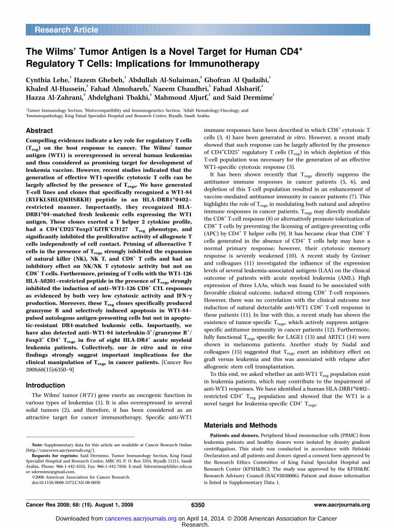
Data 1), sharing the HLA-DRB1*0402 molecule, were stimulatedrepeatedly with autologous DCs pulsed with WT1-Pepmix.Corresponding T-cell lines designated TCL 29, TCL 52, and TCL62 with specific proliferative activity against autologous WT1-Pepmix–pulsed LCLs were generated (Fig. 1A). The TCL 29 line,which gave high proliferative activity, was cloned by limitingdilution, and 48 clones were obtained and screened by proliferationassay against autologous LCL FWT1-Pepmix. Eight clones showedspecific proliferative activity against the WT1-pepmix. Four clones(TCC 29.B.9, TCC 29.B.16, TCC 29.B.19, and TCC 29.B.42), whichshowed the highest and most stable proliferative activity (Fig. 1A),were selected and expanded for further analysis. TCL 29 was thenscreened by proliferation assay against the individual 110 WT1-Pepmix peptides. A peptide designated WT1-84 with the WT1333-347RYFKLSHLQMHSRKH sequence induced the strongest specificproliferative activity of TCL 29 (Fig. 1B). Interestingly, both TCL 52and TCL 62 lines also showed specific proliferative activity againstthe WT1-84 peptide (data not shown). The rest of the peptidesdid not induce significant proliferative activity, indicating the
Figure 1. Proliferative activity of T-cell lines and clones generated against the WT1-Pepmix. A, T cells (5 � 104 per well) from three T-cell lines (TCL 29, TCL 52, TCL62) and four expanded T-cell clones (TCC 29.B.9, TCC 29.B.16, TCC 29.B.19, and TCC 29.B.42) were challenged for 72 h with irradiated (8,000 rads, XR) autologousLCL (105 per well) in the absence (gray columns ) or presence (black columns) of WT1-Pepmix. Cells were pulsed with [3H]thymidine for additional 18 h and thenharvested. T cells only (TC ; white columns ) were used as negative control. B, the TCL 29 cells (2.5 � 104 per well) were mixed with irradiated autologous PBMCs(5 � 104 per well) pulsed with the different 110 single WT1 peptides. The culture conditions were the same as described in A. C, representative T-cell clone (TCC29.B.42) showing the restriction response to the HLA-DR molecule. Experimental conditions were the same as described in A . mAbs were added to the wells containingLCLs 20 min before the addition of T cells. D, proliferative activity of a representative T-cell clone (TCC 29.B.42) against the HLA-DR0402–matched LCL-19 andthe HLA-DR0701–matched LCL-18 in the absence (gray columns ) or presence (black columns ) of the WT1-84 peptide. Experimental conditions for D were the same asdescribed in A with the exception that 104 T cells per well were challenged for 72 h with 2 � 104 per well irradiated LCLs.
Cancer Research
Cancer Res 2008; 68: (15). August 1, 2008 6352 www.aacrjournals.org
Research. on April 14, 2014. © 2008 American Association for Cancercancerres.aacrjournals.org Downloaded from

immunodominance of this epitope. All clones had specificproliferative activity to the WT1-84 peptide and a dose-dependentresponse was shown in all clones with the optimal concentration at40 Amol/L (data not shown). The HLA restriction of the clones wasdetermined in which anti–HLA-DR mAb was found to significantlyinhibit their proliferative activity, whereas their correspondingisotypes had no inhibition effect (Fig. 1C). To define more preciselythe HLA-DR restriction, two HLA-DR–matched allogeneic LCLssharing the DRB1*0402 (LCL-19) or DRB1*0701 (LCL-18) with theTCC 29.B.42 clone were used. LCL-18 failed to present the WT1-84peptide, ruling out the involvement of the DRB1*0701 in thispresentation. However, LCL-19 sharing the DRB1*0402 with theclone induced a specific proliferative activity against the WT1-84peptide, showing the requirement of DRB1*0402 restriction in thisprocess (Fig. 1D). No proliferative response was recorded when LCLpulsed with a negative control HPV3373-87 HLA-DR*0402–restrictedpeptide was used (data not shown). All clones were CD4+ (data notshown). Because CD4+ T cells can exert a cytotoxic effect (22),we used 51Cr release assay to examine cytotoxic effect againstautologous LCL FWT1-84. No lytic activity was detected (data notshown), ruling out this possibility.
Evaluation of the cytokine profile of the T-cell clones.Cytokines generated during an immune response dictate the
outcome of this response. Hence, we evaluated the cytokineprofile generated by the anti–WT1-84 clones. Various cytokines
released were determined from cell culture supernatants of T cells
cultured for 48 h with irradiated autologous PBMCs or LCL FWT1-84 or FWT1-Pepmix. All clones secreted very high amountsof IL-5 and GM-CSF, and high IL-4, specifically to WT1-84 and
WT1-Pepmix, but little or no IL-10, TGF-h1/h2, or IFN-g(Supplementary Data 3). This clearly shows a T helper 2 (Th2)-polarized immune response.
Characterization of the phenotypic profile and TCR usage ofthe T-cell clones. The cytokine profile of the clones suggested thatthey represent a Th2 phenotype. It has been recently shown (23)that human Th2 cells exert a Treg phenotype in which a generatedTh2 clone also expressed Foxp3, a specific marker for Treg lineages(24, 25). Foxp3 is also expressed transiently in activated non-Tregs(26). Therefore, we tested whether TCL 29 and clones expressFoxp3 after long-term culture. Figure 2A shows the presence ofFoxp3 mRNA in TCL 29 and two other clones. Because Tregs areelevated during pregnancy (27), we used PBMCs from a pregnant
Figure 2. Phenotypic analysis of the TCL 29 T-cell line and the expanded T-cell clones generated against the WT1-Pepmix. A, RNA expression of Foxp3 in theT-cell line (TCL 29) and clones (TCC 29.B.9 and TCC 29.B.42). PBMCs from a 7-mo pregnant women (PBMC-PR) and LCL-29 were used as a positive and a negativecontrol. PBMC-29 from which the TCL 29 was generated was used as a baseline. b-Actin acts as a housekeeping gene. Total RNA was isolated from different individualcell types and analyzed by RT-PCR. B, cytospin immunocytochemical double staining of a representative T-cell clone (TCC 29.B.9) after more than 1 mo inculture showing (right ) the nuclear expression of the Foxp3 protein (brown) and the membranous expression of the CD3 T-cell marker (blue ). Left, anti–CD3-stainedcells with an isotype Foxp3-matched negative control. Magnification, �520. C and D, FACS analysis of a representative T-cell clone (TCC 29.B.9) showing a Tregphenotype after more than 1 mo in culture.
WT1 as a New Target for Leukemia-Specific CD4+ Tregs
www.aacrjournals.org 6353 Cancer Res 2008; 68: (15). August 1, 2008
Research. on April 14, 2014. © 2008 American Association for Cancercancerres.aacrjournals.org Downloaded from
woman as a positive control. Foxp3 expression was furtherconfirmed at the protein level (Fig. 2B). We further confirmedthe protein expression in these clones by fluorescence-activatedcell sorting (FACS) analysis and found to exert a CD4+CD25+Foxp3+
Treg phenotype (Fig. 2C). Finally, we examined the clones for theexpression of other Treg-related markers. GITR (28, 29) and CTLA-4(30) molecules are constitutively expressed at high levels in Tregs.All clones expressed GITR but were negative for CTLA-4 andCD127, which has been recently used to discriminate betweenhuman regulatory CD127� and activated T cells (Fig. 2D ; ref. 31).
Altogether, these data indicate that the current anti-WT1 clonesare phenotypically Tregs.The TCR profile of the generated T-cell line and clones was
determined using a TCR Vh kit, which is used to detect the 24 mostcommon human TCR Vh molecules. All clones shared the sameTCR Vh8 chain (Supplementary Data 4A). We also determined thefrequency of TCR Vh8 in TCL 29 and PBMC-29 from the samedonor. The TCR Vh profile of PBMC-29 scattered between 1% and8% in which 5% of T cells were TCR Vh8 (Supplementary Data 4B).The frequency of other two T-cell populations with unknown
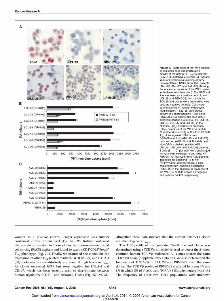
Figure 3. Expression of the WT1 antigenby leukemic cells and proliferativeactivity of the anti-WT1 Tregs to differentHLA-DR4–matched leukemias. A, cytospinimmunocytochemical staining of threerepresentative PBMCs from AML patients(AML-25, AML-27, and AML-28) showingthe nuclear expression of the WT1 proteinin the leukemic blasts (red). The K562 cellline was used as a positive control, andLCL-29 and PBMC-29, from which theTCL 29 and clones were generated, wereused as negative controls. Cells werecounterstained in instant hematoxylin.Magnification, �520. B, proliferativeactivity of a representative T-cell clone(TCC 29.B.42) against the HLA-DR04suballelic positive LCLs (LCL-06, LCL-11,LCL-10, LCL-25, and LCL-38) in theabsence (gray columns ) or presence(black columns ) of the WT1-84 peptide.C, proliferative activity of the TCC 29.B.42Treg clone against PBMCs fromDR0402-matched (AML-13 and AML-23),nonmatched (AML-27 and AML-28), andHLA-DR04 suballelic positive AML(AML-21, AML-25, and AML-29) patients.T cells (5 � 104 per well) were challengedfor 72 h with irradiated (3,000 rads, XR)PBMCs (105 per well) from AML patients,incubated for additional 18 h with[3H]thymidine, and harvested. T cellschallenged with irradiated autologousPBMC-29 in the absence or presence ofthe WT1-84 peptide served as negativeand positive control, respectively.
Cancer Research
Cancer Res 2008; 68: (15). August 1, 2008 6354 www.aacrjournals.org
Research. on April 14, 2014. © 2008 American Association for Cancercancerres.aacrjournals.org Downloaded from
specificities was increased (Vh2, 8–21%; Vh3, 7–12%) after sevenrounds of stimulation (Supplementary Data 4C). Interestingly, theTCR Vh8+ T-cell frequency was enriched to 40% in TCL 29 afterseven rounds of stimulation, indicating the immunodominance ofanti–WT1-84 TCR Vh8+ T-cell population in TCL 29.
Evaluation of WT1 expression and proliferative activity ofanti-WT1 Tregs against DR4-matched healthy and leukemiccells. We examined the expression of the WT1 mRNA in fresh AMLcells using quantitative reverse transcription-PCR (RT-PCR). WT1protein expression was also evaluated by immunostaining andscored by two independent scientists. K562 cells, known to expresshigh levels of WT1 (20), were used as a positive control. PBMCsand LCLs from normal donors served as negative controls. Higherlevels of WT1 expression were recorded in AML samples(Supplementary Data 5). Figure 3A shows immunostaining of
PBMCs from three AML patients indicating different levels of WT1expression.Because HLA-DR was the restriction element for the clones
(Fig. 1C), it was important to evaluate its expression in the AMLsamples. All AML samples expressed HLA-DR, although atdifferent levels (Supplementary Data 5). The proliferative activityof a selective clone was then measured against PBMCs from twoDR0402-matched (AML-13 and AML-23) and two non-DR0402(AML-27 and AML-28) AML patients. Specific proliferative activitywas recorded for the clone against AML-13 and AML-23 cellsexpressing the DR0402 molecule (Fig. 3B). However, this clonefailed to recognize non-DR0402 AML-27 and AML-28 cells,although they express high levels of WT1 (Fig. 3C). Percentagesof leukemic blasts used in this study are shown in SupplementaryData 4, column 2. T cells challenged with PBMC-29 F WT1-84
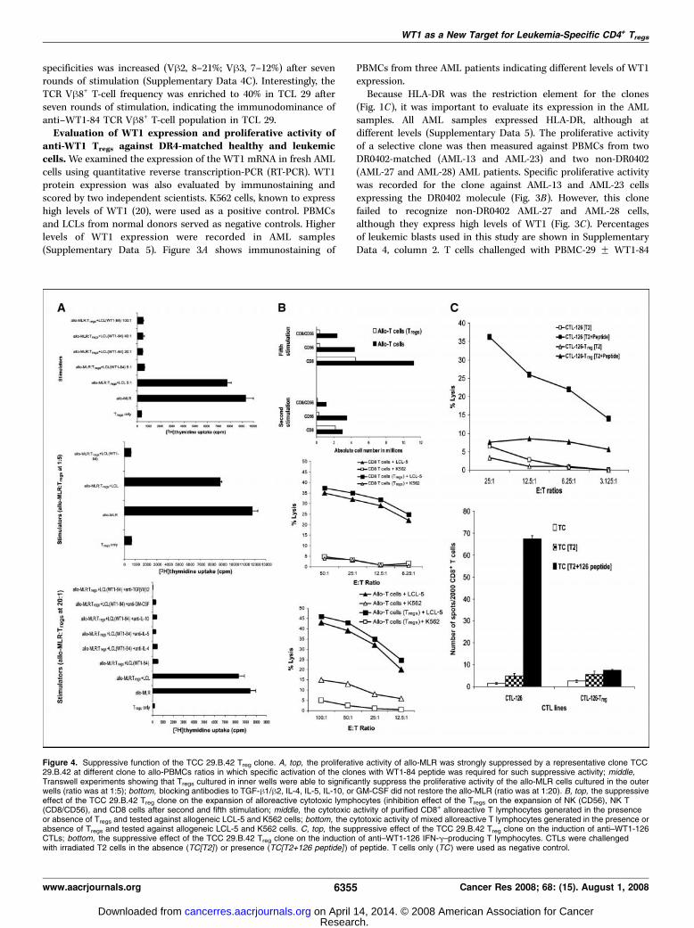
Figure 4. Suppressive function of the TCC 29.B.42 Treg clone. A, top, the proliferative activity of allo-MLR was strongly suppressed by a representative clone TCC29.B.42 at different clone to allo-PBMCs ratios in which specific activation of the clones with WT1-84 peptide was required for such suppressive activity; middle,Transwell experiments showing that Tregs cultured in inner wells were able to significantly suppress the proliferative activity of the allo-MLR cells cultured in the outerwells (ratio was at 1:5); bottom, blocking antibodies to TGF-h1/h2, IL-4, IL-5, IL-10, or GM-CSF did not restore the allo-MLR (ratio was at 1:20). B, top, the suppressiveeffect of the TCC 29.B.42 Treg clone on the expansion of alloreactive cytotoxic lymphocytes (inhibition effect of the Tregs on the expansion of NK (CD56), NK T(CD8/CD56), and CD8 cells after second and fifth stimulation; middle, the cytotoxic activity of purified CD8+ alloreactive T lymphocytes generated in the presenceor absence of Tregs and tested against allogeneic LCL-5 and K562 cells; bottom, the cytotoxic activity of mixed alloreactive T lymphocytes generated in the presence orabsence of Tregs and tested against allogeneic LCL-5 and K562 cells. C, top, the suppressive effect of the TCC 29.B.42 Treg clone on the induction of anti–WT1-126CTLs; bottom, the suppressive effect of the TCC 29.B.42 Treg clone on the induction of anti–WT1-126 IFN-g–producing T lymphocytes. CTLs were challengedwith irradiated T2 cells in the absence (TC[T2] ) or presence (TC[T2+126 peptide] ) of peptide. T cells only (TC ) were used as negative control.
WT1 as a New Target for Leukemia-Specific CD4+ Tregs
www.aacrjournals.org 6355 Cancer Res 2008; 68: (15). August 1, 2008
Research. on April 14, 2014. © 2008 American Association for Cancercancerres.aacrjournals.org Downloaded from
served as negative and positive controls. We have also showedthat the WT1-84 peptide was able to induce a specificproliferative cross-reactivity in the T-cell clones after challengingwith LCLs sharing different HLA-DR04 suballeles: DR-0401 (LCL-06 and LCL-11), DR-0403 (LCL-10, LCL-25, LCL-33, and LCL-36),and DR-0405 (LCL-38; Fig. 3B). Therefore, we tested theproliferative activity of the clones against WT1-expressing PBMCsfrom different HLA-DR04 suballelic positive AML patients (AML-21, AML-25, and AML-29). The clones recognized all AML cellsused (Fig. 3C). We conclude that leukemic cells express relativelyhigh levels of the WT1 antigen and able to process it to theWT1-84 peptide, which then presented to Tregs through the HLA-DR04 molecule.
Evaluation of the inhibition effect of the Treg clones onalloreactive T helper/cytotoxic lymphocytes and induction ofanti-WT1 CTL responses. To evaluate whether the Treg clonesexert a suppressor function on alloreactive T helper lymphocytes,
we used an allo-MLR (32) as a functional readout. All clonessignificantly suppressed the proliferative activity of the allo-MLRand showed a strong inhibition effect even at 1:100 T-cell to allo-PBMC ratio (Fig. 4A, top). Specific activation of the clones withWT1-84 peptide was required for such suppressive activity, asstimulation with LCL alone did not have a significant effect. Totest whether cell-cell contact was required for such inhibition, wecarried out experiments in Transwell plates. All clones cultured ininner wells were able to significantly suppress the proliferativeactivity of the allo-MLR cells cultured in the outer wells (Fig. 4A,middle), showing that cell-cell contact was not required for suchinhibition. This inhibition effect was not mediated with TGF-h1/h2, IL-4, IL-5, IL-10, or GM-CSF, as mAbs to these cytokines didnot restore the allo-MLR (Fig. 4A, bottom). The clones inhibitedalso the proliferation of autologous LCL (data not shown),suggesting the involvement of other soluble factors in suchinhibition.We next examined the suppressive effect of TCC 29.B.42 Treg
clone on the expansion and function of alloreactive cytotoxiclymphocytes. The presence of Tregs had a strong inhibition effect onthe expansion of both natural killer (NK; CD56) and NK T (CD8/CD56) cells in early cultures, whereas a strong inhibition effect onthe expansion of CD8 was recorded in late cultures (Fig. 4B, top).However, the cytotoxic activity of alloreactive CD8+ CTLs generatedin the presence of Tregs was comparable with that generated in theabsence of Tregs when allogeneic LCL-5 cells were used as a target(Fig. 4B, middle). Interestingly, NK activity against K562 wasrecorded only in mixed alloreactive cells generated in the absenceof Tregs (Fig. 4B, bottom).Finally, we examined the suppressive effect of TCC 29.B.42 Treg
clone on the induction of anti-WT1 CTL responses. The presence ofTregs had a strong inhibition effect on the induction of anti–WT1-126 CD8+ CTL responses as evidenced by both very low cytotoxicactivity (Fig. 4C, top) and IFN-g production (Fig. 4C, bottom)recorded for the CTL-126 Tregs after challenge with the HLA-A0201
+
T2 cell line as a target.Production of granzyme B and induction of apoptosis by
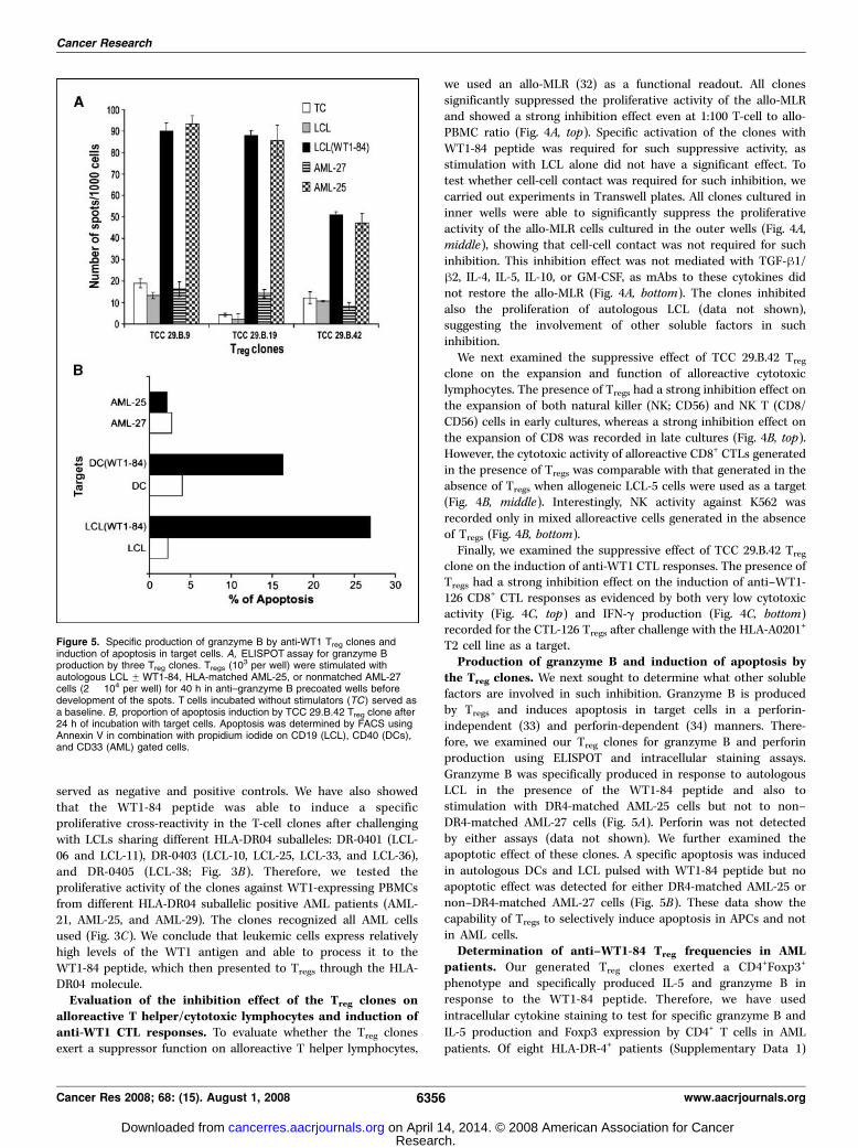
the Treg clones. We next sought to determine what other solublefactors are involved in such inhibition. Granzyme B is producedby Tregs and induces apoptosis in target cells in a perforin-independent (33) and perforin-dependent (34) manners. There-fore, we examined our Treg clones for granzyme B and perforinproduction using ELISPOT and intracellular staining assays.Granzyme B was specifically produced in response to autologousLCL in the presence of the WT1-84 peptide and also tostimulation with DR4-matched AML-25 cells but not to non–DR4-matched AML-27 cells (Fig. 5A). Perforin was not detectedby either assays (data not shown). We further examined theapoptotic effect of these clones. A specific apoptosis was inducedin autologous DCs and LCL pulsed with WT1-84 peptide but noapoptotic effect was detected for either DR4-matched AML-25 ornon–DR4-matched AML-27 cells (Fig. 5B). These data show thecapability of Tregs to selectively induce apoptosis in APCs and notin AML cells.
Determination of anti–WT1-84 Treg frequencies in AMLpatients. Our generated Treg clones exerted a CD4+Foxp3+
phenotype and specifically produced IL-5 and granzyme B inresponse to the WT1-84 peptide. Therefore, we have used
intracellular cytokine staining to test for specific granzyme B and
IL-5 production and Foxp3 expression by CD4+ T cells in AML
patients. Of eight HLA-DR-4+ patients (Supplementary Data 1)
Figure 5. Specific production of granzyme B by anti-WT1 Treg clones andinduction of apoptosis in target cells. A, ELISPOT assay for granzyme Bproduction by three Treg clones. Tregs (10
3 per well) were stimulated withautologous LCL FWT1-84, HLA-matched AML-25, or nonmatched AML-27cells (2 � 104 per well) for 40 h in anti–granzyme B precoated wells beforedevelopment of the spots. T cells incubated without stimulators (TC) served asa baseline. B, proportion of apoptosis induction by TCC 29.B.42 Treg clone after24 h of incubation with target cells. Apoptosis was determined by FACS usingAnnexin V in combination with propidium iodide on CD19 (LCL), CD40 (DCs),and CD33 (AML) gated cells.
Cancer Research
Cancer Res 2008; 68: (15). August 1, 2008 6356 www.aacrjournals.org
Research. on April 14, 2014. © 2008 American Association for Cancercancerres.aacrjournals.org Downloaded from
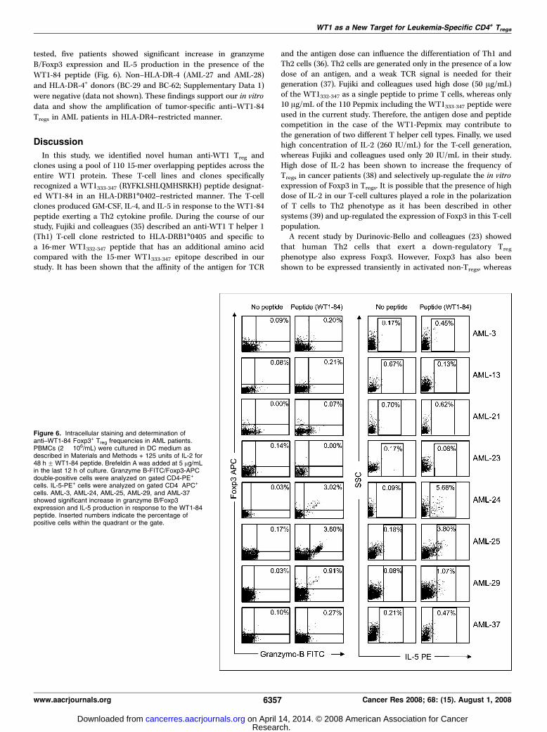
tested, five patients showed significant increase in granzyme
B/Foxp3 expression and IL-5 production in the presence of the
WT1-84 peptide (Fig. 6). Non–HLA-DR-4 (AML-27 and AML-28)
and HLA-DR-4+ donors (BC-29 and BC-62; Supplementary Data 1)
were negative (data not shown). These findings support our in vitro
data and show the amplification of tumor-specific anti–WT1-84
Tregs in AML patients in HLA-DR4–restricted manner.
Discussion
In this study, we identified novel human anti-WT1 Treg andclones using a pool of 110 15-mer overlapping peptides across theentire WT1 protein. These T-cell lines and clones specificallyrecognized a WT1333-347 (RYFKLSHLQMHSRKH) peptide designat-ed WT1-84 in an HLA-DRB1*0402–restricted manner. The T-cellclones produced GM-CSF, IL-4, and IL-5 in response to the WT1-84peptide exerting a Th2 cytokine profile. During the course of ourstudy, Fujiki and colleagues (35) described an anti-WT1 T helper 1(Th1) T-cell clone restricted to HLA-DRB1*0405 and specific toa 16-mer WT1332-347 peptide that has an additional amino acidcompared with the 15-mer WT1333-347 epitope described in ourstudy. It has been shown that the affinity of the antigen for TCR
and the antigen dose can influence the differentiation of Th1 andTh2 cells (36). Th2 cells are generated only in the presence of a lowdose of an antigen, and a weak TCR signal is needed for theirgeneration (37). Fujiki and colleagues used high dose (50 Ag/mL)of the WT1332-347 as a single peptide to prime T cells, whereas only10 Ag/mL of the 110 Pepmix including the WT1333-347 peptide wereused in the current study. Therefore, the antigen dose and peptidecompetition in the case of the WT1-Pepmix may contribute tothe generation of two different T helper cell types. Finally, we usedhigh concentration of IL-2 (260 IU/mL) for the T-cell generation,whereas Fujiki and colleagues used only 20 IU/mL in their study.High dose of IL-2 has been shown to increase the frequency ofTregs in cancer patients (38) and selectively up-regulate the in vitroexpression of Foxp3 in Tregs. It is possible that the presence of highdose of IL-2 in our T-cell cultures played a role in the polarizationof T cells to Th2 phenotype as it has been described in othersystems (39) and up-regulated the expression of Foxp3 in this T-cellpopulation.A recent study by Durinovic-Bello and colleagues (23) showed
that human Th2 cells that exert a down-regulatory Tregphenotype also express Foxp3. However, Foxp3 has also beenshown to be expressed transiently in activated non-Tregs, whereas
Figure 6. Intracellular staining and determination ofanti–WT1-84 Foxp3+ Treg frequencies in AML patients.PBMCs (2 � 106/mL) were cultured in DC medium asdescribed in Materials and Methods + 125 units of IL-2 for48 h FWT1-84 peptide. Brefeldin A was added at 5 Ag/mLin the last 12 h of culture. Granzyme B-FITC/Foxp3-APCdouble-positive cells were analyzed on gated CD4-PE+
cells. IL-5-PE+ cells were analyzed on gated CD4�APC+
cells. AML-3, AML-24, AML-25, AML-29, and AML-37showed significant increase in granzyme B/Foxp3expression and IL-5 production in response to the WT1-84peptide. Inserted numbers indicate the percentage ofpositive cells within the quadrant or the gate.
WT1 as a New Target for Leukemia-Specific CD4+ Tregs
www.aacrjournals.org 6357 Cancer Res 2008; 68: (15). August 1, 2008
Research. on April 14, 2014. © 2008 American Association for Cancercancerres.aacrjournals.org Downloaded from
it is stably expressed in Tregs (26). We have shown that thegenerated T-cell clones were stably expressing Foxp3 and exerteda CD4+CD25+Foxp3+ Treg phenotype. Furthermore, the T-cellclones were examined for the expression of other Treg-relatedmarkers (GITR, CTLA-4, and CD127) and confirmed to beCD4+CD25+Foxp3+GITR+CD127� antigen-specific Tregs. The lackof CD127 expression rules out the activation-induced Foxp3expression. These clones recognize the WT1-84 epitope throughtheir TCR Vh8 chain. The immunodominance of the anti–WT1-84/TCR Vh8+ T-cell population in the generated T-cell line mayhave had a down-regulatory effect on the generation of Th1 Tcells against other epitopes in the WT1-Pepmix. Importantly,these Treg clones recognized HLA-DR4–matched leukemic cellsexpressing the WT1 antigen, showing the natural processing andpresentation of the WT1-84 epitope. The WT1-84 peptidedescribed in this study seems to be a promiscuous HLA classII–restricted T-cell epitope as it has been reported to berecognized by both HLA-DP5–restricted cytotoxic (22) and HLA-DR0405–restricted T helper (35) CD4+ T cells.Demonstration of functional Tregs will reside eventually on their
ability to inhibit immune responses in functional assays (40).Several molecular and cellular events have been attributed to thesuppressive effect of Tregs (41). The Treg clones described in thepresent study were able to significantly inhibit the proliferativeactivity of allogeneic T cells independently of cell contact.Moreover, priming in the presence of anti–WT1-84 Tregs had astrong inhibition effect on the expansion and cytotoxic activity ofNK and NK T-cell populations. This agrees with the recent studyshowing a direct inhibitory effect of Tregs on the generation andfunction of NK cells (42). However, the presence of anti–WT1-84Tregs had a strong inhibition effect on the expansion of alloreactiveCD8 T cells but no effect on their cytotoxic activity. The animalmodel study by Edinger and colleagues (43) showing that Tregssuppress alloantigen-driven expansion of CD8 T cells withoutinhibition of their cytotoxic effect supports our observation. Moreimportantly, we have shown that the presence of Tregs stronglyinhibited the induction of anti–WT1-126 CD8+ CTL responses. Theexistence of such tumor-specific Tregs can actively suppress theWT1-specific antitumor immunity in cancer patients as it has beenshown in other systems (12).Furthermore, the present Tregs inhibited the proliferation of
LCL, which does not depend on cytokines in their proliferation,suggesting the involvement of other soluble factors. It has beenshown that production of granzyme B by Tregs results in theinduction of apoptosis in T and B cells in a perforin-independentfashion (33). The current Tregs specifically produced granzyme B,but not perforin, in response to the WT1-84 peptide and inducedapoptosis in LCL and DCs, suggesting a direct effect of granzyme
B on the suppressive activity of these Tregs. However, they failedto induce apoptosis in HLA-matched AML cells, althoughgranzyme B was produced in response to these cells. It is knownthat leukemic cells use several antiapoptotic signals to escapekilling (44). Apoptosis in response to granzyme B involvesactivation of caspase-dependent cell death pathways (45).Blockade in caspase activation pathways is a common featurein leukemia (46), and this may explain the current resistance ofAML cells to apoptosis. Our findings raise the possibility thatleukemic cells may function as APC to preferentially induce anti–WT1-specific Tregs and hence down-regulate the patients’ immuneresponse through induction of apoptosis in APCs, such as DCsand B cells (34), and/or inhibition of specific and nonspecificanti-leukemia immune responses. Another important finding isthe detection of anti–WT1-84 IL-5+/granzyme B+/Foxp3+ CD4+
Tregs in five of eight HLA-DR4+ AML patients. The recent demon-stration by Zhou and colleagues (47) that tumor-specific Tregs areamplified in vivo following cancer vaccination supports our data.Current interests in cancer immunotherapy should be focused
on determining the therapeutic implications of Tregs ‘‘regulating theregulators.’’ Interestingly, elimination of Tregs by IL-2 conjugated todiphtheria toxin (ONTAK) enhanced vaccine-mediated antitumorimmunity in cancer patients (7). However, targeting the CD25molecule may eliminate Tregs, leading to an increase in thesusceptibility to autoimmunity, and it can also deplete activatedCD25+ effector cells, which may be important for clearance ofcancer and infection (48). Therefore, studies should be tailoredtoward developing selective depletion strategies directed againstTreg-related markers. Alternatively, triggering of TLR8 or OX40, andpotentially blocking adenosine, might improve the chances ofneutralizing Treg immunosuppression in cancer patients (49). Inconclusion, our findings should open opportunity for the clinicalmanipulation of anti-WT1 Tregs for the treatment of leukemiapatients.
Disclosure of Potential Conflicts of Interest
All authors read and approved the final manuscript. The authors declare that theyhave no financial competing interests.
Acknowledgments
Received 1/8/2008; revised 5/20/2008; accepted 5/22/2008.Grant support: Research Centre and Research Advisory Council proposal grant
2030 006.The costs of publication of this article were defrayed in part by the payment of page
charges. This article must therefore be hereby marked advertisement in accordancewith 18 U.S.C. Section 1734 solely to indicate this fact.
We thank Drs. Ayodele Alaiya and Monther Al-Alwan for critical review, ManogaranPulicat for analyzing the FACS data, and Riad Youniss and Reggie Belkhadem forcollecting patients’ data.
References
1. Inoue K, Ogawa H, Sonoda Y, et al. Aberrantoverexpression of the Wilms tumor gene (WT1) inhuman leukemia. Blood 1997;89:1405–12.
2. Nakatsuka S, Oji Y, Horiuchi T, et al. Immunohisto-chemical detection of WT1 protein in a variety of cancercells. Mod Pathol 2006;19:804–14.
3. Asemissen AM, Keilholz U, Tenzer S, et al. Identifica-tion of a highly immunogenic HLA-A*01-binding T cellepitope of WT1. Clin Cancer Res 2006;12:7476–82.
4. Oka Y, Elisseeva OA, Tsuboi A, et al. Human cytotoxicT-lymphocyte responses specific for peptides of the
wild-type Wilms’ tumor gene (WT1) product. Immuno-genetics 2000;51:99–107.
5. Beyer M, Schultze JL. Regulatory T cells in cancer.Blood 2006;108:804–11.
6. Ghebeh H, Barhoush E, Tulbah A, Elkum N, Al-Tweigeri T, Dermime S. FOXP3+ Tregs and B7-H1+/PD-1+ T lymphocytes co-infiltrate the tumor tissues ofhigh-risk breast cancer patients: implication forimmunotherapy. BMC Cancer 2008;8:57.
7. Dannull J, Su Z, Rizzieri D, et al. Enhancement ofvaccine-mediated antitumor immunity in cancerpatients after depletion of regulatory T cells. J ClinInvest 2005;115:3623–33.
8. Bourgeois C, Rocha B, Tanchot C. A role for CD40expression on CD8+ T cells in the generation of CD8+ Tcell memory. Science 2002;297:2060–3.
9. Alpan O, Bachelder E, Isil E, Arnheiter H, Matzinger P.‘Educated’ dendritic cells act as messengers frommemory to naive T helper cells. Nat Immunol 2004;5:615–22.
10. Janssen EM, Lemmens EE, Wolfe T, Christen U, vonHerrath MG, Schoenberger SP. CD4+ T cells are requiredfor secondary expansion and memory in CD8+ Tlymphocytes. Nature 2003;421:852–6.
11. Greiner J, Schmitt M, Li L, et al. Expression oftumor-associated antigens in acute myeloid leukemia:
Cancer Research
Cancer Res 2008; 68: (15). August 1, 2008 6358 www.aacrjournals.org
Research. on April 14, 2014. © 2008 American Association for Cancercancerres.aacrjournals.org Downloaded from
implications for specific immunotherapeutic approaches.Blood 2006;108:4109–17.
12. Nishikawa H, Jager E, Ritter G, Old LJ, Gnjatic S. CD4+
CD25+ regulatory T cells control the induction ofantigen-specific CD4+ helper T cell responses in cancerpatients. Blood 2005;106:1008–11.
13. Wang HY, Lee DA, Peng G, et al. Tumor-specifichuman CD4+ regulatory T cells and their ligands:implications for immunotherapy. Immunity 2004;20:107–18.
14. Wang HY, Peng G, Guo Z, Shevach EM, Wang RF.Recognition of a new ARTC1 peptide ligand uniquelyexpressed in tumor cells by antigen-specific CD4+
regulatory T cells. J Immunol 2005;174:2661–70.15. Nadal E, Garin M, Kaeda J, Apperley J, Lechler R,Dazzi F. Increased frequencies of CD4(+)CD25(high)T(regs) correlate with disease relapse after allogeneicstem cell transplantation for chronic myeloid leukemia.Leukemia 2007;21:472–9.
16. Hohn H, Pilch H, Gunzel S, et al. Human papilloma-virus type 33 E7 peptides presented by HLA-DR*0402 totumor-infiltrating T cells in cervical cancer. J Virol 2000;74:6632–6.
17. Sugden B, Mark W. Clonal transformation of adulthuman leukocytes by Epstein-Barr virus. J Virol 1977;23:503–8.
18. Dermime S, Bertazzoli C, Marchesi E, et al. Lack of T-cell-mediated recognition of the fusion region of thepml/RAR-a hybrid protein by lymphocytes of acutepromyelocytic leukemia patients. Clin Cancer Res 1996;2:593–600.
19. Ghebeh H, Tulbah A, Mohammed S, et al. Expressionof B7-H1 in breast cancer patients is strongly associatedwith high proliferative Ki-67-expressing tumor cells. Int JCancer 2007;121:751–8.
20. Greiner J, Ringhoffer M, Taniguchi M, et al. mRNAexpression of leukemia-associated antigens in patientswith acute myeloid leukemia for the development ofspecific immunotherapies. Int J Cancer 2004;108:704–11.
21. Dermime S, Mavroudis D, Jiang YZ, Hensel N,Molldrem J, Barrett AJ. Immune escape from a graft-versus-leukemia effect may play a role in the relapseof myeloid leukemias following allogeneic bonemarrow transplantation. Bone Marrow Transplant1997;19:989–99.
22. Guo Y, Niiya H, Azuma T, et al. Direct recognitionand lysis of leukemia cells by WT1-specific CD4+ Tlymphocytes in an HLA class II-restricted manner.Blood 2005;106:1415–8.
23. Durinovic-Bello I, Rosinger S, Olson JA, et al.DRB1*0401-restricted human T cell clone specific for
the major proinsulin73-90 epitope expresses a down-regulatory T helper 2 phenotype. Proc Natl Acad SciU S A 2006;103:11683–8.
24. Fontenot JD, Gavin MA, Rudensky AY. Foxp3programs the development and function of CD4+CD25+
regulatory T cells. Nat Immunol 2003;4:330–6.25. Hori S, Sakaguchi S. Foxp3: a critical regulator of thedevelopment and function of regulatory T cells.Microbes Infect 2004;6:745–51.
26. Wang J, Ioan-Facsinay A, van der Voort EI, HuizingaTW, Toes RE. Transient expression of FOXP3 in humanactivated nonregulatory CD4+ T cells. Eur J Immunol2007;37:129–38.
27. Somerset DA, Zheng Y, Kilby MD, Sansom DM,Drayson MT. Normal human pregnancy is associatedwith an elevation in the immune suppressive CD25+
CD4+ regulatory T-cell subset. Immunology 2004;112:38–43.
28. McHugh RS, Whitters MJ, Piccirillo CA, et al.CD4(+)CD25(+) immunoregulatory T cells: gene expres-sion analysis reveals a functional role for the glucocor-ticoid-induced TNF receptor. Immunity 2002;16:311–23.
29. Shimizu J, Yamazaki S, Takahashi T, Ishida Y,Sakaguchi S. Stimulation of CD25(+)CD4(+) regulatoryT cells through GITR breaks immunological self-tolerance. Nat Immunol 2002;3:135–42.
30. Sakaguchi S. Naturally arising CD4+ regulatory t cellsfor immunologic self-tolerance and negative control ofimmune responses. Annu Rev Immunol 2004;22:531–62.
31. Seddiki N, Santner-Nanan B, Martinson J, et al.Expression of interleukin (IL)-2 and IL-7 receptorsdiscriminates between human regulatory and activatedT cells. J Exp Med 2006;203:1693–700.
32. Godfrey WR, Ge YG, Spoden DJ, et al. In vitro -expanded human CD4(+)CD25(+) T-regulatory cells canmarkedly inhibit allogeneic dendritic cell-stimulatedMLR cultures. Blood 2004;104:453–61.
33. Gondek DC, Lu L-F, Quezada SA, Sakaguchi S, NoelleRJ. Cutting edge: contact-mediated suppression byCD4+CD25+ regulatory cells involves a granzyme B-dependent, perforin-independent mechanism. J Immu-nol 2005;174:1783–6.
34. Zhao D-M, Thornton AM, DiPaolo RJ, Shevach EM.Activated CD4+CD25+ T cells selectively kill B lympho-cytes. Blood 2006;107:3925–32.
35. Fujiki F, Oka Y, Tsuboi A, et al. Identification andcharacterization of a WT1 (Wilms tumor gene)protein-derived HLA-DRB1*0405-restricted 16-merhelper peptide that promotes the induction andactivation of WT1-specific cytotoxic T lymphocytes. JImmunother 2007;30:282–93.
36. Constant SL, Bottomly K. Induction of Th1 and Th2CD4+ T cell responses: the alternative approaches. AnnuRev Immunol 1997;15:297–322.
37. Tao X, Constant S, Jorritsma P, Bottomly K. Strengthof TCR signal determines the costimulatory require-ments for Th1 and Th2 CD4+ T cell differentiation. JImmunol 1997;159:5956–63.
38. Ahmadzadeh M, Rosenberg SA. IL-2 administrationincreases CD4+CD25hi Foxp3+ regulatory T cells incancer patients. Blood 2006;107:2409–14.
39. Cote-Sierra J, Foucras G, Guo L, et al. Interleukin 2plays a central role in Th2 differentiation. Proc NatlAcad Sci U S A 2004;101:3880–5.
40. Spadafora-Ferreira M, Caldas C, Fae KC, et al.CD4(+)CD25(+)Foxp3(+) indirect alloreactive T cellsfrom renal transplant patients suppress both the directand indirect pathways of allorecognition. Scand JImmunol 2007;66:352–61.
41. Miyara M, Sakaguchi S. Natural regulatory T cells:mechanisms of suppression. Trends Mol Med 2007;13:108–16.
42. Giroux M, Yurchenko E, St.-Pierre J, Piccirillo CA,Perreault C. T Regulatory cells control numbers of NKcells and CD8a+ immature dendritic cells in the lymphnode paracortex. J Immunol 2007;179:4492–502.
43. Edinger M, Hoffmann P, Ermann J, et al. CD4+CD25+
regulatory T cells preserve graft-versus-tumor activitywhile inhibiting graft-versus-host disease after bonemarrow transplantation [see comment]. Nat Med 2003;9:1144–50.
44. Smith BD, Bambach BJ, Vala MS, et al. Inhibitedapoptosis and drug resistance in acute myeloidleukaemia. Br J Haematol 1998;102:1042–9.
45. Sutton VR, Wowk ME, Cancilla M, Trapani JA.Caspase activation by granzyme B is indirect, andcaspase autoprocessing requires the release of pro-apoptotic mitochondrial factors. Immunity 2003;18:319–29.
46. Schimmer AD, Pedersen IM, Kitada S, et al.Functional blocks in caspase activation pathways arecommon in leukemia and predict patient response toinduction chemotherapy. Cancer Res 2003;63:1242–8.
47. Zhou G, Drake CG, Levitsky HI. Amplification oftumor-specific regulatory T cells following therapeuticcancer vaccines. Blood 2006;107:628–36.
48. Curtin JF, Candolfi M, Fakhouri TM, et al. Tregdepletion inhibits efficacy of cancer immunotherapy:implications for clinical trials. PLoS ONE 2008;3:e1983.
49. Colombo MP, Piconese S. Regulatory-T-cell inhibitionversus depletion: the right choice in cancer immuno-therapy. Nat Rev Cancer 2007;7:880–7.
WT1 as a New Target for Leukemia-Specific CD4+ Tregs
www.aacrjournals.org 6359 Cancer Res 2008; 68: (15). August 1, 2008
Research. on April 14, 2014. © 2008 American Association for Cancercancerres.aacrjournals.org Downloaded from