Biomarkers of Response and Resistance to Immunotherapy in ...
31
Cancers 2022, 14, 2241. https://doi.org/10.3390/cancers14092241 www.mdpi.com/journal/cancers Review Biomarkers of Response and Resistance to Immunotherapy in Microsatellite Stable Colorectal Cancer: Toward a New Personalized Medicine Nicolas Huyghe 1 , Elena Benidovskaya 1 , Philippe Stevens 1 and Marc Van den Eynde 1,2, * 1 Institut de Recherche Clinique et Expérimentale (Pole MIRO), UCLouvain, 1200 Brussels, Belgium; [email protected] (N.H.); [email protected] (E.B.); [email protected] (P.S.) 2 Department of Medical Oncology and Gastroenterology, Institut Roi Albert II, Cliniques Universitaires St-Luc, 1200 Brussels, Belgium * Correspondence: [email protected] Simple Summary: Immune Checkpoint Inhibitors (ICIs) have demonstrated clinical efficacy in Mi- crosatellite Instability High Colorectal Cancer (MSI-H CRC). However, in Microsatellite Stable (MSS) CRC, ICIs monotherapy provides limited clinical benefit. Therefore, efforts must be made to understand the highly heterogeneous CRC microenvironment and to find predictive biomarkers of response in order to adequately select CRC patients who may respond to ICIs-based therapies. Abstract: Immune Checkpoint Inhibitors (ICIs) are well recognized as a major immune treatment modality for multiple types of solid cancers. However, for colorectal cancer (CRC), ICIs are only approved for the treatment of Mismatch-Repair-Deficient and Microsatellite Instability-High (dMMR/MSI-H) tumors. For the vast majority of CRC, that are not dMMR/MSI-H, ICIs alone pro- vide limited to no clinical benefit. This discrepancy of response between CRC and other solid can- cers suggests that CRC may be inherently resistant to ICIs alone. In translational research, efforts are underway to thoroughly characterize the immune microenvironment of CRC to better under- stand the mechanisms behind this resistance and to find new biomarkers of response. In the clinic, trials are being set up to study biomarkers along with treatments targeting newly discovered im- mune checkpoint molecules or treatments combining ICIs with other existing therapies to improve response in MSS CRC. In this review, we will focus on the characteristics of response and resistance to ICIs in CRC, and discuss promising biomarkers studied in recent clinical trials combining ICIs with other therapies. Keywords: colorectal cancer; immunotherapy; Immune Checkpoint Inhibitors; immune checkpoint resistance; immune microenvironment; biomarker 1. Introduction Although there has been recent progress in management, treatment, and screening, Colorectal Cancer (CRC) remains a major public health issue. Worldwide, CRC is esti- mated to be the third most frequent cancer and the second cause of cancer-related death [1]. Despite a major emphasis on CRC screening, approximately 20% of CRC is metastatic at diagnosis and 30% of treated non-metastatic patients will develop metastasis during the follow-up of their disease [2]. Metastatic CRC (mCRC) has a poor prognosis with a 5- year survival rate of 14.2% (95% CI, 13.7–14.7) [3]. Treatment options for CRC include surgery, chemotherapy, radiation, targeted therapy, and, more recently, immunotherapy for a selected molecular subgroup of tumor [4]. Citation: Huyghe, N.; Benidovskaya, E.; Stevens, P.; Van den Eynde, M. Biomarkers of Response and Resistance to Immunotherapy in Microsatellite Stable Colorectal Cancer: Toward a New Personalized Medicine. Cancers 2022, 14, 2241. https://doi.org/10.3390/ cancers14092241 Academic Editors: Julien Péron and Amélie Boespflug Received: 8 April 2022 Accepted: 27 April 2022 Published: 29 April 2022 Publisher’s Note: MDPI stays neu- tral with regard to jurisdictional claims in published maps and institu- tional affiliations. Copyright: © 2022 by the authors. Li- censee MDPI, Basel, Switzerland. This article is an open access article distributed under the terms and con- ditions of the Creative Commons At- tribution (CC BY) license (https://cre- ativecommons.org/licenses/by/4.0/).
-
Upload
khangminh22 -
Category
Documents
-
view
0 -
download
0
Transcript of Biomarkers of Response and Resistance to Immunotherapy in ...

Cancers 2022, 14, 2241. https://doi.org/10.3390/cancers14092241 www.mdpi.com/journal/cancers
Review
Biomarkers of Response and Resistance to Immunotherapy in Microsatellite Stable Colorectal Cancer: Toward a New Personalized Medicine Nicolas Huyghe 1, Elena Benidovskaya 1, Philippe Stevens 1 and Marc Van den Eynde 1,2,*
1 Institut de Recherche Clinique et Expérimentale (Pole MIRO), UCLouvain, 1200 Brussels, Belgium; [email protected] (N.H.); [email protected] (E.B.); [email protected] (P.S.)
2 Department of Medical Oncology and Gastroenterology, Institut Roi Albert II, Cliniques Universitaires St-Luc, 1200 Brussels, Belgium
* Correspondence: [email protected]
Simple Summary: Immune Checkpoint Inhibitors (ICIs) have demonstrated clinical efficacy in Mi-crosatellite Instability High Colorectal Cancer (MSI-H CRC). However, in Microsatellite Stable (MSS) CRC, ICIs monotherapy provides limited clinical benefit. Therefore, efforts must be made to understand the highly heterogeneous CRC microenvironment and to find predictive biomarkers of response in order to adequately select CRC patients who may respond to ICIs-based therapies.
Abstract: Immune Checkpoint Inhibitors (ICIs) are well recognized as a major immune treatment modality for multiple types of solid cancers. However, for colorectal cancer (CRC), ICIs are only approved for the treatment of Mismatch-Repair-Deficient and Microsatellite Instability-High (dMMR/MSI-H) tumors. For the vast majority of CRC, that are not dMMR/MSI-H, ICIs alone pro-vide limited to no clinical benefit. This discrepancy of response between CRC and other solid can-cers suggests that CRC may be inherently resistant to ICIs alone. In translational research, efforts are underway to thoroughly characterize the immune microenvironment of CRC to better under-stand the mechanisms behind this resistance and to find new biomarkers of response. In the clinic, trials are being set up to study biomarkers along with treatments targeting newly discovered im-mune checkpoint molecules or treatments combining ICIs with other existing therapies to improve response in MSS CRC. In this review, we will focus on the characteristics of response and resistance to ICIs in CRC, and discuss promising biomarkers studied in recent clinical trials combining ICIs with other therapies.
Keywords: colorectal cancer; immunotherapy; Immune Checkpoint Inhibitors; immune checkpoint resistance; immune microenvironment; biomarker
1. Introduction Although there has been recent progress in management, treatment, and screening,
Colorectal Cancer (CRC) remains a major public health issue. Worldwide, CRC is esti-mated to be the third most frequent cancer and the second cause of cancer-related death [1]. Despite a major emphasis on CRC screening, approximately 20% of CRC is metastatic at diagnosis and 30% of treated non-metastatic patients will develop metastasis during the follow-up of their disease [2]. Metastatic CRC (mCRC) has a poor prognosis with a 5-year survival rate of 14.2% (95% CI, 13.7–14.7) [3]. Treatment options for CRC include surgery, chemotherapy, radiation, targeted therapy, and, more recently, immunotherapy for a selected molecular subgroup of tumor [4].
Citation: Huyghe, N.; Benidovskaya,
E.; Stevens, P.; Van den Eynde, M.
Biomarkers of Response and
Resistance to Immunotherapy in
Microsatellite Stable Colorectal
Cancer: Toward a New Personalized
Medicine. Cancers 2022, 14, 2241.
https://doi.org/10.3390/
cancers14092241
Academic Editors: Julien Péron and
Amélie Boespflug
Received: 8 April 2022
Accepted: 27 April 2022
Published: 29 April 2022
Publisher’s Note: MDPI stays neu-
tral with regard to jurisdictional
claims in published maps and institu-
tional affiliations.
Copyright: © 2022 by the authors. Li-
censee MDPI, Basel, Switzerland.
This article is an open access article
distributed under the terms and con-
ditions of the Creative Commons At-
tribution (CC BY) license (https://cre-
ativecommons.org/licenses/by/4.0/).

Cancers 2022, 14, 2241 2 of 31
Based on genetic alterations such as mutations in V-Ki-ras2 Kirsten Rat Sarcoma viral oncogene homolog (KRAS) and proto-oncogene B-Raf (BRAF), and mutations or methyl-ation of Mismatch Repair (MMR) genes, several specific treatments of molecular CRC sub-groups have emerged over recent years [5]. Immune Checkpoint Inhibitors (ICIs) demon-strated good efficacy in deficient-Mismatch-Repair Microsatellite Instability-High (dMMR/MSI-H) CRC, providing clinical benefits superior to standard treatments and leading to the Food and Drug Administration (FDA) and European Medicines Agency (EMA) approval of anti-Programmed cell Death protein 1 (anti-PD-1) ICIs for the treat-ment of metastatic or unresectable dMMR/MSI-H CRC [6–8]. Nonetheless, for the vast majority of Microsatellite Stable (MSS) CRC, ICIs failed to provide clinical benefit in un-selected cohorts [9]. Therefore, it is crucial to better understand the genetic, epigenetic, transcriptomic, and Tumor Microenvironment (TME) characteristics of the MSS CRC.
In this review, we will discuss the genomic, transcriptomic, and tumor microenvi-ronment classifications of CRC in an immunogenic way. This will help in understanding the different biomarkers of response and resistance to ICIs investigated so far as well as promising clinical trials recently developed combining these biomarkers with therapies to improve the adaptive immune response of the tumor and the benefit of ICIs to the patient.
2. Colorectal Cancer Subtypes and Immunity Several classifications have been performed and are now available to understand
CRC development and progression, estimate prognosis, and select patients potentially able to respond to specific treatments. In this section, we discuss genomic, epigenetic, transcriptomic, and TME alterations and their relationship to CRC immunity.
2.1. Genomic and Epigenomic Classifications At the genomic level, it is well recognized that the majority of CRC (85%) presents a
Chromosomal Instability (CIN) and is MSS while a minority of CRC (15% in early stages) is characterized by an MSI-H phenotype (Figure 1) [10]. The microsatellite instability oc-curs through a deficiency in the mismatch repair mechanism that corrects single nucleo-tide base mispairings and small insertions or deletions (indels) appearing during DNA replication [11], leading to the accumulation of such alterations across the genome, favor-ing the development of cancer [12]. This loss of function of any of the DNA mismatch machinery proteins, including MutS Homolog 2 (MSH2), MutS Homolog 6 (MSH6), MutL Homolog 1 (MLH1), and Postmeiotic Segregation Increased 2 (PMS2) [13] leads to a “hy-permutated” phenotype. These tumors are characterized by high Tumor Mutational Bur-den (TMB) leading to a high number of Mutation-Associated Neoantigens (MANA) and by consequence an inflammatory TME, comprising Tumor-Infiltrating Lymphocytes (TILs), and notably memory cells and Cytotoxic T Lymphocytes (CTLs). In 12% of the cases, the deficiency is sporadic and occurs through the silencing of the MLH1 gene by promoter hypermethylation. In 3% of the cases, the deficiency occurs through germline gene mutation (Lynch syndrome) and, in rare cases, through somatic bi-allelic mutations [10,14]. In a systematic review including 1277 MSI-H out of 7642 stage I–III cases, it has been shown that the pooled Hazard Ratio (HR) for Overall Survival (OS) was 0.65 (95% CI, 0.59–0.71) for MSI-H versus MSS, suggesting a better prognosis for MSI-H patients [15].
Cancers 2022, 14, 2241 3 of 31
Figure 1. Chromosomal Instability (CIN) versus genomic instability. In CRC tumor cells, charac-terized by chromosomal instability, the majority of insertions and deletions (indels) occurring dur-ing DNA replication are repaired by a functional mismatch-repair mechanism and the DNA Poly-merase Exonuclease Domain (POLE/POLD1). A few Mutation-Associated Neoantigens (MANAs) are presented to the cell surface by the Major Histocompatibility Complex (MHC) and recognized by lymphocytes. In CRC tumor cells characterized by genomic instability and CpG Island Methyla-tor Phenotype (CIMP), the majority of indels occurring during DNA replication are not repaired. A high number of mutant proteins are translated, inducing a high number of MANAs presented and recognized by lymphocytes. mRNA: messenger RNA; TCR: T Cell Receptor; TME: Tumor Microen-vironment.
The DNA Polymerase Exonuclease Domain (POLE/POLD1) mutations could also in-duce a high MANA load and immune infiltration (Figure 1). These mutations can affect the proofreading function of the polymerase, resulting in an “ultramutator” phenotype and patients harboring POLE/POLD1 mutations are prone to developing CRC [16,17]. The frequency of POLE/POLD1 mutations in CRC is near 1%, explaining the lack of robust clinical and translational data on this subgroup of CRC [18,19]. However, in a recent re-port, it has been shown that similarly to MSI-H tumors, POLE/POLD1-mutated tumors correlate with a higher density of cytotoxic T cells (CD8+) and memory T cells (CD45RO+) compared to POLE/POLD1 wild-type tumors [20].
Mutation in the KRAS driver oncogene has recently been shown to impact immuno-modulation [21]. KRAS-mutant CRC patients have more T regulatory lymphocytes (Tregs) and fewer activated CD4+ memory T cells as compared to KRAS wild-type patients, re-sulting in a more immunosuppressive TME [22]. The loss of Adenomatous Polyposis Coli (APC) tumor-suppressor gene, involved in the cellular transition from G1 to S phase, can result in the sustained activation of the proto-oncogene Wnt (Wnt) signaling pathway and, thus, increased nuclear β-catenin [23] and decreased T cell tumor infiltration [24]. Activat-ing mutations in Phosphatidylinositol-4,5-Bisphosphate 3-Kinase Catalytic Subunit Alpha (PIK3CA), occurring in 25% of CRC cases [25], are associated with CD8+ T cell infiltration and Programmed Death-Ligand 1 (PD-L1) expression [26]. CRC studies also reported that a loss of Phosphatase and TENsin homolog (PTEN), regulating PI3K/AKT signaling path-way, results in increased PD-L1 expression [27], decreased TILs presence, and an immu-nosuppressive TME [28].

Cancers 2022, 14, 2241 4 of 31
Epigenetic regulation is an important hallmark of cancer cells [29]. In CRC, epigenetic alterations may account for the distorted expression patterns of certain genes without ge-netic alteration through hypermethylation or hypomethylation. Hypermethylation occurs when methyl groups are covalently bound in regions of CG dinucleotides or CpG-rich areas of DNA in promoter regions. In instances of normal gene expression, CpG regions are normally maintained in the unmethylated state. Methylation of these promoter re-gions can lead to gene silencing in tumor-suppressor genes, which is denoted as CpG Is-land Methylator Phenotype (CIMP). The CIMP has been defined and associated with the MSI-H phenotype, older age at diagnosis, right-sided location, mucinous histology, BRAF mutation, and a high T cell infiltration (Figure 1) [30]. Nevertheless, its use is limited by the absence of consensus regarding which genes should be included in the CIMP panels [31]. Global DNA hypomethylation can also lead to tumorigenesis and chromosomal in-stability in CRC [32]. Various preclinical studies investigate treatments targeting epige-netic changes since these latter are reversible [33,34]. Nevertheless, few clinical studies consider the potential synergic effects of methylation inhibitors. A phase I/II clinical trial studying the association of 5-azacitidine (DNA methyltransferase (DNMT) inhibitors) with capecitabine and oxaliplatin (CAPOX) in refractory CIMP-high mCRC demonstrated the safety and efficacy (high rate of disease control rate) of this association [35]. The use of DNMT inhibitors is limited by its lack of specificity targeting the global methylation of normal and oncogenic genes [36].
2.2. Transcriptomics Classification Based on gene expression data from 18 public data sets and The Cancer Genome At-
las (TCGA) proprietary data sets, four subgroups of CRC have been identified by combin-ing six different CRC subtyping algorithms forming the Consensus Molecular Subtypes (CMS) of CRC [16]. Briefly, CMS1 subtype (“immune subtype”, 14% of CRC tumors) is characterized by a high TMB and MANA load, high immune infiltration, T helper 1 (Th1) signaling, BRAFV600E mutations, and an overexpression of immune checkpoint mole-cules such as PD-1, Cytotoxic T-Lymphocyte-Associated protein 4 (CTLA-4), and In-doleamine 2,3-Dioxygenase 1 (IDO1). This subtype is mainly composed of MSI-H tumors, but it has been shown that around 16% of MSS tumors show characteristics of CMS1 such as high immune infiltration [37]. The CMS2 subtype (37% of CRC tumors) is called “ca-nonical”, highlighting its epithelial features and activation of the WNT and Myelocyto-matosis oncogene Myc (MYC) pathways. This group is enriched by CRC with the lowest MSI-H rate (less than 2%) [38]. Due to the low TMB, the infiltration of immune cells is very low, making them known as the “immune-desert” CRC subtype [16,39]. The CMS3 sub-type (“metabolic subtype”, 13% of CRC tumors) is characterized by a perturbation of met-abolic pathways and a high prevalence of RAS mutations. Immune cell infiltration in CMS3 is slightly higher than CMS2 but is still low and has an immunologically inactive TME, referred to as “immune-excluded” subtype (10). Eventually, CMS4 tumors (23% of CRC tumors) are called “mesenchymal type” because they have mesenchymal properties such as strong endothelial-mesenchymal transition activity and high stromal content with cancer-associated fibroblasts [16]. They are highly infiltrated with immunosuppressive cells such as Tregs, M2 macrophages, and Myeloid-Derived Suppressor Cells (MDSCs), and the presence of antitumor immune cells such as Dendritic Cells (DCs), activated Nat-ural Killer (NK) cells, Th1 cells, and CD8+ T cells in their TME is very low. In addition, CMS4 tumors have activated Vascular Endothelial Growth Factor (VEGF), Transforming Growth Factor beta (TGF-β), and Chemokine (CXC motif) Ligand 12 (CXCL12) signaling pathways, all of which cause, among the four CMS categories, higher risk of relapse and worst prognosis of CMS4 CRC after surgery [16].
However, these four CMS subtypes have been identified using expression data from primary lesions and it is difficult to transpose this classification in mCRC [40]. In a recent study, authors applied the CMS classification on resected CRC Liver Metastasis (CRCLM) and reported that, conversely to primary CRC, the CMS classification in CRCLM was not

Cancers 2022, 14, 2241 5 of 31
associated with OS. Using transcriptional data of messenger RNA (mRNA) and micro RNA (miRNA), they derived three subtypes of CRCLM (subtype 1: canonical, subtype2: immune, subtype 3: stromal) with different clinical risk stratification for Disease-Free Sur-vival (DFS) and OS [40]. Another study reported a similar depletion of CMS1 and CMS3 subtypes in 295 CRCLMs [41]. Due to tumor heterogeneity [42–44], they also observed frequent CMS switches between CRCLM of the same patients [41]. Together, these reports suggest that the CMS classification may not be suitable for mCRC.
2.3. Classification Regarding the Tumor Microenvironment 2.3.1. The Tumor Immune Microenvironment
Besides molecular classification, CRCs can also be classified based on immunological properties. As mentioned, the mutational burden is a hallmark for the response to immu-notherapy. However, the resistance of some MSI-H tumors and the appropriate response of some MSS tumors suggest that a high TMB is not responsible alone for the response to immunotherapy [45]. CRCs with high immune infiltration are not only composed of hy-permutated tumors and CMS1 subtypes [46]. The CRC TME is a heterogeneous microen-vironment containing a variety of immune cells. Numerous studies have linked high in-filtration of CD8+ cytotoxic T cells, Th1, follicular helper T cells, M1 macrophages, NK cells, and DCs with good prognosis in CRC. Contrarily, high infiltration of MDSCs, B cells, M2 macrophages, and CD4+ type-17 helper T (Th17) cells is associated with poor progno-sis [47–49].
The characterization of the immune infiltration became an important tool to classify the CRC tumor subtypes. In 2006, J. Galon et al. provided strong evidence that the type, density, location, and functional orientation of T cells infiltration was associated with fa-vorable DFS and OS after primary CRC (stage I–III) resection [50–53]. They developed the Immunoscore, simple and reproducible scoring that could be used routinely to predict patient clinical outcome. The Immunoscore (I) is computed by using the density of T cells (CD3+) and cytotoxic (CD8+) T cells in the Tumor Center (CT) and the Invasive Margin (IM) of the tumor. The Immunoscore ranges from I0 to I4. Low density of both CD3+ and CD8+ T cells in the CT and the IM is associated with I0 while high density is associated with I4 [51,52,54].
In 2018, an international consortium of 14 centers in 13 countries, led by the Society for Immunotherapy of Cancer, assessed the Immunoscore assay in 2681 patients with TNM stage I–III colon cancer (CC). The Immunoscore assay showed a high level of repro-ducibility between observers and centers (r = 0·97 for colon tumor; r = 0.97 for invasive margin; p < 0·0001). Of 1434 patients with stage II cancer, the difference in risk of recur-rence at 5 years was significant (HR for high vs. low Immunoscore: 0.33, 95% CI 0.21–0.52; p < 0·0001), including in the Cox multivariable analysis (p < 0·0001). Immunoscore had the highest relative contribution to the risk of all clinical parameters, including the American Joint Committee on Cancer and Union for International Cancer Control TNM classifica-tion system [55]. Similar results were found for stage III CC [55]. The IDEA-France pro-spective study [55], together with another phase 3 randomized clinical trial (N0147) [56], validated the value of Immunoscore in prognostication of relapse and death in stage III CC patients treated with adjuvant treatment combining fluoropyrimidine and oxaliplatin. The IDEA France clinical trial, evaluating 3 versus 6 months of oxaliplatin-based adjuvant chemotherapy, demonstrated the predictive value of Immunoscore for treatment dura-tion. Immunoscore predicted response to 6 months folinic acid, fluorouracil, and oxali-platin (FOLFOX) chemotherapy both in low- and high-risk stage III patients. Low-risk patients (T1-3, N1) with High-Immunoscore had 3-year DFS of 91.4% when treated with the 6-month FOLFOX, and only 80.8% with the 3-month regimen. These results and recent guidelines argue for the benefit of implementing the Immunoscore in clinical practice and for its introduction in a new TNM-Immune (TNM-I) classification system.
Cancers 2022, 14, 2241 6 of 31
In mCRC, high immune and genetic heterogeneity between the different synchro-nous and metachronous metastases of the same patients has been reported [37,57]. High T cell infiltration and Immunoscore measured in the least-infiltrated metastasis were as-sociated with a significantly lower number of metastases, larger metastasis, and pro-longed survival while patients with increased metastatic burden generally had a lower Immunoscore [57].
2.3.2. The Cancer-Associated Microbiome It is becoming clear that microbes exist outside of the gastrointestinal tract, and the
interplay of gut, circulating, and tissue-resident microbiomes with the development and treatment of malignancy is being explored. Among seven tumor types outside the gastro-intestinal tract, Nejman et al. [58] assessed the microbiome of 1526 human cancers or ad-jacent normal tissue, taking multiple measures to avoid contamination and using 5R mul-tiplexed bacterial 16S ribosomal DNA (rDNA) Polymerase Chain Reaction (PCR) sequenc-ing. They found that each tumor type has a distinct microbiome composition. The intra-tumor bacteria are mostly intracellular and are present in both cancer and immune cells (CD45+ and CD68+ cells). Authors also reported correlations between intratumor bacteria and tumor types and subtypes and response to immunotherapy. These results are con-sistent with a recent publication which reexamined whole-genome and whole-transcrip-tome sequencing studies in the TCGA of 33 types of cancer for microbial reads [59]. Au-thors found unique microbial signatures in tissue and blood within and between most major types of cancer which remained predictive when applied to patients with non-met-astatic or early cancer and cancers lacking any genomic alterations.
CRC patients with microbiotas enriched for pathogenic bacteria such as Fusobacte-rium nucleatum (Fn) and Bacteroides fragilis (Bf) generally present distinct phenotypic (fre-quent association with MSI-H, BRAF mutation, right-sided location) and clinical features (impaired chemotherapy response and poor outcome) [60]. One single paper [61] reported that Fn, a Gram-negative anaerobic bacterium, and other associated bacteria (Bf, Seleno-monas, …) were, next to the primary tumor, also present inside cancer cells of CRC metas-tases. They observed that mouse xenografts of human primary CRC were found to retain viable bacteria including Fn through successive passages and that treatment with metro-nidazole reduced Fn load, cancer cell proliferation, and tumor growth. Experiments in vitro showed that Fn triggered innate immune signaling and induced specific genomic loss of miRNAs miR-18* and miR-4802 targeting Unc-51-Like Autophagy-Activating Ki-nase 1 (ULK1) and Autophagy-Related 7 (ATG7), respectively. Thus, Fn causes chemo-resistance by selectively targeting specific miRNAs and autophagy pathways [62]. A re-cent study observed that Fn persistence in locally advanced rectal cancer after preopera-tive chemoradiotherapy was associated with higher risk of cancer relapse after surgery [63]. Interestingly, authors suggest a possible immunological mechanism for worse out-come. Tumors that turned Fn-negative after preoperative Chemoradiotherapy (CRT) had a strong increase in CD8+ T cells, while those that remained Fn-positive after treatment lacked CD8+ T cells induction as compared to baseline. This suggested that Fn may pro-mote a lack of immune cytotoxicity activation [64] and may favor metastatic spread. The influence of the CRC microbiome and its relationship with anticancer immunity raises new questions from preclinical and clinical standpoints. However, besides these initial insights in microbiota-primary tumor interaction, the role of bacteria in CRC metastases remains obscure.
As shown in Figure 2 (adapted from [65]), all these classifications of CRC overlap strongly. Each represents a unique way of representing and subdividing the different sub-groups of CRC.

Cancers 2022, 14, 2241 7 of 31
Figure 2. Colorectal cancer classifications: Th: T-helper lymphocytes; IFN: Interferon; MDSC: My-eloid-Derived Suppressor Cell; IL: Interleukin; TGF: Transforming Growth Factor; MSI: Microsatel-lite Instability; MSS: Microsatellite Stable; CIN: Chromosomal Instability; CMS: Consensus Molecu-lar Subtype; TME: Tumor Microenvironment.
3. Immune Checkpoint Inhibitors in Colorectal Cancer Immune Checkpoint Inhibitors (ICIs) have been developed to block co-inhibitory sig-
nals that regulate the effector T cells response. These co-inhibitory signals are called “im-mune checkpoint” and ICIs are treatments, usually monoclonal antibodies, which block co-inhibitory signals and improve immune activity in the tumor and the blood of the pa-tients by preventing the dysfunction and apoptosis of T effectors [66,67]. The most ex-ploited therapeutic targets are PD-1, PD-L1, and CTLA-4, but there is a plethora of other co-inhibitory or co-stimulatory checkpoint molecules such as Lymphocyte-Activation Gene 3 (LAG-3), T cell Immunoglobulin and Mucin-containing protein-3 (TIM-3), and Tu-mor Necrosis Factor Receptor superfamily, member 4, also known as OX40 receptor, that can be targeted and are currently under investigation in several trials [45]. In this section, we summarize the current efficacy of ICIs (anti-PD-1-L1 treatment combined or not with anti-CTLA-4) in MSI-H and MSS CRC.
3.1. ICIs in MSI-H CRC The first durable Complete Response (CR) observed with ICIs was observed in a
phase I study evaluating nivolumab (anti-PD-1) in the treatment of refractory solid tu-mors. The CR lasted longer than 3 years and the patient had in fact MSI-H mCRC, under-lying the potential of ICIs in this subset of CRC [68,69].
Following these initial findings, other trials (KEYNOTE-016, KEYNOTE-164, KEY-NOTE-158, KEYNOTE-012, and KEYNOTE-28) evaluating pembrolizumab (anti-PD-1) for the treatment of refractory MSI-H mCRC have been conducted (Table 1). In total, 90 patients were evaluated. The Overall Response Rate (ORR) was 39.6% and lasted over 6 months in 78% of patients. These results led in 2017 to the fast FDA approval of pembroli-zumab for MSI-H chemo refractory mCRC [7].

Cancers 2022, 14, 2241 8 of 31
Table 1. Selected ICIs trial results in MSI-H CRC.
Clinical Trial Phase Treatment Setting
Primary Endpoints OS PFS ORR HR
KEY-NOTE-
016 II
Pembroli-zumab
Refractory mCRC Cohort A: MSI-H
CRC Cohort B: MSS CRC
Cohort C: MSI-H non-CRC
ORR PFS
Median OS not reached (A, C); median OS of 5 months in co-
hort B
A: 78% B: 11% C: 67%
A: 40% B: 0%
C: 71%
A vs. B (for death) (0.22; 95% CI 0.05–1.00; p <
0.001) A vs. B (for progression or
death) (0.04; 95% CI 0.01–0.21; p <
0.001) KEY-
NOTE-016
Update
II Pembroli-
zumab
Refractory MSI-H cancers
Cohort A: CRC Cohort B: non-CRC
ORR PFS
Median not reached yet
Median not reached yet
A: 52% B: 54%
NA
KEY-NOTE-
164
II Pembroli-
zumab
MSI-H refractory mCRC
Cohort A: ≥2 prior lines
Cohort B: ≥1 prior lines
ORR
A: 55% (24 months)
B: 63% (24 months)
A: 31% (24 months)
B: 37% (24 months)
A: 33% B: 33%
NA
KEY-NOTE-
177 III
Pembroli-zumab
Treatment naive MSI-H mCRC
Cohort A: Pembroli-zumab
Cohort B: SOC
PFS OS
A: 61% (36 months)
B: 50% (36 months)
A: 42 % (36 months)
B: 11 % (36 months)
A: 69 % B: 51 %
OS: (0.74; 95% CI 0.53–1.03; p = 0.036)
PFS: (0.61; 95% CI 0.44–0.83; p = 0.0008)
Check-Mate 142
II Nivolumab Refractory MSI-H
mCRC ORR
73% (12 months)
50% (12 months)
31% NA
Check-Mate 142
II
Nivolumab +
Ipilimumab Nivolumab
Refractory MSI-H mCRC
Nivolumab (3 mg/kg)
Ipilimumab (1 mg/kg × 4)
Nivolumab (3 mg/kg every 2 weeks)
ORR 85% (12 months)
71% (12 months)
55% NA
Check-Mate 142
II Nivolumab
+ Ipilimumab
Treatment-naïve MSI-H mCRC
(Nivolumab 3 mg/kg every 2 weeks + Ipili-
mumab 1 mg/kg every 6 weeks)
ORR 79% (24 months)
74% (24 months)
69% NA
NICHE II Nivolumab
+ Ipilimumab
Resectable stage I–III MSI-H and MSS
CRC
Safety Feasibility
NA NA
Pathologic response
rate MSI-H: 100%
MSS: 27%
NA
The non-randomized multicohort CheckMate 142 trial, evaluating the safety and ef-ficacy of nivolumab 3 mg/kg combined or not with ipilimumab 1 mg/kg 4 doses (anti-CTLA-4) once every 3 weeks in chemo refractory mCRC, reported an ORR and 1-year OS rate of 55% and 85% for the treatment with nivolumab—ipilimumab compared to, respec-tively, 31% and 73.4% for nivolumab monotherapy [70,71]. Grade 3 to 4 treatment-related adverse events (AEs) occurred in 32% of patients treated with the combo compared to 21% with nivolumab monotherapy, and were manageable. This trial suggests that the combi-nation of ipilimumab with nivolumab could be superior to nivolumab monotherapy for

Cancers 2022, 14, 2241 9 of 31
the treatment of MSI-H chemo refractory mCRC. The FDA also approved nivolumab, with or without combination with ipilimumab for the treatment of previously treated MSI-H CRC.
The randomized phase III KEYNOTE-177 trial evaluated the safety and efficacy of pembrolizumab in treatment-naïve MSI-H mCRC patients (47). Patients were treated ei-ther with pembrolizumab 200 mg every 3 weeks or with chemotherapy ± cetuximab or bevacizumab as Standard of Care (SOC). Treatment-related adverse events of grade 3 or higher occurred in 22% of the patients in the pembrolizumab group, as compared with 66% in the chemotherapy group. At final analysis, median overall survival was not reached (NR; 95% CI 49·2–NR) with pembrolizumab vs. 36·7 months (27·6–NR) with chemotherapy (HR 0·74; 95% CI 0·53–1·03; p = 0·036). The estimated median progression-free survival (PFS) was 16.5 months (95% CI: 5.4–32.4) versus 8.2 months (95% CI: 6.1–10.2) in the pembrolizumab and SOC arms, respectively (HR: 0.60; 95% CI 0.45–0.80; two-sided p = 0.0004) leading to the FDA approval of pembrolizumab in June 2020 for the first-line treatment of metastatic or unresectable MSI-H CRC [72–74]. Another cohort of the CheckMate 142 trial evaluating nivolumab plus low-dose ipilimumab in the first-line ther-apy reported an ORR of 69% (95% CI, 53 to 82) with 13% complete response rate. Median PFS and median OS were not reached with minimum follow-up of 24.2 months (24-month rates, 74% and 79%, respectively) [73]. These encouraging findings pave the way for ad-ditional phase III trials (NCT04008030, NCT02997228) currently investigating the added value of combined anti-CTLA or chemotherapy and targeted therapies to an anti-PD-1-L1 for the first-line treatment of MSI-H mCRC patients.
In the NICHE phase I/II trial [75], the effect of neoadjuvant immunotherapy by dou-blet ICIs (one single dose of ipilimumab and two doses of nivolumab 6 weeks prior to surgery) was investigated in a cohort of 40 patients with operable CC. Both MSI-H (21 patients) and MSS (20 patients) cancers were included, of which 35 were evaluable for efficacy (20 MSI-H and 15 MSS). The treatment was well tolerated, and all patients under-went radical resections without delay. Pathological response was observed in 20/20 of MSI-H tumors, with 19 major pathological responses (defined as ≤10% residual viable tu-mor on histopathology) and 12 (60%) pathological complete response (CR). The NICHE study data indicate that neoadjuvant immunotherapy has the potential to become the SOC for defined groups of CC patients when validated in larger studies. These study results are corroborated by early reports in rectal MSI-H cancer [76]. Several ongoing trials cur-rently evaluate the benefit of ICIs combined or not with chemotherapy in the neoadjuvant and adjuvant settings of MSI-H CRC [77].
3.2. ICIs in MSS CRC The clinical benefit of ICIs was observed for MSI-H CRC while the vast majority of
MSS CRCs did not respond to this treatment. This could suggest that ICIs alone would not be sufficient to treat MSS tumors. Initial studies reported that only a very low propor-tion of MSS chemo refractory mCRCs benefit from anti-PD-1 combined or not with anti-CTLA-4. No MSS mCRC patients enrolled in the KEYNOTE-028 or KEYNOTE-016 trials responded to pembrolizumab treatment [78]. In CheckMate 142, only one MSS CRC pa-tient achieved a partial response from a combination of nivolumab and ipilimumab [73]. The randomized phase 2 CCTG CO.26 trial suggested that combined immune checkpoint inhibition with durvalumab (anti-PD-L1) plus tremelimumab (anti-CTLA-4) may be asso-ciated with prolonged OS in patients with advanced refractory mCRC compared to best supportive care (HR: 0.72; 90% CI, 0.54–0.97; p = 0.07) [79]. Elevated plasma TMB (≥28 mut/mb, 21% of MSS mCRC) may select patients most likely to benefit from durvalumab and tremelimumab treatment. Interestingly, among the MSS tumors included in the NICHE trial [75], 4 of 15 had pathological responses (3 major and 1 partial response). The difference in response between MSS and MSI-H cancers was mainly attributed to a differ-ence in TMB, MANA, and T cell infiltration. Notably, CD8+PD-1+ T cell infiltration was

Cancers 2022, 14, 2241 10 of 31
predictive of response in MSS tumors, suggesting that some MSS tumors are immune-responsive despite not demonstrating MSI-H at the molecular level.
As MSI-H tumors, POLE/POLD1 CRC are characterized by high TMB and are poten-tially highly infiltrated by TILs. For this reason, treatment of POLE/POLD1 mutant with ICIs is currently under investigation but is limited by the very low frequency (around 1%) of POLE/POLD1 mutant in the CRC population. Recently, in a multi-national trial, five out of seven enrolled patients with POLE/POLD1 mutant CRC achieved a clinical response to nivolumab in monotherapy [80].
Together, these findings suggest that a combination of different ICIs has marginal efficacy in MSS CRC but, most importantly, it underlies the lack of clear biomarkers, ex-cept for POLE/POLD1 mutation, that could help to select MSS tumor subtypes that would be prone to respond to ICIs. Novel strategies are developed under the rationale of over-coming immune resistance and developing an effective immune response against tumor cells, such as combined strategies of immune checkpoint inhibition, immunotherapy-based combinations with chemotherapy and targeted therapy, radiation therapy, vac-cines, and intratumoral strategies such as oncolytic viruses and bispecific antibodies. These numerous approaches are currently being evaluated in clinical trials [45,81].
4. Integration of Biomarkers of Immune Response and Resistance for the Develop-ment of Clinical Research Strategies for MSS CRC Immunotherapy
This section specifically discusses the current knowledge and clinical research strat-egies integrating predictive biomarkers of response and resistance to immune therapy (Figure 3) together with combined treatment able to overcome this CRC immune re-sistance. For easier comprehension, we describe, here, separately each biomarker with re-ported clinical efficacy and research development. However, all these markers are often linked and represent, as already highlighted, one of the pieces of the immune reactive pathway of CRC.
Figure 3. Prognostic and predictive tissue and blood biomarkers of response to immune check-point inhibitor therapy: PD-1: Programmed Cell Death protein 1; PD-L1: Programmed Death-Lig-and 1; CMS: Consensus Molecular Subtype; MSI: Microsatellite Instability; TMB: Tumor Mutational Burden; TCR: T Cell Receptor.
4.1. PD-1/PD-L1 Expression PD-L1, expressed, among others, on tumor cells, can bind to its ligand, PD-1, ex-
pressed at the cell surface of activated T cells, NK cells, and B-cells [82]. Over the last few

Cancers 2022, 14, 2241 11 of 31
years, PD-L1 expression, evaluated by Immunohistochemistry (IHC) has been extensively evaluated as a predictive biomarker of response to ICIs in several solid cancers such as gastric cancer, esophageal tumors, and Non-Small-Cell Lung Carcinoma (NSCLC) [83]. However, the expression of PD-L1 is well recognized as a dynamic process and may vary according to TME, treatment, and stage of the disease. Moreover, PD-L1 expression is also induced by constitutive oncogene activation and Interferon-γ (IFN-γ), produced by acti-vated lymphocytes [84,85]. Therefore, the assessment of PD-L1 expression by IHC is highly dependent on spatial heterogeneity and sampling. Several primary antibodies and staining conditions can be used for PD-L1 detection, thus inducing heterogeneity between laboratories. The image analysis and threshold used for quantitative detection on tumor and immune cells are also often different. Altogether, this biological and technical heter-ogeneity limits the use of PD-L1 as a predictive biomarker and efforts to harmonize PD-L1 staining and image analysis need to be made [86].
In CRC, PD-L1 expression was poorly correlated with MSI-H status [87] and was not found to be associated with response or survival in the registration studies [88]. A recent meta-analysis revealed that PD-L1 expression can serve as a significant biomarker for neg-ative prognosis that is not related to clinicopathological characteristics [89]. Another meta-analysis reported that PD-L1 expressed on immune cells was associated with good prog-nosis, while PD-L1 expression on tumor cells has heterogeneous outcomes and does not meet requirements of a prognostic marker due to absence of standardization [90]. Few studies evaluating anti-PD-1/PD-L1 treatment in MSS mCRC reported PD-L1 expression on tumor samples. In the randomized phase III trial evaluating atezolizumab (anti-PD-L1) combined or not with cobimetinib (anti-Mitogen-Activated Protein Kinase 1 (MEK)), the ORR (3%) was not associated with PD-L1 expression [91].
Cytotoxic lymphocytes (CD8+) expressing PD-1, characterized by a memory/ex-hausted transcriptome, suggesting an antitumor T cell repertoire, are leader actors of the T-cell-mediated antitumor immunity [92]. In NSCLC, cytotoxic PD-1 high cell infiltration has been associated with clinical response to anti-PD-1 [93,94]. In MSS mCRC, it has been shown that cytotoxic PD-1 high infiltration without Th17 infiltration in tumors that ex-press PD-L1 has a TME similar to MSI-H CRC and was associated with anti-PD1 benefit [92], as already reported in melanoma and digestive cancers [93].
Some ongoing studies (Table 2) with ICIs treatments for mCRC are currently inves-tigating PD-1/PD-L1 expression, T cell proportions, and gene expression on blood samples or serial tumor biopsies as a dynamic biomarker (Table 2). One ongoing trial evaluates the combination of pembrolizumab together with favezelimab (anti-LAG-3) in PD-L1-posi-tive mCRC (NCT05064059). LAG-3 (CD223) is another immune checkpoint molecule ex-pressed at the cell surface of activated T lymphocytes, NK cells, B-lymphocytes, and plas-mocytoïd dendritic cells which binds on the class II major histocompatibility complex (MHC) receptor.
Table 2. Non-exhaustive list of clinical trials including MSS mCRC patients and investigating PD-L1 expression together with ICIs-based treatment.
Clinical Trial Immunotherapy Target Other Therapy Biomarkers Clinical Indication
NCT03927898 Toripalimab PD-1 SBRT PD-1, PD-L1, Ki-67, TCR-reper-
toire mCRC
NCT01772004 Avelumab PD-L1 NA PD-L1 Adv. Solid tumors
NCT04432857 Pembrolizumab PD-1 AN0025
(EP4 antagonist) PD-L1 Adv. Solid tumors
NCT02888743 Durvalumab
Tremelimumab PD-L1
CTLA-4 RT (low dose)
PD-L1 T cells infiltration
RNA-seq TMB
Circulating immune cells pop-ulations
mCRC
Cancers 2022, 14, 2241 12 of 31
NCT04713891 Atezolizumab PD-L1 KF-0210
(PGE4 antagonist) PD-L1
CD3+ CD8+ Adv. Solid tumors
NCT05064059 Favezelimab
Pembrolizumab LAG3 PD-1
NA PD-L1 mCRC
NCT02947165 PDR001 PD-1 NIS793
(anti-TGF-β) TILs
PD-L1 Adv. Malignancies
4.2. POLE/POLD1 Mutation In CRC patients, the application of POLE/POLD1 mutation as a molecular marker for
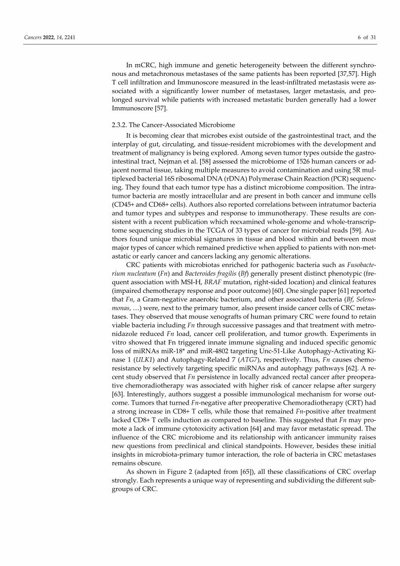
ICI treatment is being researched. In most cancers, the TMB of patients who carried POLE/POLD1 mutations was significantly higher than that of non-carriers. Among pa-tients treated with ICIs, the OS of patients who carried POLE/POLD1 mutations was sig-nificantly better than that of non-carriers [95]. This study also found that 26% of the pa-tients who had POLE/POLD1 mutations also showed the MSI-H phenotype. After remov-ing this subset of patients, the remaining patients with MSS were also found to benefit significantly from ICIs treatment. Multivariate Cox regression analysis showed that POLE/POLD1 mutation was an independent factor determining which solid tumor pa-tients may benefit from ICI treatment. At present, there are several clinical studies focus-ing on POLE/POLD1 mutation and ICI treatment (Table 3), and more evidence supporting the use of POLE/POLD1 mutation as molecular markers is expected.
Table 3. Non-exhaustive list of clinical trials including MSS mCRC patients and investigating ge-nomic, epigenomic, and transcriptomic biomarkers together with ICIs-based treatment.
Clinical Trial Immunotherapy Target Other Therapy Biomarkers Clinical Indication
NCT03436563 Bintrafusp Alfa Anti-PD-1/TGF-β trap NA CMS4 mCRC MSS CMS4, MSI-H mCRC, MSI-H non-CRC
NCT03152565 Avelumab PD-L1 Autologous dendritic cell vac-cine
Dynamic CMS modification MSS mCRC
NCT04695470 Sintilimab PD-1 Fruquitinib (VEGFR inhibitor)
TMB-H (≥5 mut/Mb) MSS mCRC
NCT03638297 BAT1306 or Pembrolizumab
PD-1 Aspirin/Celebrex (COX inhibitor)
TMB-H or MSI-H mCRC
NCT02842125 Pembrolizumab Nivolumab
PD-1 Ad-p53 (adenovirus) Chemotherapy
TMB Immune cells PD-L1, PD-L2
mCRC
NCT02628067 Pembrolizumab PD-1 NA TMB Adv. Solid tumors NCT04866862 Camrezilumab PD-1 Fruquitinib TMB Refractory MSS CRC NCT03150706 Avelumab PD-L1 NA POLE/POLD1 mCRC NCT03435107 Durvalumab PD-L1 NA POLE/POLD1 mCRC NCT03810339 Toripalimab PD-1 NA POLE/POLD1 Adv. Solid tumors
NCT03461952 Nivolumab Ipilimumab
PD-1 CTLA-4
NA POLE/POLD1 Adv. Solid tumors with POLE/POLD1 mutations
NCT03767075 Atezolizumab PD-L1 NA POLE/POLD1 Adv. Solid tumors with POLE/POLD1 mutations
NCT03832621 Nivolumab Ipilimumab
PD-1 CTLA-4
Temozolomide MGMT methylation TMB
MSS MGMT silenced mCRC
NCT03519412 Pembrolizumab PD-1 Temozolomide TMB MSS (TMB ≥ 20 mut/Mb) or MSI-H mCRC
NCT04457284 Nivolumab PD-1 Temozolomide Cisplatin
NA MSS CRC
4.3. Tumor Mutational Burden As MSI-H tumors harbor a high number of mutations and a high MANA load, the
TMB, expressed as the number of non-synonymous somatic mutations per trillion bases, emerged as a predictive biomarker of response to ICIs. In CRC, TMB-High (TMB-H) cor-relates with MSI-H and is associated with a high MANA load and immunogenicity [96]. It has been reported that 97% of MSI-H are TMB-H, as defined by a cutoff of 10 mutations
Cancers 2022, 14, 2241 13 of 31
per megabase [97]. However, only 16% of TMB-H are MSI-H, suggesting that MSS TMB-H CRC is more common than MSI-H CRC and could benefit from ICIs [98,99]. In KEY-NOTE-158, enrolling non-CRC MSI-H patients, a correlation between the antitumor activ-ity of the anti-PD-1 pembrolizumab and TMB-H has been reported. The ORR of TMB-H tumors was 29%, while the ORR of non-TMB-H tumors was 6% [100]. Following these observations, the FDA approved pembrolizumab for the treatment of TMB-H refractory advanced solid tumors, highlighting the promising value of TMB as an independent pre-dictive biomarker of response to ICIs. Moreover, in a recent report, Fabrizio et al. found that the ability of TMB to identify the CRC subgroup of patients that may respond to ICIs outperformed that of MSI status [101].
However, as a pan-cancer marker, a fixed cutoff of TMB that can be applied to differ-ent tumors was difficult to identify [99]. In a meta-analysis, the researchers summarized the most common cutoff values of TMB, which were 10, 16, and 20 mut/Mb. Schrock et al. reported that for 22 MSI-H mCRC patients treated with PD-1 or PD-L1 inhibitors, the op-timal cutoff value of TMB associated with better outcomes was between 37 and 41 mut/Mb [102]. Another trial evaluating regorafenib and nivolumab treatment in chemo refractory MSS mCRC (REGONIVO trial) reported an optimal TMB cutoff value of 22.55 mut/Mb for OS benefit [103]. Elevated plasma TMB (≥28 mut/Mb) may select patients most likely to benefit from durvalumab and tremelimumab treatment in the phase 2 CCTG CO.26 trial [79]. This suggests that a unique and optimal TMB high threshold does not exist for all cancers but more so, it could differ within different molecular subgroups of tumors as highlighted here for CRC.
The methodology for TMB evaluation is also an important characteristic to consider. Whole-Exome Sequencing (WES) is the gold standard for TMB assessment, but this tech-nique is expensive and lacks uniformity [104]. Next-Generation Sequencing (NGS) is a widely used and cheaper method regarding its convenience and applicability, but it intro-duces bias and errors related to the used panel size [105]. A correlation between TMB predicted by NGS and WES is reported [106]. Nevertheless, the methods used to calculate TMB in NGS and WES also affect the TMB results. Blood-based TMB is currently a valua-ble substitute for tissue TMB because of its facility in sampling and high consistency with tissue TMB in the predicted results [107]. However, assessment of TMB on circulating tu-mor DNA (ctDNA) requires the sequencing of a large panel of genes and, ideally, high coverage is also required to be able to distinguish an increase of TMB from an increase of ctDNA. If these conditions are not fulfilled, the assessment of TMB on blood sample could be very volatile and difficult to interpret correctly.
Several trials are currently recruiting CRC patients harboring high TMB tumors to be treated by ICIs-based therapies. One trial (NCT04695470) is combining sintilimab (anti-PD-1) with fruquitinib (VEGFR-1, -2, -3 inhibitor) for the treatment of refractory MSS mCRC with high TMB. In this trial, the TMB is assessed by NGS on plasma samples (TMB ≥ 5 mutations/Mb). Another trial (NCT03638297) recruiting CRC patients with MSI-H or high TMB tumors evaluates the association of a Cyclooxygenase (COX) inhibitor (BAT1306) with pembrolizumab treatment. Studies with preclinical models reported that COX inhibitors could act with PD-1 antibody in mice and control disease progress. In ad-dition, COX-2 could drive constitutive expression of IDO1 in human tumor cells. This could contribute to overcoming the lack of T cell infiltration and render the tumor more immunogenic [108].
4.4. DNA Methylation O6-Methylguanine–DNA Methyltransferase (MGMT) is a key protein in the DNA
repair mechanism of damages induced by alkylating agents and, as illustrated in glioblas-toma, the epigenetic silencing of MGMT is a mechanism that potentiates the effect of Te-mozolomide (TMZ) [109]. Despite the fact that MGMT methylation, inducing a lack of MGMT protein expression, is found in 40% of CRC patients, TMZ and its analog dacarba-

Cancers 2022, 14, 2241 14 of 31
zine provided limited clinical activity with an ORR under ten percent in MGMT-methyl-ated mCRC [110]. As demonstrated in CRC models and mCRC patients [111], resistance to TMZ, observed in almost all TMZ-sensitive tumors [112], may be related to the hyper-mutated phenotype and the emergence of other mutations in DNA repair mechanisms such as MSH6 [113]. In the proof-of-concept trial MAYA, designed to assess the potential role of TMZ as an inducer of hypermutated phenotype and immune-sensitizing agent in MSS MGMT-silenced mCRC tumors, eligible patients received two cycles of TMZ fol-lowed by its combination with anti-CTLA-4 ipilimumab in the absence of progression [114]. In this trial, among the 204 eligible patients, 135 started TMZ treatment and 102 of them were discontinued due to disease progression or death. Among the 33 patients who achieved disease control and received ipilimumab, 36% reached the primary objective of eight-month PFS rate. The ORR was 45% and the median OS and PFS were 18.4 and 7 months, respectively. This proof-of-concept trial provided new insight regarding the strat-egy of turning cold tumors into hot tumors through the use of TMZ inducing hypermu-tated phenotype. However, further investigation on treatment regimen, optimization, and patient selection is needed in order to maximize the success of this therapeutic approach. Similar to the MAYA trial, the ARETHUSA (NCT03519412) trial is currently investigating the efficacy of pembrolizumab in patients that reach >20 mutations/Mb after TMZ prim-ing. Moreover, another group is currently conducting a phase Ib trial (NCT04689347) com-bining fluorouracil, leucovorin, and irinotecan/bevacizumab (FLIRT/BEV) with escalating doses of TMZ in untreated MSS MGMT-silenced mCRC in order to investigate the optimal dosing of this new triplet and the role of maintenance immunotherapy in patients with disease control after the FLIRT/BEV regimen.
Previous studies have demonstrated that epigenetic modulation by DNMT inhibitor modifying the expression of genes related to innate immunity, adaptive immunity, and immune evasion in tumor tissues [115–117] may enhance the antitumor immune response by promoting increased TILs, although specific mechanisms by which this occurs have not been established [118]. In this way, a phase 2 single-arm trial has evaluated activity and tolerability of pembrolizumab plus azacytidine (DNMT inhibitor) in patients with chemotherapy-refractory mCRC [119]. This treatment combination provided modest ac-tivity (ORR: 3%, median PFS: 1.9 months, median OS: 6.3 months); correlative studies sug-gest that tumor DNA demethylation and immunomodulation occur. While not sufficient for antitumor activity, this immunomodulatory approach may contribute to future strat-egies to overcome immune resistance in patients with mCRC.
4.5. Gene Expression Profile and Consensus Molecular Subtypes As discussed, the CMS1 and CMS4 groups are both characterized as CRC infiltrated
by immune cells and could be potential predictive biomarkers. However, their immune environment is very different and could be summarized as immunoreactive for CMS1 and immunosuppressive for CMS4. If CMS1 tumors have a TME which may benefit from ICIs, the situation could be more complex for CMS4 CRC. The TME of these tumors with the presence of Tregs, MDSC, M2 macrophages, Th17 cells, and IFN-γ signature suggests the potential for immune therapy benefit but with additional efforts to overcome this immu-nosuppressive microenvironment [16,37,46].
To our knowledge, no ongoing clinical trials directly use CMS1 subtype as a predic-tive biomarker of response to ICIs. This is mainly explained by the fact that the CMS1 subgroup often comprises other recognized molecular biomarkers such as MSI-H status, POLE/POLD1 mutation, and high TMB which are easier to assess. Interestingly, a recent phase 2 study [120] evaluated the combination of encorafenib (BRAF inhibitor), cetuximab (anti-Endothelial Growth Factor Receptor (EGFR) monoclonal antibody), and nivolumab in patients with MSS, BRAF-V600E-mutated mCRC, a mutation frequently associated with CMS1 subgroup. Preclinical models of MSS CRC showed that BRAF combined with EGFR inhibition induced a transient MSI-H phenotype [120]. BRAF V600E inhibitor may prime these tumors for response to anti-PD-1 antibodies. In this trial, the 26 enrolled patients

Cancers 2022, 14, 2241 15 of 31
experienced an ORR of 45% with a median PFS of 7.3 months and OS of 11.4 months. A follow-up randomized phase II trial (SWOG 2107) to evaluate encorafenib/cetuximab with or without nivolumab in patients with MSS, BRAF-V600E-mutated metastatic CRC will begin in 2022.
CMS4 CRC features increased TGF-β signaling, which may account for de novo re-sistance to immunotherapy for patients with MSS mCRC. One recent phase 2 trial (NCT03436563) [121] evaluated bintrafusp alfa, a dual PD-L1 antibody/TGF-β trap, with radiation therapy to a single metastatic lesion with abscopal intent (8 Gy) for the treatment of CMS4 mCRC. No patients achieved a tumor response and median PFS and OS were 1.6 months and 5.0 months, respectively. Although the efficacy for bintrafusp alfa and radio-therapy was deceiving, changes in IFN-γ signature provide a potential signal for refining therapeutic strategies based upon TGF-β enrichment in patients with mCRC. Another co-hort from the same trial (NCT03436563) [121], focusing on MSI-H cancers refractory to ICIs did not demonstrate significant antitumor activity. Ongoing correlative studies may inform on the effect of TGF-β and PD-L1 modulation by bintrafusp alfa within the TME. Another phase I/II trial investigated the efficacy of avelumab plus the autologous den-dritic cell vaccine in pretreated MSS mCRC patients [122]. An interim analysis (Simon design first-stage) recommended early termination because 11% (2/19) of patients were progression-free at 6 months and no patients experienced tumor response. Four patients (21%) experienced hyper-progressive disease. Stimulation of immune response was ob-served with changes of cytokine levels after treatment. The evaluation of transcriptomic immune-metabolic signature did not correlate with clinical outcomes. Hyper-progressive disease was observed in different immune-metabolic micro-environments.
Although CMS classification appears deceiving for biomarker development, ongoing trials focusing on different compounds (TGF-β, Tregs, MDSCs…) of the immune suppres-sive TME enriched in CMS4 CRC are still ongoing.
4.6. Tumor-Infiltrating Lymphocytes and Immunoscore Tumor infiltration of cytotoxic T cells and Th1 cells and IFN-γ upregulation predict
a favorable prognosis in CRC [123] and also serve as markers indicating a good response to Immune Checkpoint Inhibitors [124], as IFN-γ can upregulate PD-L1 and Major Histo-compatibility Complex-I (MHC-I) expression by tumor cells [125]. In addition, co-locali-zation of PD-L1+ cells with tumor-infiltrating CD8+ T cells has been widely reported as a predictive biomarker for ICI treatment [126–128]. The hypothesis is that TILs induce adap-tive immune resistance, accompanied by increased PD-L1 expression. Indeed, CD8 and PD-L1 expression is significantly higher in responders to anti-PD-1 therapy than in non-responders [126,129,130].
Immunoscore has been developed, extensively evaluated in several cohorts of pa-tients, and recognized as a robust prognostic biomarker [131]. However, its use as a pre-dictive biomarker is still under investigation. Moreover, the use of Immunoscore on biop-sies is difficult since it limits the analysis to a restricted part often limited to the core of the tumor. In this way, the prognostic and the predictive values of a biopsy-adapted Im-munoscore (ISB) were evaluated in a recent study. Pre-therapeutic biopsies from patients with locally advanced rectal cancer treated with CRT followed by radical surgery were stained for CD3+ and CD8+ T lymphocytes in two independent cohorts. Density of CD3+ and CD8+ T cells was used to derive an ISB [132]. Authors reported that a high ISB posi-tively correlated with cytotoxic immune response, Th1-orientated gene expression signa-ture, and histologic response after treatment. In addition, patients with high ISB were at lower risk of relapse or death compared with low ISB. Today, some trials continue to in-vestigate Immunoscore as a prognostic biomarker (NCT04938986, NCT01688232, NCT0342260, NCT02274753) for disease-free survival stratification and detection of risk of recurrence (Table 4).

Cancers 2022, 14, 2241 16 of 31
Table 4. Non-exhaustive list of clinical trials including MSS mCRC patients and investigating Im-munoscore and immune infiltration as prognostic biomarker or predictive biomarker together with ICIs-based treatment.
Clinical Trial Immunotherapy Target Other Therapy Biomarker Clinical Indication NCT04938986 NA NA SOC Immunoscore Non-metastatic CRC NCT01688232 NA NA SOC Immunoscore CRC NCT03422601 NA NA Oxaliplatin Immunoscore Stage III
NCT02274753 NA NA SOC Immunoscore
NGS miRNA
CRC
NCT04262687 Pembrolizumab PD-1 Bevacizumab Oxaliplatin
Immunoscore High immune infiltrate
MSS mCRC
NCT02646748 Pembrolizumab PD-1 Itacitinib TILs (CD8+, FOXP3+) Adv. Solid tumors
NCT02512172 Pembrolizumab PD-1 Azacitidine Romidepsin
TILs (CD4+ CD8+) Adv. Solid tumors
NCT02837263 Pembrolizumab PD-1 SBRT TILs Liver metastatic mCRC
Recent studies suggest using Immunoscore to predict the response to immunother-apy [38,45,133], but besides Immunoscore, TILs and immune cell populations in the tumor could also be used as prognostic or predictive biomarker. The POCHI trial (NCT04262687) is currently recruiting MSS mCRC with high immune infiltration evaluated by Im-munoscore, among others, in an experimental arm combining XELOX plus bevacizumab and pembrolizumab (Table 4). This proof-of-concept study could pave the way for the use of Immunoscore as a predictive biomarker of ICIs soon.
Beyond CD3+ and CD8+ T cells, other immune cells within the TME, such as Th17 and memory T cells, have gained interest in the past years. Th17 cells, secreting IL-17, modulate the expression of other cytokines such as TGF-β, IFN-γ, IL-6, IL-21, and IL-22. The role of Th17 cells in tumor immunity and development remains controversial, mainly attributed to the plasticity of Th17 cells. In CRC, Th17 seems to play a role in carcinogen-esis and could decrease the antitumor activity of CD8+ T cells [134]. Intratumoral IL17-mediated signaling may preclude responses to immunotherapy. A recent paper reported that both IL-17 low and high immunoreactive MSS CRC are associated with features of adaptive immunosuppression (CD8/IFN-γ and PD-L1/IDO1 co-localization). Neverthe-less, only patients with a Th17 low MSS CRC had a TME resembling that of patients with mCRC responsive to anti-PD-1 treatment [92]. Several studies reported that the presence of memory T cells (CD45RO+) within the TME was associated with better prognosis and lower risk of CRC metastasis [50,135–137]. CD8+ resident memory T cells are found in greater abundance in MSI-H CRC, suggesting an important role in the antitumor immu-nogenicity of MSI-H CRC [138]. Their presence in MSS CRC could be an additional marker suggesting the immunoreactiveness of the tumor and the possible response to immune therapy.
4.7. The Gut and Cancer Microbiome It is now well established that gut microbiome is strongly involved in the develop-
ment and maintenance of the host immune system. In this regard, seminal papers have highlighted different responses to ICIs in cancer patients depending on the composition of their gut microbiome [139,140]. For instance, specific bacterial species have been asso-ciated with better prognosis and response to anti-PD-1 ICI in melanoma patients [141–144]. In NSCLC, the authors reported that Akkermansia muciniphila (Akk) was associated with increased ORR and survival after anti-PD1 ICI [145]. Another study recruiting pa-tients with gastrointestinal cancer found a significant different Prevotella/Bacteroides spe-cies ratio associated with ICIs responses [146]. A potential mechanism by which the dis-tant microbiota might benefit from tumor immunotherapy is through bacterial metabo-

Cancers 2022, 14, 2241 17 of 31
lites. Recently, authors report that a collection of eleven bacteria from human gut micro-biota appeared to be able to robustly induce IFN-γ, producing CD8+ T cells in the gut and enhancing antitumor immunity [147]. Furthermore, Mager et al. reported, in mouse mod-els, that the metabolite inosine derived from intestinal populations of Bifidobacteria, Lacto-bacillus, and Olsenella was associated with increased numbers of CD8 +IFN-γ + T cells and control of tumor growth [148].
Several ongoing trials currently study the gut microbiome. The NCT02960282 trial studies the gut microbiome in fecal samples from mCRC patients treated with chemother-apy or immunotherapy. An upcoming phase II study (NCT05279677) will evaluate the efficacy and safety of fecal microbiota transplantation plus sintilimab and fruquitinib in chemo refractory mCRC.
Beyond the gut microbiome, a diverse microbial community is also present within the CRC. Among them, Fusobacterium nucleatum (Fn), a tumor-resident bacteria in CRC, is of growing interest. Several publications have reported correlations between intra-tu-moral detection of Fn and poor prognosis, shorter PFS, and higher tumor recurrence [60] [149–151]. In addition, a recent publication reported that high Fn levels correlated with better therapeutic response to PD-1 blockade in CRC patients, regardless of MSI status [152]. Furthermore, Fn induced PD-L1 expression by activating Stimulator Of Interferon (STING) signaling and increased the accumulation of IFN-γ+ CD8+ TILs during treatment with PD-L1 blockade, thereby increasing tumor sensitivity to PD-L1 blockade. Due to its recent discovery and the lack of sufficient knowledge, there are currently no ongoing trials evaluating the cancer tissue microbiome as a biomarker of response to ICIs.
4.8. Circulating Biomarkers mCRC is characterized by the important inter- and intra-patient heterogeneity of its
intratumoral immune microenvironment [57]. Even though Immunoscore, gene expres-sion profiles, or TMB have predictive or prognostic value [153], their practical information is limited by tumor tissue accessibility and spatial and temporal heterogeneity during the CRC evolution. Due to the tumor’s dynamic behavior, the study of tumor characteristics requires the evaluation of accessible biomarkers that can reflect tumor modifications dur-ing treatment without the need for biopsy. Therefore, identifying biomarkers in body flu-ids that can accurately, quickly, and cost-effectively reflect the stage and characteristics of the tumor is desired. Circulating exosomes, microRNAs, tumor cells (CTC), and tumor DNA can be ideal indicators for tumor heterogeneity changes during treatment [154]. The similarity of the genetic profile of CTCs with tumors has been reported in 50–77% of cases [155,156]. Interestingly, the genetic profile similarity between cell-free DNA (cfDNA) or ctDNA and tumors has been reported to be more than 90% [157,158].
New liquid biomarkers have been recently investigated. Circulating tumor DNA, flow/mass cytometry, and blood T Cell Receptor sequencing (TCR-seq) allow sensitive tracking of changes in antigen-specific T cells at the clonal level, with unprecedented in-sight into mechanisms by which ICIs alter T cell responses [159]. Serial sampling and com-bination of these approaches will likely become a key element to provide an overview of the genetic makeup of the tumor and adaptive immunity of the patient.
4.8.1. Circulating Tumor DNA Commonly designated as “liquid biopsies”, the analysis of the ctDNA by NGS or
digital droplet PCR (ddPCR) is currently extensively investigated in several cancers [160]. Liquid biopsies have emerged as a diagnostic tool to assess the presence of tumoral mu-tations and to monitor the emergence of mutations over time. In CRC, the assessment of ctDNA alteration of genes belonging to the EGFR pathway predicted response to anti-EGFR treatment [158]. The emergence over time of such alteration during anti-EGFR treat-ment is associated with acquired resistance [161]. The MSI-H status as well as the TMB can also be assessed on ctDNA with similar predictability for response to ICIs [162]. De-tection of ctDNA in the follow-up of the treated patient is also used to detect minimal

Cancers 2022, 14, 2241 18 of 31
residual disease and often reveals earlier recurrence compared to standard radiology [163].
A study including 1000 patients with advanced or metastatic tumors treated with ICIs reported that on-treatment ctDNA dynamics appear to be predictive of the long-term benefit of ICI across tumor types. ctDNA dynamics could help to select patients who will ultimately derive benefit from immunotherapy [164]. A prospective clinical trial [165] also revealed the correlation between the level of ctDNA and the efficacy of ICI treatment in five different cancer types. Authors measured the ctDNA level after three cycles of pem-brolizumab and found that patients with a decrease of ctDNA level showed better clinical efficacy during the treatment. A study of MSS CRC patients treated with regorafenib and PD-1 inhibitors found that ctDNA may be predictive of early therapeutic efficacy. Specif-ically, ten patients with rising ctDNA levels or emergence of new clones four weeks after treatment experienced progressive disease after two months, whereas three patients with declining ctDNA experienced stable disease [166].
Several ongoing clinical trials currently use ctDNA to select molecular subgroups of CRC (MSI-H, TMB-H, or specific mutation of interest) more easily than tissue biopsy (Ta-ble 5). ctDNA is also used to map precise disease evolution; dynamic change in ctDNA is measured to detect response and early resistance to ICIs (NCT03946917, NCT04046445, NCT05240950, and NCT02997228).
Table 5. Non-exhaustive list of clinical trials investigating liquid biomarkers together with ICIs-based treatment.
Clinical Trial Immunotherapy Target Other Therapy Biomarkers Clinical Indication
NCT03946917 JS001 PD-1 Regorafenib
(Kinase inhibitor) ctDNA Adv. CRC
NCT04046445 BI754091 PD-1 ATP128
(Vaccine) ctDNA MSS mCRC
NCT02997228 Atezolizumab PD-L1 Bevacizumab
Chemotherapy
ctDNA Dynamic TCR repertoire
PD-L1 MLH1
MSI-H mCRC
NCT03927898 Toripalimab PD-1 SBRT Dynamic TCR repertoire
PD-L1 tumor cells PD-1, Ki-67 T cells
mCRC
NCT04714983 DNX-2440 OX40 T cells infiltration
Dynamic TCR repertoire mCRC
NCT02713373 Pembrolizumab PD-1 Cetuximab T cells populations (Flow cytometry)
mCRC
NCT03984578 Pembrolizumab PD-1 Chemotherapy T cells populations (Flow cytometry)
CRC
NCT02851004 Pembrolizumab PD-1 Napabucasin
(STAT3 inhibitor)
T cells populations (Flow cytometry)
CMS MSS/MSI mCRC
NCT05086692 ICI NA MDNA11
(IL-2 superkine) T cells populations (Flow cytometry)
Adv. Solid tumors
NCT04348643 CAR T cells CEA NA T cells populations (Flow cytometry)
CEA+ CRC
NCT02349724 CAR T cells CEA NA CAR T cells
(Flow cytometry) CEA+ CRC
NCT04513431 CAR T cells CEA NA CAR T cells
(Flow cytometry) CEA+ CRC
NCT03638206 CAR T cells c-MET NA CAR T cells
(Flow cytometry CRC
Cancers 2022, 14, 2241 19 of 31
4.8.2. T Cell Receptor Repertoire
The specificity of the tumor immune response is linked to the antigen-specific T Cell Receptor (TCR). The analysis of the TCR repertoire on Peripheral Blood Mononuclear Cells (PBMCs) and TILs could be used as a prognostic and predictive biomarker of re-sponse to ICIs [167]. T cell receptors recognize specific antigens presented by MHC class I (for CD8+ T cells) or MHC class II (for CD4+ T cells) molecules. A process called genetic recombination occurs in T cells to rearrange the DNA at three T Cell Receptor Beta Loci (TRBV, TRBD, and TRBJ) to develop TCRs that are specific for certain antigens. The T cell repertoire refers to all of the unique TCR genetic rearrangements within the adaptive im-mune system; thus, the TME T cell repertoire refers to all of the unique TCR genetic rear-rangements within the TME. Not surprisingly, having a diverse TME T cell repertoire is associated with better outcomes in response to immunotherapy [159]. As for liquid biop-sies, TCR repertoire can be assessed pre-treatment but also over time to measure dynamic changes of the repertoire in response to ICIs. The TCR repertoire analysis can be per-formed on all PBMCs found in the blood (bulk TCR) or performed on specific subsets of lymphocytes sorted by flow/mass cytometry such as CD8+PD-1+ T cells that are the main target of PD-1 blockade [159]. In melanoma, it has been shown that a high pre-treatment TCR diversity of blood CD8+PD-1+ T cells and a reduced post-treatment diversity are as-sociated with a longer PFS after anti-PD-1 therapy [168,169]. Still in melanoma, single cell analysis of peripheral CD8+ T cells revealed that responders to both anti-PD-1 and anti-PD-1 + anti-CTLA-4 presented more expanded clones than non-responders. Similarly, it has been shown that after one cycle of ICIs, responders exhibited clonally expanded CD27+ C-C Motif Chemokine Receptor 7 (CCR7)+ memory T cells. How TCR diversity impacts adaptive immunity in CRC patients remains unclear for ICI treatment. Very lim-ited data on TCR repertoire and ICIs are available for CRC. One report [170] showed that mCRC patients treated with chemotherapy and with either high baseline TCR diversity or TCR diversity that dropped during therapy had significantly better treatment re-sponses. In a TCR repertoire analysis of advanced CRC patients treated with a combina-tion of five cancer peptide vaccines and oxaliplatin-based chemotherapy, high TCR diver-sity scores were associated with improved response [171]. The expansion of tumor-asso-ciated TCRs in the blood underscores the continuity of the tumor and blood compartments and suggests that the activity of PD-L1 blockade may involve circulating T cells more than previously thought. It raises the possibility that antitumor T cells may home from the pe-riphery into the tumor before later recirculating. In addition, a recent report suggested a significant difference in the usage of TRBV and TRBJ genes between CRC patients and healthy controls, supporting its use as an additional TCR-based predictive biomarker in CRC [172].
TCR repertoire is currently investigated in several CRC trials involving ICIs (Table 5). In one trial (NCT03927898) combining toripalimab (anti-PD-1) together with stereotac-tic body radiotherapy for the treatment of oligometastatic CRC, investigators will analyze dynamic TCR repertoire changes in peripheral blood as well as PD-1 and Ki67 expression on T cells and PD-L1 expression on circulating tumor cells. Moreover, in another window of opportunity trial (NCT04714983), the immunotherapeutic response after OX40-ligand expressing oncolytic adenovirus will be evaluated by TCR repertoire changes in blood and tissue samples.
4.8.3. Flow/Mass Cytometry Flow cytometry and mass cytometry (allowed to cover up to 40 markers) can provide
interesting data on the frequency of T cells subpopulations and their variation during treatment. Flow or mass cytometry permit one to obtain a comprehensive overview of tumor-resident and circulating immune populations. Several simultaneous staining pan-els (e.g., CD45, CD3, CD8, CD4, CD11B, CD14, CD20, CD25, FOXP3, PD-1, TIGIT, …) can

Cancers 2022, 14, 2241 20 of 31
be performed, allowing a global characterization of the circulating immune subpopula-tions and in the TME [173]. In melanoma and Merkel cell carcinoma, it has been shown that an increase of the frequency of PD-1+TIGIT+CD8+ circulating cells after one month of anti-PD-1 therapy was associated with OS and clinical response [174]. In another trial on melanoma, authors demonstrated that patients who respond to anti-PD-1 treatment showed a decrease of circulating PD-1+ Tregs (CD4+CD25+CD127-) [175]. In CRC, Th1 (CD126-CD4+) cells were more abundant in the blood of patients responding to anti-PD-1 compared with non-responders [176]. Pre-treatment Tregs frequency was higher in non-responders. Moreover, a decrease of the frequency of Th1 cells during the treatment was observed in patients with acquired resistance to treatment. The analysis of blood Th1 cells together with Tregs could represent a blood biomarker of response to ICIs in CRC.
The relative distribution of immune cells subtypes and immune checkpoint mole-cules expression on tumor cells and immune cells, assessed by flow cytometry in blood and/or tissue samples, is currently investigated in several trials (NCT05086692, NCT03984578, NCT02851004) combining pembrolizumab with other treatments (chemo-therapy, cetuximab, STAT3 inhibitor). Another trial (NCT05086692) also using flow cy-tometry, evaluates a treatment with MDNA11, a drug designed to bind IL-2 beta receptor on immune cells, combined or not with ICI (Table 5).
Flow cytometry is also heavily used in clinical trials involving Chimeric Antigen Re-ceptor (CAR) T cells. This tool allows the investigator to follow in vivo, in peripheral blood, the rate of CAR T cells in a dynamic way to evaluate the proliferation and long-term survival of the cells during the therapy (Table 5). CAR T cells are cells that have been genetically engineered to produce artificial chimeric T cell receptors [177]. CAR T cells have demonstrated great success in treating CD19-positive B cell malignancies [178]. To-day, CAR T cells are also investigated in solid tumors such as CRC, either targeting, among others, Carcinoembryonic Antigen (CEA) (NCT04348643, NCT02349724, NCT04513431) or tyrosine-kinase Met (c-Met) (NCT03638206).
4.8.4. Cytokines Cytokines play a key role in both pro- and antitumor immune responses and can be
secreted by either the tumor cells or immune cells [179]. The cytokine A Proliferation-Inducing Ligand (APRIL), produced by tumor cells, and B-cell Activating Factor (BAFF), IL-8, and Matrix Metallopeptidase 2 (MMP2), produced by a variety of tissue and blood cells, have been reported to be inversely correlated with immune cell infiltration and ex-pression of CD163, a marker of M2 macrophages in CRC [180]. Authors reported that the significantly decreased APRIL and increased BAFF, IL-8, and MMP2 expression was tu-mor-specific and deserves consideration in the development of new treatments [180]. An-other study revealed, in MSS CRC cell lines and tissues, that IL-17A, secreted by Th17 cells, increased expression of PD-L1 on CRC cells and that inhibition of IL-17A improved the efficacy of anti-PD-1 therapy in a murine MSS CRC model [181]. IL-17A might serve as a therapeutic target to sensitize patients with MSS CRC to ICI therapy. Nevertheless, further investigation is needed to use cytokine as a biomarker of selection in clinical re-search.
Cancer-Associated Fibroblasts (CAFs) are cells within the TME promoting tumor-igenic features by initiating the remodeling of the extracellular matrix or by secreting cy-tokines such as TGF-β, IL-6, IL-8, CXCL14, CXCL12, and VEGF [182]. Therefore, CAFs may act on cancer development, metastasis process, and tumor immunity. However, due to the transcriptional and functional heterogeneity of the CAFs, their use as therapeutic targets or biomarkers is still controversial and further investigation is needed to better understand their complexity [183].
4.9. Clinical Tumor Burden, Metastases Location, and Characteristics Accumulating evidence suggests that a high tumor burden, defined as the total
amount of tumoral tissue in the body estimated by imaging, liquid biopsy (circulating

Cancers 2022, 14, 2241 21 of 31
tumor DNA or cells), or biological tumor derivatives (e.g., Lactate Dehydrogenase (LDH), CEA) is negatively correlated with antitumor immunity and ICI response [184–187]. An enlarging tumor implies inefficacy of the immune system at containing its growth, while smaller tumor burdens may be partly immune-controlled.
Beside tumor burden, the impact of Liver Metastases (LM) on ICI effectiveness ap-pears important. Tumeh et al. initially reported that PD-1 blockade was much less effec-tive in melanoma or NSCLC patients with LM (ORR and median PFS of 47.5% and 5.1 months for patients with LM compared to 70.8% and 20.1 months for those without LM) [188]. This observation was subsequently confirmed by other clinical reports [189–191]. A proposed mechanism of this “systemic immune suppression” involves CD11b+ suppres-sive macrophages generated in and that delete CD8+ T cells through Fas Ligand [191]. In addition, Treg activation (CTLA-4, PD-1, and ICOS- high) was observed and contributed to the distal immunosuppression [192]. Preclinical modeling suggests that LM resection remodels systemic antitumor immunity by decreasing immunosuppression [193,194]. Correlative studies in patients undergoing resection of liver tumors have mainly focused on acute postoperative immunosuppression that underlies perioperative increased infec-tion risk and have not evaluated the impact on the adaptive immune system [195]. How-ever, the clinical benefits of resection are apparent. The resection of oligometastatic CRCLM in select patients has been associated with prolonged OS [196]. An ongoing clin-ical trial is examining the resection of CRCLM in combination with immunotherapy in CRC (NCT03844750). Radiofrequency ablation, cryotherapy, transarterial chemo- or ra-dio-embolization, and stereotactic body radiotherapy are minimally invasive approaches that have demonstrated clinical utility in the management of CRCLM. Preclinical evalua-tions have shown that these treatments can induce an in situ vaccination which potentiates spontaneous and therapeutic antitumor immunity [193]. Many clinical trials are examin-ing whether these ablative techniques of CRCLM are safe and efficacious in combination with immunotherapy.
Finally, several confounding clinical factors, such as sex/gender, age, obesity, race, alcohol and tobacco consumption, exercise, and psycho-emotional stress have been re-ported to possibly influence the magnitude of benefit to ICIs. All these factors may directly or indirectly act on the immune system, thus influencing ICIs response [197]. Therefore, all of these parameters should also be considered when analyzing the response to ICIs in CRC patients.
5. Conclusions and Future Direction The current clinical use of the anti-PD-1 inhibitors pembrolizumab and nivolumab
and the anti-CTLA-4 therapy ipilimumab has revolutionized the survival potential of many patients with MSI-H CRC and indicates the significant anticancer potential of ICIs for CRC. There are many areas of current research that aim to improve clinical response rates and generalize these treatments to all CRC. The immune microenvironment of MSS CRC is highly heterogeneous, which compromises the current development of immune therapies. Several genomic, transcriptomic, and TME classifications have been performed to distinguish and characterize the different immune subtypes of CRC. While immune-competent CRC often responds to ICI treatment, the situation remains more complex for immunocompromised or suppressed CRC, requiring innovative strategies based on im-munotherapy, often combining ICI with chemotherapy, radiotherapy, or targeted thera-pies. These strategies aim at triggering an effective and adequate immune response against tumor cells.
The elucidation of the different pathways mediating immune resistance in MSS CRC remains an ongoing challenge for clinical research. Improving patient selection through effective biomarkers identifying the different immune subtypes of CRC remains essential for future research development. Although Immunoscore, gene expression profiles, or TMB have predictive or prognostic value, their practical application remains limited by the accessibility of tumor tissues and the spatial and temporal heterogeneity during CRC
Cancers 2022, 14, 2241 22 of 31
progression. Therefore, circulating biomarkers that can accurately, rapidly, and cost-ef-fectively reflect the dynamic behavior of the tumor during treatment without the need for biopsy are of great interest for future research. Serial sampling and the combination of tissue and liquid biopsy approaches will likely become a key component in providing a comprehensive view of the genetic makeup of the tumor and the patient’s adaptive im-munity.
The development of prospective clinical trials combining ICIs with other therapies and aiming at proving the predictive value of selected biomarkers is crucial. Such trials would help the clinical and scientific communities to better understand the complex in-teractions of the cancer cells with their surrounding TME and to optimally use new com-bination approaches. Finally, although MSI-H CRCs are known to respond to ICIs, not all benefit from these treatments. Therefore, the development of new combination ap-proaches when ICIs resistance occurs and the use of biomarkers for understanding must continue. Hopefully, this will allow scientists and oncologists to exploit the full potential of immunotherapy in the treatment of CRC.
Author Contributions: Writing—original draft preparation, E.B. and P.S.; writing—review and ed-iting, N.H., E.B., P.S. and M.V.d.E.; E.B. and P.S. equally contributed to the writing of some sections of the review. All authors have read and agreed to the published version of the manuscript.
Funding: Nicolas Huyghe is a research fellow supported by a grant from the Belgian National Fund for Scientific Research [Télévie/FNRS 7460918F]. Philippe Stevens is a research fellow supported by a grant from the Belgian National Fund for Scientific Research [Télévie/FNRS 7.4611.19].
Institutional Review Board Statement: Not applicable.
Acknowledgments: The authors would like to thank Derouane Françoise for her help in figure mak-ing. The four figures have been created with “BioRender.com”.
Conflicts of Interest: The authors declare no conflict of interest.
References 1. Sung, H.; Ferlay, J.; Siegel, R.L.; Laversanne, M.; Soerjomataram, I.; Jemal, A.; Bray, F. Global Cancer Statistics 2020: GLOBOCAN
Estimates of Incidence and Mortality Worldwide for 36 Cancers in 185 Countries. CA Cancer J. Clin. 2021, 71, 209–249. https://doi.org/10.3322/caac.21660.
2. Eadens, M.J.; Grothey, A. Curable metastatic colorectal cancer. Curr. Oncol. Rep. 2011, 13, 168–176. https://doi.org/10.1007/s11912-011-0157-0.
3. Crooke, H.; Kobayashi, M.; Mitchell, B.; Nwokeji, E.; Laurie, M.; Kamble, S.; McKenna, M.; Masood, A.; Korytowsky, B. Estimating 1- and 5-year relative survival trends in colorectal cancer (CRC) in the United States: 2004 to 2014. J. Clin. Oncol. 2018, 36, 587–587. https://doi.org/10.1200/JCO.2018.36.4_suppl.587.
4. Biller, L.H.; Schrag, D. Diagnosis and Treatment of Metastatic Colorectal Cancer: A Review. JAMA 2021, 325, 669–685. https://doi.org/10.1001/jama.2021.0106.
5. Di Nicolantonio, F.; Vitiello, P.P.; Marsoni, S.; Siena, S.; Tabernero, J.; Trusolino, L.; Bernards, R.; Bardelli, A. Precision oncology in metastatic colorectal cancer—From biology to medicine. Nat. Rev. Clin. Oncol. 2021, 18, 506–525. https://doi.org/10.1038/s41571-021-00495-z.
6. Casak, S.J.; Marcus, L.; Fashoyin-Aje, L.; Mushti, S.L.; Cheng, J.; Shen, Y.L.; Pierce, W.F.; Her, L.; Goldberg, K.B.; Theoret, M.R.; et al. FDA Approval Summary: Pembrolizumab for the First-line Treatment of Patients with MSI-H/dMMR Advanced Unresectable or Metastatic Colorectal Carcinoma. Clin. Cancer Res. 2021, 27, 4680–4684. https://doi.org/10.1158/1078-0432.CCR-21-0557.
7. Marcus, L.; Lemery, S.J.; Keegan, P.; Pazdur, R. FDA Approval Summary: Pembrolizumab for the Treatment of Microsatellite Instability-High Solid Tumors. Clin. Cancer Res. 2019, 25, 3753–3758. https://doi.org/10.1158/1078-0432.CCR-18-4070.
8. Trullas, A.; Delgado, J.; Genazzani, A.; Mueller-Berghaus, J.; Migali, C.; Muller-Egert, S.; Zander, H.; Enzmann, H.; Pignatti, F. The EMA assessment of pembrolizumab as monotherapy for the first-line treatment of adult patients with metastatic microsatellite instability-high or mismatch repair deficient colorectal cancer. ESMO Open 2021, 6, 100145. https://doi.org/10.1016/j.esmoop.2021.100145.
9. Le, D.T.; Uram, J.N.; Wang, H.; Bartlett, B.R.; Kemberling, H.; Eyring, A.D.; Skora, A.D.; Luber, B.S.; Azad, N.S.; Laheru, D.; et al. PD-1 Blockade in Tumors with Mismatch-Repair Deficiency. N. Engl. J. Med. 2015, 372, 2509–2520. https://doi.org/10.1056/NEJMoa1500596.
10. Boland, C.R.; Goel, A. Microsatellite instability in colorectal cancer. Gastroenterology 2010, 138, 2073–2087.e2073. https://doi.org/10.1053/j.gastro.2009.12.064.
Cancers 2022, 14, 2241 23 of 31
11. Chung, D.C.; Rustgi, A.K. DNA mismatch repair and cancer. Gastroenterology 1995, 109, 1685–1699. https://doi.org/10.1016/0016-5085(95)90660-6.
12. Herman, J.G.; Umar, A.; Polyak, K.; Graff, J.R.; Ahuja, N.; Issa, J.P.; Markowitz, S.; Willson, J.K.; Hamilton, S.R.; Kinzler, K.W.; et al. Incidence and functional consequences of hMLH1 promoter hypermethylation in colorectal carcinoma. Proc. Natl. Acad. Sci. USA 1998, 95, 6870–6875. https://doi.org/10.1073/pnas.95.12.6870.
13. Li, G.M. Mechanisms and functions of DNA mismatch repair. Cell Res. 2008, 18, 85–98. https://doi.org/10.1038/cr.2007.115. 14. Haraldsdottir, S.; Einarsdottir, H.M.; Smaradottir, A.; Gunnlaugsson, A.; Halfdanarson, T.R. Colorectal cancer—Review.
Laeknabladid 2014, 100, 75–82. https://doi.org/10.17992/lbl.2014.02.531. 15. Popat, S.; Hubner, R.; Houlston, R.S. Systematic review of microsatellite instability and colorectal cancer prognosis. J. Clin. Oncol.
2005, 23, 609–618. https://doi.org/10.1200/JCO.2005.01.086. 16. Guinney, J.; Dienstmann, R.; Wang, X.; de Reynies, A.; Schlicker, A.; Soneson, C.; Marisa, L.; Roepman, P.; Nyamundanda, G.;
Angelino, P.; et al. The consensus molecular subtypes of colorectal cancer. Nat. Med. 2015, 21, 1350–1356. https://doi.org/10.1038/nm.3967.
17. Mur, P.; Garcia-Mulero, S.; Del Valle, J.; Magraner-Pardo, L.; Vidal, A.; Pineda, M.; Cinnirella, G.; Martin-Ramos, E.; Pons, T.; Lopez-Doriga, A.; et al. Role of POLE and POLD1 in familial cancer. Genet. Med. 2020, 22, 2089–2100. https://doi.org/10.1038/s41436-020-0922-2.
18. Domingo, E.; Freeman-Mills, L.; Rayner, E.; Glaire, M.; Briggs, S.; Vermeulen, L.; Fessler, E.; Medema, J.P.; Boot, A.; Morreau, H.; et al. Somatic POLE proofreading domain mutation, immune response, and prognosis in colorectal cancer: A retrospective, pooled biomarker study. Lancet Gastroenterol. Hepatol. 2016, 1, 207–216. https://doi.org/10.1016/S2468-1253(16)30014-0.
19. Hino, H.; Shiomi, A.; Kusuhara, M.; Kagawa, H.; Yamakawa, Y.; Hatakeyama, K.; Kawabata, T.; Oishi, T.; Urakami, K.; Nagashima, T.; et al. Clinicopathological and mutational analyses of colorectal cancer with mutations in the POLE gene. Cancer Med. 2019, 8, 4587–4597. https://doi.org/10.1002/cam4.2344.
20. Mo, S.; Ma, X.; Li, Y.; Zhang, L.; Hou, T.; Han-Zhang, H.; Qian, J.; Cai, S.; Huang, D.; Peng, J. Somatic POLE exonuclease domain mutations elicit enhanced intratumoral immune responses in stage II colorectal cancer. J. Immunother. Cancer 2020, 8, e000881. https://doi.org/10.1136/jitc-2020-000881.
21. Hamarsheh, S.; Gross, O.; Brummer, T.; Zeiser, R. Immune modulatory effects of oncogenic KRAS in cancer. Nat. Commun. 2020, 11, 5439. https://doi.org/10.1038/s41467-020-19288-6.
22. Liu, J.; Huang, X.; Liu, H.; Wei, C.; Ru, H.; Qin, H.; Lai, H.; Meng, Y.; Wu, G.; Xie, W.; et al. Immune landscape and prognostic immune-related genes in KRAS-mutant colorectal cancer patients. J. Transl Med. 2021, 19, 27. https://doi.org/10.1186/s12967-020-02638-9.
23. Zhang, L.; Shay, J.W. Multiple Roles of APC and its Therapeutic Implications in Colorectal Cancer. J. Natl. Cancer Inst. 2017, 109, djw332. https://doi.org/10.1093/jnci/djw332.
24. Grasso, C.S.; Giannakis, M.; Wells, D.K.; Hamada, T.; Mu, X.J.; Quist, M.; Nowak, J.A.; Nishihara, R.; Qian, Z.R.; Inamura, K.; et al. Genetic Mechanisms of Immune Evasion in Colorectal Cancer. Cancer Discov. 2018, 8, 730–749. https://doi.org/10.1158/2159-8290.CD-17-1327.
25. Samuels, Y.; Wang, Z.; Bardelli, A.; Silliman, N.; Ptak, J.; Szabo, S.; Yan, H.; Gazdar, A.; Powell, S.M.; Riggins, G.J.; et al. High frequency of mutations of the PIK3CA gene in human cancers. Science 2004, 304, 554. https://doi.org/10.1126/science.1096502.
26. Alfano, C.M.; Jefford, M.; Maher, J.; Birken, S.A.; Mayer, D.K. Building Personalized Cancer Follow-up Care Pathways in the United States: Lessons Learned From Implementation in England, Northern Ireland, and Australia. Am. Soc. Clin. Oncol. Educ Book 2019, 39, 625–639. https://doi.org/10.1200/EDBK_238267.
27. Vidotto, T.; Melo, C.M.; Castelli, E.; Koti, M.; Dos Reis, R.B.; Squire, J.A. Emerging role of PTEN loss in evasion of the immune response to tumours. Br. J. Cancer 2020, 122, 1732–1743. https://doi.org/10.1038/s41416-020-0834-6.
28. Lin, Z.; Huang, L.; Li, S.L.; Gu, J.; Cui, X.; Zhou, Y. PTEN loss correlates with T cell exclusion across human cancers. BMC Cancer 2021, 21, 429. https://doi.org/10.1186/s12885-021-08114-x.
29. Dawson, M.A.; Kouzarides, T. Cancer epigenetics: From mechanism to therapy. Cell 2012, 150, 12–27. https://doi.org/10.1016/j.cell.2012.06.013.
30. Advani, S.M.; Swartz, M.D.; Loree, J.; Davis, J.S.; Sarsashek, A.M.; Lam, M.; Lee, M.S.; Bressler, J.; Lopez, D.S.; Daniel, C.R.; et al. Epidemiology and Molecular-Pathologic Characteristics of CpG Island Methylator Phenotype (CIMP) in Colorectal Cancer. Clin. Colorectal. Cancer 2021, 20, 137–147.e131. https://doi.org/10.1016/j.clcc.2020.09.007.
31. Jia, M.; Gao, X.; Zhang, Y.; Hoffmeister, M.; Brenner, H. Different definitions of CpG island methylator phenotype and outcomes of colorectal cancer: A systematic review. Clin. Epigenetics 2016, 8, 25. https://doi.org/10.1186/s13148-016-0191-8.
32. Suter, C.M.; Martin, D.I.; Ward, R.L. Hypomethylation of L1 retrotransposons in colorectal cancer and adjacent normal tissue. Int. J. Colorectal. Dis. 2004, 19, 95–101. https://doi.org/10.1007/s00384-003-0539-3.
33. Humeniuk, R.; Mishra, P.J.; Bertino, J.R.; Banerjee, D. Epigenetic reversal of acquired resistance to 5-fluorouracil treatment. Mol. Cancer 2009, 8, 1045–1054. https://doi.org/10.1158/1535-7163.MCT-08-0717.
34. Ikehata, M.; Ogawa, M.; Yamada, Y.; Tanaka, S.; Ueda, K.; Iwakawa, S. Different effects of epigenetic modifiers on the cytotoxicity induced by 5-fluorouracil, irinotecan or oxaliplatin in colon cancer cells. Biol. Pharm. Bull. 2014, 37, 67–73. https://doi.org/10.1248/bpb.b13-00574.

Cancers 2022, 14, 2241 24 of 31
35. Overman, M.J.; Morris, V.; Moinova, H.; Manyam, G.; Ensor, J.; Lee, M.S.; Eng, C.; Kee, B.; Fogelman, D.; Shroff, R.T.; et al. Phase I/II study of azacitidine and capecitabine/oxaliplatin (CAPOX) in refractory CIMP-high metastatic colorectal cancer: Evaluation of circulating methylated vimentin. Oncotarget 2016, 7, 67495–67506. https://doi.org/10.18632/oncotarget.11317.
36. Baretti, M.; Azad, N.S. The role of epigenetic therapies in colorectal cancer. Curr. Probl. Cancer 2018, 42, 530–547. https://doi.org/10.1016/j.currproblcancer.2018.03.001.
37. Angelova, M.; Mlecnik, B.; Vasaturo, A.; Bindea, G.; Fredriksen, T.; Lafontaine, L.; Buttard, B.; Morgand, E.; Bruni, D.; Jouret-Mourin, A.; et al. Evolution of Metastases in Space and Time under Immune Selection. Cell 2018, 175, 751–765.e716. https://doi.org/10.1016/j.cell.2018.09.018.
38. Picard, E.; Verschoor, C.P.; Ma, G.W.; Pawelec, G. Relationships Between Immune Landscapes, Genetic Subtypes and Responses to Immunotherapy in Colorectal Cancer. Front. Immunol. 2020, 11, 369. https://doi.org/10.3389/fimmu.2020.00369.
39. Karpinski, P.; Rossowska, J.; Sasiadek, M.M. Immunological landscape of consensus clusters in colorectal cancer. Oncotarget 2017, 8, 105299–105311. https://doi.org/10.18632/oncotarget.22169.
40. Pitroda, S.P.; Khodarev, N.N.; Huang, L.; Uppal, A.; Wightman, S.C.; Ganai, S.; Joseph, N.; Pitt, J.; Brown, M.; Forde, M.; et al. Integrated molecular subtyping defines a curable oligometastatic state in colorectal liver metastasis. Nat. Commun. 2018, 9, 1793. https://doi.org/10.1038/s41467-018-04278-6.
41. Eide, P.W.; Moosavi, S.H.; Eilertsen, I.A.; Brunsell, T.H.; Langerud, J.; Berg, K.C.G.; Rosok, B.I.; Bjornbeth, B.A.; Nesbakken, A.; Lothe, R.A.; et al. Metastatic heterogeneity of the consensus molecular subtypes of colorectal cancer. NPJ Genom. Med. 2021, 6, 59. https://doi.org/10.1038/s41525-021-00223-7.
42. Mamlouk, S.; Childs, L.H.; Aust, D.; Heim, D.; Melching, F.; Oliveira, C.; Wolf, T.; Durek, P.; Schumacher, D.; Blaker, H.; et al. DNA copy number changes define spatial patterns of heterogeneity in colorectal cancer. Nat. Commun. 2017, 8, 14093. https://doi.org/10.1038/ncomms14093.
43. Binder, H.; Hopp, L.; Schweiger, M.R.; Hoffmann, S.; Juhling, F.; Kerick, M.; Timmermann, B.; Siebert, S.; Grimm, C.; Nersisyan, L.; et al. Genomic and transcriptomic heterogeneity of colorectal tumours arising in Lynch syndrome. J. Pathol. 2017, 243, 242–254. https://doi.org/10.1002/path.4948.
44. Budinska, E.; Popovici, V.; Tejpar, S.; D'Ario, G.; Lapique, N.; Sikora, K.O.; Di Narzo, A.F.; Yan, P.; Hodgson, J.G.; Weinrich, S.; et al. Gene expression patterns unveil a new level of molecular heterogeneity in colorectal cancer. J. Pathol. 2013, 231, 63–76. https://doi.org/10.1002/path.4212.
45. Ganesh, K.; Stadler, Z.K.; Cercek, A.; Mendelsohn, R.B.; Shia, J.; Segal, N.H.; Diaz, L.A., Jr. Immunotherapy in colorectal cancer: Rationale, challenges and potential. Nat. Rev. Gastroenterol.Hepatol. 2019, 16, 361–375. https://doi.org/10.1038/s41575-019-0126-x.
46. Angelova, M.; Charoentong, P.; Hackl, H.; Fischer, M.L.; Snajder, R.; Krogsdam, A.M.; Waldner, M.J.; Bindea, G.; Mlecnik, B.; Galon, J.; et al. Characterization of the immunophenotypes and antigenomes of colorectal cancers reveals distinct tumor escape mechanisms and novel targets for immunotherapy. Genome Biol. 2015, 16, 64. https://doi.org/10.1186/s13059-015-0620-6.
47. Tosolini, M.; Kirilovsky, A.; Mlecnik, B.; Fredriksen, T.; Mauger, S.; Bindea, G.; Berger, A.; Bruneval, P.; Fridman, W.H.; Pages, F.; et al. Clinical impact of different classes of infiltrating T cytotoxic and helper cells (Th1, th2, treg, th17) in patients with colorectal cancer. Cancer Res. 2011, 71, 1263–1271. https://doi.org/10.1158/0008-5472.CAN-10-2907.
48. Ge, P.; Wang, W.; Li, L.; Zhang, G.; Gao, Z.; Tang, Z.; Dang, X.; Wu, Y. Profiles of immune cell infiltration and immune-related genes in the tumor microenvironment of colorectal cancer. Biomed. Pharm. 2019, 118, 109228. https://doi.org/10.1016/j.biopha.2019.109228.
49. Ye, L.; Zhang, T.; Kang, Z.; Guo, G.; Sun, Y.; Lin, K.; Huang, Q.; Shi, X.; Ni, Z.; Ding, N.; et al. Tumor-Infiltrating Immune Cells Act as a Marker for Prognosis in Colorectal Cancer. Front. Immunol. 2019, 10, 2368. https://doi.org/10.3389/fimmu.2019.02368.
50. Galon, J.; Costes, A.; Sanchez-Cabo, F.; Kirilovsky, A.; Mlecnik, B.; Lagorce-Pages, C.; Tosolini, M.; Camus, M.; Berger, A.; Wind, P.; et al. Type, density, and location of immune cells within human colorectal tumors predict clinical outcome. Science 2006, 313, 1960–1964. https://doi.org/10.1126/science.1129139.
51. Mlecnik, B.; Bindea, G.; Angell, H.K.; Maby, P.; Angelova, M.; Tougeron, D.; Church, S.E.; Lafontaine, L.; Fischer, M.; Fredriksen, T.; et al. Integrative Analyses of Colorectal Cancer Show Immunoscore Is a Stronger Predictor of Patient Survival Than Microsatellite Instability. Immunity 2016, 44, 698–711. https://doi.org/10.1016/j.immuni.2016.02.025.
52. Galon, J.; Mlecnik, B.; Bindea, G.; Angell, H.K.; Berger, A.; Lagorce, C.; Lugli, A.; Zlobec, I.; Hartmann, A.; Bifulco, C.; et al. Towards the introduction of the 'Immunoscore' in the classification of malignant tumours. J. Pathol. 2014, 232, 199–209. https://doi.org/10.1002/path.4287.
53. Sun, G.; Dong, X.; Tang, X.; Qu, H.; Zhang, H.; Zhao, E. The prognostic value of immunoscore in patients with colorectal cancer: A systematic review and meta-analysis. Cancer Med. 2019, 8, 182–189. https://doi.org/10.1002/cam4.1921.
54. Pages, F.; Kirilovsky, A.; Mlecnik, B.; Asslaber, M.; Tosolini, M.; Bindea, G.; Lagorce, C.; Wind, P.; Marliot, F.; Bruneval, P.; et al. In situ cytotoxic and memory T cells predict outcome in patients with early-stage colorectal cancer. J. Clin. Oncol. 2009, 27, 5944–5951. https://doi.org/10.1200/JCO.2008.19.6147.
55. Pages, F.; Mlecnik, B.; Marliot, F.; Bindea, G.; Ou, F.S.; Bifulco, C.; Lugli, A.; Zlobec, I.; Rau, T.T.; Berger, M.D.; et al. International validation of the consensus Immunoscore for the classification of colon cancer: A prognostic and accuracy study. Lancet 2018, 391, 2128–2139. https://doi.org/10.1016/S0140-6736(18)30789-X.
56. Sinicrope, F.A.; Shi, Q.; Hermitte, F.; Zemla, T.J.; Mlecnik, B.; Benson, A.B.; Gill, S.; Goldberg, R.M.; Kahlenberg, M.S.; Nair, S.G.; et al. Contribution of Immunoscore and Molecular Features to Survival Prediction in Stage III Colon Cancer. JNCI Cancer Spectr. 2020, 4, pkaa023. https://doi.org/10.1093/jncics/pkaa023.

Cancers 2022, 14, 2241 25 of 31
57. Van den Eynde, M.; Mlecnik, B.; Bindea, G.; Fredriksen, T.; Church, S.E.; Lafontaine, L.; Haicheur, N.; Marliot, F.; Angelova, M.; Vasaturo, A.; et al. The Link between the Multiverse of Immune Microenvironments in Metastases and the Survival of Colorectal Cancer Patients. Cancer Cell 2018, 34, 1012–1026 e1013. https://doi.org/10.1016/j.ccell.2018.11.003.
58. Nejman, D.; Livyatan, I.; Fuks, G.; Gavert, N.; Zwang, Y.; Geller, L.T.; Rotter-Maskowitz, A.; Weiser, R.; Mallel, G.; Gigi, E.; et al. The human tumor microbiome is composed of tumor type-specific intracellular bacteria. Science 2020, 368, 973–980. https://doi.org/10.1126/science.aay9189.
59. Poore, G.D.; Kopylova, E.; Zhu, Q.; Carpenter, C.; Fraraccio, S.; Wandro, S.; Kosciolek, T.; Janssen, S.; Metcalf, J.; Song, S.J.; et al. Microbiome analyses of blood and tissues suggest cancer diagnostic approach. Nature 2020, 579, 567–574. https://doi.org/10.1038/s41586-020-2095-1.
60. Hamada, T.; Zhang, X.; Mima, K.; Bullman, S.; Sukawa, Y.; Nowak, J.A.; Kosumi, K.; Masugi, Y.; Twombly, T.S.; Cao, Y.; et al. Fusobacterium nucleatum in Colorectal Cancer Relates to Immune Response Differentially by Tumor Microsatellite Instability Status. Cancer Immunol. Res. 2018, 6, 1327–1336. https://doi.org/10.1158/2326-6066.CIR-18-0174.
61. Bullman, S.; Pedamallu, C.S.; Sicinska, E.; Clancy, T.E.; Zhang, X.; Cai, D.; Neuberg, D.; Huang, K.; Guevara, F.; Nelson, T.; et al. Analysis of Fusobacterium persistence and antibiotic response in colorectal cancer. Science 2017, 358, 1443–1448. https://doi.org/10.1126/science.aal5240.
62. Yu, T.; Guo, F.; Yu, Y.; Sun, T.; Ma, D.; Han, J.; Qian, Y.; Kryczek, I.; Sun, D.; Nagarsheth, N.; et al. Fusobacterium nucleatum Promotes Chemoresistance to Colorectal Cancer by Modulating Autophagy. Cell 2017, 170, 548–563.e516. https://doi.org/10.1016/j.cell.2017.07.008.
63. Serna, G.; Ruiz-Pace, F.; Hernando, J.; Alonso, L.; Fasani, R.; Landolfi, S.; Comas, R.; Jimenez, J.; Elez, E.; Bullman, S.; et al. Fusobacterium nucleatum persistence and risk of recurrence after preoperative treatment in locally advanced rectal cancer. Ann. Oncol. 2020, 31, 1366–1375. https://doi.org/10.1016/j.annonc.2020.06.003.
64. Borowsky, J.; Haruki, K.; Lau, M.C.; Dias Costa, A.; Vayrynen, J.P.; Ugai, T.; Arima, K.; da Silva, A.; Felt, K.D.; Zhao, M.; et al. Association of Fusobacterium nucleatum with Specific T-cell Subsets in the Colorectal Carcinoma Microenvironment. Clin. Cancer Res. 2021, 27, 2816–2826. https://doi.org/10.1158/1078-0432.CCR-20-4009.
65. Dienstmann, R.; Vermeulen, L.; Guinney, J.; Kopetz, S.; Tejpar, S.; Tabernero, J. Consensus molecular subtypes and the evolution of precision medicine in colorectal cancer. Nat. Rev. Cancer 2017, 17, 79–92. https://doi.org/10.1038/nrc.2016.126.
66. Sharma, P.; Allison, J.P. The future of immune checkpoint therapy. Science 2015, 348, 56–61, doi:doi:10.1126/science.aaa8172. 67. Topalian, S.L.; Drake, C.G.; Pardoll, D.M. Immune checkpoint blockade: A common denominator approach to cancer therapy.
Cancer Cell 2015, 27, 450–461. https://doi.org/10.1016/j.ccell.2015.03.001. 68. Brahmer, J.R.; Drake, C.G.; Wollner, I.; Powderly, J.D.; Picus, J.; Sharfman, W.H.; Stankevich, E.; Pons, A.; Salay, T.M.; McMiller,
T.L.; et al. Phase I study of single-agent anti-programmed death-1 (MDX-1106) in refractory solid tumors: Safety, clinical activity, pharmacodynamics, and immunologic correlates. J. Clin. Oncol. 2010, 28, 3167–3175. https://doi.org/10.1200/JCO.2009.26.7609.
69. Lipson, E.J.; Sharfman, W.H.; Drake, C.G.; Wollner, I.; Taube, J.M.; Anders, R.A.; Xu, H.; Yao, S.; Pons, A.; Chen, L.; et al. Durable cancer regression off-treatment and effective reinduction therapy with an anti-PD-1 antibody. Clin. Cancer Res. 2013, 19, 462–468. https://doi.org/10.1158/1078-0432.CCR-12-2625.
70. Overman, M.J.; McDermott, R.; Leach, J.L.; Lonardi, S.; Lenz, H.J.; Morse, M.A.; Desai, J.; Hill, A.; Axelson, M.; Moss, R.A.; et al. Nivolumab in patients with metastatic DNA mismatch repair-deficient or microsatellite instability-high colorectal cancer (CheckMate 142): An open-label, multicentre, phase 2 study. Lancet Oncol. 2017, 18, 1182–1191. https://doi.org/10.1016/S1470-2045(17)30422-9.
71. Overman, M.J.; Lonardi, S.; Wong, K.Y.M.; Lenz, H.J.; Gelsomino, F.; Aglietta, M.; Morse, M.A.; Van Cutsem, E.; McDermott, R.; Hill, A.; et al. Durable Clinical Benefit With Nivolumab Plus Ipilimumab in DNA Mismatch Repair-Deficient/Microsatellite Instability-High Metastatic Colorectal Cancer. J. Clin. Oncol. 2018, 36, 773–779. https://doi.org/10.1200/JCO.2017.76.9901.
72. Andre, T.; Shiu, K.K.; Kim, T.W.; Jensen, B.V.; Jensen, L.H.; Punt, C.; Smith, D.; Garcia-Carbonero, R.; Benavides, M.; Gibbs, P.; et al. Pembrolizumab in Microsatellite-Instability-High Advanced Colorectal Cancer. N. Engl. J. Med. 2020, 383, 2207–2218. https://doi.org/10.1056/NEJMoa2017699.
73. Lenz, H.J.; Van Cutsem, E.; Luisa Limon, M.; Wong, K.Y.M.; Hendlisz, A.; Aglietta, M.; Garcia-Alfonso, P.; Neyns, B.; Luppi, G.; Cardin, D.B.; et al. First-Line Nivolumab Plus Low-Dose Ipilimumab for Microsatellite Instability-High/Mismatch Repair-Deficient Metastatic Colorectal Cancer: The Phase II CheckMate 142 Study. J. Clin. Oncol. 2022, 40, 161–170. https://doi.org/10.1200/JCO.21.01015.
74. Diaz, L.A., Jr.; Shiu, K.K.; Kim, T.W.; Jensen, B.V.; Jensen, L.H.; Punt, C.; Smith, D.; Garcia-Carbonero, R.; Benavides, M.; Gibbs, P.; et al. Pembrolizumab versus chemotherapy for microsatellite instability-high or mismatch repair-deficient metastatic colorectal cancer (KEYNOTE-177): Final analysis of a randomised, open-label, phase 3 study. Lancet Oncol. 2022, in press. https://doi.org/10.1016/S1470-2045(22)00197-8.
75. Chalabi, M.; Fanchi, L.F.; Van den Berg, J.G.; Beets, G.L.; Lopez-Yurda, M.; Aalbers, A.G.; Grootscholten, C.; Snaebjornsson, P.; Maas, M.; Mertz, M.; et al. Neoadjuvant ipilimumab plus nivolumab in early stage colon cancer [ESMO abstract LBA37_PR]. In Proceedings of the Annals of Oncology, Munich, Germany, 23 Octobre 2018.
76. Demisse, R.; Damle, N.; Kim, E.; Gong, J.; Fakih, M.; Eng, C.; Oesterich, L.; McKenny, M.; Ji, J.; Liu, J.; et al. Neoadjuvant Immunotherapy-Based Systemic Treatment in MMR-Deficient or MSI-High Rectal Cancer: Case Series. J. Natl. Compr. Canc. Netw. 2020, 18, 798–804. https://doi.org/10.6004/jnccn.2020.7558.

Cancers 2022, 14, 2241 26 of 31
77. Agarwal, P.; Le, D.T.; Boland, P.M. Immunotherapy in colorectal cancer. Adv. Cancer Res. 2021, 151, 137–196. https://doi.org/10.1016/bs.acr.2021.03.002.
78. O'Neil, B.H.; Wallmark, J.M.; Lorente, D.; Elez, E.; Raimbourg, J.; Gomez-Roca, C.; Ejadi, S.; Piha-Paul, S.A.; Stein, M.N.; Abdul Razak, A.R.; et al. Safety and antitumor activity of the anti-PD-1 antibody pembrolizumab in patients with advanced colorectal carcinoma. PLoS ONE 2017, 12, e0189848. https://doi.org/10.1371/journal.pone.0189848.
79. Chen, E.X.; Jonker, D.J.; Loree, J.M.; Kennecke, H.F.; Berry, S.R.; Couture, F.; Ahmad, C.E.; Goffin, J.R.; Kavan, P.; Harb, M.; et al. Effect of Combined Immune Checkpoint Inhibition vs Best Supportive Care Alone in Patients With Advanced Colorectal Cancer: The Canadian Cancer Trials Group CO.26 Study. JAMA Oncol. 2020, 6, 831–838. https://doi.org/10.1001/jamaoncol.2020.0910.
80. Rousseau, B.J.; Bieche, I.; Pasmant, E.; Simmet, V.; Hamzaoui, N.; Masliah-Planchon, J.; Pouessel, D.; Bruyas, A.; Augereau, P. High activity of nivolumab in patients with pathogenic exonucleasic domain POLE (edPOLE) mutated Mismatch Repair proficient (MMRp) advanced tumours Ann. Oncol. 2020, 31, S463.
81. Baraibar, I.; Mirallas, O.; Saoudi, N.; Ros, J.; Salva, F.; Tabernero, J.; Elez, E. Combined Treatment with Immunotherapy-Based Strategies for MSS Metastatic Colorectal Cancer. Cancers 2021, 13, 6311. https://doi.org/10.3390/cancers13246311.
82. Gandini, S.; Massi, D.; Mandala, M. PD-L1 expression in cancer patients receiving anti PD-1/PD-L1 antibodies: A systematic review and meta-analysis. Crit. Rev. Oncol. Hematol. 2016, 100, 88–98. https://doi.org/10.1016/j.critrevonc.2016.02.001.
83. Garon, E.B.; Rizvi, N.A.; Hui, R.; Leighl, N.; Balmanoukian, A.S.; Eder, J.P.; Patnaik, A.; Aggarwal, C.; Gubens, M.; Horn, L.; et al. Pembrolizumab for the treatment of non-small-cell lung cancer. N. Engl. J. Med. 2015, 372, 2018–2028. https://doi.org/10.1056/NEJMoa1501824.
84. Patel, S.P.; Kurzrock, R. PD-L1 Expression as a Predictive Biomarker in Cancer Immunotherapy. Mol. Cancer 2015, 14, 847–856. https://doi.org/10.1158/1535-7163.MCT-14-0983.
85. Aguiar, P.N., Jr.; Santoro, I.L.; Tadokoro, H.; de Lima Lopes, G.; Filardi, B.A.; Oliveira, P.; Mountzios, G.; de Mello, R.A. The role of PD-L1 expression as a predictive biomarker in advanced non-small-cell lung cancer: A network meta-analysis. Immunotherapy 2016, 8, 479–488. https://doi.org/10.2217/imt-2015-0002.
86. Wang, X.; Teng, F.; Kong, L.; Yu, J. PD-L1 expression in human cancers and its association with clinical outcomes. OncoTargets Ther. 2016, 9, 5023–5039.
87. Morse, M.A.; Hochster, H.; Benson, A. Perspectives on Treatment of Metastatic Colorectal Cancer with Immune Checkpoint Inhibitor Therapy. Oncologist 2020, 25, 33–45. https://doi.org/10.1634/theoncologist.2019-0176.
88. André, T.; Overman, M.; Lonardi, S.; Aglietta, M.; McDermott, R.; Wong, K.Y.M.; Morse, M.; Hendlisz, A.; Moss, R.A.; Ledeine, J.M.; et al. Analysis of tumor PD-L1 expression and biomarkers in relation to clinical activity in patients (pts) with deficient DNA mismatch repair (dMMR)/high microsatellite instability (MSI-H) metastatic colorectal cancer (mCRC) treated with nivolumab (NIVO) + ipilimumab (IPI): CheckMate 142. Ann. Oncol. 2017, 28, v163. https://doi.org/10.1093/annonc/mdx393.011.
89. Yang, L.; Xue, R.; Pan, C. Prognostic and clinicopathological value of PD-L1 in colorectal cancer: A systematic review and meta-analysis. Onco Targets Ther. 2019, 12, 3671–3682.
90. Alexander, P.G.; McMillan, D.C.; Park, J.H. A meta-analysis of CD274 (PD-L1) assessment and prognosis in colorectal cancer and its role in predicting response to anti-PD-1 therapy. Crit. Rev. Oncol. Hematol. 2021, 157, 103147. https://doi.org/10.1016/j.critrevonc.2020.103147.
91. Eng, C.; Kim, T.W.; Bendell, J.; Argiles, G.; Tebbutt, N.C.; Di Bartolomeo, M.; Falcone, A.; Fakih, M.; Kozloff, M.; Segal, N.H.; et al. Atezolizumab with or without cobimetinib versus regorafenib in previously treated metastatic colorectal cancer (IMblaze370): A multicentre, open-label, phase 3, randomised, controlled trial. Lancet Oncol. 2019, 20, 849–861. https://doi.org/10.1016/S1470-2045(19)30027-0.
92. Llosa, N.J.; Luber, B.; Tam, A.J.; Smith, K.N.; Siegel, N.; Awan, A.H.; Fan, H.; Oke, T.; Zhang, J.; Domingue, J.; et al. Intratumoral Adaptive Immunosuppression and Type 17 Immunity in Mismatch Repair Proficient Colorectal Tumors. Clin. Cancer. Res. 2019, 25, 5250–5259.
93. Gros, A.; Robbins, P.F.; Yao, X.; Li, Y.F.; Turcotte, S.; Tran, E.; Wunderlich, J.R.; Mixon, A.; Farid, S.; Dudley, M.E.; et al. PD-1 identifies the patient-specific CD8(+) tumor-reactive repertoire infiltrating human tumors. J. Clin. Investig. 2014, 124, 2246–2259. https://doi.org/10.1172/JCI73639.
94. Thommen, D.S.; Koelzer, V.H.; Herzig, P.; Roller, A.; Trefny, M.; Dimeloe, S.; Kiialainen, A.; Hanhart, J.; Schill, C.; Hess, C.; et al. A transcriptionally and functionally distinct PD-1(+) CD8(+) T cell pool with predictive potential in non-small-cell lung cancer treated with PD-1 blockade. Nat. Med. 2018, 24, 994–1004. https://doi.org/10.1038/s41591-018-0057-z.
95. Wang, F.; Zhao, Q.; Wang, Y.N.; Jin, Y.; He, M.M.; Liu, Z.X.; Xu, R.H. Evaluation of POLE and POLD1 Mutations as Biomarkers for Immunotherapy Outcomes Across Multiple Cancer Types. JAMA Oncol. 2019, 5, 1504–1506. https://doi.org/10.1001/jamaoncol.2019.2963.
96. Zang, Y.S.; Dai, C.; Xu, X.; Cai, X.; Wang, G.; Wei, J.; Wu, A.; Sun, W.; Jiao, S.; Xu, Q. Comprehensive analysis of potential immunotherapy genomic biomarkers in 1000 Chinese patients with cancer. Cancer Med. 2019, 8, 4699–4708. https://doi.org/10.1002/cam4.2381.
97. Chalmers, Z.R.; Connelly, C.F.; Fabrizio, D.; Gay, L.; Ali, S.M.; Ennis, R.; Schrock, A.; Campbell, B.; Shlien, A.; Chmielecki, J.; et al. Analysis of 100,000 human cancer genomes reveals the landscape of tumor mutational burden. Genome Med. 2017, 9, 34. https://doi.org/10.1186/s13073-017-0424-2.

Cancers 2022, 14, 2241 27 of 31
98. Goodman, A.M.; Sokol, E.S.; Frampton, G.M.; Lippman, S.M.; Kurzrock, R. Microsatellite-Stable Tumors with High Mutational Burden Benefit from Immunotherapy. Cancer Immunol. Res. 2019, 7, 1570–1573. https://doi.org/10.1158/2326-6066.CIR-19-0149.
99. Samstein, R.M.; Lee, C.H.; Shoushtari, A.N.; Hellmann, M.D.; Shen, R.; Janjigian, Y.Y.; Barron, D.A.; Zehir, A.; Jordan, E.J.; Omuro, A.; et al. Tumor mutational load predicts survival after immunotherapy across multiple cancer types. Nat. Genet. 2019, 51, 202–206. https://doi.org/10.1038/s41588-018-0312-8.
100. Marabelle, A.; Fakih, M.; Lopez, J.; Shah, M.; Shapira-Frommer, R.; Nakagawa, K.; Chung, H.C.; Kindler, H.L.; Lopez-Martin, J.A.; Miller, W.H., Jr.; et al. Association of tumour mutational burden with outcomes in patients with advanced solid tumours treated with pembrolizumab: Prospective biomarker analysis of the multicohort, open-label, phase 2 KEYNOTE-158 study. Lancet Oncol. 2020, 21, 1353–1365. https://doi.org/10.1016/S1470-2045(20)30445-9.
101. Fabrizio, D.A.; George, T.J., Jr.; Dunne, R.F.; Frampton, G.; Sun, J.; Gowen, K.; Kennedy, M.; Greenbowe, J.; Schrock, A.B.; Hezel, A.F.; et al. Beyond microsatellite testing: Assessment of tumor mutational burden identifies subsets of colorectal cancer who may respond to immune checkpoint inhibition. J. Gastrointest. Oncol. 2018, 9, 610–617. https://doi.org/10.21037/jgo.2018.05.06.
102. Schrock, A.B.; Ouyang, C.; Sandhu, J.; Sokol, E.; Jin, D.; Ross, J.S.; Miller, V.A.; Lim, D.; Amanam, I.; Chao, J.; et al. Tumor mutational burden is predictive of response to Immune Checkpoint Inhibitors in MSI-high metastatic colorectal cancer. Ann. Oncol. 2019, 30, 1096–1103. https://doi.org/10.1093/annonc/mdz134.
103. Fukuoka, S.; Hara, H.; Takahashi, N.; Kojima, T.; Kawazoe, A.; Asayama, M.; Yoshii, T.; Kotani, D.; Tamura, H.; Mikamoto, Y.; et al. Regorafenib Plus Nivolumab in Patients With Advanced Gastric or Colorectal Cancer: An Open-Label, Dose-Escalation, and Dose-Expansion Phase Ib Trial (REGONIVO, EPOC1603). J. Clin. Oncol. 2020, 38, 2053–2061. https://doi.org/10.1200/JCO.19.03296.
104. Zeng, Z.; Yang, B.; Liao, Z. Biomarkers in Immunotherapy-Based Precision Treatments of Digestive System Tumors. Front. Oncol. 2021, 11, 650481. https://doi.org/10.3389/fonc.2021.650481.
105. Budczies, J.; Allgauer, M.; Litchfield, K.; Rempel, E.; Christopoulos, P.; Kazdal, D.; Endris, V.; Thomas, M.; Frohling, S.; Peters, S.; et al. Optimizing panel-based tumor mutational burden (TMB) measurement. Ann. Oncol. 2019, 30, 1496–1506. https://doi.org/10.1093/annonc/mdz205.
106. Fares, J.; Fares, M.Y.; Khachfe, H.H.; Salhab, H.A.; Fares, Y. Molecular principles of metastasis: A hallmark of cancer revisited. Signal Transduct. Target. 2020, 5, 28. https://doi.org/10.1038/s41392-020-0134-x.
107. Vinayak, S.; Tolaney, S.M.; Schwartzberg, L.; Mita, M.; McCann, G.; Tan, A.R.; Wahner-Hendrickson, A.E.; Forero, A.; Anders, C.; Wulf, G.M.; et al. Open-label Clinical Trial of Niraparib Combined With Pembrolizumab for Treatment of Advanced or Metastatic Triple-Negative Breast Cancer. JAMA Oncol. 2019, 5, 1132–1140. https://doi.org/10.1001/jamaoncol.2019.1029.
108. Hennequart, M.; Pilotte, L.; Cane, S.; Hoffmann, D.; Stroobant, V.; Plaen, E.; Van den Eynde, B.J. Constitutive IDO1 Expression in Human Tumors Is Driven by Cyclooxygenase-2 and Mediates Intrinsic Immune Resistance. Cancer Immunol. Res. 2017, 5, 695–709. https://doi.org/10.1158/2326-6066.CIR-16-0400.
109. Hegi, M.E.; Diserens, A.C.; Gorlia, T.; Hamou, M.F.; de Tribolet, N.; Weller, M.; Kros, J.M.; Hainfellner, J.A.; Mason, W.; Mariani, L.; et al. MGMT gene silencing and benefit from temozolomide in glioblastoma. N. Engl. J. Med. 2005, 352, 997–1003. https://doi.org/10.1056/NEJMoa043331.
110. Morano, F.; Corallo, S.; Niger, M.; Barault, L.; Milione, M.; Berenato, R.; Moretto, R.; Randon, G.; Antista, M.; Belfiore, A.; et al. Temozolomide and irinotecan (TEMIRI regimen) as salvage treatment of irinotecan-sensitive advanced colorectal cancer patients bearing MGMT methylation. Ann. Oncol. 2018, 29, 1800–1806. https://doi.org/10.1093/annonc/mdy197.
111. Germani, M.M.; Moretto, R. Immune Checkpoint Inhibitors in Mismatch Repair Proficient/Microsatellite Stable Metastatic Colorectal Cancer Patients: Insights from the AtezoTRIBE and MAYA Trials. Cancers 2021, 14, 52. https://doi.org/10.3390/cancers14010052.
112. Campbell, B.B.; Light, N.; Fabrizio, D.; Zatzman, M.; Fuligni, F.; de Borja, R.; Davidson, S.; Edwards, M.; Elvin, J.A.; Hodel, K.P.; et al. Comprehensive Analysis of Hypermutation in Human Cancer. Cell 2017, 171, 1042–1056 e1010. https://doi.org/10.1016/j.cell.2017.09.048.
113. Hong, C.S.; Kuzmik, G.A.; Kundishora, A.J.; Elsamadicy, A.A.; Koo, A.B.; McGuone, D.; Blondin, N.A.; DiLuna, M.L.; Erson-Omay, E.Z. Hypermutated phenotype in gliosarcoma of the spinal cord. NPJ Precis. Oncol. 2021, 5, 8. https://doi.org/10.1038/s41698-021-00143-w.
114. Morano, F.; Raimondi, A.; Pagani, F.; Lonardi, S.; Salvatore, L.; Cremolini, C.; Murgioni, S.; Randon, G.; Palermo, F.; Antonuzzo, L.; et al. Temozolomide Followed by Combination With Low-Dose Ipilimumab and Nivolumab in Patients With Microsatellite-Stable, O(6)-Methylguanine-DNA Methyltransferase-Silenced Metastatic Colorectal Cancer: The MAYA Trial. J. Clin. Oncol. 2022, JCO21.02583. https://doi.org/10.1200/JCO.21.02583.
115. Li, H.; Chiappinelli, K.B.; Guzzetta, A.A.; Easwaran, H.; Yen, R.W.; Vatapalli, R.; Topper, M.J.; Luo, J.; Connolly, R.M.; Azad, N.S.; et al. Immune regulation by low doses of the DNA methyltransferase inhibitor 5-azacitidine in common human epithelial cancers. Oncotarget 2014, 5, 587–598. https://doi.org/10.18632/oncotarget.1782.
116. Chiappinelli, K.B.; Strissel, P.L.; Desrichard, A.; Li, H.; Henke, C.; Akman, B.; Hein, A.; Rote, N.S.; Cope, L.M.; Snyder, A.; et al. Inhibiting DNA Methylation Causes an Interferon Response in Cancer via dsRNA Including Endogenous Retroviruses. Cell 2015, 162, 974–986. https://doi.org/10.1016/j.cell.2015.07.011.
117. Roulois, D.; Loo Yau, H.; Singhania, R.; Wang, Y.; Danesh, A.; Shen, S.Y.; Han, H.; Liang, G.; Jones, P.A.; Pugh, T.J.; et al. DNA-Demethylating Agents Target Colorectal Cancer Cells by Inducing Viral Mimicry by Endogenous Transcripts. Cell 2015, 162, 961–973. https://doi.org/10.1016/j.cell.2015.07.056.
Cancers 2022, 14, 2241 28 of 31
118. Yu, G.; Wu, Y.; Wang, W.; Xu, J.; Lv, X.; Cao, X.; Wan, T. Low-dose decitabine enhances the effect of PD-1 blockade in colorectal cancer with microsatellite stability by re-modulating the tumor microenvironment. Cell Mol. Immunol. 2019, 16, 401–409. https://doi.org/10.1038/s41423-018-0026-y.
119. Kuang, C.; Park, Y.; Augustin, R.C.; Lin, Y.; Hartman, D.J.; Seigh, L.; Pai, R.K.; Sun, W.; Bahary, N.; Ohr, J.; et al. Pembrolizumab plus azacitidine in patients with chemotherapy refractory metastatic colorectal cancer: A single-arm phase 2 trial and correlative biomarker analysis. Clin. Epigenetics 2022, 14, 3. https://doi.org/10.1186/s13148-021-01226-y.
120. Morris, V.K.; Parseghian, C.M.; Escano, M.; Johnson, B.; Raghav, K.P.S.; Dasari, A.; Huey, R.; Overman, M.J.; Willis, J.; Lee, M.S.; et al. Phase I/II trial of encorafenib, cetuximab, and nivolumab in patients with microsatellite stable, BRAFV600E metastatic colorectal cancer. J. Clin. Oncol. 2022, 40, 12. https://doi.org/10.1200/JCO.2022.40.4
121. Sarshekeh, A.M.; Lam, M.; Zorrilla, I.R.; Holliday, E.B.; Das, P.; Kee, B.K.; Overman, M.J.; Parseghian, C.M.; Shen, J.P.Y.; Tam, A.; et al. Consensus molecular subtype (CMS) as a novel integral biomarker in colorectal cancer: A phase II trial of bintrafusp alfa in CMS4 metastatic CRC. J. Clin. Oncol. 2020, 38, 4084. https://doi.org/10.1200/JCO.2020.38.15_suppl.4084.
122. Español Rego, M.; Alonso, V.; Aparicio, J.; Elez Fernandez, E.; Escudero, P.; Fernández-Martos, C.; Rodríguez, N.; Ruiz Casado, A.; Cid, J.; Cabezón, R.; et al. AVEVAC: A phase I-II trial with avelumab plus autologous dendritic cell (ADC) vaccine in pre-treated mismatch repair-proficient (MSS) metastatic colorectal cancer (mCRC) patients (GEMCAD 16-02). Ann. Oncol. 2018, 29, viii203. https://doi.org/10.1093/annonc/mdy281.156.
123. Zhang, Y.; Luo, Y.; Qin, S.L.; Mu, Y.F.; Qi, Y.; Yu, M.H.; Zhong, M. The clinical impact of ICOS signal in colorectal cancer patients. Oncoimmunology 2016, 5, e1141857. https://doi.org/10.1080/2162402X.2016.1141857.
124. Masucci, G.V.; Cesano, A.; Hawtin, R.; Janetzki, S.; Zhang, J.; Kirsch, I.; Dobbin, K.K.; Alvarez, J.; Robbins, P.B.; Selvan, S.R.; et al. Validation of biomarkers to predict response to immunotherapy in cancer: Volume I—Pre-analytical and analytical validation. J. Immunother. Cancer 2016, 4, 76. https://doi.org/10.1186/s40425-016-0178-1.
125. Zhang, J.; Dang, F.; Ren, J.; Wei, W. Biochemical Aspects of PD-L1 Regulation in Cancer Immunotherapy. Trends Biochem. Sci. 2018, 43, 1014–1032. https://doi.org/10.1016/j.tibs.2018.09.004.
126. Wimberly, H.; Brown, J.R.; Schalper, K.; Haack, H.; Silver, M.R.; Nixon, C.; Bossuyt, V.; Pusztai, L.; Lannin, D.R.; Rimm, D.L. PD-L1 Expression Correlates with Tumor-Infiltrating Lymphocytes and Response to Neoadjuvant Chemotherapy in Breast Cancer. Cancer Immunol. Res. 2015, 3, 326–332. https://doi.org/10.1158/2326-6066.CIR-14-0133.
127. Nowicki, T.S.; Akiyama, R.; Huang, R.R.; Shintaku, I.P.; Wang, X.; Tumeh, P.C.; Singh, A.; Chmielowski, B.; Denny, C.; Federman, N.; et al. Infiltration of CD8 T Cells and Expression of PD-1 and PD-L1 in Synovial Sarcoma. Cancer Immunol. Res. 2017, 5, 118–126. https://doi.org/10.1158/2326-6066.CIR-16-0148.
128. Ogura, A.; Akiyoshi, T.; Yamamoto, N.; Kawachi, H.; Ishikawa, Y.; Mori, S.; Oba, K.; Nagino, M.; Fukunaga, Y.; Ueno, M. Pattern of programmed cell death-ligand 1 expression and CD8-positive T-cell infiltration before and after chemoradiotherapy in rectal cancer. Eur. J. Cancer 2018, 91, 11–20. https://doi.org/10.1016/j.ejca.2017.12.005.
129. Powles, T.; Eder, J.P.; Fine, G.D.; Braiteh, F.S.; Loriot, Y.; Cruz, C.; Bellmunt, J.; Burris, H.A.; Petrylak, D.P.; Teng, S.L.; et al. MPDL3280A (anti-PD-L1) treatment leads to clinical activity in metastatic bladder cancer. Nature 2014, 515, 558–562. https://doi.org/10.1038/nature13904.
130. Tumeh, P.C.; Harview, C.L.; Yearley, J.H.; Shintaku, I.P.; Taylor, E.J.; Robert, L.; Chmielowski, B.; Spasic, M.; Henry, G.; Ciobanu, V.; et al. PD-1 blockade induces responses by inhibiting adaptive immune resistance. Nature 2014, 515, 568–571. https://doi.org/10.1038/nature13954.
131. Pages, F.; Andre, T.; Taieb, J.; Vernerey, D.; Henriques, J.; Borg, C.; Marliot, F.; Ben Jannet, R.; Louvet, C.; Mineur, L.; et al. Prognostic and predictive value of the Immunoscore in stage III colon cancer patients treated with oxaliplatin in the prospective IDEA France PRODIGE-GERCOR cohort study. Ann. Oncol. 2020, 31, 921–929. https://doi.org/10.1016/j.annonc.2020.03.310.
132. El Sissy, C.; Kirilovsky, A.; Van den Eynde, M.; Musina, A.M.; Anitei, M.G.; Romero, A.; Marliot, F.; Junca, A.; Doyen, J.; Mlecnik, B.; et al. A Diagnostic Biopsy-Adapted Immunoscore Predicts Response to Neoadjuvant Treatment and Selects Patients with Rectal Cancer Eligible for a Watch-and-Wait Strategy. Clin. Cancer Res. 2020, 26, 5198–5207. https://doi.org/10.1158/1078-0432.CCR-20-0337.
133. Angell, H.K.; Bruni, D.; Barrett, J.C.; Herbst, R.; Galon, J. The Immunoscore: Colon Cancer and Beyond. Clin. Cancer Res. 2020, 26, 332–339. https://doi.org/10.1158/1078-0432.CCR-18-1851.
134. Marques, H.S.; de Brito, B.B.; da Silva, F.A.F.; Santos, M.L.C.; de Souza, J.C.B.; Correia, T.M.L.; Lopes, L.W.; Neres, N.S.d.M.; Dórea, R.S.D.M.; Dantas, A.C.S.; et al. Relationship between Th17 immune response and cancer. World J. Clin. Oncol. 2021, 12, 845–867. https://doi.org/10.5306/wjco.v12.i10.845.
135. Malka, D.; Lièvre, A.; André, T.; Taïeb, J.; Ducreux, M.; Bibeau, F. Immune scores in colorectal cancer: Where are we? Eur. J. Cancer 2020, 140, 105–118. https://doi.org/10.1016/j.ejca.2020.08.024.
136. Pages, F.; Berger, A.; Camus, M.; Sanchez-Cabo, F.; Costes, A.; Molidor, R.; Mlecnik, B.; Kirilovsky, A.; Nilsson, M.; Damotte, D.; et al. Effector memory T cells, early metastasis, and survival in colorectal cancer. N. Engl. J. Med. 2005, 353, 2654–2666. https://doi.org/10.1056/NEJMoa051424.
137. Mlecnik, B.; Tosolini, M.; Kirilovsky, A.; Berger, A.; Bindea, G.; Meatchi, T.; Bruneval, P.; Trajanoski, Z.; Fridman, W.H.; Pages, F.; et al. Histopathologic-based prognostic factors of colorectal cancers are associated with the state of the local immune reaction. J. Clin. Oncol. 2011, 29, 610–618. https://doi.org/10.1200/JCO.2010.30.5425.

Cancers 2022, 14, 2241 29 of 31
138. Toh, J.W.T.; Ferguson, A.L.; Spring, K.J.; Mahajan, H.; Palendira, U. Cytotoxic CD8+ T cells and tissue resident memory cells in colorectal cancer based on microsatellite instability and BRAF status. World J. Clin. Oncol. 2021, 12, 238–248. https://doi.org/10.5306/wjco.v12.i4.238.
139. Round, J.L.; Mazmanian, S.K. The gut microbiota shapes intestinal immune responses during health and disease. Nat. Rev. Immunol. 2009, 9, 313–323. https://doi.org/10.1038/nri2515.
140. Caruso, R.; Lo, B.C.; Nunez, G. Host-microbiota interactions in inflammatory bowel disease. Nat. Rev. Immunol. 2020, 20, 411–426. https://doi.org/10.1038/s41577-019-0268-7.
141. Matson, V.; Fessler, J.; Bao, R.; Chongsuwat, T.; Zha, Y.; Alegre, M.L.; Luke, J.J.; Gajewski, T.F. The commensal microbiome is associated with anti-PD-1 efficacy in metastatic melanoma patients. Science 2018, 359, 104–108. https://doi.org/10.1126/science.aao3290.
142. Gopalakrishnan, V.; Spencer, C.N.; Nezi, L.; Reuben, A.; Andrews, M.C.; Karpinets, T.V.; Prieto, P.A.; Vicente, D.; Hoffman, K.; Wei, S.C.; et al. Gut microbiome modulates response to anti-PD-1 immunotherapy in melanoma patients. Science 2018, 359, 97–103. https://doi.org/10.1126/science.aan4236.
143. McCulloch, J.A.; Davar, D.; Rodrigues, R.R.; Badger, J.H.; Fang, J.R.; Cole, A.M.; Balaji, A.K.; Vetizou, M.; Prescott, S.M.; Fernandes, M.R.; et al. Intestinal microbiota signatures of clinical response and immune-related adverse events in melanoma patients treated with anti-PD-1. Nat. Med. 2022, 28, 545–556. https://doi.org/10.1038/s41591-022-01698-2.
144. Lee, K.A.; Thomas, A.M.; Bolte, L.A.; Björk, J.R.; de Ruijter, L.K.; Armanini, F.; Asnicar, F.; Blanco-Miguez, A.; Board, R.; Calbet-Llopart, N.; et al. Cross-cohort gut microbiome associations with immune checkpoint inhibitor response in advanced melanoma. Nat. Med. 2022, 28, 535–544. https://doi.org/10.1038/s41591-022-01695-5.
145. Derosa, L.; Routy, B.; Thomas, A.M.; Iebba, V.; Zalcman, G.; Friard, S.; Mazieres, J.; Audigier-Valette, C.; Moro-Sibilot, D.; Goldwasser, F.; et al. Intestinal Akkermansia muciniphila predicts clinical response to PD-1 blockade in patients with advanced non-small-cell lung cancer. Nat. Med. 2022, 28, 315–324. https://doi.org/10.1038/s41591-021-01655-5.
146. Peng, Z.; Cheng, S.; Kou, Y.; Wang, Z.; Jin, R.; Hu, H.; Zhang, X.; Gong, J.F.; Li, J.; Lu, M.; et al. The Gut Microbiome Is Associated with Clinical Response to Anti-PD-1/PD-L1 Immunotherapy in Gastrointestinal Cancer. Cancer Immunol. Res. 2020, 8, 1251–1261. https://doi.org/10.1158/2326-6066.CIR-19-1014.
147. Tanoue, T.; Morita, S.; Plichta, D.R.; Skelly, A.N.; Suda, W.; Sugiura, Y.; Narushima, S.; Vlamakis, H.; Motoo, I.; Sugita, K.; et al. A defined commensal consortium elicits CD8 T cells and anti-cancer immunity. Nature 2019, 565, 600–605. https://doi.org/10.1038/s41586-019-0878-z.
148. Mager, L.F.; Burkhard, R.; Pett, N.; Cooke, N.C.A.; Brown, K.; Ramay, H.; Paik, S.; Stagg, J.; Groves, R.A.; Gallo, M.; et al. Microbiome-derived inosine modulates response to checkpoint inhibitor immunotherapy. Science 2020, 369, 1481–1489. https://doi.org/10.1126/science.abc3421.
149. Kostic, A.D.; Gevers, D.; Pedamallu, C.S.; Michaud, M.; Duke, F.; Earl, A.M.; Ojesina, A.I.; Jung, J.; Bass, A.J.; Tabernero, J.; et al. Genomic analysis identifies association of Fusobacterium with colorectal carcinoma. Genome Res. 2012, 22, 292–298. https://doi.org/10.1101/gr.126573.111.
150. Lee, J.B.; Kim, K.A.; Cho, H.Y.; Kim, D.; Kim, W.K.; Yong, D.; Lee, H.; Yoon, S.S.; Han, D.H.; Han, Y.D.; et al. Association between Fusobacterium nucleatum and patient prognosis in metastatic colon cancer. Sci. Rep. 2021, 11, 20263. https://doi.org/10.1038/s41598-021-98941-6.
151. Mima, K.; Nishihara, R.; Qian, Z.R.; Cao, Y.; Sukawa, Y.; Nowak, J.A.; Yang, J.; Dou, R.; Masugi, Y.; Song, M.; et al. Fusobacterium nucleatum in colorectal carcinoma tissue and patient prognosis. Gut 2016, 65, 1973–1980. https://doi.org/10.1136/gutjnl-2015-310101.
152. Gao, Y.; Bi, D.; Xie, R.; Li, M.; Guo, J.; Liu, H.; Guo, X.; Fang, J.; Ding, T.; Zhu, H.; et al. Fusobacterium nucleatum enhances the efficacy of PD-L1 blockade in colorectal cancer. Signal Transduct. Target 2021, 6, 398. https://doi.org/10.1038/s41392-021-00795-x.
153. Fountzilas, E.; Kurzrock, R.; Hiep Vo, H.; Tsimberidou, A.M. Wedding of Molecular Alterations and Immune Checkpoint Blockade: Genomics as a Matchmaker. J. Natl. Cancer Inst. 2021, 113, 1634–1647. https://doi.org/10.1093/jnci/djab067.
154. Molinari, C.; Marisi, G.; Passardi, A.; Matteucci, L.; De Maio, G.; Ulivi, P. Heterogeneity in Colorectal Cancer: A Challenge for Personalized Medicine? Int. J. Mol. Sci. 2018, 19, 3733. https://doi.org/10.3390/ijms19123733.
155. Sekulic, A.; Migden, M.R.; Basset-Seguin, N.; Garbe, C.; Gesierich, A.; Lao, C.D.; Miller, C.; Mortier, L.; Murrell, D.F.; Hamid, O.; et al. Long-term safety and efficacy of vismodegib in patients with advanced basal cell carcinoma: Final update of the pivotal ERIVANCE BCC study. BMC Cancer 2017, 17, 332. https://doi.org/10.1186/s12885-017-3286-5.
156. Denis, J.A.; Patroni, A.; Guillerm, E.; Pepin, D.; Benali-Furet, N.; Wechsler, J.; Manceau, G.; Bernard, M.; Coulet, F.; Larsen, A.K.; et al. Droplet digital PCR of circulating tumor cells from colorectal cancer patients can predict KRAS mutations before surgery. Mol. Oncol. 2016, 10, 1221–1231. https://doi.org/10.1016/j.molonc.2016.05.009.
157. Vidal, J.; Muinelo, L.; Dalmases, A.; Jones, F.; Edelstein, D.; Iglesias, M.; Orrillo, M.; Abalo, A.; Rodriguez, C.; Brozos, E.; et al. Plasma ctDNA RAS mutation analysis for the diagnosis and treatment monitoring of metastatic colorectal cancer patients. Ann. Oncol. 2017, 28, 1325–1332. https://doi.org/10.1093/annonc/mdx125.
158. Schmiegel, W.; Scott, R.J.; Dooley, S.; Lewis, W.; Meldrum, C.J.; Pockney, P.; Draganic, B.; Smith, S.; Hewitt, C.; Philimore, H.; et al. Blood-based detection of RAS mutations to guide anti-EGFR therapy in colorectal cancer patients: Concordance of results from circulating tumor DNA and tissue-based RAS testing. Mol. Oncol. 2017, 11, 208–219. https://doi.org/10.1002/1878-0261.12023.

Cancers 2022, 14, 2241 30 of 31
159. Kidman, J.; Principe, N.; Watson, M.; Lassmann, T.; Holt, R.A.; Nowak, A.K.; Lesterhuis, W.J.; Lake, R.A.; Chee, J. Characteristics of TCR Repertoire Associated With Successful Immune Checkpoint Therapy Responses. Front. Immunol. 2020, 11, 587014. https://doi.org/10.3389/fimmu.2020.587014.
160. Poulet, G.; Massias, J.; Taly, V. Liquid Biopsy: General Concepts. Acta Cytol. 2019, 63, 449–455. https://doi.org/10.1159/000499337. 161. Interno, V.; Tucci, M.; Pezzicoli, G.; Silvestris, F.; Porta, C.; Mannavola, F. Liquid Biopsy as a Tool Exploring in Real-Time Both
Genomic Perturbation and Resistance to EGFR Antagonists in Colorectal Cancer. Front. Oncol. 2020, 10, 581130. https://doi.org/10.3389/fonc.2020.581130.
162. Fenizia, F.; Pasquale, R.; Roma, C.; Bergantino, F.; Iannaccone, A.; Normanno, N. Measuring tumor mutation burden in non-small cell lung cancer: Tissue versus liquid biopsy. Transl. Lung Cancer Res. 2018, 7, 668–677. https://doi.org/10.21037/tlcr.2018.09.23.
163. Honore, N.; Galot, R.; van Marcke, C.; Limaye, N.; Machiels, J.P. Liquid Biopsy to Detect Minimal Residual Disease: Methodology and Impact. Cancers 2021, 13, 5364. https://doi.org/10.3390/cancers13215364.
164. Zhang, Q.; Luo, J.; Wu, S.; Si, H.; Gao, C.; Xu, W.; Abdullah, S.E.; Higgs, B.W.; Dennis, P.A.; van der Heijden, M.S.; et al. Prognostic and Predictive Impact of Circulating Tumor DNA in Patients with Advanced Cancers Treated with Immune Checkpoint Blockade. Cancer Discov. 2020, 10, 1842–1853. https://doi.org/10.1158/2159-8290.CD-20-0047.
165. Bratman, S.V.; Yang, S.Y.C.; Iafolla, M.A.J.; Liu, Z.; Hansen, A.R.; Bedard, P.L.; Lheureux, S.; Spreafico, A.; Razak, A.A.; Shchegrova, S.; et al. Personalized circulating tumor DNA analysis as a predictive biomarker in solid tumor patients treated with pembrolizumab. Nat. Cancer 2020, 1, 873–881. https://doi.org/10.1038/s43018-020-0096-5.
166. Wang, C.; Chevalier, D.; Saluja, J.; Sandhu, J.; Lau, C.; Fakih, M. Regorafenib and Nivolumab or Pembrolizumab Combination and Circulating Tumor DNA Response Assessment in Refractory Microsatellite Stable Colorectal Cancer. Oncologist 2020, 25, e1188–e1194. https://doi.org/10.1634/theoncologist.2020-0161.
167. Schrama, D.; Ritter, C.; Becker, J.C. T cell receptor repertoire usage in cancer as a surrogate marker for immune responses. Semin. ImmunoPathol. 2017, 39, 255–268. https://doi.org/10.1007/s00281-016-0614-9.
168. Gros, A.; Parkhurst, M.R.; Tran, E.; Pasetto, A.; Robbins, P.F.; Ilyas, S.; Prickett, T.D.; Gartner, J.J.; Crystal, J.S.; Roberts, I.M.; et al. Prospective identification of neoantigen-specific lymphocytes in the peripheral blood of melanoma patients. Nat. Med. 2016, 22, 433–438. https://doi.org/10.1038/nm.4051.
169. Huang, A.C.; Postow, M.A.; Orlowski, R.J.; Mick, R.; Bengsch, B.; Manne, S.; Xu, W.; Harmon, S.; Giles, J.R.; Wenz, B.; et al. T-cell invigoration to tumour burden ratio associated with anti-PD-1 response. Nature 2017, 545, 60–65. https://doi.org/10.1038/nature22079.
170. Chen, Y.T.; Hsu, H.C.; Lee, Y.S.; Liu, H.; Tan, B.C.; Chin, C.Y.; Chang, I.Y.; Yang, C.Y. Longitudinal High-Throughput Sequencing of the T-Cell Receptor Repertoire Reveals Dynamic Change and Prognostic Significance of Peripheral Blood TCR Diversity in Metastatic Colorectal Cancer During Chemotherapy. Front. Immunol. 2021, 12, 743448. https://doi.org/10.3389/fimmu.2021.743448.
171. Tamura, K.; Hazama, S.; Yamaguchi, R.; Imoto, S.; Takenouchi, H.; Inoue, Y.; Kanekiyo, S.; Shindo, Y.; Miyano, S.; Nakamura, Y.; et al. Characterization of the T cell repertoire by deep T cell receptor sequencing in tissues and blood from patients with advanced colorectal cancer. Oncol. Lett. 2016, 11, 3643–3649. https://doi.org/10.3892/ol.2016.4465.
172. Liu, X.; Cui, Y.; Zhang, Y.; Liu, Z.; Zhang, Q.; Wu, W.; Zheng, Z.; Li, S.; Zhang, Z.; Li, Y. A comprehensive study of immunology repertoires in both preoperative stage and postoperative stage in patients with colorectal cancer. Mol. Genet. Genom. Med. 2019, 7, e504. https://doi.org/10.1002/mgg3.504.
173. de Vries, N.L.; van Unen, V.; Ijsselsteijn, M.E.; Abdelaal, T.; van der Breggen, R.; Farina Sarasqueta, A.; Mahfouz, A.; Peeters, K.; Hollt, T.; Lelieveldt, B.P.F.; et al. High-dimensional cytometric analysis of colorectal cancer reveals novel mediators of antitumour immunity. Gut 2020, 69, 691–703. https://doi.org/10.1136/gutjnl-2019-318672.
174. Simon, S.; Voillet, V.; Vignard, V.; Wu, Z.; Dabrowski, C.; Jouand, N.; Beauvais, T.; Khammari, A.; Braudeau, C.; Josien, R.; et al. PD-1 and TIGIT coexpression identifies a circulating CD8 T cell subset predictive of response to anti-PD-1 therapy. J. Immunother. Cancer 2020, 8, e001631. https://doi.org/10.1136/jitc-2020-001631.
175. Gambichler, T.; Schroter, U.; Hoxtermann, S.; Susok, L.; Stockfleth, E.; Becker, J.C. Decline of programmed death-1-positive circulating T regulatory cells predicts more favourable clinical outcome of patients with melanoma under immune checkpoint blockade. Br. J. Derm. 2020, 182, 1214–1220. https://doi.org/10.1111/bjd.18379.
176. Kagamu, H.; Kitano, S.; Yamaguchi, O.; Yoshimura, K.; Horimoto, K.; Kitazawa, M.; Fukui, K.; Shiono, A.; Mouri, A.; Nishihara, F.; et al. CD4(+) T-cell Immunity in the Peripheral Blood Correlates with Response to Anti-PD-1 Therapy. Cancer Immunol. Res. 2020, 8, 334–344. https://doi.org/10.1158/2326-6066.CIR-19-0574.
177. Hong, M.; Clubb, J.D.; Chen, Y.Y. Engineering CAR-T Cells for Next-Generation Cancer Therapy. Cancer Cell 2020, 38, 473–488. https://doi.org/10.1016/j.ccell.2020.07.005.
178. Turtle, C.J.; Hanafi, L.A.; Berger, C.; Gooley, T.A.; Cherian, S.; Hudecek, M.; Sommermeyer, D.; Melville, K.; Pender, B.; Budiarto, T.M.; et al. CD19 CAR-T cells of defined CD4+:CD8+ composition in adult B cell ALL patients. J. Clin. Investig. 2016, 126, 2123–2138. https://doi.org/10.1172/JCI85309.
179. Klampfer, L. Cytokines, inflammation and colon cancer. Curr. Cancer Drug Targets 2011, 11, 451–464. https://doi.org/10.2174/156800911795538066.
180. Calu, V.; Ionescu, A.; Stanca, L.; Geicu, O.I.; Iordache, F.; Pisoschi, A.M.; Serban, A.I.; Bilteanu, L. Key biomarkers within the colorectal cancer related inflammatory microenvironment. Sci. Rep. 2021, 11, 7940. https://doi.org/10.1038/s41598-021-86941-5.

Cancers 2022, 14, 2241 31 of 31
181. Liu, C.; Liu, R.; Wang, B.; Lian, J.; Yao, Y.; Sun, H.; Zhang, C.; Fang, L.; Guan, X.; Shi, J.; et al. Blocking IL-17A enhances tumor response to anti-PD-1 immunotherapy in microsatellite stable colorectal cancer. J. Immunother. Cancer 2021, 9, e001895. https://doi.org/10.1136/jitc-2020-001895.
182. Liu, T.; Han, C.; Wang, S.; Fang, P.; Ma, Z.; Xu, L.; Yin, R. Cancer-associated fibroblasts: An emerging target of anti-cancer immunotherapy. J. Hematol Oncol. 2019, 12, 86. https://doi.org/10.1186/s13045-019-0770-1.
183. Deng, L.; Jiang, N.; Zeng, J.; Wang, Y.; Cui, H. The Versatile Roles of Cancer-Associated Fibroblasts in Colorectal Cancer and Therapeutic Implications. Front. Cell Dev. Biol. 2021, 9, 2772. https://doi.org/10.3389/fcell.2021.733270.
184. Dall'Olio, F.G.; Marabelle, A.; Caramella, C.; Garcia, C.; Aldea, M.; Chaput, N.; Robert, C.; Besse, B. Tumour burden and efficacy of immune-checkpoint inhibitors. Nat. Rev. Clin. Oncol. 2022, 19, 75–90. https://doi.org/10.1038/s41571-021-00564-3.
185. Davis, E.J.; Perez, M.C.; Ayoubi, N.; Zhao, S.; Ye, F.; Wang, D.Y.; Sosman, J.A.; Al-Rohil, R.N.; Eroglu, Z.; Johnson, D.B. Clinical Correlates of Response to Anti-PD-1-based Therapy in Patients With Metastatic Melanoma. J. Immunother. 2019, 42, 221–227. https://doi.org/10.1097/CJI.0000000000000258.
186. Hakozaki, T.; Hosomi, Y.; Kitadai, R.; Kitagawa, S.; Okuma, Y. Efficacy of immune checkpoint inhibitor monotherapy for patients with massive non-small-cell lung cancer. J. Cancer Res. Clin. Oncol. 2020, 146, 2957–2966. https://doi.org/10.1007/s00432-020-03271-1.
187. Inoue, H.; Yokota, T.; Hamauchi, S.; Onozawa, Y.; Kawakami, T.; Shirasu, H.; Notsu, A.; Yasui, H.; Onitsuka, T. Pre-treatment tumor size impacts on response to nivolumab in head and neck squamous cell carcinoma. Auris Nasus Larynx 2020, 47, 650–657. https://doi.org/10.1016/j.anl.2020.01.003.
188. Tumeh, P.C.; Hellmann, M.D.; Hamid, O.; Tsai, K.K.; Loo, K.L.; Gubens, M.A.; Rosenblum, M.; Harview, C.L.; Taube, J.M.; Handley, N.; et al. Liver Metastasis and Treatment Outcome with Anti-PD-1 Monoclonal Antibody in Patients with Melanoma and NSCLC. Cancer Immunol. Res. 2017, 5, 417–424. https://doi.org/10.1158/2326-6066.CIR-16-0325.
189. Topalian, S.L.; Hodi, F.S.; Brahmer, J.R.; Gettinger, S.N.; Smith, D.C.; McDermott, D.F.; Powderly, J.D.; Sosman, J.A.; Atkins, M.B.; Leming, P.D.; et al. Five-Year Survival and Correlates Among Patients With Advanced Melanoma, Renal Cell Carcinoma, or Non-Small Cell Lung Cancer Treated With Nivolumab. JAMA Oncol. 2019, 5, 1411–1420. https://doi.org/10.1001/jamaoncol.2019.2187.
190. Pires da Silva, I.; Lo, S.; Quek, C.; Gonzalez, M.; Carlino, M.S.; Long, G.V.; Menzies, A.M. Site-specific response patterns, pseudoprogression, and acquired resistance in patients with melanoma treated with ipilimumab combined with anti-PD-1 therapy. Cancer 2020, 126, 86–97. https://doi.org/10.1002/cncr.32522.
191. Yu, J.; Green, M.D.; Li, S.; Sun, Y.; Journey, S.N.; Choi, J.E.; Rizvi, S.M.; Qin, A.; Waninger, J.J.; Lang, X.; et al. Liver metastasis restrains immunotherapy efficacy via macrophage-mediated T cell elimination. Nat. Med. 2021, 27, 152–164. https://doi.org/10.1038/s41591-020-1131-x.
192. Lee, J.C.; Mehdizadeh, S.; Smith, J.; Young, A.; Mufazalov, I.A.; Mowery, C.T.; Daud, A.; Bluestone, J.A. Regulatory T cell control of systemic immunity and immunotherapy response in liver metastasis. Sci. Immunol. 2020, 5, eaba0759. https://doi.org/10.1126/sciimmunol.aba0759.
193. Lee, J.C.; Green, M.D.; Huppert, L.A.; Chow, C.; Pierce, R.H.; Daud, A.I. The Liver-Immunity Nexus and Cancer Immunotherapy. Clin. Cancer Res. 2022, 28, 5–12. https://doi.org/10.1158/1078-0432.CCR-21-1193.
194. Allen, B.M.; Hiam, K.J.; Burnett, C.E.; Venida, A.; DeBarge, R.; Tenvooren, I.; Marquez, D.M.; Cho, N.W.; Carmi, Y.; Spitzer, M.H. Systemic dysfunction and plasticity of the immune macroenvironment in cancer models. Nat. Med. 2020, 26, 1125–1134. https://doi.org/10.1038/s41591-020-0892-6.
195. Efron, P.A.; Matsumoto, T.; McAuliffe, P.F.; Scumpia, P.; Ungaro, R.; Fujita, S.; Moldawer, L.L.; Foley, D.; Hemming, A.W. Major hepatectomy induces phenotypic changes in circulating dendritic cells and monocytes. J. Clin. Immunol. 2009, 29, 568–581. https://doi.org/10.1007/s10875-009-9291-y.
196. Stewart, C.L.; Warner, S.; Ito, K.; Raoof, M.; Wu, G.X.; Kessler, J.; Kim, J.Y.; Fong, Y. Cytoreduction for colorectal metastases: Liver, lung, peritoneum, lymph nodes, bone, brain. When does it palliate, prolong survival, and potentially cure? Curr. Probl. Surg. 2018, 55, 330–379. https://doi.org/10.1067/j.cpsurg.2018.08.004.
197. Deshpande, R.P.; Sharma, S.; Watabe, K. The Confounders of Cancer Immunotherapy: Roles of Lifestyle, Metabolic Disorders and Sociological Factors. Cancers 2020, 12, 2983. https://doi.org/10.3390/cancers12102983.