
Resting-State Functional Connectivity in Treatment-Resistant Depression
7
Article 642 ajp.psychiatryonline.org Am J Psychiatry 168:6, June 2011 (Am J Psychiatry 2011; 168:642–648) such as Alzheimer’s disease (16), schizophrenia (17), and acute psychological trauma (18). However, no study has yet determined whether patients with refractory and non- refractory depression can be distinguished by differential functional integration within specifc neural networks. Resting-state functional connectivity MRI (fcMRI) (19) has been increasingly used to investigate the integration of neural networks at a resting state when no task is per- formed (20). Low-frequency (0.01–0.08 Hz) fuctuations of the blood-oxygen-level-dependent (BOLD) signal in the resting state are considered to be physiologically meaningful and related to spontaneous neural activity (21). While task-based functional MRI (fMRI) studies can assess disturbances in functional connectivity when pa- tients perform a particular task, assessment of resting- state connectivity has different and potentially broader signifcance, because it requires minimal patient compli- ance, can be obtained under anesthesia, and is well suited for translation into the clinical realm (19). This technique has been successfully used to detect abnormal functional integration in major depressive disorder (22). As different regional alterations have been observed in patients with refractory and nonrefractory depression (1), we hypothesized that different systems-level disturbances would be observed in distributed brain networks. Our pur- Resting-State Functional Connectivity in Treatment-Resistant Depression Su Lui, M.D., Ph.D. Qizhu Wu, Ph.D. Lihua Qiu, M.D. Xun Yang, Ph.D. Weihong Kuang, M.D. Raymond C.K. Chan, Ph.D. Xiaoqi Huang, M.D. Graham J. Kemp, M.A., D.M. Andrea Mechelli, Ph.D. Qiyong Gong, M.D., Ph.D. Objective: The authors used resting- state functional connectivity MRI to evalu- ate brain networks in patients with refrac- tory and nonrefractory major depressive disorder. Method: In a cross-sectional study, 28 pa- tients with refractory major depression, 32 patients with nonrefractory major de- pression, and 48 healthy comparison sub- jects underwent scanning using a gradi- ent-echo echo-planar imaging sequence on a 3-T MR system. Thirteen regions of interest that have been identifed in the literature as relevant to mood regulation were selected as seed areas. A reference time series was extracted for each seed and used for voxel-wise correlation analy- sis with the rest of the brain. Voxel-based comparisons of z-value maps among the three groups were performed using one- way analysis of variance followed by post hoc t tests with age and duration of illness as covariates of no interest. Results: Relative to healthy comparison subjects, both patient groups showed signifcantly reduced connectivity in prefrontal-limbic-thalamic areas bilater- ally. However, the nonrefractory group showed a more distributed decrease in connectivity than the refractory group, especially in the anterior cingulate cortex and in the amygdala, hippocampus, and insula bilaterally; in contrast, the refrac- tory group showed disrupted functional connectivity mainly in prefrontal areas and in thalamus areas bilaterally. Conclusions: Refractory depression is associated with disrupted functional con- nectivity mainly in thalamo-cortical cir- cuits, while nonrefractory depression is as- sociated with more distributed decreased connectivity in the limbic-striatal-pallidal- thalamic circuit. These results suggest that nonrefractory and refractory depression are characterized by distinct functional defcits in distributed brain networks. Substantial efforts have been made in the past decade to elucidate the neural basis of major depressive disor- der. Structural and functional neuroimaging studies of patients with depression have revealed a complex neuro- pathophysiology involving regional defcits in the limbic- thalamo-prefrontal and limbic-striatal-pallidal-thalamic systems (1–9). About 30% of patients do not respond to standard antidepressant treatment and are classifed as having refractory depression, while those who respond have nonrefractory depression (10). Little is known about how these two clinical subtypes differ at the neuronal level. We investigated the functional defcits in these two subtypes in the hope that noninvasive measurements might eventually make it possible to distinguish them at an early stage of clinical intervention. We recently (1) identifed regional cerebral perfusion differences between these groups: the refractory group showed reduced perfu- sion in prefrontal and thalamic areas, while the nonrefrac- tory group showed reduced perfusion in left frontal areas and increased perfusion in limbic-striatal areas. The ef- fects of these regional alterations in resting perfusion on systems-level disturbances in distributed brain networks are of course impossible to predict. There is increasing evidence that neural networks are disrupted in depression (11–15) as well as in other neuropsychiatric conditions, Downloaded From: http://ajp.psychiatryonline.org/ on 06/29/2012
-
Upload
independent -
Category
Documents
-
view
7 -
download
0
Transcript of Resting-State Functional Connectivity in Treatment-Resistant Depression
Article
642 ajp.psychiatryonline.org Am J Psychiatry 168:6, June 2011
(Am J Psychiatry 2011; 168:642–648)
such as Alz heimer’s disease (16), schizophrenia (17), and acute psychological trauma (18). However, no study has yet determined whether patients with refractory and non-refractory depression can be distinguished by differential functional integration within specifc neural networks.
Resting-state functional connectivity MRI (fcMRI) (19) has been increasingly used to investigate the integration of neural networks at a resting state when no task is per-formed (20). Low-frequency (0.01–0.08 Hz) fuctuations of the blood-oxygen-level-dependent (BOLD) signal in the resting state are considered to be physiologically meaningful and related to spontaneous neural activity (21). While task-based functional MRI (fMRI) studies can assess disturbances in functional connectivity when pa-tients perform a particular task, assessment of resting-state connectivity has different and potentially broader signifcance, because it requires minimal patient compli-ance, can be obtained under anesthesia, and is well suited for translation into the clinical realm (19). This technique has been successfully used to detect abnormal functional integration in major depressive disorder (22).
As different regional alterations have been observed in patients with refractory and nonrefractory depression (1), we hypothesized that different systems-level disturbances would be observed in distributed brain networks. Our pur-
Resting-State Functional Connectivity in treatment-Resistant Depression
Su Lui, M.D., Ph.D.
Qizhu Wu, Ph.D.
Lihua Qiu, M.D.
Xun Yang, Ph.D.
Weihong Kuang, M.D.
Raymond C.K. Chan, Ph.D.
Xiaoqi Huang, M.D.
Graham J. Kemp, M.A., D.M.
Andrea Mechelli, Ph.D.
Qiyong Gong, M.D., Ph.D.
Objective: The authors used resting-state functional connectivity MRI to evalu-ate brain networks in patients with refrac-tory and nonrefractory major depressive disorder.
Method: In a cross-sectional study, 28 pa-tients with refractory major depression, 32 patients with nonrefractory major de-pression, and 48 healthy comparison sub-jects underwent scanning using a gradi-ent-echo echo-planar imaging sequence on a 3-T MR system. Thirteen regions of interest that have been identifed in the literature as relevant to mood regulation were selected as seed areas. A reference time series was extracted for each seed and used for voxel-wise correlation analy-sis with the rest of the brain. Voxel-based comparisons of z-value maps among the three groups were performed using one-way analysis of variance followed by post hoc t tests with age and duration of illness as covariates of no interest.
Results: Relative to healthy comparison subjects, both patient groups showed signifcantly reduced connectivity in prefrontal-limbic-thalamic areas bilater-ally. However, the nonrefractory group showed a more distributed decrease in connectivity than the refractory group, especially in the anterior cingulate cortex and in the amygdala, hippocampus, and insula bilaterally; in contrast, the refrac-tory group showed disrupted functional connectivity mainly in prefrontal areas and in thalamus areas bilaterally.
Conclusions: Refractory depression is associated with disrupted functional con-nectivity mainly in thalamo-cortical cir-cuits, while nonrefractory depression is as-sociated with more distributed decreased connectivity in the limbic-striatal-pallidal-thalamic circuit. These results suggest that nonrefractory and refractory depression are characterized by distinct functional defcits in distributed brain networks.
Substantial efforts have been made in the past decade to elucidate the neural basis of major depressive disor-der. Structural and functional neuroimaging studies of patients with depression have revealed a complex neuro-pathophysiology involving regional defcits in the limbic-thalamo-prefrontal and limbic-striatal-pallidal-thalamic systems (1–9). About 30% of patients do not respond to standard antidepressant treatment and are classifed as having refractory depression, while those who respond have nonrefractory depression (10). Little is known about how these two clinical subtypes differ at the neuronal level. We investigated the functional defcits in these two subtypes in the hope that noninvasive measurements might eventually make it possible to distinguish them at an early stage of clinical intervention. We recently (1) identifed regional cerebral perfusion differences between these groups: the refractory group showed reduced perfu-sion in prefrontal and thalamic areas, while the nonrefrac-tory group showed reduced perfusion in left frontal areas and increased perfusion in limbic-striatal areas. The ef-fects of these regional alterations in resting perfusion on systems-level disturbances in distributed brain networks are of course impossible to predict. There is increasing evidence that neural networks are disrupted in depression (11–15) as well as in other neuropsychiatric conditions,
Downloaded From: http://ajp.psychiatryonline.org/ on 06/29/2012
Administrator
文本框
Am J Psychiatry期刊影响因子IF=12.539
lUI, WU, Q IU, et Al.
Am J Psychiatry 168:6, June 2011 ajp.psychiatryonline.org 643
mm (no slice gap) with a matrix size of 64×64 and a feld of view of 240×240 mm2, resulting in a voxel size of 3.75×3.75×5 mm3. Each brain volume comprised 30 axial slices, and each functional run contained 200 image volumes.
Data Processing and Analysis
Preprocessing and statistical analysis of functional images were carried out using the SPM2 software package (www.fl.ion.ucl.ac.uk/spm/). For each participant, EPI images were slice-time corrected, realigned to the frst image in the frst series, and un-warped to correct for artifacts due to susceptibility-by-movement interaction. The resulting images were spatially normalized to the Montreal Neurological Institute (MNI) EPI template in SPM2, and each voxel was resampled to 3×3×3 mm3. The processed images were smoothed with an isotropic Gaussian kernel (full-width at half-maximum=8 mm). Functional connectivity was examined using the Resting-State fMRI Data Analysis Toolkit (REST) soft-ware package (http://resting-fmri.sourceforge.net) using a seed voxel correlation approach (28, 29). As structural and functional studies in patients with depression have revealed regional def-cits in the limbic-thalamo-prefrontal and limbic-striatal-pallidal-thalamic systems (1–9, 11–15), we selected as seeds 13 areas that constitute these: the left and right hippocampus, insula, dorsal lateral prefrontal areas, amygdala, putamen, and thalamus and the anterior cingulate cortex. Using REST, after bandpass flter-ing (0.01–0.08 Hz) (21) and linear trend removal, a reference time series for each seed was extracted by averaging the fcMRI time se-ries of voxels within each region of interest as defned in the WFU (Wake Forest University) PickAtlas (30, 31). The Ideal Filter was used for bandpass fltering by transforming the time series into the frequency domain by discrete Fourier transform, assigning a value of zero to the excluded frequency, and then transforming back to the time domain by inverse discrete Fourier transform. Thirteen correlation analyses were performed voxel-wise be-tween each seed reference and the rest of the brain. Finally, the correlation coeffcients in each voxel were transformed to z-value images using the Fisher r-to-z transformation to improve nor-mality before averaging across subjects. Using SPM2, the impact of potential physiological artifacts such as cardiac or respiratory noise (32, 33) was minimized by regressing out components with high correlations with CSF or white matter or low correlations with gray matter.
For the patient data, individual z-value maps were analyzed with a random-effects one-sample t test to identify voxels show-ing a signifcant positive or negative correlation with the seed time series, the correlations being thresholded using a p<0.05 family-wise error correction for multiple comparisons. Voxel-based comparison of z-value maps among the three groups was performed using a design model of one-way analysis of variance with age and disease duration as covariates followed by post hoc two-sample t tests. The statistical signifcance of each region was estimated by distributional approximations from the theory of random Gaussian felds (34). In this method, clusters in smooth areas are shrunk while those in rough areas are expanded to ac-count for differences in smoothness (34). Signifcance thresholds were set at 0.05 after family-wise error correction with an extent of more than fve contiguous voxels. MNI coordinates were trans-formed to Talairach coordinates using mni2tal (http://imaging.mrc-cbu.cam.acuk/ download /MNI2tal).
Results
Age, sex, and handedness were not signifcantly dif-ferent between the patient groups and the comparison group. Depression severity (HAM-D score) was not signif-cantly different between the refractory and nonrefractory
pose, therefore, was to use resting-state fcMRI to quantify functional connectivity in 28 patients with refractory de-pression, 32 patients with nonrefractory depression, and 48 healthy comparison subjects.
Method
Participants
This study was approved by the local ethical committee, and written informed consent was obtained from all participants. The patients in the present analysis were part of a large cohort study of major depression in the Chinese population of Han national-ity. Patients were recruited consecutively, and the diagnosis of major depressive disorder was made with the Structured Clini-cal Interview for DSM-IV Axis I Disorders (SCID) (23). Exclusion criteria were bipolar disorder, any history of major illness, previ-ous psychiatric therapy, cardiovascular disease, age less than 18 or over 60 years, use of vasoactive medications, and alcohol or drug abuse. Originally 82 right-handed patients were recruited, of whom 22 were excluded by the above criteria. Finally, 60 patients were included in the study, none of whom had received antide-pressant treatment before enrollment. Severity of depression was quantifed using the 17-item Hamilton Depression Rating Scale (HAM-D) (24) and the Clinical Global Impressions (CGI) sever-ity item (25). To be included in the study, patients had to have a HAM-D total score ≥18 and a CGI severity score ≥4 on the day of MR scanning.
After MR imaging, antidepressant treatment was started for all patients. Three classes of antidepressants were used: tricyclics, typical serotonin-norepinephrine reuptake inhibitors, and typi-cal selective serotonin reuptake inhibitors. All antidepressants were empirically prescribed according to the clinical judgment of the treating psychiatrist. No patient was treated with ECT or had received ECT in the past. Refractory depression is defned as a poor response after at least two trials with antidepressants from different classes, with adequate dosages, duration (6 weeks for each trial), and compliance (26, 27). A poor response is defned as a reduction of <50% in HAM-D score with a minimum dosage of 150 mg/day of imipramine or the equivalent for 6 weeks. This outcome measure was chosen because it allows simple analyses that aid interpretation, particularly from a clinical perspective. Nonrefractory patients are those who had a reduction >50% in HAM-D score after treatment.
In addition, 48 right-handed healthy comparison subjects were recruited from the local area by poster advertisements. Compari-son subjects were screened using the non-patient edition of the SCID to confrm the lifetime absence of a history of psychiatric or neurological illness and were interviewed to exclude any family history of psychiatric illness.
All participants were found by two experienced radiologists to have no abnormalities on conventional MRI.
MRI Scanning
Patients and comparison subjects underwent scanning using a GE Signa EXCITE 3-T MR system (GE Healthcare, Milwaukee) with an 8-channel phased array head coil. During scanning, par-ticipants were instructed to relax with their eyes closed without falling asleep; after the experiment, each participant confrmed not having fallen asleep during scanning. Participants were ftted with soft earplugs and positioned carefully in the coil with com-fortable support. MR images sensitive to changes in BOLD signal (repetition time=2,000, echo time=30 msec, fip angle=90 de-grees) were obtained with a gradient-echo echo-planar imaging (EPI) sequence. Five dummy scans were discarded to remove the impact of magnetization stabilization. The slice thickness was 5
Downloaded From: http://ajp.psychiatryonline.org/ on 06/29/2012
ReStING-StAte FUNCtIoNAl CoNNeCtIv ItY IN tReAtMeNt-ReSIStANt DePReSSIoN
644 ajp.psychiatryonline.org Am J Psychiatry 168:6, June 2011
control over limbic regions, is thought to be at the root of the pathogenesis of emotional, behavioral, cognitive, and endocrine changes in depression (38, 39). Consistent with this hypothesis, reduced fronto-limbic connectivity has been reported in both task (40, 41) and resting-state (42) fMRI studies in patients with depression, although results have been inconsistent, with reports of both increased and decreased connectivity. Our results confrmed the de-crease in connectivity involving the prefrontal cortex in a cohort of 60 patients with depression. Furthermore, this decreased connectivity was more widespread in the group with nonrefractory depression than in the group with re-fractory depression.
The limbic system has widespread connections to the prefrontal cortex, amygdala, and thalamus (43), and it plays a critical role in anxiety and depressive states (44) in addition to its contribution to learning and memory. In the patients with nonrefractory depression, connectivity was decreased among distributed limbic areas, particu-larly in the anterior cingulate cortex and in the prefrontal and insula regions bilaterally (Figure 1A). The same net-work of regions was identifed in a recent meta-analysis of cortical-subcortical interactions in emotion processing (45). Thus, it may be that decreased connectivity in this network underlies emotional dysregulation in these pa-tients. The insula is thought to mediate interpretation of sensory information from the body (interoception) that contributes to emotional states (46). Decreased connec-tivity in this circuit might therefore underlie such depres-sive symptoms as somatic complaints and negative bias in interpreting bodily feedback.
This decreased functional connectivity between pre-frontal and limbic networks in the group with nonrefrac-tory depression may also account for the inverse relation-ship of activation between prefrontal lobe and limbic regions reported in previous studies (1, 40). Prefrontal cortical-limbic connectivity serves as an inhibitory link between those regions and is reduced in depression (47). The consequent disinhibition might account for the over-activity of the limbic system in the group with nonrefrac-tory depression. This in turn might stimulate the hypo-thalamic-pituitary-adrenal axis (48, 49), and consequent glucocorticoid oversecretion could contribute to loss of
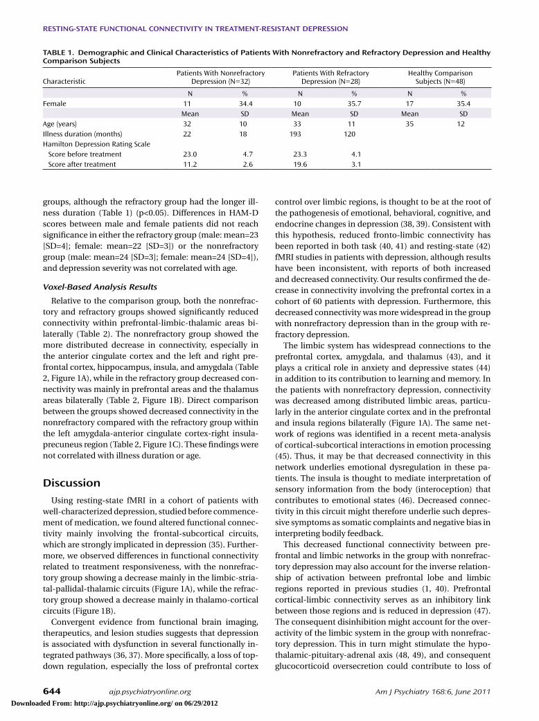
groups, although the refractory group had the longer ill-ness duration (Table 1) (p<0.05). Differences in HAM-D scores between male and female patients did not reach signifcance in either the refractory group (male: mean=23 [SD=4]; female: mean=22 [SD=3]) or the nonrefractory group (male: mean=24 [SD=3]; female: mean=24 [SD=4]), and depression severity was not correlated with age.
Voxel-Based Analysis Results
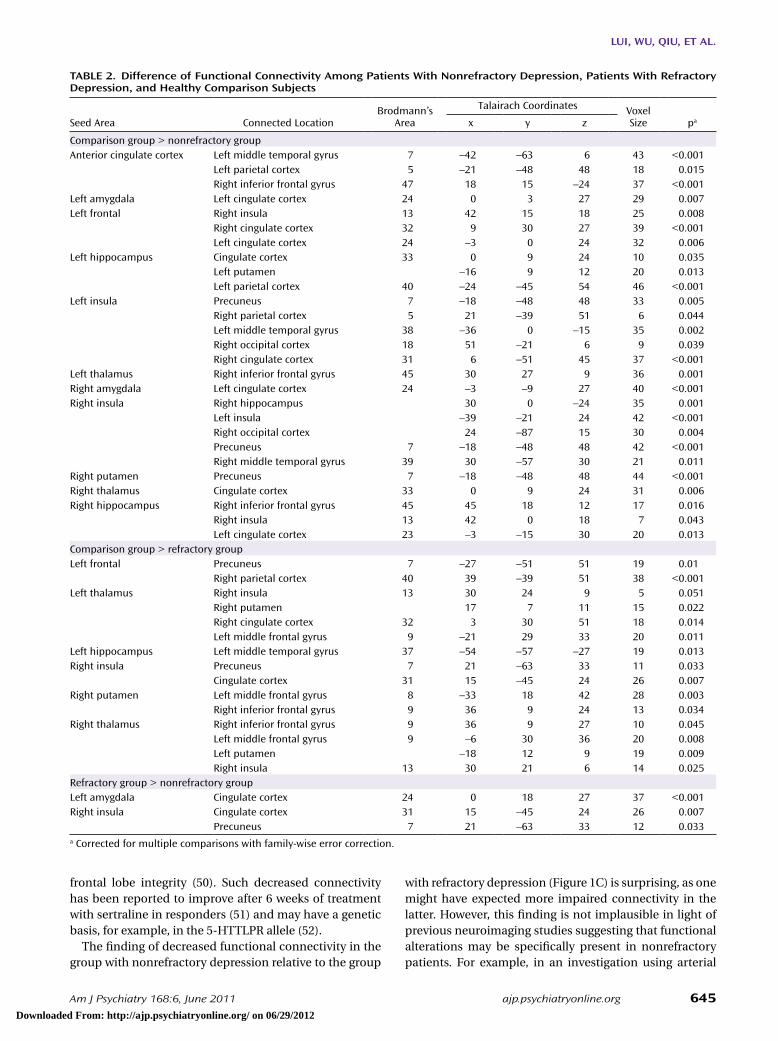
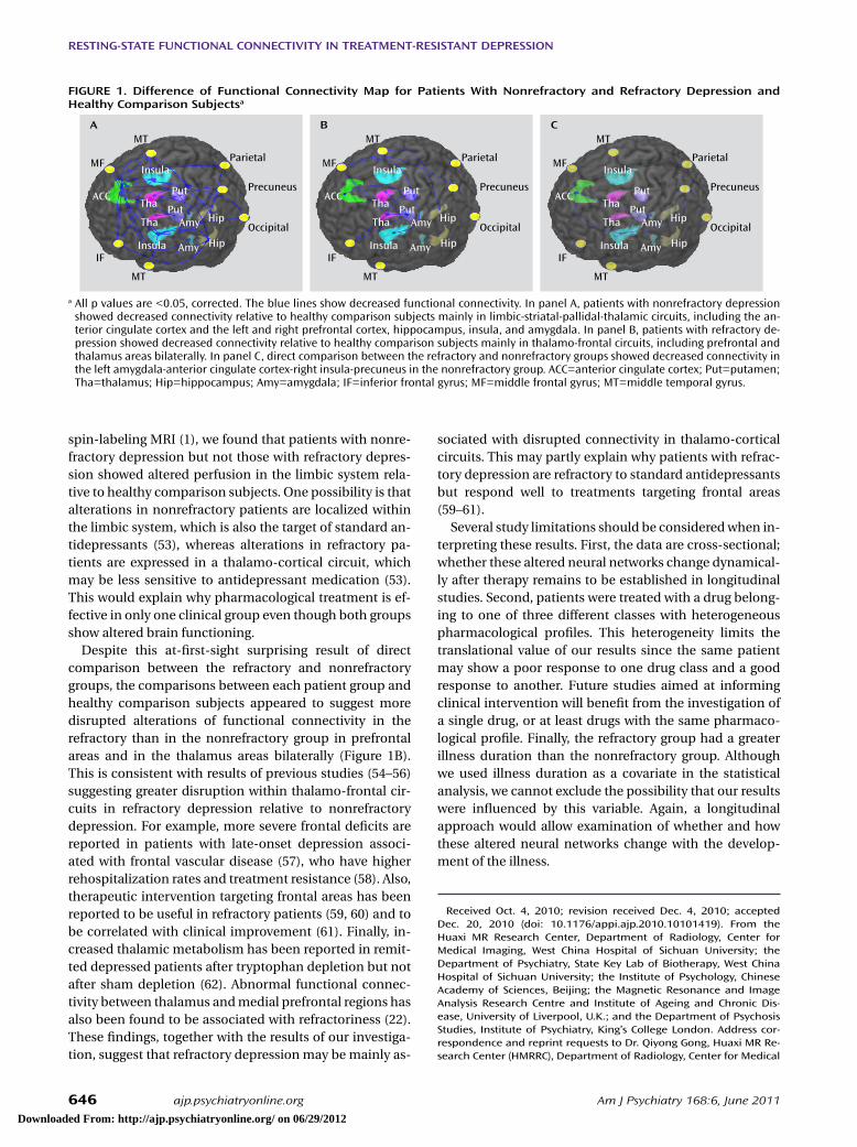
Relative to the comparison group, both the nonrefrac-tory and refractory groups showed signifcantly reduced connectivity within prefrontal-limbic-thalamic areas bi-laterally (Table 2). The nonrefractory group showed the more distributed decrease in connectivity, especially in the anterior cingulate cortex and the left and right pre-frontal cortex, hippocampus, insula, and amygdala (Table 2, Figure 1A), while in the refractory group decreased con-nectivity was mainly in prefrontal areas and the thalamus areas bilaterally (Table 2, Figure 1B). Direct comparison between the groups showed decreased connectivity in the nonrefractory compared with the refractory group within the left amygdala-anterior cingulate cortex-right insula-precuneus region (Table 2, Figure 1C). These fndings were not correlated with illness duration or age.
Discussion
Using resting-state fMRI in a cohort of patients with well-characterized depression, studied before commence-ment of medication, we found altered functional connec-tivity mainly involving the frontal-subcortical circuits, which are strongly implicated in depression (35). Further-more, we observed differences in functional connectivity related to treatment responsiveness, with the nonrefrac-tory group showing a decrease mainly in the limbic-stria-tal-pallidal-thalamic circuits (Figure 1A), while the refrac-tory group showed a decrease mainly in thalamo-cortical circuits (Figure 1B).
Convergent evidence from functional brain imaging, therapeutics, and lesion studies suggests that depression is associated with dysfunction in several functionally in-tegrated pathways (36, 37). More specifcally, a loss of top-down regulation, especially the loss of prefrontal cortex
tABle 1. Demographic and Clinical Characteristics of Patients With Nonrefractory and Refractory Depression and Healthy Comparison Subjects
CharacteristicPatients With Nonrefractory
Depression (N=32)Patients With Refractory
Depression (N=28)Healthy Comparison
Subjects (N=48)
N % N % N %Female 11 34.4 10 35.7 17 35.4
Mean SD Mean SD Mean SDAge (years) 32 10 33 11 35 12Illness duration (months) 22 18 193 120Hamilton Depression Rating Scale
Score before treatment 23.0 4.7 23.3 4.1Score after treatment 11.2 2.6 19.6 3.1
Downloaded From: http://ajp.psychiatryonline.org/ on 06/29/2012
lUI, WU, Q IU, et Al.
Am J Psychiatry 168:6, June 2011 ajp.psychiatryonline.org 645
with refractory depression (Figure 1C) is surprising, as one might have expected more impaired connectivity in the latter. However, this fnding is not implausible in light of previous neuroimaging studies suggesting that functional alterations may be specifcally present in nonrefractory patients. For example, in an investigation using arterial
frontal lobe integrity (50). Such decreased connectivity has been reported to improve after 6 weeks of treatment with sertraline in responders (51) and may have a genetic basis, for example, in the 5-HTTLPR allele (52).
The fnding of decreased functional connectivity in the group with nonrefractory depression relative to the group
tABle 2. Difference of Functional Connectivity Among Patients With Nonrefractory Depression, Patients With Refractory Depression, and Healthy Comparison Subjects
Brodmann’s Area
Talairach Coordinates Voxel SizeSeed Area Connected Location x y z pa
Comparison group > nonrefractory groupAnterior cingulate cortex Left middle temporal gyrus 7 –42 –63 6 43 <0.001
Left parietal cortex 5 –21 –48 48 18 0.015Right inferior frontal gyrus 47 18 15 –24 37 <0.001
Left amygdala Left cingulate cortex 24 0 3 27 29 0.007Left frontal Right insula 13 42 15 18 25 0.008
Right cingulate cortex 32 9 30 27 39 <0.001Left cingulate cortex 24 –3 0 24 32 0.006
Left hippocampus Cingulate cortex 33 0 9 24 10 0.035Left putamen –16 9 12 20 0.013Left parietal cortex 40 –24 –45 54 46 <0.001
Left insula Precuneus 7 –18 –48 48 33 0.005Right parietal cortex 5 21 –39 51 6 0.044Left middle temporal gyrus 38 –36 0 –15 35 0.002Right occipital cortex 18 51 –21 6 9 0.039Right cingulate cortex 31 6 –51 45 37 <0.001
Left thalamus Right inferior frontal gyrus 45 30 27 9 36 0.001Right amygdala Left cingulate cortex 24 –3 –9 27 40 <0.001Right insula Right hippocampus 30 0 –24 35 0.001
Left insula –39 –21 24 42 <0.001Right occipital cortex 24 –87 15 30 0.004Precuneus 7 –18 –48 48 42 <0.001Right middle temporal gyrus 39 30 –57 30 21 0.011
Right putamen Precuneus 7 –18 –48 48 44 <0.001Right thalamus Cingulate cortex 33 0 9 24 31 0.006Right hippocampus Right inferior frontal gyrus 45 45 18 12 17 0.016
Right insula 13 42 0 18 7 0.043Left cingulate cortex 23 –3 –15 30 20 0.013
Comparison group > refractory groupLeft frontal Precuneus 7 –27 –51 51 19 0.01
Right parietal cortex 40 39 –39 51 38 <0.001Left thalamus Right insula 13 30 24 9 5 0.051
Right putamen 17 7 11 15 0.022Right cingulate cortex 32 3 30 51 18 0.014Left middle frontal gyrus 9 –21 29 33 20 0.011
Left hippocampus Left middle temporal gyrus 37 –54 –57 –27 19 0.013Right insula Precuneus 7 21 –63 33 11 0.033
Cingulate cortex 31 15 –45 24 26 0.007Right putamen Left middle frontal gyrus 8 –33 18 42 28 0.003
Right inferior frontal gyrus 9 36 9 24 13 0.034Right thalamus Right inferior frontal gyrus 9 36 9 27 10 0.045
Left middle frontal gyrus 9 –6 30 36 20 0.008Left putamen –18 12 9 19 0.009Right insula 13 30 21 6 14 0.025
Refractory group > nonrefractory groupLeft amygdala Cingulate cortex 24 0 18 27 37 <0.001Right insula Cingulate cortex 31 15 –45 24 26 0.007
Precuneus 7 21 –63 33 12 0.033a Corrected for multiple comparisons with family-wise error correction.
Downloaded From: http://ajp.psychiatryonline.org/ on 06/29/2012
ReStING-StAte FUNCtIoNAl CoNNeCtIv ItY IN tReAtMeNt-ReSIStANt DePReSSIoN
646 ajp.psychiatryonline.org Am J Psychiatry 168:6, June 2011
sociated with disrupted connectivity in thalamo-cortical circuits. This may partly explain why patients with refrac-tory depression are refractory to standard antidepressants but respond well to treatments targeting frontal areas (59–61).
Several study limitations should be considered when in-terpreting these results. First, the data are cross-sectional; whether these altered neural networks change dynamical-ly after therapy remains to be established in longitudinal studies. Second, patients were treated with a drug belong-ing to one of three different classes with heterogeneous pharmacological profles. This heterogeneity limits the translational value of our results since the same patient may show a poor response to one drug class and a good response to another. Future studies aimed at informing clinical intervention will beneft from the investigation of a single drug, or at least drugs with the same pharmaco-logical profle. Finally, the refractory group had a greater illness duration than the nonrefractory group. Although we used illness duration as a covariate in the statistical analysis, we cannot exclude the possibility that our results were infuenced by this variable. Again, a longitudinal approach would allow examination of whether and how these altered neural networks change with the develop-ment of the illness.
Received Oct. 4, 2010; revision received Dec. 4, 2010; accepted Dec. 20, 2010 (doi: 10.1176/appi.ajp.2010.10101419). From the Huaxi M R Research Center, Department of Radiology, Center for M edical Imaging, West China Hospital of Sichuan University; the Department of Psychiatry, State Key Lab of Biotherapy, West China Hospital of Sichuan University; the Institute of Psychology, Chinese Academy of Sciences, Beijing; the M agnetic Resonance and Image Analysis Research Centre and Institute of Ageing and Chronic Dis-ease, University of Liverpool, U.K .; and the Department of Psychosis Studies, Institute of Psychiatry, K ing ’s College London. Address cor-respondence and reprint requests to Dr. Q iyong Gong, Huaxi M R Re-search Center (HM RRC), Department of Radiology, Center for M edical
spin-labeling MRI (1), we found that patients with nonre-fractory depression but not those with refractory depres-sion showed altered perfusion in the limbic system rela-tive to healthy comparison subjects. One possibility is that alterations in nonrefractory patients are localized within the limbic system, which is also the target of standard an-tidepressants (53), whereas alterations in refractory pa-tients are expressed in a thalamo-cortical circuit, which may be less sensitive to antidepressant medication (53). This would explain why pharmacological treatment is ef-fective in only one clinical group even though both groups show altered brain functioning.
Despite this at-frst-sight surprising result of direct comparison between the refractory and nonrefractory groups, the comparisons between each patient group and healthy comparison subjects appeared to suggest more disrupted alterations of functional connectivity in the refractory than in the nonrefractory group in prefrontal areas and in the thalamus areas bilaterally (Figure 1B). This is consistent with results of previous studies (54–56) suggesting greater disruption within thalamo-frontal cir-cuits in refractory depression relative to nonrefractory depression. For example, more severe frontal defcits are reported in patients with late-onset depression associ-ated with frontal vascular disease (57), who have higher rehospitalization rates and treatment resistance (58). Also, therapeutic intervention targeting frontal areas has been reported to be useful in refractory patients (59, 60) and to be correlated with clinical improvement (61). Finally, in-creased thalamic metabolism has been reported in remit-ted depressed patients after tryptophan depletion but not after sham depletion (62). Abnormal functional connec-tivity between thalamus and medial prefrontal regions has also been found to be associated with refractoriness (22). These fndings, together with the results of our investiga-tion, suggest that refractory depression may be mainly as-
FIGURe 1. Difference of Functional Connectivity Map for Patients With Nonrefractory and Refractory Depression and Healthy Comparison Subjectsa
MT
MF
ACC
IF
Insula
PutTha
Amy
Amy
Hip
Parietal
Occipital
Precuneus
MT
HipInsula
Tha
Put
MT
MF
ACC
IF
Insula
PutTha
Amy
Amy
Hip
Parietal
Occipital
Precuneus
MT
HipInsula
Tha
Put
MT
MF
ACC
IF
Insula
PutTha
Amy
Amy
Hip
Parietal
Occipital
Precuneus
MT
HipInsula
Tha
Put
A B C
a All p values are <0.05, corrected. The blue lines show decreased functional connectivity. In panel A, patients with nonrefractory depression showed decreased connectivity relative to healthy comparison subjects mainly in limbic-striatal-pallidal-thalamic circuits, including the an-terior cingulate cortex and the left and right prefrontal cortex, hippocampus, insula, and amygdala. In panel B, patients with refractory de-pression showed decreased connectivity relative to healthy comparison subjects mainly in thalamo-frontal circuits, including prefrontal and thalamus areas bilaterally. In panel C, direct comparison between the refractory and nonrefractory groups showed decreased connectivity in the left amygdala-anterior cingulate cortex-right insula-precuneus in the nonrefractory group. ACC=anterior cingulate cortex; Put=putamen; Tha=thalamus; Hip=hippocampus; Amy=amygdala; IF=inferior frontal gyrus; MF=middle frontal gyrus; MT=middle temporal gyrus.
Downloaded From: http://ajp.psychiatryonline.org/ on 06/29/2012
lUI, WU, Q IU, et Al.
Am J Psychiatry 168:6, June 2011 ajp.psychiatryonline.org 647
and the medial prefrontal cortex activity. J Affect Disord 2010; 122:76–85
15. Bluhm R, Williamson P, Lanius R, Theberge J, Densmore M, Bar-tha R, Neufeld R, Osuch E: Resting state default-mode network connectivity in early depression using a seed region-of-interest analysis: decreased connectivity with caudate nucleus. Psychi-atry Clin Neurosci 2009; 63:754–761
16. Greicius MD, Srivastava G, Reiss AL, Menon V: Default-mode network activity distinguishes Alzheimer’s disease from healthy aging: evidence from functional MRI. Proc Natl Acad Sci USA 2004; 101:4637–4642
17. Lui S, Deng W, Huang X, Jiang L, Ma X, Chen H, Zhang T, Li X, Li D, Zou L, Tang H, Zhou XJ, Mechelli A, Collier DA, Sweeney JA, Li T, Gong Q: Association of cerebral defcits with clinical symp-toms in antipsychotic-naive frst-episode schizophrenia: an op-timized voxel-based morphometry and resting state functional connectivity study. Am J Psychiatry 2009; 166:196–205
18. Lui S, Huang X, Chen L, Tang H, Zhang T, Li X, Li D, Kuang W, Chan RC, Mechelli A, Sweeney JA, Gong Q: High-feld MRI reveals an acute impact on brain function in survivors of the magnitude 8.0 earthquake in China. Proc Natl Acad Sci USA 2009; 106:15412–15417
19. Fox MD, Greicius M: Clinical applications of resting state func-tional connectivity. Front Syst Neurosci 2010; 4:19
20. Raichle ME, Mintun MA: Brain work and brain imaging. Annu Rev Neurosci 2006; 29:449–476
21. Cordes D, Haughton VM, Arfanakis K, Carew JD, Turski PA, Moritz CH, Quigley MA, Meyerand ME: Frequencies contributing to func-tional connectivity in the cerebral cortex in “resting-state” data. AJNR Am J Neuroradiol 2001; 22:1326–1333
22. Greicius MD, Flores BH, Menon V, Glover GH, Solvason HB, Ken-na H, Reiss AL, Schatzberg AF: Resting-state functional connec-tivity in major depression: abnormally increased contributions from subgenual cingulate cortex and thalamus. Biol Psychiatry 2007; 62:429–437
23. First MB, Spitzer RL, Gibbon M, Williams JBW: Structured Clini-cal Interview for DSM-IV Axis I Disorders (SCID). Washington, DC, American Psychiatric Press, 1997
24. Williams JBW: A structured interview guide for the Hamilton Depression Rating Scale. Arch Gen Psychiatry 1988; 45:742–747
25. Guy W (ed): ECDEU Assessment Manual for Psychopharmacol-ogy: Publication ADM 76-338. Washington, DC, US Department of Health, Education, and Welfare, 1976, pp 218–222
26. World Psychiatric Association: Symposium on Therapy-Resis-tant Depression. Pharmacopsychiatry 1974; 7:69–224
27. Berlim MT, Turecki G: Defnition, assessment, and staging of treatment-resistant refractory major depression: a review of current concepts and methods. Can J Psychiatry 2007; 52:46–54
28. Horwitz B, Rumsey JM, Donohue BC: Functional connectivity of the angular gyrus in normal reading and dyslexia. Proc Natl Acad Sci USA 1998; 95:8939–8944
29. Friston KJ: Functional and effective connectivity in neuroimag-ing: a synthesis. Hum Brain Mapp 1994; 2:56–57
30. Lancaster JL, Woldorff MG, Parsons LM, Liotti M, Freitas CS, Rainey L, Kochunov PV, Nickerson D, Mikiten SA, Fox PT: Au-tomated Talairach atlas labels for functional brain mapping. Hum Brain Mapp 2000; 10:120–131
31. Maldjian JA, Laurienti PJ, Kraft RA, Burdette JH: An automated method for neuroanatomic and cytoarchitectonic atlas-based interrogation of fMRI data sets. Neuroimage 2003; 19:1233–1239
32. Dagli MS, Ingeholm JE, Haxby JV: Localization of cardiac-in-duced signal change in fMRI. Neuroimage 1999; 9:407–415
33. Windischberger C, Langenberger H, Sycha T, Tschernko EM, Fuchsjager-Mayerl G, Schmetterer L, Moser E: On the origin of respiratory artifacts in BOLD-EPI of the human brain. Magn Reson Imaging 2002; 20:575–582
Imaging, West China Hospital of Sichuan University, Chengdu, China; [email protected] .cn (e-mail).
All authors report no fnancial relationships w ith commercial in-terests.
Supported by grants 81030027, 30625024, and 30900361 from the National Natural Science Foundation of China and grant 2007CB512305/2 from the 973 Program of the National Research Program of China.
References
1. Lui S, Parkes LM, Huang X, Zou K, Chan RC, Yang H, Zou L, Li D, Tang H, Zhang T, Li X, Wei Y, Chen L, Sun X, Kemp GJ, Gong QY: Depressive disorders: focally altered cerebral perfusion mea-sured with arterial spin-labeling MR imaging. Radiology 2009; 251:476–484
2. Bench CJ, Friston KJ, Brown RG, Scott LC, Frackowiak RS, Dolan RJ: The anatomy of melancholia: focal abnormalities of ce-rebral blood fow in major depression. Psychol Med 1992; 22:607–615
3. de Asis JM, Stern E, Alexopoulos GS, Pan H, Van Gorp W, Blum-berg H, Kalayam B, Eidelberg D, Kiosses D, Silbersweig DA: Hip-pocampal and anterior cingulate activation defcits in patients with geriatric depression. Am J Psychiatry 2001; 158:1321–1323
4. Saxena S, Brody AL, Ho ML, Alborzian S, Ho MK, Maidment KM, Huang SC, Wu HM, Au SC, Baxter LR Jr: Cerebral metabo-lism in major depression and obsessive-compulsive disorder occurring separately and concurrently. Biol Psychiatry 2001; 50:159–170
5. Ebmeier KP, Cavanagh JT, Moffoot AP, Glabus MF, O’Carroll RE, Goodwin GM: Cerebral perfusion correlates of depressed mood. Br J Psychiatry 1997; 170:77–81
6. Bielau H, Trubner K, Krell D, Agelink MW, Bernstein HG, Stauch R, Mawrin C, Danos P, Gerhard L, Bogerts B, Baumann B: Volume defcits of subcortical nuclei in mood disorders: a postmortem study. Eur Arch Psychiatry Clin Neurosci 2005; 255:401–412
7. Campbell S, Marriott M, Nahmias C, MacQueen GM: Lower hippocampal volume in patients suffering from depression: a meta-analysis. Am J Psychiatry 2004; 161:598–607
8. Coryell W, Nopoulos P, Drevets W, Wilson T, Andreasen NC: Sub-genual prefrontal cortex volumes in major depressive disorder and schizophrenia: diagnostic specifcity and prognostic impli-cations. Am J Psychiatry 2005; 162:1706–1712
9. Kim MJ, Hamilton JP, Gotlib IH: Reduced caudate gray matter volume in women with major depressive disorder. Psychiatry Res 2008; 164:114–122
10. Stimpson N, Agrawal N, Lewis G: Randomised controlled trials investigating pharmacological and psychological interventions for treatment-refractory depression: systematic review. Br J Psychiatry 2002; 181:284–294
11. Friedel E, Schlagenhauf F, Sterzer P, Park SQ, Bermpohl F, Strohle A, Stoy M, Puls I, Hagele C, Wrase J, Buchel C, Heinz A: 5-HTT genotype effect on prefrontal-amygdala coupling differs between major depression and controls. Psychopharmacology (Berl) 2009; 205:261–271
12. Harrison NA, Brydon L, Walker C, Gray MA, Steptoe A, Critchley HD: Infammation causes mood changes through alterations in subgenual cingulate activity and mesolimbic connectivity. Biol Psychiatry 2009; 66:407–414
13. Holmes AJ, Pizzagalli DA: Spatiotemporal dynamics of error processing dysfunctions in major depressive disorder. Arch Gen Psychiatry 2008; 65:179–188
14. Yoshimura S, Okamoto Y, Onoda K, Matsunaga M, Ueda K, Su-zuki S, Yamawaki S: Rostral anterior cingulate cortex activity mediates the relationship between the depressive symptoms
Downloaded From: http://ajp.psychiatryonline.org/ on 06/29/2012
ReStING-StAte FUNCtIoNAl CoNNeCtIv ItY IN tReAtMeNt-ReSIStANt DePReSSIoN
648 ajp.psychiatryonline.org Am J Psychiatry 168:6, June 2011
dependent behavioral effects in nonhuman primates. Proc Natl Acad Sci USA 2002; 99:15749–15754
50. Gold SM, Dziobek I, Rogers K, Bayoumy A, McHugh PF, Convit A: Hypertension and hypothalamo-pituitary-adrenal axis hy-peractivity affect frontal lobe integrity. J Clin Endocrinol Metab 2005; 90:3262–3267
51. Anand A, Li Y, Wang Y, Wu J, Gao S, Bukhari L, Mathews VP, Kalnin A, Lowe MJ: Antidepressant effect on connectivity of the mood-regulating circuit: an fMRI study. Neuropsychopharma-cology 2005; 30:1334–1344
52. Pezawas L, Meyer-Lindenberg A, Drabant EM, Verchinski BA, Munoz KE, Kolachana BS, Egan MF, Mattay VS, Hariri AR, Wein-berger DR: 5-HTTLPR polymorphism impacts human cingu-late-amygdala interactions: a genetic susceptibility mechanism for depression. Nat Neurosci 2005; 8:828–834
53. Lopez JF, Chalmers DT, Little KY, Watson SJ: Regulation of sero-tonin
1A, glucocorticoid, and mineralocorticoid receptor in rat
and human hippocampus: implications for the neurobiology of depression. Biol Psychiatry 1998; 43:547–573
54. Clark CP, Brown GG, Archibald SL, Fennema-Notestine C, Braun DR, Thomas LS, Sutherland AN, Gillin JC: Does amygdalar perfu-sion correlate with antidepressant response to partial sleep de-privation in major depression? Psychiatry Res 2006; 146:43–51
55. Clark CP, Brown GG, Frank L, Thomas L, Sutherland AN, Gil-lin JC: Improved anatomic delineation of the antidepressant response to partial sleep deprivation in medial frontal cortex using perfusion-weighted functional MRI. Psychiatry Res 2006; 146:213–222
56. Kohn Y, Freedman N, Lester H, Krausz Y, Chisin R, Lerer B, Bonne O: 99mTc-HMPAO SPECT study of cerebral perfusion af-ter treatment with medication and electroconvulsive therapy in major depression. J Nucl Med 2007; 48:1273–1278
57. Matsuo K, Onodera Y, Hamamoto T, Muraki K, Kato N, Kato T: Hypofrontality and microvascular dysregulation in remit-ted late-onset depression assessed by functional near-infrared spectroscopy. Neuroimage 2005; 26:234–242
58. Fujikawa T, Yokota N, Muraoka M, Yamawaki S: Response of patients with major depression and silent cerebral infarction to antidepressant drug therapy, with emphasis on central ner-vous system adverse reactions. Stroke 1996; 27:2040–2042
59. Fregni F, Ono CR, Santos CM, Bermpohl F, Buchpiguel C, Bar-bosa ER, Marcolin MA, Pascual-Leone A, Valente KD: Effects of antidepressant treatment with rTMS and fuoxetine on brain perfusion in PD. Neurology 2006; 66:1629–1637
60. Isenberg K, Downs D, Pierce K, Svarakic D, Garcia K, Jarvis M, North C, Kormos TC: Low frequency rTMS stimulation of the right frontal cortex is as effective as high frequency rTMS stimulation of the left frontal cortex for antidepressant-free, treatment-resistant depressed patients. Ann Clin Psychiatry 2005; 17:153–159
61. Dougherty DD, Weiss AP, Cosgrove GR, Alpert NM, Cassem EH, Nierenberg AA, Price BH, Mayberg HS, Fischman AJ, Rauch SL: Cerebral metabolic correlates as potential predictors of re-sponse to anterior cingulotomy for treatment of major depres-sion. J Neurosurg 2003; 99:1010–1017
62. Neumeister A, Nugent AC, Waldeck T, Geraci M, Schwarz M, Bonne O, Bain EE, Luckenbaugh DA, Herscovitch P, Charney DS, Drevets WC: Neural and behavioral responses to tryptophan de-pletion in unmedicated patients with remitted major depressive disorder and controls. Arch Gen Psychiatry 2004; 61:765–773
34. Worsley KJ: An improved theoretical p value for SPMs based on discrete local maxima. Neuroimage 2005; 28:1056–1062
35. Drevets WC, Raichle ME: Neuroanatomical circuits in depres-sion: implications for treatment mechanisms. Psychopharma-col Bull 1992; 28:261–274
36. Anand A, Li Y, Wang Y, Wu J, Gao S, Bukhari L, Mathews VP, Kal-nin A, Lowe MJ: Activity and connectivity of brain mood regu-lating circuit in depression: a functional magnetic resonance study. Biol Psychiatry 2005; 57:1079–1088
37. Mayberg HS: Modulating dysfunctional limbic-cortical circuits in depression: towards development of brain-based algo-rithms for diagnosis and optimised treatment. Br Med Bull 2003; 65:193–207
38. Savitz JB, Drevets WC: Imaging phenotypes of major depressive disorder: genetic correlates. Neuroscience 2009; 164:300–330
39. Savitz J, Drevets WC: Bipolar and major depressive disorder: neuroimaging the developmental-degenerative divide. Neuro-sci Biobehav Rev 2009; 33:699–771
40. Siegle GJ, Steinhauer SR, Thase ME, Stenger VA, Carter CS: Can’t shake that feeling: event-related fMRI assessment of sustained amygdala activity in response to emotional information in de-pressed individuals. Biol Psychiatry 2002; 51:693–707
41. Bhagwagar Z, Wylezinska M, Jezzard P, Evans J, Boorman E, Matthews PM, Cowen PJ: Low GABA concentrations in occipital cortex and anterior cingulate cortex in medication-free, recov-ered depressed patients. Int J Neuropsychopharmacol 2008; 11:255–260
42. Anand A, Li Y, Wang Y, Lowe MJ, Dzemidzic M: Resting state corticolimbic connectivity abnormalities in unmedicated bi-polar disorder and unipolar depression. Psychiatry Res 2009; 171:189–198
43. Frodl T, Meisenzahl EM, Zetzsche T, Born C, Groll C, Jäger M, Leinsinger G, Bottlender R, Hahn K, Möller H-J: Hippocampal changes in patients with a frst episode of major depression. Am J Psychiatry 2002; 159:1112–1118
44. Fountoulakis KN, Iacovides A, Gerasimou G, Fotiou F, Ioanni-dou C, Bascialla F, Grammaticos P, Kaprinis G: The relationship of regional cerebral blood fow with subtypes of major de-pression. Prog Neuropsychopharmacol Biol Psychiatry 2004; 28:537–546
45. Kober H, Barrett LF, Joseph J, Bliss-Moreau E, Lindquist K, Wa-ger TD: Functional grouping and cortical-subcortical interac-tions in emotion: a meta-analysis of neuroimaging studies. Neuroimage 2008; 42:998–1031
46. Craig AD: How do you feel? interoception: the sense of the physiological condition of the body. Nat Rev Neurosci 2002; 3:655–666
47. Dannlowski U, Ohrmann P, Konrad C, Domschke K, Bauer J, Ku-gel H, Hohoff C, Schoning S, Kersting A, Baune BT, Mortensen LS, Arolt V, Zwitserlood P, Deckert J, Heindel W, Suslow T: Re-duced amygdala-prefrontal coupling in major depression: as-sociation with MAOA genotype and illness severity. Int J Neuro-psychopharmacol 2009; 12:11–22
48. Sanchez MM, Young LJ, Plotsky PM, Insel TR: Autoradiographic and in situ hybridization localization of corticotropin-releasing factor 1 and 2 receptors in nonhuman primate brain. J Comp Neurol 1999; 408:365–377
49. Strome EM, Wheler GH, Higley JD, Loriaux DL, Suomi SJ, Dou-det DJ: Intracerebroventricular corticotropin-releasing factor increases limbic glucose metabolism and has social context-
Downloaded From: http://ajp.psychiatryonline.org/ on 06/29/2012