Application of positron emission tomography in psychiatry
11
REVIEW ARTICLE OPEN Application of positron emission tomography in psychiatry— methodological developments and future directions Simon Cervenka 1,2 ✉ , Andreas Frick 1 , Robert Bodén 1 and Mark Lubberink 3 © The Author(s) 2022 Mental disorders represent an increasing source of disability and high costs for societies globally. Molecular imaging techniques such as positron emission tomography (PET) represent powerful tools with the potential to advance knowledge regarding disease mechanisms, allowing the development of new treatment approaches. Thus far, most PET research on pathophysiology in psychiatric disorders has focused on the monoaminergic neurotransmission systems, and although a series of discoveries have been made, the results have not led to any material changes in clinical practice. We outline areas of methodological development that can address some of the important obstacles to fruitful progress. First, we point towards new radioligands and targets that can lead to the identification of processes upstream, or parallel to disturbances in monoaminergic systems. Second, we describe the development of new methods of PET data quantification and PET systems that may facilitate research in psychiatric populations. Third, we review the application of multimodal imaging that can link molecular imaging data to other aspects of brain function, thus deepening our understanding of disease processes. Fourth, we highlight the need to develop imaging study protocols to include longitudinal and interventional paradigms, as well as frameworks to assess dimensional symptoms such that the field can move beyond cross-sectional studies within current diagnostic boundaries. Particular effort should be paid to include also the most severely ill patients. Finally, we discuss the importance of harmonizing data collection and promoting data sharing to reach the desired sample sizes needed to fully capture the phenotype of psychiatric conditions. Translational Psychiatry (2022)12:248 ; https://doi.org/10.1038/s41398-022-01990-2 BACKGROUND In a given year, one out of six individuals in Europe will suffer from a mental health problem, and it is estimated that the total cost to European societies for these disorders is 600 billion euros [1]. Recovery rates for schizophrenia have not improved for several decades [2], and similarly the global disease burden of depressive and anxiety disorders, both ranked among the top 25 leading causes of burden, has not diminished since 1990 [3]. Importantly, the identification of new treatment approaches that could improve this situation is hampered by a lack of understanding of the pathophysiology of mental disorders. Molecular imaging methods such as positron emission tomography (PET) offer a unique opportunity to measure brain biochemical markers in vivo, and have since their advent in the 1980:s been considered a key approach to advance the field. Psychiatry was indeed an early focus in the field of quantitative PET, with the very first clinical studies being conducted in patients with schizophrenia. Demonstrations of the antipsychotic mechan- ism of action using dopamine D2 receptor (D2-R) radioligands such as [ 11 C]raclopride was followed by a series of occupancy studies outlining thresholds for treatment efficacy and side effects [4, 5]. Arguably, the resulting change in dosing regimens remains the most significant contribution of PET research to clinical psychiatry to date. Since then, a wealth of studies have attempted to investigate the pathophysiology of psychiatric disorders, mainly based on disease models involving monoamine neurotransmis- sion systems. To name a few examples, studies of dopaminergic function in psychosis and schizophrenia have collectively shown increased dopamine synthesis and release capacity as indexed by increased [ 18 F]DOPA uptake and more pronounced amphetamine- induced decreases in D2-R receptor binding, a small increase in striatal D2-R availability and lower D2-R availability in the thalamus [6, 7]. In major depressive disorder (MDD), findings point to reduced serotonin transporter [8] and serotonin 1A receptor (5HT1A-R) availability [9], whereas in patients with anxiety disorders the evidence suggests decreases in 5HT1A and benzodiazepine receptors [10]. Several new radioligands for monoaminergic neurotransmission targets have been developed in recent years, such as tracers with increased sensitivity for endogenous neurotransmitter levels [11–14], that may further advance our understanding of the involvement of these systems. However, thus far this line of research has not led to new diagnostic tools or treatments for clinical use, prompting research based on new models of disease pathophysiology. In order to advance PET research to the point of improving the care of psychiatric patients, some important challenges remain. First, there is a need to identify disease-related processes that act upstream or parallel to the observed abnormalities in Received: 22 December 2021 Revised: 20 May 2022 Accepted: 25 May 2022 1 Department of Medical Sciences, Psychiatry, Uppsala University, Uppsala, Sweden. 2 Centre for Psychiatry Research, Department of Clinical Neuroscience, Karolinska Institutet and Stockholm Health Care Services, Region Stockholm, Stockholm, Sweden. 3 Department of Surgical Sciences, Uppsala University, Uppsala, Sweden. ✉ email: [email protected] www.nature.com/tp Translational Psychiatry 1234567890();,:
-
Upload
khangminh22 -
Category
Documents
-
view
4 -
download
0
Transcript of Application of positron emission tomography in psychiatry

REVIEW ARTICLE OPEN
Application of positron emission tomography in psychiatry—methodological developments and future directionsSimon Cervenka 1,2✉, Andreas Frick 1, Robert Bodén 1 and Mark Lubberink3
© The Author(s) 2022
Mental disorders represent an increasing source of disability and high costs for societies globally. Molecular imaging techniquessuch as positron emission tomography (PET) represent powerful tools with the potential to advance knowledge regarding diseasemechanisms, allowing the development of new treatment approaches. Thus far, most PET research on pathophysiology inpsychiatric disorders has focused on the monoaminergic neurotransmission systems, and although a series of discoveries havebeen made, the results have not led to any material changes in clinical practice. We outline areas of methodological developmentthat can address some of the important obstacles to fruitful progress. First, we point towards new radioligands and targets that canlead to the identification of processes upstream, or parallel to disturbances in monoaminergic systems. Second, we describe thedevelopment of new methods of PET data quantification and PET systems that may facilitate research in psychiatric populations.Third, we review the application of multimodal imaging that can link molecular imaging data to other aspects of brain function,thus deepening our understanding of disease processes. Fourth, we highlight the need to develop imaging study protocols toinclude longitudinal and interventional paradigms, as well as frameworks to assess dimensional symptoms such that the field canmove beyond cross-sectional studies within current diagnostic boundaries. Particular effort should be paid to include also the mostseverely ill patients. Finally, we discuss the importance of harmonizing data collection and promoting data sharing to reach thedesired sample sizes needed to fully capture the phenotype of psychiatric conditions.
Translational Psychiatry (2022) 12:248 ; https://doi.org/10.1038/s41398-022-01990-2
BACKGROUNDIn a given year, one out of six individuals in Europe will suffer froma mental health problem, and it is estimated that the total cost toEuropean societies for these disorders is 600 billion euros [1].Recovery rates for schizophrenia have not improved for severaldecades [2], and similarly the global disease burden of depressiveand anxiety disorders, both ranked among the top 25 leadingcauses of burden, has not diminished since 1990 [3]. Importantly,the identification of new treatment approaches that couldimprove this situation is hampered by a lack of understandingof the pathophysiology of mental disorders. Molecular imagingmethods such as positron emission tomography (PET) offer aunique opportunity to measure brain biochemical markers in vivo,and have since their advent in the 1980:s been considered a keyapproach to advance the field.Psychiatry was indeed an early focus in the field of quantitative
PET, with the very first clinical studies being conducted in patientswith schizophrenia. Demonstrations of the antipsychotic mechan-ism of action using dopamine D2 receptor (D2-R) radioligandssuch as [11C]raclopride was followed by a series of occupancystudies outlining thresholds for treatment efficacy and side effects[4, 5]. Arguably, the resulting change in dosing regimens remainsthe most significant contribution of PET research to clinicalpsychiatry to date. Since then, a wealth of studies have attempted
to investigate the pathophysiology of psychiatric disorders, mainlybased on disease models involving monoamine neurotransmis-sion systems. To name a few examples, studies of dopaminergicfunction in psychosis and schizophrenia have collectively shownincreased dopamine synthesis and release capacity as indexed byincreased [18F]DOPA uptake and more pronounced amphetamine-induced decreases in D2-R receptor binding, a small increase instriatal D2-R availability and lower D2-R availability in the thalamus[6, 7]. In major depressive disorder (MDD), findings point toreduced serotonin transporter [8] and serotonin 1A receptor(5HT1A-R) availability [9], whereas in patients with anxietydisorders the evidence suggests decreases in 5HT1A andbenzodiazepine receptors [10]. Several new radioligands formonoaminergic neurotransmission targets have been developedin recent years, such as tracers with increased sensitivity forendogenous neurotransmitter levels [11–14], that may furtheradvance our understanding of the involvement of these systems.However, thus far this line of research has not led to newdiagnostic tools or treatments for clinical use, prompting researchbased on new models of disease pathophysiology.In order to advance PET research to the point of improving the
care of psychiatric patients, some important challenges remain.First, there is a need to identify disease-related processes that actupstream or parallel to the observed abnormalities in
Received: 22 December 2021 Revised: 20 May 2022 Accepted: 25 May 2022
1Department of Medical Sciences, Psychiatry, Uppsala University, Uppsala, Sweden. 2Centre for Psychiatry Research, Department of Clinical Neuroscience, Karolinska Institutetand Stockholm Health Care Services, Region Stockholm, Stockholm, Sweden. 3Department of Surgical Sciences, Uppsala University, Uppsala, Sweden.✉email: [email protected]
www.nature.com/tpTranslational Psychiatry
1234567890();,:

monoaminergic dysfunction. To accomplish this, radioligands fornew targets need to be developed, and a wider methodologicalscope is required to outline molecular pathways from geneticsand environmental factors to the dysfunctional informationprocessing that underlies psychiatric symptoms. Second, psychia-tric populations are characterized by considerable heterogeneityin clinical phenotype—likely corresponding to heterogeneity inpathophysiological mechanisms. It is generally acknowledged thatcurrent diagnostic boundaries are ill-matched to the underlyingbiology, presenting a critical challenge to advancing knowledge[15]. Third, psychiatric patients are a vulnerable group, calling forthe simplification of clinical research protocols. The most severelyill patients are typically not included in research studies at all,owing to difficulties in adhering to cumbersome study proceduresor not being able to provide informed consent, limiting ourunderstanding of the full phenotype associated with mentaldisorders.In this review, we aim to describe recent advancements in
methodological development in PET that may address thesechallenges, and to what extent they have been applied in clinicalpsychiatric research.
New targets and radioligandsA key bottleneck in clinical PET research in general is theavailability of radioligands for targets of interest. In the following,we highlight areas of development with specific relevance topsychiatric disorders.
Intracellular targetsOne approach to go beyond the level of neurotransmissionsignalling is to target intracellular enzymes in neurons [16–19].This approach allows for investigating the activation state ofgroups of neurons during a certain condition, and may alsoelucidate if a dysfunction of that specific intracellular mechanismis part of the underlying pathophysiology of specific disorders[20]. Examples of this strategy are studies of the cyclic nucleotidephosphodiesterase (PDE) family of enzymes, which metabolizecyclic nucleotides and thereby regulate the signalling of thesesecond messenger systems. PDE10A has been of recent interest inschizophrenia research [17,18]. In the brain, PDE10A is highlylocalized to the striatum, where it regulates the output of thedirect and indirect striatal pathways that are implicated in severalpsychiatric conditions. PDE10A radioligands have thus far onlybeen used in small samples of schizophrenia patients, showinginconclusive results [17,18].Another enzyme in the PDE family, PDE4, is the main enzyme in
the brain responsible for metabolizing 3′,5′-monophosphate(cAMP) into its inactive state. The cAMP signalling pathway is amajor second messenger pathway of G-coupled receptors,involved in a range of intracellular processes including propagatingneuron-neuron signals, and is hence of high relevance for manypsychiatric conditions. The radioligand [11C]rolipram, a nonselec-tive PDE4 antagonist, was used to show reduced cAMP activity inMDD compared to controls, and an upregulation after selectiveserotonin reuptake inhibitor (SSRI) treatment [19]. Indeed, PDE4inhibitors have been suggested as a treatment in psychiatricconditions based on positive effects on neuroplasticity andneuroinflammation, but adverse effects hamper their implementa-tion [21]. More selective targeting of PDE4 subtypes (i.e. PDE4 A/B/C/D) and increased understanding of the regulation of conforma-tional states may be keys to overcome the adverse effects. In thisrespect, recent efforts have been made to develop radioligandswith higher specificity for certain PDE4 subtypes [22], but thesehave yet to be applied in MDD or other psychiatric conditions.
Immune markersGenetic and epidemiologic data support a role for the immunesystem in psychotic disorders, depression and anxiety disorders as
well as neurodevelopmental disorders. During the last decade,intensive efforts have aimed to identify PET markers fordysregulated immune function which for instance could be usedto stratify patients for treatments targeting the immune system.To date, the most established method is to target the 18kDtranslocator protein (TSPO), which is expressed in activatedmicroglia and astrocytes. Whereas initial studies in schizophreniausing the first generation TSPO radioligand [11C]PK-11195 showedincreased binding in patients, this could not be confirmed inensuing studies using TSPO radioligands with increased sensitivitysuch as [11C]PBR28 and [18F]FEPPA. Instead, data has convergedtowards lower binding across several brain regions [23, 24]. Incontrast, six out of seven TSPO studies in MDD have shownincreased binding compared with controls [24].TSPO is not specific to microglia [25, 26], and in vitro studies
have shown conflicting results regarding the specificity of TSPOfor pro-inflammatory cells [27, 28]. There are currently severalradioligands under investigation to find more specific markers forneuroinflammatory processes. Radioligands such as [11C]deprenyland [¹¹C]SL25.1188, targeting the enzyme MAO-B, have beenconsidered markers of astroglial activation. The latter radioligandhas been used to show increased binding in the prefrontal cortexin patients with MDD [29], whereas a small study found nodifference in binding in post-traumatic stress disorder (PTSD)patients [30]. With regard to microglial and proinflammatorymarkers, some promising candidates have been developed forCox-1 and Cox-2, CSF1R and P2X7 [24], but to our knowledge,none of these have so farbeen used to study psychiatric disorders.
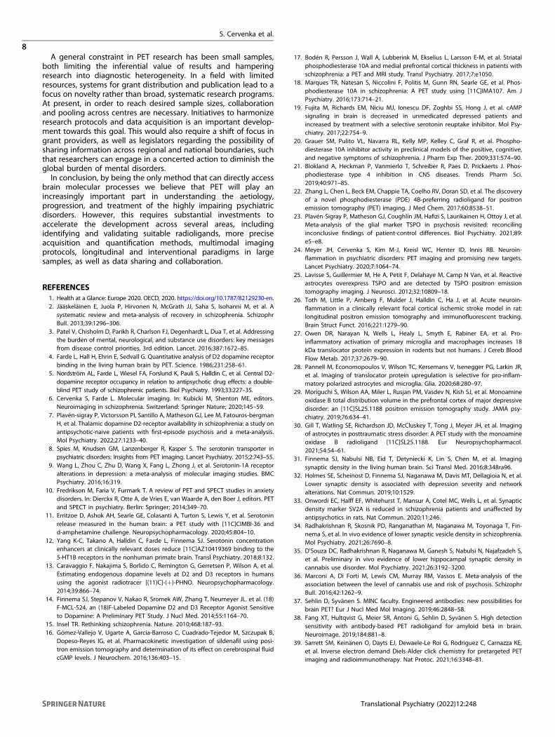
Synaptic density. A wealth of literature supports aberrantconnectivity in several psychiatric conditions, as indicatedindirectly using functional magnetic resonance imaging (MRI)modalities and post-mortem studies. In order to investigateconnectivity at the molecular level, there has been an increasinginterest in identifying targets for synaptic density. The PETradioligand [11C]UCB-J binds to the synaptic vesicle glycoprotein2A (SV2A), which has shown high correspondence to theestablished post-mortem marker synaptophysin in non-humanprimate studies [31]. [11C]UCB-J has been applied in patients withMDD and/or PTSD, showing an inverse correlation betweendepressive symptoms and SV2A density, as well as an associationbetween SV2A density and functional connectivity in prefrontalcortex as measured using resting-state functional MR (fMRI) [32].The radioligand has been used in medicated patients with chronicschizophrenia, showing decreases in SV2A binding across severalcortical regions [33, 34]. Recently, a small study found decreasedSV2A binding in the hippocampus in users of cannabis [35], a drugconsidered to be a major risk factor for schizophrenia [36]. Animportant aim following the observations in MDD and schizo-phrenia is to investigate if changes in synaptic density are presentalready in the early stages of psychiatric disorders (Fig. 1).
Future developments. PET neuroreceptor ligands are generallybased on small, lipophilic molecules that can easily cross theblood-brain barrier (BBB). In oncological PET, the use of labelledantibodies with high specificity and affinity has long been ofinterest. To enable brain uptake sufficient for imaging, recentdevelopments have shown the possibility of designing bi-specificantibodies that undergo active transport across the BBB bytransferrin-receptor-mediated transcytosis visualizing, e.g.,amyloid-beta [37]. Although these methods have only beendemonstrated in small-animal studies at this point [38] and havebeen developed primarily for application in neurodegenerativedisorders, there could be potential applications in psychiatry giventhe interest in immune-related disease mechanisms. A limitationwith labelled antibodies is their slow clearance from the bloodwhich may require long-lived isotopes such as 79Zr for labelling,resulting in high radiation doses. A novel approach to overcome
S. Cervenka et al.
2
Translational Psychiatry (2022) 12:248

this is so-called pre-targeting using bioorthogonal chemistry, i.e.,administration of an intermediate compound such as an antibodythat binds to the target of interest, which can in turn be targetedby small molecules labelled with short-lived PET isotopes after thecirculating antibody has cleared [39]. Hence, the antibody islabelled after it has bound to its target in the body and clearedfrom the blood, improving image contrast and reducing radiationdose compared to direct imaging of labelled antibodies. Achallenge with this approach that will need to be solved is howto measure target availability, as the PET signal will be dependenton the kinetics of both the antibody and the tracer. If applied topsychiatry, his approach could potentially also be used to increasethe ecological validity of studies in conditions with a fluctuatingcourse, i.e., by administering the pre-targeting compoundimmediately when patients are experiencing symptoms, allowingfor PET scanning to be done at a later time.
Radioligand validation and availability. When exploring newradioligands for use in psychiatry, it should be kept in mind thatPET tracers are typically designed to fulfil a set of specific imagingproperties [40]. The development involves validating the specifi-city of the tracer for its target using blocking or displacementstudies, identifying the optimal method for quantitative analysisby studies on tracer kinetic analysis, and establishing the precisionof binding measures using test-retest studies. However, the factthat a tracer has been validated for one application does notnecessarily mean that it can be utilized for other applications. Forinstance, a utility of the medium affinity D2-R radioligand [11C]raclopride, which has primarily been used to investigate high-density striatal regions, has been suggested also for measure-ments in low-density cortical brain regions based on test-retestreliability studies and between-sample comparisons. Importantly,competition studies and within-subject comparisons to high-affinity radioligands showed low validity for this approach [41, 42].Similarly, high image quality and accurate quantification ofbinding potential during the time typically deemed practical fordynamic PET scans require preferably a high extraction, a relativelyshort time to (transient) equilibrium, and fast clearance of thetracer. These characteristics may not be optimal for steady-statePET/fMRI protocols aiming to quantify neurotransmitter releasewith the high temporal resolution, where both a fast associationand dissociation from the target as well as a fast clearance of non-bound tracer from the brain are ideally required.Another general challenge associated with the use of PET in
clinical research is the availability and hence cost of PET tracers.Few centres around the world are able to produce 11C-labelledligands, and these cannot be distributed to other hospitals
because of the 20-min radioactive half-life. Moreover, productionusually results in amounts of radioactivity that are sufficient foronly one scanning time. The development of 18F-labelled ligands,that allow for scanning of a larger number of subjects for eachproduction run as well as for transportation of tracers to otherhospitals, could facilitate the more widespread and routine use ofPET in clinical research. For instance, 18F-labelled analogues ofUCB-J, such as [18F]SynVesT-1, have recently been developed forSV2A [43, 44]. It should be noted, however, that the longer half-life18F-labelled radioligands render them unsuitable for multipleexaminations during the same day, e.g., before and after anintervention, due to carry-over effects.
PET data quantificationBesides identifying new radioligands and targets, optimizingmethods for data quantification and harmonizing across centresare other areas that could accelerate the development of clinicalPET research in psychiatry, as well as in other fields. Forradioligands where there is no true region devoid of target, ametabolite-corrected arterial input function is required forquantification of radioligand binding, imposing limitations onclinical research in particular for vulnerable patient groups. Inattempts to increase the clinical utility of TSPO PET, the use of apseudoreference region (i.e., a region containing specific binding,hence not fulfilling the criteria for a true reference region) hasbeen applied in several studies, however, the reliability andvalidity of this approach has shown to be low in healthy subjects[45, 46]. Although pseudoreference regions may prove to beuseful in certain neurological conditions where the pathology canbe assumed to be more restricted [47, 48], their use is morequestionable in psychiatric populations where changes in immunefunction are expected to be subtle and diffuse. For SV2A PET awhite-matter reference region has been proposed [49], and evenmore simplified approaches have also been evaluated [50] butcompetition studies showing specific binding indicate thatreference tissue approaches may introduce a bias in bindingestimates [51]. To ease the experimental burden, image-basedinput function is one area under investigation [52, 53] but thepoor resolution of current PET systems, as well as head movementartefacts, limit their utility. Importantly, even if the accurateestimation of image-based input functions is achieved, apart froma few exceptions (notably [18F]FDG and [15O]water) most tracersundergo peripheral metabolism and arterial sampling for meta-bolite analysis will therefore still be required to assess the amountof intact tracer in blood. The possibility of using venous instead ofarterial samples for metabolite analysis would result in a lessinvasive procedure. Although metabolite analysis based on
Fig. 1 PET SV2A in early stage psychosis. An image of mean [11C]UCB-J uptake across frames in a first episode psychosis patient, horizontal(A), sagittal (B) and coronal (C) projections. Courtesy of Karolinska Institutet PET Centre.
S. Cervenka et al.
3
Translational Psychiatry (2022) 12:248

venous sampling has been published for several tracers, directcomparison to arterial sampling has only been done in few studieswith varying results [54, 55].When full quantification using an input function is performed,
the typical outcome (total distribution volume) is the sum ofspecific binding and non-specific and free radioligand (=non-displaceable binding). For tracers requiring this method, novelapproaches to separate specific from non-displaceable bindingcould potentially increase sensitivity, leading to increased powerin clinical studies [56, 57]. Importantly, non-displaceable bindinghas also shown to be a potential confounder in clinicalpopulations, including alcohol use disorder [58]. Finally, simulta-neous multifactor estimation of binding parameters usingBayesian analysis has in initial evaluations shown promise forfurther reducing measurement error, although the computationalcost needs to be addressed for sufficient generalizability [59].Ideally, the development of methods for quantification should beperformed using open source code, to increase transparency andharmonization [60].
PET systemsTo further enhance the utility of PET in clinical studies, there is aneed for new PET systems with increased sensitivity to allow forboth more precise measures, increasing statistical power, andrepeated measurements by means of reduced radioactive doses.The current state of the art for brain-PET imaging, the High-
resolution research tomograph (HRRT, Siemens) was designed inthe early 2000s [61]. Its spatial resolution, although high, is limitedby its sensitivity, i.e., the spatial resolution that can theoretically beachieved based on detector size cannot be realized in practicebecause of limited counts. Recent years have seen a steadyincrease in the axial field of view (FOV) of PET/CT systems, from astandard axial length of 15 cm about 10 years ago, to 25 or even30 cm as standard options from most vendors [62, 63]. Since thesensitivity of PET systems is proportional to the square of theiraxial FOV, a factor two increase in axial FOV leads to a fourfoldincrease in sensitivity. More recently, total-body PET scanners havebeen launched, such as the uEXPLORER (United Imaging) coveringthe entire body with an axial FOV of 194 cm [64], or the Quadra(Siemens) head-to-thigh with an axial FOV of 106 cm [65]. Inaddition to an order of magnitude increase in sensitivity, thesesystems offer the possibility of using the left ventricle of the heartor the aorta to define image-derived input functions, and ofsimultaneously measuring tracer kinetics in the brain and body,allowing for studies of the brain-body connection in psychiatricdisorders.In addition to increases in sensitivity due to larger axial FOV,
improved performance of time-of-flight (TOF) PET results inincreases in signal to noise ratio [66]. Whereas the first commercialTOF-capable PET/CT systems from around 2010 [67] were mainlybeneficial to whole-body oncological imaging due to high spatialuncertainty, the latest generation of digital PET/CT scanners has aTOF resolution of slightly over 200 ps [63], which corresponds to aspatial uncertainty of 3 cm resulting in image quality improve-ments also for brain measurements. Combining these lines ofdevelopment, the proposed neuroEXPLORER system has an axialFOV of about 50 cm and a high TOF resolution of <250 ps withimproved spatial resolution due to the reduction of detectordimensions [68]. This may result in a tenfold increase of effectivesensitivity compared to the HRRT, enabling also higher spatialresolution images. The large FOV and sensitivity will for instanceenable high-resolution imaging of the carotid arteries, whichfinally may allow for quantitatively accurate measurement ofimage-derived input functions from these arteries despite theirsmall size.The typical effective radiation dose [69] due to PET scans with
11C- and 18F-labelled ligands is around 2mSv and between 5 and10mSv, respectively, limiting the possibility of performing
multiple examinations in the same subjects, as the maximumeffective dose in clinical research is typically limited to 10mSvdepending on the expected impact of the research. Apart fromimproved image quality and resolution, the development of PETsystems with higher sensitivity also allows for proportionalreductions in the amount of administered radioactivity. Thiswould enable longitudinal imaging protocols with increased timeresolution or studies with multiple radioligands, allowing forinvestigation of interactions between different molecular pro-cesses [70–72], and also the inclusion of young adults or evenchildren.
Multimodal approachesIn order to enhance the interpretation of findings from molecularimaging in psychiatry, a growing insight is that PET measuresneed to be aligned to other aspects of brain function, such asfunctional connectivity and neurophysiological measures (for arecent review, see [73]). Combining PET with blood-oxygenation-level dependent (BOLD) fMRI opens up for research on associa-tions between molecular targets and neural activity in response tospecific tasks. Early attempts have provided important insightsinto the serotonergic involvement in amygdala activity to threat-related cues [74], and more recently, advanced modellingapproaches of the coupling between neurotransmitters andneural activity (fMRI) have shown how basic changes inneurotransmitter systems may affect large-scale brain networkfunctioning and give rise to psychiatric symptoms [75]. Thus far,these studies have mainly performed PET scanning and fMRI atseparate occasions, a design that has many drawbacks (e.g.,different physiological states due to sleep, menstrual cycle, andfood intake; and habituation or learning effects from performingthe same task twice in the case of functional PET). The advent ofcombined PET/MR systems has enabled the simultaneousacquisition of both modalities, allowing for the exploration ofmore precise relations between molecular targets and hemody-namic responses. As an example, measurements of endogenousdopamine release were recently combined with fMRI to studyPavlovian fear conditioning, an experimental and translationalmodel of anxiety disorders and PTSD [76].As mentioned above, an obstacle to the realization of the full
potential of combined fPET/fMRI is the lack of suitable radi-oligands for measuring changes in neurotransmitter levels duringtask or pharmacological challenge paradigms. In waiting for suchradioligands, existing radioligands have been put to creative use.Hahn and co-workers used [18F]DOPA and a constant infusiondesign, where task-related deviation from expected linear increasein tracer uptake was calculated to study the dopaminergicunderpinnings of reward processing, thereby making the tem-poral resolution of neurotransmitter release more equal to thelevel of fMRI [77]. For tracers with reversible kinetics, steady-statemethods using bolus-infusion protocols can be used to measurechanges in binding after a pharmacological challenge. In caseswhere a reference tissue is available, linear parametric neuro-transmitter PET (lp-ntPET) has shown to be able to quantify andestimate the kinetics of neurotransmitter release with hightemporal resolution [78].Apart from studies on brain activation, further insight may be
gained by adding functional magnetic resonance spectroscopy(fMRS) to assess neurometabolite concentrations in multimodalPET/MRS investigations. MRS is a non-invasive MR technique thatexploits the change in MR signal exerted by the specific molecularsurrounding of each biochemical (i.e., chemical shift) to produce aspectrum where specific peaks are associated with specificbiochemicals (often called metabolites). Using MRS it is possibleto quantify concentrations of a range of biochemicals includingGABA, glutamate, choline, and N-acetyl-aspartate within ananatomically pre-defined brain region. However, neuroreceptorsand transporters cannot be measured, and the relatively lower
S. Cervenka et al.
4
Translational Psychiatry (2022) 12:248

concentrations of, e.g., dopamine and serotonin and overlap ofpeaks with other more abundant chemicals hampers detection ofthese neurotransmitters. Moreover, most of the MRS studies todate have used single-voxel MRS with relative large voxels(>1 cm3) that need to be defined before data collection. MRSimaging (MRSI) can be used to produce a 2 or 3-dimensional mapof spectra, but has not been applied to the same extent due toincreased artefacts and longer scan times. Hence, although bothPET and MRS are used to assess neurochemical processes, theydiffer in spatial resolution and targets. In a recent example ofusing MRS in conjunction with PET, measurements of prefrontalGABA with MRS and whole-brain GABA-receptor availability with[11C]Flumazenil PET were performed in patients with MDDcompared to healthy controls [79]. A negative correlation betweenreceptor and neurotransmitter availability was observed, whichmay be reflective of homoeostatic regulation. Of special interest inthis context is the recent development of functional protocols tomeasure task-based changes in glutamate [80]. This opens up forthe application of interleaved fMRS/fMRI acquisition, allowing fortrimodal data collection of, e.g., glutamate and dopamine releasetogether with hemodynamic measures of neural activity within ahybrid PET/MRI system. However, to the best of our knowledge,this approach is still to be realized.Another emerging strategy to capture a richer picture of what
molecular imaging is reflecting in terms of brain function is tocombine PET with measurements of neural activity usingneurophysiological techniques [79, 81]. In a recent example ofthis approach, the PET tracer [11C]Lu AE92686 was combined withboth fMRI-derived striatal activity and neurophysiological striatalfunction measured using the mismatch negativity (MMN)response to show a link between intracellular enzyme levels,regional brain activity and behavioural brain function [81]. Thecombination of PET with electroencephalogram (EEG) recordingshas been applied also to the serotonin system, by examining5HT1A-R levels in relation to measures of loudness dependence ofauditory evoked potentials (LDAEP) [82]. This design enabled atranslation of knowledge from animal studies on the serotonergicbasis of LDAEP to a clinical sample of patients with unipolar orbipolar depressive disorder. In a recent trimodal study, simulta-neous EEG, molecular PET and fMRI was used to investigatefunctional microstates in cortical hubs in healthy participants [83].This approach has yet to be applied to psychiatric populations.There are also other neurophysiological techniques that to thebest of our knowledge so far have not been combined withmolecular imaging in psychiatric samples, such as magnetoence-phalography and functional near infrared spectroscopy.
Longitudinal and interventional studiesDetermining changes in imaging markers over time is animportant strategy to establish causal mechanisms as well asidentifying individuals with specific disease trajectories that wouldbenefit from directed and/or early interventions. In the field ofneurodegenerative disease, the identification of early stages ofAlzheimer’s disease by assessing mild cognitive impairment andamyloid depositions has prompted studies investigating theassociation of biomarkers to disease progression [84]. Earlyattempts in psychiatric populations include longitudinal [18F]DOPA examinations in individuals with clinical high risk forpsychosis, showing an increase in uptake in those individuals thatconverted to psychosis, and the conversion was in turn predictedby higher levels of [18F]DOPA at baseline [85–87]. Despite theintense focus on serotonergic markers in the field of depressionand anxiety disorders, there is a lack of longitudinal studies in thisarea, and we are not aware of any longitudinal psychiatric PETstudies that include individuals before adulthood.An additional approach to infer causal links between molecular
imaging measures and clinical symptoms is to perform measure-ments before and after an intervention, comparing changes in the
biomarker to changes in symptoms level. Ideally, the interventionshould not directly target the marker of interest. A handful ofstudies have assessed changes in brain neurotransmissionfollowing psychotherapy, showing increases in serotonin markersafter treatment in patients with MDD [88, 89], a relationshipbetween reductions in symptoms of social phobia and changes inD2-R binding following cognitive behavioural therapy (CBT) [90]and reduced serotonin 1B receptor (5HT1B-R) binding after CBT inpatients with MDD [91]. Similarly, PET has been applied before andafter electroconvulsive therapy (ECT), showing changes in bindingto the 5HT1B-R as well as the dopamine transporter [92, 93]whereas no change or inconclusive results were observed in smallstudies investigating the 5HT1A and dopamine D2 receptors [94–96]. A recent study of how expectations may modulate treatmenteffects of SSRI in social anxiety disorder revealed an influence onthe dopamine system as shown using PET measurements of thedopamine transporter [72].In repetitive transcranial stimulation (rTMS) used for MDD, a
variety of treatment protocols have been associated to changes inmetabolic activity in remote brain areas not subjected to theactual stimulation, including frontal areas and anterior cingulatecortex (ACC) [97]. In healthy volunteers, effects of rTMS was shownon dopamine signalling as measured using [11C]PHNO PET [98, 99].We could not identify any longitudinal or non-pharmacologicalinterventional studies using radioligands for intracellular targets,immune markers or synaptic density.
PET and precision medicineAn important step towards the application of molecular imagingin clinical practice is to find biomarkers that predict treatmentresponse. With regard to specific molecular targets, a six-monthlongitudinal study in first-episode psychosis patients found thatelevated [18F]DOPA uptake predicted a positive treatmentresponse to antipsychotics [100]. Similarly, PET TSPO levels wererecently shown to predict treatment response to celecoxib, a non-steroidal anti-inflammatory drug, in patients with the depressivedisorder [101]. Earlier attempts focusing on brain metabolisminclude studies in obsessive-compulsive disorder where respon-ders to paroxetine treatment showed decreased metabolism infrontal and subcortical regions [102], and a meta-analysis hassuggested that metabolic activity in the ACC may have apredictive value for treatment response in geriatric depression[103]. Extending this paradigm to study treatment-specificpredictors, McGrath et al. employed [18F]FDG PET in a sample ofpatients with the major depressive disorder before beingrandomized to either CBT or SSRI treatment. For CBT, hypometa-bolism in the insula (relative to the whole-brain mean) predictedremission and hypermetabolism in the same region predictedpoor response, whereas the opposite pattern was noted for theSSRI escitalopram [104]. With regard to other treatment mod-alities, an early pilot study showed that insular [18F]FDG levelspredicted treatment response to vagus nerve stimulation [105],and metabolism in the subgenual ACC has been observed topredict response to deep-brain stimulation (DBS) [106].Importantly, taking the next step into true precision medicine
requires evaluating the utility of a candidate biomarker forselecting treatment in prospective trials. However, these are stillvery scarce. Following the results by McGrath et al., the samegroup recently assigned treatment based on pre-treatment insular[18F]FDG levels, but could not provide support for insulametabolism as a treatment selection biomarker [107], and anotherrelatively large retrospective trial including over 60 patients withMDD failed to identify any [18F]FDG PET derived cerebralbiomarkers of citalopram or placebo response [108]. PET mightalso be used to identify individualized targets for brain stimulationtherapies such as rTMS, DBS and transcranial focused ultrasound.However, early attempts using [18F]FDG PET for localization oftargets for rTMS have not been successful [109, 110]. Arguably,
S. Cervenka et al.
5
Translational Psychiatry (2022) 12:248

more specific molecular targets that show a closer link topathophysiological mechanisms are needed for treatment predic-tion studies, as well as the addition of multimodal functionalmeasurements.Once a causal mechanistic model has been established, adding
relevant genetic information or blood and cerebrospinal fluidbiomarkers to PET data could be a way of further enhancing theperformance of prediction models. An important general aspectthat needs to be taken into consideration when it comes to theuse of PET in precision psychiatry is the cost-benefit trade-offincluding both the cost and patient radiation exposure.
Transdiagnostic studies. An important overarching limitation inresearch on the biological underpinnings of psychiatric disordersis the reliance on current diagnostic boundaries. Over a century ofresearch has led to the insight that disturbances in human brainfunction and subsequent behaviour does not translate into thecurrent diagnostic systems. As a consequence, trying to identifyneurobiological determinants based on heterogeneous clinicaldiagnoses becomes an inefficient strategy [15], instead it isconsidered that the field should move towards employingtransdiagnostic samples selected on more specific symptoms orbehaviours. An extensive and ambitious initiative to aid research-ers in this pursuit is the Research Domain Criteria (RDoC)framework [111], which consists of five domains of humanbehaviour that are proposed to reflect similar underlyingneurobiology, with several dimensions that would allow theinvestigation of disease pathways from genes, to molecules,circuits and symptoms. This brings clear theoretical advantages fortranslational studies, but thus far no consensus exists on how tospecifically assess these domains in clinical samples. Moreover,since the criteria were launched in 2011 only a minority of the first48 published proposals of RDoC-related studies included morethan one diagnostic category in the study sample, and only 10examined more than one domain [112]. To date, there are only afew PET studies fully embracing the RDoC-strategy. Langeneckeret al. studied patients with MDD, finding that lowered 5HT1A-Rbinding corresponded to a subtype with the decreased engage-ment of the cognitive control network and impaired resolution ofinterfering cognitive stimuli [113].
Approaches to increase sample sizeAnother general limitation in psychiatric PET research, as in otherareas of molecular imaging, is the use of small samples. Notably,this gives rise not only to low sensitivity for true effects but alsoincreases the risk for type II errors [114]. Given the high costs andsometimes cumbersome research protocols, data sharing acrosscentres is an important way to increase sample sizes. For instance,in the field of TSPO, data from 140 healthy individuals examinedusing [11C]PBR28 was pooled to show the effects of gender, ageand body mass index which had not been conclusively demon-strated before [115]. Similarly, employing an individual participantdata metanalysis approach, Plavén-Sigray et al. synthesized TSPOdata from psychosis and schizophrenia data collected at sixdifferent centres, using three different second-generation radi-oligands. The best fit was obtained was for the statistical modelwhere differences in standardized binding did not vary betweencentres, indicating that methodological differences in datacollection were not a key factor [23, 116].In order to examine both within-diagnosis heterogeneity as well
as cross-diagnostic research questions, combining samplesbecomes even more essential. To facilitate this development, itwould of great value to collect data on dimensional symptoms, toallow for diagnostic-agnostic investigations of the biological basisfor symptoms, across samples. Similarly, it is advised to include inthe informed consent that data may be shared as part ofinternational collaborations [117]. Data sharing has since longbeen employed in the field of dementia in the form of the
Alzheimer’s Disease Neuroimaging Initiative, generating morethan 3500 publications [118], as well as for MR imaging andgenetics in psychiatry through the ENIGMA consortium [119]. Inthe field of PET, harmonized protocols for data acquisition, pre-processing and data structure, for instance, using the BrainImaging Data Structure (BIDS) specifications could additionallyfacilitate data sharing [120].
Ethical issuesAnother challenge when performing molecular imaging inpsychiatry is the population of severely ill patients who may notbe able to provide informed consent to research participation. Anexample is catatonia, a striking clinical presentation of a severepsychomotor disturbance, which can occur across diagnosticcategories. It has been hypothesized that the symptom may havedistinct neurobiological underpinnings, based on the dramaticeffects of interventions such as lorazepam injection or ECT [121].Yet, our understanding of the neurobiology of catatonia isprecluded by the lack of systematic large-scale studies. Casereports reporting molecular imaging findings have pointed to afrontal-parietal hypofunction [122], however, all these studiesinvestigated patients only after having been successfully treatedwith lorazepam. In one of the few studies using informed consentfrom peers instead of from the patients, patients were investi-gated during the actual catatonic state before and after ECT. Inthis study another pattern emerged, with increased blood flow inparietal, temporal, and occipital regions [123], suggesting thatexamining patients during the actual state can provide morerelevant information. Difficulties of investigating severe illnesses inpatients who cannot provide informed consent are shared withother areas of research such as emergency research and dementiastudies, where there are recommendations available on situationswhere a consent waiver could be applied for intervention studies[124].Other areas where ethical issues can be present are studies of
paediatric populations or young individuals with prodromal signsof psychotic illness. Studies using molecular imaging early in thedevelopment of a brain disorder can be critical for the under-standing of key disease mechanisms and how they may bemodified or even prevented. An example of this is studies ofautism during childhood and adolescence where an aberrantdevelopment of the serotonergic system has been revealed,suggesting new possible interventions [125]. Hence, whereas itcould be considered ethical to refrain from studying individualswho cannot provide informed consent from an individual level, itcould at the same time be considered unethical not to study thedisorder from a patient population level.
CONCLUSIONS AND FUTURE DIRECTIONSThe contribution of mental disorders to the global disease burdenhas increased from 3.1 to 4.9% between 1990 and 2019,corresponding to 125.3 million disability-adjusted life-years(DALYs) [126]. This disability is distributed across all age groups,with a peak between 25 and 34 years. Notably, these estimates didnot include substance use disorders which together account forsimilar proportions of disability [127]. For Alzheimer’s disease, abrain disorder causing approximately 5.2 million DALYs [128], theestimated research budget for the National Institute of Health inthe US for 2022 was 3.2 billion USD, whereas for all mentaldisorders (except substance use disorders) the correspondingamount was 1.7 billion USD [129]. Although not necessarilyreflecting global priorities, these figures indicate that an increasedfocus on research on mental disorders is highly warranted,including investing in the development of methods specificallydesigned for this purpose (Fig. 2).The discovery and marketing of antipsychotics, acting mainly on
the dopamine system, in the 1950s and serotonergic
S. Cervenka et al.
6
Translational Psychiatry (2022) 12:248

antidepressants in the 1980s, was the main basis for two majordisease models in psychiatry – the dopamine hypothesis ofschizophrenia, and the serotonin hypothesis of depression. Theseconceptualizations have been a powerful driver of research intopathophysiology, resulting in a strong focus of psychiatricmolecular imaging research on monoaminergic brain neurotrans-mission systems. The lack of resulting diagnostic tools or newtreatment approaches indicate that causal mechanisms mayinvolve other aspects of brain function, hence identifying newtargets and radioligands is a critical step to advance psychiatricPET research. A concern is that programs for radioliganddevelopment are often driven by the pharmaceutical industry tovalidate compounds based on existing disease models, such asthose above, or aimed towards other disease areas such asneurodegeneration or oncology, rather than being informed bynew hypotheses regarding psychiatric disorders. The immunesystem has been implicated in both genetic and biomolecularstudies across several psychiatric diagnoses [24], and with regardto schizophrenia, recent genetic and experimental data havesuggested a mechanistic model involving immune-neuronalinteractions, converging on aberrant synaptic development andplasticity [130–132]. These new disease models have indeed led toa shift of focus, and new radioligands for immune markers andsynaptic density have to some extent been applied in psychiatricconditions, but much remains unexplored. For instance, we couldnot identify any longitudinal, intervention or treatment predictionstudies involving these radioligands.Psychiatric conditions in general are likely not caused by loss of
neurons or tissue damage. Instead, symptoms are thought toreflect changes in dynamic brain function and plasticity, leading toaberrant information processing. Hence, these aspects need to beincorporated in order to fully understand the results of molecularimaging studies. A growing number of studies are combining PETwith other imaging modalities such as fMRI as well asneurophysiological measures. These paradigms have mainlytargeted monoaminergic brain neurotransmission systems, andshould now also be extended to new putative markers ofpathophysiology. In addition, there is increasing evidenceindicating that psychiatric symptoms may be a result of aninterplay between the brain and other organs such as the gut[133], suggesting that molecular imaging studies in some casescould be extended to include also whole-body examinations.
Whereas some psychiatric conditions are by definition con-sidered neurodevelopmental disorders (autism spectrum disordersand attention-deficit/hyperactivity disorder), an emerging viewalso for other diagnoses is that genetic and environmental riskfactors interact over longer time periods before symptoms present(e.g. refs. [134, 135]). Hence, for many conditions, investigationsmay need to be initiated already during childhood or adolescenceto fully capture the entire disease course. One important aspect ofmethodological development is to increase sensitivity such thatradioactivity dosing can be reduced, allowing both for repeatedexaminations and studies in early life, as well as investigatingmultiple targets in the same individual. Moreover, diseasetrajectories are often heterogeneous, spanning from continuoussymptom presentation and disability to relapsing-remittingcourses. There is a clear lack of molecular imaging studies inpsychiatric patients over longer time periods, for instancecomparing periods of active disease to periods of higherfunctioning. Again, an important requisite for this developmentis a new methodology to allow for the administration of loweramounts of radioactivity.Examining individuals with severe mental disorders involves
both practical and ethical challenges. In order to gain informationregarding these conditions, it is vital to simplify experimentalprocedures as well as limit the number of research visits. Methodsto quantify radioligand binding without the use of an arterial inputfunction, methodology allowing for shorter times of acquisition, aswell as simultaneous multimodal measurements are importantsteps in this direction.The definition of psychiatric diagnoses has essentially
remained the same since before the advent of genetic researchand modern neuroscience. Despite that psychiatric diagnosesare generally acknowledged to not reflect underlying biology,most research using molecular imaging have aimed to comparespecific diagnostic populations to unaffected individuals.Initiatives such as the RDoC [111] have been applied to a verylimited extent in psychiatric PET research, limiting the disen-tangling of specific symptom domains from the imposeddiagnostic framework. Ideally, researchers should strive toinclude both diagnostic information and assessment of dimen-sional symptoms in studies. This would allow both for theidentification of subgroups within diagnoses and commonbiological aberrations across disorders.
Fig. 2 Summary of proposed areas of development in psychiatric PET research. Figure created with BioRender.com.
S. Cervenka et al.
7
Translational Psychiatry (2022) 12:248
A general constraint in PET research has been small samples,both limiting the inferential value of results and hamperingresearch into diagnostic heterogeneity. In a field with limitedresources, systems for grant distribution and publication lead to afocus on novelty rather than broad, systematic research programs.At present, in order to reach desired sample sizes, collaborationand pooling across centres are necessary. Initiatives to harmonizeresearch protocols and data acquisition is an important develop-ment towards this goal. This would also require a shift of focus ingrant providers, as well as legislators regarding the possibility ofsharing information across regional and national boundaries, suchthat researchers can engage in a concerted action to diminish theglobal burden of mental disorders.In conclusion, by being the only method that can directly access
brain molecular processes we believe that PET will play anincreasingly important part in understanding the aetiology,progression, and treatment of the highly impairing psychiatricdisorders. However, this requires substantial investments toaccelerate the development across several areas, includingidentifying and validating suitable radioligands, more preciseacquisition and quantification methods, multimodal imagingprotocols, longitudinal and interventional paradigms in largesamples, as well as data sharing and collaboration.
REFERENCES1. Health at a Glance: Europe 2020. OECD, 2020. https://doi.org/10.1787/82129230-en.2. Jääskeläinen E, Juola P, Hirvonen N, McGrath JJ, Saha S, Isohanni M, et al. A
systematic review and meta-analysis of recovery in schizophrenia. SchizophrBull. 2013;39:1296–306.
3. Patel V, Chisholm D, Parikh R, Charlson FJ, Degenhardt L, Dua T, et al. Addressingthe burden of mental, neurological, and substance use disorders: key messagesfrom disease control priorities, 3rd edition. Lancet. 2016;387:1672–85.
4. Farde L, Hall H, Ehrin E, Sedvall G. Quantitative analysis of D2 dopamine receptorbinding in the living human brain by PET. Science. 1986;231:258–61.
5. Nordström AL, Farde L, Wiesel FA, Forslund K, Pauli S, Halldin C, et al. Central D2-dopamine receptor occupancy in relation to antipsychotic drug effects: a double-blind PET study of schizophrenic patients. Biol Psychiatry. 1993;33:227–35.
6. Cervenka S, Farde L. Molecular imaging. In: Kubicki M, Shenton ME, editors.Neuroimaging in schizophrenia. Switzerland: Springer Nature; 2020;145–59.
7. Plavén-sigray P, Victorsson PI, Santillo A, Matheson GJ, Lee M, Fatouros-bergmanH, et al. Thalamic dopamine D2-receptor availability in schizophrenia: a study onantipsychotic-naive patients with first-episode psychosis and a meta-analysis.Mol Psychiatry. 2022;27:1233–40.
8. Spies M, Knudsen GM, Lanzenberger R, Kasper S. The serotonin transporter inpsychiatric disorders: Insights from PET imaging. Lancet Psychiatry. 2015;2:743–55.
9. Wang L, Zhou C, Zhu D, Wang X, Fang L, Zhong J, et al. Serotonin-1A receptoralterations in depression: a meta-analysis of molecular imaging studies. BMCPsychiatry. 2016;16:319.
10. Fredrikson M, Faria V, Furmark T. A review of PET and SPECT studies in anxietydisorders. In: Dierckx R, Otte A, de Vries E, van Waarde A, den Boer J, editors. PETand SPECT in psychiatry. Berlin: Springer; 2014;349–70.
11. Erritzoe D, Ashok AH, Searle GE, Colasanti A, Turton S, Lewis Y, et al. Serotoninrelease measured in the human brain: a PET study with [11C]CIMBI-36 andd-amphetamine challenge. Neuropsychopharmacology. 2020;45:804–10.
12. Yang K-C, Takano A, Halldin C, Farde L, Finnema SJ. Serotonin concentrationenhancers at clinically relevant doses reduce [11C]AZ10419369 binding to the5-HT1B receptors in the nonhuman primate brain. Transl Psychiatry. 2018;8:132.
13. Caravaggio F, Nakajima S, Borlido C, Remington G, Gerretsen P, Wilson A, et al.Estimating endogenous dopamine levels at D2 and D3 receptors in humansusing the agonist radiotracer [(11)C]-(+)-PHNO. Neuropsychopharmacology.2014;39:866–74.
14. Finnema SJ, Stepanov V, Nakao R, Sromek AW, Zhang T, Neumeyer JL. et al. (18)F-MCL-524, an (18)F-Labeled Dopamine D2 and D3 Receptor Agonist Sensitiveto Dopamine: A Preliminary PET Study. J Nucl Med. 2014;55:1164–70.
15. Insel TR. Rethinking schizophrenia. Nature. 2010;468:187–93.16. Gómez-Vallejo V, Ugarte A, García-Barroso C, Cuadrado-Tejedor M, Szczupak B,
Dopeso-Reyes IG, et al. Pharmacokinetic investigation of sildenafil using posi-tron emission tomography and determination of its effect on cerebrospinal fluidcGMP levels. J Neurochem. 2016;136:403–15.
17. Bodén R, Persson J, Wall A, Lubberink M, Ekselius L, Larsson E-M, et al. Striatalphosphodiesterase 10A and medial prefrontal cortical thickness in patients withschizophrenia: a PET and MRI study. Transl Psychiatry. 2017;7:e1050.
18. Marques TR, Natesan S, Niccolini F, Politis M, Gunn RN, Searle GE, et al. Phos-phodiesterase 10A in schizophrenia: A PET study using [11C]IMA107. Am JPsychiatry. 2016;173:714–21.
19. Fujita M, Richards EM, Niciu MJ, Ionescu DF, Zoghbi SS, Hong J, et al. cAMPsignaling in brain is decreased in unmedicated depressed patients andincreased by treatment with a selective serotonin reuptake inhibitor. Mol Psy-chiatry. 2017;22:754–9.
20. Grauer SM, Pulito VL, Navarra RL, Kelly MP, Kelley C, Graf R, et al. Phospho-diesterase 10A inhibitor activity in preclinical models of the positive, cognitive,and negative symptoms of schizophrenia. J Pharm Exp Ther. 2009;331:574–90.
21. Blokland A, Heckman P, Vanmierlo T, Schreiber R, Paes D, Prickaerts J. Phos-phodiesterase type 4 inhibition in CNS diseases. Trends Pharm Sci.2019;40:971–85.
22. Zhang L, Chen L, Beck EM, Chappie TA, Coelho RV, Doran SD, et al. The discoveryof a novel phosphodiesterase (PDE) 4B-preferring radioligand for positronemission tomography (PET) imaging. J Med Chem. 2017;60:8538–51.
23. Plavén-Sigray P, Matheson GJ, Coughlin JM, Hafizi S, Laurikainen H, Ottoy J, et al.Meta-analysis of the glial marker TSPO in psychosis revisited: reconcilinginconclusive findings of patient-control differences. Biol Psychiatry. 2021;89:e5–e8.
24. Meyer JH, Cervenka S, Kim M-J, Kreisl WC, Henter ID, Innis RB. Neuroin-flammation in psychiatric disorders: PET imaging and promising new targets.Lancet Psychiatry. 2020;7:1064–74.
25. Lavisse S, Guillermier M, He A, Petit F, Delahaye M, Camp N Van, et al. Reactiveastrocytes overexpress TSPO and are detected by TSPO positron emissiontomography imaging. J Neurosci. 2012;32:10809–18.
26. Toth M, Little P, Arnberg F, Mulder J, Halldin C, Ha J, et al. Acute neuroin-flammation in a clinically relevant focal cortical ischemic stroke model in rat:longitudinal positron emission tomography and immunofluorescent tracking.Brain Struct Funct. 2016;221:1279–90.
27. Owen DR, Narayan N, Wells L, Healy L, Smyth E, Rabiner EA, et al. Pro-inflammatory activation of primary microglia and macrophages increases 18kDa translocator protein expression in rodents but not humans. J Cereb BloodFlow Metab. 2017;37:2679–90.
28. Pannell M, Economopoulos V, Wilson TC, Kersemans V, Isenegger PG, Larkin JR,et al. Imaging of translocator protein upregulation is selective for pro‐inflam-matory polarized astrocytes and microglia. Glia. 2020;68:280–97.
29. Moriguchi S, Wilson AA, Miler L, Rusjan PM, Vasdev N, Kish SJ, et al. Monoamineoxidase B total distribution volume in the prefrontal cortex of major depressivedisorder: an [11C]SL25.1188 positron emission tomography study. JAMA psy-chiatry. 2019;76:634–41.
30. Gill T, Watling SE, Richardson JD, McCluskey T, Tong J, Meyer JH, et al. Imagingof astrocytes in posttraumatic stress disorder: A PET study with the monoamineoxidase B radioligand [11C]SL25.1188. Eur Neuropsychopharmacol.2021;54:54–61.
31. Finnema SJ, Nabulsi NB, Eid T, Detyniecki K, Lin S, Chen M, et al. Imagingsynaptic density in the living human brain. Sci Transl Med. 2016;8:348ra96.
32. Holmes SE, Scheinost D, Finnema SJ, Naganawa M, Davis MT, Dellagioia N, et al.Lower synaptic density is associated with depression severity and networkalterations. Nat Commun. 2019;10:1529.
33. Onwordi EC, Halff EF, Whitehurst T, Mansur A, Cotel MC, Wells L, et al. Synapticdensity marker SV2A is reduced in schizophrenia patients and unaffected byantipsychotics in rats. Nat Commun. 2020;11:246.
34. Radhakrishnan R, Skosnik PD, Ranganathan M, Naganawa M, Toyonaga T, Fin-nema S, et al. In vivo evidence of lower synaptic vesicle density in schizophrenia.Mol Psychiatry. 2021;26:7690–8.
35. D’Souza DC, Radhakrishnan R, Naganawa M, Ganesh S, Nabulsi N, Najafzadeh S,et al. Preliminary in vivo evidence of lower hippocampal synaptic density incannabis use disorder. Mol Psychiatry. 2021;26:3192–3200.
36. Marconi A, Di Forti M, Lewis CM, Murray RM, Vassos E. Meta-analysis of theassociation between the level of cannabis use and risk of psychosis. SchizophrBull. 2016;42:1262–9.
37. Sehlin D, Syvänen S. MINC faculty. Engineered antibodies: new possibilities forbrain PET? Eur J Nucl Med Mol Imaging. 2019;46:2848–58.
38. Fang XT, Hultqvist G, Meier SR, Antoni G, Sehlin D, Syvänen S. High detectionsensitivity with antibody-based PET radioligand for amyloid beta in brain.Neuroimage. 2019;184:881–8.
39. Sarrett SM, Keinänen O, Dayts EJ, Dewaele-Le Roi G, Rodriguez C, Carnazza KE,et al. Inverse electron demand Diels-Alder click chemistry for pretargeted PETimaging and radioimmunotherapy. Nat Protoc. 2021;16:3348–81.
S. Cervenka et al.
8
Translational Psychiatry (2022) 12:248

40. Laruelle M, Slifstein M, Huang Y. Relationships between radiotracer propertiesand image quality in molecular imaging of the brain with positron emissiontomography. Mol Imaging Biol. 2003;5:363–75.
41. Svensson JE, Schain M, Plavén-Sigray P, Cervenka S, Tiger M, Nord M, et al.Validity and reliability of extrastriatal [11C]raclopride binding quantification inthe living human brain. Neuroimage. 2019;202:116143.
42. Freiburghaus T, Svensson JE, Matheson GJ, Plavén-Sigray P, Lundberg J, Farde L,et al. Low convergent validity of [11C]raclopride binding in extrastriatal brainregions: A PET study of within-subject correlations with [11C]FLB 457. Neuro-image. 2021;226:117523.
43. Li S, Naganawa M, Pracitto R, Najafzadeh S, Holden D, Henry S, et al. Assessmentof test-retest reproducibility of [18F]SynVesT-1, a novel radiotracer for PETimaging of synaptic vesicle glycoprotein 2A. Eur J Nucl Med Mol Imaging.2021;48:1327–38.
44. Naganawa M, Li S, Nabulsi N, Henry S, Zheng MQ, Pracitto R, et al. First-in-Human Evaluation of 18 F-SynVesT-1, a Radioligand for PET Imaging of SynapticVesicle Glycoprotein 2A. J Nucl Med. 2021;62:561–7.
45. Matheson GJ, Plavén-Sigray P, Forsberg A, Varrone A, Farde L, Cervenka S.Assessment of simplified ratio-based approaches for quantification of PET [11C]PBR28 data. EJNMMI Res. 2017;7:58.
46. Plavén-sigray P, Matheson GJ, Cselenyi Z, Jučaite A, Farde L, Cervenka S. Test-retest reliability and convergent validity of (R) - [11 C] PK11195 outcomemeasures from pseudo-reference region, supervised cluster analysis and stan-dardized uptake values. 2018;8:102.
47. Gershen LD, Zanotti-Fregonara P, Dustin IH, Liow J-S, Hirvonen J, Kreisl WC,et al. Neuroinflammation in temporal lobe epilepsy measured using positronemission tomographic imaging of translocator protein. JAMA Neurol.2015;72:882–8.
48. Datta G, Colasanti A, Rabiner EA, Gunn RN, Malik O, Ciccarelli O, et al. Neu-roinflammation and its relationship to changes in brain volume and whitematter lesions in multiple sclerosis. Brain. 2017;140:2927–38.
49. Koole M, van Aalst J, Devrome M, Mertens N, Serdons K, Lacroix B, et al.Quantifying SV2A density and drug occupancy in the human brain using [11C]UCB-J PET imaging and subcortical white matter as reference tissue. Eur J NuclMed Mol Imaging. 2019;46:396–406.
50. Naganawa M, Gallezot J-D, Finnema SJ, Matuskey D, Mecca A, Nabulsi NB, et al.Simplified quantification of 11C-UCB-J PET evaluated in a large human cohort. JNucl Med. 2021;62:418–21.
51. Rossano S, Toyonaga T, Finnema SJ, Naganawa M, Lu Y, Nabulsi N, et al.Assessment of a white matter reference region for 11C-UCB-J PET quantification.J Cereb Blood Flow Metab. 2020;40:1890–901.
52. Kanegawa N, Schain M, Collste K, Amini N, Takano A, Halldin C, et al. Towardsless-invasive quantification of [11C]PBR28: image-derived and population-basedinput functions. In: International Conference on Quantification of Brain Functionwith PET. 2015.
53. Mabrouk R, Rusjan PM, Mizrahi R, Jacobs MF, Koshimori Y, Houle S, et al. Imagederived input function for [18F]-FEPPA: application to quantify translocatorprotein (18 kDa) in the human brain. PLoS ONE. 2014;9:e115768.
54. Greuter H, Lubberink M, Hendrikse NH, van der Veldt A, Wong Y, Schuit R, et al.Venous versus arterial blood samples for plasma input pharmacokinetic analysisof different radiotracer PET studies. J Nucl Med. 2011;52:1974.
55. Ng Y, Moberly SP, Mather KJ, Brown-Proctor C, Hutchins GD, Green MA.Equivalence of arterial and venous blood for [11C]CO2-metabolite analysis fol-lowing intravenous administration of 1-[11C]acetate and 1-[11C]palmitate. NuclMed Biol. 2013;40:361–5.
56. Plavén-Sigray P, Schain M, Zanderigo F, Farde L, Halldin C, Forsberg A, et al.Accuracy and reliability of [11 C]PBR28 specific binding estimated without theuse of a reference region. Neuroimage. 2019;188:102–10.
57. Schain M, Zanderigo F, Ogden RT, Kreisl WC. Non-invasive estimation of [11C]PBR28 binding potential. Neuroimage. 2018;169:278–85.
58. Laurell GL, Plavén-Sigray P, Jucaite A, Varrone A, Cosgrove KP, Svarer C, et al.Nondisplaceable binding is a potential confounding factor in 11C-PBR28translocator protein PET studies. J Nucl Med. 2021;62:412–7.
59. Matheson GJ, Ogden RT. Simultaneous Multifactor Bayesian Analysis (SiMBA) ofPET Time Activity Curve Data. Neuroimage. 2022;256:119195.
60. Tjerkaski J, Cervenka S, Farde L, Matheson GJ. Kinfitr – an open source tool forreproducible PET modelling: validation and evaluation of test-retest reliability.EJNMMI Res. 2020;10:77.
61. de Jong HWAM, van Velden FHP, Kloet RW, Buijs FL, Boellaard R, LammertsmaAA. Performance evaluation of the ECAT HRRT: an LSO-LYSO double layer highresolution, high sensitivity scanner. Phys Med Biol. 2007;52:1505–26.
62. Hsu DFC, Ilan E, Peterson WT, Uribe J, Lubberink M, Levin CS. Studies of a next-generation silicon-photomultiplier-based time-of-flight PET/CT system. J NuclMed. 2017;58:1511–8.
63. Van Sluis J, De Jong J, Schaar J, Noordzij W, Van Snick P, Dierckx R, et al. Per-formance characteristics of the digital biograph vision PET/CT system. J NuclMed. 2019;60:1031–6.
64. Spencer BA, Berg E, Schmall JP, Omidvari N, Leung EK, Abdelhafez YG, et al.Performance evaluation of the uEXPLORER total-body PET/CT scanner based onNEMA NU 2-2018 with additional tests to characterize PET scanners with a longaxial field of view. J Nucl Med. 2021;62:861–70.
65. Prenosil GA, Sari H, Fürstner M, Afshar-Oromieh A, Shi K, Rominger A, et al.Performance characteristics of the biograph vision quadra PET/CT system withlong axial field of view using the NEMA NU 2-2018 standard. J Nucl Med.2022;63:476–84.
66. Surti S. Update on time-of-flight PET imaging. J Nucl Med. 2015;56:98–105.67. Surti S, Kuhn A, Werner ME, Perkins AE, Kolthammer J, Karp JS. Performance of
Philips Gemini TF PET/CT scanner with special consideration for its time-of-flightimaging capabilities. J Nucl Med. 2007;48:471–80.
68. Carson R, Berg E, Badawi R, Cherry S, Du J, Feng T, et al. Design of the Neu-roEXPLORER, a next-generation ultra-high performance human brain PET ima-ger. J Nucl Med. 2021;62:(supplement 1)1120.
69. ICRP. The 2007 recommendations of the international commission on radi-ological protection. ICRP Publication 103. Ann ICRP. 2007;37:2–4.
70. Frick A, Åhs F, Palmquist ÅM, Pissiota A, Wallenquist U, Fernandez M, et al.Overlapping expression of serotonin transporters and neurokinin-1 receptors inposttraumatic stress disorder: a multi-tracer PET study. Mol Psychiatry.2016;21:1400–7.
71. Hjorth OR, Frick A, Gingnell M, Hoppe JM, Faria V, Hultberg S, et al. Expressionand co-expression of serotonin and dopamine transporters in social anxietydisorder: a multitracer positron emission tomography study. Mol Psychiatry.2021;26:3970–9.
72. Hjorth OR, Frick A, Gingnell M, Hoppe JM, Faria V, Hultberg S, et al. Expectancyeffects on serotonin and dopamine transporters during SSRI treatment of socialanxiety disorder: a randomized clinical trial. Transl Psychiatry. 2021;11:559.
73. Sander CY, Hansen HD, Wey H-Y. Advances in simultaneous PET/MR for imagingneuroreceptor function. J Cereb Blood Flow Metab. 2020;40:1148–66.
74. Fisher PM, Meltzer CC, Ziolko SK, Price JC, Moses-Kolko EL, Berga SL, et al.Capacity for 5-HT1A-mediated autoregulation predicts amygdala reactivity. NatNeurosci. 2006;9:1362–3.
75. Kringelbach ML, Cruzat J, Cabral J, Knudsen GM, Carhart-Harris R, Whybrow PC,et al. Dynamic coupling of whole-brain neuronal and neurotransmitter systems.Proc Natl Acad Sci USA. 2020;117:9566–76.
76. Frick A, Björkstrand J, Lubberink M, Eriksson A, Fredrikson M, Åhs F. Dopamineand fear memory formation in the human amygdala. Mol Psychiatry.2022;27:1704–11.
77. Hahn A, Reed MB, Pichler V, Michenthaler P, Rischka L, Godbersen GM, et al.Functional dynamics of dopamine synthesis during monetary reward andpunishment processing. J Cereb Blood Flow Metab. 2021;41:2973–85.
78. Cosgrove KP, Wang S, Kim S-J, McGovern E, Nabulsi N, Gao H, et al. Sex dif-ferences in the brain’s dopamine signature of cigarette smoking. J Neurosci.2014;34:16851–5.
79. Persson J, Wall A, Weis J, Gingnell M, Antoni G, Lubberink M, et al. Inhibitory andexcitatory neurotransmitter systems in depressed and healthy: A positronemission tomography and magnetic resonance spectroscopy study. PsychiatryRes Neuroimaging. 2021;315:111327.
80. Mullins PG. Towards a theory of functional magnetic resonance spectroscopy(fMRS): a meta-analysis and discussion of using MRS to measure changes inneurotransmitters in real time. Scand J Psychol. 2018;59:91–103.
81. Persson J, Szalisznyó K, Antoni G, Wall A, Fällmar D, Zora H, et al. Phospho-diesterase 10A levels are related to striatal function in schizophrenia: a com-bined positron emission tomography and functional magnetic resonanceimaging study. Eur Arch Psychiatry Clin Neurosci. 2020;270:451–9.
82. Pillai RLI, Bartlett EA, Ananth MR, Zhu C, Yang J, Hajcak G, et al. Examining theunderpinnings of loudness dependence of auditory evoked potentials withpositron emission tomography. Neuroimage. 2020;213:116733.
83. Rajkumar R, Régio Brambilla C, Veselinović T, Bierbrier J, Wyss C, Ramkiran S,et al. Excitatory-inhibitory balance within EEG microstates and resting-state fMRInetworks: assessed via simultaneous trimodal PET-MR-EEG imaging. TranslPsychiatry. 2021;11:60.
84. Jack CR, Wiste HJ, Schwarz CG, Lowe VJ, Senjem ML, Vemuri P, et al. Long-itudinal tau PET in ageing and Alzheimer’s disease. Brain. 2018;141:1517–28.
85. Howes O, Bose S, Turkheimer F, Valli I, Egerton A, Stahl D, et al. Progressiveincrease in striatal dopamine synthesis capacity as patients develop psychosis: aPET study. Mol Psychiatry. 2011;16:885–6.
86. Howes OD, Bose SK, Turkheimer F, Valli I, Egerton A, Valmaggia LR, et al.Dopamine synthesis capacity before onset of psychosis: a prospective [18 F]-DOPA PET imaging study. Am J Psychiatry. 2011;168:1311–7.
S. Cervenka et al.
9
Translational Psychiatry (2022) 12:248

87. Egerton A, Chaddock CA, Winton-Brown TT, Bloomfield MAP, Bhattacharyya S,Allen P, et al. Presynaptic striatal dopamine dysfunction in people at ultra-highrisk for psychosis: findings in a second cohort. Biol Psychiatry. 2013;74:106–12.
88. Lehto SM, Tolmunen T, Joensuu M, Saarinen PI, Valkonen-Korhonen M, Vanni-nen R, et al. Changes in midbrain serotonin transporter availability in atypicallydepressed subjects after one year of psychotherapy. Prog Neuropsycho-pharmacol Biol Psychiatry. 2008;32:229–37.
89. Karlsson H, Hirvonen J, Kajander J, Markkula J, Rasi-Hakala H, Salminen JK, et al.Research letter: Psychotherapy increases brain serotonin 5-HT1A receptors inpatients with major depressive disorder. Psychol Med. 2010;40:523–8.
90. Cervenka S, Hedman E, Ikoma Y, Djurfeldt DR, Rück C, Halldin C, et al. Changes indopamine D2-receptor binding are associated to symptom reduction afterpsychotherapy in social anxiety disorder. Transl Psychiatry. 2012;2:e120.
91. Tiger M, Rück C, Forsberg A, Varrone A, Lindefors N, Halldin C, et al. Reduced 5-HT(1B) receptor binding in the dorsal brain stem after cognitive behaviouraltherapy of major depressive disorder. Psychiatry Res. 2014;223:164–70.
92. Masuoka T, Tateno A, Sakayori T, Tiger M, Kim W, Moriya H, et al. Electro-convulsive therapy decreases striatal dopamine transporter binding in patientswith depression: a positron emission tomography study with [18F]FE-PE2I.Psychiatry Res Neuroimaging. 2020;301:111086.
93. Tiger M, Gärde M, Tateno A, Matheson GJ, Sakayori T, Nogami T, et al. A positronemission tomography study of the serotonin1B receptor effect of electro-convulsive therapy for severe major depressive episodes. J Affect Disord.2021;294:645–51.
94. Tiger M, Svensson J, Liberg B, Saijo T, Schain M, Halldin C, et al. [11 C]raclopridepositron emission tomography study of dopamine-D2/3 receptor binding inpatients with severe major depressive episodes before and after electro-convulsive therapy and compared to control subjects. Psychiatry Clin Neurosci.2020;74:263–9.
95. Saijo T, Takano A, Suhara T, Arakawa R, Okumura M, Ichimiya T, et al. Effect ofelectroconvulsive therapy on 5-HT1A receptor binding in patients withdepression: a PET study with [11C]WAY 100635. Int J Neuropsychopharmacol.2010;13:785–91.
96. Saijo T, Takano A, Suhara T, Arakawa R, Okumura M, Ichimiya T, et al. Electro-convulsive therapy decreases dopamine D2receptor binding in the anteriorcingulate in patients with depression: a controlled study using positron emis-sion tomography with radioligand [11C]FLB 457. J Clin Psychiatry. 2010;71:793–9.
97. Tremblay S, Tuominen L, Zayed V, Pascual-Leone A, Joutsa J. The study of non-invasive brain stimulation using molecular brain imaging: a systematic review.Neuroimage. 2020;219:117023.
98. Cho SS, Strafella AP. rTMS of the left dorsolateral prefrontal cortex modulatesdopamine release in the ipsilateral anterior cingulate cortex and orbitofrontalcortex. PLoS ONE. 2009;4:e6725.
99. Malik S, Jacobs M, Cho SS, Boileau I, Blumberger D, Heilig M, et al. Deep TMS ofthe insula using the H-coil modulates dopamine release: a crossover [11 C]PHNO-PET pilot trial in healthy humans. Brain Imaging Behav. 2018;12:1306–17.
100. Jauhar S, Veronese M, Nour MM, Rogdaki M, Hathway P, Turkheimer FE, et al.Determinants of treatment response in first-episode psychosis: an 18F-DOPAPET study. Mol Psychiatry. 2019;24:1502–12.
101. Attwells S, Setiawan E, Rusjan PM, Xu C, Hutton C, Rafiei D, et al. Translocatorprotein distribution volume predicts reduction of symptoms during open-labeltrial of celecoxib in major depressive disorder. Biol Psychiatry. 2020;88:649–56.
102. Saxena S, Brody AL, Maidment KM, Dunkin JJ, Colgan M, Alborzian S, et al.Localized orbitofrontal and subcortical metabolic changes and predictors ofresponse to paroxetine treatment in obsessive-compulsive disorder. Neu-ropsychopharmacology. 1999;21:683–93.
103. De Crescenzo F, Ciliberto M, Menghini D, Treglia G, Ebmeier KP, Janiri L. Is 18F-FDG-PET suitable to predict clinical response to the treatment of geriatricdepression? A systematic review of PET studies. Aging Ment Health.2017;21:889–94.
104. McGrath CL, Kelley ME, Holtzheimer PE, Dunlop BW, Craighead WE, Franco AR,et al. Toward a neuroimaging treatment selection biomarker for majordepressive disorder. JAMA Psychiatry. 2013;70:821–9.
105. Conway CR, Chibnall JT, Gangwani S, Mintun MA, Price JL, Hershey T, et al.Pretreatment cerebral metabolic activity correlates with antidepressant efficacyof vagus nerve stimulation in treatment-resistant major depression: a potentialmarker for response? J Affect Disord. 2012;139:283–90.
106. Brown EC, Clark DL, Forkert ND, Molnar CP, Kiss ZHT, Ramasubbu R. Metabolicactivity in subcallosal cingulate predicts response to deep brain stimulation fordepression. Neuropsychopharmacology. 2020;45:1681–8.
107. Kelley ME, Choi KS, Rajendra JK, Craighead WE, Rakofsky JJ, Dunlop BW, et al.Establishing evidence for clinical utility of a neuroimaging biomarker in majordepressive disorder: prospective testing and implementation challenges. BiolPsychiatry. 2021;90:236–42.
108. Hill KR, Gardus JD, Bartlett EA, Perlman G, Parsey RV, DeLorenzo C. Measuringbrain glucose metabolism in order to predict response to antidepressant orplacebo: a randomized clinical trial. NeuroImage Clin. 2021;32:102858.
109. Paillère Martinot M-L, Galinowski A, Ringuenet D, Gallarda T, Lefaucheur J-P,Bellivier F, et al. Influence of prefrontal target region on the efficacy of repetitivetranscranial magnetic stimulation in patients with medication-resistantdepression: a [(18)F]-fluorodeoxyglucose PET and MRI study. Int J Neu-ropsychopharmacol. 2010;13:45–59.
110. Garcia-Toro M, Salva J, Daumal J, Andres J, Romera M, Lafau O, et al. High(20-Hz) and low (1-Hz) frequency transcranial magnetic stimulation asadjuvant treatment in medication-resistant depression. Psychiatry Res.2006;146:53–7.
111. Insel T, Cuthbert B, Garvey M, Heinssen R, Pine DS, Quinn K, et al. ResearchDomain Criteria (RDoC): Toward a new classification framework for research onmental disorders. Am J Psychiatry. 2010;167:748–51.
112. Carcone D, Ruocco AC. Six Years of Research on the National Institute of MentalHealth’s Research Domain Criteria (RDoC) Initiative: A Systematic Review. FrontCell Neurosci. 2017;11:46.
113. Langenecker SA, Mickey BJ, Eichhammer P, Sen S, Elverman KH, Kennedy SE,et al. Cognitive control as a 5-HT1A-based domain that is disrupted in majordepressive disorder. Front Psychol. 2019;10:691.
114. Button KS, Ioannidis JPA, Mokrysz C, Nosek BA, Flint J, Robinson ESJ, et al. Powerfailure: why small sample size undermines the reliability of neuroscience. NatRev Neurosci. 2013;14:365–76.
115. Tuisku J, Plavén-Sigray P, Gaiser EC, Airas L, Al-Abdulrasul H, Brück A, et al.Effects of age, BMI and sex on the glial cell marker TSPO — a multicentre [11C]PBR28 HRRT PET study. Eur J Nucl Med Mol Imaging. 2019;46:2329–38.
116. Plavén-Sigray P, Matheson GJ, Collste K, Ashok AH, Coughlin JM, Howes OD,et al. Positron emission tomography studies of the glial cell marker translocatorprotein in patients with psychosis: a meta-analysis using individual participantdata. Biol Psychiatry. 2018;84:433–42.
117. Matheson GJ, Plavén-sigray P, Tuisku J, Rinne J, Matuskey D, Cervenka S. Clinicalbrain PET research must embrace multi-centre collaboration and data sharing orrisk its demise. Eur J Nucl Med Mol Imaging. 2020;47:502–4.
118. Veitch DP, Weiner MW, Aisen PS, Beckett LA, DeCarli C, Green RC, et al. Using theAlzheimer’s disease neuroimaging initiative to improve early detection, diag-nosis, and treatment of Alzheimer’s disease. Alzheimers Dement.2022;18:824–57.
119. Thompson PM, Jahanshad N, Schmaal L, Turner JA, Winkler AM, ThomopoulosSI, et al. The Enhancing neuroimaging genetics through meta-analysis con-sortium: 10 years of global collaborations in human brain mapping. Hum BrainMapp. 2022;43:15–22.
120. Knudsen GM, Ganz M, Appelhoff S, Boellaard R, Bormans G, Carson RE, et al.Guidelines for the content and format of PET brain data in publications andarchives: a consensus paper. J Cereb Blood Flow Metab. 2020;40:1576–85.
121. Fink M. Rediscovering catatonia: the biography of a treatable syndrome. ActaPsychiatr Scand Suppl. 2013:1–47.
122. Haroche A, Rogers J, Plaze M, Gaillard R, Williams SC, Thomas P, et al. Brainimaging in catatonia: systematic review and directions for future research.Psychol Med. 2020;50:1585–97.
123. Escobar R, Rios A, Montoya ID, Lopera F, Ramos D, Carvajal C, et al. Clinical andcerebral blood flow changes in catatonic patients treated with ECT. J PsychosomRes. 2000;49:423–9.
124. Rebers S, Aaronson NK, van Leeuwen FE, Schmidt MK. Exceptions to the rule ofinformed consent for research with an intervention. BMC Med Ethics. 2016;17:9.
125. Hwang BJ, Mohamed MA, Brašić JR. Molecular imaging of autism spectrumdisorder. Int Rev Psychiatry. 2017;29:530–54.
126. GBD 2019 Mental Disorders Collaborators. Global, regional, and national burdenof 12 mental disorders in 204 countries and territories, 1990–2019: a systematicanalysis from the Global Burden of Disease Study. Lancet Psychiatry.2022;9:137–50.
127. Degenhardt L, Charlson F, Ferrari A, Santomauro D, Erskine H, Mantilla-Herrara A,et al. The global burden of disease attributable to alcohol and drug use in 195countries and territories, 1990–2016: a systematic analysis for the Global Burdenof Disease Study 2016. Lancet Psychiatry. 2018;5:987–1012.
128. Deuschl G, Beghi E, Fazekas F, Varga T, Christoforidi KA, Sipido E, et al. Theburden of neurological diseases in Europe: an analysis for the Global Burden ofDisease Study 2017. Lancet Public Heal. 2020;5:e551–67.
129. National Institute of health. Estimates of Funding for Various Research, Condi-tion, and Disease Categories (RCDC). 2021. https://report.nih.gov/funding/categorical-spending#/9).
130. Sekar A, Bialas AR, de Rivera H, Davis A, Hammond TR, Kamitaki N, et al. Schi-zophrenia risk from complex variation of complement component 4. Nature.2016;530:177–83.
S. Cervenka et al.
10
Translational Psychiatry (2022) 12:248
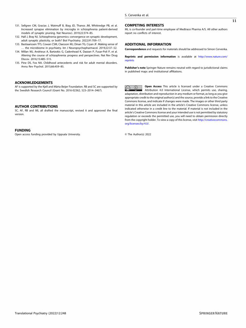
131. Sellgren CM, Gracias J, Watmuff B, Biag JD, Thanos JM, Whittredge PB, et al.Increased synapse elimination by microglia in schizophrenia patient-derivedmodels of synaptic pruning. Nat Neurosci. 2019;22:374–85.
132. Hall J, Bray NJ. Schizophrenia genomics: convergence on synaptic development,adult synaptic plasticity, or both? Biol Psychiatry. 2022;91:709–17.
133. Bastiaanssen TFS, Cowan CSM, Claesson MJ, Dinan TG, Cryan JF. Making sense of… the microbiome in psychiatry. Int J Neuropsychopharmacol. 2019;22:37–52.
134. Millan MJ, Andrieux A, Bartzokis G, Cadenhead K, Dazzan P, Fusar-Poli P, et al.Altering the course of schizophrenia: progress and perspectives. Nat Rev DrugDiscov. 2016;15:485–515.
135. Pine DS, Fox NA. Childhood antecedents and risk for adult mental disorders.Annu Rev Psychol. 2015;66:459–85.
ACKNOWLEDGEMENTSAF is supported by the Kjell and Märta Beijer Foundation. RB and SC are supported bythe Swedish Research Council (Grant No. 2016-02362, 523–2014–3467).
AUTHOR CONTRIBUTIONSSC, AF, RB and ML all drafted the manuscript, revised it and approved the finalversion.
FUNDINGOpen access funding provided by Uppsala University.
COMPETING INTERESTSML is co-founder and part-time employee of Medtrace Pharma A/S. All other authorsreport no conflicts of interest.
ADDITIONAL INFORMATIONCorrespondence and requests for materials should be addressed to Simon Cervenka.
Reprints and permission information is available at http://www.nature.com/reprints
Publisher’s note Springer Nature remains neutral with regard to jurisdictional claimsin published maps and institutional affiliations.
Open Access This article is licensed under a Creative CommonsAttribution 4.0 International License, which permits use, sharing,
adaptation, distribution and reproduction in anymedium or format, as long as you giveappropriate credit to the original author(s) and the source, provide a link to the CreativeCommons license, and indicate if changes were made. The images or other third partymaterial in this article are included in the article’s Creative Commons license, unlessindicated otherwise in a credit line to the material. If material is not included in thearticle’s Creative Commons license and your intended use is not permitted by statutoryregulation or exceeds the permitted use, you will need to obtain permission directlyfrom the copyright holder. To view a copy of this license, visit http://creativecommons.org/licenses/by/4.0/.
© The Author(s) 2022
S. Cervenka et al.
11
Translational Psychiatry (2022) 12:248