Apiaceae Essential Oils: Boosters of Terbinafine Activity ...
17
plants Article Apiaceae Essential Oils: Boosters of Terbinafine Activity against Dermatophytes and Potent Anti-Inflammatory Effectors Adriana Trifan 1, * , Simon Vlad Luca 1,2 , Andra-Cristina Bostănaru 3, *, Mihai Brebu 4 , Alexandra Jităreanu 5 , Romeo-Teodor Cristina 6 , Krystyna Skalicka-Wo´ zniak 7 , Sebastian Granica 8 , Monika E. Czerwi ´ nska 9,10 , Aleksandra Kruk 8 ,Hélène Greige-Gerges 11 , Elwira Sieniawska 7 and Mihai Mares , 3 Citation: Trifan, A.; Luca, S.V.; Bost˘ anaru, A.-C.; Brebu, M.; Jit˘ areanu, A.; Cristina, R.-T.; Skalicka-Wo´ zniak, K.; Granica, S.; Czerwi ´ nska, M.E.; Kruk, A.; et al. Apiaceae Essential Oils: Boosters of Terbinafine Activity against Dermatophytes and Potent Anti-Inflammatory Effectors. Plants 2021, 10, 2378. https://doi.org/ 10.3390/plants10112378 Academic Editor: Stefania Garzoli Received: 15 October 2021 Accepted: 1 November 2021 Published: 4 November 2021 Publisher’s Note: MDPI stays neutral with regard to jurisdictional claims in published maps and institutional affil- iations. Copyright: © 2021 by the authors. Licensee MDPI, Basel, Switzerland. This article is an open access article distributed under the terms and conditions of the Creative Commons Attribution (CC BY) license (https:// creativecommons.org/licenses/by/ 4.0/). 1 Department of Pharmacognosy, Faculty of Pharmacy, Grigore T. Popa University of Medicine and Pharmacy Iasi, 700115 Iasi, Romania 2 Biothermodynamics, TUM School of Life and Food Sciences, Technical University of Munich, 85354 Freising, Germany; [email protected] 3 Laboratory of Antimicrobial Chemotherapy, Faculty of Veterinary Medicine, “Ion Ionescu de la Brad” Iasi University of Life Sciences, 700489 Iasi, Romania; [email protected] 4 Physical Chemistry of Polymers Laboratory, Petru Poni Institute of Macromolecular Chemistry, 700481 Iasi, Romania; [email protected] 5 Department of Toxicology, Faculty of Pharmacy, Grigore T. Popa University of Medicine and Pharmacy Iasi, 700115 Iasi, Romania; alexandra.jitareanu@umfiasi.ro 6 Department of Pharmacology, The Banat University of Agricultural Sciences and Veterinary Medicine, 300645 Timisoara, Romania; [email protected] 7 Department of Natural Products Chemistry, Medical University of Lublin, 20-093 Lublin, Poland; [email protected] (K.S.-W.); [email protected] (E.S.) 8 Microbiota Lab, Centre for Preclinical Studies, Department of Pharmacognosy and Molecular Basis of Phytotherapy, Medical University of Warsaw, 02-097 Warsaw, Poland; [email protected] (S.G.); [email protected] (A.K.) 9 Department of Biochemistry and Pharmacogenomics, Faculty of Pharmacy, Medical University of Warsaw, 02-097 Warsaw, Poland; [email protected] 10 Centre for Preclinical Research, Medical University of Warsaw, 02-097 Warsaw, Poland 11 Bioactive Molecules Research Laboratory, Department of Chemistry and Biochemistry, Faculty of Sciences, Section II, Lebanese University, Jdaidet el-Matn B.P. 90656, Lebanon; [email protected] * Correspondence: adriana.trifan@umfiasi.ro (A.T.); [email protected] (A.-C.B.) Abstract: Dermatophyte infections represent an important public health concern, affecting up to 25% of the world’s population. Trichophyton rubrum and T. mentagrophytes are the predominant dermato- phytes in cutaneous infections, with a prevalence accounting for 70% of dermatophytoses. Although terbinafine represents the preferred treatment, its clinical use is hampered by side effects, drug– drug interactions, and the emergence of resistant clinical isolates. Combination therapy, associating terbinafine and essential oils (EOs), represents a promising strategy in the treatment of dermatophy- tosis. In this study, we screened the potential of selected Apiaceae EOs (ajowan, coriander, caraway, and anise) to improve the antifungal activity of terbinafine against T. rubrum ATCC 28188 and T. men- tagrophytes ATCC 9533. The chemical profile of EOs was analyzed by gas chromatography. The minimal inhibitory concentration (MIC) and minimal fungicidal concentration (MFC) of EOs/main compounds were determined according to EUCAST-AFST guidelines, with minor modifications. The checkerboard microtiter method was used to identify putative synergistic combinations of EOs/main constituents with terbinafine. The influence of EOs on the viability and pro-inflammatory cytokine production (IL-1β, IL-8 and TNF-α) was determined using an ex vivo human neutrophils model. The binary associations of tested EOs with terbinafine were found to be synergistic against T. rubrum, with FICI values of 0.26–0.31. At the tested concentrations (6.25–25 mg/L), EOs did not exert cyto- toxic effects towards human neutrophils. Anise EO was the most potent inhibitor of IL-1β release (46.49% inhibition at 25 mg/L), while coriander EO displayed the highest inhibition towards IL-8 and TNF-α production (54.15% and 54.91%, respectively). In conclusion, the synergistic combinations of terbinafine and investigated Apiaceae EOs could be a starting point in the development of novel topical therapies against T. rubrum-related dermatophytosis. Plants 2021, 10, 2378. https://doi.org/10.3390/plants10112378 https://www.mdpi.com/journal/plants
-
Upload
khangminh22 -
Category
Documents
-
view
1 -
download
0
Transcript of Apiaceae Essential Oils: Boosters of Terbinafine Activity ...
plants
Article
Apiaceae Essential Oils: Boosters of Terbinafine Activity againstDermatophytes and Potent Anti-Inflammatory Effectors
Adriana Trifan 1,* , Simon Vlad Luca 1,2, Andra-Cristina Bostănaru 3,*, Mihai Brebu 4 , Alexandra Jităreanu 5,Romeo-Teodor Cristina 6 , Krystyna Skalicka-Wozniak 7 , Sebastian Granica 8 , Monika E. Czerwinska 9,10 ,Aleksandra Kruk 8, Hélène Greige-Gerges 11 , Elwira Sieniawska 7 and Mihai Mares, 3
�����������������
Citation: Trifan, A.; Luca, S.V.;
Bostanaru, A.-C.; Brebu, M.; Jitareanu,
A.; Cristina, R.-T.; Skalicka-Wozniak,
K.; Granica, S.; Czerwinska, M.E.;
Kruk, A.; et al. Apiaceae Essential
Oils: Boosters of Terbinafine Activity
against Dermatophytes and Potent
Anti-Inflammatory Effectors. Plants
2021, 10, 2378. https://doi.org/
10.3390/plants10112378
Academic Editor: Stefania Garzoli
Received: 15 October 2021
Accepted: 1 November 2021
Published: 4 November 2021
Publisher’s Note: MDPI stays neutral
with regard to jurisdictional claims in
published maps and institutional affil-
iations.
Copyright: © 2021 by the authors.
Licensee MDPI, Basel, Switzerland.
This article is an open access article
distributed under the terms and
conditions of the Creative Commons
Attribution (CC BY) license (https://
creativecommons.org/licenses/by/
4.0/).
1 Department of Pharmacognosy, Faculty of Pharmacy, Grigore T. Popa University of Medicine and PharmacyIasi, 700115 Iasi, Romania
2 Biothermodynamics, TUM School of Life and Food Sciences, Technical University of Munich,85354 Freising, Germany; [email protected]
3 Laboratory of Antimicrobial Chemotherapy, Faculty of Veterinary Medicine, “Ion Ionescu de la Brad” IasiUniversity of Life Sciences, 700489 Iasi, Romania; [email protected]
4 Physical Chemistry of Polymers Laboratory, Petru Poni Institute of Macromolecular Chemistry,700481 Iasi, Romania; [email protected]
5 Department of Toxicology, Faculty of Pharmacy, Grigore T. Popa University of Medicine and Pharmacy Iasi,700115 Iasi, Romania; [email protected]
6 Department of Pharmacology, The Banat University of Agricultural Sciences and Veterinary Medicine,300645 Timisoara, Romania; [email protected]
7 Department of Natural Products Chemistry, Medical University of Lublin, 20-093 Lublin, Poland;[email protected] (K.S.-W.); [email protected] (E.S.)
8 Microbiota Lab, Centre for Preclinical Studies, Department of Pharmacognosy and Molecular Basis ofPhytotherapy, Medical University of Warsaw, 02-097 Warsaw, Poland; [email protected] (S.G.);[email protected] (A.K.)
9 Department of Biochemistry and Pharmacogenomics, Faculty of Pharmacy, Medical University of Warsaw,02-097 Warsaw, Poland; [email protected]
10 Centre for Preclinical Research, Medical University of Warsaw, 02-097 Warsaw, Poland11 Bioactive Molecules Research Laboratory, Department of Chemistry and Biochemistry, Faculty of Sciences,
Section II, Lebanese University, Jdaidet el-Matn B.P. 90656, Lebanon; [email protected]* Correspondence: [email protected] (A.T.); [email protected] (A.-C.B.)
Abstract: Dermatophyte infections represent an important public health concern, affecting up to 25%of the world’s population. Trichophyton rubrum and T. mentagrophytes are the predominant dermato-phytes in cutaneous infections, with a prevalence accounting for 70% of dermatophytoses. Althoughterbinafine represents the preferred treatment, its clinical use is hampered by side effects, drug–drug interactions, and the emergence of resistant clinical isolates. Combination therapy, associatingterbinafine and essential oils (EOs), represents a promising strategy in the treatment of dermatophy-tosis. In this study, we screened the potential of selected Apiaceae EOs (ajowan, coriander, caraway,and anise) to improve the antifungal activity of terbinafine against T. rubrum ATCC 28188 and T. men-tagrophytes ATCC 9533. The chemical profile of EOs was analyzed by gas chromatography. Theminimal inhibitory concentration (MIC) and minimal fungicidal concentration (MFC) of EOs/maincompounds were determined according to EUCAST-AFST guidelines, with minor modifications. Thecheckerboard microtiter method was used to identify putative synergistic combinations of EOs/mainconstituents with terbinafine. The influence of EOs on the viability and pro-inflammatory cytokineproduction (IL-1β, IL-8 and TNF-α) was determined using an ex vivo human neutrophils model. Thebinary associations of tested EOs with terbinafine were found to be synergistic against T. rubrum,with FICI values of 0.26–0.31. At the tested concentrations (6.25–25 mg/L), EOs did not exert cyto-toxic effects towards human neutrophils. Anise EO was the most potent inhibitor of IL-1β release(46.49% inhibition at 25 mg/L), while coriander EO displayed the highest inhibition towards IL-8and TNF-α production (54.15% and 54.91%, respectively). In conclusion, the synergistic combinationsof terbinafine and investigated Apiaceae EOs could be a starting point in the development of noveltopical therapies against T. rubrum-related dermatophytosis.
Plants 2021, 10, 2378. https://doi.org/10.3390/plants10112378 https://www.mdpi.com/journal/plants
Plants 2021, 10, 2378 2 of 17
Keywords: essential oil; Trichophyton rubrum; checkerboard assay; Coriandrum sativum; cytokines;Trachyspermum ammi; terbinafine; Carum carvi; synergy; Pimpinella anisum
1. Introduction
Dermatophytes are filamentous fungi with a high affinity for the keratinized tissueof the skin, nails, and hair, causing superficial infections with different degrees of in-flammation known as dermatophytoses [1,2]. According to data from the World HealthOrganization, dermatophytosis has become a public health issue, affecting up to a quarterof the global population; its incidence is influenced by different factors, e.g., age, sex,the season, socioeconomic status, and geographical region [3]. Trichophyton species arethe main causative agents of dermatophytosis, accounting for up to 70% of total cases;Trichophyton rubrum is the major etiological agent of the cutaneous infection of the feet,nails, and body, followed by T. mentagrophytes [4,5].
Terbinafine, a synthetic allylamine derivative, is currently the “golden standard” treat-ment in dermatophytosis and acts as a potent inhibitor of the fungal squalene epoxidase, anenzyme involved in ergosterol biosynthesis [6]. Still, its oral use is hampered by commonside effects such as gastrointestinal complaints, rash, headache, and loss of taste. Theemergence of recalcitrant dermatophytoses that require a long-term treatment regimen canlead to severe hepatotoxicity, making the monitoring of the liver function mandatory [6–8].Moreover, in numerous cases, systemic therapy is not recommended due to putative drug–drug interactions and patient co-morbidities [9]. In addition, the topical use of terbinafinein onychomycosis is hampered by its poor nail penetration and low efficacy, as revealedin several clinical trials, which evaluated the potency of terbinafine nail solutions andsprays [10,11]. Even though, in the late 2010s, terbinafine-resistant dermatophytes wereseldomly reported, nowadays an alarming number of clinical isolates that are resistant toterbinafine have emerged as a consequence of misused or repeated topical and systemictreatments. Thus, dermatophytosis cases have become refractory to antifungal drugs, withfrequent relapses and the chronicity of fungal infection [6,12,13].
Combinatorial strategies associating conventional antifungals with plant-derivedproducts represent a promising approach to increase the efficacy of extant treatment reg-imens in dermatophytosis and to overcome fungal resistance [14,15]. In this context,synergistic combinations can exert a multi-target antifungal activity, thus increasing the po-tency of conventional drugs and tackling multi-drug microbial resistance. In addition, suchassociations have the advantage of reducing the effective doses of conventional drugs, thusminimizing their toxicity and side effects [16,17]. Among plant-derived products, essentialoils (EOs) possess valuable biological properties and their potent and broad-spectrumantimicrobial activities generate impressive reports in the scientific literature [18–20]. Dis-tillation or pressing are physical processes used to obtain EOs, which comprise a blendof small volatile molecules that act as signaling agents in plants (e.g., attraction of pol-linators, antifeedant agents, and protection against microorganisms) [21,22]. The anti-dermatophytic propensities of EOs are also well-documented and include the disruptionof the cell wall/membrane; the inhibition of spore germination, ergosterol and cellularproteins synthesis; efflux pumps, and biofilm formation [23,24]. Moreover, EOs exhibitanti-inflammatory effects, thus supporting lesion healing and alleviating symptoms relatedto dermatophytosis [14,15].
Apiaceae (Umbelliferae) family comprises 446 genera of 3540 herbaceous species dis-tributed worldwide, many of them used in the food, pharmaceutical, and cosmetic indus-tries [25,26]. A characteristic feature of Apiaceae members is the presence of volatile com-pounds which are found both in aerial parts and the underground systems of plants [27].Apiaceae-derived essential oils are rich sources of monoterpenes, sesquiterpenes, andphenylpropanoids endowed with significant antibacterial, antifungal, and anti-inflammatoryactivities [28–31].
Plants 2021, 10, 2378 3 of 17
Among Apiaceae species, ajowan (Trachyspermum ammi L.), coriander (Coriandrumsativum L.), caraway (Carum carvi L.), and anise (Pimpinella anisum L.) are widely used asspices and flavoring agents and are also employed as remedies to treat various humanailments [20,27,29,31–36]. Their fruits are rich sources of EOs endowed with significant an-tibacterial and antifungal properties against a broad spectrum of microorganisms, includingdermatophytes [20,29,31,37–40].
The treatment failure and resistance to terbinafine are key elements in driving re-search into alternative therapies for dermatophyte infections. As mentioned above, theeffective strategies to treat recalcitrant infections include combination therapy. Consideringthe known antidermatophytic effects of selected Apiaceae species, the association withterbinafine might display a multi-target activity, increasing its potency and tackling fungalresistance. Hence, the aim of the present study was to assess, for the first time, the potentialof EOs derived from the fruits of ajowan, coriander, caraway, anise, and their main con-stituents, and to enhance the terbinafine activity against T. rubrum and T. mentagrophytes. Inaddition, the influence of EOs on the viability and pro-inflammatory cytokine productionin an ex vivo human neutrophils model was evaluated.
2. Materials and Methods2.1. Chemicals
The following chemicals were purchased from Sigma-Aldrich (Steinheim, Germany):4-(2-hydroxyethyl)-1-piperazineethanesulfonic acid) (HEPES), 3-(N-morpholino) propane-sulphonic acid (MOPS), anethole, anhydrous glucose, carvone, chloramphenicol, cyclohex-imide, dimethyl sulfoxide (DMSO), dextran, fetal bovine serum (FBS), L-glutamine, linalool,limonene, propidium iodide, RPMI 1640 medium, terbinafine hydrochloride, thymol andurolithin A. Phosphate-buffered saline (PBS) was bought from Gibco (Hong Kong, China).The (Ca2+)-free PBS was acquired from Biomed (Lublin, Poland). The Human Pancoll(1.077 g/mL) was from PAA Laboratories (Pasching, Austria). Penicillin-streptomycin waspurchased from Biowest (Nauillé, France). Lipopolysaccharide (LPS) from Escherichia coli0111:B4 was purchased from Merck (Kenilworth, NJ, USA). Human ELISA sets (IL-1β, IL-8,TNF-α) were acquired from BD Biosciences (Franklin Lake, NJ, USA). Sabouraud dextroseagar (SDA) was bought from Biolab (Budapest, Hungary).
2.2. Plant Material
Dried plant materials (fruits of ajowan, coriander, caraway, and anise) were purchasedfrom local pharmacies and their botanical identity was confirmed by one of the authors(A.T.). Voucher specimens (AJ 1/2019, CO 2/2019, CA 3/2019, AN 4/2019) were depositedin the Department of Pharmacognosy, Faculty of Pharmacy, “Grigore T. Popa” Universityof Medicine and Pharmacy Iasi, Romania.
2.3. Essential Oil Extraction
The powdered plant materials (100 g each) were subjected to hydrodistillation for 3 hin a Clevenger apparatus. EOs were dried over anhydrous sodium sulfate and stored indark glass tubes at a temperature of 4 ◦C until further analysis.
2.4. Essential Oils Analysis
EOs analysis was undertaken by gas chromatography (GC) using an Agilent 6890Ngas chromatograph coupled with a mass spectrometer (MS) detector (Agilent model 5975inert XL) and a flame ionization detector (FID). TRB-5MS capillary columns (30 m length,0.25 mm internal diameter, 0.25 µm film thickness) were used for each detector. The GC-MSconditions were as follows: injection volume of 0.2 µL, EOs samples being injected in thesplit mode (1:200); helium as carrier gas, with a flow rate of 0.5 mL/min; injector/detectortemperature of 230 ◦C. Oven temperature was raised from 60 ◦C to 220 ◦C at a rate of4 ◦C/min, and then to 320 ◦C at 20 ◦C/min; the final temperature was isothermally heldfor 7.5 min. MS was operated in full scan mode, with an ionization energy of 70 eV, and

Plants 2021, 10, 2378 4 of 17
mass spectra were recorded in the range m/z 15–450. GC-FID conditions were similar tothose used in GC-MS analysis, except for the carrier gas flow that was equal to 1.8 mL/min,and the splitting ratio (1:100). Retention indices were calculated for individual compoundsusing a standard solution of n-alkanes C8–C20 (Sigma-Aldrich). EOs constituents wereidentified by comparing their mass spectra with those from the NIST 11 mass spectra library,accepting only the quality of recognition above 80%, and by comparing their retentionindices with those from the literature [41,42]. The relative percentages of individualconstituents were obtained from the FID peak areas without correction factors.
2.5. Microbial Strains
The antifungal activities of the EOs (ajowan, coriander, caraway, and anise), theirmain constituents (thymol, linalool, carvone, limonene, and anethole), and terbinafinewere investigated against standard strains from the American Type Culture Collection(Trichophyton rubrum ATCC 28188, Trichophyton mentagrophytes ATCC 9533). Prior to testing,fungi were grown on SDA and incubated up to 15 days at 30 ◦C. All strains were checkedduring incubation, and they were used when a maximal number of conidia were formed.
2.6. Antifungal Susceptibility Testing
The MIC values were assessed by the broth microdilution method using 96-well plates,according to The European Committee on Antimicrobial Susceptibility Testing Subcom-mittee on Antifungal Susceptibility Testing (EUCAST-AFST) guidelines [43] and followingthe methodology described by Saunte et al. [12] and Markantonatou et al. [44], with slightmodifications. Serial double dilutions of EOs and volatile compounds (thymol, linalool,carvone, limonene, anethole), ranging from 4 to 2048 mg/L, were prepared in a RPMI1640 medium 2% glucose buffered with 0.165 M MOPS, and supplemented with chloram-phenicol 50 mg/L and cycloheximide 300 mg/L, followed by inoculation (105 CFU/mL).Final concentration of DMSO did not exceed 1%. Terbinafine was used as reference drug(concentration range 0.015–8 mg/L). Fungi growth control and sterility control were usedin order to assure the reliability of the tests. The incubation was performed at 30 ◦C for7 days. The MIC was considered as the lowest concentration with no visual growth of thetested fungi. The MFC was assessed by inoculating 10 µL from wells that did not showgrowth in the MIC assay onto SDA plates, followed by incubation at 30 ◦C for 72 h. TheMFC was considered as the lowest concentration with no growth or less than five coloniesin the case of subculture (corresponding to ~99.9% killing activity). Experiments were runin triplicate.
2.7. Checkerboard Assay
The checkerboard broth microdilution method was used to evaluate putative inter-actions between EOs/volatile compounds (thymol, linalool, carvone, limonene, anethole)and terbinafine against T. rubrum and T. mentagrophytes. Serial double dilutions of EOs,volatile compounds and terbinafine were prepared as previously described for determina-tion of MIC. EOs/volatile compounds were dispensed in increasing concentrations alongthe X-axis (1/128xMIC–4xMIC), while terbinafine was dispensed in increasing concentra-tions along the Y-axis (1/16xMIC–4xMIC), followed by inoculation (105 CFU/mL) [12,45].The plates were incubated at 30 ◦C for 7 days. Assays were performed in duplicate andrepeated at least three times. The interactions between EOs/volatile compounds andterbinafine were interpreted based on the fractional inhibitory concentration index (FICI)values determined using the following equation:
FICI = FICEO/compound + FICterbinafine
where:
FICEO/compound = MICEO/compound in combination/MICEO/compound alone
Plants 2021, 10, 2378 5 of 17
and:FICterbinafine = MICterbinafine in combination/MICterbinafine alone
Synergy was defined by FICI ≤ 0.5: addition was considered when 0.5 > FICI ≤ 1;was indifference considered if 1 > FICI ≤ 4 and antagonism for FICI > 4 [17,22].
2.8. Evaluation of Cytokine Production in Human Neutrophils2.8.1. Neutrophils Isolation
The protocol used to prepare the buffy coat was in accordance with the principlesstated in the Declaration of Helsinki; peripheral venous blood was collected from healthymale volunteers (ages 20–35 years) at the Warsaw Regional Blood Centre. Human neu-trophils were isolated by dextran sedimentation and Pancol centrifugation [46], allowinga neutrophil preparation with over 97% purity. Neutrophils were suspended in PBS andkept at a temperature of 4 ◦C until subsequent analysis.
2.8.2. Evaluation of Neutrophils Viability
Flow cytometry using propidium iodide (PI) staining was undertaken to evaluatethe influence of Apiaceae EOs on neutrophils viability, following a previously describedmethod [47]. Briefly, neutrophils (2.0 × 10⁶/mL) were cultured for 24 h in RPMI 1640medium (supplemented with 10% FBS, 2 mM L-glutamine and 10 mM HEPES) in theabsence/presence of EOs (6.25–25 mg/L) added 30 min before stimulation with 100 ng/mLLPS. After incubation, cells were centrifuged, washed, and re-suspended in PI solution0.5 µg/mL, followed by incubation in the dark at room temperature (15 min). The neu-trophils were analyzed using a BD FACSCalibur flow cytometer (BD Biosciences, San Jose,CA, USA) by recording 10.000 events for each sample. Cells showing high permeabilityto PI were considered PI+ cells. Neutrophils viability was calculated using the followingformula: 100% − PI+ cells%. Triton X-100 (0.1%) was used as positive control.
2.8.3. Evaluation of IL-1β, IL-8 and TNF-α Secretion
The evaluation of cytokine release from LPS-stimulated neutrophils was carried outfollowing a protocol previously described [47]. Thus, neutrophils were incubated at 37 ◦C ina 96 well-plate in RPMI 1640 medium (prepared as described in Section 2.8.2), with/withoutthe tested EOs (concentrations of 6.25, 12.5, and 25 mg/L) added 30 min prior to neutrophilsstimulation with LPS (100 ng/mL). After 24 h, the plates were centrifuged, the supernatantswere collected, and the cytokines release was determined by ELISA assay kits using aSynergy 4 microplate reader, according to the manufacturer’s instructions (BD Biosciences,San Jose, CA, USA). The effect on cytokine production was determined as percentage ofreleased cytokines relative to the control without the investigated EOs. Urolithin A (asubstance with known anti-inflammatory properties [48,49]) at concentrations of 12.5, 25,and 50 µM was used as a positive control.
2.9. Statistical Analyses
The results were calculated as a mean ± standard error of the mean (SEM) of theindicated number of experiments. One-way analysis of variance (ANOVA) with Tukey’sand Dunnet’s post hoc tests was used to evaluate the statistical significance that was set atp < 0.05.
3. Results3.1. Yield and Chemical Composition of Essential Oils
The highest essential oil yield, as calculated on the basis of the weight of the dry fruits(triplicate experiments, percentage ± SEM), was determined for ajowan (7.23 ± 0.09%),followed by anise (3.13 ± 0.07%), caraway (2.83 ± 0.03%), and coriander (2.13 ± 0.09%).The phytochemical compositions of the four Apiaceae EOs assessed by GC are summarizedin Table 1.
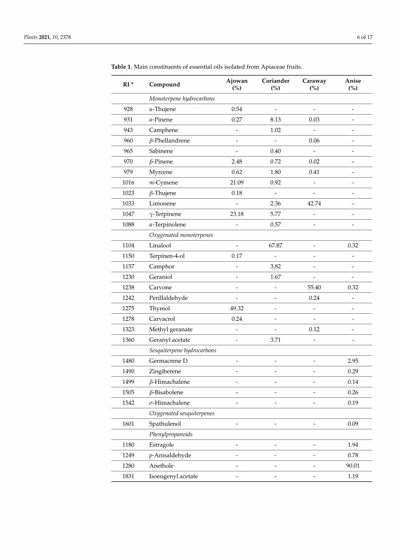
Plants 2021, 10, 2378 6 of 17
Table 1. Main constituents of essential oils isolated from Apiaceae fruits.
RI * Compound Ajowan(%)
Coriander(%)
Caraway(%)
Anise(%)
Monoterpene hydrocarbons
928 α-Thujene 0.54 - - -
931 α-Pinene 0.27 8.13 0.03 -
943 Camphene - 1.02 - -
960 β-Phellandrene - - 0.06 -
965 Sabinene - 0.40 - -
970 β-Pinene 2.48 0.72 0.02 -
979 Myrcene 0.62 1.80 0.41 -
1016 m-Cymene 21.09 0.92 - -
1023 β-Thujene 0.18 - - -
1033 Limonene - 2.36 42.74 -
1047 γ-Terpinene 23.18 5.77 - -
1088 α-Terpinolene - 0.57 - -
Oxygenated monoterpenes
1104 Linalool - 67.87 - 0.32
1150 Terpinen-4-ol 0.17 - - -
1157 Camphor - 3.82 - -
1230 Geraniol - 1.67 - -
1238 Carvone - - 55.40 0.32
1242 Perillaldehyde - - 0.24 -
1275 Thymol 49.32 - - -
1278 Carvacrol 0.24 - - -
1323 Methyl geranate - - 0.12 -
1360 Geranyl acetate - 3.71 - -
Sesquiterpene hydrocarbons
1480 Germacrene D - - - 2.95
1490 Zingiberene - - - 0.29
1499 β-Himachalene - - - 0.14
1505 β-Bisabolene - - - 0.26
1542 σ-Himachalene - - - 0.19
Oxygenated sesquiterpenes
1601 Spathulenol - - - 0.09
Phenylpropanoids
1180 Estragole - - - 1.94
1249 p-Anisaldehyde - - - 0.78
1280 Anethole - - - 90.01
1831 Isoeugenyl acetate - - - 1.19

Plants 2021, 10, 2378 7 of 17
Table 1. Cont.
RI * Compound Ajowan(%)
Coriander(%)
Caraway(%)
Anise(%)
Monoterpene hydrocarbons 48.36 21.69 43.26 -
Oxygenated monoterpenes 49.73 77.07 55.76 0.64
Sesquiterpenehydrocarbons - - - 3.83
Oxygenated sesquiterpenes - - - 0.09
Phenylpropanoids - - - 93.92
Total 98.09 98.76 99.02 98.48* Retention indices relative to a series of C8–C20 n-alkanes calculated on TRB-5MS column. “-” not detected.
In the case of ajowan EO, up to 98.09% of the total constituents were identified, withthymol as the main compound (49.32%), followed by γ-terpinene (23.18%), and m-cymene(21.09%). From the total constituents, 98.76% were identified in coriander EO; linaloolwas the major detected compound (67.87%), alongside several minor constituents, suchas α-pinene (8.13%), γ-terpinene (5.77%), camphor (3.82%), and geranyl acetate (3.71%).Carvone and limonene were the main compounds identified in caraway EO (55.40% and42.74%, respectively), plus additional volatiles found in trace amounts, together making upto 99.02% of the total constituents. Anise EO was characterized by the presence of anetholeas the major compound (90.01%), followed by germacrene (2.95%), estragole (1.28%), andother minor constituents, representing 98.48% of the total volatile fraction. The EOs weremainly characterized by the presence of monoterpenes (monoterpene hydrocarbons andoxygenated monoterpenes) in the case of ajowan, coriander, and caraway EOs; meanwhile,the anise EO comprised phenylpropanoids and sesquiterpene hydrocarbons.
3.2. Antifungal Susceptibility Results
The antifungal activity of Apiaceae EOs and their major constituents (thymol, linalool,carvone, limonene, anethole) was assessed using the microdilution method. The MIC andMFC values obtained for EO/main compounds against T. rubrum and T. mentagrophytes areshown in Table 2.
Table 2. Antifungal activity (MIC, MFC expressed as mg/L) of Apiaceae essential oils and theirmain constituents.
Sample/Positive Control Trichophyton rubrum ATCC28188
Trichophyton mentagrophytesATCC 9533
MIC MFC MIC MFC
Ajowan essential oil 256 512 256 512Coriander essential oil 512 1024 512 1024Caraway essential oil 512 512 512 512Anise essential oil 1024 2048 1024 2048Thymol 1024 1024 1024 1024Linalool 1024 1024 1024 2048Carvone 1024 2048 1024 2048Limonene 2048 2048 2048 2048Anethole 2048 2048 2048 2048Terbinafine 0.031 0.031 0.031 0.031
MIC = minimum inhibitory concentration; MFC = minimum fungicidal concentration.
Ajowan EO exhibited the highest antifungal potential against the tested strains, withMICs of 256 mg/L. Coriander and caraway EOs showed a similar antifungal activitypattern, inhibiting the growth of both dermatophytes, with MIC values of 512 mg/L.Anise EO was the least active among the investigated EOs, displaying fungistatic effects at1024 mg/L. With respect to the antidermatophytic activity of EOs, the main compounds,

Plants 2021, 10, 2378 8 of 17
thymol, linalool, and carvone, acted similarly towards both Trichophyton strains, withMICs of 1024 mg/mL; meanwhile, limonene and anethole exhibited a lower degree ofdermatophytic growth inhibition (MIC 2048 mg/L). Compared to the investigated EOsand their main constituents, terbinafine was highly active against dermatophytes (MIC0.031 mg/L). Based on the MFC values, all EOs/main compounds were found to display afungicidal activity against strains of T. rubrum and T. mentagrophytes (Table 2).
3.3. In Vitro Effects of Essential Oils/Main Constituents and Terbinafine Combinationsagainst Dermatophytes
In order to evaluate the putative synergistic combinations of EOs and their mainconstituents with terbinafine, the checkerboard microtiter assay was performed. Con-sidering their high sensitivity to terbinafine, in vitro experiments using T. rubrum andT. mentagrophytes strains were undertaken. The combinatorial effects of the investigatedEOs/compounds with terbinafine are reported in Table 3. As revealed by the checkerboardtesting, the binary associations of all the tested EOs with terbinafine were found to besynergistic against the T. rubrum strain, with FICI values of 0.26 (coriander and aniseEOs), 0.28 (caraway EO), and 0.31 (ajowan EO), respectively. Moreover, EOs significantlypotentiated the activity of terbinafine in combinatorial therapy, with a 4-fold reduction intheir MIC, from 0.031 to 0.007 mg/L. In addition, the MIC of EOs significantly decreasedin combination with terbinafine; a 64-fold reduction in the case of coriander and aniseEOs, while MICs of caraway and ajowan EOs underwent a 32-fold and a 16-fold reduction,respectively. The main constituents of the EOs showed only additive effects in combinationwith terbinafine (FICI values range of 0.53–0.56). Similar additive interactions were dis-played by the associations between the EOs/volatile compounds and terbinafine againstT. mentagrophytes, with FICI values ranging from 0.56 to 0.75.
Table 3. Combinatorial effects (FIC and FICI values) between Apiaceae essential oils/main constituents and terbinafineassessed by checkerboard method.
Combination
Trichophyton rubrumATCC 28188
INTTrichophyton mentagrophytes ATCC 9533
INTMIC *
in combination(mg/L)
FIC FICI MIC *in combination
(mg/L)FIC FICI
Ajowan essential oil 16 0.060.31 S
16 0.060.56 Ad
Terbinafine 0.007 0.25 0.015 0.50
Coriander essential oil 8 0.010.26 S
32 0.060.56 Ad
Terbinafine 0.007 0.25 0.015 0.50
Caraway essential oil 16 0.030.28 S
64 0.120.62 Ad
Terbinafine 0.007 0.25 0.015 0.50
Anise essential oil 16 0.010.26 S
128 0.120.62 Ad
Terbinafine 0.007 0.25 0.015 0.50
Thymol 64 0.060.56 Ad
128 0.120.62 Ad
Terbinafine 0.015 0.5 0.015 0.5
Linalool 32 0.030.53 Ad
256 0.250.75 Ad
Terbinafine 0.015 0.5 0.015 0.5
Carvone 32 0.030.53 Ad
256 0.250.75 Ad
Terbinafine 0.015 0.5 0.015 0.5
Limonene 64 0.030.53 Ad
256 0.120.62 Ad
Terbinafine 0.015 0.5 0.015 0.5
Anethole 128 0.060.56 Ad
512 0.250.75 Ad
Terbinafine 0.015 0.5 0.015 0.5
Ad—addition; FIC—fractional inhibitory concentration; FICI—fractional inhibitory concentration index; INT—interpretation; S—synergism.* MIC of tested agent in most effective combination.

Plants 2021, 10, 2378 9 of 17
3.4. Effects on Cytokine Production in Human Neutrophils
Based on the MIC values obtained from the synergistic combinations with terbinafine,the four EOs (concentration range 6.25–25 mg/L) were next assessed for their ability toinfluence the viability of human neutrophils and the production of pro-inflammatorycytokines in LPS-stimulated neutrophils.
3.4.1. Effects on Neutrophils Viability
None of the tested EOs concentrations induced cytotoxic effects in human neutrophilsas compared to negative control cells (LPS-) (Figure 1). For instance, at 25 mg/L, the cellviabilities ranged from 96.82% to 97.68%. In addition, the activation of neutrophils withLPS did not significantly reduce their viability (cell viability: 92.20%). Furthermore, thetreatment with urolithin A, used as the positive control in the following cytokine productionassays, did not display cytotoxic effects towards neutrophils (viability of neutrophils97.62–98.06%). Meanwhile, Triton X-100 (0.1%) caused a very strong reduction in the cellviability, with only 1.63% viable cells (data not shown).
Figure 1. Neutrophils viability (%) following 24 h-treatment with essential oils of ajowan, corian-der, caraway, and anise (6.25–25 mg/L), as well as urolithin A (12.5, 25 and 50 µM). Results areexpressed as mean ± SEM of three separate experiments performed with cells isolated from fourindependent donors.
3.4.2. Effects on Pro-Inflammatory Cytokine Secretion
The effects of EOs on the release of pro-inflammatory cytokines, namely IL-1β, IL-8,and TNF-α, were assessed by ELISA in an ex vivo, LPS-stimulated neutrophil model(Figure 2).
When tested at non-cytotoxic concentrations (6.25, 12.5, and 25 mg/L), the fourApiaceae EOs exhibited a concentration-dependent reduction in IL-1β release (Figure 2a).At 25 mg/L, the EOs reduced the IL-1β levels to 66.64% (ajowan), 55.62% (coriander),47.50% (caraway), and 46.49% (anise), when compared to LPS-stimulated neutrophils.Nevertheless, the EOs potency was lower when compared to urolithin A, which decreasedthe IL-1β production to 13.64% of the LPS+ control at 50 µM. The potential of EOs to act asIL-8 inhibitors is depicted in Figure 2b. Out of the four tested EOs, the coriander EO wasthe most active, interfering with IL-8 release in a concentration-dependent manner; the IL-8levels at 25 mg/L were decreased to 54.15% of LPS+ control. A similar effect was observedfor urolithin A at 25 µM (57.61% of LPS+ control). Ajowan EO was found to slightly inhibitthe IL-8 production at the highest tested concentration (76.74% of LPS+ control). Theeffects of the preincubation of human neutrophils with Apiaceae EOs upon TNF-α releaseafter stimulation with LPS is presented in Figure 2c. At the highest tested concentration(25 mg/L), only coriander and caraway EOs were able to produce a slight to moderate
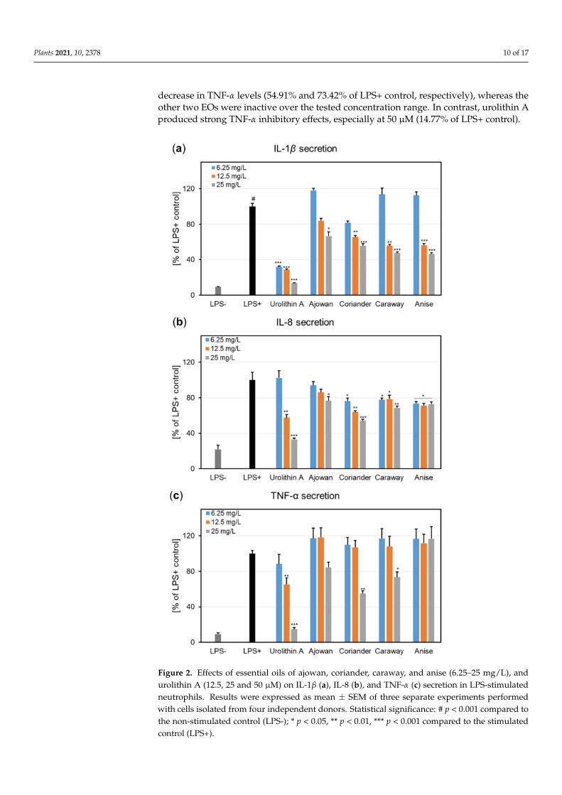
Plants 2021, 10, 2378 10 of 17
decrease in TNF-α levels (54.91% and 73.42% of LPS+ control, respectively), whereas theother two EOs were inactive over the tested concentration range. In contrast, urolithin Aproduced strong TNF-α inhibitory effects, especially at 50 µM (14.77% of LPS+ control).
Figure 2. Effects of essential oils of ajowan, coriander, caraway, and anise (6.25–25 mg/L), andurolithin A (12.5, 25 and 50 µM) on IL-1β (a), IL-8 (b), and TNF-α (c) secretion in LPS-stimulatedneutrophils. Results were expressed as mean ± SEM of three separate experiments performedwith cells isolated from four independent donors. Statistical significance: # p < 0.001 compared tothe non-stimulated control (LPS-); * p < 0.05, ** p < 0.01, *** p < 0.001 compared to the stimulatedcontrol (LPS+).

Plants 2021, 10, 2378 11 of 17
4. Discussion
Even though combinatorial strategies including antifungal drugs and EOs are alreadyrecommended to improve monotherapy, the literature reports regarding terbinafine’sassociation with these plant-derived products are scarce [50–52]. Our study aimed toidentify putative synergistic combinations of terbinafine using the selected Apiaceae EOs(ajowan, coriander, caraway, anise) and their major compounds (thymol, linalool, carvone,limonene, and anethole) against the main causative agents of dermatophytosis, T. rubrum,and T. mentagrophytes.
Therefore, the antifungal potential was first tested using broth microdilution method.According to de Oliveira Lima et al. [53], the antimicrobial potential of plant extracts iscorrelated with their MIC values, as follows: strong activity (MIC 50–500 mg/L), moderateactivity (500 mg/L > MIC < 1500 mg/L), and weak activity (MIC > 1500 mg/L). Thus,ajowan EO was highly active against all tested fungi (MIC 256 mg/L), while coriander,caraway, and anise EOs displayed a moderate activity (MIC > 500 mg/L). Among the testedvolatile compounds, thymol, linalool, and carvone also had a moderate antifungal potential(MIC 1024 mg/L); meanwhile, limonene and anethole possessed a low antimicrobialactivity (MIC 2048 mg/L). Compared to the parent EOs, the antifungal potential of theirmain constituents was lower, suggesting that other minor compounds might contribute,via phytosynergic interactions, to the overall bioactivity of EOs. Even though severalstudies assessed the antidermatophytic potential of investigated Apiaceae EOs and theirmajor components [54–59], a direct comparison between the MICs obtained in the presentstudy and the literature data is hampered as a consequence of the implementation ofdifferent protocols (e.g., obtaining the serial dilutions, fungi growth phase and vitality,inoculum volume, culture medium and pH, temperature, and incubation time). In addition,our results showed that EOs and their main compounds acted as fungicides against bothT. rubrum and T. mentagrophytes strains. This is of the highest importance in clinical settingsof recurrent and multi-drug resistant dermatophytoses, as the use of fungistatic agents iscommonly associated with fungal resistance [10,14].
Still, even if the antifungal effects of the investigated EOs do not justify their use asstand-alone antimicrobial agents, it is noteworthy that, at significantly lower subinhibitoryconcentrations (1/16xMIC–1/64xMIC), EOs induced a 4-fold enhancement of terbinafineactivity against T. rubrum. The observed synergistic effects may be due to the abilityof EOs to facilitate terbinafine entry into the cell with an enhancement in its inhibitoryeffects towards ergosterol biosynthesis. The Apiaceae EOs investigated in our studymostly comprised monoterpene hydrocarbons and oxygenated monoterpenes (ajowan,coriander, and caraway EOs), and phenylpropanoids (anise EO). It is known that suchcompounds are lipophilic and have a small molecular size which enable their passivediffusion through fungi membranes [16]. Volatile compounds exert their fungicidal activitythrough different mechanisms of action that include, among others, the perturbation ofthe lipid membrane organization [60] followed by the increased permeability of the fungalcell wall/membrane, the extravasation of cell constituents, the inhibition of ergosterolsynthesis, and cell lysis [15,23]. Moreover, it was reported that several main constituentsof the Apiaceae EOs included in our study acted on the virulence factors and resistancemechanisms of T. rubrum. For example, Ponte et al. [54] showed that linalool, the majorcompound of coriander EO, acts as an efflux pump inhibitor and is able to increase thesusceptibility to azoles of Trichophyton multi-drug resistant strains. Thymol, the mainconstituent of ajowan EO, enhanced fluconazole efficacy against the clinical isolates ofT. rubrum by inhibiting the activity of proteinases (elastase and keratinase) that contributeto fungal virulence [61]. Furthermore, thymol displayed an inhibitory activity towardsefflux pumps, which are one of the fungal resistance mechanisms [62]. Obaid et al. [63]demonstrated that anethole, identified as the main compound in the anise EO, was able todown-regulate the keratinase gene expression in T. rubrum strains.
Herein, we reported for the first time the synergistic combinatorial effects of terbinafinewith EOs derived from the fruits of ajowan, coriander, caraway, and anise. As of now, the
Plants 2021, 10, 2378 12 of 17
literature data on terbinafine’s synergistic associations with EOs is scarce. To the best ofour knowledge, only two studies reported on the ability of Lavandula luisieri and Schinuslentiscifolius-derived EOs to enhance terbinafine’s activity against the clinical isolates ofT. rubrum [49,50]. One must note that these species grow spontaneously in specific habitats:L. luisieri is endemic to the Iberic Peninsula, while S. lentiscifolius grows in southern Brazil,therefore representing a limited supply that could hamper their industrial use [51,52]. Ourstudy included species which are commonly used as spices and are cultivated on a largescale [23]. Thus, the plant material for the essential oil isolation is readily available and canbe constantly supplied to the pharmaceutical industry. In addition, the development ofApiaceae EOs-based formulations should also consider the quality and standardization ofEOs. It is known that the chemical variability of EOs is mainly due to the plant phenotype,pedoclimatic conditions, harvesting and storage conditions. To overcome batch-to-batchvariability, valuable cultivars might supply a plant material with a constant chemicalprofile, thus a reliable source for the pharmaceutical industry. Our results are of utmostimportance in the case of recalcitrant dermatophytoses when higher doses of terbinafineand long-term treatment are required, which in turn entail hepatotoxic events and a pooradherence to therapy regimens [6]. Therefore, the association with EOs could minimizethe side effects of the dose-related toxicity of terbinafine and increase the efficacy of thetreatment compared to monotherapy. In addition, such combinations could prove usefulin the topical treatment of onychomycosis, as EOs are blends of molecules with a lowmolecular weight that act as nail permeation enhancers for terbinafine, thus ensuring a ahigher efficacy of the treatment [18,64,65]. Moreover, considering that EOs act concurrentlytowards different microbial targets due to their multicomponent nature, the selection ofresistant fungal strains can be overcome [16,66].
Based on the MIC values obtained from the synergistic combinations with terbinafine(Table 3), Apiaceae EOs were evaluated for their ability to influence the secretion of pro-inflammatory cytokines in ex vivo-stimulated human neutrophils. Human neutrophilsare primarily involved in the host defense mechanism against T. rubrum, as both clinicalsetups and animal model experiments revealed a dense infiltration of neutrophils ininfected areas [67]. Following recruitment from the bloodstream, neutrophils’ activationand responses to a fungi attack include: phagocytosis, an oxidative burst by reactive oxygenspecies production, the secretion of proteases, the release of extracellular traps, the secretionof pro-inflammatory cytokines (e.g., IL-1β, IL-6, IL-8, and TNF-α), chemokines, and growthfactors [1,68,69]. However, the prolonged activation of neutrophils hinders the resolution ofinfection, sustaining a chronic inflammation which in turn can contribute to colonization ofthe neighboring host tissue [70]. Therefore, therapeutic alternatives that combine selectiveantifungal and anti-inflammatory activities should also be considered to tune the finebalance between pro- and anti-inflammatory signals in human–host fungi interactions.
First, the potential deleterious effects of Apiaceae EOs were investigated towards hu-man neutrophils, obtained ex vivo from healthy volunteers. EOs did not affect neutrophilsviability, as no cytotoxic effects were recorded at the tested concentrations (6.25–25 mg/L)(Figure 1), thus proving the safety of EOs for pharmaceutical and cosmetic use. Moreover,neutrophils showed a good viability and released functional, pro-inflammatory cytokinesfollowing LPS-stimulation, such as IL-1β, IL-8, and TNF-α (Figure 2). Our results revealedthat EOs inhibited, in different degrees of potency, the production of cytokines in LPS-stimulated neutrophils. It was observed that the inhibition of IL-1β release by EOs wasconcentration-dependent (Figure 2a), with the anise EO displaying the most potent in-hibitory activity, followed by caraway, coriander, and ajowan EOs. Regarding IL-8 andTNF-α, coriander EO proved the highest ability to lower their production in stimulatedneutrophils (Figure 2b,c).
To the best of our knowledge, we reported herein for the first time the inhibitoryactivity of EOs isolated from the fruits of ajowan, coriander, caraway, and anise, towardsIL-1β, IL-8, and TNF-α secretion in an ex vivo human neutrophils model. Our results arein accordance with previously published data regarding the influence of the investigated

Plants 2021, 10, 2378 13 of 17
Apiaceae EOs and their main compounds on pro-inflammatory cytokine production. Thus,ajowan EO was shown to possess anti-inflammatory effects as assessed by in vitro studieson murine macrophage cells, by inhibiting cyclooxygenase-2, inducible nitric oxide syn-thase, and heme oxygenase-1 [71]. Coriander EO exhibited anti-inflammatory effects in anin vivo model of colitis as determined by histo-pathologic evaluation and myeloperoxidaseactivity [72]; meanwhile, its main constituent linalool was found to inhibit the productionof pro-inflammatory cytokines (e.g., TNF-α, IL-1β, IL-6, IL-8) in both in vitro and in vivosettings [73,74]. Caraway EO has shown a protective activity in animal models of colitisand renal injury by the inhibition of cytokine production and the induction of endogenousantioxidant enzymatic systems [75,76]; its beneficial effects are mostly due to limoneneand carvone as revealed by various in vitro and in vivo studies [77–79]. As for anise EO,it has proven in vitro anti-inflammatory propensities by the inhibition of IL-1β and IL-8secretion in a bronchial epithelial cell line [80], while its main constituent, anethole wasshown to decrease IL-6 and TNF-α serum concentrations in an animal model of chronicand obstructive pulmonary disease [81].
Correlating the results from the checkerboard testing, where Apiaceae EOs showed po-tent synergistic combinations with terbinafine, alongside the inhibition of pro-inflammatorycytokines release, we can conclude that the further development of formulations includingthe investigated EOs and terbinafine are justified.
Considering the lipophilic nature of EOs components, their high volatility and chemi-cal instability, the encapsulation in nanoformulated delivery systems could be an approachto overcome such limitations and boost the antidermatophytic activity of EOs [15,22,82].The additional data regarding their skin and nail permeation, elucidation of their mech-anism of activity, and whether in vitro results translate into similar outcomes in in vivomodels, are in high demand. In addition, the development of pharmaceutical formulationsshould consider several parameters, including the stability of such combinations, theirpharmacokinetic profiles, and their safety upon administration. Moreover, the mode ofadministration significantly influences the availability of EOs at the infection site. Thus, atopical use of Apiaceae EOs–terbinafine combinations is readily envisioned.
5. Conclusions
The investigated combinations of Apiaceae EOs/main compounds and terbinafineshowed synergistic and additive effects against T. rubrum and T. mentagrophytes, thusreducing the active concentration of the antifungal drug when used together. Therefore,the use of such mixtures might provide a better outcome than monotherapy in terms ofside effects and toxicity, but also in decreasing the emergence of resistant strains. Basedon their antifungal and anti-inflammatory activities, these combinatorial strategies couldbe complementary to conventional therapy by alleviating symptoms, aiding the healingprocess, and preventing dermatophytosis dissemination. Overall, our study highlightsthe putative use of synergistic combinations of terbinafine and investigated Apiaceae EOs(ajowan, coriander, caraway, and anise) as a starting point for the development of noveltopical formulations for T. rubrum-related dermatophytosis.
Author Contributions: Conceptualization, A.T., M.M., S.V.L., S.G. and K.S.-W.; methodology, A.T.,M.B., A.-C.B., M.E.C., S.G., A.K., A.J. and R.-T.C.; software, M.B. and S.V.L.; validation, A.T., S.G., A.J.and A.-C.B.; formal analysis, A.T., S.V.L., E.S. and H.G.-G.; investigation, A.T., A.-C.B., S.V.L. andA.K.; resources, A.T., S.G., R.-T.C. and M.B.; data curation, A.T., S.V.L. and M.E.C.; writing—originaldraft preparation, A.T., A.-C.B., M.B. and S.V.L.; writing—review and editing, A.T., M.B., S.V.L.,M.M., K.S.-W., M.E.C., H.G.-G. and E.S.; visualization, M.E.C., S.G. and H.G.-G.; supervision, A.T.,M.M. and S.G.; project administration, A.T., S.V.L. and M.M; funding acquisition, A.T. and M.M. Allauthors have read and agreed to the published version of the manuscript.
Funding: A.T. acknowledges the financial support of “Grigore T. Popa”, University of Medicine andPharmacy Iasi, Romania (contract no. 7246/12.04.2018), and of the Executive Agency for the HigherEducation, Research, Development and Innovation Funding (UEFISCDI, Romania) mobility projectfor researchers PN-III-P1-1.1-MC-2019-1056.
Plants 2021, 10, 2378 14 of 17
Institutional Review Board Statement: Not applicable.
Informed Consent Statement: Not applicable.
Data Availability Statement: Not applicable.
Conflicts of Interest: The authors declare no conflict of interest.
References1. Burstein, V.L.; Beccacece, I.; Guasconi, L.; Mena, C.J.; Cervi, L.; Chiapello, L.S. Skin immunity to dermatophytes: From
experimental infection models to human disease. Front. Immunol. 2020, 11, 605644. [CrossRef]2. Gnat, S.; Łagowski, D.; Nowakiewicz, A. Major challenges and perspectives in the diagnostics and treatment of dermatophyte
infections. J. Appl. Microbiol. 2020, 129, 212–232. [CrossRef] [PubMed]3. de Hoog, G.S.; Dukik, K.; Monod, M.; Packeu, A.; Stubbe, D.; Hendrickx, M.; Kupsch, C.; Stielow, J.B.; Freeke, J.; Göker, M.
Toward a novel multilocus phylogenetic taxonomy for the dermatophytes. Mycopathologia 2017, 182, 5–31. [CrossRef]4. Nenoff, P.; Verma, S.B.; Vasani, R.; Burmester, A.; Hipler, U.C.; Wittig, F.; Krüger, C.; Nenoff, K.; Wiegand, C.; Saraswat, A. The
current Indian epidemic of superficial dermatophytosis due to Trichophyton mentagrophytes—A molecular study. Mycoses 2019, 62,336–356. [CrossRef]
5. Zhan, P.; Liang, G.; Liu, W. Dermatophytes and dermatophytic infections worldwide. In Dermatophytes and Dermatophytoses;Bouchara, J.P., Nenoff, P., Gupta, A.K., Chaturvedi, V., Eds.; Springer: Cham, Switzerland, 2021; pp. 15–40.
6. Gupta, A.K.; Renaud, H.J.; Quinlan, E.M.; Shear, N.H.; Piguet, V. The growing problem of antifungal resistance in onychomycosisand other superficial mycoses. Am. J. Clin. Dermatol. 2021, 22, 149–157. [CrossRef] [PubMed]
7. Monod, M.; Feuermann, M.; Yamada, T. Terbinafine and itraconazole resistance in dermatophytes. In Dermatophytes andDermatophytoses; Bouchara, J.P., Nenoff, P., Gupta, A.K., Chaturvedi, V., Eds.; Springer: Cham, Switzerland, 2021; pp. 415–429.
8. Kramer, O.; Albrecht, J. Clinical presentation of terbinafine-induced severe liver injury and the value of laboratory monitoring: ACritically Appraised Topic. Brit. J. Dermatol. 2017, 177, 1279–1284. [CrossRef]
9. Durdu, M.; Ilkit, M.; Tamadon, Y.; Tolooe, A.; Rafati, H.; Seyedmousavi, S. Topical and systemic antifungals in dermatologypractice. Expert Rev. Clin. Pharmacol. 2017, 10, 225–237. [CrossRef] [PubMed]
10. Gupta, A.K.; Venkataraman, M.; Quinlan, E.M. New antifungal agents and new formulations against dermatophytes. InDermatophytes and Dermatophytoses; Bouchara, J.P., Nenoff, P., Gupta, A.K., Chaturvedi, V., Eds.; Springer: Cham, Switzerland,2021; pp. 433–471.
11. Elewski, B.; Ghannoum, M.; Mayser, P.; Gupta, A.; Korting, H.C.; Shouey, R.; Baker, D.; Rich, P.; Ling, M.; Hugot, S. Efficacy,safety and tolerability of topical terbinafine nail solution in patients with mild to moderate toenail onychomycosis: Results fromthree randomized studies using double blind vehicle controlled and open label active controlled designs. J. Eur. Acad. Dermatol.Venereol. 2013, 27, 287–294. [CrossRef]
12. Saunte, D.M.; Hare, R.K.; Jørgensen, K.M.; Jørgensen, R.; deleuran, M.; Zachariae, C.O.; Thomsen, S.F.; Bjørnskov-Halkier, L.;Kofoed, K.; Arendrup, M.C. Emerging terbinafine resistance in Trichophyton: Clinical characteristics, squalene epoxidase genemutations, and a reliable EUCAST method for detection. Antimicrob. Agents Chemother. 2019, 63, e01126-19. [CrossRef] [PubMed]
13. Khurana, A.; Sardana, K.; Chowdhary, A. Antifungal resistance in dermatophytes: Recent trends and therapeutic implications.Fungal Genet. Biol. 2019, 132, 103255. [CrossRef]
14. Lopes, G.; Pinto, E.; Salgueiro, L. Natural products: An alternative to conventional therapy for dermatophytosis? Mycopathologia2017, 182, 143–167. [CrossRef] [PubMed]
15. Brescini, L.; Fioriti, S.; Morroni, G.; Barchiesi, F. Antifungal combinations in dermatophytes. J. Fungi 2021, 7, 727. [CrossRef][PubMed]
16. Zuzarte, M.; Lopes, G.; Pinto, E.; Salgueiro, L. Are natural products an alternative therapy for dermatophytosis? In Dermatophytesand Dermatophytoses; Bouchara, J.P., Nenoff, P., Gupta, A.K., Chaturvedi, V., Eds.; Springer: Cham, Switzerland, 2021; pp. 473–519.
17. Van Vuuren, S.; Viljoen, A. Plant-based antimicrobial studies–methods and approaches to study the interaction between naturalproducts. Planta Med. 2011, 77, 1168–1182. [CrossRef] [PubMed]
18. Adorjan, B.; Buchbauer, G. Biological properties of essential oils: An updated review. Flavour Fragr. J. 2010, 25, 407–426. [CrossRef]19. Hammer, K.A.; Carson, C.F. Antibacterial and antifungal activities of essential oils. In Lipids and Essential Oils as Antimicrobial
Agents; Thormar, H., Ed.; John Wiley & Sons: Chichester, UK, 2011; pp. 255–295.20. Bakkali, F.; Averbeck, S.; Averbeck, D.; Idaomar, M. Biological effects of essential oils—A review. Food Chem. Toxicol. 2008, 46,
446–475. [CrossRef]21. Bruneton, J. Pharmacognosy, Phytochemistry, Medicinal Plants, 2nd ed.; Lavoisier: Paris, France, 2008; p. 484.22. Trifan, A.; Luca, S.V.; Greige-Gerges, H.; Miron, A.; Gille, E.; Aprotosoaie, A.C. Recent advances in tackling microbial multidrug
resistance with essential oils: Combinatorial and nano-based strategies. Crit. Rev. Microbiol. 2020, 46, 338–357. [CrossRef][PubMed]
23. Lopes, A.I.; Tavaria, F.K.; Pintado, M.E. Conventional and natural compounds for the treatment of dermatophytosis. Med. Mycol.2020, 58, 707–720. [CrossRef]
24. D’agostino, M.; Tesse, N.; Frippiat, J.P.; Machouart, M.; debourgogne, A. Essential oils and their natural active compoundspresenting antifungal properties. Molecules 2019, 24, 3713. [CrossRef]
Plants 2021, 10, 2378 15 of 17
25. Simpson, M.G. Plant Systematics; Elsevier Academic Press: Amsterdam, The Netherlands, 2019; p. 829.26. Acimovic, M. Nutraceutical potential of Apiaceae. In Bioactive Molecules in Food. Reference Series in Phytochemistry; Mérillon, J.M..,
Ramawat, K., Eds.; Springer: Cham, Switzerland, 2017. [CrossRef]27. Heinrich, M.; Williamson, E.M.; Gibbons, S.; Barnes, J.; Prieto-Garcia, J. Fundamentals of Pharmacognosy and Phytotherapy, 2nd ed.;
Elsevier: Edinbourg, UK, 2012; p. 34.28. Widelski, J.; Graikou, K.; Ganos, C.; Skalicka-Wozniak, K.; Chinou, I. Volatiles from selected Apiaceae species cultivated in
Poland—Antimicrobial activities. Processes 2021, 9, 695. [CrossRef]29. Khalil, N.; Ashour, M.; Fikry, S.; Singab, A.N.; Salama, O. Chemical composition and antimicrobial activity of the essential oils of
selected Apiaceous fruits. FJPS 2018, 4, 88–92. [CrossRef]30. Elshafie, H.S.; Camele, I. An overview of the biological effects of some mediterranean essential oils on human health. BioMed Res.
Int. 2017, 2017. [CrossRef]31. Sayed-Ahmad, B.; Talou, T.; Saad, Z.; Hijazi, A.; Merah, O. The Apiaceae: Ethnomedicinal family as source for industrial uses.
Ind. Crops Prod. 2017, 109, 661–671. [CrossRef]32. Trifan, A.; Bostănaru, A.-C.; Luca, S.V.; Grădinaru, A.C.; Jităreanu, A.; Aprotosoaie, A.C.; Miron, A.; Cioancă, O.; Hăncianu, M.;
Ochiuz, L.; et al. Antifungal potential of Pimpinella anisum, Carum carvi and Coriandrum sativum extracts. A comparative studywith focus on the phenolic composition. Farmacia 2020, 68, 22–27. [CrossRef]
33. Grădinaru, A.; Trifan, A.; Spac, A.; Brebu, M.; Miron, A.; Aprotosoaie, A. Antibacterial activity of traditional spices against lowerrespiratory tract pathogens: Combinatorial effects of Trachyspermum ammi essential oil with conventional antibiotics. Lett. Appl.Microbiol. 2018, 67, 449–457. [CrossRef] [PubMed]
34. Trifan, A.; Aprotosoaie, A.C.; Cioanca, O.; Hancianu, M.; Jitareanu, A.; Gille, E.; Miron, A. Antioxidant activity of essential oilfrom Carum carvi L. cultivated in North-eastern Romania. Med.-Surg. J. 2016, 120, 732–736.
35. Trifan, A.; Miron, A.; Aprotosoaie, A.C.; Hancianu, M.; Cioanca, O.; Gille, E.; Stanescu, U. Phytotoxicity assessment of polypheno-lic extracts from Carum carvi L. fruits. Farmacia 2013, 61, 12–19.
36. Trifan, A.; Aprotosoaie, A.C.; Spac, A.; Hăncianu, M.; Miron, A.; Stănescu, U. Contributions to the chemical study of the essentialoil isolated from coriander (Omagiu cultivar) fruits. Farmacia 2012, 60, 177–183.
37. Jain, N.; Sharma, M.; Joshi, S.; Kaushik, U. Chemical composition, toxicity and antidermatophytic activity of essential oil ofTrachyspermum ammi. Indian J. Pharm. Sci. 2018, 80, 135–142. [CrossRef]
38. Swamy, M.K.; Akhtar, M.S.; Sinniah, U.R. Antimicrobial properties of plant essential oils against human pathogens and theirmode of action: An updated review. Evid.-Based Complementary Altern. Med. 2016, 2016. [CrossRef]
39. Laribi, B.; Kouki, K.; M’Hamdi, M.; Bettaieb, T. Coriander (Coriandrum sativum L.) and its bioactive constituents. Fitoterapia 2015,103, 9–26. [CrossRef]
40. Bairwa, R.; Sodha, R.; Rajawat, B. Trachyspermum ammi. Pharmacogn. Rev. 2012, 6, 56. [CrossRef]41. Navarro-Rocha, J.; Andrés, M.F.; Díaz, C.E.; Burillo, J.; González-Coloma, A. Composition and biocidal properties of essential oil
from pre-domesticated Spanish Satureja montana. Ind. Crops Prod. 2020, 145, 111958. [CrossRef]42. NIST Chemistry WebBook. NIST Standard Reference Database Number 69. Available online: https://webbook.nist.gov/
chemistry (accessed on 20 January 2021).43. Arendrup, M.; Meletiadis, J.; Mouton, J.; Lagrou, K.; Hamal, P.; Guinea, J. Eucast Definitive Document E DEF. 9.3.1. Method for
the Determination of Broth Dilution Minimum Inhibitory Concentrations of Antifungal Agents for Conidia Forming Moulds.London: European Committee on Antimicrobial Susceptibility Testing. Available online: https://www.eucast.org/ast_of_fungi/(accessed on 4 June 2020).
44. Markantonatou, A.-M.; Samaras, K.; Zachrou, E.; Vyzantiadis, T.-A. Comparison of four methods for the in vitro susceptibilitytesting of dermatophytes. Front. Microbiol. 2020, 11, 1593. [CrossRef]
45. Verma, P. Methods for determining bactericidal activity and antimicrobial interactions: Synergy testing, time-kill curves, andpopulation analysis. In Antimicrobial Susceptibility Testing Protocols; Schwalbe, R., Steele-Moore, L., Goodwin, A.C., Eds.; CRCPress: Boca Raton, FL, USA, 2007; pp. 275–298.
46. Czerwinska, M.E.; Dudek, M.K.; Pawłowska, K.A.; Prus, A.; Ziaja, M.; Granica, S. The influence of procyanidins isolated fromsmall-leaved lime flowers (Tilia cordata Mill.) on human neutrophils. Fitoterapia 2018, 127, 115–122. [CrossRef]
47. Trifan, A.; Skalicka-Wozniak, K.; Granica, S.; Czerwinska, M.E.; Kruk, A.; Marcourt, L.; Wolfender, J.-L.; Wolfram, E.; Esslinger, N.;Grubelnik, A. Symphytum officinale L.: Liquid-liquid chromatography isolation of caffeic acid oligomers and evaluation of theirinfluence on pro-inflammatory cytokine release in LPS-stimulated neutrophils. J. Ethnopharmacol. 2020, 262, 113169. [CrossRef]
48. Rønning, S.B.; Voldvik, V.; Bergum, S.K.; Aaby, K.; Borge, G.I.A. Ellagic acid and urolithin A modulate the immune responsein LPS-stimulated U937 monocytic cells and THP-1 differentiated macrophages. Food Funct. 2020, 11, 7946–7959. [CrossRef][PubMed]
49. Fu, X.; Gong, L.F.; Wu, Y.F.; Lin, Z.; Jiang, B.J.; Wu, L.; Yu, K.H. Urolithin A targets the PI3K/Akt/NF-κB pathways and preventsIL-1β-induced inflammatory response in human osteoarthritis: In vitro and in vivo studies. Food Funct. 2019, 10, 6135–6146.[CrossRef]
50. Danielli, L.J.; Pippi, B.; Soares, K.D.; Duarte, J.A.; Maciel, A.J.; Machado, M.M.; Oliveira, L.F.S.; Bordignon, S.A.; Fuentefria, A.M.;Apel, M.A. Chemosensitization of filamentous fungi to antifungal agents using Nectandra Rol. ex Rottb. species essential oils. Ind.Crops Prod. 2017, 102, 7–15. [CrossRef]
Plants 2021, 10, 2378 16 of 17
51. Danielli, L.J.; Pippi, B.; Duarte, J.A.; Maciel, A.J.; Lopes, W.; Machado, M.M.; Oliveira, L.F.S.; Vainstein, M.H.; Teixeira, M.L.;Bordignon, S.A. Antifungal mechanism of action of Schinus lentiscifolius Marchand essential oil and its synergistic effect in vitrowith terbinafine and ciclopirox against dermatophytes. J. Pharm. Pharmacol. 2018, 70, 1216–1227. [CrossRef] [PubMed]
52. Dias, N.; Dias, M.; Cavaleiro, C.; Sousa, M.; Lima, N.; Machado, M. Oxygenated monoterpenes-rich volatile oils as potentialantifungal agents for dermatophytes. Nat. Prod. Res. 2017, 31, 460–464. [CrossRef] [PubMed]
53. de Oliveira Lima, M.; de Medeiros, A.A.; Silva, K.S.; Cardoso, G.; de Oliveira Lima, E.; de Oliveira Pereira, F. Investigation ofthe antifungal potential of linalool against clinical isolates of fluconazole resistant Trichophyton rubrum. J. Mycol. Med. 2017, 27,195–202. [CrossRef] [PubMed]
54. Ponte, H.A.S.; Lima, M.I.D.O.; Lima, E.D.O.; Pereira, F.D.O. Linalool modulates dermatophyte susceptibility to azole drugs. Med.Mycol. 2020, 58, 272–274. [CrossRef] [PubMed]
55. Jain, N.; Sharma, M. Inhibitory effect of some selected essential oil terpenes on fungi causing superficial infection in humanbeings. J. Essent. Oil Bear. Plants 2020, 23, 862–869. [CrossRef]
56. Obaid, A.J.; Al-Janabi, J.K.A.; Taj-Aldin, W.R. Chemical composition and bioactivity characteristics of Pimpinella anisum essentialoil against Trichophyton rubrum. J. Glob. Pharma Technol. 2017, 8, 44–56.
57. Pinto, E.; Gonçalves, M.-J.; Cavaleiro, C.; Salgueiro, L. Antifungal activity of Thapsia villosa essential oil against Candida,Cryptococcus, Malassezia, Aspergillus and dermatophyte species. Molecules 2017, 22, 1595. [CrossRef]
58. Inouye, S.; Uchida, K.; Abe, S. Vapor activity of 72 essential oils against a Trichophyton mentagrophytes. J. Infect. Chemother. 2006, 12,210–216. [CrossRef] [PubMed]
59. Lim, S.; Shin, S.-W. Synergism in antifungal activity against Candida and Trichophyton species in combination with the essential oilof Coriandrum sativum L. and antibiotics. Nat. Prod. Sci. 2007, 13, 85–89.
60. Gharib, R.; Auezova, L.; Charcosset, C.; Greige-Gerges, H. Effect of a series of essential oil molecules on DPPC membrane fluidity:A biophysical study. J. Iran. Chem. Soc. 2018, 15, 75–84. [CrossRef]
61. Khan, M.S.A.; Ahmad, I.; Cameotra, S.S. Carum copticum and Thymus vulgaris oils inhibit virulence in Trichophyton rubrum andAspergillus spp. Braz. J. Microbiol. 2014, 45, 523–531. [CrossRef]
62. de Melo, J.O.; Bitencourt, T.A.; Fachin, A.L.; Cruz, E.M.O.; de Jesus, H.C.R.; Alves, P.B.; de Fátima Arrigoni-Blank, M.; de CastroFranca, S.; Beleboni, R.O.; Fernandes, R.P.M. Antidermatophytic and antileishmanial activities of essential oils from Lippia gracilisSchauer genotypes. Acta Trop. 2013, 128, 110–115. [CrossRef]
63. Obaid, A.J.; Al-Janabi, J.; Taj-Aldin, W.R. Bioactivities of anethole, astragalin and cryptochlorogenic acid extracted from anise oiland Moringa oleifera on the keratinase gene expression of Trichophyton rubrum. J Pure Appl. Microbiol. 2020, 14, 615–626. [CrossRef]
64. Flores, F.C.; Beck, R.C.; Da Silva, C.D.B. Essential oils for treatment for onychomycosis: A mini-review. Mycopathologia 2016, 181,9–15. [CrossRef] [PubMed]
65. Vörös-Horváth, B.; Das, S.; Salem, A.; Nagy, S.; Böszörményi, A.; Koszegi, T.; Pál, S.; Széchenyi, A. Formulation of tioconazole andMelaleuca alternifolia essential oil pickering emulsions for onychomycosis topical treatment. Molecules 2020, 25, 5544. [CrossRef][PubMed]
66. Christenson, J.K.; Peterson, G.M.; Naunton, M.; Bushell, M.; Kosari, S.; Baby, K.E.; Thomas, J. Challenges and opportunities in themanagement of onychomycosis. J. Fungi 2018, 4, 87. [CrossRef] [PubMed]
67. Calderon, R.; Hay, R. Fungicidal activity of human neutrophils and monocytes on dermatophyte fungi, Trichophyton quinckeanumand Trichophyton rubrum. Immunology 1987, 61, 289. [PubMed]
68. Van der Linden, M.; Meyaard, L. Fine-tuning neutrophil activation: Strategies and consequences. Immunol. Lett. 2016, 178, 3–9.[CrossRef] [PubMed]
69. Zhao, G.; Usui, M.L.; Lippman, S.I.; James, G.A.; Stewart, P.S.; Fleckman, P.; Olerud, J.E. Biofilms and inflammation in chronicwounds. Adv. Wound Care 2013, 2, 389–399. [CrossRef] [PubMed]
70. Romani, L. Immunity to fungal infections. Nat. Rev. Immunol. 2011, 11, 275–288. [CrossRef] [PubMed]71. Bahuguna, A.; Ramalingam, S.; Arumugam, A.; Natarajan, D.; Kim, M. Molecular and in silico evidences explain the anti-
inflammatory effect of Trachyspermum ammi essential oil in lipopolysaccharide induced macrophages. Process Biochem. 2020, 96,138–145. [CrossRef]
72. Heidari, B.; Sajjadi, S.E.; Minaiyan, M. Effect of Coriandrum sativum hydroalcoholic extract and its essential oil on acetic acid-induced acute colitis in rats. Avicenna J. Phytomed. 2016, 6, 205.
73. Ma, J.; Xu, H.; Wu, J.; Qu, C.; Sun, F.; Xu, S. Linalool inhibits cigarette smoke-induced lung inflammation by inhibiting NF-κBactivation. Int. Immunopharmacol. 2015, 29, 708–713. [CrossRef] [PubMed]
74. Huo, M.; Cui, X.; Xue, J.; Chi, G.; Gao, R.; deng, X.; Guan, S.; Wei, J.; Soromou, L.W.; Feng, H. Anti-inflammatory effects of linaloolin RAW 264.7 macrophages and lipopolysaccharide-induced lung injury model. J. Surg. Res. 2013, 180, e47–e54. [CrossRef][PubMed]
75. Keshavarz, A.; Minaiyan, M.; Ghannadi, A.; Mahzouni, P. Effects of Carum carvi L.(caraway) extract and essential oil onTNBS-induced colitis in rats. Res. Pharm. Sci. 2013, 8, 1. [PubMed]
76. Dadkhah, A.; Fatemi, F. Heart and kidney oxidative stress status in septic rats treated with caraway extracts. Pharm. Biol. 2011, 49,679–686. [CrossRef] [PubMed]
77. Zhao, M.; Du, J. Anti-inflammatory and protective effects of D-carvone on lipopolysaccharide (LPS)-induced acute lung injury inmice. J. King Saud Univ. Sci. 2020, 32, 1592–1596. [CrossRef]
Plants 2021, 10, 2378 17 of 17
78. Vieira, A.J.; Beserra, F.P.; Souza, M.; Totti, B.; Rozza, A. Limonene: Aroma of innovation in health and disease. Chem. Biol. Interact.2018, 283, 97–106. [CrossRef] [PubMed]
79. Andrade, L.N.; de Sousa, D.P. A review on anti-inflammatory activity of monoterpenes. Molecules 2013, 18, 1227–1254.80. Iannarelli, R.; Marinelli, O.; Morelli, M.B.; Santoni, G.; Amantini, C.; Nabissi, M.; Maggi, F. Aniseed (Pimpinella anisum L.) essential
oil reduces pro-inflammatory cytokines and stimulates mucus secretion in primary airway bronchial and tracheal epithelial celllines. Ind. Crops Prod. 2018, 114, 81–86. [CrossRef]
81. Kim, K.Y.; Lee, H.S.; Seol, G.H. Anti-inflammatory effects of trans-anethole in a mouse model of chronic obstructive pulmonarydisease. Biomed. Pharmacother. 2017, 91, 925–930. [CrossRef]
82. Sebaaly, C.; Trifan, A.; Sieniawska, E.; Greige-Gerges, H. Chitosan-coating effect on the characteristics of liposomes: A focus onbioactive compounds and essential oils: A review. Processes 2021, 9, 445. [CrossRef]