Antinociception by Spinal and Systemic Oxycodone: Why Does the Route Make a Difference?: In Vitro...
12
Anesthesiology 2006; 105:801–12 Copyright © 2006, the American Society of Anesthesiologists, Inc. Lippincott Williams & Wilkins, Inc. Antinociception by Spinal and Systemic Oxycodone: Why Does the Route Make a Difference? In Vitro and In Vivo Studies in Rats Kim K. Lemberg, D.D.S.* Vesa K. Kontinen, M.D., Ph.D.,† Antti O. Siiskonen, M.Sc.,‡ Kaarin M. Viljakka, M.D.,§ Jari T. Yli-Kauhaluoma, Ph.D., Esa R. Korpi, M.D., Ph.D.,# Eija A. Kalso, M.D., Ph.D.** Background: The pharmacology of oxycodone is poorly un- derstood despite its growing clinical use. The discrepancy be- tween its good clinical effectiveness after systemic administra- tion and the loss of potency after spinal administration led the authors to study the pharmacodynamic effects of oxycodone and its metabolites using in vivo and in vitro models in rats. Methods: Male Sprague-Dawley rats were used in hot-plate, tail-flick, and paw-pressure tests to study the antinociceptive properties of morphine, oxycodone, and its metabolites oxy- morphone and noroxycodone. -Opioid receptor agonist–stim- ulated GTP[ 35 S] autoradiography was used to study G-protein activation induced by morphine, oxycodone, and oxymor- phone in the rat brain and spinal cord. Spontaneous locomotor activity was measured to assess possible sedation or motor dysfunction. Naloxone and the selective -opioid receptor an- tagonist nor-binaltorphimine were used to study the opioid receptor selectivity of the drugs. Results: Oxycodone showed lower efficacy and potency to stimulate GTP[ 35 S] binding in the spinal cord and periaqueduc- tal gray compared with morphine and oxymorphone. This could relate to the fact that oxycodone produced only weak naloxone-reversible antinociception after intrathecal adminis- tration. It also suggests that the metabolites may have a role in oxycodone-induced analgesia in rats. Intrathecal oxymorphone produced strong long-lasting antinociception, whereas noroxy- codone produced antinociception with very high doses only. Subcutaneous administration of oxycodone and oxymorphone produced thermal and mechanical antinociception that was reversed by naloxone but not by nor-binaltorphimine. Oxymor- phone was more potent than oxycodone, particularly in the hot-plate and paw-pressure tests. Conclusions: The low intrathecal potency of oxycodone in rats seems be related to its low efficacy and potency to stimulate -opioid receptor activation in the spinal cord. OXYCODONE is an opioid analgesic with high oral bio- availability in humans ( 60%) 1 compared with mor- phine (19 –30%), 2 whereas its physicochemical proper- ties are similar to those of morphine. 3 Oxycodone is increasingly used worldwide to treat acute and chronic pain. Opioids bind to specific G protein– coupled -, -, and -opioid and nociceptin/orphanin FQ receptors. 4 –10 Oxycodone shows -opioid receptor selectivity, 11 but it has a lower binding affinity to the -opioid receptor compared with morphine in the rat brain homoge- nates. 12 Oxycodone has also been shown to act as a -opioid receptor agonist in the mouse paraphenylqui- none writhing, hot-plate, and tail-flick assays. 13 In humans, intravenous, intramuscular, and oral admin- istration of oxycodone produces pain relief similar to that produced by other -opioid receptor agonists. 1,14 In postoperative pain, the equianalgesic parenteral dose ratio of oxycodone to morphine has previously been reported as 2:3. 1 Intravenous oxycodone was found to be more potent than intravenous morphine in postoper- ative pain. 15 Interestingly, Backlund et al. 16 found that the epidural dose ratio between morphine and oxyc- odone was 1:9.8 in humans after abdominal surgery. Another study indicated that twice the dose of oxyc- odone compared with morphine was needed to achieve comparable analgesia after epidural administration fol- lowing gynecologic surgery. 17 In rats, both intrathe- cal 18,19 and intracerebroventricular 20 administered mor- phine has been shown to induce significantly greater antinociception compared with oxycodone. After subcu- taneous and intraperitoneal administration, oxycodone was found to be two to four times more potent than morphine in rats. 19 These results suggest an active role for the metabolites of oxycodone. Oxycodone is metabolized in humans by hepatic cyto- chrome P450 (CYP) isoenzymes. The main pathways of metabolism include oxidation to oxymorphone and no- roxycodone, conjugation to -D-glucuronic acid, and conversion to 6-oxycodol. 21–25 O-demethylation by CYP2D6 leads to the formation of the main active me- tabolite oxymorphone. Oxymorphone induces antinoci- ception in rodents 26,27 and humans. 28 Noroxycodone is formed through N-demethylation by CYP3A4. It has a poor antinociceptive effect after intracerebroventricular injection in rats. 20 It has been suggested that at least part of the analgesia after oxycodone administration is medi- ated by active metabolites such as oxymorphone. 29 The aim of the current project was to study the antino- * Ph.D. Student, # Professor, Department of Pharmacology, Institute of Bio- medicine, ‡ Ph.D. Student, Professor, Faculty of Pharmacy, Division of Phar- maceutical Chemistry, University of Helsinki. † Senior Researcher, § Ph.D. Student, ** Professor, Department of Pharmacology, Institute of Biomedicine, University of Helsinki, and Department of Anesthesia and Intensive Care Medi- cine, Helsinki University Central Hospital. Received from the Department of Pharmacology, Institute of Biomedicine, University of Helsinki, Helsinki, Finland. Submitted for publication March 14, 2006. Accepted for publication June 6, 2006. Supported by a grant from the Finnish Association for the Study of Pain, Helsinki, Finland, and funds from the University of Helsinki, Helsinki, Finland. The authors do not have any financial or other relationships that might lead to a conflict of interest. Dr. Kalso has received an unrestricted grant for clinical research from Mundifarma Oy, Vantaa, Finland, and she has received honoraria from Mundipharma Oy, Vantaa, Finland; Janssen- Cilag Oy, Espoo, Finland; Pfizer Oy, Helsinki, Finland; and Astra Zeneca Oy, Espoo, Finland. Dr. Kontinen has received honoraria from Janssen-Cilag Oy, Espoo, Finland. Address correspondence to Dr. Lemberg: Institute of Biomedicine/Pharmacology, P.O. Box 63, FIN-00014 University of Helsinki, Finland. kim.lemberg@helsinki.fi. Individual article reprints may be purchased through the Journal Web site, www. anesthesiology.org. Anesthesiology, V 105, No 4, Oct 2006 801 Downloaded From: http://anesthesiology.pubs.asahq.org/pdfaccess.ashx?url=/data/journals/jasa/931073/ on 02/04/2017
Transcript of Antinociception by Spinal and Systemic Oxycodone: Why Does the Route Make a Difference?: In Vitro...

Anesthesiology 2006; 105:801–12 Copyright © 2006, the American Society of Anesthesiologists, Inc. Lippincott Williams & Wilkins, Inc.
Antinociception by Spinal and Systemic Oxycodone: WhyDoes the Route Make a Difference?
In Vitro and In Vivo Studies in RatsKim K. Lemberg, D.D.S.* Vesa K. Kontinen, M.D., Ph.D.,† Antti O. Siiskonen, M.Sc.,‡ Kaarin M. Viljakka, M.D.,§Jari T. Yli-Kauhaluoma, Ph.D.,� Esa R. Korpi, M.D., Ph.D.,# Eija A. Kalso, M.D., Ph.D.**
Background: The pharmacology of oxycodone is poorly un-derstood despite its growing clinical use. The discrepancy be-tween its good clinical effectiveness after systemic administra-tion and the loss of potency after spinal administration led theauthors to study the pharmacodynamic effects of oxycodoneand its metabolites using in vivo and in vitro models in rats.
Methods: Male Sprague-Dawley rats were used in hot-plate,tail-flick, and paw-pressure tests to study the antinociceptiveproperties of morphine, oxycodone, and its metabolites oxy-morphone and noroxycodone. �-Opioid receptor agonist–stim-ulated GTP�[35S] autoradiography was used to study G-proteinactivation induced by morphine, oxycodone, and oxymor-phone in the rat brain and spinal cord. Spontaneous locomotoractivity was measured to assess possible sedation or motordysfunction. Naloxone and the selective �-opioid receptor an-tagonist nor-binaltorphimine were used to study the opioidreceptor selectivity of the drugs.
Results: Oxycodone showed lower efficacy and potency tostimulate GTP�[35S] binding in the spinal cord and periaqueduc-tal gray compared with morphine and oxymorphone. Thiscould relate to the fact that oxycodone produced only weaknaloxone-reversible antinociception after intrathecal adminis-tration. It also suggests that the metabolites may have a role inoxycodone-induced analgesia in rats. Intrathecal oxymorphoneproduced strong long-lasting antinociception, whereas noroxy-codone produced antinociception with very high doses only.Subcutaneous administration of oxycodone and oxymorphoneproduced thermal and mechanical antinociception that wasreversed by naloxone but not by nor-binaltorphimine. Oxymor-phone was more potent than oxycodone, particularly in thehot-plate and paw-pressure tests.
Conclusions: The low intrathecal potency of oxycodone inrats seems be related to its low efficacy and potency to stimulate�-opioid receptor activation in the spinal cord.
OXYCODONE is an opioid analgesic with high oral bio-availability in humans (� 60%)1 compared with mor-
phine (19–30%),2 whereas its physicochemical proper-ties are similar to those of morphine.3 Oxycodone isincreasingly used worldwide to treat acute and chronicpain.
Opioids bind to specific G protein–coupled �-, �-, and�-opioid and nociceptin/orphanin FQ receptors.4–10
Oxycodone shows �-opioid receptor selectivity,11 but ithas a lower binding affinity to the �-opioid receptorcompared with morphine in the rat brain homoge-nates.12 Oxycodone has also been shown to act as a�-opioid receptor agonist in the mouse paraphenylqui-none writhing, hot-plate, and tail-flick assays.13
In humans, intravenous, intramuscular, and oral admin-istration of oxycodone produces pain relief similar tothat produced by other �-opioid receptor agonists.1,14 Inpostoperative pain, the equianalgesic parenteral doseratio of oxycodone to morphine has previously beenreported as 2:3.1 Intravenous oxycodone was found tobe more potent than intravenous morphine in postoper-ative pain.15 Interestingly, Backlund et al.16 found thatthe epidural dose ratio between morphine and oxyc-odone was 1:9.8 in humans after abdominal surgery.Another study indicated that twice the dose of oxyc-odone compared with morphine was needed to achievecomparable analgesia after epidural administration fol-lowing gynecologic surgery.17 In rats, both intrathe-cal18,19 and intracerebroventricular20 administered mor-phine has been shown to induce significantly greaterantinociception compared with oxycodone. After subcu-taneous and intraperitoneal administration, oxycodonewas found to be two to four times more potent thanmorphine in rats.19 These results suggest an active rolefor the metabolites of oxycodone.
Oxycodone is metabolized in humans by hepatic cyto-chrome P450 (CYP) isoenzymes. The main pathways ofmetabolism include oxidation to oxymorphone and no-roxycodone, conjugation to �-D-glucuronic acid, andconversion to 6-oxycodol.21–25 O-demethylation byCYP2D6 leads to the formation of the main active me-tabolite oxymorphone. Oxymorphone induces antinoci-ception in rodents26,27 and humans.28 Noroxycodone isformed through N-demethylation by CYP3A4. It has apoor antinociceptive effect after intracerebroventricularinjection in rats.20 It has been suggested that at least partof the analgesia after oxycodone administration is medi-ated by active metabolites such as oxymorphone.29
The aim of the current project was to study the antino-
* Ph.D. Student, # Professor, Department of Pharmacology, Institute of Bio-medicine, ‡ Ph.D. Student, � Professor, Faculty of Pharmacy, Division of Phar-maceutical Chemistry, University of Helsinki. † Senior Researcher, § Ph.D.Student, ** Professor, Department of Pharmacology, Institute of Biomedicine,University of Helsinki, and Department of Anesthesia and Intensive Care Medi-cine, Helsinki University Central Hospital.
Received from the Department of Pharmacology, Institute of Biomedicine,University of Helsinki, Helsinki, Finland. Submitted for publication March 14,2006. Accepted for publication June 6, 2006. Supported by a grant from theFinnish Association for the Study of Pain, Helsinki, Finland, and funds from theUniversity of Helsinki, Helsinki, Finland. The authors do not have any financial orother relationships that might lead to a conflict of interest. Dr. Kalso has receivedan unrestricted grant for clinical research from Mundifarma Oy, Vantaa, Finland,and she has received honoraria from Mundipharma Oy, Vantaa, Finland; Janssen-Cilag Oy, Espoo, Finland; Pfizer Oy, Helsinki, Finland; and Astra Zeneca Oy,Espoo, Finland. Dr. Kontinen has received honoraria from Janssen-Cilag Oy,Espoo, Finland.
Address correspondence to Dr. Lemberg: Institute of Biomedicine/Pharmacology,P.O. Box 63, FIN-00014 University of Helsinki, Finland. [email protected] article reprints may be purchased through the Journal Web site, www.anesthesiology.org.
Anesthesiology, V 105, No 4, Oct 2006 801
Downloaded From: http://anesthesiology.pubs.asahq.org/pdfaccess.ashx?url=/data/journals/jasa/931073/ on 02/04/2017
ciceptive properties of oxycodone and its metabolitesoxymorphone and noroxycodone after different routesof administration using various nociceptive tests in rats.We were particularly interested to explore further theeffectiveness of oxycodone after spinal administration.For this purpose, G-protein activation by oxycodone andoxymorphone was compared with morphine in an invitro study using GTP�[35S] autoradiography in the spi-nal cord and brain sections from rats.
Materials and Methods
AnimalsThe research was conducted according to the guide-
lines of local authorities and the International Associa-tion for the Study of Pain.30 The protocol of the studywas approved by the institutional animal investigationcommittee University of Helsinki and the provincial gov-ernment of Southern Finland (Uudenmaan laaninhallitus,Helsinki, Finland).
Male Sprague-Dawley rats (n � 56; Harlan, Horst, Neth-erlands) weighing 175–200 g were used (n � 7 or 8 inthe behavioral studies and n � 5 or 6 in the GTP�[35S]binding study). The animals were housed in clear plasticcages with a 12-h/12-h artificial light–dark cycle. Waterand laboratory chow were available ad libitum. Beforethe nociceptive tests, the animals were habituated to thetesting environment for 30 min/day for 3 days. A 3-daywashout was used between the nociceptive tests. In thespontaneous motility test, habituation was not per-formed. After the tests, the animals were killed.
DrugsOxycodone hydrochloride was purchased from the Uni-
versity Pharmacy (Helsinki, Finland); naloxone hydrochlo-ride and naltrexone hydrochloride were purchased fromRBI (Natick, MA); and nor-binaltorphimine hydrochlorideand [d-Ala2,N-Me-Phe4,Gly5-ol]-enkephalin (DAMGO) werepurchased from Tocris (Bristol, United Kingdom). Oxymor-phone hydrochloride and noroxycodone hydrochloridewere synthesized from oxycodone hydrochloride. All drugswere dissolved in saline and administered subcutaneously orintrathecally. Saline served as control. Naloxone and nor-bin-altorphimine were administered 30 min before the testing.
Synthesis of Noroxycodone HydrochlorideOxycodone (1.20 g, 3.81 mmol) was dissolved in ace-
tic anhydride (15 ml). The reaction mixture was stirredat 100°C for 2 h, after which it was evaporated todryness in vacuo. Saturated aqueous NaHCO3 (20 ml)was added, and the mixture was extracted with CH2Cl2(3 � 20 ml). The combined organic layers were washedwith water (20 ml) and brine (20 ml), dried over MgSO4,and evaporated to afford 14-acetyloxycodone as a solid.The yield was 1.27 g (93%). The product had proton
nuclear magnetic resonance spectroscopy (1H NMR)data identical to those reported before.31
At 0°C, 1-chloroethyl chloroformate (2.50 g, 17.5mmol) was slowly added to a mixture of 14-acetyloxyc-odone (1.26 g, 3.53 mmol) and KHCO3 (3.50 g, 35.0mmol) in 1,2-dichloroethane (20 ml). The reaction mix-ture was refluxed for 25 h, allowed to cool, and filteredthrough a pad of Celite 545 (Aldrich, Steinheim, Ger-many). The filtrate was evaporated to give a brownishoil. Methanol (30 ml) was added, and the reaction mix-ture was refluxed for 1 h. The solvent was evaporated,and the resulting solids were dissolved in 6 M aqueousHCl (20 ml). The reaction mixture was stirred at 85°C for42 h. Water was evaporated in vacuo, and saturatedaqueous NaHCO3 was added. The resulting mixture wasextracted with CH2Cl2 (4 � 50 ml). The combined or-ganic layers were washed with water (30 ml) and brine(30 ml), dried over MgSO4, and evaporated. The crudeproduct was dissolved in methanol, and HCl (5 ml, 1.25M in 2-propanol) was added. The solvents were evapo-rated, and the resulting crude hydrochloride was recrys-tallized from MeOH/Et2O to afford noroxycodone hydro-chloride as a white solid. The yield was 350 mg (30%from oxycodone). The product had 1H NMR data iden-tical to those reported.31
Synthesis of Oxymorphone HydrochlorideBoron tribromide (12 ml, 1 M in CH2Cl2) was added
drop-wise under argon at 0°C to a solution of oxycodone(630 mg, 2.00 mmol) in CH2Cl2 (10 ml). The reactionmixture was stirred for 1 h and poured into a mixture ofice and water (approximately 20 ml). The resulting mix-ture was made alkaline with concentrated aqueousNH4OH, and the layers were separated. The aqueouslayer was extracted with CH2Cl2 (8 � 20 ml). The com-bined organic layers were dried over MgSO4 and evapo-rated. The resulting yellowish solid was dissolved inaqueous HCl (approximately 1 M) and stirred for 1 h, andthe solvent was evaporated. The resulting hydrochloridewas recrystallized from ethanol/ethyl acetate to affordoxymorphone hydrochloride as a brownish solid (138mg, 23%). The product had 1H NMR data identical tothose reported.32,33
Intrathecal CannulationIntrathecal cannulation was performed during anesthesia
with 5.0 mg/kg subcutaneous midazolam (Midazolam Alp-harma; Alpharma, Oslo, Norway) and 1.0 ml/kg Hypnorm®
(0.2 mg/ml fentanyl and 10 mg/ml fluanisone; Janssen Phar-maceutica, Beerse, Belgium). A thin polyethene cannula(Portex Ltd., Hythe, Kent, United Kingdom) was insertedthrough the cisterna magna to the lumbar subarachnoidspace with the tip of the cannula at 8 cm from the inser-tion. The cannula was fixed with a suture to the paraver-tebral muscles.34 The condition of the animals waschecked after surgery, and the animals were housed indi-
802 LEMBERG ET AL.
Anesthesiology, V 105, No 4, Oct 2006
Downloaded From: http://anesthesiology.pubs.asahq.org/pdfaccess.ashx?url=/data/journals/jasa/931073/ on 02/04/2017
vidually in clear plastic cages. Animals with any neurologicdisturbances were immediately killed. Proper placement ofthe polyethene cannula was verified with an intrathecalinjection of 10 �l lidocaine, 10 mg/ml (Lidocain®; OrionPharma, Espoo, Finland), 5 days after cannulation. Only ratswith reversible symmetrical paralysis of both hind limbsafter injection were used in the experiments, which started10 days after the cannulation.
Nociceptive TestsFor the paw-pressure test, performed with a paw-pres-
sure apparatus (Ugo Basile, Milan, Italy), the rats weregently wrapped in a towel. The left hind paw of the ratwas placed under the weight of the apparatus, and thetest was started. A brisk foot withdrawal of the hind legafter constantly increasing pressure terminated the mea-surement, and the pressure was recorded.
Hot-plate latencies were tested with a Harvard ApparatusLtd. hot plate (Edenbridge, Kent, United Kingdom). In thetest, the rats were kept inside a transparent plastic cage ona hot plate (52° � 0.3°C). Licking or shaking the hind pawor jumping were considered as signs of thermal nocicep-tion. To avoid tissue damage, 60 s was set as the cutofftime. Tail-flick latencies were tested with a Ugo Basile(Comerio, Italy) apparatus. In the test, the rats were placedin transparent hard plastic tubes. The tests were repeatedthree times (with a 15-s interval) at every time point. Thecutoff was set at 8 s to avoid tissue damage.
The results of the nociceptive tests where a cutoffvalue was used (tail-flick, hot-plate, and paw-pressuretests) are shown as mean of the maximum percentageeffect (MPE%), calculated as [(post value � pre value)/(cutoff � pre value)] � 100%.
Spontaneous Motor ActivityPossible sedative effects of the drugs were tested by
measuring spontaneous motor activity. The rats wereplaced one at a time in a measurement box (70 � 70 �35 cm) that was covered and isolated from sounds andlight. Photocells were located at two different levelsinside the box (2 and 12 cm) above its bottom. Theyautomatically detected horizontal and vertical move-ments of the rat inside. A 30-min period of testing wasundertaken 30 min after subcutaneous drug injection.Because there is usually very little motor activity 15–30min after the animal has been placed in the box, themean percentage of inhibition at 0–15 min after the drugadministration was used for the dose–response analysisand calculated as
%inhibition � {mean [AUCsal0–15 � (AUCdrug0–15)]/mean(AUCsal0–15)} � 100%,
where (AUCdrug0–15) is the area under the curve from 0to 15 min after the drug administration, and mean (AUC-
sal0–15) is the average of the same AUC:s measured afterthe saline injections.
GTP�[35S] AutoradiographyAfter decapitation, the brains and the lumbar spinal
cords of the opioid-naive rats were carefully dissected.Tissues were rinsed in ice-cold saline, frozen on dry ice,and stored at �80°C. Brain and lumbar spinal cord sec-tions (14 �m thick) were cut in a cryostat (Leica CM3050S; Leica Microsystems, Nussloch, Germany). After this,they were mounted on poly-L-lysine–coated slides thatwere kept desiccated at �80°C until used. TheGTP�[35S] autoradiography was conducted essentially asdescribed before.35,36 Briefly, sections were first prein-cubated with 0.95 ml of 50 mM Tris-HCl, 100 mM NaCl, 5mM MgCl2, and 1 mM EDTA (pH 7.4) per slide for 20 minat 20°C, followed by a second preincubation (0.95 mlper slide for 60 min at 20°C) with the same buffer withpresence of 48 mM GDP and 24 mM DPCPX (Tocris). Thefinal incubation (90 min at 20°C) was performed usingthe buffer supplemented with 20 mM DTT, 48 mM GDP,24 mM DPCPX, and 42 pM GTP�[35S] (PerkinElmer Inc.,Boston, MA) supplemented with various concentrationsof the ligands studied. Nonspecific binding was deter-mined in the presence of 10 �M unlabeled GTP�S.DAMGO, 10 �M, represented as maximal binding, andnon-�-opioid receptor–mediated binding of the ligandsstudied were determined in the presence of 10 �M nal-trexone. Slides were washed twice for 5 min in ice-coldwashing buffer (50 mM Tris-HCl, pH 7.4, 5 mM MgCl2),followed by one 30-s rinse in distilled water at 0°C anddrying under cool air. Slides were exposed to Kodak BioMax MR film (Eastman Kodak, Rochester, NY). After 5days of exposure together with 14C-plastic standards (GEHealthcare, Buckinghamshire, United Kingdom), thefilms were developed using a Kodak GBX developer(Eastman Kodak) for 5 min, H2O rinse for 5 s, then fixedwith a Kodak GBX fixer (Eastman Kodak) for 4 min at20°C, and finally rinsed under cold running tap water.Films were further analyzed and quantified with DageMTI-apparatus (DAGE-MTI Inc., Michigan City, IN) com-bined with MCID Elite M5� version 4.0 (Imaging Re-search Inc., St. Catharines, Ontario, Canada). The bind-ing densities in the studied central nervous system (CNS)regions were converted to nCi/g values with the help ofthe simultaneously exposed standards. Nonspecific bind-ing was subtracted from all values. Finally, all values forthe various ligands were expressed as percentages of the10 �M DAMGO–stimulated maximal values. All chemi-cals were purchased from Sigma-Aldrich (St. Louis, MO),if not otherwise stated.
Statistical AnalysisPaired t test and nonparametric analyses of variance
(the Kruskal–Wallis and Mann–Whitney tests), of Stat-View 5.5 (SAS Institute Inc., Cary, NC), with Bonferroni
803ANTINOCICEPTION BY OXYCODONE AND ITS METABOLITES
Anesthesiology, V 105, No 4, Oct 2006
Downloaded From: http://anesthesiology.pubs.asahq.org/pdfaccess.ashx?url=/data/journals/jasa/931073/ on 02/04/2017

correction for multiple comparisons, when appropriate,were used for the statistical analysis.
Results
Antinociception and the Route of DrugAdministrationSaline injections had no significant effects on the pain
thresholds in any of the tests. In the tail-flick test, sub-cutaneous oxycodone (fig. 1A) and oxymorphone (fig.1B) produced dose-related antinociception (significantlydifferent from the saline group) with a maximum effect30 min after the injection. A 100% MPE was achievedwith 2.5 mg/kg oxycodone and 0.15 mg/kg oxymor-phone. Subcutaneous administration of 25 mg/kg no-roxycodone (fig. 1C) caused a 60% MPE after 30 min ofthe drug administration (significantly different from thesaline group).
In the hot-plate test, a subcutaneous injection of oxy-codone (fig. 2A) did not induce a 100% MPE, and the
effect of 76% MPE was reached 30 min after the admin-istration of 2.5 mg/kg oxycodone (significantly differentfrom the saline group). Subcutaneous injection of oxy-morphone (fig. 2B) produced clear dose-related antino-ciception with a maximum effect 30 min after the injec-tion (significantly different from the saline group). A100% MPE was achieved with 0.63 mg/kg oxymorphone.Subcutaneous administration of 25 mg/kg noroxycodone(fig. 2C) caused a 13% MPE.
In the paw-pressure test, a subcutaneous injection of2.5 mg/kg oxycodone (fig. 3A) induced statistically sig-nificant dose-related antinociception, and a 73% MPEwas reached 30 min after the administration. Subcutane-ous injection of oxymorphone (fig. 3B) produced cleardose-related antinociception (significantly different fromthe saline group) with a maximum effect 30 min afterthe injection. A 100% MPE was achieved with 0.63mg/kg oxymorphone. Subcutaneous administration of25 mg/kg noroxycodone (fig. 3C) caused a 22% MPE 60min after the injection.
Fig. 1. Effects of subcutaneous administra-tion of the drugs studied in the tail-flicktest. Time course of the antinociceptive ef-fect of oxycodone (A), oxymorphone (B),and noroxycodone (C) after subcutaneousadministration in the tail-flick test. Themean of the maximum possible effect(MPE%) � SEM is plotted over time (inminutes). * Statistically significant differ-ences (P < 0.05) as compared with thesaline control.
804 LEMBERG ET AL.
Anesthesiology, V 105, No 4, Oct 2006
Downloaded From: http://anesthesiology.pubs.asahq.org/pdfaccess.ashx?url=/data/journals/jasa/931073/ on 02/04/2017
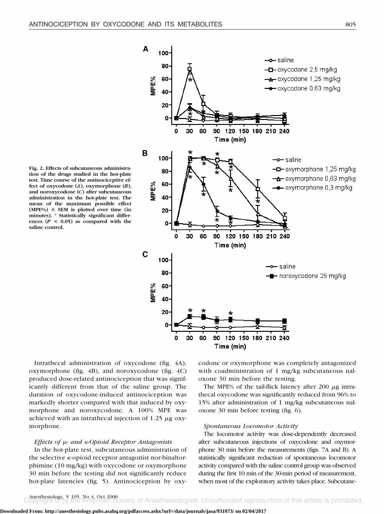
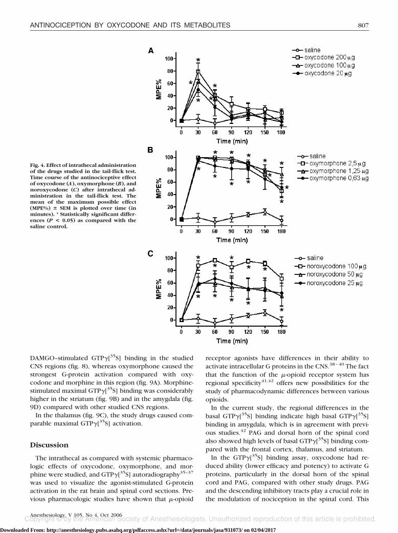
Intrathecal administration of oxycodone (fig. 4A),oxymorphone (fig. 4B), and noroxycodone (fig. 4C)produced dose-related antinociception that was signif-icantly different from that of the saline group. Theduration of oxycodone-induced antinociception wasmarkedly shorter compared with that induced by oxy-morphone and noroxycodone. A 100% MPE wasachieved with an intrathecal injection of 1.25 �g oxy-morphone.
Effects of �- and �-Opioid Receptor AntagonistsIn the hot-plate test, subcutaneous administration of
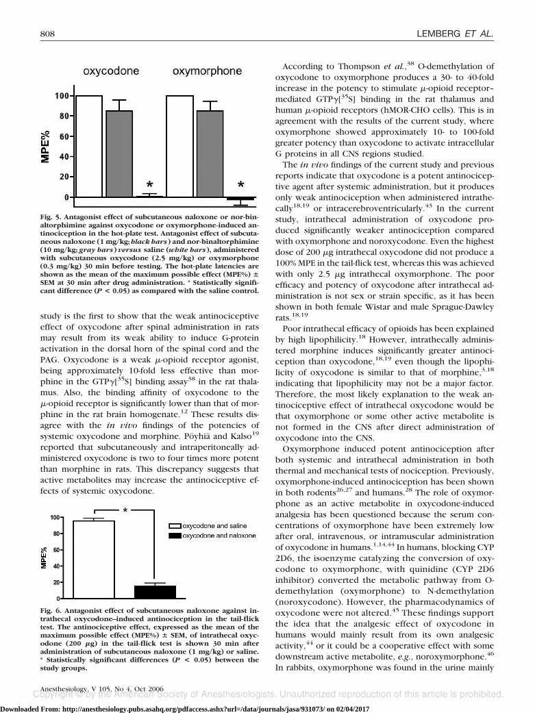
the selective �-opioid receptor antagonist nor-binaltor-phimine (10 mg/kg) with oxycodone or oxymorphone30 min before the testing did not significantly reducehot-plate latencies (fig. 5). Antinociception by oxy-
codone or oxymorphone was completely antagonizedwith coadministration of 1 mg/kg subcutaneous nal-oxone 30 min before the testing.
The MPE% of the tail-flick latency after 200 �g intra-thecal oxycodone was significantly reduced from 96% to15% after administration of 1 mg/kg subcutaneous nal-oxone 30 min before testing (fig. 6).
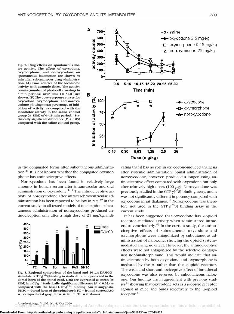
Spontaneous Locomotor ActivityThe locomotor activity was dose-dependently decreased
after subcutaneous injections of oxycodone and oxymor-phone 30 min before the measurements (figs. 7A and B). Astatistically significant reduction of spontaneous locomotoractivity compared with the saline control group was observedduring the first 10 min of the 30-min period of measurement,when most of the exploratory activity takes place. Subcutane-
Fig. 2. Effects of subcutaneous administra-tion of the drugs studied in the hot-platetest. Time course of the antinociceptive ef-fect of oxycodone (A), oxymorphone (B),and noroxycodone (C) after subcutaneousadministration in the hot-plate test. Themean of the maximum possible effect(MPE%) � SEM is plotted over time (inminutes). * Statistically significant differ-ences (P < 0.05) as compared with thesaline control.
805ANTINOCICEPTION BY OXYCODONE AND ITS METABOLITES
Anesthesiology, V 105, No 4, Oct 2006
Downloaded From: http://anesthesiology.pubs.asahq.org/pdfaccess.ashx?url=/data/journals/jasa/931073/ on 02/04/2017

ous administration of 25 mg/kg noroxycodone had no statisti-cally significant sedative effect compared with the saline control.
Opioid Receptor Signaling�-Opioid receptor–mediated G-protein activation was
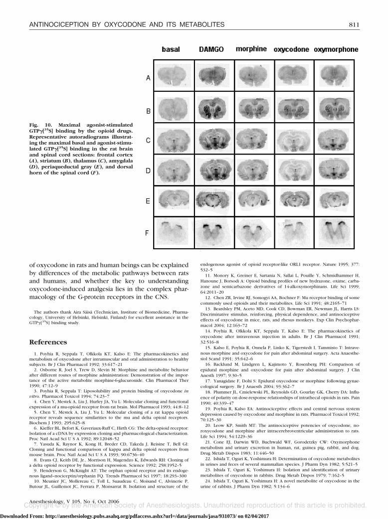
differentially observed in all studied rat brain regions andin the dorsal horn of the spinal cord with all study drugs,and it was absent in the presence of 10 �M naltrexone.There were also regional differences in the basalGTP�[35S] binding: The amygdala, periaqueductal gray(PAG), and dorsal horn of the spinal cord showed in-creased levels of basal GTP�[35S] binding compared withthe frontal cortex, thalamus, and striatum (figs. 8–10).
DAMGO-stimulated (10 �M) GTP�[35S] binding washigh in the striatum, amygdala, and dorsal horn of thespinal cord (fig. 8 and autoradiographs in figs. 10B, D,and F). Moderate stimulation of the GTP�[35S] binding
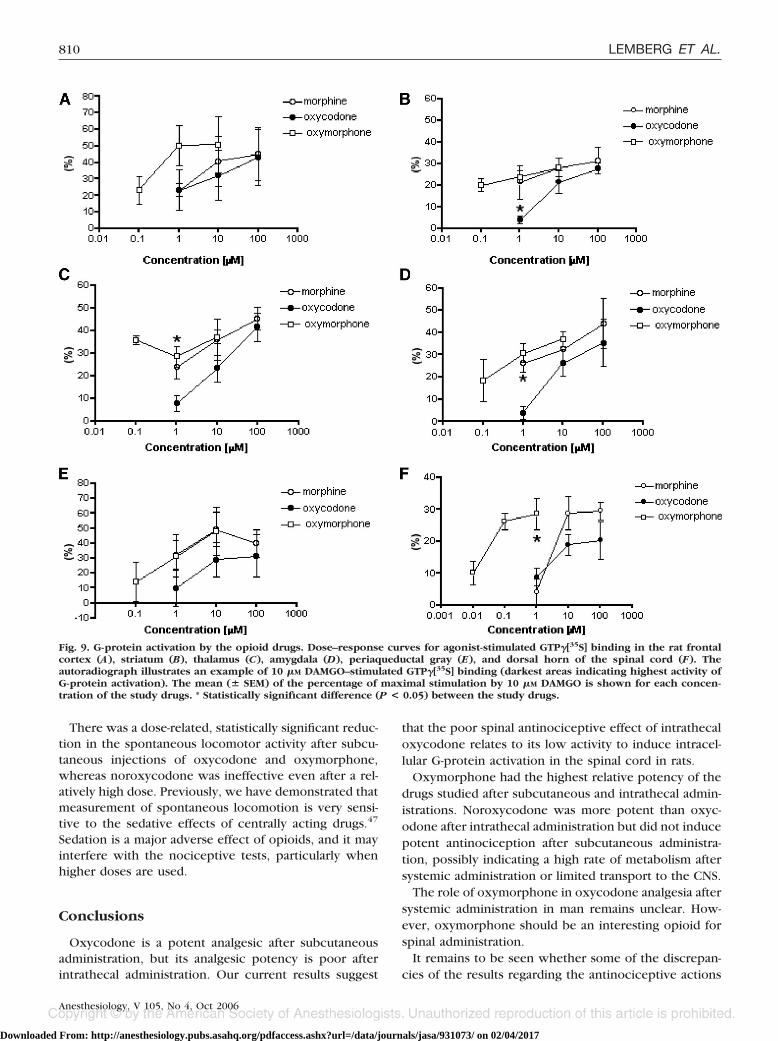
was observed in the thalamus and PAG (fig. 8 and auto-radiographs in figs. 10C and E). Low stimulation wasfound in the frontal cortex (fig. 8 and autoradiograph infig. 10A). In the spinal cord, �-opioid receptor–mediatedG-protein activation was mainly located in the mostsuperficial layers of the dorsal horn (fig. 10F). The max-imal G-protein activation in the spinal cord induced byoxycodone was lower than that induced by oxymor-phone and morphine (fig. 9F). The weakest effect ofoxycodone compared with morphine and oxymorphonewas observed in the PAG and the spinal cord (fig. 9E, F).
Oxymorphone was the most potent of the drugs in allof the CNS regions studied. All drugs studied were ap-parently partial agonists. Oxymorphone was in all re-gions as efficacious as morphine, but it needed approx-imately 10- to 100-fold lower concentrations thanoxycodone. The frontal cortex showed the lowest 10 �M
Fig. 3. Effects of subcutaneous administra-tion of the drugs studied in the paw-pres-sure test. Time course of the antinocicep-tive effect of oxycodone (A), oxymorphone(B), and noroxycodone (C) after subcuta-neous administration in the paw-pressuretest. The mean of the maximum possibleeffect (MPE%) � SEM is plotted over time(in minutes). * Statistically significant dif-ferences (P < 0.05) as compared with thesaline control.
806 LEMBERG ET AL.
Anesthesiology, V 105, No 4, Oct 2006
Downloaded From: http://anesthesiology.pubs.asahq.org/pdfaccess.ashx?url=/data/journals/jasa/931073/ on 02/04/2017
DAMGO–stimulated GTP�[35S] binding in the studiedCNS regions (fig. 8), whereas oxymorphone caused thestrongest G-protein activation compared with oxy-codone and morphine in this region (fig. 9A). Morphine-stimulated maximal GTP�[35S] binding was considerablyhigher in the striatum (fig. 9B) and in the amygdala (fig.9D) compared with other studied CNS regions.
In the thalamus (fig. 9C), the study drugs caused com-parable maximal GTP�[35S] activation.
Discussion
The intrathecal as compared with systemic pharmaco-logic effects of oxycodone, oxymorphone, and mor-phine were studied, and GTP�[35S] autoradiography35–37
was used to visualize the agonist-stimulated G-proteinactivation in the rat brain and spinal cord sections. Pre-vious pharmacologic studies have shown that �-opioid
receptor agonists have differences in their ability toactivate intracellular G proteins in the CNS.38–40 The factthat the function of the �-opioid receptor system hasregional specificity41,42 offers new possibilities for thestudy of pharmacodynamic differences between variousopioids.
In the current study, the regional differences in thebasal GTP�[35S] binding indicate high basal GTP�[35S]binding in amygdala, which is in agreement with previ-ous studies.42 PAG and dorsal horn of the spinal cordalso showed high levels of basal GTP�[35S] binding com-pared with the frontal cortex, thalamus, and striatum.
In the GTP�[35S] binding assay, oxycodone had re-duced ability (lower efficacy and potency) to activate Gproteins, particularly in the dorsal horn of the spinalcord and PAG, compared with other study drugs. PAGand the descending inhibitory tracts play a crucial role inthe modulation of nociception in the spinal cord. This
Fig. 4. Effect of intrathecal administrationof the drugs studied in the tail-flick test.Time course of the antinociceptive effectof oxycodone (A), oxymorphone (B), andnoroxycodone (C) after intrathecal ad-ministration in the tail-flick test. Themean of the maximum possible effect(MPE%) � SEM is plotted over time (inminutes). * Statistically significant differ-ences (P < 0.05) as compared with thesaline control.
807ANTINOCICEPTION BY OXYCODONE AND ITS METABOLITES
Anesthesiology, V 105, No 4, Oct 2006
Downloaded From: http://anesthesiology.pubs.asahq.org/pdfaccess.ashx?url=/data/journals/jasa/931073/ on 02/04/2017
study is the first to show that the weak antinociceptiveeffect of oxycodone after spinal administration in ratsmay result from its weak ability to induce G-proteinactivation in the dorsal horn of the spinal cord and thePAG. Oxycodone is a weak �-opioid receptor agonist,being approximately 10-fold less effective than mor-phine in the GTP�[35S] binding assay38 in the rat thala-mus. Also, the binding affinity of oxycodone to the�-opioid receptor is significantly lower than that of mor-phine in the rat brain homogenate.12 These results dis-agree with the in vivo findings of the potencies ofsystemic oxycodone and morphine. Poyhia and Kalso19
reported that subcutaneously and intraperitoneally ad-ministered oxycodone is two to four times more potentthan morphine in rats. This discrepancy suggests thatactive metabolites may increase the antinociceptive ef-fects of systemic oxycodone.
According to Thompson et al.,38 O-demethylation ofoxycodone to oxymorphone produces a 30- to 40-foldincrease in the potency to stimulate �-opioid receptor–mediated GTP�[35S] binding in the rat thalamus andhuman �-opioid receptors (hMOR-CHO cells). This is inagreement with the results of the current study, whereoxymorphone showed approximately 10- to 100-foldgreater potency than oxycodone to activate intracellularG proteins in all CNS regions studied.
The in vivo findings of the current study and previousreports indicate that oxycodone is a potent antinocicep-tive agent after systemic administration, but it producesonly weak antinociception when administered intrathe-cally18,19 or intracerebroventricularly.43 In the currentstudy, intrathecal administration of oxycodone pro-duced significantly weaker antinociception comparedwith oxymorphone and noroxycodone. Even the highestdose of 200 �g intrathecal oxycodone did not produce a100% MPE in the tail-flick test, whereas this was achievedwith only 2.5 �g intrathecal oxymorphone. The poorefficacy and potency of oxycodone after intrathecal ad-ministration is not sex or strain specific, as it has beenshown in both female Wistar and male Sprague-Dawleyrats.18,19
Poor intrathecal efficacy of opioids has been explainedby high lipophilicity.18 However, intrathecally adminis-tered morphine induces significantly greater antinoci-ception than oxycodone,18,19 even though the lipophi-licity of oxycodone is similar to that of morphine,3,18
indicating that lipophilicity may not be a major factor.Therefore, the most likely explanation to the weak an-tinociceptive effect of intrathecal oxycodone would bethat oxymorphone or some other active metabolite isnot formed in the CNS after direct administration ofoxycodone into the CNS.
Oxymorphone induced potent antinociception afterboth systemic and intrathecal administration in boththermal and mechanical tests of nociception. Previously,oxymorphone-induced antinociception has been shownin both rodents26,27 and humans.28 The role of oxymor-phone as an active metabolite in oxycodone-inducedanalgesia has been questioned because the serum con-centrations of oxymorphone have been extremely lowafter oral, intravenous, or intramuscular administrationof oxycodone in humans.1,14,44 In humans, blocking CYP2D6, the isoenzyme catalyzing the conversion of oxy-codone to oxymorphone, with quinidine (CYP 2D6inhibitor) converted the metabolic pathway from O-demethylation (oxymorphone) to N-demethylation(noroxycodone). However, the pharmacodynamics ofoxycodone were not altered.45 These findings supportthe idea that the analgesic effect of oxycodone inhumans would mainly result from its own analgesicactivity,44 or it could be a cooperative effect with somedownstream active metabolite, e.g., noroxymorphone.46
In rabbits, oxymorphone was found in the urine mainly
Fig. 5. Antagonist effect of subcutaneous naloxone or nor-bin-altorphimine against oxycodone or oxymorphone-induced an-tinociception in the hot-plate test. Antagonist effect of subcuta-neous naloxone (1 mg/kg; black bars) and nor-binaltorphimine(10 mg/kg; gray bars) versus saline (white bars), administeredwith subcutaneous oxycodone (2.5 mg/kg) or oxymorphone(0.3 mg/kg) 30 min before testing. The hot-plate latencies areshown as the mean of the maximum possible effect (MPE%) �SEM at 30 min after drug administration. * Statistically signifi-cant difference (P < 0.05) as compared with the saline control.
Fig. 6. Antagonist effect of subcutaneous naloxone against in-trathecal oxycodone–induced antinociception in the tail-flicktest. The antinociceptive effect, expressed as the mean of themaximum possible effect (MPE%) � SEM, of intrathecal oxyc-odone (200 �g) in the tail-flick test is shown 30 min afteradministration of subcutaneous naloxone (1 mg/kg) or saline.* Statistically significant differences (P < 0.05) between thestudy groups.
808 LEMBERG ET AL.
Anesthesiology, V 105, No 4, Oct 2006
Downloaded From: http://anesthesiology.pubs.asahq.org/pdfaccess.ashx?url=/data/journals/jasa/931073/ on 02/04/2017
in the conjugated forms after subcutaneous administra-tion.23 It is not known whether the conjugated oxymor-phone has antinociceptive effects.
Noroxycodone has been found in relatively largeamounts in human serum after intramuscular and oraladministration of oxycodone.1,44 The antinociceptive ac-tivity of noroxycodone after intracerebroventricular ad-ministration has been reported to be low in rats.20 In thecurrent study, in all tested models of nociception subcu-taneous administration of noroxycodone produced an-tinociception only after a high dose of 25 mg/kg, indi-
cating that it has no role in oxycodone-induced analgesiaafter systemic administration. Spinal administration ofnoroxycodone, however, produced a longer-lasting an-tinociceptive effect compared with oxycodone but onlyafter relatively high doses (100 �g). Noroxycodone waspreviously studied in the GTP�[35S] binding assay, and itwas not significantly different in potency compared withoxycodone in rat thalamus.38 Noroxycodone was there-fore not used in the GTP�[35S] binding assay in thecurrent study.
It has been suggested that oxycodone has �-opioidreceptor–mediated activity when administered intrac-erebroventricularly.43 In the current study, the antino-ciceptive effects of subcutaneous oxycodone andoxymorphone were antagonized by subcutaneous ad-ministration of naloxone, showing the opioid system–mediated analgesic effect. However, the antinociceptiveeffects were not antagonized by the selective �-antago-nist nor-binaltorphimine. This would indicate that an-tinociception by both oxycodone and oxymorphone ismediated by the �- rather than the �-opioid receptor.The weak and short antinociceptive effect of intrathecaloxycodone was also reversed by subcutaneous nalox-one. Our findings are in agreement with previous stud-ies13 showing that oxycodone acts as a �-opioid receptoragonist in mice and binds selectively to the �-opioidreceptor.11
Fig. 7. Drug effects on spontaneous mo-tor activity. The effects of oxycodone,oxymorphone, and noroxycodone onspontaneous locomotion are shown 30min after subcutaneous drug administra-tion. (A) Time courses of the locomotoractivity with example doses. The activitycounts (number of photocell crossings in5-min periods) over time (� SEM) areshown. (B) The dose–response curves foroxycodone, oxymorphone, and noroxy-codone plotting mean percentage of inhi-bition of activity, as compared with thelocomotor activity in the saline controlgroup (� SEM) of 0–15 min period. * Sta-tistically significant difference (P < 0.05)compared with the saline control group.
Fig. 8. Regional comparison of the basal and 10 �M DAMGO–stimulated GTP�[35S] binding in studied brain regions and in thedorsal horn of the spinal cord. Data are expressed as mean (�SEM) in nCi/g. * Statistically significant difference (P < 0.05) ascompared with the basal GTP�[35S] binding. Am � amygdala;DHSC � dorsal horn of the spinal cord; FC � frontal cortex; PAG� periaqueductal gray; Str � striatum; Th � thalamus.
809ANTINOCICEPTION BY OXYCODONE AND ITS METABOLITES
Anesthesiology, V 105, No 4, Oct 2006
Downloaded From: http://anesthesiology.pubs.asahq.org/pdfaccess.ashx?url=/data/journals/jasa/931073/ on 02/04/2017
There was a dose-related, statistically significant reduc-tion in the spontaneous locomotor activity after subcu-taneous injections of oxycodone and oxymorphone,whereas noroxycodone was ineffective even after a rel-atively high dose. Previously, we have demonstrated thatmeasurement of spontaneous locomotion is very sensi-tive to the sedative effects of centrally acting drugs.47
Sedation is a major adverse effect of opioids, and it mayinterfere with the nociceptive tests, particularly whenhigher doses are used.
Conclusions
Oxycodone is a potent analgesic after subcutaneousadministration, but its analgesic potency is poor afterintrathecal administration. Our current results suggest
that the poor spinal antinociceptive effect of intrathecaloxycodone relates to its low activity to induce intracel-lular G-protein activation in the spinal cord in rats.
Oxymorphone had the highest relative potency of thedrugs studied after subcutaneous and intrathecal admin-istrations. Noroxycodone was more potent than oxyc-odone after intrathecal administration but did not inducepotent antinociception after subcutaneous administra-tion, possibly indicating a high rate of metabolism aftersystemic administration or limited transport to the CNS.
The role of oxymorphone in oxycodone analgesia aftersystemic administration in man remains unclear. How-ever, oxymorphone should be an interesting opioid forspinal administration.
It remains to be seen whether some of the discrepan-cies of the results regarding the antinociceptive actions
Fig. 9. G-protein activation by the opioid drugs. Dose–response curves for agonist-stimulated GTP�[35S] binding in the rat frontalcortex (A), striatum (B), thalamus (C), amygdala (D), periaqueductal gray (E), and dorsal horn of the spinal cord (F). Theautoradiograph illustrates an example of 10 �M DAMGO–stimulated GTP�[35S] binding (darkest areas indicating highest activity ofG-protein activation). The mean (� SEM) of the percentage of maximal stimulation by 10 �M DAMGO is shown for each concen-tration of the study drugs. * Statistically significant difference (P < 0.05) between the study drugs.
810 LEMBERG ET AL.
Anesthesiology, V 105, No 4, Oct 2006
Downloaded From: http://anesthesiology.pubs.asahq.org/pdfaccess.ashx?url=/data/journals/jasa/931073/ on 02/04/2017
of oxycodone in rats and human beings can be explainedby differences of the metabolic pathways between ratsand humans, and whether the key to understandingoxycodone-induced analgesia lies in the complex phar-macology of the G-protein receptors in the CNS.
The authors thank Aira Saisa (Technician, Institute of Biomedicine, Pharma-cology, University of Helsinki, Helsinki, Finland) for excellent assistance in theGTP�[35S] binding study.
References
1. Poyhia R, Seppala T, Olkkola KT, Kalso E: The pharmacokinetics andmetabolism of oxycodone after intramuscular and oral administration to healthysubjects. Br J Clin Pharmacol 1992; 33:617–21
2. Osborne R, Joel S, Trew D, Slevin M: Morphine and metabolite behaviorafter different routes of morphine administration: Demonstration of the impor-tance of the active metabolite morphine-6-glucuronide. Clin Pharmacol Ther1990; 47:12–9
3. Poyhia R, Seppala T: Liposolubility and protein binding of oxycodone invitro. Pharmacol Toxicol 1994; 74:23–7
4. Chen Y, Mestek A, Liu J, Hurley JA, Yu L: Molecular cloning and functionalexpression of a mu-opioid receptor from rat brain. Mol Pharmacol 1993; 44:8–12
5. Chen Y, Mestek A, Liu J, Yu L: Molecular cloning of a rat kappa opioidreceptor reveals sequence similarities to the mu and delta opioid receptors.Biochem J 1993; 295:625–8
6. Kieffer BL, Befort K, Gaveriaux-Ruff C, Hirth CG: The delta-opioid receptor:Isolation of a cDNA by expression cloning and pharmacological characterization.Proc Natl Acad Sci U S A 1992; 89:12048–52
7. Yasuda K, Raynor K, Kong H, Breder CD, Takeda J, Reisine T, Bell GI:Cloning and functional comparison of kappa and delta opioid receptors frommouse brain. Proc Natl Acad Sci U S A 1993; 90:6736–40
8. Evans CJ, Keith DE, Jr., Morrison H, Magendzo K, Edwards RH: Cloning ofa delta opioid receptor by functional expression. Science 1992; 258:1952–5
9. Henderson G, McKnight AT: The orphan opioid receptor and its endoge-nous ligand–nociceptin/orphanin FQ. Trends Pharmacol Sci 1997; 18:293–300
10. Meunier JC, Mollereau C, Toll L, Suaudeau C, Moisand C, Alvinerie P,Butour JL, Guillemot JC, Ferrara P, Monsarrat B: Isolation and structure of the
endogenous agonist of opioid receptor-like ORL1 receptor. Nature 1995; 377:532–5
11. Monory K, Greiner E, Sartania N, Sallai L, Pouille Y, Schmidhammer H,Hanoune J, Borsodi A: Opioid binding profiles of new hydrazone, oxime, carba-zone and semicarbazone derivatives of 14-alkoxymorphinans. Life Sci 1999;64:2011–20
12. Chen ZR, Irvine RJ, Somogyi AA, Bochner F: Mu receptor binding of somecommonly used opioids and their metabolites. Life Sci 1991; 48:2165–71
13. Beardsley PM, Aceto MD, Cook CD, Bowman ER, Newman JL, Harris LS:Discriminative stimulus, reinforcing, physical dependence, and antinociceptiveeffects of oxycodone in mice, rats, and rhesus monkeys. Exp Clin Psychophar-macol 2004; 12:163–72
14. Poyhia R, Olkkola KT, Seppala T, Kalso E: The pharmacokinetics ofoxycodone after intravenous injection in adults. Br J Clin Pharmacol 1991;32:516–8
15. Kalso E, Poyhia R, Onnela P, Linko K, Tigerstedt I, Tammisto T: Intrave-nous morphine and oxycodone for pain after abdominal surgery. Acta Anaesthe-siol Scand 1991; 35:642–6
16. Backlund M, Lindgren L, Kajimoto Y, Rosenberg PH: Comparison ofepidural morphine and oxycodone for pain after abdominal surgery. J ClinAnesth 1997; 9:30–5
17. Yanagidate F, Dohi S: Epidural oxycodone or morphine following gynae-cological surgery. Br J Anaesth 2004; 93:362–7
18. Plummer JL, Cmielewski PL, Reynolds GD, Gourlay GK, Cherry DA: Influ-ence of polarity on dose-response relationships of intrathecal opioids in rats. Pain1990; 40:339–47
19. Poyhia R, Kalso EA: Antinociceptive effects and central nervous systemdepression caused by oxycodone and morphine in rats. Pharmacol Toxicol 1992;70:125–30
20. Leow KP, Smith MT: The antinociceptive potencies of oxycodone, no-roxycodone and morphine after intracerebroventricular administration to rats.Life Sci 1994; 54:1229–36
21. Cone EJ, Darwin WD, Buchwald WF, Gorodetzky CW: Oxymorphonemetabolism and urinary excretion in human, rat, guinea pig, rabbit, and dog.Drug Metab Dispos 1983; 11:446–50
22. Ishida T, Oguri K, Yoshimura H: Determination of oxycodone metabolitesin urines and feces of several mammalian species. J Pharm Dyn 1982; 5:521–5
23. Ishida T, Oguri K, Yoshimura H: Isolation and identification of urinarymetabolites of oxycodone in rabbits. Drug Metab Dispos 1979; 7:162–5
24. Ishida T, Oguri K, Yoshimura H: A novel metabolite of oxycodone in theurine of rabbits. J Pharm Dyn 1982; 5:134–6
Fig. 10. Maximal agonist-stimulatedGTP�[35S] binding by the opioid drugs.Representative autoradiograms illustrat-ing the maximal basal and agonist-stimu-lated GTP�[35S] binding in the rat brainand spinal cord sections: frontal cortex(A), striatum (B), thalamus (C), amygdala(D), periaqueductal gray (E), and dorsalhorn of the spinal cord (F).
811ANTINOCICEPTION BY OXYCODONE AND ITS METABOLITES
Anesthesiology, V 105, No 4, Oct 2006
Downloaded From: http://anesthesiology.pubs.asahq.org/pdfaccess.ashx?url=/data/journals/jasa/931073/ on 02/04/2017
25. Weinstein SH, Gaylord JC: Determination of oxycodone in plasma andidentification of a major metabolite. J Pharm Sci 1979; 68:527–8
26. Briggs SL, Sawyer DC, Rech RH, Galligan JJ: Oxymorphone-induced anal-gesia and colonic motility measured in colorectal distension. Pharmacol BiochemBehav 1995; 52:561–3
27. Bolan EA, Tallarida RJ, Pasternak GW: Synergy between mu opioid ligands:Evidence for functional interactions among mu opioid receptor subtypes.J Pharmacol Exp Ther 2002; 303:557–62
28. Beaver WT, Feise GA: A comparison of the analgesic effect of oxymor-phone by rectal suppository and intramuscular injection in patients with post-operative pain. J Clin Pharmacol 1977; 17:276–91
29. Kalso E, Vainio A, Mattila MJ, Rosenberg PH, Seppala T: Morphine andoxycodone in the management of cancer pain: Plasma levels determined bychemical and radioreceptor assays. Pharmacol Toxicol 1990; 67:322–8
30. Zimmermann M: Ethical guidelines for investigations of experimental painin conscious animals. Pain 1983; 16:109–10
31. Cheng CY, Hsin LW, Lin YP, Tao PL, Jong TT: N-cubylmethyl substitutedmorphinoids as novel narcotic antagonists. Bioorg Med Chem 1996; 4:73–80
32. Iijima I, Minamikawa J, Jacobson AE, Brossi A, Rice KC: Studies in the(�)-morphinan series: 5. Synthesis and biological properties of (�)-naloxone.J Med Chem 1978; 21:398–400
33. Coop A, Janetka JW, Lewis JW, Rice KC: L-Selectride as a general reagentfor the O-demethylation and N-decarbomethoxylation of opium alkaloids andderivates. J Org Chem 1998; 63:4392–6
34. Yaksh TL, Rudy TA: Chronic catheterization of the spinal subarachnoidspace. Physiol Behav 1976; 17:1031–6
35. Laitinen JT: Selective detection of adenosine A1 receptor-dependent G-protein activity in basal and stimulated conditions of rat brain [35S]guanosine5=-(gamma-thio)triphosphate autoradiography. Neuroscience 1999; 90:1265–79
36. Laitinen JT, Jokinen M: Guanosine 5=-(gamma-[35S]thio)triphosphate au-toradiography allows selective detection of histamine H3 receptor-dependent Gprotein activation in rat brain tissue sections. J Neurochem 1998; 71:808–16
37. Sim LJ, Selley DE, Childers SR: In vitro autoradiography of receptor-
activated G proteins in rat brain by agonist-stimulated guanylyl 5=-[gamma-[35S]thio]-triphosphate binding. Proc Natl Acad Sci U S A 1995; 92:7242–6
38. Thompson CM, Wojno H, Greiner E, May EL, Rice KC, Selley DE: Activa-tion of G-proteins by morphine and codeine congeners: insights to the relevanceof O- and N-demethylated metabolites at mu- and delta-opioid receptors. J Phar-macol Exp Ther 2004; 308:547–54
39. Selley DE, Sim LJ, Xiao R, Liu Q, Childers SR: Mu-opioid receptor-stimu-lated guanosine-5=-O-(gamma-thio)-triphosphate binding in rat thalamus and cul-tured cell lines: Signal transduction mechanisms underlying agonist efficacy. MolPharmacol 1997; 51:87–96
40. Traynor JR, Nahorski SR: Modulation by mu-opioid agonists of guanosine-5=-O-(3-[35S]thio)triphosphate binding to membranes from human neuroblas-toma SH-SY5Y cells. Mol Pharmacol 1995; 47:848–54
41. Maher CE, Selley DE, Childers SR: Relationship of mu opioid receptorbinding to activation of G-proteins in specific rat brain regions. Biochem Phar-macol 2000; 59:1395–401
42. Sim LJ, Selley DE, Dworkin SI, Childers SR: Effects of chronic morphineadministration on mu opioid receptor-stimulated [35S]GTP�S autoradiography inrat brain. J Neurosci 1996; 16:2684–92
43. Ross FB, Smith MT: The intrinsic antinociceptive effects of oxycodoneappear to be kappa-opioid receptor mediated. Pain 1997; 73:151–7
44. Kaiko RF, Benziger DP, Fitzmartin RD, Burke BE, Reder RF, GoldenheimPD: Pharmacokinetic-pharmacodynamic relationships of controlled-release oxy-codone. Clin Pharmacol Ther 1996; 59:52–61
45. Heiskanen T, Olkkola KT, Kalso E: Effects of blocking CYP2D6 on thepharmacokinetics and pharmacodynamics of oxycodone. Clin Pharmacol Ther1998; 64:603–11
46. Lalovic B, Phillips B, Risler LL, Howald W, Shen DD: Quantitative contri-bution of CYP2D6 and CYP3A to oxycodone metabolism in human liver andintestinal microsomes. Drug Metab Dispos 2004; 32:447–54
47. Xu M, Wei H, Kontinen VK, Kalso E, Pertovaara A: The dissociation ofsedative from spinal antinociceptive effects following administration of a novelalpha-2-adrenoceptor agonist, MPV-2426, in the locus coeruleus in the rat. ActaAnaesth Scand 2000; 44:648–55
812 LEMBERG ET AL.
Anesthesiology, V 105, No 4, Oct 2006
Downloaded From: http://anesthesiology.pubs.asahq.org/pdfaccess.ashx?url=/data/journals/jasa/931073/ on 02/04/2017