Neurophysiological effects of spinal manipulation - CiteSeerX
15
The Spine Journal 2 (2002) 357–371 1529-9430/02/$ – see front matter © 2002 Elsevier Science Inc. All rights reserved. PII: S1529-9430(02)00400-X Review Article Neurophysiological effects of spinal manipulation Joel G. Pickar, DC, PhD* Palmer Center for Chiropractic Research, 1000 Brady Street, Davenport, IA 52803, USA Received 23 February 2001; accepted 15 May 2002 Abstract Background context: Despite clinical evidence for the benefits of spinal maniputation and the appar- ent wide usage of it, the biological mechanisms underlying the effects of spinal manipulation are not known. Although this does not negate the clinical effects of spinal manipulation, it hinders acceptance by the wider scientific and health-care communities and hinders rational strategies for improving the delivery of spinal manipulation. Purpose: The purpose of this review article is to examine the neurophysiological basis for the effects of spinal manipulation. Study desig n : A review article discussing primarily basic science literature and clinically oriented basic science studies. Methods: This review article draws primarily from the peer-reviewed literature available on Med- line. Several textbook publications and reports are referenced. A theoretical model is presented de- scribing the relationships between spinal manipulation, segmental biomechanics, the nervous system and end-organ physiology. Experimental data for these relationships are presented. Results: Biomechanical changes caused by spinal manipulation are thought to have physiological con- sequences by means of their effects on the inflow of sensory information to the central nervous system. Muscle spindle afferents and Golgi tendon organ afferents are stimulated by spinal manipulation. Smaller-diameter sensory nerve fibers are likely activated, although this has not been demonstrated di- rectly. Mechanical and chemical changes in the intervertebral foramen caused by a herniated interver- tebral disc can affect the dorsal roots and dorsal root ganglia, but it is not known if spinal manipulation directly affects these changes. Individuals with herniated lumbar discs have shown clinical improve- ment in response to spinal manipulation. The phenomenon of central facilitation is known to increase the receptive field of central neurons, enabling either subthreshold or innocuous stimuli access to cen- tral pain pathways. Numerous studies show that spinal manipulation increases pain tolerance or its threshold. One mechanism underlying the effects of spinal manipulation may, therefore, be the manip- ulation’s ability to alter central sensory processing by removing subthreshold mechanical or chemical stimuli from paraspinal tissues. Spinal manipulation is also thought to affect reflex neural outputs to both muscle and visceral organs. Substantial evidence demonstrates that spinal manipulation evokes paraspinal muscle reflexes and alters motoneuron excitability. The effects of spinal manipulation on these somatosomatic reflexes may be quite complex, producing excitatory and inhibitory effects. Whereas substantial information also shows that sensory input, especially noxious input, from paraspinal tissues can reflexively elicit sympathetic nerve activity, knowledge about spinal manipu- lation’s effects on these reflexes and on end-organ function is more limited. Conclusions: A theoretical framework exists from which hypotheses about the neurophysiological effects of spinal manipulation can be developed. An experimental body of evidence exists indicating that spinal manipulation impacts primary afferent neurons from paraspinal tissues, the motor control system and pain processing. Experimental work in this area is warranted and should be encouraged to help better understand mechanisms underlying the therapeutic scope of spinal manipulation. © 2002 Elsevier Science Inc. All rights reserved. Keywords: Spinal manipulation; Neurophysiology; Manual therapy; Manual medicine; Chiropractic; Osteopathy FDA device/drug status: not applicable. Nothing of value received from a commercial entity related to this research. This study was supported by the Consortial Center for Chiropractic Research through cooperative agreement 1-U24AR5166 funded by National Institutes of Health, National Center for Complementary and Alternative Medicine and by the National Institute of Neurological Disease and Stroke Grant NS35300 on behalf of the Office of Alternative Medicine. * Corresponding author. Palmer Center for Chiropractic Research, 1000 Brady Street, Davenport, IA 52803, USA. Tel.: (563) 884-5219; fax: (563) 884-5227. E-mail address: [email protected] (J.G. Pickar)
-
Upload
khangminh22 -
Category
Documents
-
view
2 -
download
0
Transcript of Neurophysiological effects of spinal manipulation - CiteSeerX

The Spine Journal 2 (2002) 357–371
1529-9430/02/$ – see front matter © 2002 Elsevier Science Inc. All rights reserved.PII: S1529-9430(02)00400-X
Review Article
Neurophysiological effects of spinal manipulation
Joel G. Pickar, DC, PhD*
Palmer Center for Chiropractic Research, 1000 Brady Street, Davenport, IA 52803, USA
Received 23 February 2001; accepted 15 May 2002
Abstract
Background context:
Despite clinical evidence for the benefits of spinal maniputation and the appar-ent wide usage of it, the biological mechanisms underlying the effects of spinal manipulation are notknown. Although this does not negate the clinical effects of spinal manipulation, it hinders acceptanceby the wider scientific and health-care communities and hinders rational strategies for improving thedelivery of spinal manipulation.
Purpose:
The purpose of this review article is to examine the neurophysiological basis for the effectsof spinal manipulation.
Study desig
n
:
A review article discussing primarily basic science literature and clinically orientedbasic science studies.
Methods:
This review article draws primarily from the peer-reviewed literature available on Med-line. Several textbook publications and reports are referenced. A theoretical model is presented de-scribing the relationships between spinal manipulation, segmental biomechanics, the nervous systemand end-organ physiology. Experimental data for these relationships are presented.
Results:
Biomechanical changes caused by spinal manipulation are thought to have physiological con-sequences by means of their effects on the inflow of sensory information to the central nervous system.Muscle spindle afferents and Golgi tendon organ afferents are stimulated by spinal manipulation.Smaller-diameter sensory nerve fibers are likely activated, although this has not been demonstrated di-rectly. Mechanical and chemical changes in the intervertebral foramen caused by a herniated interver-tebral disc can affect the dorsal roots and dorsal root ganglia, but it is not known if spinal manipulationdirectly affects these changes. Individuals with herniated lumbar discs have shown clinical improve-ment in response to spinal manipulation. The phenomenon of central facilitation is known to increasethe receptive field of central neurons, enabling either subthreshold or innocuous stimuli access to cen-tral pain pathways. Numerous studies show that spinal manipulation increases pain tolerance or itsthreshold. One mechanism underlying the effects of spinal manipulation may, therefore, be the manip-ulation’s ability to alter central sensory processing by removing subthreshold mechanical or chemicalstimuli from paraspinal tissues. Spinal manipulation is also thought to affect reflex neural outputs toboth muscle and visceral organs. Substantial evidence demonstrates that spinal manipulation evokesparaspinal muscle reflexes and alters motoneuron excitability. The effects of spinal manipulation onthese somatosomatic reflexes may be quite complex, producing excitatory and inhibitory effects.Whereas substantial information also shows that sensory input, especially noxious input, fromparaspinal tissues can reflexively elicit sympathetic nerve activity, knowledge about spinal manipu-lation’s effects on these reflexes and on end-organ function is more limited.
Conclusions:
A theoretical framework exists from which hypotheses about the neurophysiologicaleffects of spinal manipulation can be developed. An experimental body of evidence exists indicatingthat spinal manipulation impacts primary afferent neurons from paraspinal tissues, the motor controlsystem and pain processing. Experimental work in this area is warranted and should be encouraged tohelp better understand mechanisms underlying the therapeutic scope of spinal manipulation. © 2002Elsevier Science Inc. All rights reserved.
Keywords:
Spinal manipulation; Neurophysiology; Manual therapy; Manual medicine; Chiropractic; Osteopathy
FDA device/drug status: not applicable.Nothing of value received from a commercial entity related to this research.This study was supported by the Consortial Center for Chiropractic
Research through cooperative agreement 1-U24AR5166 funded by NationalInstitutes of Health, National Center for Complementary and Alternative
Medicine and by the National Institute of Neurological Disease and StrokeGrant NS35300 on behalf of the Office of Alternative Medicine.
* Corresponding author. Palmer Center for Chiropractic Research,1000 Brady Street, Davenport, IA 52803, USA. Tel.: (563) 884-5219; fax:(563) 884-5227.
E-mail address
: [email protected] (J.G. Pickar)

358
J.G. Pickar / The Spine Journal 2 (2002) 357–371
Introduction
Recent reports estimate that 7.7% to 8.3% of the US pop-ulation uses some form of complementary or alternativemedicine [1–3]. Approximately 30% to 40% of these indi-viduals likely receive spinal manipulation [1]. Strong evi-dence supports using spinal manipulation to help patientswith acute low back pain and neck pain [4,5]. The benefitsof spinal manipulation for other disorders, such as chroniclow back pain and visceral disorders, are less clear, althoughbenefits have been noted [4,6–8]. Despite the clinical evi-dence for the benefits of and the apparent wide usage of spi-nal manipulation, the biological mechanisms underlying theeffects of spinal manipulation are not known. Although thisdoes not negate the clinical effects of spinal manipulation, ithinders acceptance by the wider scientific and health-carecommunities and hinders rational strategies for improvingthe delivery of spinal manipulation. The purpose of this re-view article is to examine the neurophysiological basis forand the neurophysiological effects of spinal manipulation.
Biomechanical considerations of spinal manipulation
Spinal manipulation by its very nature is a mechanicalinput to tissues of the vertebral column. Chiropractors de-liver more than 90% of these manipulations in the UnitedStates [9]. Spinal manipulation is distinguished from spinalmobilization in several ways [10]. During spinal manipula-tion, the practitioner delivers a dynamic thrust (impulse) toa specific vertebra. The clinician controls the velocity, mag-nitude and direction of the impulse [11]. The art or skill ofspinal manipulation lies in the clinician’s ability to controlthese three factors once the specific contact with a vertebrais made. Mobilization techniques are sometimes used prepa-ratory to the manipulation. Manipulation is also distinguishedfrom mobilization in that it is delivered at or near the end ofthe physiological range of motion (the so-called paraphysio-logical range [12]) but not exceeding the anatomical limits ofmotion. A cracking or popping sound often, but not necessar-ily, accompanies the manipulation, because gapping the jointcreates fluid cavitation [13,14].
The most common form of spinal manipulation used bychiropractors is the short-lever, high-velocity and low-amplitude thrust [15]. The clinician usually delivers thedynamic thrust through a short-lever arm by manually con-tacting paraspinal tissues overlying the spinous, transverseor mammillary processes of the vertebra being manipulated.Alternatively, the clinician contacts tissues overlying thelamina or articular pillar of the vertebra. To manipulate thepelvis, the iliac spine or the ischial spine is used [10]. Spinalmanipulation may also be delivered through a long-leverarm. While one hand may contact a specific area over thevertebra being manipulated, the second hand contacts anarea of the body distant from the specific contact. Force isdeveloped through this long-lever arm. However, using ashort-lever arm applied directly over the vertebra minimizes
the force necessary to accomplish the manipulation [10] byreducing the amount of compliant tissue through which theforce must be transmitted.
Several laboratories have studied biomechanical featuresof short-lever, high-velocity and low-amplitude manipula-tion. Herzog’s group [16] was the first to report the biome-chanical features of a spinal manipulation in an indexedjournal. They identified two characteristics common to thedelivery of a spinal manipulation: 1) a preload force fol-lowed by 2) a larger impulse force. Using two chiropractors,they quantified the preload and peak impulse forces appliedperpendicular to the contact point and the impulse durationduring manipulation of the sacroiliac joint. Preload loadforces ranged from 20 to 180 N, and peak forces ranged from220 to 550 N. Often the preload was approximately 25% ofimpulse load. The duration of the high-velocity impulseranged from 200 to 420 ms.
A number of studies have confirmed the force–time pro-file initially described by Hessel et al. [16]. Herzog et al.[17] showed the time to peak impulse was similar duringmanipulation of the thoracic spine and sacroiliac joint (ap-proximately 150 ms
�
77 ms, mean
�
SD). The perpendicu-larly applied preload and peak impulse forces were also similarduring spinal manipulations applied to the thoracic (139
�
46 Nvs. 88
�
78 N, respectively) and sacroiliac (328
�
78 N vs.399
�
119 N, respectively) regions. Studies of the cervicalspine indicate that preload, peak impulse force and time topeak impulse are less compared with the thoracic and lum-bosacral spine [17–19]. Depending upon the type of cervicalmanipulative technique used, preload forces range from 0 toapproximately 50 N, and peak impulse forces range fromapproximately 40 N to approximately 120 N. The forces de-livered during cervical manipulations develop faster thanduring manipulation of the thoracic spine and sacroiliac joint.Impulse duration lasts from approximately 30 ms to approxi-mately 120 ms. The large variability in the applied forces anddurations should be recognized. The impact of this variabilityon the biological mechanisms that could contribute to theclinical effects of manipulation is unknown.
A complete understanding of the biomechanics of spi-nal manipulation requires knowing the manner in whichmanipulative loads are transmitted to a specific vertebra.Experimentally, this is substantially more difficult and morecomplex compared with measuring applied loads. Transmit-ted loads may be different from applied loads because of theeffects of patient positioning and the contributions from in-ertial loads, loading moments and the active and passiveproperties of the intervening connective and muscle tissues.Triano and Schultz [20] calculated peak transmitted loads ata lumbar segment by measuring loads transmitted to a forceplate placed under the subject. The force plate was capableof transducing forces and moments about three orthogonalaxes. Peak forces transmitted to a lumbar segment during aside posture spinal manipulation tended to be higher thanpeak forces applied during a prone thoracic or sacroiliacmanipulation measured by Herzog et al. [17]. Transmitted

J.G. Pickar / The Spine Journal 2 (2002) 357–371
359
impulse durations were similar to applied impulse durationsmeasured by Herzog et al. [17]. Peak transmitted momentswere approximately three to four times less than peak trans-mitted forces. The transmitted loads were considered belowa threshold level capable of injuring the lumbar spine (see[20] for further discussion).
In addition to applied and transmitted loads, the relative dis-placement or movement between contiguous vertebrae duringa spinal manipulation has been studied. Nathan and Keller [21]measured intervertebral lumbar motion using pins inserted intolumbar spinous processes. Manipulations were delivered usinga mechanical adjusting device (Activator Adjusting Instru-ment, Activator Methods International, Ltd., Phoenix, AZ[22]). Impulse duration using this device is approximately5 ms, an impulse duration shorter than that from manual ma-nipulation. Impulses delivered to the L2 spinous produced 1.62mm
�
1.06 mm peak axial displacement (in the longitudinalplane), 0.48
�
0.1 mm shear displacement (in the transverseplane) and 0.89
�
0.49° of rotation between L3 and L4 [21].Smith et al. [23] measured similar vertebral displacements inthe lumbar spine of the dog. L2 translated 0.71
�
0.03 mm androtated 0.53
�
0.15° on L3 with impulse loads of 53 N. Gal etal. [24] performed measurements in the thoracic spine, but theirresults are difficult to compare with those reported above forthe lumbar spine. Nonetheless, the movements induced duringa spinal manipulative load suggest that mechanical processesmay play a role in the biological effects of spinal manipulation.
Neurophysiological and biomechanical mechanisms underlying the effects of spinal manipulation
Numerous theories have been proposed to explain the ef-fects of spinal manipulation [25,26]. A thread common tomany of these theories is that changes in the normal anatomi-cal, physiological or biomechanical dynamics of contiguousvertebrae can adversely affect function of the nervous system.[27,28]. Spinal manipulation is thought to correct these changes.
Accordingly, a number of biomechanical changes pro-duced by vertebral movement during a spinal manipulationhave been hypothesized. The mechanical force introducedinto the vertebral column during a spinal manipulation maydirectly alter segmental biomechanics by releasing trappedmeniscoids, releasing adhesions or by reducing distortion ofthe annulus fibrosus [29–33]. In addition, individual motionsegments can buckle, thereby producing relatively large ver-tebral motions that achieve a new position of stable equilib-rium [34]. The mechanical changes elicited by manipulationmay provide sufficient energy to restore a buckled segment toa lower energy level, thus reducing mechanical stress orstrain on soft and hard paraspinal tissues [35]. A major con-sequence of these hypothesized mechanical changes elicitedby manipulation could be the restoration of zygapophysealjoint mobility and joint play [31]. In fact, authoritative dis-cussion of spinal manipulation considers “the goal of ma-nipulation to restore maximal, pain-free movement of themusculoskeletal system” (from [35a] and see [31,36,37]).
Biomechanical changes caused by the manipulation arethought to have physiological consequences by means oftheir effects on the inflow of sensory information to the cen-tral nervous system [25,36]. By releasing trapped meniscoids,discal material or segmental adhesions, or by normalizing abuckled segment, the mechanical input may ultimately re-duce nociceptive input from receptive nerve endings in inner-vated paraspinal tissues. This would be consistent with theobservation that spinal manipulation is not painful when ad-ministered correctly. In addition, the mechanical thrust couldeither stimulate or silence nonnociceptive, mechanosensitivereceptive nerve endings in paraspinal tissues, including skin,muscle, tendons, ligaments, facet joints and intervertebraldisc [28,38,39]. These neural inputs may influence pain-producing mechanisms as well as other physiological sys-tems controlled or influenced by the nervous system.
Fig. 1 diagrams the theoretical relationships between spi-nal manipulation, segmental biomechanics, the nervous sys-tem and end-organ physiology. A biomechanical alterationbetween vertebral segments hypothetically produces a bio-mechanical overload the effects of which may alter the sig-naling properties of mechanically or chemically sensitiveneurons in paraspinal tissues. These changes in sensory in-put are thought to modify neural integration either by di-rectly affecting reflex activity and/or by affecting centralneural integration within motor, nociceptive and possiblyautonomic neuronal pools. Either of these changes in sen-sory input may elicit changes in efferent somatomotor andvisceromotor activity. Pain, discomfort, altered muscle func-tion or altered visceromotor activities comprise the signs orsymptoms that might cause patients to seek spinal manipula-tion. Spinal manipulation, then, theoretically alters the inflowof sensory signals from paraspinal tissues in a manner thatimproves physiological function. This explanation comprisesone of the most rational neurophysiological bases for themechanisms underlying the effects of spinal manipulation.Experimental efforts to understand sensory processing fromparaspinal tissues and the effects of spinal manipulation onthis sensory processing is receiving increasing attention, asdescribed below. Each of the following sections addresses acomponent of the theoretical relationship depicted in Fig. 1with each section’s number corresponding to a numberedcomponent in the figure.
1. The effects of spinal manipulation on sensory receptors in paraspinal tissues
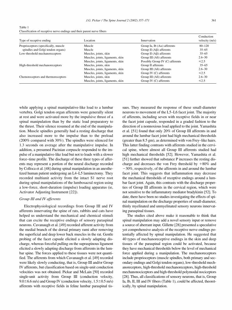
Group I and II afferents (proprioceptive afferents)
Korr [36] proposed that spinal manipulation increasesjoint mobility by producing a barrage of impulses in musclespindle afferents and smaller-diameter afferents ultimatelysilencing facilitated
�
motoneurons. Fig. 2 shows the neuralcircuitry of the
�
loop. He hypothesized that
�
-motoneurondischarge is elevated in muscles of vertebral segments re-sponding to spinal manipulation. The high gain of the
�

360
J.G. Pickar / The Spine Journal 2 (2002) 357–371
loop would impair joint mobility by sensitizing the stretchreflex to abnormally small changes in muscle length. Korrfurther hypothesized that spinal manipulation stimulatesmuscle spindle afferents, that is, Group Ia and possiblyGroup II afferents (Table 1). The barrage of impulses fromthese afferents produced by the spinal manipulation wouldreduce the gain of the
�
loop through an undetermined neu-ral pathway. Although portions of this mechanism remainspeculative, the contribution of proprioceptive afferents tospine function and the neurophysiological effects of spinalmanipulation on these afferents are receiving increasing at-tention.
The importance of paraspinal proprioceptive input to thefunction of the vertebral column, and of the lumbar spine in
particular, has been demonstrated recently in humans. Severalstudies indicate that muscle spindle input from the lumbarmultifidus helps to accurately position the pelvis and lum-bosacral spine. Healthy individuals can accurately repositiontheir lumbosacral spine, but their repositioning ability is im-paired when the multifidus muscle is vibrated [40]. Vibrationstimulates muscle spindles and creates a sensory illusion thatthe multifidus is stretched and therefore that the spine isflexed more than it actually is. The repositioning error occursbecause of the misperception of vertebral position. Interest-ingly, lumbosacral-repositioning ability is impaired in indi-viduals with a history of low back pain, even in the absenceof vibration [41]. This finding was associated with alteredproprioceptive input from muscle spindles [41]. In addition,paraspinal muscles in individuals with a history of low backpain also have longer response times to sudden loads, whichalso suggests the presence of abnormal paraspinal proprio-ceptive input in these individuals [42–44].
Two experimental models have been developed recentlythat should enhance our neurophysiological understandingof the lumbar and cervical spine in general and of spinal ma-nipulation specifically [45,46]. The experimental preparationsenable the recording of neural activity from paraspinal tissuesunder conditions where controlled mechanical loads can beapplied to an individual vertebra. The discharge properties ofprimary afferents with receptive fields in paraspinal tissuesand the effects of these sensory inputs on neurons in the spi-nal cord can be determined. The preparations isolate thespinous process of a cervical [45] or lumbar [46] vertebra anduse a servo-driven motor to control the displacement of orforce applied to the spinous process. These preparations willenable neurophysiological studies not possible in humans.
Recent findings using one of the experimental modelsdescribed above [46] demonstrate that spinal manipulationmodifies the discharge of Group I and II afferents. Pickarand Wheeler [47] recorded single-unit activity from musclespindle and Golgi tendon organ afferents having receptivefields in the lumbar multifidus and longissimus muscles
Fig. 1. A theoretical model showing components that describe the relation-ships between spinal manipulation, segmental biomechanics, the nervoussystem and physiology. The neurophysiological effects of spinal manipula-tion could be mediated at any of the numbered boxes.
Fig. 2. Schematic showing the sensory pathways that could modulate � motoneuron discharge. High-frequency discharge from muscle spindles input mayaffect descending input to the � motoneurons. In addition, input from the smaller-diameter Group III and IV neurons may affect � motoneurons.
J.G. Pickar / The Spine Journal 2 (2002) 357–371
361
while applying a spinal manipulative-like load to a lumbarvertebra. Golgi tendon organ afferents were generally silentat rest and were activated more by the impulsive thrust of aspinal manipulation than by the static load preparatory tothe thrust. Their silence resumed at the end of the manipula-tion. Muscle spindles generally had a resting discharge thatalso increased more to the impulse than to the preload(200% compared with 30%). The spindles were silenced for1.3 seconds on average after the manipulative impulse. Inaddition, a presumed Pacinian corpuscle responded to the im-pulse of a manipulative-like load but not to loads with a slowerforce–time profile. The discharge of these three types of affer-ents may represent a portion of the neural discharge recordedby Colloca et al. [48] during spinal manipulation in an anesthe-tized human patient undergoing an L4–L5 laminectomy. Theyrecorded multiunit activity from the intact S1 nerve rootduring spinal manipulations of the lumbosacral region usinga low-force, short-duration (impulse) loading apparatus (ie,Activator Adjusting Instrument [22]).
Group III and IV afferents
Electrophysiological recordings from Group III and IVafferents innervating the spine of rats, rabbits and cats havehelped us understand the mechanical and chemical stimulithat can excite the receptive endings of sensory paraspinalneurons. Cavanaugh et al. [49] recorded afferent activity fromthe medial branch of the dorsal primary rami after removingthe superficial and deep lower back muscles in the rat. Gentleprobing of the facet capsule elicited a slowly adapting dis-charge, whereas forceful pulling on the supraspinous ligamentelicited a slowly adapting discharge from afferents in the lum-bar spine. The forces applied to these tissues were not quanti-fied. The afferents from which Cavanaugh et al. [49] recordedwere likely slowly conducting, that is, Group III and/or GroupIV afferents, but classification based on single-unit conductionvelocities was not obtained. Pickar and McLain [50] recordedsingle-unit activity from Group III (conduction velocity,9.0
�
6.6 m/s) and Group IV (conduction velocity, 1.5
�
0.5 m/s)afferents with receptive fields in feline lumbar paraspinal tis-
sues. They measured the response of these small-diameterneurons to movement of the L5–L6 facet joint. The majorityof afferents, including seven with receptive fields in or nearthe facet joint capsule, responded in a graded fashion to thedirection of a nonnoxious load applied to the joint. Yamashitaet al. [51] found that only 20% of Group III afferents in andaround the lumbar facet joint had high mechanical thresholds(greater than 8.5 gm), as determined with von Frey–like hairs.This latter finding contrasts with afferents studied in the cervi-cal spine, where almost all Group III afferents studied hadhigh mechanical thresholds [52]. However, Yamashita et al.[51] further showed that substance P increases the resting dis-charge and decreases the von Frey threshold by
�
80% and
�
30%, respectively, of the afferents in and around the lumbarfacet joint. This suggests that inflammation may decreasethe mechanical thresholds of receptive endings around a lum-bar facet joint. Again, this contrasts with the discharge proper-ties of Group III afferents in the cervical region, which werenot sensitive to the inflammatory mediator bradykinin [52]. Todate, there have been no studies investigating the effects of spi-nal manipulation on the discharge properties of small-diameter,thinly myelinated and unmyelinated sensory neurons innervat-ing paraspinal tissues.
The studies cited above make it reasonable to think thatspinal manipulation may add a novel sensory input or removea source of aberrant input. Gillette [28] presented a speculativeyet comprehensive analysis of the receptive nerve endings po-tentially affected by spinal manipulation. He suggested that40 types of mechanoreceptive endings in the skin and deeptissues of the paraspinal region could be activated, becausethey have mechanical thresholds below the level of mechanicalforce applied during a manipulation. The mechanoreceptorsinclude proprioceptors (muscle spindles, both primary and sec-ondary endings and Golgi tendon organs), low-threshold mech-anoreceptors, high-threshold mechanoreceptors, high-thresholdmechanonociceptors and high-threshold polymodal nociceptors[28]. Thus, all classifications of sensory neurons, that is, GroupIa, Ib, II, III and IV fibers (Table 1), could be affected, theoret-ically, by spinal manipulation.
Table 1Classification of receptive nerve endings and their parent nerve fibers
Type of receptive ending Location InnervationConductionvelocity (m/s)
Proprioceptors (specifically, musclespindles and Golgi tendon organs)
MuscleMuscle
Group Ia, Ib (A
�
) afferentsGroup II (A
�
) afferents80–12035–65
Low-threshold mechanoreceptors Muscles, joints, skinMuscles, joints, ligaments, skinMuscles, joints, ligaments, skin
Group II (A
�
) afferentsGroup III (A
�
) afferentsPossibly Group IV (C) afferents
35–652.6–30
2.5High-threshold mechanoreceptors Muscles, joints, skin
Muscles, joints, ligaments, skinMuscles, joints, ligaments, skin
Group II afferentsGroup III (A
�
) afferentsGroup IV (C) afferents
35–652.6–30
2.5Chemoreceptors and thermoreceptors Muscles, joints, skin
Muscles, joints, ligaments, skinGroup III (A
�
) afferentsGroup IV (C) afferents
2.6–30
2.5

362
J.G. Pickar / The Spine Journal 2 (2002) 357–371
2. The effects of spinal manipulation on neural tissue within the intervertebral foramen
The spinal roots within the intervertebral foramen (IVF)possess unusual anatomical properties, having less connec-tive tissue support and protection compared with peripheralnerve [53,54]. As the peripheral nerve trunk enters the IVF,its epineurium separates from the trunk and becomes con-tinuous with the dura mater. Perineurium surrounding indi-vidual fascicles is lost as the fascicles separate into ventraland dorsal roots. Endoneurium surrounding the individualSchwann cells that ensheath both the myelinated and unmyeli-nated axons continue into the nerve roots, but the endoneu-rium’s collagen content becomes less dense and is no longerorganized as a protective sheath [55]. In addition, the densityof Na
+
channels in the soma and initial segment of dorsalroot ganglia cells is relatively high, suggesting these regionsmay be unusually excitable [56]. These properties may renderneural tissue within the IVF vulnerable to effects of mechanicalcompression and the chemical environment produced bychanges in the intervertebral disc or facet joints [57].
Substantial evidence demonstrates that the dorsal roots(DRs) and dorsal root ganglia (DRG) are more susceptible tothe effects of mechanical compression than are the axons ofperipheral nerves, because impaired or altered function isproduced at substantially lower pressures [57,58]. Compres-sive loads as low as 10 mg applied rapidly to the DRs slightlyincreases the discharge of Group I, II, III and IV afferents[58]. Slowly repeated loads or gradually increasing loadsproduce conduction block [58,59]. Maintained compressivepressures as low as 20 mm Hg applied to the DRs cause con-duction block [60]. Although the DRs are not as sensitive asthe DRGs to mechanical pressure, prior mechanical injurygreatly increases resting DR discharge. In contrast, onlyslight mechanical compression applied to the DRG is suffi-cient to produce large, prolonged increases in the discharge ofGroup I, II, III and IV afferents even in the absence of priormechanical injury [58,59,61].
Mechanical compression of the DRs or DRG, in addition toaltering impulse-based neural transmission (ie, action poten-tials), may alter non–impulse-based mechanisms (eg, axoplas-mic transport). This biological concept was introduced into theliterature of spinal manipulation nearly a quarter century ago[60]. Applying as little as 10 mm Hg pressure to the DRs re-duces by 20% to 30% nutritional transport to the peripheralaxons as measured by tracer-labeled glucose [62]. DR com-pression reduces the transport rate of the neuropeptide sub-tance P but not vasointestinal peptide [63]. In addition, DRGcompression increases endoneurial fluid pressure and is ac-companied by edema and hemorrhage within the DRG [64].
Compression studies, like those described above, laid ex-perimental groundwork for investigating how herniated in-tervertebral discs affect nerve root function. Clearly, theidea that a herniated disc could directly compress the DRsor DRG is straightforward. Recently, pressure between aherniated disc and the nerve root was measured in 34 hu-
mans undergoing surgery for lumbar disc herniation [65].Mean pressures of 53 mm Hg (range, 7 to 256 mm Hg) weremeasured. A second idea describing how herniated interverte-bral discs could affect nerve root function suggests that its ef-fects are mediated indirectly by the release of neuroactivechemicals [66]. This mechanism would help explain thecommon observation that, even in the absence of compres-sion, herniated discs are accompanied by neurological find-ings. Recent studies demonstrate that the application of nu-cleus pulposus to a lumbar nerve root causes mechanicalhyperalgesia in the distal limb and causes swelling in and de-creased blood flow to the DRG [67,68]. In addition, phospho-lipase A
2
(PLA
2
), an inflammatory mediator associated withdisc herniation [66,69], is neurotoxic in high doses to Group I,II, III and IV [61]. In moderate doses it increases mechanicalsensitivity of the DRs, producing long-lasting discharge, and itincreases the discharge of previously silent DRG cells [61,70].
Whereas increasing evidence demonstrates that the mechan-ical and chemical consequences of a herniated disc can affectneural tissue within the IVF, no studies were found investigat-ing the effects of spinal manipulation on the mechanical orchemical environment of the IVF. Whether spinal manipulationcan alter neural function by mechanically changing compres-sional pressures or reducing the concentration of metabolites inthe IVF is unknown. However, several case studies [35,71,72]and randomized clinical studies [73,74] show that spinal ma-nipulation of patients with herniated intervertebral discs canbe followed by clinical improvements. These findings war-rant further investigation. Without adequate basic sciencestudies, it will be difficult to determine the mechanism ofaction underlying observed clinical improvements.
3. The effects of spinal manipulation on central facilitation
Central facilitation (also called central sensitization) refers tothe increased excitability or enhanced responsiveness of dorsalhorn neurons to an afferent input. Central facilitation can bemanifested by increased spontaneous central neural activity, byenhanced discharge of central neurons to an afferent input or bya change in the receptive field properties of central neurons [75].
Denslow et al. [76] were one of the first groups of inves-tigators to systematically study the neural organization oftender areas in paraspinal tissues. Their findings lead to oneof the predominant rationales for the clinical use of spinalmanipulation, namely, the premise that persistent alterationsin normal sensory input from a functional spinal unit increasesthe excitability of neuronal cells or circuits in the spinal cord[25,36,76]. They observed that muscles with firm texture,which accompany postural abnormalities, show electromyo-graphic (EMG) characteristics different from muscles withnormal texture. Either spontaneous EMG activity was presentor EMG activity could be induced unlike the normal area[77,78]. In subsequent studies, Denslow et al. [76,79] showedthat reflex erector spinae activity evoked by pressure placed
J.G. Pickar / The Spine Journal 2 (2002) 357–371
363
against paraspinal tissues varied between subjects and be-tween vertebral segments. The patterns they observed sug-gested that
�
motoneurons could be held in a facilitated statebecause of sensory bombardment from segmentally relatedparaspinal structures. The motor reflex thresholds also corre-lated with pain thresholds, further suggesting that some sensorypathways were also sensitized or facilitated in the abnormalsegment [76].
We currently know that the phenomenon of central facil-itation increases the receptive field of central neurons andallows innocuous mechanical stimuli access to central painpathways [80]. In other words, subthreshold mechanicalstimuli may initiate pain, because central neurons have be-come sensitized. Removal of these subthreshold stimulishould be clinically beneficial. One mechanism underlyingthe clinical effects of spinal manipulation may be the re-moval of subthreshold stimuli induced by changes in jointmovement or joint play (see previous section: Neurophysio-logical and biomechanical mechanisms underlying the ef-fects of spinal manipulation). In addition, nonnoxious me-chanical inputs themselves can also have therapeutic effect.The gate control theory of Melzack and Wall [81] drew at-tention to the active role of the dorsal horn of the spinalcord. The dorsal horn is not simply a passive relay stationfor sensory messages but can modulate the messages aswell. Numerous studies inspired by Melzack and Wall’stheory clearly demonstrate that nonnoxious mechanical in-puts travelling by means of the large, myelinated A fiberneurons can inhibit the response of dorsal horn neurons tonociceptive stimuli from C fibers (reviewed in [82]). Natu-ral activation of A-
�
and A-
�
fibers (Table 1) has beenshown to reduce chronic pain and increase pain thresholdlevels (reviewed in [82]). If such a gate mechanism contrib-utes to the effects of spinal manipulation, the means bywhich such a short-lasting nonnoxious mechanical inputproduces a long-lasting effect needs to be understood.
Effects on pain and pain processing
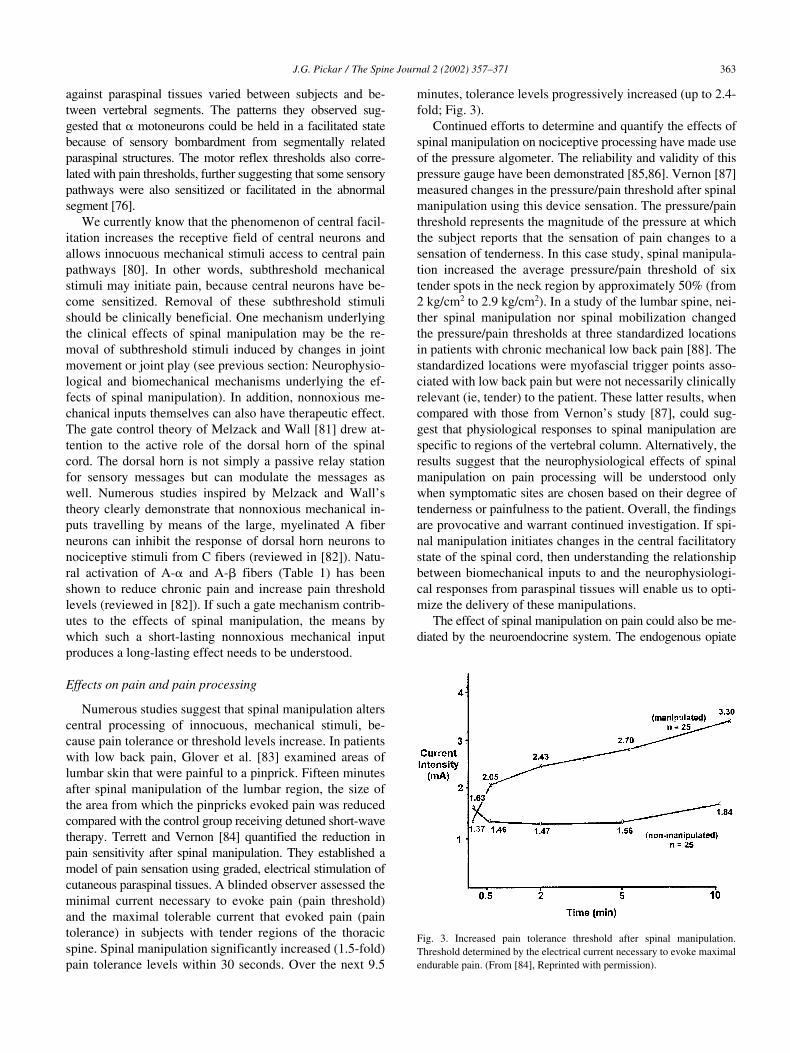
Numerous studies suggest that spinal manipulation alterscentral processing of innocuous, mechanical stimuli, be-cause pain tolerance or threshold levels increase. In patientswith low back pain, Glover et al. [83] examined areas oflumbar skin that were painful to a pinprick. Fifteen minutesafter spinal manipulation of the lumbar region, the size ofthe area from which the pinpricks evoked pain was reducedcompared with the control group receiving detuned short-wavetherapy. Terrett and Vernon [84] quantified the reduction inpain sensitivity after spinal manipulation. They established amodel of pain sensation using graded, electrical stimulation ofcutaneous paraspinal tissues. A blinded observer assessed theminimal current necessary to evoke pain (pain threshold)and the maximal tolerable current that evoked pain (paintolerance) in subjects with tender regions of the thoracicspine. Spinal manipulation significantly increased (1.5-fold)pain tolerance levels within 30 seconds. Over the next 9.5
minutes, tolerance levels progressively increased (up to 2.4-fold; Fig. 3).
Continued efforts to determine and quantify the effects ofspinal manipulation on nociceptive processing have made useof the pressure algometer. The reliability and validity of thispressure gauge have been demonstrated [85,86]. Vernon [87]measured changes in the pressure/pain threshold after spinalmanipulation using this device sensation. The pressure/painthreshold represents the magnitude of the pressure at whichthe subject reports that the sensation of pain changes to asensation of tenderness. In this case study, spinal manipula-tion increased the average pressure/pain threshold of sixtender spots in the neck region by approximately 50% (from2 kg/cm
2
to 2.9 kg/cm
2
). In a study of the lumbar spine, nei-ther spinal manipulation nor spinal mobilization changedthe pressure/pain thresholds at three standardized locationsin patients with chronic mechanical low back pain [88]. Thestandardized locations were myofascial trigger points asso-ciated with low back pain but were not necessarily clinicallyrelevant (ie, tender) to the patient. These latter results, whencompared with those from Vernon’s study [87], could sug-gest that physiological responses to spinal manipulation arespecific to regions of the vertebral column. Alternatively, theresults suggest that the neurophysiological effects of spinalmanipulation on pain processing will be understood onlywhen symptomatic sites are chosen based on their degree oftenderness or painfulness to the patient. Overall, the findingsare provocative and warrant continued investigation. If spi-nal manipulation initiates changes in the central facilitatorystate of the spinal cord, then understanding the relationshipbetween biomechanical inputs to and the neurophysiologi-cal responses from paraspinal tissues will enable us to opti-mize the delivery of these manipulations.
The effect of spinal manipulation on pain could also be me-diated by the neuroendocrine system. The endogenous opiate
Fig. 3. Increased pain tolerance threshold after spinal manipulation.Threshold determined by the electrical current necessary to evoke maximalendurable pain. (From [84], Reprinted with permission).

364
J.G. Pickar / The Spine Journal 2 (2002) 357–371
system is known to modify pain processes [89], and a numberof therapeutic modalities, including acupuncture [90], transcu-taneous nerve stimulation [91] and exercise [92], are thought toexert pain-relieving effects through activation of this system.Several studies have investigated the effect on spinal manipula-tion on circulating levels of
�
-endorphin. The findings havebeen inconsistent for possible reasons discussed by Rosner[93]. Vernon et al. [94] reported an 8% increase in plasma
�
-endorphin levels 5 minutes after spinal manipulation butnot after control interventions. Christian et al. [95] did notfind any change in plasma
�
-endorphin levels, but their as-say would have been unable to detect an 8% increase be-cause their between-assay variation was greater than the8%. On the other hand, Sanders et al. [96] did not find anychange in plasma
�
-endorphin levels despite a reduction inthe visual analog pain scale in the group receiving spinal ma-nipulation. Anti–pain-producing effects of
�
-endorphin canbe mediated by their ability to bind to membrane-bound re-ceptors on sensory nerve endings in the periphery as well asto receptors in the spinal cord and brain. However, the rela-tionship between circulating levels of
�
-endorphin and therelease of
�
-endorphin in the spinal cord is not known [97].Thus, while the experiments cited may indicate a responsemediated by peripheral receptors, the effects of spinal ma-nipulation on
�
-endorphin release within the central ner-vous system are unknown.
4. The effects of spinal manipulation on somatosomatic (muscle) reflexes
Substantial evidence demonstrates that spinal manipula-tion evokes paraspinal muscle reflexes and alters motoneu-ron excitability. In asymptomatic patients. Herzog’s group[98,99] showed that posterior to anterior spinal manipula-tive treatments applied to the cervical, thoracic lumbar andsacroiliac regions increased paraspinal EMG activity in apattern related to the region of the spine that was manipu-lated. The EMG response latencies occur within 50 to 200ms after initiation of the manipulative thrust. Similarly, spi-nal manipulation using an Activator Adjusting Instrumentapplied to a transverse process elicits paraspinal EMG ac-tivity at the same segmental level but within 2 to 3 ms [22].Colloca and Keller [100] confirmed these latter findings insymptomatic patients with low back pain. In addition, theyreported that the increased EMG activity, while beginningwithin 2 to 3 ms of the manipulation, reached its peak within50 to 100 ms. EMG activity representing a strong reflex re-sponse in terms of peak amplitude was relatively long lived(greater than 273 ms), whereas EMG activity representingweak reflex responses was more short lived (less than 273 ms).Paraspinal EMG responses were greatest in magnitudewhen the manipulation was delivered close to the electrodesite and, interestingly, the more chronic the low back pain,the less the EMG response. It is important to note that theEMG electrodes were not placed relative to any physicalfinding associated with the low back, for example, a pre-
sumed site of muscle spasm or a site of muscle pain or ten-derness.
The effect of spinal manipulation on paraspinal muscleactivity is not only excitatory. In one symptomatic patientwith spontaneous muscle activity in the thoracic spine,Suter et al. [99] observed reduced paraspinal EMG activitywithin 1 second after a thoracic spinal manipulation. De-Vocht obtained similar findings in a symptomatic patientwith low back pain (Fig. 4, unpublished observations). Heplaced EMG electrodes over palpably taut lumbar paraspi-nal muscles and often observed a decrease in spontaneousEMG activity after spinal manipulation using an ActivatorAdjusting Instrument and treatment protocol. The decreasedmuscle activity did not occur instantaneously.
The effects of spinal manipulation on somatomotor activitymay be quite complex, producing excitatory and inhibitory ef-fects. It is worth noting that many of the individual humanstudies cited above were performed on either symptomatic orasymptomatic individuals but not both. In addition, EMG re-cordings were sometimes obtained from standardized sitesand in other studies were obtained relative to clinical findingsof taut muscle fibers. Paradoxical findings may be reconciled iffuture studies compare the effects of spinal manipulation onsymptomatic versus asymptomatic subjects and on anatomicalsites with clinically identified or quantified signs. Clearly, thepotential for spinal manipulation to inhibit motor activity can
Fig. 4. Original data from a subject showing spontaneous paraspinal mus-cle activity in the lower lumbar spine and its response to spinal manipula-tion. Electromyographic (EMG) activity (top trace) decreased in responseto spinal manipulation using an Activator Adjusting Instrument and treat-ment protocol. A tripolar, disposable, adhesive EMG electrode was placedapproximately 2 to 3 cm to the right of the L4 spinous process overparaspinal muscle that was taut when palpated. Bottom trace is the outputfrom an accelerometer attached to the head of the Activator AdjustingInstrument. Large accelerometer spikes represent the onset of each spinalmanipulative impulse. Spinal manipulation was applied three times to thesacral base (third manipulation not obtained from the accelerometer) andsuccessively applied to the L5, L4 and L3 mammillary processes. The largespikes in the top trace are likely mechanical artifacts from the manipulativeimpulse. (Unpublished observations.)

J.G. Pickar / The Spine Journal 2 (2002) 357–371
365
be determined only under experimental conditions where mus-cle activity is spontaneously present or has been evoked.
The effects of spinal manipulation on paraspinal EMGactivity may be associated with increases in muscle strengthmeasured after spinal manipulation. Suter et al. [101] stud-ied symptomatic patients with sacroiliac joint dysfunction,anterior knee pain and evidence of motor inhibition to kneeextensor muscles. A side posture spinal manipulation ap-plied to the sacroiliac joint significantly decreased the inhi-bition of the knee extensors on the side of the body to whichthe manipulation was applied. Similarly, Keller and Collocafound that erector spinae isometric strength (assessed usingEMG activity) was increased after spinal manipulation com-pared with sham manipulation [102]. In neurophysiologicalterms, these two studies indicate that spinal manipulation im-proves muscle function either through facilitation or disinhibi-tion of neural pathways.
A series of studies has sought to understand how spinalmanipulation affects central processing of motor control in-formation. The studies indicate that spinal manipulation canboth increase the excitability of motor pathways in the spi-nal cord and depress the inflow of sensory information frommuscle spindles. In asymptomatic patients, Dishman et al.[103] showed that spinal manipulation increases central mo-tor excitability (Fig. 5). EMG activity from gastrocnemiusmuscle evoked by direct activation of descending corticospinaltracts using transcranial magnetic stimulation was largerafter lumbar spinal manipulation compared with simplypositioning the patient but not applying the manipulation.Spinal manipulation also depresses the H reflex. Manipulationapplied to the sacroiliac joint in a posterior to anterior direc-tion decreased the magnitude of the tibial nerve H reflex forup to 15 minutes in asymptomatic humans [104]. Similarly,side-posture lumbar manipulation of L5–S1 joint inhibitedthe H reflex from the tibial nerve [105]. The effects of mobi-lization alone applied to the same joint were similar, but theeffects of manipulation tended to be greater. After manipula-tion alone, the inhibition lasted for approximately 20 secondsbut lasted up to 1 minute when manipulation was preceded byspinal mobilization. These contrasting effects on EMG ac-tivity, between methodologies using motor evoked poten-tials versus the H reflex, may reflect the differential effectsof sensory input evoked by spinal manipulation on postsyn-aptic processing versus presynaptic inhibition, respectively(see, for extensive discussion, Dishman et al. [103].
One possible mechanism contributing to spinal manipu-lation’s inhibitory effects on the H reflex and on spontane-ous paraspinal EMG activity is suggested by recent experi-ments. Sensory input from facet joint tissues stimulatedduring spinal manipulation might reflexively decreaseparaspinal muscle activity. Indahl et al. [106] elicited reflexlongissimus and multifidus muscle (EMG) activity byelectrically stimulating the intervertebral disc in a porcinepreparation. Stretching the facet joint by injecting 1 mlphysiological saline abolished the EMG activity.
There is reason to believe that stretching the facet jointcapsule and surrounding tissues likely occurs during spinal
manipulation, although this has received little study [107].Using magnetic resonance imaging scans in human sub-jects, Cramer et al. [108] demonstrated that a side-posturespinal manipulation, accompanied by cavitation, gaps thefacet joints. The synovial space of the lumbar facet joints in-creased in width by up to 0.7 mm in individuals receivingmanipulation compared with nonmanipulated controls. Thelength of time between manipulation and the magnetic reso-nance imaging scan was not reported. In a study of the meta-carpophalangeal joint, 5 minutes after cavitation joint separa-tion was still increased by 0.4 mm and did not return toprecavitation dimensions until 10 minutes after “cracking”[109]. It remains to be shown if joint separations of thesemagnitudes are sufficient to load the facet joint tissues. Ifso, this raises the possibility that tissues surrounding thefacet joint could be stretched for periods of time longer thanthe duration of the manipulation itself. Graded sensory inputfrom tissues surrounding the facet joint [50] could elicit re-flex muscle responses similar to that measured by Indahl etal. [106].
Changes in muscle spindle input produced by spinal ma-nipulation could also contribute to the inhibition of soma-tosomatic reflexes. Using magnetic stimulation, Zhu et al.[110,111] stimulated lumbar paraspinal muscles and recordedthe evoked cerebral potentials. Stimulation of paraspinal mus-
Fig. 5. Effects of spinal manipulation on motor evoked potentials in the gas-trocnemius muscle. Muscle activity was evoked using transcranial magneticstimulation applied near the vertex of the skull. Side posture spinal manipula-tion was applied on the right to L5–S1. During control, individuals were posi-tioned in side posture but not manipulated. MEPmotor evoked potential;Prebefore manipulation or positioning; SMspinal manipulation. (Reprintedwith permission from J Manipulative Physiol Ther [103]).

366
J.G. Pickar / The Spine Journal 2 (2002) 357–371
cle spindles using vibration reduced the magnitude of thecerebral potentials. Similarly, muscle spasm in human patientsreduced the magnitude of the paraspinal muscle–evoked cere-bral potentials. Spinal manipulation reversed these effects,improving muscle spasm and restoring the magnitude of theevoked cerebral potentials [111], suggesting that increasedsensory input from paraspinal muscle spindles during mus-cle spasm may contribute to the reduced magnitude of theevoked cerebral potentials. It is worthwhile recalling Korr’sideas [36] that spinal manipulation increases joint mobilityby producing a barrage of impulses in muscle spindle afferentsand smaller-diameter afferents, ultimately silencing facilitated
�
motoneurons (see previous section: The effects of spinal ma-nipulation on sensory neurons innervating paraspinal tissues;Group I and II afferents [proprioceptive afferents]).
At first it seems counterintuitive that muscle spindle dis-charge is increased during muscle spasm, because one couldanticipate muscle shortening and spindle unloading duringspasm. However, extensive studies from Proske’s laboratory(reviewed in [112]) show that a maintained joint position ormaintained muscle shortening, even for short durations, altersmuscle spindle sensitivity to subsequent joint movement ormuscle stretch. For example, from a given muscle length, mus-cle spindles respond more to a slow stretch when a leg musclehas previously been held at a shortened length compared withhaving been previously held at a long length for as little as 10seconds [113]. Recently, Pickar and Kang [114] observed thesame phenomenon in the lumbar longissimus and multifidusmuscles (Fig. 6). Muscle spindle activity in response to aslow vertebral translation that stretched the muscle spindledepended on whether the muscle had previously been short-
ened for as little as 5 seconds (by linearly displacing the L6vertebra dorsalward) or had previously been stretched (by lin-early displacing the L6 vertebra ventralward). If paraspinalmuscle spasm results in muscle shortening, or if segmentalbuckling results in muscle shortening ipsilaterally and mus-cle lengthening contralaterally, then for the same change inmuscle length subsequent stretch or vibration of the affectedmuscles would increase spindle discharge more than ex-pected. Because spinal manipulation has been shown tostimulate muscle spindles (Fig. 7), spinal manipulation maynormalize spindle biomechanics and return muscle spindledischarge to normal.
5. The effects of spinal manipulation on somatovisceral reflexes
A number of animal experiments provide evidence sup-porting the link between altered paraspinal sensory inputand a somatovisceral change shown in Fig. 1. Sensory inputfrom paraspinal tissues can evoke visceral reflexes affectingthe sympathetic nervous system and may alter end-organfunction. In general, nonnoxious paraspinal sensory inputappears to have an inhibitory effect on sympathetic outflow,whereas noxious input appears to have an excitatory effect.However, insufficient experiments have been conducted todetermine the regional variation of this effect, that is, thechange in sympathetic outflow to different organs. Nonethe-less, the data are provocative, indicating that neural inputfrom axial tissues can evoke somatovisceral reflexes.
Sato and Swenson [115] applied a nonnoxious mechani-cal stimulus to several vertebrae in the thoracic and lumbar
Fig. 6. Paraspinal muscle spindle sensitivity to identical changes in vertebral displacement is determined by the previous short-term history of its muscle’s length. Thereceptive field of the muscle spindle was in the lumbar multifidus muscle. Top panel shows the discharge frequency of the muscle spindle afferent recorded from theL6 dorsal root. Instantaneous discharge frequency was averaged into 25-ms bins. The bottom panel shows the amount the L6 vertebra was displaced during flexion(negative displacement) and extension (positive displacement). These displacements shortened and stretched the multifidus muscle, respectively, because holding theL6 vertebra in an extended position (12 to 15 seconds) increased spindle discharge frequency compared with control. Conversely, holding the L6 vertebra in a flexedposition (11 to 15 seconds) decreased spindle discharge frequency compared with control. Spindle discharge was the same at the start of the protocols (control, 0 to 2seconds). The rapid extensions and flexions at the start of each protocol provided the same initial conditions. (Spindle discharge is not shown during these displace-ments). Top panel shows the change in muscle spindle sensitivity to the slow vertebral extension (18.5 to 40 seconds) after holding the multifidus muscle in shortened� and stretched � positions. Note that vertebral position was held for as little as 5 seconds. (Reprinted with permission from J Neuromusculoskel Sys, Data TracePublishing Company [114]).

J.G. Pickar / The Spine Journal 2 (2002) 357–371
367
spine of rats by applying a force to the lateral aspects of theirspinous processes. Renal and adrenal sympathetic nerve ac-tivities were recorded. Because the paraspinal musculaturewas removed, the sensory input was derived presumablyfrom the facet joints, intervertebral discs and/or interverte-bral ligaments. The mechanical stimulus reflexively decreasedthe level of renal and adrenal sympathetic nerve activity by25% to 40%. The stimuli were short in duration (approxi-mately 30 seconds), and the responses attenuated rapidly.The sensory input from the paraspinal tissues had access tocenters at least as high as the upper cervical spinal cord,because C1–C2 spinal cord transection abolished the inhibi-tion. Sato and Swenson concluded that nonnoxious mechani-cal stimuli applied to the spine reflexively inhibit the level ofsympathetic nerve activity by means of a supraspinal reflex.
Budgell et al. [116,117] also stimulated paraspinal structuresusing noxious and nonnoxious chemical stimuli. Injectionswere placed into the lumbar facet joints or lumbar interspinoustissues. Blood pressure and sciatic nerve blood flow were mea-sured [116]. A small volume (20 ul) of a nonnoxious chemical(physiological saline 0.9%) injected into the interspinousligament produced a depressor response and a concomitantdecrease in sciatic nerve blood flow. A similar volume oflow-dose capsaicin (2 ug), which activates nociceptive neurons[118], caused an initial increase in blood pressure and sciaticnerve blood flow. However, when injected into the facet joint,capsaicin produced a depressor response. The results from theinterspinous ligament are consistent with the suggestion of-fered by Sato and Swenson [115] that stimulation of recep-tive endings sensitive to innocuous mechanical stimuli inthe paraspinal tissues produce inhibitory somaticsympa-thetic reflexes. The findings from the facet joints suggestedto the authors that capsaicin might more effectively produceinnocuous mechanical changes in the facet joint comparedwith the interspinous ligament by increasing the permeabil-ity of the synovial membrane’s microvasculature. Similar tothe cardiovascular effects produced by capsaicin injection
into the lumbar interspinous ligament, capsaicin injectioninto the lumbar interspinous tissues also increased adrenalsympathetic nerve activity and catecholamine secretion[117], whereas physiological saline injection had no effect.Thus, noxious stimulation of paraspinal tissues can produceexcitatory somatic-sympathetic reflexes.
More recently, Pickar et al. [119], in a preliminary re-port, showed that mustard oil, a nociceptive substance thatalso produces inflammation, injected into the lumbar multi-fidus muscle increases the discharge of sympathetic nervesto the kidney and spleen. The response is a reflex mediatedby segmental branches of the dorsal ramus and is integratedby centers at least as high as the upper cervical spinal cord.This reflex organization is similar to that found by Sato andSwenson [115] for the sympathetic nerves to the kidney andadrenal gland. Interestingly, animal studies have also shownthat increased splenic sympathetic nerve discharge is immu-nosuppressive, decreasing the number of natural killer cellsreleased. Somatovisceral reflex stimulation of the sympa-thetic outflow to the spleen may contribute to the depressedlevels of natural killer cells measured in individuals withlow back pain [120].
Mechanical stimulation of paraspinal tissues can be suf-ficient to inhibit gastric motility. Myoelectric activity fromthe wall of the gastrointestinal tract in conscious rabbits wasdecreased by sustained (2.5 minutes) mechanical inputs[121]. In these experiments, it was unclear if the mechanicalstimulation was noxious or innocuous, but the inhibition ofgastric motility was greatest when the mechanical stimulationwas applied to the sixth thoracic vertebra, and it decreased asthe mechanical stimulation was applied further cranial or cau-dal. These results were confirmed by Budgell and Suzuki[122]. Noxious chemical stimulation inhibited gastric motil-ity, and the effect tended to be greater when the stimulus wasapplied to the mid-thoracic region compared with the lumbarregion. In addition, the inhibitory response was shown to bea reflex predominated by changes in sympathetic outflowand to a lesser extent vagal outflow.
It is important to note that these studies do not provide evi-dence for the unique potential of paraspinal tissues to elicit so-matosympathetic reflexes. Substantial evidence shows thatnoxious stimulation of tissues in the appendicular skeleton alsoevokes somatosympathetic reflexes [123], but nothing isknown about the relative magnitudes of somatosympathetic re-flexes elicited by axial versus appendicular tissues. Althoughthe data on gastric motility suggest segmental specificity, it isnot certain the degree to which segmental input from paraspi-nal tissues produce regionally specific changes in sympatheticnerve activity.
Very few laboratory or clinically oriented basic sciencestudies have been conducted to determine the effects of spi-nal manipulation on the sympathetic nervous system. Re-cently, Budgell and Hirano [124] measured changes in heartrate variability after upper cervical versus sham spinal ma-nipulation. Power spectral analysis of heart rate variabilityshowed that manipulation increased the ratio of low fre-
Fig. 7. Original tracing of a muscle spindle’s response to a spinal manipu-lative-like load. The single unit activity was obtained from a muscle spin-dle afferent in the L6 dorsal root. The muscle spindle was located in thelumbar paraspinal muscles. Inset shows the spindle’s discharge on anexpanded time scale immediately before, during and shortly after theimpulse. (From [47] Reprinted with permission.)
368
J.G. Pickar / The Spine Journal 2 (2002) 357–371
quency to high frequency components indicating a possibleshift in the balance of autonomic control of the heart towardthe parasympathetic nervous system.
Spinal manipulation may alter the response of immunologiccells as well as the production of immunomodulatory and neu-romodulatory cytokines. In a series of studies on human sub-jects in the 1990s, Brennan et al. [120,125,126] showed thatspinal manipulation but not sham manipulation nor soft tissuemassage primed polymorphonuclear leukocytes (PMNs) andmonocytes. Spinal manipulation enhanced the respiratoryburst (a marker for phagocytic activity) of these white bloodcells to a particulate challenge. The mechanism is unclear,although speculation on the role of substance P was discussed.Spinal manipulation also primed the polymorphonuclear leu-kocytes for enhanced production of cytokines as determined bythe release of tumor necrosis factor in response to endotoxinchallenge. The priming effect was short lived, being greater15 minutes after manipulation compared with 30 and 45 min-utes. The biological consequence of these changes have yet tobe investigated, but their changes suggested their potential use,at least, as markers of successful spinal manipulation.
Conclusion
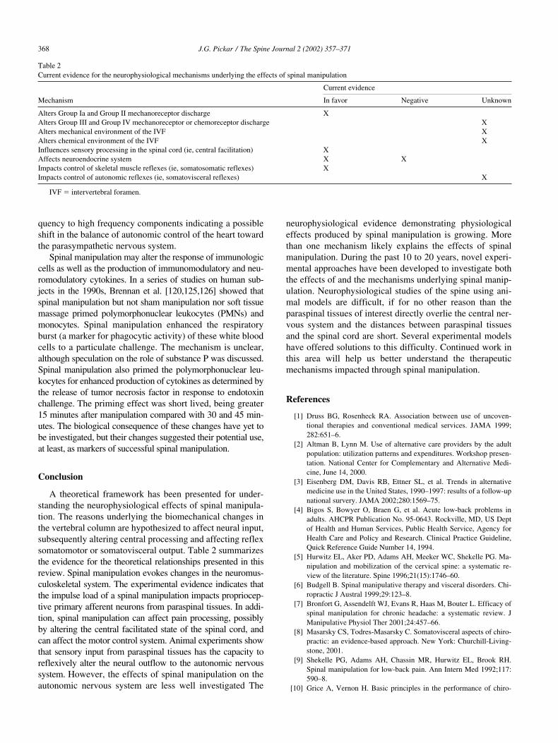
A theoretical framework has been presented for under-standing the neurophysiological effects of spinal manipula-tion. The reasons underlying the biomechanical changes inthe vertebral column are hypothesized to affect neural input,subsequently altering central processing and affecting reflexsomatomotor or somatovisceral output. Table 2 summarizesthe evidence for the theoretical relationships presented in thisreview. Spinal manipulation evokes changes in the neuromus-culoskeletal system. The experimental evidence indicates thatthe impulse load of a spinal manipulation impacts propriocep-tive primary afferent neurons from paraspinal tissues. In addi-tion, spinal manipulation can affect pain processing, possiblyby altering the central facilitated state of the spinal cord, andcan affect the motor control system. Animal experiments showthat sensory input from paraspinal tissues has the capacity toreflexively alter the neural outflow to the autonomic nervoussystem. However, the effects of spinal manipulation on theautonomic nervous system are less well investigated The
neurophysiological evidence demonstrating physiologicaleffects produced by spinal manipulation is growing. Morethan one mechanism likely explains the effects of spinalmanipulation. During the past 10 to 20 years, novel experi-mental approaches have been developed to investigate boththe effects of and the mechanisms underlying spinal manip-ulation. Neurophysiological studies of the spine using ani-mal models are difficult, if for no other reason than theparaspinal tissues of interest directly overlie the central ner-vous system and the distances between paraspinal tissuesand the spinal cord are short. Several experimental modelshave offered solutions to this difficulty. Continued work inthis area will help us better understand the therapeuticmechanisms impacted through spinal manipulation.
References
[1] Druss BG, Rosenheck RA. Association between use of uncoven-tional therapies and conventional medical services. JAMA 1999;282:651–6.
[2] Altman B, Lynn M. Use of alternative care providers by the adultpopulation: utilization patterns and expenditures. Workshop presen-tation. National Center for Complementary and Alternative Medi-cine, June 14, 2000.
[3] Eisenberg DM, Davis RB, Ettner SL, et al. Trends in alternativemedicine use in the United States, 1990–1997: results of a follow-upnational survery. JAMA 2002;280:1569–75.
[4] Bigos S, Bowyer O, Braen G, et al. Acute low-back problems inadults. AHCPR Publication No. 95-0643. Rockville, MD, US Deptof Health and Human Services, Public Health Service, Agency forHealth Care and Policy and Research. Clinical Practice Guideline,Quick Reference Guide Number 14, 1994.
[5] Hurwitz EL, Aker PD, Adams AH, Meeker WC, Shekelle PG. Ma-nipulation and mobilization of the cervical spine: a systematic re-view of the literature. Spine 1996;21(15):1746–60.
[6] Budgell B. Spinal manipulative therapy and visceral disorders. Chi-ropractic J Austral 1999;29:123–8.
[7] Bronfort G, Assendelft WJ, Evans R, Haas M, Bouter L. Efficacy ofspinal manipulation for chronic headache: a systematic review. JManipulative Physiol Ther 2001;24:457–66.
[8] Masarsky CS, Todres-Masarsky C. Somatovisceral aspects of chiro-practic: an evidence-based approach. New York: Churchill-Living-stone, 2001.
[9] Shekelle PG, Adams AH, Chassin MR, Hurwitz EL, Brook RH.Spinal manipulation for low-back pain. Ann Intern Med 1992;117:590–8.
[10] Grice A, Vernon H. Basic principles in the performance of chiro-
Table 2Current evidence for the neurophysiological mechanisms underlying the effects of spinal manipulation
Mechanism
Current evidence
In favor Negative Unknown
Alters Group Ia and Group II mechanoreceptor discharge XAlters Group III and Group IV mechanoreceptor or chemoreceptor discharge XAlters mechanical environment of the IVF XAlters chemical environment of the IVF XInfluences sensory processing in the spinal cord (ie, central facilitation) XAffects neuroendocrine system X XImpacts control of skeletal muscle reflexes (ie, somatosomatic reflexes) XImpacts control of autonomic reflexes (ie, somatovisceral reflexes) X
IVF
intervertebral foramen.

J.G. Pickar / The Spine Journal 2 (2002) 357–371
369
practic adjusting: historical review, classification, and objectives.In: Haldeman S, editor. Principles and practice of chiropractic, 2nded. Norwalk: Appleton and Lange, 1992:443–58.
[11] Bergmann TF. Short lever, specific contact articular chiropractictechnique. J Manipulative Physiol Ther 1992;15:591–5.
[12] Bartol KM. Osseous manual thrust techniques. In: Gatterman MI,editor. Foundations of chiropractic, 1st ed. St. Louis: Mosby, 1995:88–104.
[13] Conway PJW, Herzog W, Zhang Y, Hasler EM, Ladly K. Forces re-quired to cause cavitation during spinal manipulation of the thoracicspine. Clin Biomech 1993;8:210–4.
[14] Brodeur R. The audible release associated with joint manipulation. JManipulative Physiol Ther 1995;18:155–64.
[15] Haldeman S. Spinal manipulative therapy; a status report. Clin Or-thop 1983;179:62–70.
[16] Hessel BW, Herzog W, Conway PJW, McEwen MC. Experimentalmeasurement of the force exerted during spinal manipulation usingthe Thompson technique. J Manipulative Physiol Ther 1990;13:448–53.
[17] Herzog W, Conway PJ, Kawchuk GN, Zhang Y, Hasler EM. Forcesexerted during spinal manipulative therapy. Spine 1993;18:1206–12.
[18] Kawchuk GN, Herzog W, Hasler EM. Forces generated during spi-nal manipulative therapy of the cervical spine: a pilot study. J Ma-nipulative Physiol Ther 1992;15:275–8.
[19] Kawchuk GN, Herzog W. Biomechanical characterization (finger-printing) of five novel methods of cervical spine manipulation. JManipulative Physiol Ther 1993;16:573–7.
[20] Triano J, Schultz AB. Loads transmitted during lumbosacral spinalmanipulative therapy. Spine 1997;22:1955–64.
[21] Nathan M, Keller TS. Measurement and analysis of the in vivo pos-teroanterior impulse response of the human thoracolumbar spine: afeasibility study. J Manipulative Physiol Ther 1994;17:431–41.
[22] Fuhr AW, Smith DC. Accuracy of piezoelectric accelerometersmeasuring displacement of a spinal adjusting instrument. J Manipu-lative Physiol Ther 1986;9:15–21.
[23] Smith DB, Fuhr AW, Davis BP. Skin accelerometer displacementand relative bone movement of adjacent vertebrae in response tochiropractic percussion thrusts. J Manipulative Physiol Ther 1989;12:26–37.
[24] Gal J, Herzog W, Kawchuk G, Conway P, Zhang YT. Biomechani-cal studies of spinal manipulative therapy (SMT): quantifying themovements of vertebral bodies during SMT. J CCA 1994;38:11–24.
[25] Leach RA. The chiropractic theories, 3rd ed. Baltimore: Williamsand Wilkins, 1994.
[26] Gatterman MI. What’s in a word? In: Gatterman MI, editor. Foun-dations of chiropractic, 1st ed. St. Louis: Mosby, 1995:6–17.
[27] Triano J. Interaction of spinal biomechanics and physiology. In:Anonymous principles and practice of chiropractic, 2nd ed. Nor-walk: Appleton and Lange, 1992:225–57.
[28] Gillette RG. A speculative argument for the coactivation of diversesomatic receptor populations by forceful chiropractic adjustments.Manual Med 1987;3:1–14.
[29] Farfan HF. The scientific basis of manipulation procedures. In:Buchanan WW, Kahn MF, Laine V, Rodnan GP, Scott JT, ZvaiflerNJ, Grahame R, editors. Clinics in rheumatic diseases. London: WBSaunders Company, Ltd., 1980:159–77.
[30] Giles LGF. Anatomical basis of low back pain. Baltimore: Williamsand Wilkins, 1989.
[31] Lewit K. Manipulative therapy in rehabilitation of the locomotorsystem. Oxford: Butterworth-Heinemann, 1991.
[32] Haldeman S. The clinical basis for discussion of mechanisms of ma-nipulative therapy. In: Korr IM, editor. The neurobiologic mecha-nisms in manipulative therapy. New York: Plenum, 1978:53–75.
[33] Vernon H. Biological rationale for possible benefits of spinal ma-nipulation. Cherkin DC, Mootz RD. AHCPR Publication No. 98-N002, 105–115. 1997. Chiropractic in the United States: training,practice and research.
[34] Wilder DG, Pope MH, Frymoyer JW. The biomechanics of lumbardisc herniation and the effect of overload and instability. J SpinalDisord 1988;1:16–32.
[35] Triano J. The mechanics of spinal manipulation. In: Herzog W, edi-tor. Clinical biomechanics of spinal manipulation. New York:Churchill Livingstone, 2001:92–190.
[35a] Greenman PE. Principles of Manual Medicine. Baltimore: Williamsand Wilkins, 1989, p 4.
[36] Korr IM. Proprioceptors and somatic dysfunction. J Am OsteopathAssoc 1975;74:638–50.
[37] Whittingham W, Nilsson N. Active range of motion in the cervicalspine increases after spinal manipulation (toggle recoil). J Manipu-lative Physiol Ther 2001;24:552–5.
[38] Eldred E, Hutton RS, Smith JL. Nature of the persisting changes inafferent discharge from muscle following its contraction. In:Homma S, editor. Understanding the stretch reflex. New York:Elsevier Scientific Publishing Company, 1976:157–83.
[39] Buerger AA. Experimental neuromuscular models of spinal manualtechniques. Manual Med 1983;1:10–7.
[40] Brumagne S, Cordo P, Lysens R, Verschueren S, Swinnen S. Therole of paraspinal muscle spindles in lumbosacral position sense inindividuals with and without low back pain. Spine 2000;25:989–94.
[41] Brumagne S, Lysens R, Swinnen S, Verschueren S. Effect ofparaspinal muscle vibration on position sense of the lumbosacralspine. Spine 1999;24:1328–31.
[42] Wilder DG, Aleksiev AR, Magnusson ML, Pope MH, Spratt KF,Goel VK. Muscular response to sudden load. A tool to evaluate fa-tigue and rehabilitation. Spine 1996;21:2628–39.
[43] Radebold A, Cholewicki J, Panjabi MM, Patel TCh. Muscle re-sponse pattern to sudden trunk loading in healthy individuals and inpatients with chronic low back pain. Spine 2000;25:947–54.
[44] Radebold A, Cholewicki J, Polzhofer GK, Greene HS. Impairedpostural control of the lumbar spine is associated with delayed mus-cle response times in patients with chronic idiopathic low back pain.Spine 2001;26:724–30.
[45] Bolton PS, Holland CT. An in vivo method for studying afferent fi-bre activity from cervical paravertebral tissue during vertebral mo-tion in anaesthetised cats. J Neurosci Methods 1998;85:211–8.
[46] Pickar JG. An in vivo preparation for investigating neural responsesto controlled loading of a lumbar vertebra in the anesthetized cat. JNeurosci Methods 1999;89:87–96.
[47] Pickar JG, Wheeler JD. Response of muscle proprioceptors to spinalmanipulative-like loads in the anesthetized cat. J ManipulativePhysiol Ther 2001;24:2–11.
[48] Colloca CJ, Keller TS, Gunzburg R, Vandeputte K, Fuhr AW. Neu-rophysiologic response to intraoperative lumbosacral spinal manip-ulation. J Manipulative Physiol Ther 2000;23:447–56.
[49] Cavanaugh JM, El-Bohy A, Hardy WN, Getchell TV, Getchell ML,King AI. Sensory innervation of soft tissues of the lumbar spine inthe rat. J Orthop Res 1989;7:378–88.
[50] Pickar JG, McLain RF. Responses of mechanosensitive afferents tomanipulation of the lumbar facet in the cat. Spine 1995;20:2379–85.
[51] Yamashita T, Cavanaugh JM, Ozaktay CA, Avramov AI, King AI.Effect of substance P on mechanosensitive units of tissues aroundand in the lumbar facet joint. J Orthop Res 1993;11:205–14.
[52] Abrahams VC, Lynn B, Richmond FJR. Organization and sensoryproperties of small myelinated fibers in the dorsal cervical rami ofthe cat. J Physiol 1984;347:177–87.
[53] Beel JA, Stodieck LS, Luttges MW. Structural properties of spinalnerve roots: biomechanics. Exp Neurol 1986;91:30–40.
[54] Berthold CH, Carlstedt T, Corneliuson O. Anatomy of the nerveroot at the central-peripheral transitional region. In: Dyck PJ, editor.Peripheral neuropathy, 1st ed. Philadelphia: WB Saunders Com-pany, 1984:156–70.
[55] Thomas PK, Berthold CH, Ochoa J. Microscopic anatomy of the pe-ripheral nervous system. In: Dyck PJ, editor. Peripheral neuropathy,1st ed. Philadelphia: WB Saunders Company, 1993:28–91.

370
J.G. Pickar / The Spine Journal 2 (2002) 357–371
[56] Devor M, Obermayer M. Membrane differentiation in rat dorsal rootganglia and possible consequences for back pain. Neurosci Letters1984;51:341–6.
[57] Rydevik BL. The effects of compression on the physiology of nerveroots. J Manipulative Physiol Ther 1992;15:62–6.
[58] Howe JF, Loeser JD, Calvin WH. Mechanosensitivity of dorsal rootganglia and chronically injured axons:a physiological basis for theradicular pain of nerve root compression. Pain 1977;3:27–41.
[59] Wall PD, Devor M. Sensory afferent impulses originate from dorsalroot ganglia as well as from the periphery in normal and nerve in-jured rats. Pain 1983;17:321–39.
[60] Sharpless SK. Susceptibility of spinal roots to compression block.Goldstein M. (No. 15), 155–161. 1975. NINCDS-Research Status ofSpinal Manipulation Therapy.
[61] Ozaktay AC, Kallakuri S, Cavanaugh JM. Phospholipase A
2
sensi-tivity of the dorsal root and dorsal root ganglion. Spine 1998;23:1297–306.
[62] Olmarker K, Rydevik B, Hansson T, Holm S. Compression-inducedchanges of the nutritional supply to the porcine cauda equina. J Spi-nal Disord 1990;3:25–9.
[63] Cornefjord M, Olmarker K, Farley DB, Weinstein JN, Rydevik B.Neuropeptide changes in compressed spinal nerve roots. Spine1995;20:670–3.
[64] Rydevik BL, Myers RR, Powell HC. Pressure increase in the dorsalroot ganglion following mechanical compression. Closed compart-ment syndrome in nerve roots. Spine 1989;14:574–6.
[65] Takahashi K, Shima I, Porter RW. Nerve root pressure in lumbardisc herniation. Spine 1999;24:2003–6.
[66] McCarron RF, Wimpee MW, Hudkins PG, Laros GS. The inflam-matory effect of nucleus pulposus. A possible element in the patho-genesis of low-back pain. Spine 1987;12:760–4.
[67] Kawakami M, Tamaki T, Hayashi N, et al. Mechanical compressionof the lumber nerve root alters pain-related behaviors induced by thenucleus pulposus in the rat. J Orthop Res 2000;18:257–64.
[68] Yabuki S, Igarashi T, Kikuchi S. Application of nucleus pulposus tothe nerve root simultaneously reduces blood flow in dorsal root gan-glion and corresponding hindpaw in the rat. Spine 2000;25:1471–6.
[69] Nygaard OP, Mellgren SI, Osterud B. The inflammatory propertiesof contained and noncontained lumbar disc herniation. Spine 1997;22:2484–8.
[70] Chen C, Cavanaugh JM, Ozaktay AC, Kallakuri S, King AI. Effectsof phospholipase A
2
on lumbar nerve root structure and function.Spine 1997;22:1057–64.
[71] Floman Y, Liram N, Gilai AN. Spinal manipulation results in imme-diate H-reflex changes in patients with unilateral disc herniation.Eur Spine J 1997;6:398–401.
[72] Stern PJ, Côte P, Cassidy JD. A series of consecutive cases of lowback pain with radiating leg pain treated by chiropractors. J Manipu-lative Physiol Ther 1995;18:335–42.
[73] Nwuga VC. Relative therapeutic efficacy of vertebral manipulationand conventional treatment in back pain management. Am J PhysMed 1982;61:273–8.
[74] Burton AK, Tillotson KM, Cleary J. Single-blind randomised con-trolled trial of chemonucleolysis and manipulation in the treatmentof symptomatic lumbar disc herniation. Eur Spine J 2000;9:202–7.
[75] Cook AJ, Woolf CJ, Wall PD, McMahon SB. Dynamic receptivefield plasticity in rat spinal cord dorsal horn following C-primary af-ferent input. Nature 1987;325:151–3.
[76] Denslow JS, Korr IM, Krems AD. Quantitative studies of chronicfacilitation in human motoneuron pools. Am J Physiol 1947;150:229–38.
[77] Denslow JS, Clough GH. Reflex activity in the spinal extensors. JNeurophysiol 1941;4:430–7.
[78] Denslow JS, Hassett CC. The central excitatory state associatedwith postural abnormalities. J Neurophysiol 1942;5:393–401.
[79] Denslow JS. An analysis of the variability of spinal reflex thresh-olds. J Neurophysiol 1944;7:207–15.
[80] Woolf CJ. The dorsal horn: state-dependent sensory processing andthe generation of pain. In: Wall PD, Melzack R, editors. Textbookof pain, 3rd ed. Edinburgh: Churchill Livingstone, 1994:101–12.
[81] Melzack R, Wall PD. Pain mechanisms: a new theory. Science1965;150:971–9.
[82] Besson J-M, Chaouch A. Peripheral and spinal mechanisms of noci-ception. Physiol Rev 1987;67(1):67–186.
[83] Glover JR, Morris JG, Khosla T. Back pain: a randomized clinicaltrial of rotational manipulation of the trunk. Br J Indust Med 1974;31:59–64.
[84] Terrett ACJ, Vernon HT. Manipulation and pain tolerance: a con-trolled study of the effect of spinal manipulation on paraspinal cuta-neous pain tolerance levels. Am J Phys Med 1984;63:217–25.
[85] Fischer AA. Pressure algometry over normal muscles. Standard val-ues, validity and reproduceability of pressure threshold. Pain 1986;30:115–26.
[86] Reeves JL, Jaeger B, Graff-Radford SB. Reliability of the pressurealgometer as a measure of myofascial trigger point sensitivity. Pain1986;24:313–21.
[87] Vernon HT. Pressure pain threshold evaluation of the effect of spi-nal manipulation on chronic neck pain: a single case study. J CCA1988;32:191–4.
[88] Cote P, Silvano AM, Vernon H, Mior SA. The short-term effect of aspinal manipulation on pain/pressure threshold in patients withchronic mechanical low back pain. J Manipulative Physiol Ther1994;17:364–8.
[89] Fields HL, Basbaum AI. Central nervous system mechanisms ofpain modulation. In: Wall PD, Melzack R, editors. Textbook ofpain, 3rd ed. Edinburgh: Churchill Livingstone, 1994:101–12.
[90] Szczudlik A, Lypka A. Plasma immunoreactive beta-endorphin andenkephalin concentration in healthy subjects before and after elec-troacupuncture. Acupunct Electro-Ther Res 1983;8:127–37.
[91] Hughes GSJ, Lichstein PR, Whitlock D, Harker C. Response ofplasma beta-endorphins to trancutaneous electrical nerve stimula-tion in healthy subjects. Phys Ther 1984;64:1062–6.
[92] Harber VJ, Sutton JR. Endorphins and exercise. Sports Med 1984;1:154–71.
[93] Rosner A. Endocrine disorders. In: Masarsky CS, Todres-MasarskyC, editors. Somatovisceral aspects of chiropractic: an evidence-based approach. New York: Churchill-Livingstone, 2001:187–202.
[94] Vernon HT, Dhami MSI, Howley TP, Annett R. Spinal manipula-tion and beta-endorphin: a controlled study of the effect of a spinalmanipulation on plasma beta-endorphin levels in normal males. JManipulative Physiol Ther 1986;9:115–23.
[95] Christian GF, Stanton GJ, Sissons D, et al. Immunoreactive acth,b-endorphin, and cortisol levels in plasma following spinal manipu-lative therapy. Spine 1988;13:1411–7.
[96] Sanders GE, Reinert O, Tepe R, Maloney P. Chiropractic adjustivemanipulation on subjects with acute low back pain: visual analogpain scores and plasma b-endorphin levels. J Manipulative PhysiolTher 1990;13:391–5.
[97] Clement-Jones V, Lowry PJ, Rees LH, Besser GM. Developmentof a specific extracted radioimmunoassay for methionine enkepha-lin in human plasma and cerebrospinal fluid. J Endocrinol 1980;86:231–43.
[98] Herzog W, Scheele D, Conway PJ. Electromyographic responses ofback and limb muscles associated with spinal manipulative therapy.Spine 1999;24:146–53.
[99] Suter E, Herzog W, Conway PJ, Zhang YT. Reflex response associ-ated with manipulative treatment of the thoracic spine. J Neuromus-culoskel Sys 1994;2:124–30.
[100] Colloca CJ, Keller TS. Stiffness and neuromuscular reflex responseof the human spine to posteroanterior manipulative thrusts in pa-tients with low back pain. J Manipulative Physiol Ther 2001;24:489–500.
[101] Suter E, McMorland G, Herzog W, Bray R. Conservative lowerback treatment reduces inhibition in knee-extensor muscles: a ran-

J.G. Pickar / The Spine Journal 2 (2002) 357–371
371
domized controlled trial. J Manipulative Physiol Ther 2000;23:76–80.
[102] Keller TS, Colloca CJ. Mechanical force spinal manipulation in-creases trunk muscle strength assessed by electromyography: acomparative clinical trial. J Manipulative Physiol Ther 2000;23:585–95.
[103] Dishman JD, Ball KA, Burke J. Central motor excitability changesafter spinal manipulation, a transcranial magnetic stimulation study.J Manipulative Physiol Ther 2002;25:1–9.
[104] Murphy BA, Dawson NJ, Slack JR. Sacroiliac joint manipulationdecreases the H-reflex. Electromyog Clin Neurophysiol 1995;35:87–94.
[105] Dishman JD, Bulbulian R. Spinal reflex attenuation associated withspinal manipulation. Spine 2000;25:2519–25.
[106] Indahl A, Kaigle AM, Reikeras O, Holm SH. Interaction betweenthe porcine lumbar intervertebral disc, zygapophysial joints, andparaspinal muscles. Spine 1997;22:2834–40.
[107] El-Bohy AA, Goldberg SJ, King AI. Measurement of facet capsularstretch [abstract]. Biomechanics Symposium 1987; American Soci-ety of Mechanical Engineers: ASME, NY, NY; 161–4.
[108] Cramer GD, Tuck NR, Knudsen JT, et al. Effects of side-posturepositioning and side-posture adjusting on the lumbar zygapophysealjoints as evaluated by magnetic resonance imaging: a before and af-ter study with randomization. J Manipulative Physiol Ther 2000;23:380–94.
[109] Unsworth A, Dowson D, Wright V. “Cracking joints.” Ann RheumDis 1971;30:348–58.
[110] Zhu Y, Haldeman S, Starr A, Seffinger MA, Su S. Paraspinal mus-cle evoked cerebral potentials in patients with unilateral low backpain. Spine 1993;18:1096–102.
[111] Zhu Y, Haldeman S, Hsieh C-YJ, Wu P, Starr A. Do cerebral poten-tials to magnetic stimulation of paraspinal muscles reflect changesin palpable muscle spasm, low back pain, and activity scores? J Ma-nipulative Physiol Ther 2000;23:458–64.
[112] Proske U, Morgan DL, Gregory JE. Thixotropy in skeletal muscleand in muscle spindles: a review. Prog Neurobiol 1993;41:705–21.
[113] Gregory JE, Mark RF, Morgan DL, Patak A, Polus B, Proske U. Ef-fects of muscle history on the stretch reflex in cat and man. J Neuro-physiol 1990;424:93–107.
[114] Pickar JG, Kang YM. Short-lasting zstretch of lumbar paraspinalmuscle decreases muscle spindle sensitivity to subsequent musclezstretch. J Neuromusculoskel Sys 2001;9:88–96.
[115] Sato A, Swenson RS. Sympathetic nervous system response to me-chanical stress of the spinal column in rats. J Manipulative PhysiolTher 1984;7:141–7.
[116] Budgell B, Hotta H, Sato A. Spinovisceral reflexes evoked by nox-ious and innocuous stimulation of the lumbar spine. J Neuromuscu-loskel Sys 1995;3:122–30.
[117] Budgell B, Sato A, Suzuki A, Uchida S. Responses of adrenal func-tion to stimulation of lumbar and thoracic interspinous tissues in therat. Neurosci Res 1997;28:33–40.
[118] Fitzgerald M. Capsaicin and sensory neurons—a review. Pain 1983;15:109–30.
[119] Pickar JG, Kang YM, Cobb T, Kenney MJ. Mustard oil injected intolumbar multifidus muscle increases sympathetic nerve activity tospleen and kidney via a suprasegmental reflex [abstract]. Soc Neu-rosci Abstr 2001;27(818):9.
[120] Brennan PC, Graham MA, Triano JJ, Hondras MA, Anderson RJ.Lymphocyte profiles in patients with chronic low back pain enrolledin a clinical trial. J Manipulative Physiol Ther 1994;17:219–27.
[121] Deboer KF, Schutz M, McKnight ME. Acute effects of spinal ma-nipulation on gastrointestinal myoelectric activity in conscious rab-bits. Manual Med 1988;3:85–94.
[122] Budgell B, Suzuki A. Inhibition of gastric motility by noxiouschemical stimulation of interspinous tissues in the rat. J AutonomNerv Sys 2000;80:162–8.
[123] Sato A, Sato Y, Schmidt RF. The impact of somatosensory input onautonomic functions. In: Blaustein MP, Grunicke H, Pette D,Schultz G, Schweiger M, editors. Reviews of physiology, biochem-istry and pharmacology. Tokyo: Springer, 1997.
[124] Budgell B, Hirano F. Innocuous mechanical stimulation of the neckand alterations in heart-rate variability in healthy young adults. Au-ton Neurosci Basic Clin 2001;91:96–9.
[125] Brennan PC, Kokjohn K, Kaltinger CJ, et al. Enhanced phagocyticcell respiratory burst induced by spinal manipulation: potential roleof substance P. J Manipulative Physiol Ther 1991;14:399–407.
[126] Brennan PC, Triano JJ, McGregor M, Kokjohn K, Hondras MA,Brennan DC. Enhanced neutrophil respiratory burst as a biologicalmarker for manipulation forces: duration of the effect and associa-tion with substance P and tumor necrosis factor. J ManipulativePhysiol Ther 1992;15:83–9.
[127] Habler H-J, Eschenfelder S, Liu X-G, Janig W. Sympathetic-sensorycoupling after L5 spinal nerve lesion in the rat and its relation tochanges in dorsal root ganglion blood flow. Pain 2000;87:335–45.