Mechanism for alternating access in neurotransmitter transporters
BioMed CentralMolecular Pain
ss
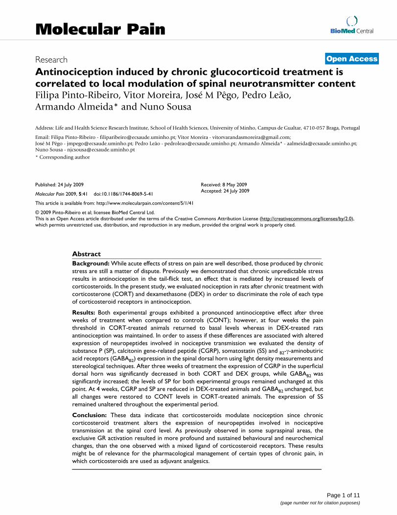
Open AcceResearchAntinociception induced by chronic glucocorticoid treatment is correlated to local modulation of spinal neurotransmitter contentFilipa Pinto-Ribeiro, Vitor Moreira, José M Pêgo, Pedro Leão, Armando Almeida* and Nuno SousaAddress: Life and Health Science Research Institute, School of Health Sciences, University of Minho, Campus de Gualtar, 4710-057 Braga, Portugal
Email: Filipa Pinto-Ribeiro - [email protected]; Vitor Moreira - [email protected]; José M Pêgo - [email protected]; Pedro Leão - [email protected]; Armando Almeida* - [email protected]; Nuno Sousa - [email protected]
* Corresponding author
AbstractBackground: While acute effects of stress on pain are well described, those produced by chronicstress are still a matter of dispute. Previously we demonstrated that chronic unpredictable stressresults in antinociception in the tail-flick test, an effect that is mediated by increased levels ofcorticosteroids. In the present study, we evaluated nociception in rats after chronic treatment withcorticosterone (CORT) and dexamethasone (DEX) in order to discriminate the role of each typeof corticosteroid receptors in antinociception.
Results: Both experimental groups exhibited a pronounced antinociceptive effect after threeweeks of treatment when compared to controls (CONT); however, at four weeks the painthreshold in CORT-treated animals returned to basal levels whereas in DEX-treated ratsantinociception was maintained. In order to assess if these differences are associated with alteredexpression of neuropeptides involved in nociceptive transmission we evaluated the density ofsubstance P (SP), calcitonin gene-related peptide (CGRP), somatostatin (SS) and B2-γ-aminobutiricacid receptors (GABAB2) expression in the spinal dorsal horn using light density measurements andstereological techniques. After three weeks of treatment the expression of CGRP in the superficialdorsal horn was significantly decreased in both CORT and DEX groups, while GABAB2 wassignificantly increased; the levels of SP for both experimental groups remained unchanged at thispoint. At 4 weeks, CGRP and SP are reduced in DEX-treated animals and GABAB2 unchanged, butall changes were restored to CONT levels in CORT-treated animals. The expression of SSremained unaltered throughout the experimental period.
Conclusion: These data indicate that corticosteroids modulate nociception since chroniccorticosteroid treatment alters the expression of neuropeptides involved in nociceptivetransmission at the spinal cord level. As previously observed in some supraspinal areas, theexclusive GR activation resulted in more profound and sustained behavioural and neurochemicalchanges, than the one observed with a mixed ligand of corticosteroid receptors. These resultsmight be of relevance for the pharmacological management of certain types of chronic pain, inwhich corticosteroids are used as adjuvant analgesics.
Published: 24 July 2009
Molecular Pain 2009, 5:41 doi:10.1186/1744-8069-5-41
Received: 8 May 2009Accepted: 24 July 2009
This article is available from: http://www.molecularpain.com/content/5/1/41
© 2009 Pinto-Ribeiro et al; licensee BioMed Central Ltd. This is an Open Access article distributed under the terms of the Creative Commons Attribution License (http://creativecommons.org/licenses/by/2.0), which permits unrestricted use, distribution, and reproduction in any medium, provided the original work is properly cited.
Page 1 of 11(page number not for citation purposes)

Molecular Pain 2009, 5:41 http://www.molecularpain.com/content/5/1/41
BackgroundNociception can be modulated at different levels of theCNS through facilitating (pronociceptive) or inhibiting(antinociceptive) central actions [1-3]. One of the levelswhere nociceptive modulation takes place is in laminae I–II of the spinal dorsal horn [4], where nociceptors synapseupon interneurons and projection neurons [5,6]. Thetransmission of nociceptive information in the dorsalhorn involves several events, neuropeptides and fibres.After peripheral noxious stimulation of unmyelinatednociceptors the release of calcitonin gene-related peptide(CGRP) [7], substance P (SP) [8] and somatostatin (SS)[4,9] is increased although it remains largely unchangedafter innocuous stimulation or stimulation of large myeli-nated fibres [8,9]. Spinal nociceptive neurons that areexcited by CGRP and SP [10,11] receive numerous synap-tic contacts from primary afferent terminals colocalizingthese neurotransmitters, whereas non-nociceptive neu-rons lack synaptic input from boutons with both peptides[12]. Spinal SS [13] and GABA [14] have an inhibitoryeffect on nociceptive neurons, being present mainly infibres belonging to local inhibitory interneurons [15].
Acute stress induces analgesia but the effects of chronicstress in nociception are still controversial, with studiesreporting hyperalgesia after prolonged stress [16], whileothers observed analgesia [17]. Recently, we demon-strated that animals submitted to chronic unpredictablestress display antinociception in the tail-flick test [18];since the plasmatic levels of corticosteroids were increasedthroughout the entire experimental period, we implicatedthese hormones in that phenomenon. Corticosteroids canbind to two types of corticosteroid receptors, mineraloco-rticoid (MR) and glucocorticoid (GR) receptors. In basalconditions, MR display greater occupancy than GR; thus,conditions resulting in elevation of corticosteroids, e.g.stress, will result mainly in increased activation of GR.Importantly, the spinal cord is a corticoid-responsive tis-sue [19] and within the spinal cord the greatest density ofGR and MR occurs in laminae I–II [20]. Of notice, CGRPand SP (but not SS) coexist with corticosteroid receptorsin neurons of dorsal root ganglia [21] and some studiesdemonstrate that an imbalanced corticosteroid milieumay affect neuropeptide content in the DRG [22,23].Importantly, corticosteroids are often used as adjuvantanalgesics in the management of several types of pain [24-26]. Taken together, these findings predict a potentialinfluence of corticosteroids in the modulation of spinalnociceptive transmission.
In the premise that a distinctive activation of MR or GRcould be responsible for altered levels of neuropeptidesinvolved in spinal nociceptive transmission and, conse-quently, for diverse pain-like effects we evaluated the den-sity of CGRP, SP, SS and GABAB2 innervation in the spinal
dorsal horn of animals submitted to prolonged adminis-tration of CORT (activating both MR and GR) and DEX (aselective ligand of GR). These data were correlated withpain-like behaviour measured through the tail-flick andhot-plate tests.
ResultsPain-like BehaviourEvolution within groups during the experimental periodAnalysis of TF and HP latency in CONT revealed no signif-icant differences between testing sessions throughout theexperimental period (ANOVArm, TF, P = 0.29 and HP, P =0.60).
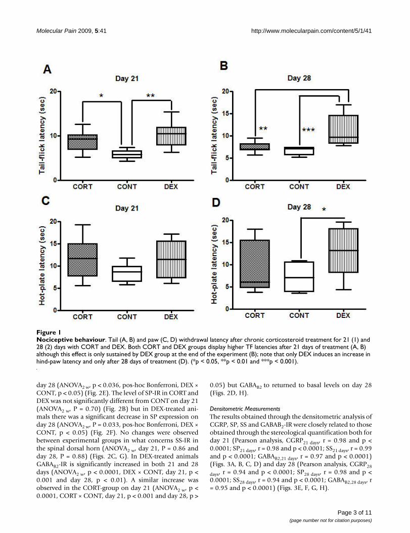
Tail-flick testThe chronic subcutaneous administration of CORT andDEX resulted in a significant decrease in pain-like behav-iour. Statistical data indicate that both CORT and DEXinduced a significant increase in TF latencies on day 21(ANOVAow, P = 0.002, pos-hoc Bonferroni, CORT × CONT,p < 0.05; DEX × CONT, p < 0.01) (Fig. 1A). However, withthe prolongation of the treatment (day 28) only subjectsunder DEX treatment maintained the significant increasein TF thresholds; in contrast in CORT-treated animalsnociceptive behaviour decreased slightly (ANOVAow, P =0.0003, pos-hoc Bonferroni, CORT × CONT, p > 0.05, DEX× CONT, p < 0.001 and DEX × CORT, p < 0.001) (Fig. 1B).
Hot-plate testThe prolonged administration of DEX but not CORTresulted in a significant decrease in nociceptive behaviour.Statistical data indicates that HP latencies are significantlyincreased in DEX-treated animals on day 28 (ANOVAow, P= 0.02, pos-hoc Bonferroni, DEX × CONT, p < 0.05) (Fig.1D). Contrary to what was observed for the TF test, no dif-ferences between groups were observed on day 21,although a trend towards an antinociceptive effect wasalready observed (ANOVAow, P = 0.07) (Fig. 1C).
Neurotransmitter Spinal InnervationAll statistical data presented in this section referring toimmunoreactivity evaluation is based on the study of thelumbar portion of the spinal cord as no differences in neu-rotransmitter-IR were found between cervical and lumbarportions.
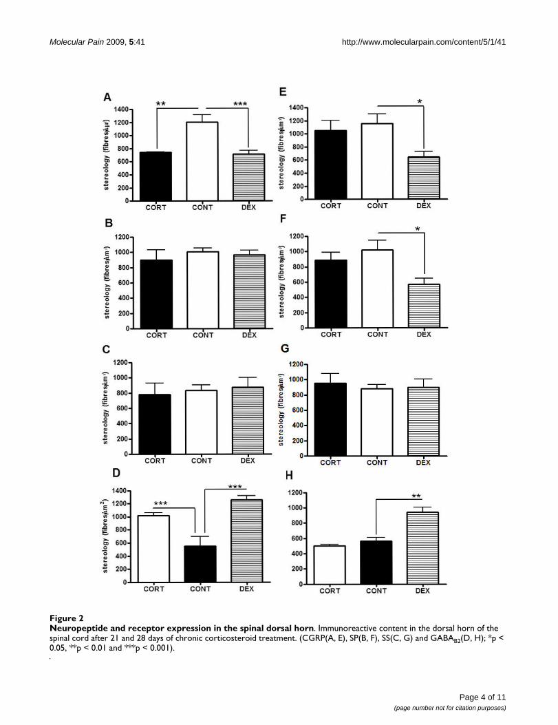
StereologyThe stereological analysis of CGRP-, SP-, SS- and GABAB2-IR in the spinal dorsal horn after prolonged CORT andDEX treatment is summarized in figure 2. The expressionof CGRP-IR was significantly decreased in both DEX andCORT-treated animals when compared to CONT on day21 (ANOVA2 w, p < 0.0001, pos-hoc Bonferroni, CORT ×CONT, p < 0.01 and DEX × CONT, p < 0.001) (Fig. 2A)although this effect was sustained only in DEX animals on
Page 2 of 11(page number not for citation purposes)
Molecular Pain 2009, 5:41 http://www.molecularpain.com/content/5/1/41
day 28 (ANOVA2 w, p < 0.036, pos-hoc Bonferroni, DEX ×CONT, p < 0.05) (Fig. 2E). The level of SP-IR in CORT andDEX was not significantly different from CONT on day 21(ANOVA2 w, P = 0.70) (Fig. 2B) but in DEX-treated ani-mals there was a significant decrease in SP expression onday 28 (ANOVA2 w, P = 0.033, pos-hoc Bonferroni, DEX ×CONT, p < 0.05) (Fig. 2F). No changes were observedbetween experimental groups in what concerns SS-IR inthe spinal dorsal horn (ANOVA2 w, day 21, P = 0.86 andday 28, P = 0.88) (Figs. 2C, G). In DEX-treated animalsGABAB2-IR is significantly increased in both 21 and 28days (ANOVA2 w, p < 0.0001, DEX × CONT, day 21, p <0.001 and day 28, p < 0.01). A similar increase wasobserved in the CORT-group on day 21 (ANOVA2 w, p <0.0001, CORT × CONT, day 21, p < 0.001 and day 28, p >
0.05) but GABAB2 to returned to basal levels on day 28(Figs. 2D, H).
Densitometric MeasurementsThe results obtained through the densitometric analysis ofCGRP, SP, SS and GABAB2-IR were closely related to thoseobtained through the stereological quantification both forday 21 (Pearson analysis, CGRP21 days, r = 0.98 and p <0.0001; SP21 days, r = 0.98 and p < 0.0001; SS21 days, r = 0.99and p < 0.0001; GABAB2,21 days, r = 0.97 and p < 0.0001)(Figs. 3A, B, C, D) and day 28 (Pearson analysis, CGRP28
days, r = 0.94 and p < 0.0001; SP28 days, r = 0.98 and p <0.0001; SS28 days, r = 0.94 and p < 0.0001; GABAB2,28 days, r= 0.95 and p < 0.0001) (Figs. 3E, F, G, H).
Nociceptive behaviourFigure 1Nociceptive behaviour. Tail (A, B) and paw (C, D) withdrawal latency after chronic corticosteroid treatment for 21 (1) and 28 (2) days with CORT and DEX. Both CORT and DEX groups display higher TF latencies after 21 days of treatment (A, B) although this effect is only sustained by DEX group at the end of the experiment (B); note that only DEX induces an increase in hind-paw latency and only after 28 days of treatment (D). (*p < 0.05, **p < 0.01 and ***p < 0.001).
Page 3 of 11(page number not for citation purposes)
Molecular Pain 2009, 5:41 http://www.molecularpain.com/content/5/1/41
Page 4 of 11(page number not for citation purposes)
Neuropeptide and receptor expression in the spinal dorsal hornFigure 2Neuropeptide and receptor expression in the spinal dorsal horn. Immunoreactive content in the dorsal horn of the spinal cord after 21 and 28 days of chronic corticosteroid treatment. (CGRP(A, E), SP(B, F), SS(C, G) and GABAB2(D, H); *p < 0.05, **p < 0.01 and ***p < 0.001).

Molecular Pain 2009, 5:41 http://www.molecularpain.com/content/5/1/41
Page 5 of 11(page number not for citation purposes)
Pearson correlation between densitometry and stereology quantification methodsFigure 3Pearson correlation between densitometry and stereology quantification methods. Pearson correlation for valida-tion of densitometric versus stereologic quantification methods on days 21(A-D) and 28(E-H). (SS(A, E), SP(B, F), CGRP(C, G) and GABAB2(D, H)).
Molecular Pain 2009, 5:41 http://www.molecularpain.com/content/5/1/41
DiscussionThe present study demonstrates that prolonged adminis-tration of corticosteroids decreases nociception. The anti-nociceptive effect reflects both a decrease ofpronociceptive neuropeptide expression and an increasedavailability of GABA receptors in laminae I–II of the spinaldorsal horn. After 21 days of treatment the decrease inpain-like behaviour was correlated with a decrease inCGRP and an increase in GABAB2 receptors in the spinalcord of CORT and DEX treated-animals. Interestingly, theantinociceptive effect in the CORT-group vanished afterfour weeks of treatment (which was paralleled by a resto-ration of CGRP and GABAB2 expression towards controllevels) while it remained unchanged in DEX-treated rats(which were correlated with a decrease in spinal contentof both CGRP and SP and increased availability ofGABAB2). These findings confirm that corticosteroidreceptors play a crucial role in the mediation of pain trans-mission at the spinal cord level.
Pain perception involves the transmission of nociceptivemessages from the periphery to the CNS. This transmis-sion can be modulated by acute [16] and chronic stress[16,17]. Recently, we showed that chronic unpredictablestress, which results in a prolonged elevation of plasmaticglucocorticoid (GC) levels, decreases pain-like behaviour[18]. Most actions mediated by chronic stress are attrib-uted to hypercortisolemia, as the increased secretion ofcorticosteroids characterizes the sustained phase of thestress response [27]. Because corticosteroids can bind totwo types of receptors we decided to further explore therole of each of these corticosteroid receptors on the noci-ceptive modulation. It is important to note at this point,that the confounding effect of drug potency has been con-sidered, as the doses of each corticosteroid were adjustedaccordingly to their glucocorticoid potency. Thus, in thisexperimental paradigm CORT treatment differs from DEXadministration basically in terms of MR activation: whileCORT treatment activates these receptors, DEX does notbind to MR and because it shuts-off the endogenous secre-tion of corticosteroids, MR remains unoccupied [28].
The results observed after prolonged daily treatment withcorticosteroids demonstrate that these steroids promoteantinociception. GR are likely to mediate this phenome-non since a similar response was observed in DEX-treatedanimals. The TF test evaluates a spinally organized reflex[29,30] mediated by C-fibres innervating the tail [31,32]and motoneurons innervating the three sets of back mus-cles that control tail movements [33-35]. In contrast, theHP test involves a supraspinally integrated response, andthus, represents a more complex behavioural response[36]. Such difference in the neuroanatomical substratesimplicated in both tests might explain why there was only
a trend towards increased HP latencies after 21 days ofCORT and DEX treatments.
It is admissible that the influence of GC upon neuropep-tidergic innervation results both from direct and indirectactions. Indirect actions may result from altered availabil-ity of GABAB2 receptors in CORT and DEX treated animalson day 21, as GABAB receptors are well known players inpain modulation [15,37]. Moreover Kangrga and col-legues [38] described that the antinociceptive effect ofGABAergic transmission in the spinal dorsal horn resultsfrom presynaptic inhibition of the release of excitatoryamino acids and neurotransmitters from the primaryafferents [14,39] which is in accordance with our observa-tions that CGRP, a pronociceptive neuropeptide, isdecreased in both CORT- and DEX-treated groups. Analternative indirect action of glucocorticoids might occurthrough the modulatory actions of arachidonic pathwayswhich down-regulate nerve growth factor (NGF); this, inturn, is known to exert an inhibitory effect in both theaccumulation and release of CGRP mRNA in nociceptors[40,41]. In parallel, the direct modulation of glucocorti-coids can be ascribed to the fact that approximately onethird of the afferents that are immunoreactive to SP orCGRP, also display immunoreactivity to GR [21]. Thus, itis plausible to assume that GR activation of nuclearresponsive elements alters the expression of such trans-mitters in spinal dorsal horn afferents. This hypothesis isfurther supported by the fact that it was recently shownthat stressors decrease CGRP expression in the frontal cor-tex, hippocampus, occipital cortex and hypothalamus[42].
Curiously, the dissimilarity in pain-like behaviourobserved between CORT and DEX groups after 28 days oftreatment, suggests that other mechanisms involving MRactivation are implicated in the modulation of pain. Infact, the behavioural differences observed between CORTand DEX treatment at 28 days were paralleled by distinctpatterns in CGRP, SP and GABAB2 expression in the super-ficial dorsal horn: while in CORT-treated animals theexpression of both CGRP and GABAB2 was restored to con-trol levels, DEX treatment resulted in a decreased expres-sion of SP and CGRP and sustained increase in GABAB2.The explanations for such discrepancy are more complex,as besides the local effects at the spinal cord level, theymight involve alterations at the supraspinal level. Indeed,there is a complex feedback system between the neuro-transmitters herein studied and GC involving supraspinalprocessing that is regulated by MR. There is evidence thatthe activation of MR is correlated with GABA modulation[43] in lamina II [44] of the spinal cord, namely ininterneurones [45], and in other supraspinal pain modu-lating areas such as the rostroventral lateral medulla(RVM) [46] or the periaductal grey matter (PAG) [47].
Page 6 of 11(page number not for citation purposes)

Molecular Pain 2009, 5:41 http://www.molecularpain.com/content/5/1/41
This effect of GABAergic transmission appears to selec-tively inhibit the release of SP, but not of CGRP, [15]which may account at least partly for the differencesobserved between the groups. Another alternative, but notexclusive, mechanism to explain the differential effect ofDEX and CORT upon neuropeptidergic spinal expressionderives from the specific modulatory effects of MR uponpreprotachykinin (PPT), the precursor of SP expression; infact, MR activation has been shown to positively regulate(up to 50%) mRNA PPT expression in the nervous tissues[48]. The more persistent changes in pain perceptioninduced by DEX treatment and measured by an increase inboth the TF and HP latency at day 28 might thereforeresult from a decrease in SP fibre innervation in the spinaldorsal horn.
Contrary to CGRP, SP and GABAB2, no effect of GC wasobserved on the spinal levels of SS. This differentialchange observed between these neuropeptides illustratesthe selectivity of this process, and is likely to be relatedwith the lack of coexistence of corticosteroid and SS in thespinal cord [21]. Interestingly, different neurotransmittersare associated with different roles in pain modulation[5,49]. In contrast to CGRP/SP, SS is a tonic inhibitor ofperipheral nociceptors [50]. Thus, the data hereinreported suggests that the effects of chronic corticosteroidtreatment on pain perception are associated with changesin the nociceptive transmitting system (CGRP/SP) butwould not involve specific alterations in the spinal intrin-sic modulatory system (SS).
In addition to their presence in the spinal dorsal horn,both glucocorticoid- [51] and mineralocorticoid- [52]receptors are present also in neurons of a large number ofsupraspinal sites along the rostrocaudal extent of the neu-raxis in the rat. These include several forebrain and brain-stem components of the supraspinal pain control system,including areas like the anterior cingulate cortex [53],amygdala [54], paraventricular hypothalamic nucleus[55], periaqueductal grey matter [56], locus coeruleus[57], rostral ventromedial medulla [58], dorsal reticularnucleus [59] and caudal ventrolateral medulla [60]. Tak-ing into account data obtained in the present study on theeffect of corticosteroid manipulation upon spinal neuro-transmitter content, future studies should explore altera-tions induced at supraspinal levels. Accordingly,profound structural, physiological and neurochemicalalterations have been observed at different forebrain areasfollowing chronic manipulation of corticosteroids [30,61-63].
ConclusionThe present study shows that corticosteroids modulatenociception by altering the expression of neuropeptidesinvolved in nociceptive transmission at the spinal cord
level. Moreover, we demonstrate differential modulatoryactions of different ligands of corticosteroid receptors,which are of relevance for the pharmacological manage-ment of those conditions involving chronic pain, inwhich corticosteroids are recommended as adjuvant anal-gesics.
MethodsSubjectsWistar Han rats obtained from Charles Rivers (Barcelona,UE), weighting between 200–240 g, at the beginning ofthe experiment, were housed in groups of three in stand-ard polycarbonate cages (45.4 × 25.5 × 20 cm). The lightcycle was 12:12 h with lights on at 9:00 am and housingwas maintained at 22°C and 30% relative humidity.Water and food were available ad libitum. All regulationsdetermined by the local veterinarian committee (inaccordance to the European Community Council Direc-tive 86/609/EEC) concerning the handling of laboratoryanimals and the international ethical guidelines for thestudy of experimental pain in conscious animals were fol-lowed [64].
Chronic corticosteroid treatmentCorticosterone, dexamethasone and sesame oil wereacquired from Sigma (St Louis, MO, USA). Subjects wereassigned to one of the following three groups (n = 24):
(i) Controls (CONT). Rats were submitted to vehicleinjection (0.5 ml sesame oil) everyday (05:00 pm), during3 weeks (n = 4) and 4 weeks (n = 4).
(ii) Corticosterone-treated (CORT). Rats were submittedover a period of 3 weeks (n = 4) and 4 weeks (n = 4) to adaily subcutaneous injection (05:00 pm) of 40 mg/kgdose of 4-Pregnene-11β,21 diol-3,20-dione in sesame oil.
(iii) Dexamethasone-treated (DEX). Rats were submittedover a period of 3 weeks (n = 4) and 4 weeks (n = 4) to adaily subcutaneous injection (05:00 pm) of 300 μmg/kgdose of 9α-fluoro-16α-methylpredenisolone in sesameoil.
Nociceptive testingPain-like behaviour was analyzed for each animal usingthe tail-flick (TF) and the hot-plate (HP) tests. In the TF(Ugo Basile, Comerio, Italy) the time spent the start of thestimulus and the withdrawal of the tail (nociceptivelatency) was recorded, whereas in the HP (Ugo Basile,Comerio, Italy) as the heating plate was kept at a constanttemperature of 54 ± 0.5°C, it was the latency for hind pawlicking or jumping was recorded.
In order to determine the nociceptive threshold, rats weretested before corticosteroid administration (day 0) and on
Page 7 of 11(page number not for citation purposes)

Molecular Pain 2009, 5:41 http://www.molecularpain.com/content/5/1/41
days 7, 14, 21 and 28 of the treatment; each testing dayanimals were submitted (11:00 am) to 3 TF tests, within a2 min interval, and 2 HP test, with 45 min interval (Fig.4). To avoid bias related with the handling and testing ofthe rats, a one-week period prior to the first nociceptivetest was established for the habituation of the animals tothe behavioural test equipment and the researcher. Ani-mals were placed daily in the test room for 2 h followedby a 10 minute handling and 1 minute training session inthe TF and the HP apparatus (without performing thetest).
ImmunocytochemistryAt the end of the experimental period (21 or 28 days), ani-mals were anesthetized intraperitoneally (sodium pento-barbital, 0.5 mg/kg) and perfused transcardially with 4%paraformaldehyde in PBS 0.1 M, pH 7.2. The spinal cordwas removed and placed in 30% sucrose for 24 h. Portionsof the cervical and lumbar spinal cord enlargements weresampled. Sections, 30 μm thick, were cut on a vibratingblade microtome (Leica, Germany) and collected insuperfrosted slides. Sections from the same region for allsubjects and treatments were exposed to the same solu-tions. Sections were permeabilized for 10 min in 0.2% Tri-ton X-100 in Tris buffer saline (TBS) and microwaved (20min) while immersed in citrate buffer (0.1 M). Endog-enous peroxidase activity was blocked with 3% H2O2 inPBS (10 min) and non-specific staining was blocked with4% bovine serum albumin (BSA) in PBS (30 min). Alter-nating sections were incubated overnight at room temper-ature in rabbit primary antibodies against CGRP (1:3000;Chemicon, USA) (Fig. 5), SP (1:3000; Chemicon, USA),SS (1:3000; Chemicon, USA) and GABAB2 (1:1000,Chemicon, USA) (Fig. 6) in 0.02% Triton X-100 (PBST).Antigen visualization was carried out using a universaldetection system (BioGenex, San Ramon, CA) and diami-
nobenzidine (DAB; 0.025% and 0.5% H2O2 in Tris-HCl0.05 M, pH 7.2).
StereologyThe stereological analysis was performed in the dorsalhorn of CGRP-, SP-, SS-and GABAB2-immunoreacted (IR)spinal cord sections using StereoInvestigator software(MicroBrightField, Williston/VT, USA). From each set ofserial sections, ten photomicrographs of areas within thespinal laminae I–II were obtained at a primary magnifica-tion of × 50 and analyzed at a final magnification of ×1000. The number of stained fibres per unit of laminae I–II volume (numerical density) was estimated using theoptical fractionator method [65]. The surface volumeoccupied by laminae I–II stained fibres was calculated onthe basis of the surface density of the fibres (surface areaper unit volume, SV) and the volume of laminae I–II. TheSV was estimated, using a 'staggered' cycloid test system inorder to obtain the total number of intersections betweencycloid arcs and stained fibres. Measurements were madeon laminae I–II regions randomly selected by the soft-ware.
Densitometric MeasurementsThe densitometric analysis was performed in the dorsalhorn of CGRP, SP, SS and GABAB2-IR spinal cord sectionsusing a Zeiss light microscope coupled to a PC, using NIH
Time course of testing and sampling sessions throughout the four week experimental periodFigure 4Time course of testing and sampling sessions throughout the four week experimental period. Within a testing session, tail-flick and hot-plate tests were performed according to the time course example for day 7.
Photomicrographs of superficial dorsal horn sectionsFigure 5Photomicrographs of superficial dorsal horn sections. Examples of photomicrographs of superficial dorsal horn sec-tions immunoreacted for CGRP (A) on days 21 (A1–3) and 28 (A4–6) for CONT (A1,4), DEX (A2,5) and CORT (A3,6).
Page 8 of 11(page number not for citation purposes)

Molecular Pain 2009, 5:41 http://www.molecularpain.com/content/5/1/41
Image 1.52 software. The sampling area for optical densitymeasurement corresponded to all the area occupied bylaminas I and II of the spinal dorsal horn, bilaterally. Den-sity levels and distribution of CGRP-, SP-, SS- andGABAB2-IR were quantified and, for all sections, back-ground density measurements were subtracted to thesevalues.
Data analysisStatistical analysis was performed using GraphPad Prismversion 4.00 for Windows (GraphPad Software, San DiegoCalifornia, USA). A two-way ANOVA (ANOVA2 w) wasused to analyze differences between groups at differenttime points, while repeated-measures ANOVA (ANO-VArm) was used to evaluate efficiency of treatment alongdifferent time points within groups; pos-hoc Bonferroni'stest was used to detect significant differences for bothANOVA analysis. Densitometric and stereological datawas compared using the Pearson correlation analysis. Dif-ferences were considered statistically significant when p <0.05. All values are presented as mean ± SD.
AbbreviationsCGRP: Calcitonin gene-related peptide; CONT: Controls;CORT: Corticosterone; DEX: Dexamethasone; GABAB2:B2-γ-aminobutiric acid receptors; GC: Glucocorticoid;GR: Glucocorticoid receptor; HP: Hot-plate test; IR:
Immunoreactivity; MR: Mineralocorticoid receptor; SP:Substance P; SS: Somatostatin; TF: Tail-flick test
Competing interestsThe authors declare that they have no competing interests.
Authors' contributionsFPR performed the statistics analysis, carried out the IHCand quantification of densitometry and drafted the paper.VM: performed behavioural tests and the stereologicalanalysis. JMP: performed corticosteroid administrationand behavioural testing. PL: performed corticosteroidadministration and stereological quantification. AA: con-ceived, designed and coordinated the study and revisedthe paper. NS: conceived, designed and coordinated thestudy and revised the paper. All authors read andapproved the final manuscript.
AcknowledgementsThis study was supported by Calouste Gulbenkian Foundation project Nr. 74551 and the Grünenthal Foundation.
References1. Pertovaara A: Plasticity in descending pain modulatory sys-
tems. Prog Brain Res 2000, 129:231-242.2. Lima D, Almeida A: The medullary dorsal reticular nucleus as a
pronociceptive centre of the pain control system. Prog Neuro-biol 2002, 66:81-108.
3. Porreca F, Ossipov MH, Gebhart GF: Chronic pain and medullarydescending facilitation. Trends Neurosci 2002, 25:319-325.
4. Morton CR, Hutchison WD, Hendry IA, Duggan AW: Somatosta-tin: evidence for a role in thermal nociception. Brain Res 1989,488:89-96.
5. Millan MJ: The induction of pain: an integrative review. ProgNeurobiol 1999, 57:1-164.
6. Millan MJ: Descending control of pain. Prog Neurobiol 2002,66:355-474.
7. Morton CR, Hutchison WD: Release of sensory neuropeptidesin the spinal cord: studies with calcitonin gene-related pep-tide and galanin. Neuroscience 1989, 31:807-815.
8. Duggan AW, Hendry IA, Morton CR, Hutchison WD, Zhao ZQ:Cutaneous stimuli releasing immunoreactive substance P inthe dorsal horn of the cat. Brain Res 1988, 451:261-273.
9. Morton CR, Hutchison WD, Hendry IA: Release of immunoreac-tive somatostatin in the spinal dorsal horn of the cat. Neu-ropeptides 1988, 12:189-197.
10. Ma W, Ribeiro-Da-Silva A, De Koninck Y, Radhakrishnan V, Henry JL,Cuello AC: Quantitative analysis of substance P-immunoreac-tive boutons on physiologically characterized dorsal hornneurons in the cat lumbar spinal cord. J Comp Neurol 1996,376:45-64.
11. Yu LC, Zheng EM, Lundeberg T: Calcitonin gene-related peptide8–37 inhibits the evoked discharge frequency of widedynamic range neurons in dorsal horn of the spinal cord inrats. Regul Pept 1999, 83:21-24.
12. Ribeiro-da-Silva A: Ultrastructural features of the colocaliza-tion of calcitonin gene related peptide with substance P orsomatostatin in the dorsal horn of the spinal cord. Can J PhysiolPharmacol 1995, 73:940-944.
13. Todd AJ, Hughes DI, Polgar E, Nagy GG, Mackie M, Ottersen OP,Maxwell DJ: The expression of vesicular glutamate transport-ers VGLUT1 and VGLUT2 in neurochemically definedaxonal populations in the rat spinal cord with emphasis onthe dorsal horn. Eur J Neurosci 2003, 17:13-27.
14. Malcangio M, Bowery NG: GABA and its receptors in the spinalcord. Trends Pharmacol Sci 1996, 17:457-462.
Photomicrographs of superficial dorsal horn sectionsFigure 6Photomicrographs of superficial dorsal horn sections. Examples of photomicrographs of superficial dorsal horn sec-tions immunoreacted for GABAB2 (B) on days 21 (B1–3) and 28 (B4–6) for CONT (B1,4), DEX (B2,5) and CORT (B3,6).
Page 9 of 11(page number not for citation purposes)

Molecular Pain 2009, 5:41 http://www.molecularpain.com/content/5/1/41
15. Bowery NG, Hudson AL, Price GW: GABAA and GABAB recep-tor site distribution in the rat central nervous system. Neuro-science 1987, 20:365-383.
16. Gamaro GD, Xavier MH, Denardin JD, Pilger JA, Ely DR, Ferreira MB,Dalmaz C: The effects of acute and repeated restraint stresson the nociceptive response in rats. Physiol Behav 1998,63:693-697.
17. Clark WC, Yang JC, Janal MN: Altered pain and visual sensitivityin humans: the effects of acute and chronic stress. Ann N YAcad Sci 1986, 467:116-129.
18. Pinto-Ribeiro F, Almeida A, Pego JM, Cerqueira J, Sousa N: Chronicunpredictable stress inhibits nociception in male rats. Neuro-sci Lett 2004, 359:73-76.
19. Gonzalez S, Moses DF, De Nicola AF: Glucocorticoid receptorsand enzyme induction in the spinal cord of rats: effects ofacute transection. J Neurochem 1990, 54:834-840.
20. Marlier LN, Csikos T, Rebaudengo N, Borboni P, Patacchioli FR,Angelucci L, Privat A, Lauro R: Distribution of glucocorticoidreceptor mRNA in the rat spinal cord. Neuroreport 1995,6:2245-2249.
21. DeLeon M, Covenas R, Chadi G, Narvaez JA, Fuxe K, Cintra A: Sub-populations of primary sensory neurons show coexistence ofneuropeptides and glucocorticoid receptors in the rat spinaland trigeminal ganglia. Brain Res 1994, 636:338-342.
22. Smith GD, Seckl JR, Sheward WJ, Bennie JG, Carroll SM, Dick H, Har-mar AJ: Effect of adrenalectomy and dexamethasone on neu-ropeptide content of dorsal root ganglia in the rat. Brain Res1991, 564:27-30.
23. Covenas R, DeLeon M, Chadi G, Cintra A, Gustafsson JA, Narvaez JA,Fuxe K: Adrenalectomy increases the number of substance Pand somatostatin immunoreactive nerve cells in the rat lum-bar dorsal root ganglia. Brain Res 1994, 640:352-356.
24. Mercadante S, Arcuri E: Pharmacological management of can-cer pain in the elderly. Drugs Aging 2007, 24:761-776.
25. Abdi S, Datta S, Trescot AM, Schultz DM, Adlaka R, Atluri SL, SmithHS, Manchikanti L: Epidural steroids in the management ofchronic spinal pain: a systematic review. Pain Physician 2007,10:185-212.
26. Knotkova H, Pappagallo M: Adjuvant analgesics. Anesthesiol Clin2007, 25:775-786. vi.
27. Herman JP, Figueiredo H, Mueller NK, Ulrich-Lai Y, Ostrander MM,Choi DC, Cullinan WE: Central mechanisms of stress integra-tion: hierarchical circuitry controlling hypothalamo-pitui-tary-adrenocortical responsiveness. Front Neuroendocrinol 2003,24:151-180.
28. Sousa N, Cerqueira JJ, Almeida OF: Corticosteroid receptors andneuroplasticity. Brain Res Rev 2008, 57:561-570.
29. Ness TJ, Gebhart GF: Centrifugal modulation of the rat tail flickreflex evoked by graded noxious heating of the tail. Brain Res1986, 386:41-52.
30. Danneman PJ, Kiritsy-Roy JA, Morrow TJ, Casey KL: Central delayof the laser-activated rat tail-flick reflex. Pain 1994, 58:39-44.
31. Fleischer E, Handwerker HO, Joukhadar S: Unmyelinated nocice-ptive units in two skin areas of the rat. Brain Res 1983,267:81-92.
32. Necker R, Hellon RF: Noxious thermal input from the rat tail:modulation by descending inhibitory influences. Pain 1978,4:231-242.
33. Brink EE, Pfaff DW: Vertebral muscles of the back and tail ofthe albino rat (Rattus norvegicus albinus). Brain Behav Evol1980, 17:1-47.
34. Grossman ML, Basbaum AI, Fields HL: Afferent and efferent con-nections of the rat tail flick reflex (a model used to analyzepain control mechanisms). J Comp Neurol 1982, 206:9-16.
35. Cargill CL, Steinman JL, Willis WD: A fictive tail flick reflex in therat. Brain Res 1985, 345:45-53.
36. Le Bars D, Gozariu M, Cadden SW: Animal models of nocicep-tion. Pharmacol Rev 2001, 53:597-652.
37. Towers S, Princivalle A, Billinton A, Edmunds M, Bettler B, Urban L,Castro-Lopes J, Bowery NG: GABAB receptor protein andmRNA distribution in rat spinal cord and dorsal root ganglia.Eur J Neurosci 2000, 12:3201-3210.
38. Kangrga I, Jiang MC, Randic M: Actions of (-)-baclofen on rat dor-sal horn neurons. Brain Res 1991, 562:265-275.
39. Smith CG, Bowery NG, Whitehead KJ: GABA transporter type 1(GAT-1) uptake inhibition reduces stimulated aspartate and
glutamate release in the dorsal spinal cord in vivo via differ-ent GABAergic mechanisms. Neuropharmacology 2007,53:975-981.
40. Supowit SC, Christensen MD, Westlund KN, Hallman DM, DiPetteDJ: Dexamethasone and activators of the protein kinase Aand C signal transduction pathways regulate neuronal calci-tonin gene-related peptide expression and release. Brain Res1995, 686:77-86.
41. Nohr D, Schafer MK, Persson S, Romeo H, Nyberg F, Post C,Ekstrom G, Weihe E: Calcitonin gene-related peptide geneexpression in collagen-induced arthritis is differentially regu-lated in primary afferents and motoneurons: influence of glu-cocorticoids. Neuroscience 1999, 93:759-773.
42. Husum H, Termeer E, Mathe AA, Bolwig TG, Ellenbroek BA: Earlymaternal deprivation alters hippocampal levels of neuropep-tide Y and calcitonin-gene related peptide in adult rats. Neu-ropharmacology 2002, 42:798-806.
43. Katona I, Sperlagh B, Sik A, Kafalvi A, Vizi ES, Mackie K, Freund TF:Presynaptically located CB1 cannabinoid receptors regulateGABA release from axon terminals of specific hippocampalinterneurons. J Neurosci 1999, 19:4544-4558.
44. Salio C, Fischer J, Franzoni MF, Conrath M: Pre- and postsynapticlocalizations of the CB1 cannabinoid receptor in the dorsalhorn of the rat spinal cord. Neuroscience 2002, 110:755-764.
45. Jennings EA, Vaughan CW, Christie MJ: Cannabinoid actions onrat superficial medullary dorsal horn neurons in vitro. J Physiol2001, 534:805-812.
46. Vaughan CW, McGregor IS, Christie MJ: Cannabinoid receptoractivation inhibits GABAergic neurotransmission in rostralventromedial medulla neurons in vitro. Br J Pharmacol 1999,127:935-940.
47. Vaughan CW, Connor M, Bagley EE, Christie MJ: Actions of can-nabinoids on membrane properties and synaptic transmis-sion in rat periaqueductal gray neurons in vitro. Mol Pharmacol2000, 57:288-295.
48. Lucas LR, Pompei P, Ono J, McEwen BS: Effects of adrenal steroidson basal ganglia neuropeptide mRNA and tyrosine hydroxy-lase radioimmunoreactive levels in the adrenalectomizedrat. J Neurochem 1998, 71:833-843.
49. Jang JH, Nam TS, Paik KS, Leem JW: Involvement of peripherallyreleased substance P and calcitonin gene-related peptide inmediating mechanical hyperalgesia in a traumatic neuropa-thy model of the rat. Neurosci Lett 2004, 360:129-132.
50. Carlton SM, Du J, Zhou S, Coggeshall RE: Tonic control of periph-eral cutaneous nociceptors by somatostatin receptors. J Neu-rosci 2001, 21:4042-4049.
51. Ahima RS, Harlan RE: Charting of type II glucocorticoid recep-tor-like immunoreactivity in the rat central nervous system.Neuroscience 1990, 39:579-604.
52. Ahima R, Krozowski Z, Harlan R: Type I corticosteroid receptor-like immunoreactivity in the rat CNS: distribution and regu-lation by corticosteroids. J Comp Neurol 1991, 313:522-538.
53. Zhang L, Zhang Y, Zhao ZQ: Anterior cingulate cortex contrib-utes to the descending facilitatory modulation of pain viadorsal reticular nucleus. Eur J Neurosci 2005, 22:1141-1148.
54. Myers B, Dittmeyer K, Greenwood-Van Meerveld B: Involvementof amygdaloid corticosterone in altered visceral and somaticsensation. Behav Brain Res 2007, 181:163-167.
55. Pinto-Ribeiro F, Ansah OB, Almeida A, Pertovaara A: Influence ofarthritis on descending modulation of nociception from theparaventricular nucleus of the hypothalamus. Brain Res 2008,1197:63-75.
56. Waters AJ, Lumb BM: Descending control of spinal nociceptionfrom the periaqueductal grey distinguishes between neuronswith and without C-fibre inputs. Pain 2008, 134:32-40.
57. Tsuruoka M, Matsutani K, Inoue T: Coeruleospinal inhibition ofnociceptive processing in the dorsal horn during unilateralhindpaw inflammation in the rat. Pain 2003, 104:353-361.
58. Zhuo M, Gebhart GF: Biphasic modulation of spinal nociceptivetransmission from the medullary raphe nuclei in the rat. JNeurophysiol 1997, 78:746-758.
59. Lima D, Almeida A: The medullary dorsal reticular nucleus as apronociceptive centre of the pain control system. Prog Neuro-biol 2002, 66:81-108.
Page 10 of 11(page number not for citation purposes)
Molecular Pain 2009, 5:41 http://www.molecularpain.com/content/5/1/41
Publish with BioMed Central and every scientist can read your work free of charge
"BioMed Central will be the most significant development for disseminating the results of biomedical research in our lifetime."
Sir Paul Nurse, Cancer Research UK
Your research papers will be:
available free of charge to the entire biomedical community
peer reviewed and published immediately upon acceptance
cited in PubMed and archived on PubMed Central
yours — you keep the copyright
Submit your manuscript here:http://www.biomedcentral.com/info/publishing_adv.asp
BioMedcentral
60. Tavares I, Lima D: The caudal ventrolateral medulla as animportant inhibitory modulator of pain transmission in thespinal cord. J Pain 2002, 3:337-346.
61. Sousa N, Almeida OF: Corticosteroids: sculptors of the hippoc-ampal formation. Rev Neurosci 2002, 13:59-84.
62. Cerqueira JJ, Catania C, Sotiropoulos I, Schubert M, Kalisch R, Alme-ida OF, Auer DP, Sousa N: Corticosteroid status influences thevolume of the rat cingulate cortex – a magnetic resonanceimaging study. J Psychiatr Res 2005, 39:451-460.
63. Cerqueira JJ, Pêgo JM, Taipa R, Bessa JM, Almeida OF, Sousa N: Mor-phological correlates of corticosteroid-induced changes inprefrontal cortex-dependent behaviors. J Neurosci 2005,25:7792-7800.
64. Zimmermann M: Ethical guidelines for investigations of exper-imental pain in conscious animals. Pain 1983, 16:109-110.
65. Gundersen HJ, Jensen EB: The efficiency of systematic samplingin stereology and its prediction. J Microsc 1987, 147:229-263.
Page 11 of 11(page number not for citation purposes)
Copyright © 2022 FDOKUMEN