Chromatin Remodeling Complexes Interact Dynamically with a Glucocorticoid Receptor-regulated...
15
Molecular Biology of the Cell Vol. 19, 3308 –3322, August 2008 Chromatin Remodeling Complexes Interact Dynamically with a Glucocorticoid Receptor–regulated Promoter Thomas A. Johnson,* † Cem Elbi,* †‡ Bhavin S. Parekh, § Gordon L. Hager,* and Sam John* *Laboratory of Receptor Biology and Gene Expression, National Cancer Institute, National Institutes of Health, Bethesda, MD 20892-5055; and § Eli Lilly and Company, Indianapolis, IN 46221 Submitted February 6, 2008; Revised April 24, 2008; Accepted May 21, 2008 Monitoring Editor: Jennifer Lippincott-Schwartz Brahma (BRM) and Brahma-related gene 1 (BRG1) are the ATP-dependent catalytic subunits of the SWI/SNF family of chromatin-remodeling complexes. These complexes are involved in essential processes such as cell cycle, growth, differentiation, and cancer. Using imaging approaches in a cell line that harbors tandem repeats of stably integrated copies of the steroid responsive MMTV-LTR (mouse mammary tumor virus–long terminal repeat), we show that BRG1 and BRM are recruited to the MMTV promoter in a hormone-dependent manner. The recruitment of BRG1 and BRM resulted in chromatin remodeling and decondensation of the MMTV repeat as demonstrated by an increase in the restriction enzyme accessibility and in the size of DNA fluorescence in situ hybridization (FISH) signals. This chromatin remodeling event was concomitant with an increased occupancy of RNA polymerase II and transcriptional activation at the MMTV promoter. The expression of ATPase-deficient forms of BRG1 (BRG1-K-R) or BRM (BRM-K-R) inhibited the remodeling of local and higher order MMTV chromatin structure and resulted in the attenuation of transcription. In vivo photo- bleaching experiments provided direct evidence that BRG1, BRG1-K-R, and BRM chromatin-remodeling complexes have distinct kinetic properties on the MMTV array, and they dynamically associate with and dissociate from MMTV chromatin in a manner dependent on hormone and a functional ATPase domain. Our data provide a kinetic and mechanistic basis for the BRG1 and BRM chromatin-remodeling complexes in regulating gene expression at a steroid hormone inducible promoter. INTRODUCTION The eukaryotic genome is organized into higher order chro- matin structures in the nucleus. The regulation of eukaryotic gene expression in the context of chromatin is a complex event and is essential for numerous cellular processes (Lemon and Tjian, 2000;Orphanides and Reinberg, 2002; Labrador and Corces, 2002; Maniatis and Reed, 2002; Felsenfeld and Groudine, 2003; Roberts and Orkin, 2004). Maintenance, establishment, and modification of global chromatin organi- zation and local chromatin structure are modulated by a large number of chromatin-binding proteins that generate transcriptionally permissive or repressed chromatin do- mains in response to environmental stimuli (Workman and Kingston, 1998; Peterson and Workman, 2000; Jones and Kadonaga, 2000; Wu and Grunstein, 2000; Wolffe and Han- sen, 2001; Becker et al., 2002). The association of linker his- tones and nonhistone and heterochromatin-specific proteins such as high mobility group proteins and HP1 play key roles in the generation of higher order chromatin structures (Ar- ents et al., 1991; Luger et al., 1997; Bustin, 1999; Eissenberg and Elgin, 2000; Hill, 2001; Thomas and Travers, 2001; Woodcock and Dimitrov, 2001; Grewal and Elgin, 2002; Bianchi and Agresti, 2005; Bustin et al., 2005; Verschure et al., 2005). The stearically restricted environment presented by chromatin is in part overcome by multisubunit protein com- plexes that enzymatically regulate chromatin structure. These complexes can either chemically modify histone tails (acetylation, phosphorylation, ubiquitylation, methylation) or disrupt histone–DNA interactions by using the energy generated through ATP hydrolysis (Strahl and Allis, 2000; Berger, 2002; Narlikar et al., 2002; Neely and Workman, 2002; Shogren-Knaak et al., 2006). The combination of chromatin modification and chromatin-remodeling complexes acting in a concerted manner modulate the accessibility of individual genes to sequence-specific transcription factors, general transcription factors, and components of the RNA pol II transcription machinery. SWI/SNF is one of three subclasses of ATP-dependent chromatin remodeling enzymes that have been identified in mammalian cells (Peterson, 2002). The SWI/SNF complex exists in one of two forms, containing either one of two highly homologous ATPases, BRG1 or BRM and several shared subunits collectively called BAFs (BRG1- or BRM- associated factors). SWI/SNF is an evolutionarily conserved, 2 MDa multisubunit complex interacting with a wide va- riety of proteins and is functionally implicated in cell cycle, differentiation, and cancer (Muchardt and Yaniv, 2001; Klochendler-Yeivin et al., 2002; Roberts and Orkin, 2004; Cho et al., 2004; Gregory and Shiekhattar, 2004; Imbalzano and Jones, 2005). Several subunits of the SWI/SNF chromatin- remodeling complex possess tumor suppressor activity and play key roles in the functional activity of other tumor suppressor genes including Rb, BRCA1, and c-MYC (Mu- This article was published online ahead of print in MBC in Press (http://www.molbiolcell.org/cgi/doi/10.1091/mbc.E08 – 02– 0123) on May 28, 2008. † These authors contributed equally to this work. ‡ Present address: Merck Research Laboratories, Boston, MA 02115. Address correspondence to: Sam John ([email protected]). 3308 © 2008 by The American Society for Cell Biology http://www.molbiolcell.org/content/suppl/2008/06/02/E08-02-0123v1.DC1.html Supplemental Material can be found at:
Transcript of Chromatin Remodeling Complexes Interact Dynamically with a Glucocorticoid Receptor-regulated...

Molecular Biology of the CellVol. 19, 3308–3322, August 2008
Chromatin Remodeling Complexes Interact Dynamicallywith a Glucocorticoid Receptor–regulated PromoterThomas A. Johnson,*† Cem Elbi,*†‡ Bhavin S. Parekh,§ Gordon L. Hager,*and Sam John*
*Laboratory of Receptor Biology and Gene Expression, National Cancer Institute, National Institutes ofHealth, Bethesda, MD 20892-5055; and §Eli Lilly and Company, Indianapolis, IN 46221
Submitted February 6, 2008; Revised April 24, 2008; Accepted May 21, 2008Monitoring Editor: Jennifer Lippincott-Schwartz
Brahma (BRM) and Brahma-related gene 1 (BRG1) are the ATP-dependent catalytic subunits of the SWI/SNF family ofchromatin-remodeling complexes. These complexes are involved in essential processes such as cell cycle, growth,differentiation, and cancer. Using imaging approaches in a cell line that harbors tandem repeats of stably integrated copiesof the steroid responsive MMTV-LTR (mouse mammary tumor virus–long terminal repeat), we show that BRG1 and BRMare recruited to the MMTV promoter in a hormone-dependent manner. The recruitment of BRG1 and BRM resulted inchromatin remodeling and decondensation of the MMTV repeat as demonstrated by an increase in the restriction enzymeaccessibility and in the size of DNA fluorescence in situ hybridization (FISH) signals. This chromatin remodeling eventwas concomitant with an increased occupancy of RNA polymerase II and transcriptional activation at the MMTVpromoter. The expression of ATPase-deficient forms of BRG1 (BRG1-K-R) or BRM (BRM-K-R) inhibited the remodelingof local and higher order MMTV chromatin structure and resulted in the attenuation of transcription. In vivo photo-bleaching experiments provided direct evidence that BRG1, BRG1-K-R, and BRM chromatin-remodeling complexes havedistinct kinetic properties on the MMTV array, and they dynamically associate with and dissociate from MMTVchromatin in a manner dependent on hormone and a functional ATPase domain. Our data provide a kinetic andmechanistic basis for the BRG1 and BRM chromatin-remodeling complexes in regulating gene expression at a steroidhormone inducible promoter.
INTRODUCTION
The eukaryotic genome is organized into higher order chro-matin structures in the nucleus. The regulation of eukaryoticgene expression in the context of chromatin is a complexevent and is essential for numerous cellular processes(Lemon and Tjian, 2000;Orphanides and Reinberg, 2002;Labrador and Corces, 2002; Maniatis and Reed, 2002; Felsenfeldand Groudine, 2003; Roberts and Orkin, 2004). Maintenance,establishment, and modification of global chromatin organi-zation and local chromatin structure are modulated by alarge number of chromatin-binding proteins that generatetranscriptionally permissive or repressed chromatin do-mains in response to environmental stimuli (Workman andKingston, 1998; Peterson and Workman, 2000; Jones andKadonaga, 2000; Wu and Grunstein, 2000; Wolffe and Han-sen, 2001; Becker et al., 2002). The association of linker his-tones and nonhistone and heterochromatin-specific proteinssuch as high mobility group proteins and HP1 play key rolesin the generation of higher order chromatin structures (Ar-ents et al., 1991; Luger et al., 1997; Bustin, 1999; Eissenbergand Elgin, 2000; Hill, 2001; Thomas and Travers, 2001;Woodcock and Dimitrov, 2001; Grewal and Elgin, 2002;
Bianchi and Agresti, 2005; Bustin et al., 2005; Verschure et al.,2005). The stearically restricted environment presented bychromatin is in part overcome by multisubunit protein com-plexes that enzymatically regulate chromatin structure.These complexes can either chemically modify histone tails(acetylation, phosphorylation, ubiquitylation, methylation)or disrupt histone–DNA interactions by using the energygenerated through ATP hydrolysis (Strahl and Allis, 2000;Berger, 2002; Narlikar et al., 2002; Neely and Workman, 2002;Shogren-Knaak et al., 2006). The combination of chromatinmodification and chromatin-remodeling complexes acting ina concerted manner modulate the accessibility of individualgenes to sequence-specific transcription factors, generaltranscription factors, and components of the RNA pol IItranscription machinery.
SWI/SNF is one of three subclasses of ATP-dependentchromatin remodeling enzymes that have been identified inmammalian cells (Peterson, 2002). The SWI/SNF complexexists in one of two forms, containing either one of twohighly homologous ATPases, BRG1 or BRM and severalshared subunits collectively called BAFs (BRG1- or BRM-associated factors). SWI/SNF is an evolutionarily conserved,�2 MDa multisubunit complex interacting with a wide va-riety of proteins and is functionally implicated in cell cycle,differentiation, and cancer (Muchardt and Yaniv, 2001;Klochendler-Yeivin et al., 2002; Roberts and Orkin, 2004; Choet al., 2004; Gregory and Shiekhattar, 2004; Imbalzano andJones, 2005). Several subunits of the SWI/SNF chromatin-remodeling complex possess tumor suppressor activity andplay key roles in the functional activity of other tumorsuppressor genes including Rb, BRCA1, and c-MYC (Mu-
This article was published online ahead of print in MBC in Press(http://www.molbiolcell.org/cgi/doi/10.1091/mbc.E08–02–0123)on May 28, 2008.† These authors contributed equally to this work.‡ Present address: Merck Research Laboratories, Boston, MA 02115.
Address correspondence to: Sam John ([email protected]).
3308 © 2008 by The American Society for Cell Biology http://www.molbiolcell.org/content/suppl/2008/06/02/E08-02-0123v1.DC1.htmlSupplemental Material can be found at:

chardt and Yaniv, 2001; Roberts and Orkin, 2004). For ex-ample, a core subunit of SWI/SNF, Snf5 (Ini1) is inactivatedin highly aggressive malignant rhabdoid tumors (Versteegeet al., 1998; Biegel et al., 1999; Sevenet et al., 1999).
Upon hormone binding, nuclear hormone receptors suchas the glucocorticoid receptor (GR) bind hormone responseelements and regulate transcription at their target genesthrough the recruitment of a variety of coactivators, core-pressors, chromatin remodeling activities, and componentsof the basal transcription machinery (Fragoso et al., 1998;Giangrande et al., 2000; Dilworth and Chambon, 2001;McKenna and O’Malley, 2002; Schaaf and Cidlowski, 2003;Belandia and Parker, 2003; Metivier et al., 2003; Hager et al.,2006; Carroll and Brown, 2006; Lee et al., 2006). To furtherunderstand the process by which chromatin-remodelingcomplexes are recruited and regulate target genes to mod-ulate transcription, we have directly visualized the sequenceof gene expression events involving the SWI/SNF complexin mouse mammary adenocarcinoma cells that contain atandem repeat of stably integrated copies of the MMTV-LTR(mouse mammary tumor virus–long terminal repeat). Thisarray which contains 800-1200 binding sites for GR can bevisualized by using green fluorescent protein (GFP)-taggedversions of steroid receptors and associated cofactors(Kramer et al., 1999; McNally et al., 2000; Rayasam et al.,2005). We have investigated the molecular basis by whichSWI/SNF regulates transcription as well as its influence onthe chromatin structure of a steroid hormone–responsivepromoter array. Our study provides an integrated view ofgene activation events demonstrating the hormone-depen-dent recruitment of SWI/SNF chromatin-remodeling com-plexes to the MMTV array and the associated chromatinremodeling, decondensation, and transcriptional events as-sociated with SWI/SNF function. Furthermore, we demon-strate the dynamic interaction of BRG1 and BRM with theMMTV array and determine for the first time, by using invivo photobleaching microscopy, that BRG1 and BRM chro-matin-remodeling complexes have distinct kinetic proper-ties on the MMTV array and dynamically associate with anddissociate from MMTV chromatin in a manner dependenton hormone and a functional ATPase domain. These resultsfurther our understanding of SWI/SNF action in chromatinremodeling and gene expression in vivo.
MATERIALS AND METHODS
Expression VectorsFlag-tagged forms of BRG1, BRM, BRG1-K-R, and BRM-K-R were providedby Anthony Imbalzano (University of Massachusetts Medical School, Worces-ter, MA). Yellow fluorescent protein (YFP)-BRG1 and YFP-BRG1-K-R weregenerated by cloning of wild-type BRG1 and mutant BRG1-K-R intopEYFP-C2 and pEGFP-C2 vectors (Clontech, Palo Alto, CA). GFP-BRM andGFP BRG1 have been previously described (Reyes et al., 1997; de la Serna et al.,2001) and was provided by Christian Muchardt, (Institute Pasteur, Paris,France). The full-length MMTV-LTR driving transcription of the luciferasegene, pRSV-GR (glucocorticoid receptor) and pCMV �-Galactosidase (inter-nal control) has been described previously (Lefebvre et al., 1991; Fragoso et al.,1998). All cloned vectors were confirmed by sequencing.
Cell Culture and Stable Cell LinesThe murine mammary adenocarcinoma cell line (3134) contains a large tan-dem array of a mouse mammary tumor virus/Harvey ras reporter (Kramer etal., 1999). In 3134 cells, 200 copies of the MMTV-LTR with 800-1200 GR-binding sites are stably integrated in a head-to-tail orientation into the cen-tromeric region of chromosome 4 (McNally et al., 2000). The 3617 cell lineexpressing GFP-GR under control of a tetracycline-repressible (Tet-Off) sys-tem is generated by stable transfection of 3134 cells (Walker et al., 1999). The3617 cell line was stably transfected with a Flag-tagged BRG1-K-R to generatethe 5555 cell line. The 1365.1 cell line is derived from NIH 3T3 mousefibroblast cells by stable transformation with a multicopy episome and con-tains multiple tandem copies of a stably integrated MMTV-LTR array (Cord-
ingley et al., 1987; Bresnick et al., 1990). Human adrenal carcinoma cells(SW13) are deficient in BRG1, BRM, and GR expression and were obtainedfrom the American Type Culture Collection (ATCC, Manassas, VA). All celllines were maintained in Dulbecco’s modified Eagle’s medium (DMEM;Invitrogen, Carlsbad, CA) supplemented with 10% fetal bovine serum (Gem-ini, Woodland, CA), 2 mM L-glutamine, 1 mM sodium pyruvate, 0.1 mMnonessential amino acids, 5 mg/ml penicillin-streptomycin, and 1 mg/mlG418 (Invitrogen) and kept at 37°C incubator with 5% CO2. Cells weretransferred to 10% charcoal-dextran–treated, heat-inactivated fetal bovineserum for 24 h before hormone treatment. 3617 and 5555 cells were supple-mented with 10 �g/ml tetracycline (FisherBiotech, Fair Lawn, NJ) to suppressGFP-GR and/or Flag-tagged mutant BRG1-K-R expression. In preparation forbiochemical and imaging experiments, cell culture medium was replacedwith the same medium without tetracycline and phenol red to induce theexpression of GFP-GR or Flag-tagged BRG1-K-R. The cells were grown for anadditional 18–24 h and GFR-GR or endogenous GR was activated usingdexamethasone at 100 nM for 30 min.
Transfections, Immunoblot Analysis, andRNA InterferenceIn the SW13 transactivation assays, cells were transfected with GR, BRG1,BRM, BRG1-K-R, BRM-K-R, MMTV-LTR-Luc, and CMV �-Gal (as an internalcontrol) using Lipofectamine 2000 according to manufacturer’s instructions(Invitrogen). SW13 cells were treated with 100 nM dexamethasone for 4 h, andwhole cell extracts prepared. Luciferase and �-galactosidase assays wereperformed by using the Dual Reporter Assay kit according to the manufac-turer’s instructions (Tropix, Bedford, MA). All transfections were done intriplicates and all experiments were repeated three times. For Western blots,5555 cells were grown as described previously in the presence or absence oftetracycline to regulate the expression of Flag-tagged BRG1-K-R. Whole cellextracts were prepared and equal amounts of total cell extracts were frac-tioned on a 7.5% SDS-PAGE gels and electrotransferred to Immobilon-P(Millipore, Billerica, MA). Mutant BRG1-K-R was detected using a polyclonalanti-Flag antibody (kindly provided by Anthony Imbalzano). Small interfer-ing RNAs (siRNAs) to BRG1 (smart pool) and scrambled siRNAs werepurchased from Dharmacon (Chicago, IL). siRNAs were transfected into 3134cells using Lipofectamine 2000 at a final concentration of 100 nM. After 3 d,cells were treated with 100 nM dexamethasone for 30 min, fixed, and pro-cessed for indirect immunofluorescence microscopy combined with RNAfluorescence in situ hybridization (RNA FISH).
Restriction Endonuclease Accessibility AssayRestriction endonuclease cleavage of MMTV chromatin was conducted aspreviously described (Mulholland et al., 2003). Nuclei were isolated anddigested with SacI restriction enzyme (New England Biolabs, Ipswich, MA).DNA from nuclei was purified and digested to completion with DpnII (NewEngland Biolabs). The digestion products were amplified linearly by primerextension using Taq polymerase and a radiolabeled primer specific to theMMTV-LTR promoter region. The extension products were run on an 8%denaturing sequencing gel and quantified on a phosphorimager using Image-Quant software (Molecular Dynamics, Sunnyvale, CA). Nuclease hypersen-sitivity and chromatin remodeling were expressed as % fractional cleavage,which was determined by dividing the intensity of the SacI digestion productby the sum of the intensities of the SacI and DpnII digestion products.
Chromatin ImmunoprecipitationChromatin immunoprecipitation (ChIP) assays were performed as previouslydescribed (Mulholland et al., 2003) with some modifications. Briefly, 3617 and5555 cells were treated with dexamethasone and tetracycline as described inFigure 4. Cells were fixed with formaldehyde and sonicated on ice with aBranson sonicator (Branson Ultrasonics, Danbury, CT) at a power setting of20–30 W. After centrifugation, the soluble material was immunoprecipitatedovernight with an anti-RNA polymerase II (pol II) antibody (provided byKevin Gardner, National Cancer Institute, Bethesda, MD). Antibody boundchromatin complexes were immunoprecipitated with protein A-agarosebeads, and the bound material was eluted. Formaldehyde cross-links werereversed at 65°C overnight and DNA was purified. DNA from each samplewas subjected to PCR (25 cycles) using primer sets specific for the MMTV-LTRnuc-B region. PCR products were run on 6% PAGE gels and stained with sybrgreen.
Immunofluorescence Microscopy3134 and 5555 mouse mammary adenocarcinoma cells were grown on 22-mm2 glass coverslips in six-well plates. The cells were fixed in 4% parafor-maldehyde and processed for indirect immunofluorescence microscopy aspreviously described (Parada et al., 2003). The primary antibodies used in thisstudy included anti-BRG1 at 1:100 (provided by Weidong Wang, NationalInstitutes of Health, Bethesda, MD, and by Anthony Imbalzano); anti-ISWI(Snf2h) at 1:200 (provided by Ramin Shiekhattar, Wistar Institute, Philadel-phia, PA); and polyclonal anti-BRM and anti-Flag at 1:100 (provided byAnthony Imbalzano). We used species-specific secondary antibodies de-
SWI/SNF Complexes Are Required for GR Action
Vol. 19, August 2008 3309

signed for simultaneous multiple labeling (Jackson ImmunoResearch Labo-ratories, West Grove, PA). Secondary antibodies were conjugated to FITC orTexas Red. Images were acquired with narrow-band-pass emission filters(Chroma Technology, Rockingham, VT). DNA was stained with DAPI (In-vitrogen), and the cells were mounted using Prolong Gold mounting solution(Invitrogen). Cells were imaged on an Olympus IE80 inverted microscopeequipped with a 100� 1.35 NA oil immersion objective (Melville, NY) and aPhotometrics CCD camera configured at 0.07-�m pixels (Tucson, AZ). Imageswere analyzed by using Metamorph software (Universal Imaging, Sunnyvale,CA). Colocalization of two distributions were verified by linescan analysis aspreviously described (Elbi et al., 2002). A line was drawn through a colocal-ized region and fluorescence intensity peaks from two distributions weremeasured and then plotted using Metamorph software. Colocalization of thesignals were confirmed by examining consecutive optical sections above andbelow the midplane optical sections covering the entire depth of the cellnuclei.
RNA FISH3134 and 5555 cell lines were grown on 22-mm2 glass coverslips in six-wellplates. Cells were fixed in 4% paraformaldehyde and processed for indirectimmunofluorescence microscopy as described above. This was followed by aRNA FISH procedure to detect MMTV transcripts as described previously(Parada et al., 2003; Rayasam et al., 2005). All images were acquired with thesame exposure times in order to compare across different treatment condi-tions. The RNA FISH signals were quantified using MetaMorph software.Thirty five cells from each treatment or control group were randomly se-lected. Background nuclear fluorescence intensity was subtracted from theRNA FISH fluorescence intensity in each cell. The regions defined by the RNAFISH fluorescence signals were identified by thresholding and the pixelintensities in the regions were averaged to compare across the differentconditions. The average integrated intensities were plotted as a bar histogram,with error bars representing SE. We performed one way ANOVA (SPSSsoftware) on all data sets. Where warranted by ANOVA results (p � 0.05),Student-Newman-Keuls post hoc tests (SPSS software) were applied to detectdifferences (p � 0.05) between experimental conditions.
DNA FISH3134 cells were grown on 22-mm2 glass coverslips in six-well plates. Cellswere fixed in 4% paraformaldehyde and processed for DNA FISH analysis todetect MMTV DNA using a probe specific for the MMTV-LTR array aspreviously described (Mueller et al., 2001). This was followed by indirectimmunofluorescence microscopy. All images were acquired with the sameexposure times in order to compare between different treatment conditions.The DNA FISH signals were quantified using MetaMorph software. Thirty-five cells from transfected or untransfected (control) group were randomlyselected. Background nuclear fluorescence intensity was subtracted fromDNA FISH fluorescence intensity in each cell. The regions defined by theDNA FISH fluorescence signals were identified by thresholding and the areasof MMTV arrays were measured. The areas from each experimental conditionwere averaged and plotted as a bar histogram, with error bars representingSE. We performed a one-way ANOVA (SPSS software) on all data sets. Wherewarranted by ANOVA results (p � 0.05), Student-Newman-Keuls post hoctests (SPSS software) were applied to detect differences (p � 0.05) betweenexperimental conditions.
Fluorescence Recovery after Photobleaching andImage Analysis1361.5 cells were grown in Lab-Tek one-well chamber slides (Nalge NuncInternational, Naperville, IL) for live cell fluorescence recovery after photo-bleaching (FRAP) experiments. Cells were transfected with YFP-BRG1, YFP-BRG1-K-R, and GFP-BRM and treated with 100 nM dexamethasone for 30min. FRAP analysis was carried out on a Zeiss 510 laser-scanning confocalmicroscope (Thornwood, NY). The stage temperature was maintained at37°C, and images were acquired with a 100� 1.3 NA oil immersion objectiveand 40 mW argon laser. Five single prebleach images were acquired followedby a brief bleach pulse of 160 ms using 458/488/514-nm laser lines at 100%laser power (laser output, 50%) without attenuation. Single optical sectionswere acquired at 490-ms intervals by using a 488-nm laser line with laserpower attenuated to 0.1%. In all FRAP experiments, signal loss during therecovery period was �5% of the initial fluorescence intensity. The bleachextent and depth were confirmed by analyses of three-dimensional imagestacks along the Z-plane of the image axis of fixed cells. Fluorescence inten-sities in the regions of interest were analyzed, and quantitative FRAP recov-ery curves were generated using LSM software and Microsoft Excel (Red-mond, WA) as previously described (Elbi et al., 2004a). Pseudocolor images ofthe MMTV array and the area of the bleached region were generated usingMetaMorph software. All FRAP recovery curves were generated from back-ground subtracted images, and all quantitative data for FRAP recovery ki-netics represent means � SE from at least 25 cells imaged in three indepen-dent experiments.
RESULTS
SWI/SNF Chromatin Remodelers Potentiate theHormone-dependent Activation of the MMTV-LTRThe SWI/SNF chromatin-remodeling complex contributesto the regulation of gene expression by either transcriptionalactivation or transcriptional repression depending on ge-netic context and cellular environment (Muchardt andYaniv, 2001; Klochendler-Yeivin et al., 2002). Although theSWI/SNF complex consists of multiple subunits, the cata-lytic subunits, BRG1 and BRM, play a key role in chromatinremodeling by utilizing ATP hydrolysis (Peterson, 2002;Narlikar et al., 2002; Roberts and Orkin, 2004). To establishthe role of the SWI/SNF complex in the transcriptionalregulation of the MMTV-LTR, we carried out transcriptionreporter assays in the human adrenal carcinoma cell line(SW13). The SW13 cell line expresses all the BAFs and theSWI/SNF complex can be reconstituted by transfectingBRG1 or BRM into this cell line (Muchardt and Yaniv, 2001;Hsiao et al., 2003). This model system has allowed us toassess the contribution of each chromatin remodeling pro-tein to the hormone-dependent regulation of MMTV tran-scription. Transfection of SW13 cells with a MMTV-Lucif-erase reporter plasmid and a GR expression plasmidresulted in an eightfold increase of MMTV transcription inthe presence of hormone (Figure 1C). However, the trans-fection of GR along with BRG1 or BRM expression plasmidspotentiated the hormone response to 33- and 77-fold, respec-tively (Figure 1C). ATPase-deficient forms of BRG1 or BRM(BRG1-K-R or BRM-K-R) contain a lysine-to-arginine muta-tion in the ATP binding pocket (Figure 1, A and B) thatabrogates ATP hydrolysis but retains the ability to efficientlyincorporate into multisubunit SWI/SNF-like complexes (Ra-yasam et al., 2005). Consequently, when overexpressed,these mutants function as dominant negatives (de la Serna etal., 2000). Transfection of SW13 cells with ATPase-deficientforms of BRG1 or BRM compromised transcription dramat-ically (to eightfold), suggesting that optimal MMTV tran-scription in SW13 cells required a chromatin remodelingcompetent BRG1 or BRM (Figure 1C). These results suggestthat the SWI/SNF family of chromatin remodelers are im-portant regulators in the hormone-dependent activation ofthe MMTV-LTR.
BRG1 and BRM Chromatin-remodeling Complexes AreSelectively Recruited to the MMTV Array in aHormone-dependent MannerTo probe the mechanisms of transcriptional activation by theSWI/SNF complex, we first determined whether BRG1 orBRM can be directly recruited to the MMTV array in vivo byusing indirect immunofluorescence microscopy combinedwith RNA FISH. In 3134 (murine mammary adenocarci-noma) cells, 200 copies of the MMTV-LTR with 800–1200GR-binding sites are stably integrated in a head-to-tail ori-entation near the centromere of chromosome 4. Previousstudies have indicated that the hormone response of theMMTV-LTR array as a unit is indistinguishable from that ofa single integrated copy of the viral LTR (Fragoso et al., 1998;Kramer et al., 1999; McNally et al., 2000). The array, there-fore, provides a good model system to examine the in vivorecruitment of chromatin-remodeling complexes to their tar-get promoter. RNA FISH analysis in 3134 cells detects a lowlevel of basal transcription from the MMTV array (Figure 2,B, J, and R); however, the intensity and size of the RNA FISHsignal increases dramatically in dexamethasone-treatedcells, indicative of GR-mediated activation of the promoter(Figure 2, F, N, V, Y, and Z). In the absence of hormone,
T. A. Johnson et al.
Molecular Biology of the Cell3310

endogenous BRG1 or BRM was diffusely distributed in thenucleoplasm and no enrichment of the remodeling proteinswas observed at the MMTV array (Figure 2, A–C and I–K).In cells treated with dexamethasone, a single bright andlarge BRG1 or BRM immunofluorescence signal was de-tected within the nucleoplasm in addition to a diffuse nu-clear distribution (Figure 2, E–G and M–O). The strongfluorescence focal signal was completely coincident with theMMTV RNA FISH signal suggesting that BRG1 and BRMare recruited at the MMTV array in a hormone-dependentmanner (Figure 2, G and O). This colocalization was con-firmed by a linescan analyses in which BRG1 or BRM fluo-rescence intensity peaks and nascent MMTV transcriptswere shown to be coincident in hormone-treated cells (Fig-ure 2, H and P). In contrast, the distribution of endogenousSnf2h, the catalytic subunit belonging to the imitation switchfamily of remodeling proteins (ISWI), remained unchanged
in the presence or absence of hormone (Figure 2, Q–S andU–W). A strong but diffuse nucleoplasmic staining patternwas noted for ISWI, but unlike BRG1 or BRM, this remod-eling protein was not enriched at the array upon dexameth-asone treatment, suggesting that hormone-dependent acti-vation of the MMTV promoter involves the selective andclass-specific recruitment of remodeling proteins (Figure 2, Tand X). The involvement of multiple members of the SWI/SNF family in transcriptional regulation is similar to whathas been previously described for the hsp70 promoter (de laSerna et al., 2000). BRG1 and BRM may have separate anddistinct roles in the transcriptional process; this is yet to bedetermined. Alternatively, the recruitment of BRG1 or BRMby GR may be mediated by proteins shared between theBRG1 and BRM complexes. This may enable GR to bringeither BRG1- or BRM-containing SWI/SNF complexes to atarget promoter in a functionally redundant manner. Recent
Figure 1. SWI/SNF chromatin remodelerspotentiate the hormone-dependent activationof the MMTV-LTR. (A) Schematic representa-tion of BRG1 and BRM with conserved do-mains. (B) Location of lysine to arginine pointmutations in the highly conserved ATPase do-main of BRG1 and BRM. This mutation abol-ishes the ability of BRG1 and BRM to hydro-lyze ATP and remodel chromatin. (C) BRG1and BRM potentiate the transcriptional activ-ity of MMTV in BRG1-, BRM-, and GR-defi-cient human adrenal carcinoma cells (SW13).SW13 cells were transfected with MMTV-LTR-Luciferase and pCMV �-galactosidase(internal control) along with pGR, pBRG1,pBRM, pBRG1-K-R, or pBRM-K-R expressionvectors. The cells were treated with 100 nMdexamethasone or vehicle control for 4 h. Lu-ciferase reporter gene activity was assayedand normalized to �-galactosidase reportergene activity. The data shown is from twoindependent experiments.
SWI/SNF Complexes Are Required for GR Action
Vol. 19, August 2008 3311
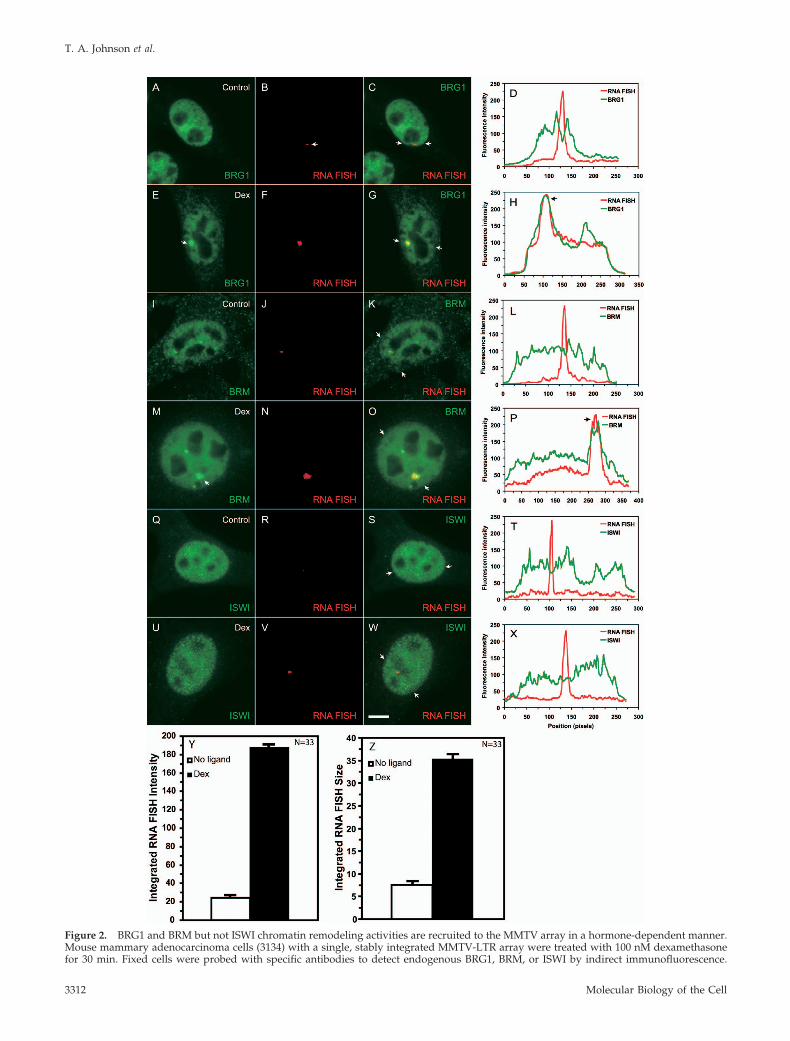
Figure 2. BRG1 and BRM but not ISWI chromatin remodeling activities are recruited to the MMTV array in a hormone-dependent manner.Mouse mammary adenocarcinoma cells (3134) with a single, stably integrated MMTV-LTR array were treated with 100 nM dexamethasonefor 30 min. Fixed cells were probed with specific antibodies to detect endogenous BRG1, BRM, or ISWI by indirect immunofluorescence.
T. A. Johnson et al.
Molecular Biology of the Cell3312

work has shown that GR makes direct contacts not neces-sarily with BRG1 but with the BRG1- or BRM-associatedfactors, BAF 57 and BAF60a (Hsiao et al., 2003).
Transcription from the MMTV-LTR Is Regulated by theATP-Hydrolysis Activity of BRG1 or BRMTo gain more insight into the functional role of BRG1 orBRM at an integrated GR responsive gene, we evaluateddirectly the effect of BRG1-K-R or BRM-K-R on transcriptionat the MMTV array in 3134 cells. 3134 cells, which expressendogenous GR, BRG1, and BRM, were transfected withFlag-tagged BRG1, BRM, BRG1-K-R, or BRM-K-R expressionvectors. The cells were treated with dexamethasone andanalyzed by quantitative RNA FISH combined with anti-Flag indirect immunofluorescence microscopy to distinguishtransfected from untransfected cells. In BRG1- or BRM-transfected cells, dexamethasone treatment yielded RNAFISH signals of comparable intensity to untransfected cells(Figure 3, A–B and E–F); hence, overexpression of wild-typeBRG1 or BRM did not significantly enhance or inhibit ex-pression from the MMTV array. In BRG1-K-R– or BRM-K-R–transfected cells, the size and the fluorescence intensity ofRNA FISH signals were greatly reduced in comparison toneighboring untransfected cells (Figure 3, C-D and G-H). Ithas been shown that the size and the fluorescence intensityof the MMTV array, as measured by RNA FISH, is correlatedwith the level of MMTV transcription (Mueller et al., 2001).Quantitation of the RNA FISH signal, from BRG1-K-R– orBRM-K-R–transfected cells, showed an 81 and 45% decreasein MMTV transcription, respectively (Figure 3I). A com-bined ANOVA analysis and Student-Newman-Keuls posthoc test showed that the BRG1-K-R– and BRM-K-R–trans-fected cells have statistically significant reduction in RNAFISH signals, whereas the wild-type BRG1-transfected cellsdo not vary from untransfected populations (p � 0.05).These results demonstrate that BRG1-K-R and BRM-K-Rfunction as dominant negative proteins by interfering withthe chromatin-remodeling function of endogenous BRG1and BRM and as a consequence inhibit hormone-dependenttranscription on an integrated MMTV promoter.
Dominant negative BRM was consistently a weaker effec-tor of transcription than dominant negative BRG1 (Figure3I). Purified BRM complexes have been previously shown tobe weaker chromatin remodelers than purified BRG1 com-plexes (Sif et al., 2001). This observation along with ourresults suggested a more pronounced role for BRG1 in thetranscriptional process at the MMTV promoter. Conse-quently, we focused our efforts on the study of BRG1 bygenerating a mouse mammary adenocarcinoma cell line that
stably expressed BRG1-K-R and GR under the control of atetracycline-repressible promoter (cell line 5555; Figure 3J).Western blot analysis of total cell lysates prepared from 5555cells showed a robust induction of BRG1-K-R expressionupon removal of tetracycline from culture medium (Figure3K). We directly assessed the functional role of dominantnegative BRG1 on MMTV transcription in vivo by growingthe 5555 cell line in the presence (Figure 3L) or absence(Figure 3M) of tetracycline and then challenged these cellswith dexamethasone. The expression of BRG1-K-R reducedthe robust hormone-dependent RNA FISH signal by 80%(Figure 3, L–N).
ANOVA analysis of the RNA FISH data demonstrated astatistically significant difference between the BRG1-K-R–expressing and nonexpressing conditions (p � 10�8).
To eliminate the possibility that the inhibition of MMTVtranscription in the 5555 cell line is merely an outcome ofoverexpression of an exogenous protein, we used siRNA-mediated gene silencing to knock down endogenous BRG1in the MMTV array–containing cell line (3134). The absenceof nuclear BRG1 immunostaining in cells transfected with apool of siRNAs designed against BRG1 (Figure 3P) validatedthe effectiveness of these siRNAs. 3134 cells transfected withsiRNAs specific to BRG1 show MMTV RNA FISH signalsthat were smaller in size and intensity to cells transfectedwith a scrambled control siRNA (Figure 3, O and P). Quan-titation of RNA FISH signal intensities obtained from eachtransfected group of 3134 cells showed an 84% decrease inthe level of MMTV transcription in BRG1 depleted cells(Figure 3Q). ANOVA analysis of the RNA FISH data dem-onstrated a statistically significant difference between BRG1siRNA-treated cells and scrambled siRNA -transfected cells(p � 10�8). Interestingly, transcriptional inhibition gener-ated by siRNA-mediated silencing of endogenous BRG1expression was very similar to the transcriptional inhibitionobtained by either the stable or transient expression of dom-inant negative BRG1 (Figure 3, I, N, and Q). We concludethat interfering with the function of endogenous BRG1 ei-ther by the expression of a dominant negative form of BRG1or by the siRNA-mediated depletion of endogenous BRG1dramatically compromises transcription from the MMTVpromoter.
BRG1 Is Required for the Hormone-dependent Remodelingof MMTV Chromatin and Loading of RNA pol II to theMMTV PromoterThe data presented in Figures 1–3 demonstrate the hor-mone-dependent recruitment of BRG1 to the MMTV arrayand the involvement of BRG1 mediated chromatin remod-eling in the transcriptional activation of the MMTV pro-moter. Data from our lab and others have demonstrated thatactivation of the MMTV promoter by hormone, results in thebinding of GR to hormone response elements (GREs) withinthe nucleosome B-C region of the MMTV promoter (Fragosoet al., 1998; Fryer and Archer, 1998; Fletcher et al., 2002). Inthe presence of hormone, this region becomes more accessi-ble to a variety of chemical and enzymatic nucleases, thehallmark of a chromatin-remodeling event. We and othershave used the restriction endonuclease, SacI, which cutswithin the nucleosome B-C region, as a measure of thischromatin transition (Fragoso et al., 1998; Fryer and Archer,1998; Fletcher et al., 2002). We compared the extent of SacIcleavage in 3617 mouse mammary adenocarcinoma cells(which stably expresses GFP-GR in a tetracycline-repressiblesystem; Walker et al., 1999) with SacI cleavage in the 5555 cellline (generated by stable transfection of 3617 cells with aFlag-tagged BRG1-K-R under the control of the same tetra-
Figure 2 (cont). MMTV RNA was detected by a RNA FISH usinga probe specific to the MMTV transcript. The arrows in E and Mpoint to the immunofluorescence detected localization of BRG1 andBRM on the MMTV array. The overlays (yellow) in C, G, K, O, S,and W indicate colocalization of the immunofluorescence and RNAFISH signals. The arrows in C, G, K, O, S, and W point to the endpositions of linescans. Linescan analyses in D, H, L, P, T, and Xquantitatively show the ligand-dependent recruitment or lackthereof of the BRG1, BRM, or ISWI chromatin-remodeling com-plexes to the MMTV-LTR array. The linescan in L runs through theRNA FISH signal that is adjacent but not coincident with the BRMfluorescence intensity peak. In H, P, and X, fluorescence intensitypeaks for BRG1 and BRM but not for ISWI coincided with MMTVRNA. Bar, 4 um. Treatment of cells with 100nM dexamethasone for30 minutes increases the intensity (Y) as well as the size (Z) of theMMTV RNA FISH signal.
SWI/SNF Complexes Are Required for GR Action
Vol. 19, August 2008 3313

cycline regulator). Both 3617 and 5555 cells express endog-enous GR and BRG1. 3617 cells grown in the presence oftetracycline (no expression of exogenous GR) showed anincrease in fractional cleavage of 9% at the SacI site, inresponse to hormone (Figure 4, A and B). Chromatin remod-eling in 3617 cells was further increased to 20% in responseto hormone when cells were grown in the absence of tetra-cycline. This increase in cutting most likely reflects the con-tribution of the additional GR that is expressed in the ab-sence of tetracycline. The additional GR can recruit moreBRG1 or BRM to the MMTV array, which in turn can makechromatin even further accessible. The extent of SacI cuttingin the presence of tetracycline was similar in both the 3617and 5555 cell lines (Figure 4, A and B). However, when 5555cells were grown in the absence of tetracycline, under con-ditions that induce the expression of BRG1-K-R (and GR),the extent of SacI cutting was diminished to 6%; in contrastto a fractional cleavage of 20% observed in 3617 cells undersimilar conditions. These results suggest that effective re-modeling of MMTV chromatin requires BRG1 with a func-tional ATPase domain.
To further establish a functional connection between re-modeling at the MMTV promoter and transcription from theMMTV promoter, we probed for pol II loading by ChIPunder the same conditions used in the chromatin accessibil-ity assay (Figure 4, A and B). 3617 and 5555 cells grown inthe presence of tetracycline (no expression of BRG1-K-R)showed a robust increase in the loading of RNA pol II inresponse to hormone treatment (Figure 4C, lane 2 vs. 4 andlane 6 vs. 8). In contrast, expression of BRG1-K-R in 5555cells, resulted in a marked decrease in the hormone-depen-dent loading of RNA pol II (cf. Figure 4C, lanes 1 and 2),whereas tetracycline withdrawal in 3617 cells, in fact,showed a modest increase in pol II loading. These resultsshow that changes in chromatin transitions correlate withchanges in the level of RNA pol II loading at the MMTVpromoter, thereby providing a functional link betweenBRG1, chromatin remodeling, and transcription at a hor-mone-activated promoter. We have extended these studiesand examined the contribution of BRG1 to other GR-regu-lated genes. We find that the robust hormone-dependentinduction of transcription from the Rgs2, regulator of G-protein silencing 2, locus (Figure 4D, lane 6 vs. 8) is alsoaccompanied by an increase in pol II occupancy (Figure 4D,lane 2 vs. 4). In contrast, expression of BRG1-K-R in 5555cells resulted in a significant decrease of hormone-depen-dent transcription (Figure 4D, lane 5 vs. 6) as well as hor-mone-dependent pol II occupancy (Figure 4D, lane 1 vs. 2) atthe Rgs2 locus. These results further confirm and extend ourfindings and implicate BRG1 in the regulation of additionalGR-regulated genes.
Large-Scale Chromatin Decondensation and CondensationAre Regulated by BRG1 and BRM Chromatin-remodelingComplexesResults obtained from chromatin accessibility experiments(Figure 4) provide limited information on the nature ofchromatin remodeling in vivo. We assessed the impactof chromatin-remodeling complexes on the large-scaleMMTV chromatin structure and topology in 3134 cells. 3134cells contain a 2-Mb stably integrated array containing 200copies of the MMTV-LTR. Combining DNA FISH and indi-rect immunofluorescence microscopy, Mueller et al. (2001)observed that hormone treatment resulted in an increase ofthe size of the array, suggesting that the array decondenses
T. A. Johnson et al.
Molecular Biology of the Cell3314
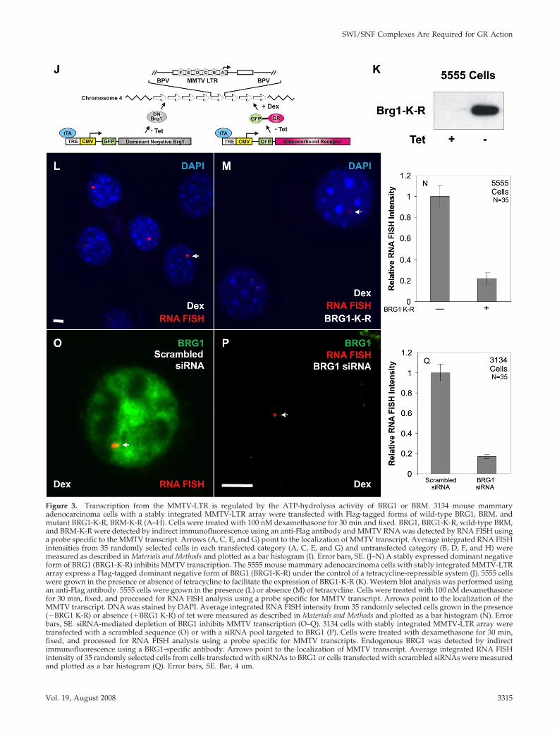
Figure 3. Transcription from the MMTV-LTR is regulated by the ATP-hydrolysis activity of BRG1 or BRM. 3134 mouse mammaryadenocarcinoma cells with a stably integrated MMTV-LTR array were transfected with Flag-tagged forms of wild-type BRG1, BRM, andmutant BRG1-K-R, BRM-K-R (A–H). Cells were treated with 100 nM dexamethasone for 30 min and fixed. BRG1, BRG1-K-R, wild-type BRM,and BRM-K-R were detected by indirect immunofluorescence using an anti-Flag antibody and MMTV RNA was detected by RNA FISH usinga probe specific to the MMTV transcript. Arrows (A, C, E, and G) point to the localization of MMTV transcript. Average integrated RNA FISHintensities from 35 randomly selected cells in each transfected category (A, C, E, and G) and untransfected category (B, D, F, and H) weremeasured as described in Materials and Methods and plotted as a bar histogram (I). Error bars, SE. (J–N) A stably expressed dominant negativeform of BRG1 (BRG1-K-R) inhibits MMTV transcription. The 5555 mouse mammary adenocarcinoma cells with stably integrated MMTV-LTRarray express a Flag-tagged dominant negative form of BRG1 (BRG1-K-R) under the control of a tetracycline-repressible system (J). 5555 cellswere grown in the presence or absence of tetracycline to facilitate the expression of BRG1-K-R (K). Western blot analysis was performed usingan anti-Flag antibody. 5555 cells were grown in the presence (L) or absence (M) of tetracycline. Cells were treated with 100 nM dexamethasonefor 30 min, fixed, and processed for RNA FISH analysis using a probe specific for MMTV transcript. Arrows point to the localization of theMMTV transcript. DNA was stained by DAPI. Average integrated RNA FISH intensity from 35 randomly selected cells grown in the presence(�BRG1 K-R) or absence (�BRG1 K-R) of tet were measured as described in Materials and Methods and plotted as a bar histogram (N). Errorbars, SE. siRNA-mediated depletion of BRG1 inhibits MMTV transcription (O–Q). 3134 cells with stably integrated MMTV-LTR array weretransfected with a scrambled sequence (O) or with a siRNA pool targeted to BRG1 (P). Cells were treated with dexamethasone for 30 min,fixed, and processed for RNA FISH analysis using a probe specific for MMTV transcripts. Endogenous BRG1 was detected by indirectimmunofluorescence using a BRG1-specific antibody. Arrows point to the localization of MMTV transcript. Average integrated RNA FISHintensity of 35 randomly selected cells from cells transfected with siRNAs to BRG1 or cells transfected with scrambled siRNAs were measuredand plotted as a bar histogram (Q). Error bars, SE. Bar, 4 um.
SWI/SNF Complexes Are Required for GR Action
Vol. 19, August 2008 3315

concomitant with transcriptional activation. To address thecontribution of remodeling proteins in array decondensa-tion, 3134 cells were transfected with BRG1, BRG1-K-R,BRM, or BRM-K-R, treated with dexamethasone, and pro-cessed for DNA FISH analysis combined with indirectimmunofluorescence microscopy. As previously ob-served, in BRG1- or BRM-transfected cells, a decondensa-tion of the array was detected in response to hormone(Figure 5, B and D; Mueller et al., 2001). In contrast, inBRG1-K-R–transfected cells we detected a relative de-crease in the size of large arrays even in the presence ofhormone, suggesting that the inability to remodel chro-matin resulted in a less pronounced decondensation event(cf. Figure 5, C to B). A similar decrease in the size of largearrays was also detected in BRM-K-R–transfected cells,although it was far less in magnitude in comparison toBRG1-K-R (cf. Figure 5, E to D). The effect on the size ofthe MMTV array was specific to cells transfected with thevarious remodelers, because in the neighboring untrans-fected cells, robust and large DNA FISH signals wereobserved (Figure 5, F–K). ANOVA analysis and Student-Newman-Keuls post hoc tests show that the BRG1-K-R–and BRM-K-R–transfected cells have a statistically signif-icant reduction in the size of DNA FISH signals, whereasthe wild-type BRG1-transfected cells do not vary fromuntransfected cells (p � 0.05). The size of the DNA FISHsignals in BRG1-K-R–transfected cells also differs signifi-cantly from the DNA FISH signals in BRM-K-R–trans-fected cells. We conclude that the expression of dominantnegative BRG1 or dominant negative BRM inhibits thehormone-induced, large-scale decondensation of the 2-MbMMTV array. The effect of BRG1 on chromatin remodel-ing appears to be more pronounced than that of BRM.These findings provide a strong in vivo correlation be-tween chromatin remodeling, chromatin decondensation,and transcription.
Chromatin-remodeling Complexes Have Distinct KineticProperties at Their Target Gene and DynamicallyAssociate with the MMTV Array in aHormone-dependent MannerThe dynamics of chromatin-remodeling complexes bound toa specific target site on a regulated promoter or the modu-lation of the kinetic properties of chromatin-remodelingcomplexes in response to hormone has never been demon-strated in living cells. In fact, this modulation is one of thekey mechanisms essential for the functional role of nuclearhormone receptors (Stenoien et al., 2001; Schaaf and Ci-dlowski, 2003; Stavreva et al., 2004; Elbi et al., 2004b; Farla etal., 2005; Rayasam et al., 2005). In vivo FRAP has been usedto study the dynamic properties of chromatin proteins andthat the FRAP recovery kinetics of chromatin proteins aredirectly related to their chromatin-binding properties (Le-febvre et al., 1991; Fragoso et al., 1998; Lever et al., 2000;Kimura and Cook, 2001; Kimura et al., 2002; Maruvada et al.,2003; Phair et al., 2004; Becker et al., 2005; Chen et al., 2005).Because BRG1 and BRM were selectively recruited to theMMTV array in response to hormone (Figures 2 and 3), weused FRAP to study the binding kinetics of chromatin-re-modeling complexes at the amplified MMTV target in vivo.Array-containing cells were transfected with YFP- or GFP-tagged BRG1, BRM, BRG1-K-R, or YFP alone and treatedwith dexamethasone for 30 min. Consistent with the immu-nofluorescence experiments (Figure 2), a clear enrichment ofYFP- or GFP-tagged BRG1, BRM, or BRG1-K-R was ob-served at the MMTV array when cells were treated withhormone (Figure 6, A–C). BRG1, BRM, or BRG1-K-R boundto the MMTV array was bleached using a brief laser pulse.The recovery of fluorescence signal in the bleached regionwas monitored using in vivo time-lapse confocal micros-copy. We correlated the dynamics of array-bound BRG1 andarray-bound BRM with the dynamics of BRG1 and BRMdistributed in the nucleoplasm. As a standard we used YFP,which moves freely in the nucleoplasm, and as anticipated
Figure 4. BRG1 is required for the hormone-dependent remodeling of MMTV chromatinand associated loading of RNA pol II at theMMTV promoter. 5555 and 3617 cells wereuntreated or treated with 100 nM dexameth-asone for 30 min and tetracycline as indicated.Nuclei were isolated and digested with SacIand DpnII restriction enzymes. Digestionproducts were detected by linear amplifica-tion using a radiolabeled primer specific tothe MMTV promoter region. Percent cleavageand the fractional change in the accessibilityof MMTV promoter to restriction enzymes areindicators of nuclease hypersensitivity andchromatin remodeling of MMTV promoter re-gion. (A) Intensity of SacI digestion product isdivided by the sum of the intensities of SacIand DpnII digestion products and presentedas percent cleavage at the bottom of eachlane. (B) Bar graph shows dexamethasone-or dexamethasone-, GR-, and BRG1-K-R–induced change in percent cleavage in SacIhypersensitivity between 5555 and 3617 cells
from A. (C) Expression of BRG1-K-R reduced the loading of RNA pol II to MMTV promoter. 5555 and 3617 cells were untreated or treatedwith 100 nM dexamethasone for 30 min and tetracycline as indicated. Chromatin was immunoprecipitated using an antibody specific forRNA pol II or no antibody (control). Immunoprecipitated and input DNA were amplified using primers specific to the MMTV promoter. (D)Expression of BRG1-K-R reduced the loading of RNA pol II to the dexamethasone-induced Rgs2 locus. 5555 cells were untreated or treatedwith 100 nM dexamethasone for 30 min and tetracycline as indicated. cDNA was prepared from RNAs isolated from the indicated conditions.Chromatin was immunoprecipitated using an antibody specific for RNA pol II or no antibody (control). Immunoprecipitated and input DNAor cDNA were amplified using primers specific to the Rgs2 coding region.
T. A. Johnson et al.
Molecular Biology of the Cell3316

the recovery of YFP fluorescence was very rapid, reaching90% of prebleach levels in �0.4 s (Figure 6E). The recoverykinetics of YFP-BRG1 on the MMTV array (Figure 6D, BRG1-Dex) in response to hormone was significantly slower thanYFP-BRG1 in the nucleoplasm (Figure 6D, BRG1-Control) inthe absence of hormone with t1/2 of 3.9 � 0.49 and 0.9 �0.25 s, respectively (p � 0.001; Figure 6, A and D). A similardifference in kinetic behavior was observed with array-bound BRM and nucleoplasmic BRM. Nucleoplasmic BRG1or BRM presumably represent nonspecific DNA-bindingevents (Karpova et al., 2004). The observed difference in therecovery kinetics of BRG1 and BRM on the MMTV array(Figure 6D, BRG1-Dex vs. BRM-Dex) in response to hor-mone was significant, with t1/2 of 3.9 � 0.49 and 1.95 �0.46 s, respectively (p � 0.001), suggesting a stronger inter-action of BRG1 with MMTV chromatin than BRM. Theseinteraction differences between BRG1 and BRM might ac-count for their differential effects on transcription and re-modeling. Interestingly, in the presence of dexamethasonethe recovery kinetics of BRG1-K-R on the MMTV array wasthe slowest, reaching 50% of prebleach levels within 5.5 s(t1/2 of 5.49 � 0.86; Figure 6, C and E, BRG1-K-R-Dex),suggesting an ATP-dependent functional interaction ofBRG1 with MMTV chromatin. Cells expressing different lev-els of the fluorescently tagged proteins showed similar
FRAP recovery curves (Supplementary Figure S1) indicatingthat differential expression levels cannot account for thekinetic differences observed in FRAP experiments. Ourphotobleaching experiments suggest that BRG1, BRM, andBRG1-K-R chromatin-remodeling complexes have distinctkinetic properties on the MMTV array and dynamicallyassociate with and dissociate from MMTV chromatin in vivoin a hormone and ATP-dependent manner.
DISCUSSION
It is well-established that the remodeling of chromatin struc-ture is an essential process that has a profound effect onbasic cellular functions including transcription, DNA recom-bination, repair, and replication (Fletcher and Hansen, 1996;Fyodorov and Kadonaga, 2001; Becker et al., 2002; Elgin andWorkman, 2002). The mechanisms of gene activation arehighly complex and because of this complexity most studiesfocus on individual events using biochemical or geneticapproaches. Here our study provides an integrated kineticview of gene activation events on a target gene in vivoinvolving hormone-dependent recruitment of chromatin-remodeling complexes, dynamic interaction of chromatin-re-modeling complexes with a target promoter, chromatin re-modeling, regulation of higher order chromatin structure,
Figure 5. Large-scale chromatin decondensation is regulated by BRG1 and BRM chromatin-remodeling complexes. 3134 cells weretransfected with Flag-tagged forms of wild-type BRG1 (A and B), mutant BRG1-K-R (C), wild-type BRM (D), and mutant BRM-K-R (E). Cellswere treated with 100 nM dexamethasone for 90 min, fixed, and processed for DNA FISH analysis. BRG1, BRG1-K-R, BRM, and BRM-K-Rwere detected by indirect immunofluorescence using an anti-Flag antibody and MMTV DNA was detected by DNA FISH using a probespecific to the entire MMTV-LTR array. The average DNA FISH signal areas obtained from 35 randomly selected cells in the transfected (A–E)and untransfected (F–J) populations were measured and plotted as a bar histogram (K). Error bars, SE. The inset rectangle shows an enlargedimage of the DNA FISH signal. Expression of BRG1-K-R and BRM-K-R prevent hormone-induced decondensation of MMTV chromatin. Bar,4 um.
SWI/SNF Complexes Are Required for GR Action
Vol. 19, August 2008 3317

RNA pol II loading, and transcriptional activation. Our resultsdemonstrate that individual gene regulatory events are coor-dinated in vivo by members of the SWI/SNF chromatin-re-modeling complex thereby providing a mechanistic basis forBRG1 and BRM chromatin-remodeling complexes in the tran-scriptional process.
The 3134 (murine mammary adenocarcinoma) cell linecontains 200 copies of the MMTV-LTR array stably inte-grated in a head-to-tail orientation at a single integrationevent near the centromere of chromosome 4 (Kramer et al.,1999). The hormone responsiveness of the MMTV array isidentical to that of a single copy MMTV promoter, therebymaking it a useful model system to directly visualize geneexpression events such as the recruitment of chromatin-remodeling complexes and nuclear receptors to a targetpromoter in real time (Fragoso et al., 1998; Fletcher et al.,2002). Belmont and colleagues (Memedula and Belmont,2003) have used an amplified gene array based on the lacoperator/repressor system to analyze the sequential recruit-ment of chromatin-remodeling complexes by the acidic ac-
tivator VP16 to a condensed chromatin locus. Tsukamoto etal. (2000) and Janicki et al. (2004) have used a modified lacoperator/repressor artificial array to demonstrate the re-cruitment of a lac repressor-VP16 chimera that resulted inchromatin decondensation. However in these studies, thecontribution of chromatin-remodeling complexes on chro-matin decondensation were not directly investigated. Herewe extended these studies by observing the in vivo func-tional link between local chromatin remodeling, higher or-der chromatin reorganization, and transcriptional activationusing various approaches including quantitative in vivomicroscopy, chromatin accessibility, and decondensation as-says as well as photobleaching approaches. Importantly, wehave determined for the first time that BRG1 and BRMchromatin-remodeling complexes have distinct kinetic prop-erties on the MMTV array, and they dynamically associatewith and dissociate from MMTV chromatin in a mannerdependent on hormone and a functional ATPase domain.
Three subclasses of ATP-dependent chromatin-remodel-ing complexes have been identified in mammalian cells:
T. A. Johnson et al.
Molecular Biology of the Cell3318

SWI/SNF, ISWI, and Mi-2/CHD (Narlikar et al., 2002). Wefind that the members of the SWI/SNF remodeling complex,BRG1 and BRM, are preferentially recruited to the MMTVpromoter in a hormone-dependent manner. Under the sameexperimental conditions, we failed to detect any enrichmentof the ISWI (Snf2h) chromatin-remodeling complex at theMMTV array (Figure 2). Although, we cannot define themolecular basis of this specificity, the subunit compositionof individual chromatin-remodeling complexes is likely acontributory factor (Hsiao et al., 2003). We have confirmedthe contribution of BRG1 and BRM ATPases in the transcrip-tional activation of the MMTV promoter by biochemical andimaging approaches in well defined genetic backgrounds.Using SW13 cells that are deficient in BRG1, BRM, and GRexpression, we find that both BRG1 and BRM potentiatedtranscription by GR on a transiently introduced MMTVreporter template (Figure 1). Furthermore, transactivationrequired a functional BRG1, BRM, and ATP hydrolysis be-cause the ATPase-deficient forms of BRG1 and BRM failed tostimulate transcription under similar conditions. The intro-duction of ATPase-deficient remodeling complexes can alsodramatically compromise transcription from the stably inte-grated MMTV repeat (Figure 3). We have further confirmedour observations by siRNA-mediated silencing of endoge-nous BRG1 expression (Figure 3). At this point, we areunable to ascertain if BRG1 and BRM make distinct contri-butions to MMTV activation. BRG1 and BRM may haveunique functions in the transcriptional process; alterna-tively, GR might be able recruit either BRG1 or BRM via
shared BAFs. The use of cell lines lacking BRG1 or BRMmight provide some insight into the complex(es) that con-tributes to MMTV activation.
ATP-dependent chromatin-remodeling complexes and hi-stone-modifying complexes dynamically modulate chroma-tin structure both at the nucleosome as well as at a higherorder level (Vignali et al., 2000; Jenuwein and Allis, 2001).We explored the consequences of ATPase-deficient remod-eling proteins on chromatin structure by using a restrictionenzyme accessibility assay to assess the disruption of localchromatin structure. Our studies demonstrated the expectedhormone-dependent increase in restriction enzyme cuttingin control cells (from 9 to 20%) compared with an inhibitionof this hormone-dependent increase in endonuclease cuttingin cells expressing the dominant negative form of BRG1(Figure 4). Interestingly, our ChIP analysis showed that thisreduction in chromatin remodeling in cells expressingBRG1-K-R was accompanied by a reduction RNA pol IIloading and transcription. These experiments provide datathat implicate chromatin remodeling by BRG1 as a necessaryprerequisite for optimal transcription of the MMTV pro-moter.
We have also used quantitative DNA FISH analysis inconjunction with indirect immunofluorescence microscopy,to examine higher order chromatin reorganization events invivo. We observed a large-scale chromatin decondensation,of the MMTV array, in response to hormone when wild-typeBRG1 and BRM is expressed, as has been previously de-scribed (Mueller et al., 2001). When BRG1-K-R or BRM-K-R
Figure 6. Chromatin remodeling complexeshave distinct kinetic properties and dynami-cally associate with MMTV-LTR array in aligand and ATPase-dependent manner. (A–C)Qualitative FRAP analysis of BRG1, mutantBRG1-K-R and BRM in 1365.1 cells. 1365.1mouse fibroblast cells were transfected withYFP-BRG1, YFP-BRG1-K-R, or GFP-BRM andtreated with 100 nM dexamethasone for 30min. BRG1 (A), BRM (B), or BRG1-K-R (C)bound to the MMTV-LTR array was imagedbefore and during recovery after photobleach-ing of the array for 120 ms. Images were ac-quired at the indicated times after the end ofthe bleach pulse. The MMTV-LTR array andthe area of the bleached region is indicated bya red rectangle and shown as an enlargedpseudocolor image in the bottom panels. (D)Quantitative FRAP analysis of YFP-BRG1 orGFP-BRM in the nucleoplasm (control) orbound to the MMTV-LTR array after treat-ment with dexamethasone for 30 min (Dex).BRG1 and BRM bound to the MMTV-LTR ar-ray showed slower recovery kinetics after li-gand treatment. (E) Quantitative FRAP analy-sis of YFP-BRG1, YFP-BRG1-K-R, or GFP-BRMbound to the MMTV-LTR array after treat-ment with dexamethasone for 30 min. Therecovery kinetics of mutant BRG1-K-R boundto the MMTV array was slower than the wild-type BRG1 or wild-type BRM bound toMMTV-LTR array. All quantitative data val-ues in the FRAP studies represent averages �SE from at least 25 cells imaged in three inde-pendent experiments. Bar, 4 um.
SWI/SNF Complexes Are Required for GR Action
Vol. 19, August 2008 3319

was expressed, the hormone-dependent decondensationevents were inhibited significantly by BRG1-K-R and less soby BRM-K-R, in keeping with the differential transcriptionaleffects of these remodeling-deficient proteins (Figure 5).These findings suggest that chromatin remodeling mediatedby BRG1 and BRM ATPases can lead to higher-order chro-matin unfolding and reorganization and this, in turn, corre-lates well with increased transcription from the MMTVarray.
The dynamics of BRG1 and BRM chromatin-remodelingcomplexes at a specific promoter and the modulation of theirkinetic properties in response to environmental stimuli havenever been demonstrated in native chromatin in living cells.In our study, we found that BRG1, BRM, and BRG1-K-Rdynamically exchange at the MMTV promoter with distinctkinetic properties in a manner dependent on hormone and afunctional ATPase domain. The dynamic exchange of re-modeling proteins on the MMTV array are consistent withour in vitro results obtained from rapid UV laser cross-linking where purified SWI/SNF binds to and is displacedfrom purified MMTV chromatin (Fletcher et al., 2002; Na-gaich et al., 2004). Because the FRAP recovery kinetics ofchromatin proteins are directly related to their chromatin-binding properties (Lefebvre et al., 1991; Fragoso et al., 1998;Lever et al., 2000; Kimura and Cook, 2001; Hager et al., 2002;Kimura et al., 2002; Maruvada et al., 2003; Phair et al., 2004;Becker et al., 2005; Chen et al., 2005), we conclude that theremodeling proteins with the slowest exchange rate residelongest on the MMTV promoter and associate most stronglywith MMTV chromatin. A comparison of the kinetic prop-erties of chromatin-remodeling complexes revealed thatBRG1 was more strongly associated with the MMTV arraythan BRM (Figure 6D). Interestingly, the remodeler with theslowest exchange rate is the dominant negative BRG1(BRG1-K-R). Molecular chaperones have been demonstratedto regulate the dynamic properties of GR and PR in thenucleus and recently the high mobility group box 1 protein,HMGB1 has been found to influence the residence time ofGR in chromatin (Stavreva et al., 2004; Wagner et al., 2004;Elbi et al., 2004a,b; Agresti et al., 2005). Considering that thedominant negative BRG1 (BRG1-K-R) is simply a singleamino acid change in the ATPase domain, our study revealsthe importance of ATP hydrolysis in the dynamic propertiesof BRG1 and BRM. Further studies will be necessary for acomplete understanding of the regulation of chromatin pro-tein dynamics and its role in gene expression.
ACKNOWLEDGMENTS
We acknowledge Jim McNally for valuable discussions on FRAP. We thankPamela Badger and Anindya Hendarwanto for technical assistance on FRAPanalysis and tissue culture. We also thank Weidong Wang (NIA, Baltimore),Tony Imbalzano (U Mass, Worcester), and Kevin Gardner (NIH, NCI) forproviding antibodies to BRG1, BRM, and pol II, respectively. We also thankChristian Muchardt (CARS, France) for providing us with BRM expressionconstructs. Imaging was carried out in the Fluorescence Imaging Facility,Laboratory of Receptor Biology and Gene Expression, National Cancer Insti-tute (NCI). This research was supported by the Intramural Research Programof the National Institutes of Health, NCI, Center for Cancer Research.
REFERENCES
Agresti, A., Scaffidi, P., Riva, A., Caiolfa, V. R., and Bianchi, M. E. (2005). GRand HMGB1 interact only within chromatin and influence each other’s resi-dence time. Mol. Cell 18, 109–121.
Arents, G., Burlingame, R. W., Wang, B. C., Love, W. E., and Moudrianakis,E. N. (1991). The nucleosomal core histone octamer at 3.1 A resolution: atripartite protein assembly and a left-handed superhelix. Proc. Natl. Acad. Sci.USA 88, 10148–10152.
Becker, M., Baumann, C. T., John, S., Walker, D., Vigneron, M., McNally, J. G.,and Hager, G. L. (2002). Dynamic behavior of transcription factors on anatural promoter in living cells. EMBO Rep. 3, 1188–1194.
Becker, M., Becker, A., Miyara, F., Han, Z., Kihara, M., Brown, D. T., Hager,G. L., Latham, K., Adashi, E. Y., and Misteli, T. (2005). Differential in vivobinding dynamics of somatic and oocyte-specific linker histones in oocytesand during ES cell nuclear transfer. Mol. Biol. Cell 16, 3887–3895.
Belandia, B., and Parker, M. G. (2003). Nuclear receptors: a rendezvous forchromatin remodeling factors. Cell 114, 277–280.
Berger, S. L. (2002). Histone modifications in transcriptional regulation. Curr.Opin. Genet. Dev. 12, 142–148.
Bianchi, M. E., and Agresti, A. (2005). HMG proteins: dynamic players in generegulation and differentiation. Curr. Opin. Genet. Dev. 15, 496–506.
Biegel, J. A., Zhou, J. Y., Rorke, L. B., Stenstrom, C., Wainwright, L. M., andFogelgren, B. (1999). Germ-line and acquired mutations of INI1 in atypicalteratoid and rhabdoid tumors. Cancer Res. 59, 74–79.
Bresnick, E. H., John, S., Berard, D. S., Lefebvre, P., and Hager, G. L. (1990).Glucocorticoid receptor-dependent disruption of a specific nucleosome on themouse mammary tumor virus promoter is prevented by sodium butyrate.Proc. Natl. Acad. Sci. USA 87, 3977–3981.
Bustin, M. (1999). Regulation of DNA-dependent activities by the functionalmotifs of the high-mobility-group chromosomal proteins. Mol. Cell Biol. 19,5237–5246.
Bustin, M., Catez, F., and Lim, J. H. (2005). The dynamics of histone H1function in chromatin. Mol. Cell 17, 617–620.
Carroll, J. S., and Brown, M. (2006). Estrogen receptor target gene: an evolvingconcept. Mol. Endocrinol. 20, 1707–1714.
Chen, D., Dundr, M., Wang, C., Leung, A., Lamond, A., Misteli, T., andHuang, S. (2005). Condensed mitotic chromatin is accessible to transcriptionfactors and chromatin structural proteins. J. Cell Biol. 168, 41–54.
Cho, K. S., Elizondo, L. I., and Boerkoel, C. F. (2004). Advances in chromatinremodeling and human disease. Curr. Opin. Genet. Dev. 14, 308–315.
Cordingley, M. G., Riegel, A. T., and Hager, G. L. (1987). Steroid-dependentinteraction of transcription factors with the inducible promoter of mousemammary tumor virus in vivo. Cell 48, 261–270.
de la Serna, I. L., Carlson, K. A., Hill, D. A., Guidi, C. J., Stephenson, R. O., Sif,S., Kingston, R. E., and Imbalzano, A. N. (2000). Mammalian SWI-SNF com-plexes contribute to activation of the hsp70 gene. Mol. Cell. Biol. 20, 2839–2851.
de la Serna, I. L., Carlson, K. A., and Imbalzano, A. N. (2001). MammalianSWI/SNF complexes promote MyoD-mediated muscle differentiation. Nat.Genet. 27, 187–190.
Dilworth, F. J., and Chambon, P. (2001). Nuclear receptors coordinate theactivities of chromatin remodeling complexes and coactivators to facilitateinitiation of transcription. Oncogene 20, 3047–3054.
Eissenberg, J. C., and Elgin, S. C. (2000). The HP1 protein family: getting a gripon chromatin. Curr. Opin. Genet. Dev. 10, 204–210.
Elbi, C., Walker, D. A., Lewis, M., Romero, G., Sullivan, W. P., Toft, D. O.,Hager, G. L., and DeFranco, D. B. (2004a). A novel in situ assay for theidentification and characterization of soluble nuclear mobility factors. ScienceSTKE 2004, PL10.
Elbi, C., Walker, D. A., Romero, G., Sullivan, W. P., Toft, D. O., Hager, G. L.,and DeFranco, D. B. (2004b). Molecular chaperones function as steroid recep-tor nuclear mobility factors. Proc. Natl. Acad. Sci. USA 101, 2876–2881.
Elbi, C., Misteli, T., and Hager, G. L. (2002). Recruitment of the DioxinReceptor to Active Transcription Sites. Mol. Biol. Cell 13, 2001–2015.
Elgin, S. C., and Workman, J. L. (2002). Chromosome and expression mech-anisms: a year dominated by histone modifications, transitory and remem-bered. Curr. Opin. Genet. Dev. 12, 127–129.
Farla, P., Hersmus, R., Trapman, J., and Houtsmuller, A. B. (2005). Antian-drogens prevent stable DNA-binding of the androgen receptor. J. Cell Sci. 118,4187–4198.
Felsenfeld, G., and Groudine, M. (2003). Controlling the double helix. Nature421, 448–453.
Fletcher, T. M., and Hansen, J. C. (1996). The nucleosomal array: structure/function relationships. Crit. Rev. Eukaryot. Gene Expr. 6, 149–188.
Fletcher, T. M., Xiao, N., Mautino, G., Baumann, C. T., Wolford, R. G., Warren,B. S., and Hager, G. L. (2002). ATP-dependent mobilization of the glucocor-ticoid receptor during chromatin remodeling. Mol. Cell. Biol. 22, 3255–3263.
Fragoso, G., Pennie, W. D., John, S., and Hager, G. L. (1998). The position andlength of the steroid-dependent hypersensitive region in the mouse mam-
T. A. Johnson et al.
Molecular Biology of the Cell3320

mary tumor virus long terminal repeat are invariant despite multiple nucleo-some B frames. Mol. Cell Biol. 18, 3633–3644.
Fryer, C. J., and Archer, T. K. (1998). Chromatin remodelling by the glucocor-ticoid receptor requires the BRG1 complex. Nature 393, 88–91.
Fyodorov, D. V., and Kadonaga, J. T. (2001). The many faces of chromatinremodeling: SWItching beyond transcription. Cell 106, 523–525.
Giangrande, P. H., Kimbrel, E. A., Edwards, D. P., and McDonnell, D. P.(2000). The opposing transcriptional activities of the two isoforms of thehuman progesterone receptor are due to differential cofactor binding. Mol.Cell. Biol. 20, 3102–3115.
Gregory, R. I., and Shiekhattar, R. (2004). Chromatin modifiers and carcino-genesis. Trends Cell Biol. 14, 695–702.
Grewal, S. I., and Elgin, S. C. (2002). Heterochromatin: new possibilities forthe inheritance of structure. Curr. Opin. Genet. Dev. 12, 178–187.
Hager, G. L., Elbi, C., Johnson, T. A., Voss, T. C., Nagaich, A. K., Schiltz, R. L.,Qiu, Y., and John, S. (2006). Chromatin dynamics and the evolution of alter-nate promoter states. Chromosome. Res. 14, 107–116.
Hager, G. L., Elbi, C. C., and Becker, M. (2002). Protein dynamics in thenuclear compartment. Curr. Opin. Genet. Dev. 12, 137–141.
Hill, D. A. (2001). Influence of linker histone H1 on chromatin remodeling.Biochem. Cell Biol. 79, 317–324.
Hsiao, P. W., Fryer, C. J., Trotter, K. W., Wang, W., and Archer, T. K. (2003).BAF60a mediates critical interactions between nuclear receptors and theBRG1 chromatin-remodeling complex for transactivation. Mol. Cell Biol. 23,6210–6220.
Imbalzano, A. N., and Jones, S. N. (2005). Snf5 tumor suppressor coupleschromatin remodeling, checkpoint control, and chromosomal stability. Can-cer Cell 7, 294–295.
Janicki, S. M., et al. (2004). From silencing to gene expression: real-timeanalysis in single cells. Cell 116, 683–698.
Jenuwein, T., and Allis, C. D. (2001). Translating the histone code. Science 293,1074–1080.
Jones, K. A., and Kadonaga, J. T. (2000). Exploring the transcription-chromatininterface. Genes Dev. 14, 1992–1996.
Karpova, T. S., Chen, T. Y., Sprague, B. L., and McNally, J. G. (2004). Dynamicinteractions of a transcription factor with DNA are accelerated by a chromatinremodeller. EMBO Rep. 5, 1064–1070.
Kimura, H., and Cook, P. R. (2001). Kinetics of core histones in living humancells: little exchange of H3 and H4 and some rapid exchange of H2B. J. CellBiol. 153, 1341–1353.
Kimura, H., Sugaya, K., and Cook, P. R. (2002). The transcription cycle of RNApolymerase II in living cells. J. Cell Biol. 159, 777–782.
Klochendler-Yeivin, A., Muchardt, C., and Yaniv, M. (2002). SWI/SNF chro-matin remodeling and cancer. Curr. Opin. Genet. Dev. 12, 73–79.
Kramer, P., Fragoso, G., Pennie, W. D., Htun, H., Hager, G. L., and Sinden,R. R. (1999). Transcriptional state of the mouse mammary tumor virus pro-moter can effect topological domain size in vivo. J. Biol. Chem. 274, 28590–28597.
Labrador, M., and Corces, V. G. (2002). Setting the boundaries of chromatindomains and nuclear organization. Cell 111, 151–154.
Lee, D. Y., Northrop, J. P., Kuo, M. H., and Stallcup, M. R. (2006). Histone H3lysine 9 methyltransferase G9a is a transcriptional coactivator for nuclearreceptors. J. Biol. Chem. 281, 8476–8485.
Lefebvre, P., Berard, D. S., Cordingley, M. G., and Hager, G. L. (1991). Tworegions of the mouse mammary tumor virus LTR regulate the activity of itspromoter in mammary cell lines. Mol. Cell Biol. 11(5), 2529–2537.
Lemon, B., and Tjian, R. (2000). Orchestrated response: a symphony of tran-scription factors for gene control. Genes Dev. 14, 2551–2569.
Lever, M. A., Th’ng, J. P., Sun, X., and Hendzel, M. J. (2000). Rapid exchangeof histone H1.1 on chromatin in living human cells. Nature 408, 873–876.
Luger, K., Mader, A. W., Richmond, R. K., Sargent, D. F., and Richmond, T. J.(1997). Crystal structure of the nucleosome core particle at 2.8 A resolution[see comments]. Nature 389, 251–260.
Maniatis, T., and Reed, R. (2002). An extensive network of coupling amonggene expression machines. Nature 416, 499–506.
Maruvada, P., Baumann, C. T., Hager, G. L., and Yen, P. M. (2003). Dynamicshuttling and intranuclear mobility of nuclear hormone receptors. J. Biol.Chem. 278, 12425–12432.
McKenna, N. J., and O’Malley, B. W. (2002). Combinatorial control of geneexpression by nuclear receptors and coregulators. Cell 108, 465–474.
McNally, J. G., Mueller, W. G., Walker, D., Wolford, R. G., and Hager, G. L.(2000). The glucocorticoid receptor: Rapid exchange with regulatory sites inliving cells. Science 287, 1262–1265.
Memedula, S., and Belmont, A. S. (2003). Sequential recruitment of HAT andSWI/SNF components to condensed chromatin by VP16. Curr. Biol. 13,241–246.
Metivier, R., Penot, G., Hubner, M. R., Reid, G., Brand, H., Kos, M., andGannon, F. (2003). Estrogen receptor-alpha directs ordered, cyclical, andcombinatorial recruitment of cofactors on a natural target promoter. Cell 115,751–763.
Muchardt, C., and Yaniv, M. (2001). When the SWI/SNF complex remodelsthe cell cycle. Oncogene 20, 3067–3075.
Mueller, W. G., Walker, D., Hager, G. L., and McNally, J. G. (2001). Large scalechromatin decondensation and recondensation in living cells and the role oftranscription. J. Cell Biol. 154, 33–48.
Mulholland, N. M., Soeth, E., and Smith, C. L. (2003). Inhibition of MMTVtranscription by HDAC inhibitors occurs independent of changes in chroma-tin remodeling and increased histone acetylation. Oncogene 22, 4807–4818.
Nagaich, A. K., Walker, D. A., Wolford, R. G., and Hager, G. L. (2004). Rapidperiodic binding and displacement of the glucocorticoid receptor duringchromatin remodeling. Mol. Cell 14, 163–174.
Narlikar, G. J., Fan, H. Y., and Kingston, R. E. (2002). Cooperation betweencomplexes that regulate chromatin structure and transcription. Cell 108, 475–487.
Neely, K. E., and Workman, J. L. (2002). Histone acetylation and chromatinremodeling: which comes first? Mol. Genet. Metab. 76, 1–5.
Orphanides, G., and Reinberg, D. (2002). A unified theory of gene expression.Cell 108, 439–451.
Parada, L. F., Elbi, C. C., Dundr, M., and Misteli, T. (2003). Gene expression.In: Cell Function, Vol. 2, ed. J. Davey and M. Lord, Oxford: Oxford UniversityPress, 47–77.
Peterson, C. L. (2002). Chromatin remodeling enzymes: taming the machines.Third in review series on chromatin dynamics. EMBO Rep. 3, 319–322.
Peterson, C. L., and Workman, J. L. (2000). Promoter targeting and chromatinremodeling by the SWI/SNF complex. Curr. Opin. Genet. Dev. 10, 187–192.
Phair, R. D., Scaffidi, P., Elbi, C., Vecerova, J., Dey, A., Ozato, K., Brown, D. T.,Hager, G. L., Bustin, M., and Misteli, T. (2004). Global nature of dynamicprotein-chromatin interactions in vivo: three-dimensional genome scanningand dynamic interaction networks of chromatin proteins. Mol. Cell. Biol. 24,6393–6402.
Rayasam, G. V., Elbi, C., Walker, D. A., Wolford, R. G., Fletcher, T. M.,Edwards, D. P., and Hager, G. L. (2005). Ligand specific dynamics of theprogesterone receptor in living cells and during chromatin remodeling invitro. Mol. Cell. Biol. 25, 2406–2418.
Reyes, J. C., Muchardt, C., and Yaniv, M. (1997). Components of the humanSWI/SNF complex are enriched in active chromatin and are associated withthe nuclear matrix. J. Cell Biol. 137, 263–274.
Roberts, C. W., and Orkin, S. H. (2004). The SWI/SNF complex–chromatinand cancer. Nat. Rev. Cancer 4, 133–142.
Schaaf, M. J., and Cidlowski, J. A. (2003). Molecular determinants of glucocor-ticoid receptor mobility in living cells: the importance of ligand affinity. Mol.Cell. Biol. 23, 1922–1934.
Sevenet, N., Sheridan, E., Amram, D., Schneider, P., Handgretinger, R., andDelattre, O. (1999). Constitutional mutations of the hSNF5/INI1 gene predis-pose to a variety of cancers. Am. J. Hum. Genet. 65, 1342–1348.
Shogren-Knaak, M., Ishii, H., Sun, J. M., Pazin, M. J., Davie, J. R., and Peterson,C. L. (2006). Histone H4–K16 acetylation controls chromatin structure andprotein interactions. Science 311, 844–847.
Sif, S., Saurin, A. J., Imbalzano, A. N., and Kingston, R. E. (2001). Purificationand characterization of mSin3A-containing Brg1 and hBrm chromatin remod-eling complexes. Genes Dev. 15, 603–618.
Stavreva, D. A., Muller, W. G., Hager, G. L., Smith, C. L., and McNally, J. G.(2004). Rapid glucocorticoid receptor exchange at a promoter is coupled totranscription and regulated by chaperones and proteasomes. Mol. Cell. Biol.24, 2682–2697.
Stenoien, D. L., Nye, A. C., Mancini, M. G., Patel, K., Dutertre, M., O’Malley,B. W., Smith, C. L., Belmont, A. S., and Mancini, M. A. (2001). Ligand-mediated assembly and real-time cellular dynamics of estrogen receptoralpha-coactivator complexes in living cells. Mol. Cell. Biol. 21, 4404–4412.
Strahl, B. D., and Allis, C. D. (2000). The language of covalent histonemodifications. Nature 403, 41–45.
SWI/SNF Complexes Are Required for GR Action
Vol. 19, August 2008 3321

Thomas, J. O., and Travers, A. A. (2001). HMG1 and 2, and related ‘architec-tural’ DNA-binding proteins. Trends Biochem. Sci. 26, 167–174.
Tsukamoto, T., Hashiguchi, N., Janicki, S. M., Tumbar, T., Belmont, A. S., andSpector, D. L. (2000). Visualization of gene activity in living cells. Nat. CellBiol. 2, 871–878.
Verschure, P. J., van, d. K., I, de, L. W., van, d., V, Carpenter, A. E., Belmont,A. S., and van Driel, R. (2005). In vivo HP1 targeting causes large-scalechromatin condensation and enhanced histone lysine methylation. Mol. CellBiol. 25, 4552–4564.
Versteege, I., Sevenet, N., Lange, J., Rousseau-Merck, M. F., Ambros, P.,Handgretinger, R., Aurias, A., and Delattre, O. (1998). Truncating mutationsof hSNF5/INI1 in aggressive pediatric cancer. Nature 394, 203–206.
Vignali, M., Hassan, A. H., Neely, K. E., and Workman, J. L. (2000). ATP-dependent chromatin-remodeling complexes. Mol. Cell Biol. 20, 1899–1910.
Wagner, S., Chiosea, S., Ivshina, M., and Nickerson, J. A. (2004). In vitro FRAPreveals the ATP-dependent nuclear mobilization of the exon junction complexprotein SRm160. J. Cell Biol. 164, 843–850.
Walker, D., Htun, H., and Hager, G. L. (1999). Using inducible vectors tostudy intracellular trafficking of GFP-tagged steroid/nuclear receptors inliving cells. Methods 19, 386–393.
Wolffe, A. P., and Hansen, J. C. (2001). Nuclear visions: functional flexibilityfrom structural instability. Cell 104, 631–634.
Woodcock, C. L., and Dimitrov, S. (2001). Higher-order structure of chromatinand chromosomes. Curr. Opin. Genet. Dev. 11, 130–135.
Workman, J. L., and Kingston, R. E. (1998). Alteration of nucleosome structureas a mechanism of transcriptional regulation. Annu. Rev. Biochem. 67, 545–579.
Wu, J., and Grunstein, M. (2000). 25 years after the nucleosome model:chromatin modifications. Trends Biochem. Sci. 25, 619–623.
T. A. Johnson et al.
Molecular Biology of the Cell3322