Antimicrobial activity of microencapsulated lemongrass essential oil and the effect of experimental...
8
This article appeared in a journal published by Elsevier. The attached copy is furnished to the author for internal non-commercial research and education use, including for instruction at the authors institution and sharing with colleagues. Other uses, including reproduction and distribution, or selling or licensing copies, or posting to personal, institutional or third party websites are prohibited. In most cases authors are permitted to post their version of the article (e.g. in Word or Tex form) to their personal website or institutional repository. Authors requiring further information regarding Elsevier’s archiving and manuscript policies are encouraged to visit: http://www.elsevier.com/copyright
Transcript of Antimicrobial activity of microencapsulated lemongrass essential oil and the effect of experimental...

This article appeared in a journal published by Elsevier. The attachedcopy is furnished to the author for internal non-commercial researchand education use, including for instruction at the authors institution
and sharing with colleagues.
Other uses, including reproduction and distribution, or selling orlicensing copies, or posting to personal, institutional or third party
websites are prohibited.
In most cases authors are permitted to post their version of thearticle (e.g. in Word or Tex form) to their personal website orinstitutional repository. Authors requiring further information
regarding Elsevier’s archiving and manuscript policies areencouraged to visit:
http://www.elsevier.com/copyright
Author's personal copy
Antimicrobial activity of microencapsulated lemongrass essential oil and the effect ofexperimental parameters on microcapsules size and morphology
Fernanda V. Leimann a,⁎, Odinei H. Gonçalves b, Ricardo A.F. Machado a, Ariovaldo Bolzan a
a Chemical and Food Engineering Department, Federal University of Santa Catarina, EQA/UFSC, C.P. 476, CEP 88040-900 Florianópolis, SC, Brazilb Material Science and Engineering Department, Federal University of Santa Catarina, CTC/UFSC, Brazil
a b s t r a c ta r t i c l e i n f o
Article history:Received 30 April 2008Received in revised form 25 August 2008Accepted 26 August 2008Available online 6 September 2008
Keywords:MicroencapsulationAntimicrobial activityCoacervationLemongrassEssential oilPoly(vinyl alcohol)
Lemongrass (Cymbopogom citratus) essential oil, known due to its broad-spectrum antimicrobial activity, wasmicroencapsulated by simple coacervation. Poly(vinyl alcohol) (PVA, 78,000 Da and 88 mol% degree ofhydrolysis) crosslinked with glutaraldehyde was used as wall-forming polymer. The influence of stirring rateand oil volume fraction on the microcapsule size distribution were evaluated. Sodium dodecil sulphate (SDS)and Poly(vinyl pirrolidone) were tested in order to avoid microcapsules agglomeration during the process.Depending on the experimental conditions, microcapsules in the range of 10 μm to 250 μm were obtained.Microcapsules presenting no agglomerationwere obtained when SDS at 0.03 wt.% was used. The compositionand the antimicrobial properties of the encapsulated oil were determined, demonstrating that the process ofmicroencapsulation did not deteriorate the encapsulated essential oil.
© 2008 Elsevier B.V. All rights reserved.
1. Introduction
Many active agents present in cosmetics and food products(essential oils and flavors) are instable compounds. They can sufferoxidation or volatilization or react with other formulation compo-nents causing skin irritation. Microencapsulation is a feasible alter-native to increase the stability of these compounds. There are severalworks available in the literature that discuss essential oil micro-encapsulation using methods as spray-drying [1–3] complex coacer-vation [4,5], simple coacervation [6–8] and extrusion [9]. The mayoradvantage of simple coacervation over the other methods is that itreadily allows the production of microcapsules containing hydro-phobic substances, such as essential oils. In this method, the wall-forming polymer plays an important role because it is responsible forthe protection of the encapsulated essential oil. Poly(vinyl alcohol)(PVA) is a hydrophilic polymer that can be used as wall-formingmaterial in microcapsules. In presence of sulphuric acid, acetic acidand methanol, PVA can be crosslinked with glutaraldehyde, forming ahydrogel. Many drug delivery systems are based on hydrogels sincethey do not dissolve in water and maintain their three-dimensionalnetworks [10]. PVA is also interesting because of its relatively simplechemical structure, ease of processing, and potential use in pharma-ceutical and biomedical fields [11].
Studies on lemongrass (Cymbopogom citratus) essential oil havebeen reported in the last years due to its applicability in food and
pharmaceutical industry. It can be use as a sedative [12] or as anantimicrobial agent [12–14]. The antimicrobial activity of the lemon-grass essential oil was investigated by Onawunmi et al. [13],demonstrating thatα-citral (geranial) and β-citral (neral) componentsindividually exhibited antibacterial action on gram-negative andgram-positive organisms. Sacchetti et al. [15] evaluated elevenessential oils for the use as a food functional ingredient concludingthat lemongrass essential oil presented the most broad-spectrumactivity and showed satisfactory effectiveness. Its minimum inhibitoryconcentration (MIC) was comparable to that provided by the referenceoil (Thymus vulgaris). The authors pointed out that geraniol and citralisomers should probably account for such efficacy.
The critical aspect in themicroencapsulation of a given essential oilis to prevent the deterioration of the oil during the encapsulation step.Baranauskien et al. [1] observed changes in the composition oforegano, citronella andmarjoram flavors after encapsulation into milkprotein-based matrices by spray-drying. Those changes could beexplained by loss of flavor during encapsulation and a high amount ofnon-entrapped flavors. Ramos [2] studied the therapeutic efficiency ofcopaiba oil encapsulated with gum arabic, showing that the efficiencyof the essential oil was not affected by the encapsulation process.
There is only a fewworks in literature [16–18] on the encapsulationof lemongrass essential oil. Some aspects of the microencapsulationprocess still need to be better understood as well as the influence ofthe process on the oil characteristics. If the encapsulation is carriedout by coacervation, it is of key importance to evaluate theantimicrobial activity of the oil after the process because potentiallyaggressive reagents are used, such as methanol, sulfuric acid and
Materials Science and Engineering C 29 (2009) 430–436
⁎ Corresponding author. Tel./fax: +55 48 37219554.E-mail address: [email protected] (F.V. Leimann).
0928-4931/$ – see front matter © 2008 Elsevier B.V. All rights reserved.doi:10.1016/j.msec.2008.08.025
Contents lists available at ScienceDirect
Materials Science and Engineering C
j ourna l homepage: www.e lsev ie r.com/ locate /msec

Author's personal copy
acetic acid. It is also important to know the influence of someoperational parameters on the microcapsule size distribution becauseit directly influences end-use properties [6]. In addition, the mechan-isms responsible for microcapsules agglomeration during coacerva-tion must be better investigated because it may reduce encapsulationefficiency and influence release characteristics.
In the present work, the microencapsulation of lemongrassessential oil by the simple coacervation method was investigated,using crosslinked PVA as the wall-forming material. The influence ofstirring rate and oil volume fraction on the microcapsule sizedistribution and on the release characteristics was evaluated. Theagglomeration of the microcapsules was investigated using an ionicsurfactant and a steric stabilizer. Observations using an opticalmicroscope were carried out during the process to determine themoment in which the microcapsules begun to agglomerate. Themicroencapsulated essential oil was extracted by hydrodistillation andits composition and minimum inhibitory concentration were com-pared to those of the pure oil.
2. Materials and methods
2.1. Materials
Poly(vinyl alcohol) (PVA, Polysciences Inc., Mw=78,000 Da, degreeof hydrolysis of 88 mol%) was used as the wall-forming polymer.Sodium sulphate (Nuclear, reagent grade) was employed as phaseseparation inducer. Lemongrass essential oil (EO) was purchased fromFerquima. A glutaraldehyde solution (Nuclear, 25 vol.%) was used ascrosslinking agent under acidic conditions (sulphuric acid from Ecibra,anhydrous methanol and glacial acetic acid from Nuclear, all ofanalytical grade). Poly(vinyl pyrrolidone) (PVP, Sigma-Aldrich,Mw=40,000 Da) was used as stabilizing agent. Sodium dodecilsulphate (SDS, Vetec, 99% purity) was used as surfactant. Ethanol(Nuclear, reagent grade) was used to wash the microcapsules. Sodiumhydroxide (Vetec, reagent grade) was used to adjust the pH. All thereagents were used as received.
2.2. Microencapsulation procedure
All experiments were carried out in a 1000ml borosilicate jacketedreactor (FGG Instruments Inc) using a propeller impeller. Reactiontemperature was controlled by a thermostatic water bath using a j-thermocouple to measure the temperature inside the reaction vessel.The reagents used are presented in Table 1.
Initially, 600 ml of a 2 wt.% PVA aqueous solutionwere added to thereactor and temperaturewas set to 10 °C. Stirring ratewas set accordingto the experiment (500, 700 or 900 rpm). Then, the essential oil (EO)wasslowly added to the reactor. Nitrogen was used to remove atmosphericoxygen from the reaction medium. Subsequently, sodium sulphate(20 wt.% aqueous solution) was added. Temperature was increased atl °C/min to 50 °C (the phase separation temperature of the PVA in thesolution is in the range of 35 °C–40 °C). When the reactor reached 50 °C,the crosslinking solution (glutaraldehyde, methanol, glacial acetic acidand sulphuric acid) was added. The systemwas allowed to react for 3 h.
After this, pH was adjusted to 7.0 using NaOH (0.5 N aqueous solution)and stored under refrigeration.
2.3. Microcapsules characterization
Optical observations of the microcapsules were carried out withthe aid of an optical microscope (Bioval L-2000A) attached to a digitalcamera. The microcapsule size distribution was determined measur-ing the microcapsules diameter using an image analysis software.About 300 microcapsules were measured for each experiment.
2.4. Equilibrium degree of swelling (EDS)
Swelling behavior of the crosslinked microcapsules was determinedby first removing the essential oil from the microcapsules with ethanolfor 24 h. After this, the microcapsules were dried and put in 50 ml ofdistilled water for 24 h. The swollen samples were removed fromwaterand theirmass (Ws)wasdetermined after removal of the excess ofwaterwith a filter paper. The sample was then dried in an oven until no massvariation could be detected. This masswas defined as dry polymermass(Wd). The equilibriumdegree of swelling (EDS) is expressed as themassof water in the hydrogel per mass of the dry polymer.
EDS gH2O=gpolymer� � ¼ Ws−Wdð Þ=Wd ð1Þ
2.5. EO release
A hydrodistillation apparatus (Clevenger) was used to evaluate theessential oil release from themicrocapsules. The sample was filtered andwashed three times with distilled water and one time with ethanol toremove any not encapsulated EO. The EO was collected from theapparatus at intervals of time. The released percentage was determinedusingEq. (2),werem∞ is the EOmass accumulated at the endof extractionandmt is the mass accumulated in the respective interval of time.
released oil kð Þ ¼ mt � 100ð Þ=m∞ ð2Þ
2.6. Gas chromatography/mass spectrometry analysis
Essential oil composition was determined using a Varian CP-3800gas-chromatograph equipped with a CP-Sil 8 CB Low Bleed/MS(30 m×0.25 mm) column. Equipment conditions were set as follows:injector temperature at 250 °C; Helium as carrier gas (flow rate of 1 ml/min); oven temperature initially at 50 °C and then raised to 240 °C at3 °C/min. Quantification was computed as the percentage contributionof each compound to the total amount present. EO constituents werethen analyzed by Mass Spectrometry — MS (ion trap temperature at220 °C; manifold temperature at 80 °C, transfer line temperature at240 °C). TheMS fragmentationpatternwas checked bymatching theMSfragmentation patterns with NIST mass spectra libraries.
2.7. Antimicrobial assay
The antibacterial activity of the lemongrass EO was investigated byemploying a microdilution method [19]. The assay was carried out withtwo bacterial species, including the Gram-negative bacteria Escherichiacoli ATCC 25922 (American Type Culture Collection) and the Gram-positive bacteria Staphylococcus aureus ATCC 25923. Both the pure EOand the microencapsulated EO (extracted from the microcapsules byhydrodistillation) were evaluated. 200 μl of dimethyl sulfoxide (DMSO)were added to 200 μl of sample previously sterilizated in an autoclave.Subsequently, 600 μl of Mueller Hinton broth were added. Serialdilutions were prepared in the concentration range from 0.349 mg/mlto 89.300 mg/ml. 100 μl of each dilution were distributed in 96-wellplates, aswell as the sterilitycontrol (growthcontrol containedMueller–Hinton broth and DMSO, without antimicrobial substance). Each test
Table 1Reagents used in the lemongrass EO microencapsulation
Reagent Mass (g)
Water 735.90Lemongrass EO 23.90/54.00PVA 12.00Sodium sulphate 12.00Glutaraldehyde 2.40Sulphuric acid 0.30Methanol 13.20Acetic acid 5.25
431F.V. Leimann et al. / Materials Science and Engineering C 29 (2009) 430–436

Author's personal copy
and growth control well was inoculated with 5 μl of a bacterialsuspension (108 CFU/ml). All experiments were performed in duplicateand the microdilution trays were incubated at 36 °C for 24 h. Bacterialgrowth was detected first by optical density determination (ELISAreader, CLX800-BioTek Instruments, Inc.) and later by addition of 20 μl ofan alcoholic solution (0.5 mg/ml) of 2-(4-iodophenyl)-3-(4-nitrophe-nyl)-5-phenyltetrazolium chloride diluted (INT) (Sigma, analyticalgrade). Minimum inhibitory concentration values were defined as thelowest concentration of each sample that completely inhibited micro-organism's growth.
3. Results and discussion
3.1. Effect of stirring rate on microcapsules morphology and EO release
The effect of stirring rate on microcapsule size distribution wasstudied in three levels: 900 rpm, 700 rpm and 500 rpm. Fig. 1 shows
the microcapsules obtained using each stirring rate and the respectivemicrocapsules size distributions.
Spherical microcapsules were obtained in all experimental condi-tions. However, in all cases, the microcapsules coagulated, formingagglomerates higher than 1 mm.
Size distributions strongly depended on the applied stirring rate.At 500 rpm,microcapsules with diameters from 20 μm to 100 μmwereobtained. Microcapsules with diameters from 10 μm to 60 μm wereobtained when the stirring rate was increased to 900 rpm. Thedifference between the diameters of themicrocapsules produced with500 rpm, 700 rpm and 900 rpm was confirmed by the analysis ofvariance as is evident from the calculated Fisher's ‘F’ value (240.80)and from the probability (p) value b0.0001 presenting statisticdifference at a significance level of 99%.
The increase in the stirring rate led to the formation of smallerparticles and narrower distributions. This effect was also observed byChatzi and Kiparissides [20] when studying the size distribution of n-
Fig. 1. Microcapsules morphology and size distributions using the stirring rates of (A) 500 rpm, (B) 700 rpm and (C) 900 rpm (EO volume fraction of 3.4 vol.%).
432 F.V. Leimann et al. / Materials Science and Engineering C 29 (2009) 430–436
Author's personal copy
butyl chloride droplets in water. According to some authors (Shinnar[21]; Tanaka [22]; Zerfa and Brooks [23,24]) the non-homogeneity ofthe turbulence levels inside the reactor vessel is responsible for theformation of droplets with different sizes.
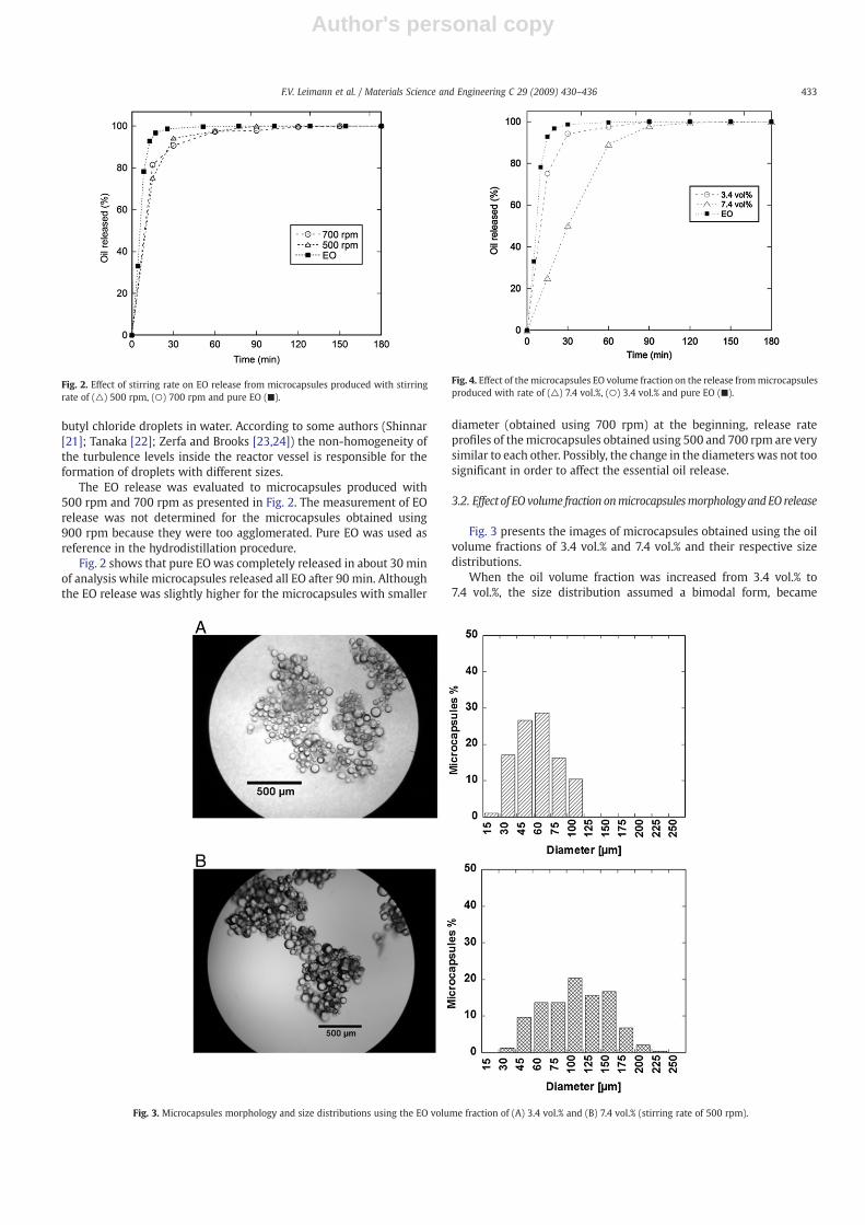
The EO release was evaluated to microcapsules produced with500 rpm and 700 rpm as presented in Fig. 2. The measurement of EOrelease was not determined for the microcapsules obtained using900 rpm because they were too agglomerated. Pure EO was used asreference in the hydrodistillation procedure.
Fig. 2 shows that pure EO was completely released in about 30 minof analysis while microcapsules released all EO after 90 min. Althoughthe EO release was slightly higher for the microcapsules with smaller
diameter (obtained using 700 rpm) at the beginning, release rateprofiles of themicrocapsules obtained using 500 and 700 rpm are verysimilar to each other. Possibly, the change in the diameters was not toosignificant in order to affect the essential oil release.
3.2. Effect of EO volume fraction onmicrocapsulesmorphologyandEO release
Fig. 3 presents the images of microcapsules obtained using the oilvolume fractions of 3.4 vol.% and 7.4 vol.% and their respective sizedistributions.
When the oil volume fraction was increased from 3.4 vol.% to7.4 vol.%, the size distribution assumed a bimodal form, became
Fig. 3. Microcapsules morphology and size distributions using the EO volume fraction of (A) 3.4 vol.% and (B) 7.4 vol.% (stirring rate of 500 rpm).
Fig. 2. Effect of stirring rate on EO release from microcapsules produced with stirringrate of (△) 500 rpm, (○) 700 rpm and pure EO (■).
Fig. 4. Effect of themicrocapsules EO volume fraction on the release frommicrocapsulesproduced with rate of (△) 7.4 vol.%, (○) 3.4 vol.% and pure EO (■).
433F.V. Leimann et al. / Materials Science and Engineering C 29 (2009) 430–436

Author's personal copy
broader and larger diameters were observed. The average micro-capsules diameter obtained to 3.4 vol.% was 47.8 μm and to 7.4 vol.%was equal to 93.6 μm. The analysis of variance using the microcapsulediameter as dependent variable showed statistic difference at asignificance level of 99% with a calculated F value of 328.68 and a pvalue b0.0001. Similar results were also found in other oil-in-watersystems (Bachtsi, Boutris and Kiparissides [6]; Zerfa and Brooks[23,24]). The authors observed that an increase in the volume fractionof the dispersed phase led to formation of larger dispersed oildroplets. Shinnar [21] stated that it occurs because the increase in the
volume fraction of the dispersed phase leads to a dampening effect,decreasing the turbulence levels inside the agitated vessel.
As observed before, the microcapsules were formed agglomeratesin all experimental conditions evaluated.
Fig. 4 presents the essential oil release for the microcapsulesobtained using the volume fractions of 3.4 vol.% and 7.4 vol.%.
The release rate of EO of the microcapsules produced with 7.4 vol.%was slower than that presented by the microcapsules produced with3.4 vol.%. This behavior can be explained by the difference in thediameters of the microcapsules, since the use of 3.4 vol.% led to the
Fig. 5. (A) Temperature profile during the experiment, (B) dispersion EO/PVA solution (T=10 °C), (C) GA addition (T=50 °C), (D) 1 h crosslinking, (E) 2 h crosslinking and (F) 3 hcrosslinking.
434 F.V. Leimann et al. / Materials Science and Engineering C 29 (2009) 430–436

Author's personal copy
production of smaller microcapsules than the use of 7.4 vol.%. Theinfluence of the capsules diameter on the release of Santosol oil wasobserved by Bachtsi and Kiparissides [7]. The authors observed thatsmallermicrocapsules exhibit a faster release rate due to the increasedsurface area of the microcapsules.
3.3. Agglomeration of the microcapsules
It was observed that the microcapsules agglomerated in allexperimental conditions evaluated. The origin of this agglomerationcould be due to simple coagulation or to crosslinking reactionbetween the microcapsules. With the aid of an optical microscope,samples were withdrawn during the microencapsulation process inorder to determine the moment when the agglomeration takes place.
The images are presented in Fig. 5 (B)–(F), while Fig. 5 (A) indicates themoment when the samples were collected.
At the beginning (Fig. 5(B)) a coarse dispersion of the essential oilcan be seen. Droplets size decreased when the temperature wasincreased to 50 °C due to the decrease in the oil viscosity [6]. Fig. 5 (D)showed that themicrocapsules were slightly agglomerated after 1 h ofreaction. After 2 h they were totally agglomerated. It is important tonote that the microcapsules were immersed in an ultrasound bath for2 h and any change in the agglomerations was observed, meaning thatsome degree of crosslinking between the microcapsules may haveoccurred.
In order to gather more information about the agglomerationprocess, experiments were carried out using a steric stabilizer (poly(vinyl pyrrolidone) — PVP) and an ionic surfactant (sodium dodecylsulphate — SDS). They were added to reaction medium 30 min afterthe addition of the crosslinking agent (glutaraldehyde). The images ofthe obtained microcapsules are presented in Fig. 6. In addition, theequilibrium degree of swelling (EDS) of the microcapsules arepresented in Fig. 7 since it is an indication of the extent of thecrosslinking reaction.
The use of PVP was not efficient in preventing microcapsulesagglomeration for both evaluated concentrations. It was expected,since both PVA and PVP present the same mechanism of stericstabilization. In addition, the use of PVP at 0.4 wt.% resulted in a highervalue of EDS, as can be observed in Fig. 7. EDS is directly influenced bythe degree of crosslink in the polymer chains, meaning that PVPdecreased the extent on the crosslinking reaction probably due to itsinfluence in the glutaraldehyde diffusion from the water phase to thepolymer microcapsule. Park et al. [25] observed the same behavior in
Fig. 6. Microcapsules morphology with the use of stabilizing agents: (A) PVP 0.1 wt.%,(B) PVP 0.4 wt.% and (C) SDS 0.03 wt.%.
Fig. 7. EDS for microcapsules produced with PVP, SDS and without stabilizing agent.
Fig. 8. EO release from microcapsules produced (■) without SDS and (○) with SDS.
435F.V. Leimann et al. / Materials Science and Engineering C 29 (2009) 430–436

Author's personal copy
the production microcapsules based on urea-formaldehyde when asteric stabilizer was used. On the other hand, no agglomeration wasobserved when SDS was used. This may be due to the fact that SDS isan ionic surfactant and stabilized the microcapsules by a chargerepulsion mechanism. Additionally, SDS did not influence the EDSvalue of the microcapsules, probably because the diffusion ofglutaraldehyde was not influenced by the presence of SDS on themicrocapsules surface.
After production, the microcapsules were washed in order toremove any trace of the SDS from the water phase and from thecapsules surface. Observations in the optical microscope showed thatthe microcapsules did not agglomerated when SDS was removed. Thissupports the hypothesis that the agglomeration was due to thecrosslinking between the microcapsules. Fig. 8 presents the EO releasefor the microcapsules obtained with and without the use of SDS.
From Fig. 8 it is evident that the presence of SDS did not influencethe EO release. The results of microcapsules morphology and EOrelease indicated that SDS can be used to avoid agglomerationwithoutinfluencing the crosslinking reaction and the release of the essentialoil from the microcapsules.
3.4. Essential oil composition (GC–MS) and antimicrobial activity
The encapsulated essential oil was removed from the microcap-sules by hydrodistillation in order to compare its properties with thepure essential oil (before encapsulation). The identified compoundsare listed in Table 2. Table 3 presents the minimum inhibitoryconcentration for the pure EO and the encapsulated EO.
It is worth noting that the two compounds with antimicrobialactivity [13], α-citral and β-citral, were not degraded during theencapsulation process, meaning that this process can be used tomicroencapsulate the lemongrass EO without losing its main activeconstituents. Both samples presented the same MIC values to E. coli(22.32 mg/ml) and to S. aureus (2.79 mg/ml) meaning that the
encapsulation process did not cause any deterioration in the essentialoil. These results indicate that there were no significant alterations inthe EO during the microencapsulation and also that the biologicalactivity of the essential oil was not affected by the microencapsulationprocess.
4. Conclusions
The production of crosslinked PVA microcapsules containinglemongrass essential oil (EO) was carried out using the simplecoacervation method. The antimicrobial activity and composition ofthe encapsulated oil were evaluated after the encapsulation process,demonstrating that the essential oil was not degraded by themicroencapsulation process.
The influence of the stirring rate and the EO volume fraction on themicrocapsule size distribution was evaluated showing that theformation of small microcapsules was favored when low stirringrates and low volume fractions were used. The decrease in themicrocapsule diameter led to an increase in the release rate of the EOdue to the increase in the total surface area.
Microcapsules agglomerated after 1 h of crosslinking when nosurfactant was used. The agglomerates could not be dispersed bysimple agitation or by the application of ultrasound. However,microcapsules agglomeration was successfully avoided by using anionic surfactant as steric stabilizer during the process. Particles did notagglomerate even when all surfactant was removed from thesuspending medium, supporting the hypothesis that agglomerationsoccurred due to the crosslink between the microcapsules.
Acknowledgment
The authors thank CNPq (Conselho Nacional de DesenvolvimentoCientífico e Tecnológico) for the financial support.
References
[1] R. Baranauskien, P.R. Venskutonis, K. Dewettinck, R. Verhé, Food Res. Int. 39 (4)(2006) 413–425.
[2] M.F. de S. Ramos, Doctoral Thesis, São Paulo University (2006).[3] J. Shaikh, R. Bhosale, R. Singhal, Food Chem. 94 (1) (2006) 105–110.[4] V. Ducel, J. Richard, P. Saulnier, Y. Popineau, F. Boury, Colloids Surf. A 232 (2004)
239–247.[5] C.-P. Chang, T.-K. Leung, S.-M. Linc, C.-C. Hsu, Colloids Surf. B. 50 (2) (2006)
136–140.[6] A.R. Bachtsi, C.J. Boutris, C. Kiparissides, J. Appl. Polym. Sci. 60 (1) (1996) 9–20.[7] A.R. Bachtsi, C. Kiparissides, J. Control. Release 38 (1) (1996) 49–58.[8] J. Lazko, Y. Popineau, J. Legrand, Colloids Surf., B 37 (1–2) (2004) 1–8.[9] S. Yuliani, P.J. Torley, B. D'Arcy, T. Nicholson, B. Bhandari, Food Res. Int. 39 (3) (2006)
318–331.[10] K. Park, W.S.W. Shalaby, H. Park, Biodegradable Hydrogels for Drug Delivery, CRC
Press, Lancaster, 1993.[11] C.M. Hassan, N.A. Peppas, Adv. Polym. Sci. 153 (2000) 37–65.[12] R.S. Pereira, T.C. Sumita, M.R. Furlan, A.O.C. Jorge,M. Ueno, Rev. Saúde Pública 38 (2)
(2004) 326–328.[13] G.O. Onawunmi, W.A. Yisak, E.O. Ogunlana, J. Ethnopharmacol. 12 (1984) 279–283.[14] L.R. Williams, J.K. Stockley, W. Yan, V.N. Home, Int. J. Aromath. 8 (4) (1998) 30–40.[15] G. Sacchetti, S. Maietti, M. Muzzoli, M. Scaglianti, S. Manfredini, M. Radice, R. Bruni,
Food Chem. 91 (4) (2005) 621–632.[16] A.M. Shana, V.B. Villa, European Patent No. 1480612 (2004).[17] G.J. Garces, P. Viladot, L. Josep, United States Patent No. 6,818,296 (2004).[18] J.N. Ness, P.V. Irving, M.J. Goodall, United States Patent No. 6,194,375 (2001).[19] S.M. Souza, F.D. Monache, A.S.J. Souza, Z. Naturforsch., C: Biosci. 60 (2005) 9–10.[20] E.G. Chatzi, C. Kiparissides, Chem. Eng. Sci. 49 (24B) (1994) 5039–5052.[21] R. Shinnar, J. Fluid Mech. 10 (2) (1961) 259–275.[22] M. Tanaka, Can. J. Chem. Eng. 63 (1985) 723–727.[23] M. Zerfa, B.W. Brooks, Chem. Eng. Sci. 51 (12) (1996) 3223–3233.[24] M. Zerfa, B.W. Brooks, J. Appl. Polym. Sci. 60 (12) (1996) 2077–2086.[25] S.-J. Park, Y.-S. Shin, J.-R. Lee, J. Colloid Interface Sci. 241 (2) (2001) 502–508.
Table 3Lemongrass EO antimicrobial activity expressed as minimum inhibitory concentration(MIC)
EO Minimum inhibitory concentration (MIC) (mg/ml)
Escherichia coli Staphylococcus aureus
Pure 22.32 2.79Microencapsulated 22.32 2.79
Table 2Composition of the main constituents of lemongrass EO before and aftermicroencapsulation
Compound Pure EOa Microencapsulated EOa
α pinene 0.136 0.1113 Carene 0.201 0.156Camphene 1.049 1.005β-citral (Neral) 36.548 32.789α-citral(Geranial) 43.356 42.411isogeraniol 0.014 0.072α cyclocitral 0.292 0.35terpineol 0.14 0.142cis-Verbenol 1.495 0.265m-Eugenol 0.014 0.7342,3-dehydro 1,8-Cineole 0.049 0.378Geranyl N-butyrate 2.661 2.619β-caryophyllene 1.998 0.055
a GC relative area percentage.
436 F.V. Leimann et al. / Materials Science and Engineering C 29 (2009) 430–436