Antidiabetic activity of some pentacyclic acid triterpenoids, roleof PTP1B: in vitro, in silico, and...
9
Original article Antidiabetic activity of some pentacyclic acid triterpenoids, role of PTPe1B: In vitro, in silico, and in vivo approaches q Juan José Ramírez-Espinosa a , Maria Yolanda Rios b , Sugey López-Martínez b , Fabian López-Vallejo c , José L. Medina-Franco c , Paolo Paoli d , Guido Camici d , Gabriel Navarrete-Vázquez a , Rolffy Ortiz-Andrade e , Samuel Estrada-Soto a, * a Facultad de Farmacia, Universidad Autónoma del Estado de Morelos, Avenida Universidad 1001, Col. Chamilpa, Cuernavaca, Morelos 62209, Mexico b Centro de Investigaciones Químicas, Universidad Autónoma del Estado de Morelos, Avenida Universidad 1001, Col. Chamilpa, Cuernavaca, Morelos 62209, Mexico c Torrey Pines Institute for Molecular Studies, Port St. Lucie, FL 34987, USA d Dipartimento di Scienze Biochimiche, Universitá degli Studi di Firenze, Viale Morgani 50, 50134 Firenze, Italy e Facultad de Química, Universidad Autónoma de Yucatán, Mérida, Calle 421 No. 41 x 26 y 28 Col. Industrial, C.P. 97150 Mérida Yucatán 97150, Mexico article info Article history: Received 27 October 2010 Received in revised form 28 February 2011 Accepted 1 March 2011 Available online 10 March 2011 Keywords: Diabetes Pentacyclic triterpenoids PTP-1B Docking Enzyme inhibition abstract The aim of the current study was to investigate the oral antidiabetic activity of four structurally-related tri- terpenic acids: ursolic (RE-01), oleanolic (RE-02), moronic (RE-03) and morolic (RE-04) acids. STZ-nicotin- amide diabetic rats were treated with these triterpenes (50 mg/kg) and the antidiabetic effects in acute experiment were determined. All compounds showed significant antidiabetic activity in comparison with control group (p < 0.05). The in vitro inhibitory activity of compounds against protein tyrosine phosphatase 1B (PTPe1B) was also evaluated. At 50 mM, the enzymatic activity was almost completely inhibited. All compounds were docked with a crystal structure of PTPe1B. Docking results suggested the potential binding of the triterpenic acids in a binding pocket next to the catalytic site. An extensive hydrogen bond network with the carboxyl group and Van der Waals interactions stabilize the protein-ligand complexes. Ó 2011 Elsevier Masson SAS. All rights reserved. 1. Introduction Diabetes mellitus (DM) is a chronic disease that occurs when the pancreas does not produce enough insulin or when the body cannot effectively use it, resulting in hyperglycemia. Over time this condition can cause serious damage to several body systems, in particular the nerves and blood vessels. Type 2 DM, or non insulin dependent diabetes, results from ineffective use of insulin as consequence of excess body weight, physical inactivity, and genetic susceptibility [1]. This disease has been becoming more common because of a lifestyle and diet that promotes obesity; it’s particu- larly prevalent in Hispanic and aging populations [2]. Treatment of diabetes involves lowering blood glucose through different mech- anisms, including insulin secretion, glucose absorption, and metabolism [3]. Activation of PPAR-a and PPAR-g by rosiglitazone [4] or inhibition of dipeptidyl peptidase-4 (DPP-4) by sitagliptin [5] are novel approaches for the treatment of diabetes. Protein tyrosine phosphatase 1B (PTPe1B) has recently shown an important influ- ence over insulin sensitivity since it is implicated in modulating insulin signal transduction becoming a key regulator of insulin- receptor activity and downstream signaling pathways [6]. This is related with a dephosphorylation of active insulin receptor (IR) [7]. Studies carried out with knock-out mice suggest that the lack of PTPe1B enzyme would result in activated insulin receptors, improved sensibility to insulin, and stimulated glucose uptake [8]. The study of medicinal plants has lead to the discovery of new chemical structures for potential development as drugs that act over new or known therapeutic targets. Thus, the Phoradendron genus has been used in Mexico since ancient times for the treat- ment of diabetes, hypertension, among other diseases [9]. Penta- cyclic acid triterpenoids and other compounds isolated from Phoradendron reichenbachianum, a parasitic herbal of Holm oaks, have shown significant cytotoxic [10] and antiviral activities [11]. In an attempt to find novel antidiabetic drugs from medicinal plants, herein we report in vivo, in silico, and in vitro studies of a series of structurally-related pentacyclic acid triterpenoids (Fig. 1) q Taken in part from Master in Pharmacy thesis of J.J. Ramírez-Espinosa (Phar- macological Research) and Postdoctoral stay of S. López (Phytochemical investigation). * Corresponding author. Tel./fax: þ52 777 329 7089. E-mail address: [email protected] (S. Estrada-Soto). Contents lists available at ScienceDirect European Journal of Medicinal Chemistry journal homepage: http://www.elsevier.com/locate/ejmech 0223-5234/$ e see front matter Ó 2011 Elsevier Masson SAS. All rights reserved. doi:10.1016/j.ejmech.2011.03.005 European Journal of Medicinal Chemistry 46 (2011) 2243e2251
-
Upload
independent -
Category
Documents
-
view
6 -
download
0
Transcript of Antidiabetic activity of some pentacyclic acid triterpenoids, roleof PTP1B: in vitro, in silico, and...
lable at ScienceDirect
European Journal of Medicinal Chemistry 46 (2011) 2243e2251
Contents lists avai
European Journal of Medicinal Chemistry
journal homepage: http: / /www.elsevier .com/locate/ejmech
Original article
Antidiabetic activity of some pentacyclic acid triterpenoids, role of PTPe1B:In vitro, in silico, and in vivo approachesq
Juan José Ramírez-Espinosa a, Maria Yolanda Rios b, Sugey López-Martínez b, Fabian López-Vallejo c,José L. Medina-Franco c, Paolo Paoli d, Guido Camici d, Gabriel Navarrete-Vázquez a,Rolffy Ortiz-Andrade e, Samuel Estrada-Soto a,*
a Facultad de Farmacia, Universidad Autónoma del Estado de Morelos, Avenida Universidad 1001, Col. Chamilpa, Cuernavaca, Morelos 62209, MexicobCentro de Investigaciones Químicas, Universidad Autónoma del Estado de Morelos, Avenida Universidad 1001, Col. Chamilpa, Cuernavaca, Morelos 62209, Mexicoc Torrey Pines Institute for Molecular Studies, Port St. Lucie, FL 34987, USAdDipartimento di Scienze Biochimiche, Universitá degli Studi di Firenze, Viale Morgani 50, 50134 Firenze, Italye Facultad de Química, Universidad Autónoma de Yucatán, Mérida, Calle 421 No. 41 x 26 y 28 Col. Industrial, C.P. 97150 Mérida Yucatán 97150, Mexico
a r t i c l e i n f o
Article history:Received 27 October 2010Received in revised form28 February 2011Accepted 1 March 2011Available online 10 March 2011
Keywords:DiabetesPentacyclic triterpenoidsPTP-1BDockingEnzyme inhibition
q Taken in part from Master in Pharmacy thesis omacological Research) and Postdoctoral stay ofinvestigation).* Corresponding author. Tel./fax: þ52 777 329 7089
E-mail address: [email protected] (S. Estrada-Soto)
0223-5234/$ e see front matter � 2011 Elsevier Masdoi:10.1016/j.ejmech.2011.03.005
a b s t r a c t
The aim of the current study was to investigate the oral antidiabetic activity of four structurally-related tri-terpenic acids: ursolic (RE-01), oleanolic (RE-02), moronic (RE-03) and morolic (RE-04) acids. STZ-nicotin-amide diabetic rats were treated with these triterpenes (50 mg/kg) and the antidiabetic effects in acuteexperiment were determined. All compounds showed significant antidiabetic activity in comparison withcontrol group (p<0.05). The invitro inhibitoryactivityof compoundsagainstprotein tyrosinephosphatase1B(PTPe1B) was also evaluated. At 50 mM, the enzymatic activity was almost completely inhibited. Allcompoundswere dockedwith a crystal structure of PTPe1B. Docking results suggested the potential bindingof the triterpenicacids inabindingpocketnext to the catalytic site.Anextensivehydrogenbondnetworkwiththe carboxyl group and Van der Waals interactions stabilize the protein-ligand complexes.
� 2011 Elsevier Masson SAS. All rights reserved.
1. Introduction
Diabetesmellitus (DM) is a chronic disease that occurs when thepancreas does not produce enough insulin or when the bodycannot effectively use it, resulting in hyperglycemia. Over time thiscondition can cause serious damage to several body systems, inparticular the nerves and blood vessels. Type 2 DM, or non insulindependent diabetes, results from ineffective use of insulin asconsequence of excess body weight, physical inactivity, and geneticsusceptibility [1]. This disease has been becoming more commonbecause of a lifestyle and diet that promotes obesity; it’s particu-larly prevalent in Hispanic and aging populations [2]. Treatment ofdiabetes involves lowering blood glucose through different mech-anisms, including insulin secretion, glucose absorption, andmetabolism [3]. Activation of PPAR-a and PPAR-g by rosiglitazone
f J.J. Ramírez-Espinosa (Phar-S. López (Phytochemical
..
son SAS. All rights reserved.
[4] or inhibition of dipeptidyl peptidase-4 (DPP-4) by sitagliptin [5]are novel approaches for the treatment of diabetes. Protein tyrosinephosphatase 1B (PTPe1B) has recently shown an important influ-ence over insulin sensitivity since it is implicated in modulatinginsulin signal transduction becoming a key regulator of insulin-receptor activity and downstream signaling pathways [6]. This isrelated with a dephosphorylation of active insulin receptor (IR) [7].Studies carried out with knock-out mice suggest that the lack ofPTPe1B enzyme would result in activated insulin receptors,improved sensibility to insulin, and stimulated glucose uptake [8].
The study of medicinal plants has lead to the discovery of newchemical structures for potential development as drugs that actover new or known therapeutic targets. Thus, the Phoradendrongenus has been used in Mexico since ancient times for the treat-ment of diabetes, hypertension, among other diseases [9]. Penta-cyclic acid triterpenoids and other compounds isolated fromPhoradendron reichenbachianum, a parasitic herbal of Holm oaks,have shown significant cytotoxic [10] and antiviral activities [11].
In an attempt to find novel antidiabetic drugs from medicinalplants, herein we report in vivo, in silico, and in vitro studies ofa series of structurally-related pentacyclic acid triterpenoids (Fig. 1)

Fig. 1. Compounds evaluated.
J.J. Ramírez-Espinosa et al. / European Journal of Medicinal Chemistry 46 (2011) 2243e22512244
using the streptozotocin-nicotinamide induced diabetic rat model.In addition, we report the results of in vitro enzymatic and in silicoexperiments and discuss the possible mechanism of action of thesemetabolites through the inhibition of PTPe1B.
2. Results and discussion
2.1. Chemistry
Ursolic (RE-01), oleanolic (RE-02), moronic (RE-03), andmorolic(RE-04) acids were purified and characterized from acetone extractof leaves and stems of Phoradendron reichenbachianum (AEPr) aspreviously reported in Reference [10].
2.2. In vivo antidiabetic activity
Table 1 shows that i.g. administration of AEPr (100 mg/kg) andRE-01, RE-02, RE-03 and RE-04 (50 mg/kg), induced, in general,a significant decrease in plasma glucose concentration in diabeticrats (p< 0.05) during acute time periods (7 h) as comparedwith thecontrol group (vehicle). All compounds showed significant antidia-betic activity, inparticularRE-01 andRE-03 showed the best activityas compared with the control group (p < 0.05). RE-01 and RE-03produced a sustained decrease of blood glucose levels, close to 25%,during all the experiment. The in vivo antidiabetic activity of RE-01and RE-02 on streptozotocin-induced diabetic rats has been
Table 1Percentage of variation of blood glucose concentration on streptozotocin-nicotinamide i
Test samples Dose (mg/Kg) Percentage of variation of glycemia
Zero hour First hour
Vehicle e 0 � 0.0 9.1 � 10.98Glibenclamide 5 0 � 0.0 �20.43 � 8.17*AEPr 100 0 � 0.0 �20.3 � 6.17*RE-01 50 0 � 0.0 �13.46 � 7.7*RE-02 50 0 � 0.0 �26.92 � 5.7*RE-03 50 0 � 0.0 �17.3 � 5.6*RE-04 50 0 � 0.0 �1.54 � 5.02*
* Values represent the mean � S.E.M. (n ¼ 6). p < 0.05 compared with control group. Th
described in References [12,13]. Also, RE-02 significantly (p < 0.05)improved the glucose tolerance test at the 30 min period withamaximal decline at 10mg/kg. Compared tovehicle-treated control,RE-02 lowered the blood glucose levels by 30, 37, and 22%, respec-tively, for the respective doses [14]. These previous reports are inagreement with our results made on the non insulin-dependentdiabetes model. On the other hand, AEPr showed the same potencyas pure compounds, which suggests a possible additive action oftriterpenic acids contained there. Moreover, this is the first reportconcerning the in vivo antidiabetic effect of AEPr, RE-03, and RE-04.
The in vitro enzymatic activity of RE-01 and RE-02 as antidia-betic agents has been reported; both triterpenic acids induceda significant PTPe1B [15,16] and 11b-HSD1 [17,18] enzymatic inhi-bition. RE-02 enhanced the IRb tyrosine kinase activity and alsoenhanced the effect of insulin on translocation of glucose trans-porter 4 in 3T3eL1 adipocytes, a classical insulin-sensitive cell line[19]. Therefore, we decided to determine the in vitro enzymaticPTPe1B inhibition of RE-03 and RE-04 as structural related analogsof RE-01 and RE-02. We also conducted automated docking studiesin order to propose a binding model with PTPe1B.
2.3. In vitro PTPe1B assay
To test the ability of each compound to inhibit PTPe1B, aliquots ofstock solutions (dissolved in DMSO to prepare 20 mM) were dilutedwith the assay buffer, containing 2.5 mM p-nitrophenyl phosphate
nduced diabetic rats treated with AEPr and pentacyclic acid triterpenoids.
� S.E.M. (mg/dL)
Third hour Fifth hour Seventh hour
�1.71 � 5.11 �0.51 � 5.11 �5.67 � 6.09�36.34 � 7.55* �37.48 � 7.16* �43.64 � 9.9*�17.55 � 3.15* �26.42 � 10.87* �28.53 � 6.62*�24.45 � 10.9* �25.66 � 9.78* �38.01 � 5.0*�14.1 � 6.78* �23.2 � 9.9* �21.92 � 8.9*�26.3 � 5.9* �15.3 � 3.6* �29.2 � 4.0*
�15.23 � 3.84* �8.77 � 4.86 �39.18 � 2.72*
e negative value (�) indicates decrease in glycemia.
DMSO10
uM50
uM10
uM50
uM10
uM50
uM10
uM50
uM
0
20
40
60
80
100
120
RE-04RE-03RE-02RE-01
Res
idua
l act
ivity
(%)
Fig. 2. Assay of inhibition activity of ursolic (RE-01), oleanolic (RE-02), moronic (RE-03), and morolic (RE-04) acids at 10 and 50 mM against PTP. All assays were performedin 0.075 M b,b-dimethylglutarate buffer pH 7.0, containing 1 mM EDTA and 1 mMdithiothreitol. The final concentration of the substrate (pNPP) in each assay was2.5 mM. All experiments were performed in triplicate. The data reported represent themean � S.E.M.
DMSORE-0
1
RE-02
RE-03RE-04
0
20
40
60
80
100
120
Res
idua
l act
ivity
(%)
Fig. 4. Inhibition reversibility assay. Aliquots of PTP were incubated in the presence ofa fixed concentration of each compound for 1 h at 37 �C (RE-01, 10 mM; RE-02, 25 mM;RE-03, 35 mM; RE-04, 25 mM). Then, the enzyme was diluted 400-fold with the assaysolution to measure the residual activity (37 �C, 10 mM pNPP). Control experimentswere carried out adding DMSO. All tests were performed in triplicate. The datarepresent the mean � S.E.M.
J.J. Ramírez-Espinosa et al. / European Journal of Medicinal Chemistry 46 (2011) 2243e2251 2245
(pNPP), to obtain twodifferent inhibitor concentrations. Sampleswereincubated at 37 �C, and the reactions were initiated by adding anappropriate enzyme aliquot. Fig. 2 shows the preliminary results. Thisfigure clearly shows that all compounds induced a significant enzymeinhibition at both evaluated concentrations (10 and 50 mM), and theinhibitioneffectsweremorepronouncedat thehighest concentration.
In order to determine the IC50 value of each compound, theenzyme activity was measured at a fixed substrate concentration
1E-3 0.01 0.1 1 10 100 1000
0.0
0.2
0.4
0.6
0.8
1.0
1.2
v i/vo
[RE-01] µM
IC50 = 2.3 ± 0.1
1E-3 0.01 0.1 1 10 100 1000
0.0
0.2
0.4
0.6
0.8
1.0
1.2
v i/vo
[RE-02] µM
IC50 = 9.5 ± 0.5
A
B
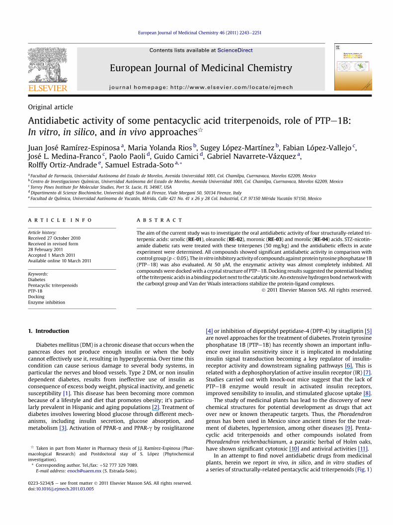
Fig. 3. AeD: IC50 determination. The IC50 value for compounds RE-01 (A), RE-02 (B), RE-03of the PTP against the inhibitor concentration. 15e18 different inhibitor concentrations weremean � S.E.M.
(corresponding to the Km) and varying inhibitor concentrations.IC50 values were calculated by fitting experimental data in thefollowing equation, using a non-nonlinear fitting program (FigSys,Biosoft, UK):
y ¼ Max�Min
1þ
xIC50
!slopeþMin
1E-3 0.01 0.1 1 10 100 1000
0.0
0.2
0.4
0.6
0.8
1.0
1.2
v i/vo
[RE-03] µM
IC50 = 13.2 ± 0.7
1E-3 0.01 0.1 1 10 100 1000
0.0
0.2
0.4
0.6
0.8
1.0
1.2
v i/vo
[RE-04] µM
IC50 = 9.1 ± 0.5
C
D
(C) and RE-04 (D) were determined by plotting the data relative to the residual activityused for each inhibitor. All assays were performed in quadruplicate. Data represent the
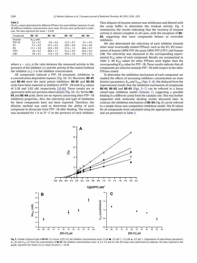
Table 2The IC50 values determined for different PTPases. For each inhibitor, between 15 and18 different inhibitor concentrations were used. All tests were performed in tripli-cate. The data represent the mean � S.E.M.
Compound REL01 REL02 REL03 REL04
Enzyme IC50 (mM)PTP-1B 2.3 � 0.1 9.5 � 0.5 13.2 � 0.7 9.1 � 0.5IF1 7.2 � 0.2 21.2 � 0.5 20.6 � 0.6 13.4 � 0.4IF2 11.1 � 0.3 22.9 � 0.9 27.4 � 1.5 30.6 � 0.7LTP1 20 � 0.7 44.8 � 1.7 51.2 � 0.9 52.0 � 0.3LAR 3.8 � 0.1 11.8 � 1.0 45.6 � 3.0 35.9 � 8.3
J.J. Ramírez-Espinosa et al. / European Journal of Medicinal Chemistry 46 (2011) 2243e22512246
where y ¼ vi/vo is the ratio between the measured activity in thepresence of the inhibitor (vi) and the activity of the control withoutthe inhibitor (vo). x is the inhibitor concentration.
All compounds induced a PTPe1B enzymatic inhibition ina concentration-dependent manner (Fig. 3AeD). Moreover, RE-01and RE-04 were the most potent inhibitors. RE-01 and RE-02acids have been reported as inhibitors of PTPe1B with IC50 valuesof 3.28 and 3.02 mM, respectively [15,16]. These results are inagreement with our previous observations (Fig. 3AeD). Yet for RE-03 and RE-04 acids, there are no reports concerning their PTPe1Binhibitory properties. Also, the selectivity and type of inhibitionfor these compounds have not been reported. Therefore, thedilution method was used to determine the ability of eachcompound to dissociate from PTPe1B after binding. The enzymewas incubated for 1 h at 37 �C in the presence of each inhibitor.
Fig. 5. Double reciprocal plot of RE-01 (1/v versus 1/[S]) (A), the inhibitor concentrations weKm (B) and Vmax (C) from the concentration of RE-01, the inhibitor concentrations were: 0,graph, represent the means (A) or values (B and C) � S.E.M.
Then aliquots of enzyme mixture was withdrawn and diluted withthe assay buffer to determine the residual activity. Fig. 4summarizes the results indicating that the recovery of enzymeactivity is almost complete in all cases, with the exception of RE-02, suggesting that most compounds behave as reversibleinhibitors.
We also determined the selectivity of each inhibitor towardsother none structurally related PTPases, such as the IF1, IF2 isoen-zymes of human LMW-PTP, the yeast LMW-PTP (LTP1) and humanLAR. The selectivity was measured at the corresponding experi-mental IC50 value of each compound. Results are summarized inTable 2. All IC50 values for other PTPases were higher than thecorresponding IC50 value for PTPe1B. These results indicate that allcompounds are selective towards PTPe1B with respect to the otherPTPases tested.
To determine the inhibition mechanism of each compound, westudied the effects of increasing inhibitors concentration on mainkinetics parameters, Km and Vmax (Figs. 5e8). We deduced from theexperimental results that the inhibition mechanism of compoundsRE-01, RE-02 and RE-03 (Figs. 5e7) can be referred to a linearmixed-type inhibition model (Scheme 1), suggesting a possiblebinding in a different cavity from the catalytic site. This was furthersupported with molecular docking results discussed later. Incontrast, the inhibition mechanism of RE-04 (Fig. 8) can be referredto a simple linear non-competitive inhibition model. The Ki valuesfor all compounds were calculated using the appropriate equationsand are presented in Table 3.
re: 0 mM C; 2.5 mM B; 3.5 mM :; 4.5 mM 7. Dependence of main kinetic parameters2.5, 3.5 and 4.5 mM. All assays were performed by triplicate. The data reported in the
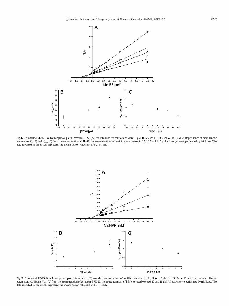
Fig. 6. Compound RE-02. Double reciprocal plot (1/v versus 1/[S]) (A), the inhibitor concentrations were: 0 mM -; 6.5 mM B; 10.5 mM :; 14.5 mM 7. Dependence of main kineticparameters Km (B) and Vmax (C) from the concentration of RE-02, the concentrations of inhibitor used were: 0, 6.5, 10.5 and 14.5 mM. All assays were performed by triplicate. Thedata reported in the graph, represent the means (A) or values (B and C) � S.E.M.
Fig. 7. Compound RE-03. Double reciprocal plot (1/v versus 1/[S]) (A), the concentrations of inhibitor used were: 0 mM -; 10 mM B; 15 mM :. Dependence of main kineticparameters Km (B) and Vmax (C) from the concentration of compound RE-03, the concentrations of inhibitor used were: 0, 10 and 15 mM. All assays were performed by triplicate. Thedata reported in the graph, represent the means (A) or values (B and C) � S.E.M.
J.J. Ramírez-Espinosa et al. / European Journal of Medicinal Chemistry 46 (2011) 2243e2251 2247
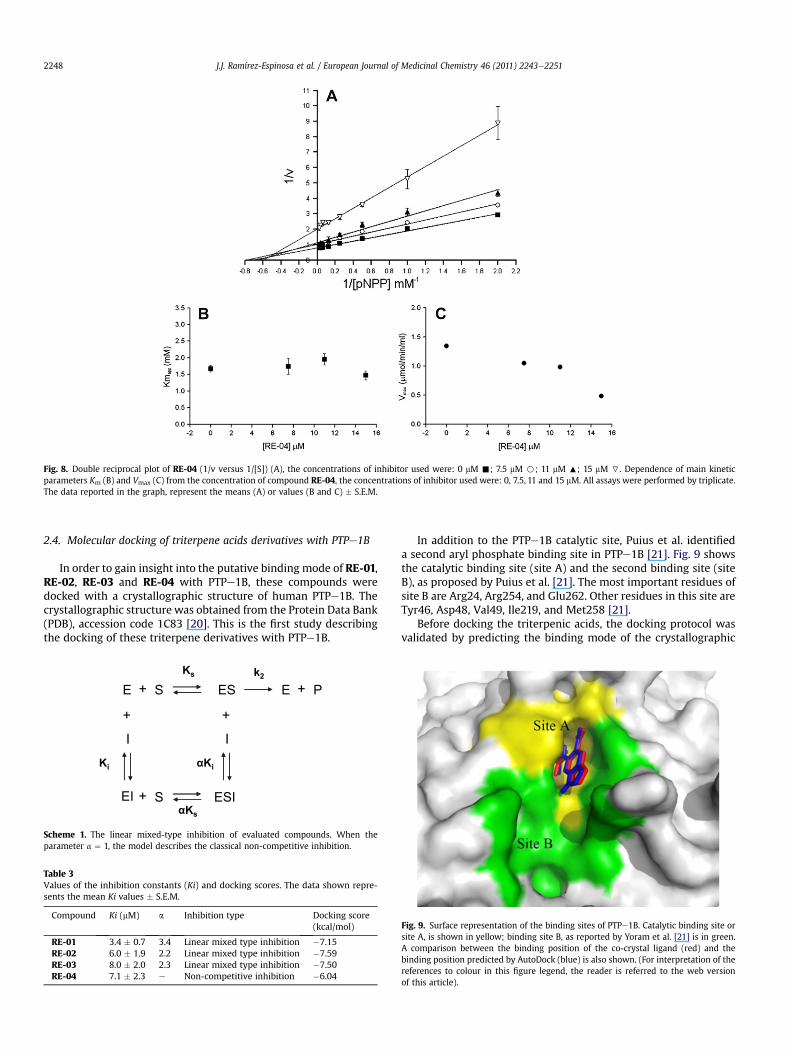
Fig. 8. Double reciprocal plot of RE-04 (1/v versus 1/[S]) (A), the concentrations of inhibitor used were: 0 mM -; 7.5 mM B; 11 mM :; 15 mM 7. Dependence of main kineticparameters Km (B) and Vmax (C) from the concentration of compound RE-04, the concentrations of inhibitor used were: 0, 7.5, 11 and 15 mM. All assays were performed by triplicate.The data reported in the graph, represent the means (A) or values (B and C) � S.E.M.
J.J. Ramírez-Espinosa et al. / European Journal of Medicinal Chemistry 46 (2011) 2243e22512248
2.4. Molecular docking of triterpene acids derivatives with PTPe1B
In order to gain insight into the putative binding mode of RE-01,RE-02, RE-03 and RE-04 with PTPe1B, these compounds weredocked with a crystallographic structure of human PTPe1B. Thecrystallographic structure was obtained from the Protein Data Bank(PDB), accession code 1C83 [20]. This is the first study describingthe docking of these triterpene derivatives with PTPe1B.
E S
I
+ ES
Ki
E P+
+
EI S+ ESI
I
+
αKi
Ks
αKs
k2
Scheme 1. The linear mixed-type inhibition of evaluated compounds. When theparameter a ¼ 1, the model describes the classical non-competitive inhibition.
Table 3Values of the inhibition constants (Ki) and docking scores. The data shown repre-sents the mean Ki values � S.E.M.
Compound Ki (mM) a Inhibition type Docking score(kcal/mol)
RE-01 3.4 � 0.7 3.4 Linear mixed type inhibition �7.15RE-02 6.0 � 1.9 2.2 Linear mixed type inhibition �7.59RE-03 8.0 � 2.0 2.3 Linear mixed type inhibition �7.50RE-04 7.1 � 2.3 e Non-competitive inhibition �6.04
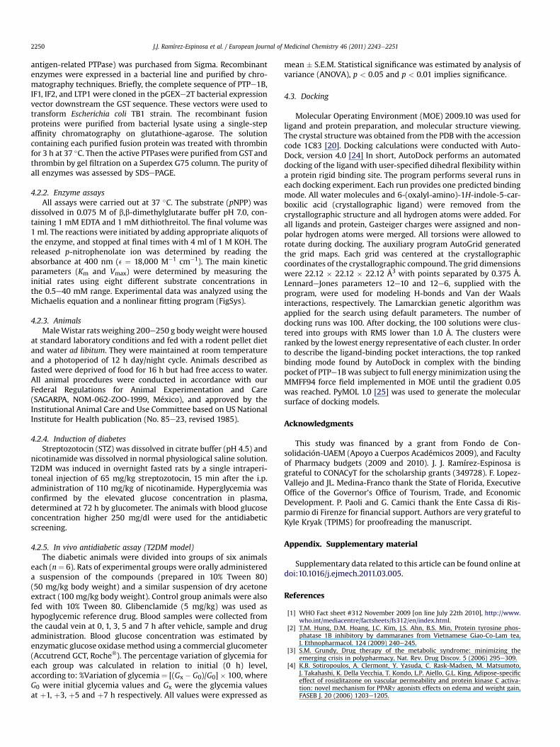
In addition to the PTPe1B catalytic site, Puius et al. identifieda second aryl phosphate binding site in PTPe1B [21]. Fig. 9 showsthe catalytic binding site (site A) and the second binding site (siteB), as proposed by Puius et al. [21]. The most important residues ofsite B are Arg24, Arg254, and Glu262. Other residues in this site areTyr46, Asp48, Val49, Ile219, and Met258 [21].
Before docking the triterpenic acids, the docking protocol wasvalidated by predicting the binding mode of the crystallographic
Fig. 9. Surface representation of the binding sites of PTPe1B. Catalytic binding site orsite A, is shown in yellow; binding site B, as reported by Yoram et al. [21] is in green.A comparison between the binding position of the co-crystal ligand (red) and thebinding position predicted by AutoDock (blue) is also shown. (For interpretation of thereferences to colour in this figure legend, the reader is referred to the web versionof this article).

Fig. 10. Docking of triterpenic acids with PTPe1B. A) Overlay of all triterpenic bindingmodes. B) Surface representation of PTBe1B showing the binding mode of RE-01 intothe binding site B; residues forming hydrogen bonds with RE-01 are shown in red.Residues making Van der Waals contacts with the ligand are shown in green.Figure generated with PyMOL 1.0 [24]. C) Three-dimensional binding model of RE-01.Hydrogen bonds are indicated with yellow dashes. D) Two-dimensional interactiondiagram of the predicted binding mode of RE-01. The binding site residues are rep-resented as follows: polar residues in pink, acidic residues with a red contour ring,basic residues with a blue contour ring. Green arrows indicate hydrogen bonding toside chain atoms, respectively. The ligand proximity contour is depicted with a dottedline. The ligand solvent exposure is represented with blue circles; larger and darkercircles on ligand atoms indicate more solvent exposure. The receptor solvent exposure
J.J. Ramírez-Espinosa et al. / European Journal of Medicinal Chemistry 46 (2011) 2243e2251 2249
6-(oxalyl-amino)-1H-indole-5-carboxilic acid, a competitive inhib-itor of PTPe1B [20]. Fig. 9 shows a comparison between bindingmode of the crystallographic ligand and the binding mode predictedby AutoDock. Fig. 9 clearly shows that AutoDock successfully pre-dicted the binding mode of crystallographic ligand with a root-meansquare (RMS) deviation of 0.35 Å. Predicted binding energies for thetriterpenes are summarized in Table 3. The four compounds showedcomparable energies as calculated by AutoDock. Fig. 10 summarizesthe binding modes of the triterpenic acids predicted by AutoDock.According to the dockingmodels, all four compoundswere predictedto bind into the site B, thus sharing a very similar binding mode(Fig. 10A and B). Interestingly, no binding poses were found byAutoDock into the catalytic binding site. Fig. 10C depicts the opti-mized binding mode of RE-01, the most active triterpene. In thisbinding model, the carboxylic group of RE-01 at C28 forms anextensive hydrogen bond network with Arg24, Arg254 and Gln262;the surface of these residues is showed in red in Fig. 10B. Otherresidues that form van der Waals contacts with the triterpene areTyr46, Asp48, Val49, Met258, and Gly259 (showed in green inFig. 10B). Fig. 10D shows the corresponding two-dimensional inter-action diagram of the optimized binding model generated with theprogram Molecular Operating Environment (MOE) [22].
3. Conclusions
We obtained four pentacyclic triterpenic acids from P. reich-enbachianum with significant in vivo antidiabetic activity on non-insulin dependent diabetic rat model. Moreover, the main mode ofaction of triterpenic acids was produced by PTPe1B enzymaticinhibition with potent, reversible, selective, and linear mixed-typeinhibition model. According to the docking results, the proposedbinding mode of the triterpene derivatives in a second binding site(site B) of PTPe1B suggests a new strategy to obtain compoundswith higher affinity and specificity. Arg254 and Gln262 areconserved among all PTPases, though other residues such asGln262, Met258 and Gly259 are less conserved. Finally, pentacyclictriterpenic acids could be potential drugs for the treatment of type2 diabetes as insulin sensitizer [23].
4. Experimental
4.1. Chemistry
Glibenclamide, nicotinamide, streptozotocin (STZ) and glucose(GLU) were purchased from SigmaeAldrich Co. (St. Louis, MO, USA).Pentobarbital (Anestesal�) was obtained from Smith Kline Co.(Mexico City, Mexico). Test evaluations for GLU were acquired fromRoche (ACCUTREND) (MexicoCity,Mexico).Ursolic (RE-01),Oleanolic(RE-02), Moronic (RE-03) and Morolic (RE-04) acids were purifiedand characterized from acetone extract of leaves and stems of P.reichenbachianum (AEPr) as previously reported in Reference [10].
4.2. Biological assays
4.2.1. Expression and purification of recombinant PTPasesAll experiments were performed using recombinant enzymes.
Human PTPe1B, the IF1 and IF2 isoenzymes of LMW-PTP, and theyeast LMW-PTP (LTP1) were prepared and purified in our labora-tory, whereas the recombinant catalytic domain of LAR (Leukocyte
differencesdin the presence and absence of the liganddare represented by the sizeand intensity of the turquoise discs surrounding the residues; larger and darker discsindicate residues highly exposed to solvent in the active site when the ligand is absent.(For interpretation of the references to colour in this figure legend, the reader isreferred to the web version of this article).
J.J. Ramírez-Espinosa et al. / European Journal of Medicinal Chemistry 46 (2011) 2243e22512250
antigen-related PTPase) was purchased from Sigma. Recombinantenzymes were expressed in a bacterial line and purified by chro-matography techniques. Briefly, the complete sequence of PTPe1B,IF1, IF2, and LTP1 were cloned in the pGEXe2T bacterial expressionvector downstream the GST sequence. These vectors were used totransform Escherichia coli TB1 strain. The recombinant fusionproteins were purified from bacterial lysate using a single-stepaffinity chromatography on glutathione-agarose. The solutioncontaining each purified fusion protein was treated with thrombinfor 3 h at 37 �C. Then the active PTPases were purified fromGSTandthrombin by gel filtration on a Superdex G75 column. The purity ofall enzymes was assessed by SDSePAGE.
4.2.2. Enzyme assaysAll assays were carried out at 37 �C. The substrate (pNPP) was
dissolved in 0.075 M of b,b-dimethylglutarate buffer pH 7.0, con-taining 1 mM EDTA and 1 mM dithiothreitol. The final volume was1 ml. The reactions were initiated by adding appropriate aliquots ofthe enzyme, and stopped at final times with 4 ml of 1 M KOH. Thereleased p-nitrophenolate ion was determined by reading theabsorbance at 400 nm (e ¼ 18,000 M�1 cm�1). The main kineticparameters (Km and Vmax) were determined by measuring theinitial rates using eight different substrate concentrations inthe 0.5e40 mM range. Experimental data was analyzed using theMichaelis equation and a nonlinear fitting program (FigSys).
4.2.3. AnimalsMaleWistar rats weighing 200e250 g bodyweight were housed
at standard laboratory conditions and fed with a rodent pellet dietand water ad libitum. They were maintained at room temperatureand a photoperiod of 12 h day/night cycle. Animals described asfasted were deprived of food for 16 h but had free access to water.All animal procedures were conducted in accordance with ourFederal Regulations for Animal Experimentation and Care(SAGARPA, NOM-062-ZOO-1999, México), and approved by theInstitutional Animal Care and Use Committee based on US NationalInstitute for Health publication (No. 85e23, revised 1985).
4.2.4. Induction of diabetesStreptozotocin (STZ) was dissolved in citrate buffer (pH 4.5) and
nicotinamide was dissolved in normal physiological saline solution.T2DM was induced in overnight fasted rats by a single intraperi-toneal injection of 65 mg/kg streptozotocin, 15 min after the i.p.administration of 110 mg/kg of nicotinamide. Hyperglycemia wasconfirmed by the elevated glucose concentration in plasma,determined at 72 h by glucometer. The animals with blood glucoseconcentration higher 250 mg/dl were used for the antidiabeticscreening.
4.2.5. In vivo antidiabetic assay (T2DM model)The diabetic animals were divided into groups of six animals
each (n¼ 6). Rats of experimental groups were orally administereda suspension of the compounds (prepared in 10% Tween 80)(50 mg/kg body weight) and a similar suspension of dry acetoneextract (100 mg/kg body weight). Control group animals were alsofed with 10% Tween 80. Glibenclamide (5 mg/kg) was used ashypoglycemic reference drug. Blood samples were collected fromthe caudal vein at 0, 1, 3, 5 and 7 h after vehicle, sample and drugadministration. Blood glucose concentration was estimated byenzymatic glucose oxidase method using a commercial glucometer(Accutrend GCT, Roche�). The percentage variation of glycemia foreach group was calculated in relation to initial (0 h) level,according to: %Variation of glycemia¼ [(Gx � G0)/G0]� 100, whereG0 were initial glycemia values and Gx were the glycemia valuesat þ1, þ3, þ5 and þ7 h respectively. All values were expressed as
mean � S.E.M. Statistical significance was estimated by analysis ofvariance (ANOVA), p < 0.05 and p < 0.01 implies significance.
4.3. Docking
Molecular Operating Environment (MOE) 2009.10 was used forligand and protein preparation, and molecular structure viewing.The crystal structure was obtained from the PDB with the accessioncode 1C83 [20]. Docking calculations were conducted with Auto-Dock, version 4.0 [24] In short, AutoDock performs an automateddocking of the ligand with user-specified dihedral flexibility withina protein rigid binding site. The program performs several runs ineach docking experiment. Each run provides one predicted bindingmode. All water molecules and 6-(oxalyl-amino)-1H-indole-5-car-boxilic acid (crystallographic ligand) were removed from thecrystallographic structure and all hydrogen atoms were added. Forall ligands and protein, Gasteiger charges were assigned and non-polar hydrogen atoms were merged. All torsions were allowed torotate during docking. The auxiliary program AutoGrid generatedthe grid maps. Each grid was centered at the crystallographiccoordinates of the crystallographic compound. The grid dimensionswere 22.12 � 22.12 � 22.12 Å3 with points separated by 0.375 Å.LennardeJones parameters 12e10 and 12e6, supplied with theprogram, were used for modeling H-bonds and Van der Waalsinteractions, respectively. The Lamarckian genetic algorithm wasapplied for the search using default parameters. The number ofdocking runs was 100. After docking, the 100 solutions were clus-tered into groups with RMS lower than 1.0 Å. The clusters wereranked by the lowest energy representative of each cluster. In orderto describe the ligand-binding pocket interactions, the top rankedbinding mode found by AutoDock in complex with the bindingpocket of PTPe1B was subject to full energyminimization using theMMFF94 force field implemented in MOE until the gradient 0.05was reached. PyMOL 1.0 [25] was used to generate the molecularsurface of docking models.
Acknowledgments
This study was financed by a grant from Fondo de Con-solidación-UAEM (Apoyo a Cuerpos Académicos 2009), and Facultyof Pharmacy budgets (2009 and 2010). J. J. Ramírez-Espinosa isgrateful to CONACyT for the scholarship grants (349728). F. Lopez-Vallejo and JL. Medina-Franco thank the State of Florida, ExecutiveOffice of the Governor’s Office of Tourism, Trade, and EconomicDevelopment. P. Paoli and G. Camici thank the Ente Cassa di Ris-parmio di Firenze for financial support. Authors are very grateful toKyle Kryak (TPIMS) for proofreading the manuscript.
Appendix. Supplementary material
Supplementary data related to this article can be found online atdoi:10.1016/j.ejmech.2011.03.005.
References
[1] WHO Fact sheet #312 November 2009 [on line July 22th 2010], http://www.who.int/mediacentre/factsheets/fs312/en/index.html.
[2] T.M. Hung, D.M. Hoang, J.C. Kim, J.S. Ahn, B.S. Min, Protein tyrosine phos-phatase 1B inhibitory by dammaranes from Vietnamese Giao-Co-Lam tea,J. Ethnopharmacol. 124 (2009) 240e245.
[3] S.M. Grundy, Drug therapy of the metabolic syndrome: minimizing theemerging crisis in polypharmacy, Nat. Rev. Drug Discov. 5 (2006) 295e309.
[4] K.B. Sotiropoulos, A. Clermont, Y. Yasuda, C. Rask-Madsen, M. Matsumoto,J. Takahashi, K. Della Vecchia, T. Kondo, L.P. Aiello, G.L. King, Adipose-specificeffect of rosiglitazone on vascular permeability and protein kinase C activa-tion: novel mechanism for PPARg agonist�s effects on edema and weight gain,FASEB J. 20 (2006) 1203e1205.

J.J. Ramírez-Espinosa et al. / European Journal of Medicinal Chemistry 46 (2011) 2243e2251 2251
[5] A.J. Scheen, L.F. Van Gaal, Sitagliptine (Januvia): incretin enhancer potentiatinginsulin secretion for the treatment of type 2 diabetes, Rev. Med. Liege 63(2008) 105e109.
[6] T.O. Johnson, J. Ermolieff, M.R. Jirousek, Protein tyrosine phosphatase 1Binhibitors for diabetes, Nat. Rev. Drug Discov. 1 (2002) 696e709.
[7] Y. Romsicki, M. Reece, J.Y. Gauthier, E. Asante-Appiah, B.P. Kennedy, Proteintyrosine phosphatase dephosphorylation of the insulin receptor occurs ina perinuclear endosome compartment in human embryonic kidney 293 cells,J. Biol. Chem. 279 (2004) 12868e12875.
[8] M. Elchebly, P. Payette, E. Michaliszyn, W. Cromlish, S. Collins, A.L. Loy,D. Normandin, A. Cheng, J. Himms-Hagen, C.C. Chan, C. Ramachandran,M.J. Gresser, M.L. Tremblay, B.P. Kennedy, Increased insulin sensitivity andobesity resistance in mice lacking the protein tyrosine phosphatase gene,Science 283 (1999) 1544e1548.
[9] A. Andrade-Cetto, M. Heinrich, Mexican plants with hypoglycaemic effectused in the treatment of diabetes, J. Ethnophamacol. 99 (2005) 325e348.
[10] M.Y. Rios, D. Salinas, M.L. Villareal, Cytotoxic activity of moronic acid andidentification of the new triterpene 3,4-seco-olean-18-ene-3,28-dioic acidfrom Phoradendron reichenbachianum, Planta Med. 67 (2000) 443e446.
[11] M. Kurokawa, P. Basnet, M. Ohsugi, T. Hozumi, S. Kadota, T. Namba, T. Kawana,K. Shiraki, Anti-herpes simplex virus activity of moronic acid purified fromRhus javanica in vitro and in vivo, J. Pharmacol. Exp. Ther. 289 (1999) 72e78.
[12] S.M. Jang, M.J. Kim, M.S. Choi, E.Y. Kwon, M.K. Lee, Inhibitory effects ofursolic acid on hepatic polyol pathway and glucose production in strepto-zotocin-induced diabetic mice, Metabolism 59 (2010) 512e519.
[13] R.M. Perez Gutierrez, R. Vargas Solis, E. Garcia Baez, E. Gallardo Navarro,Hypoglycemic activity of constituents from Astianthus viminalis in normal andstrptozotocin-induced diabetic mice, J. Nat. Med. 63 (2009) 393e401.
[14] C.L. De Melo, M.G.R. Queiroz, S.G.C. Fonseca, A.M.C. Bizerra, T.L.G. Lemos,T.S. Melo, F.A. Santos, V.S. Rao, Oleanolic acid, a natural triterpenoid improvesblood glucose tolerance in normal mice and ameliorates visceral obesity inmice fed a high-fat diet, Chem. Biol. Interact. 185 (2010) 59e65.
[15] W. Zhang, D. Hong, Y. Zhou, Y. Zhang, Q. Shen, J.Y. Li, L.H. Hu, J. Li, Ursolic acidand its derivative inhibit protein tyrosine phosphatase 1B, enhancing insulin
receptor phosphorylation and stimulating glucose uptake, Biochim. Biophys.Acta 1760 (2006) 1505e1512.
[16] Y.N. Zhang, W. Zhang, D. Hong, L. Shi, Q. Shen, J.Y. Li, J. Li, L.H. Hu, Oleanolicacid and its derivatives: new inhibitor of protein tyrosine phosphatase 1Bwith cellular activities, Bioorg. Med. Chem. 16 (2008) 8697e8705.
[17] A. Blum, A.D. Favia, E. Maser, 11beta-Hydroxysteroid dehydrogenase type 1inhibitors with oleanan and ursan scaffolds, Mol. Cell. Endocrinol. 301 (2009)132e136.
[18] J.M. Rollinger, D.V. Kratschmar, D. Schuster, P.H. Pfisterer, C. Gumy,E.M. Aubry, S. Brandstötter, H. Stuppner, G. Wolber, A. Odermatt, 11beta-Hydroxysteroid dehydrogenase 1 inhibiting constituents from Eriobotryajaponica revealed by bioactivity-guided isolation and computationalapproaches, Bioorg. Med. Chem. 18 (2010) 1507e1515.
[19] S.H. Jung, Y.J. Ha, E.K. Shim, S.Y. Choi, J.L. Jin, H.S. Yun-Choi, J.R. Lee, Insulin-mimetic and insulin-sensitizing activities of a pentacyclic triterpenoid insulinreceptor activator, Biochem. J. 403 (2007) 243e250.
[20] H.S. Andersen, L.F. Iversen, C.B. Jeppesen, S. Branner, K. Norris,H.B. Rasmussen, K.B. Møller, N.P. Møller, 2-(oxalylamino)-benzoic acid isa general, competitive inhibitor of protein-tyrosine phosphatases, J. Biol.Chem. 275 (2000) 7101e7108.
[21] Y.A. Puius, Y. Zhao, M. Sullivan, D.S. Lawrence, S.C. Almo, Z.-Y. Zhang, Iden-tification of a second aryl phosphate-binding site in protein-tyrosine phos-phatase 1b: a paradigm for inhibitor design, Proc. Natl. Acad. Sci. U.S.A. 94(1997) 13420e13425.
[22] Molecular Operating Environment (MOE), Version 2009.10, ChemicalComputing Group Inc., Montreal, Quebec, Canada. Available at: http://www.chemcomp.com.
[23] R. Sarabu, J. Tilley, Recent advances in therapeutic approaches to type 2 dia-betes. in: A.M. Doherty (Ed.), Annual Reports in Medicinal Chemistry, vol. 40.Elsevier: Academic Press, 2005, pp. 167e181.
[24] R. Huey, G.M. Morris, A.J. Olson, D.S. Goodsell, A semiempirical free energy forcefield with charge-based desolvation, J. Comput. Chem. 28 (2007) 1145e1152.
[25] W.L. DeLano "The PyMOL Molecular Graphics System." DeLano Scientific LLC,San Carlos, CA, USA. Available at: http://www.pymol.org.