Sequence determinants of GLUT1 oligomerization: analysis by homology-scanning mutagenesis
Hindawi Publishing CorporationEvidence-Based Complementary and Alternative MedicineVolume 2011, Article ID 167684, 9 pagesdoi:10.1093/ecam/neq004
Original Article
Antidiabetic Activities of Abutilon indicum (L.)Sweet Are Mediated by Enhancement of Adipocyte Differentiationand Activation of the GLUT1 Promoter
Chutwadee Krisanapun,1, 2 Seong-Ho Lee,1 Penchom Peungvicha,2
Rungravi Temsiririrkkul,3 and Seung Joon Baek1
1 Department of Pathobiology, College of Veterinary Medicine, University of Tennessee, 2407 River Drive, Knoxville,TN 37996, USA
2 Department of Physiology, Faculty of Pharmacy, Mahidol University, Bangkok, Thailand3 Department of Pharmaceutical Botany, Faculty of Pharmacy, Mahidol University, Bangkok, Thailand
Correspondence should be addressed to Seung Joon Baek, [email protected]
Received 20 August 2009; Accepted 1 January 2010
Copyright © 2011 Chutwadee Krisanapun et al. This is an open access article distributed under the Creative Commons AttributionLicense, which permits unrestricted use, distribution, and reproduction in any medium, provided the original work is properlycited.
Abutilon indicum (L.) Sweet is an Asian phytomedicine traditionally used to treat several disorders, including diabetes mellitus.However, molecular mechanisms supporting the antidiabetic effect of A. indicum L. remain unknown. The aim of this studywas to evaluate whether extract of A. indicum L. improves insulin sensitivity. First, we observed the antidiabetic activity of aqueousextract of the entire plant (leaves, twigs and roots) of A. indicum L. on postprandial plasma glucose in diabetic rats. The subsequentexperiments revealed that butanol fractions of the extract bind to PPARγ and activate 3T3-L1 differentiation. To measure glucoseuptake enhanced by insulin-like activity, we used rat diaphragm incubated with various concentrations of the crude extract andfound that the extract enhances glucose consumption in the incubated solution. Our data also indicate that the crude extractand the fractions (water and butanol) did not affect the activity of kinases involved in Akt and GSK-3β pathways; however, thereporter assay showed that the crude extract could activate glucose transporter 1 (GLUT1) promoter activity. These results suggestthat the extract from A. indicum L. may be beneficial for reducing insulin resistance through its potency in regulating adipocytedifferentiation through PPARγ agonist activity, and increasing glucose utilization via GLUT1.
1. Introduction
Diabetes mellitus (DM) is a group of metabolic diseasescharacterized by hyperglycemia resulting from defects ininsulin secretion, insulin action, or both; the incidence ofdiabetes is increasing worldwide. Diabetes or obesity causessubstantial morbidity, mortality and long-term complica-tions, and remains an important risk factor for cardiovas-cular disease [1]. There exists a genetic predisposition todiabetes; however, environmental factors such as a sedentarylifestyle and obesity are the most prominent risk factors [2].Insulin resistance is a key feature in type 2 DM as well as inobesity and plays an important pathophysiological role [3].Insulin resistance is characterized by a diminished reactionof insulin-sensitive tissues and a marked decrease in glucosemetabolism in response to insulin. In fact, impairment of
insulin action has also been observed in syndromes includinginflammatory and chronic infectious diseases in addition totype 2 DM [4]. Recent data suggest that activation of PPARγby using plant extracts improves lipid metabolism andmitigates insulin resistance [5]. Throughout the world, manytraditional plant treatments for diabetes are employed thatare frequently considered to be less toxic and have fewer sideeffects than synthetic drug treatments [1, 6]. According to amedicinal survey report in Thailand, traditional medicinesor herbs are used 31% of the time in the treatment ofdiabetic patients [7]. However, traditional antidiabetic plantspossess less adequate scientific or medicinal data supportin spite of the fact that the World Health Organization stillrecommends further evaluation of their accepted use [8, 9].
The native Southeast Asian plant A. indicum (L.) Sweet isan erect, branched shrub of 0.5–1 m height, having the Thai

2 Evidence-Based Complementary and Alternative Medicine
name Krob-Fun-Si, Fun-Si or Ma-Kong-Khaao. A. indicumL. is in the family Malvaceae, is easily identified by its cog-like fruits, and is found abundantly in wastelands. Thisplant has a long history of being used medicinally as anantidiabetic remedy, and phytochemical screening of theplant revealed that it contained alkaloids, flavonoids, tannins,saponins and glycosides [10, 11]. This medicinal plant playsan important role in folk medicine; in Thailand, it hasbeen used as a blood tonic, carminative, antipyretic, anti-cough, diuretic, anti-inflammatory, laxative and antidiabetic[12], whereas in India and China, it has been used forurinary disease, gonorrhea, jaundice, rheumatism, highfever, mumps, pulmonary tuberculosis, bronchitis, lack ofurination and some nervous and ear problems [13, 14].According to many scientific reports, the leaf extracts possesshypoglycemic, hepatoprotective, antibacterial and larvicidalproperties [10, 13, 15, 16]. Analgesic principles from thisplant were also isolated [17], and its polyherbal formulationshave been reported as being effective in treating diabetes andhyperlipidemia, and effective as free radical scavengers [18–20].
With respect to its traditional use, A. indicum L. has beenknown to contain an active ingredient against diabetes andis believed to reduce some symptoms of diabetic compli-cations. In this study, we investigated whether the extractsof A. indicum L. could reduce postprandial plasma glucoselevels in an animal model of diabetes, since postprandialhyperglycemia is one of the common complications seenin diabetic patients [21]. We found that A. indicum L.ameliorates insulin resistance via PPARγ activation andenhances glucose uptake.
2. Methods
2.1. Plant Material. The leaves, twigs and roots of A. indicum(L.) Sweet were collected from Mahidol University, SalayaCampus, Nakorn Pathom Province, Thailand, during May2004, and were authenticated by Rungravi Temsiririrkkul,Department of Pharmaceutical Botany, Faculty of Pharmacy,Mahidol University. A voucher specimen (PBM 04765) wasdeposited at the Pharmaceutical Botany Mahidol Universityherbarium. All fresh parts were dried at 50◦C in a hotair oven and then pulverized by an electric blender. Crudeextract was obtained by boiling 1 kg of plant powder (leaves261.2 g, twigs 559.7 g and root 179.1 g) in 10 l of distilledwater for 10 min. The boiling process was repeated twice,and the combined extract was filtered across cotton wooland gauze. The filtrate was lyophilized in a freeze drysystem resulting in a dark brown powder of 11.44% w/w.Lyophilized extract was preserved in an airtight bottle atroom temperature and protected from light until use. Thecrude extract (10 g) was subsequently partitioned betweenbutan-1-ol (3 × 0.2 l) and distilled water (0.2 l). The butanolfraction was concentrated under reduced pressure in a rotaryevaporator and lyophilized to give a total semi-solid residue(19% of dried crude extract). The resulting water fractionwas freeze-dried, followed by generating brown powder(65.4% of dried crude extract).
2.2. Animals. Male Wistar rats, weighing 140–180 g, werepurchased from the National Laboratory Animal Center,Mahidol University, Salaya, Nakorn Pathom Province, Thai-land. They were housed in a temperature-controlled room(22–25◦C) with a 12 h light-dark cycle for at least 1 week toacclimatization. The animals were fed with free access to acommercial pellet diet (C.P. Thailand) and water ad libitum.The animal protocol was approved by the Animal EthicsCommittee, Faculty of Pharmacy, Mahidol University (No.5/2548).
2.3. Preparation of Diabetic Rats. Diabetes was induced inthe 16 h-fasted male Wistar rats with a single intraperi-toneal injection of 75 mg kg−1 streptozotocin (STZ), freshlydissolved in isotonic saline. Five days after injection, diabeticrats were identified by measuring fasting plasma glucose(FPG) levels using PGO enzyme (Sigma). Rats with FPGlevels over 200 mg dl−1 were used in the experiment [22, 23].
2.4. Antidiabetic Activity of A. indicum Extract on Postpran-dial Plasma Glucose. The STZ-induced diabetic rats wererandomly allocated into four groups (6–9 rats per group)A. indicum crude extract and glibenclamide were dissolvedin distilled water and administered orally once a day for 2weeks as follows: Group 1: diabetic rats treated orally withdistilled water (control), Group 2: diabetic rats treated orallywith glibenclamide 5 mg kg−1 body weight (positive control),Groups 3 and 4: diabetic rats treated orally with crudeextract 0.25 or 0.5 g kg−1 body weight. Blood samples (60 μlof each) were collected from the tail vein using Hematocrit-capillary tube, after 2 h fasting on day 0 (1 day beforeoral administration), day 7 and day 14 for plasma glucosedetermination. The animals were weighed on day 0, 4, 7, 11and 14 for corrected doses of medication.
2.5. Cell Cultures. HCT-116, HepG2, C2C12, 3T3-L1 and L6cells were purchased from American Type Culture Collection(Manassas, VA, USA). HCT-116 cells were maintained inMcCoy’s 5A medium supplemented with 10% FBS; allothers were maintained in DMEM supplemented with 10%FBS. Cells were grown at 37◦C in a humidified, 5% CO2
environment. For adipocyte differentiation, differentiationmedium containing 0.5 mM isobutyl methylxanthine, 1 μMdexamethasone and 1 μg ml−1 insulin was added to theculture. The differentiation media was changed every 2 daysuntil day 4. Thereafter, DMEM containing 10% FBS andinsulin was subsequently replaced every 2 days until day 8.C2C12 and L6 myoblasts were differentiated with DMEMcontaining 2% horse serum for 6 and 2 days, respectively.
2.6. Binding Activity of A. indicum Extract to PPARα andPPARγ. HCT-116 cells were plated in 12-well plates at aconcentration of 2 × 10⁵ cells/well. After growth for 18 h,transient transfections were performed using lipofectamine(Invitrogen, Carlsbad, CA, USA) according to the manufac-turer’s instructions. MH 100×4-TK-LUC plasmid, pCMX-Gal-mPPARα-LBD, and pCMX-Gal-mPPARγ-LBD (0.25 μgeach) were co-transfected with pRL-null vector (0.05 μg) into
Evidence-Based Complementary and Alternative Medicine 3
the cells. The transfected cells were cultured in the absenceor presence of various concentrations of crude extract, wateror butanol fraction for 24 h. Cells were harvested in 1×luciferase lysis buffer, and luciferase activity was normalizedto the pRL null luciferase activity using a dual luciferase assaykit (Promega, Madison, WI, USA). Assays were performed intriplicate for each experiment group as previously described[24].
2.7. Analysis of mRNA Expression during Adipocyte Differ-entiation. Total RNA was extracted from 3T3-L1 cells atday 0 (before differentiation), 2, 4, 6 and 8 after differen-tiation using the PerfectPure RNA purification kit accord-ing to the manufacturer’s instructions (5Prime, Gaithers-burg, MD, USA). Reverse transcription (RT) was per-formed on 1 μg of RNA samples using an iScript cDNAsynthesis kit in a final volume of 20 μl, according tothe manufacturer’s instructions (Bio Rad, Hercules, CA,USA). Specific oligonucleotide primers were designedusing the Primer3 program (http://frodo.wi.mit.edu/cgi-bin/primer3/primer3 www.cgi). Quantitative changes inmRNA expression were assessed with a semi-quantitative RT-PCR, using specific primers (Table 1). The PCR productswere separated electrophoretically in a 2% agarose gel andstained with ethidium bromide.
2.8. Glucose Consumption Assay. Adult male Wistar ratsweighing 180–200 g were used in this experiment. Ani-mals were fasted for 36 h and sacrificed by inhalation ofdiethyl ether. The thorax was opened, and the diaphragmwas removed. The diaphragm was immediately placedin oxygenated nutrient solution (125 ml of aerated 1.3%NaHCO3 was added to 750 ml of 0.16 M NaCl, 5.4 mM KCl,2.7 mM CaCl2, 4.2 mM NaHCO3, 1.4 mM MgSO4·7H2O,and 1.5 mM KH2PO4). The diaphragm was cleaned of fatand connective tissue and cut into 6 pieces before beingsuspended in a nutrient solution organ bath containing300 mg dl−1 of glucose and bubbled with carbogen at 37◦C[25]. Then, the crude extracts or insulin (used as the positivecontrol) was added as follows: Group 1: incubated in onlynutrient solution (control group), Group 2: crude extractat a concentration of 0.04 mg dl−1, Group 3: crude extractat a concentration of 0.2 mg dl−1, Group 4: crude extractat a concentration of 1.0 mg dl−1, Group 5: crude extractat a concentration of 5.0 mg dl−1, and Group 6: insulin1 unit ml−1 (positive control). After 90 min of incubation,the concentration of remaining glucose in the solution wasmeasured, and each piece of diaphragm was dried. Theresults were expressed as glucose consumption per 10 mg drydiaphragm.
2.9. GLUT1 Luciferase Reporter Assay. L6 myocytes in 48-well plates were co-transfected using FuGENE6 (Roche,Indianapolis, IN, USA) with 0.25 μg GLUT1 promoter(generously provided by Dr Birnbaum at the University ofPennsylvania, Philadelphia, PA, USA) and 0.025 μg of pRL-SV40 plasmid. After 30 h of transfection, cells were exposed
800
2h
post
-pra
ndi
alpl
asm
a
glu
cose
(mg/
dl)
700
600
Control
∗∗∗∗ ∗
∗ ∗ ∗
500
Glibenclamide
400
Extract 0.25 g/kgExtract 0.5 g/kg
300
200
100
0
Day 0 Day 7 Day 14
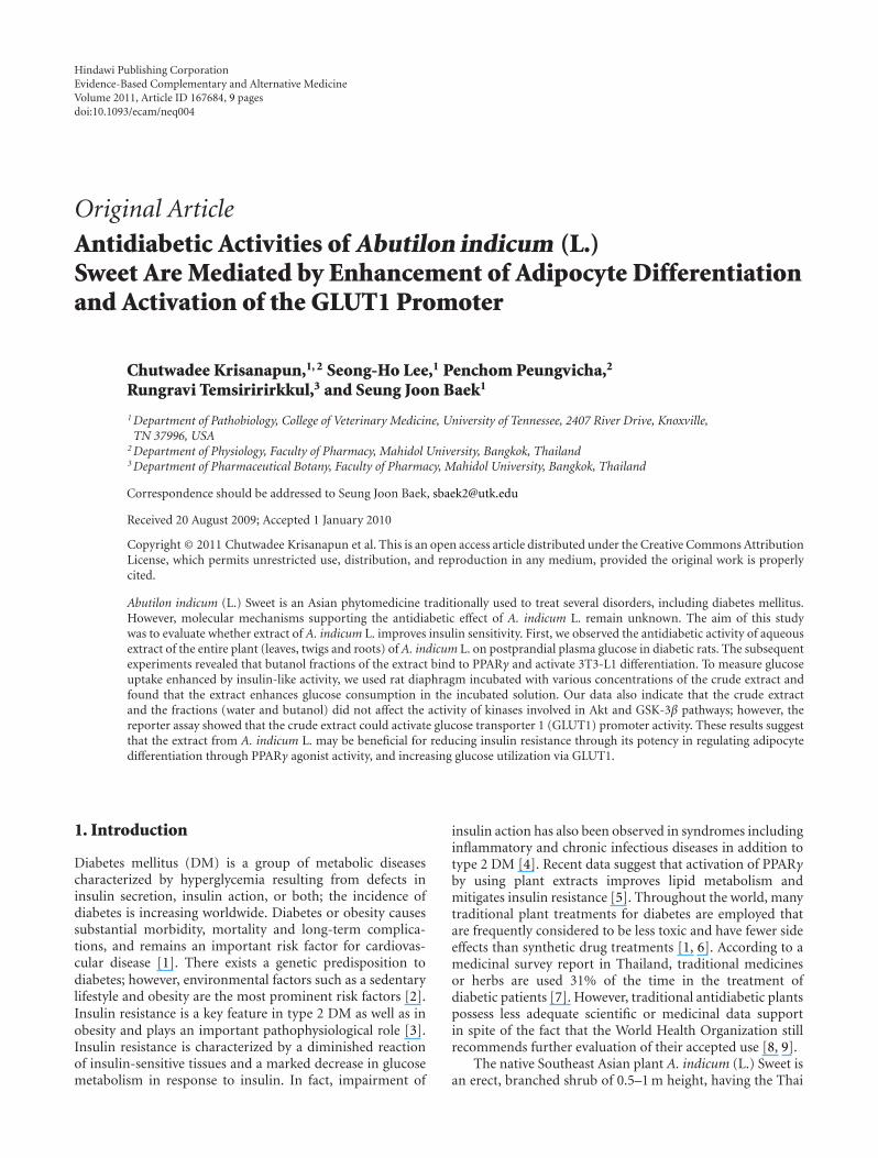
Figure 1: The effect of crude extract of A. indicum on 2 hpostprandial plasma glucose in STZ-induced diabetic rats. Theresults show the mean ± SEM, (n = 6–9). Asterisks representsstatistical significance (P < .05) compared with control at eachday. Double asterisk represent statistical significance (P < .05) bothcompared with control at each day and day 0 within the group.
to various concentrations of crude extract in HEPES-buffered saline, pH 7.4, containing 2% horse serum with5 mM glucose for 16 h. The cells were washed twice with PBSand then assayed for luciferase activity as shown above.
2.10. Statistical Analysis. The data were presented as themean ± standard error of the mean (SEM). Statisticalanalysis was performed using the Student’s t-test or analysisof variance (ANOVA) followed by the least significantdifferences (LSD) test. The significance of differences was setat P-value < .05.
3. Results
3.1. Postprandial Plasma Glucose in STZ-Induced DiabeticRats. As shown in Figure 1, administration of A. indicumcrude extract significantly lowered (P < .05) 2 h postpran-dial plasma glucose concentrations of treated groups incomparison to those of control STZ-diabetic rats at thecorresponding time. A similar significant decrease was alsoobserved in rats treated with glibenclamide. However, priorto treatment administration (day 0), basal plasma glucoseconcentration did not significantly differ between groups.
3.2. Binding Activity to Peroxisome Proliferator-ActivatedReceptors. To examine whether A. indicum extracts activateperoxisome proliferator-activated receptor (PPAR) respon-siveness, HCT-116 cells were transfected with four copies of aGal4 binding site (MH100x4-TK-LUC) and chimeric recep-tors (pCMX-Gal-mPPARα-LBD or pCMX-Gal-mPPARγ-LBD). In this system, when a compound binds to theligand binding domain (LBD) of the PPARα or PPARγchimeric receptor, the DNA binding domain of the yeastGal4 binds to the co-transfected Gal4 binding site andinitiates transcription of the firefly luciferase (LUC). After

4 Evidence-Based Complementary and Alternative Medicine
Table 1: Primer sequences used for RT-PCR.
Gene Sense Sequence
AdiponectinForward 5′-TGC ACA GCT CCG TGT ACT TC-3′
Reverse 5′-CAC CTG CAC AGA GTC GTC AT-3′
aP2Forward 5′-AAG AAG TGG GAG TGG GCT TT-3′
Reverse 5′-CTT GTG GAA GTC ACG CCT TT-3′
LPLForward 5′-GGA TCC GTG GCC GCA GCA GAC GCA GGA AGA-3′
Reverse 5′-GAA TTC CAT CCA GTT GAT GAA TCT GGC CAC-3′
PPARγForward 5′-GGT GAA ACT CTG GGA GAT TC-3′
Reverse 5′-CAA CCA TTG GGT CAG CTC TT-3′
GAPDHForward 5′-CAG GAG CGA GAC CCC ACT AAC AT-3′
Reverse 5′-GTC AGA TCC ACG ACG GAC ACA TT-3′
cells were treated with crude extract for 24 h, luciferaseactivities were measured to assess the transactivation for eachPPAR receptor. As shown in Figures 2(a) and 3(a), crudeextract 100 μg ml−1 showed 1.6- and 1.4-fold increases oftransactivation to PPARα and PPARγ, respectively, indicatingthat crude extract contains compounds that bind to PPARαand PPARγ.
The activation of PPARα and PPARγ using water andbutanol fractions was also investigated. The results revealedthat the water fraction has a minimal effect on PPARαand PPARγ activation, whereas butanol fractions showed adose-dependent increase of PPARα and PPARγ activation(Figures 2(b) and 3(b)). The concentration of butanolfraction (100 μg ml−1) showed 5.5- and 3.2-fold increasesof transactivation to PPARα and PPARγ, respectively. Theseresults suggest that the butanol fraction, but not the waterfraction, contains a substance that activates PPARα andPPARγ.
3.3. mRNA Expression during Preadipocyte Differentiation.Since butanol fraction binds to PPARγ and transactivatesPPARγ activity, gene expression profiles were observed toelucidate whether the butanol fraction actually inducesadipocyte differentiation through PPARγ activation. Twenty-four hours before and during differentiation, butanol frac-tion at 100 μg ml−1 was added to the medium for observationof its effects on gene expression during 3T3-L1 adipocytedifferentiation. Dimethyl sulfoxide (DMSO) was used asvehicle control. As shown in Figure 4, cells treated with thebutanol fraction increased the expression of PPARγ, aP2,and adiponectin at day 8, consistent with the previous dataindicating that butanol fraction enhances PPARγ activity(Figure 3(b)). However, there was no change in mRNA levelof lipoprotein lipase (LPL) throughout the differentiation.
3.4. Glucose Consumption. Crude extract of A. indicum wastested for insulin-like effect using diaphragm of rat. Pieces ofdiaphragms were incubated with various concentrations ofthe crude extract for 1.5 h. As shown in Figure 5, the extractenhanced glucose uptake in a concentration-dependentmanner. The extract between concentrations of 1–5 mg ml−1
significantly increased glucose uptake and showed an almostmaximal effect on glucose uptake at the concentration of
1 mg ml−1. Insulin was used as a positive control and alsoshowed an ∼1.7-fold induction over the untreated group.
3.5. Protein Expression in Insulin Signaling Pathway andGLUT1 Promoter Activation. Since protein kinases B (Akt)and glycogen synthase kinase 3β (GSK3β) are major kinasesof the insulin signaling pathway [26, 27], the effects ofcrude extract and butanol and water fractions on phospho-rylation of Akt and GSK3β in C2C12 myotubes and 3T3-L1 adipocytes were examined. However, no changes weredetected in Akt phosphorylation or GSK3β phosphorylation,although insulin activated these two biochemical markers(data not shown), suggesting that extracts and fractions donot affect the Akt/GSK3β pathway. To determine the effectof crude extract on the transcriptional activity of Glut1, apGL3-GLUT1 luciferase construct that contains –2100 bpof the rat Glut1 promoter region was transfected into L6cells. After transfection, the cells were treated with variousconcentrations of crude extract for 16 h, and luciferaseactivity was measured. As shown in Figure 6, the cells treatedwith the extract increased relative luciferase unit (RLU) ina dose-response manner. A significant fold increase againstuntreated cells was found in the cells treated with 100 and500 μg ml−1 of crude extract.
4. Discussion
Much research effort is currently directed toward develop-ment of adipogenesis and lipogenesis studies, and a numberof antidiabetic compounds have been developed and testedfor their effects. For example, thiazolidinediones (TZDs),synthetic PPARγ ligands, are a novel class of antidiabeticdrugs for patients with type 2 diabetes, and two of these,rosiglitazone and pioglitazone, are currently available forclinical use [28]. Originally, TZDs were identified basedon their antihyperglycemic activity, but they are also ableto improve other abnormalities associated with type 2diabetes, such as hyperlipidemia, atherosclerosis, hyperten-sion, chronic inflammation and fibrinolytic state. It is wellestablished that PPARγ agonists such as TZDs promotethe adipogenesis of 3T3-L1 cells, which have been usedfor the development of antidiabetic compounds [29–31].Since troglitazone (TGZ) was voluntarily withdrawn from

Evidence-Based Complementary and Alternative Medicine 5
1413 ∗
∗
121110
9876543210
RLU
(fol
din
crea
se)
X 10
Control WY14643 1μg/mL 100μg/mL
Crude extract
(a)
876543210
1110
9
Con
trol
WY
1464
3
1μ
g/m
L
10μ
g/m
L
100μ
g/m
L
1μ
g/m
L
10μ
g/m
L
100μ
g/m
L
Water fraction Butanol fraction
∗
∗
∗X 10
RLU
(fol
din
crea
se)
(b)
Figure 2: (a) The effect of A. indicum crude extract on luciferaseactivity of PPARα. After the cells were transiently transfectedwith four copies of a Gal4 binding site (MH100×4-TK-LUC)and chimeric receptors pCMX-Gal-mPPARα-LBD, the cells weretreated with vehicle (DMSO, control), WY14643 (positive control)or different concentrations of crude extract for 24 h and luciferaseactivity was measured. The data are presented as relative luciferaseactivity (firefly luciferase signal/renilla luciferase signal). The resultsshow the mean ± SEM of three independent transfections. ∗P <.01 versus vehicle-treated cells. (b) The effect of water and butanolfractions on luciferase activity of PPARα. The data are presented asrelative luciferase activity (firefly luciferase signal/renilla luciferasesignal). The results show the mean ± SEM of three independenttransfections. ∗P < .01 versus vehicle-treated cells.
the market in March 2000 due to occurrence of severeidiosyncratic liver injury, it is necessary to establish a safercompound that shows antidiabetic activity. Many researchershave focused on the study of dietary phytochemicals foundin plants. In this report, we examined whether the extractof A. indicum has antidiabetic activity in STZ-induceddiabetic rats. The results demonstrated that administra-tion of A. indicum extract at 0.25 and 0.5 g kg−1 resultedin significant decreases of postprandial plasma glucose,as seen in glibenclamide-treated rats. Although gliben-clamide lowers plasma glucose level, mainly by stimulatinginsulin secretion from pancreatic β-cells [32], destruction of
∗
∗
1.0
0.5
0
X 102.5
2.0
1.5
Control TGZ 1μg/mL 100μg/mL
Crude extract
RLU
(fol
din
crea
se)
(a)
∗
∗∗∗∗
121110
9876543210
RLU
(fol
din
crea
se)
Con
trol
TG
Z
1μ
g/m
L
10μ
g/m
L
100μ
g/m
L
1μ
g/m
L
10μ
g/m
L
100μ
g/m
L
Water fraction Butanol fraction
131415
(b)
Figure 3: (a) The effect of A. indicum crude extract on luciferaseactivity of PPARγ. After the cells were transiently transfectedwith four copies of a Gal4 binding site (MH100×4-TK-LUC) andchimeric receptors pCMX-Gal-mPPARγ-LBD, they were treatedwith vehicle (DMSO, control), TGZ (positive control) or differentconcentrations of crude extract for 24 h and luciferase activity wasmeasured. The data are presented as relative luciferase activity(firefly luciferase signal/renilla luciferase signal). The results showthe mean ± SEM of three independent transfections. ∗P < .01versus vehicle-treated cells. (b) The effect of water and butanolfractions on luciferase activity of PPARγ. The data are presented asrelative luciferase activity (firefly luciferase signal/renilla luciferasesignal). The results show the mean ± SEM of three independenttransfections. ∗P < .01 versus vehicle-treated cells.
β-cells are observed in STZ-induced diabetes model (type 1diabetes) [33]. Thus, extrapancreatic action such as stimu-lating liver and muscle fructose-2,6-bisphosphate formationmay be involved in the effect of both glibenclamide andextracts used in this study rather than insulin secretion.In fact, it has been known that the extrapancreatic actionof glibenclamide has an important role to inhibit glucoseproduction by liver and improve carbohydrate metabolismin diabetes [34, 35], and increases glucose disposal bystabilizing GLUT1 protein at the plasma membrane ofmuscle [36]. Subsequently, we found that crude extract of A.indicum acts as a PPARγ ligand. Further, the data indicated
6 Evidence-Based Complementary and Alternative Medicine
Butanol fraction
80 2 4 680 2 4 6 Days
PPARγ
LPL
aP2
Adiponectin
GAPDH
DMSO
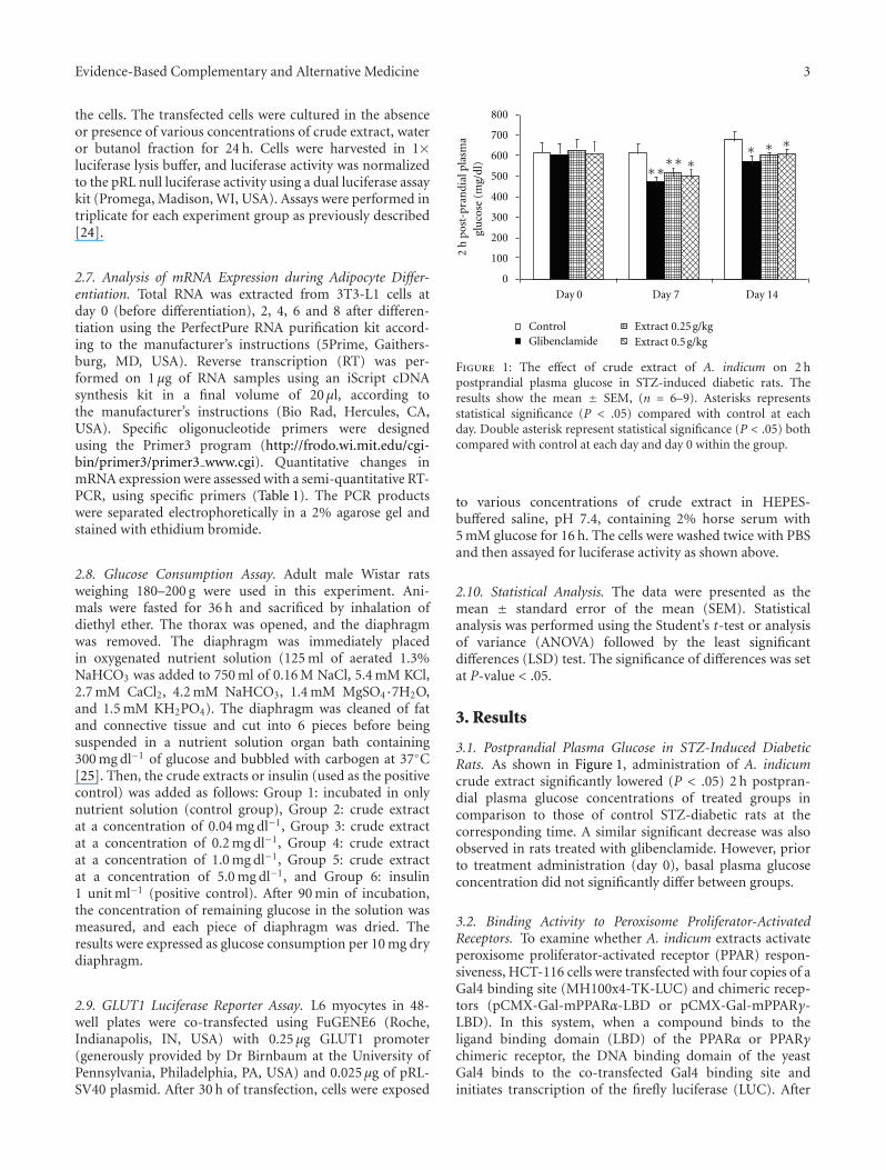
Figure 4: The effect of butanol fraction of A. indicum to inducemRNA expression of the PPARγ target gene. 3T3-L1 preadipocytecells were differentiated. Twenty-four hours before and duringdifferentiation, cells were treated with vehicle control (DMSO) or100 μg ml−1 of butanol fraction. mRNA expression of PPARγ, LPL,aP2, adiponectin and glyceraldehyde 3-phosphate dehydrogenase(GAPDH) were measured at Days 0, 2, 4, 6 and 8 of differentiation.
2.5
2.0
Fold
incr
ease
1.5
1.0
0.5
0Control
∗∗
0.04 0.2
Crude extract (mg/mL)
∗
51 Insulin
Figure 5: Effect of A. indicum crude extract on muscle glucoseconsumption. Diaphragms were isolated from fasted Wistar ratsand incubated with 300 mg dl−1 of glucose in the absence orpresence of crude extract for 90 min. The glucose consumption wasexpressed as disappeared glucose into diaphragm per 10 mg drydiaphragm. Data are presented as means ± SEM of eight replicates.Statistical analysis was performed using ANOVA followed by LSD.∗P < .05 compared with the untreated group.
that the butanol fraction (non-polar part) may contain acompound that induces PPARγ activity (Figure 3(b)). Thepresent study also demonstrates that the butanol fractionenhances 3T3-L1 adipocyte differentiation by an increase ofaccumulation of differentiation and fat cell marker genes(Figure 4). The accumulation of these mRNA reflects boththe rate of initiation of transcription and the half time ofdegradation of those transcripts.
At the molecular level, adipogenesis is driven by acomplex transcriptional cascade involving the sequentialactivation of CCAAT/enhancer binding proteins (C/EBPs)and PPARγ [37]. Loss-of-function studies have convincinglyshown that PPARγ is necessary as well as sufficient topromote adipogenesis and that C/EBPα is influential bymaintaining the expression of PPARγ and promoting full
2.5
∗2.0 ∗
∗
1.5
1.0
0.5
0Empty Control 10 50 100 500 Positive
controlCrude extract (μg/mL)
RLU
(fol
din
crea
se)
Figure 6: The effect of A. indicum crude extract on the transcriptionlevel of Glut1. L6 myocytes were transfected with GLUT1 promoterand pRL-SV40 plasmid. Then, cells were exposed to indicateconcentrations of crude extract for 16 h as described in the‘Methods’ section. The luciferase activity was expressed as a ratioof firefly to renilla luciferase activity. Transfections and assays wereperformed in triplicate for each experiment group. Data showedmean ± SEM of fold increase of three replicates against controlgroup. ∗P < .05 compared with untreated sample group. The pGL3-Basic vector (empty) was used as a negative control, and pGL3-control vector was used for a positive control.
insulin sensitivity [38]. Our data indicate that exposing 3T3-L1 preadipocytes to the butanol fraction during adipogenesisincreases PPARγ mRNA, as well as the adipocyte-specificfatty acid binding protein (aP2) mRNA level; aP2 is oneof the genes that characterizes the adipocyte phenotype[37, 39]. Interestingly, the mRNA level of adiponectin in thebutanol fraction-treated group was higher than the controlthroughout the differentiation. Adiponectin is exclusivelyexpressed by mature adipocytes and is the most abundantcirculating adipokine. Thus, these data imply that thebutanol fraction of A. indicum extract enhances adipogenesisthrough PPARγ activation. Hypoadiponectinemia appears toplay an important causal role in insulin resistance, type 2DM and metabolic syndrome [40, 41]. We could not excludethe possibility that butanol fraction affects mRNA stability ofthose genes, and further studies may require elucidating themolecular mechanism by which butanol fraction affects geneinduction during adipogenesis. Since LPL (a PPARγ targetgene) did not increase in the presence of butanol fraction, itis possible that butanol fraction may affect gene inductionin a PPARγ-independent manner. The data also showed thatbutanol fraction increased the activity of PPARα; however,further work to investigate expression of the PPARα targetgene is required to claim that the extract of A. indicumacts as a PPARα/γ dual agonist. Indeed, PPARα agonists,such as fibrates, have been used to treat hypertriglyceridemiaand reduce cardiovascular risk [42]. Dual PPARα/γ agonistshold promise to be able to combine the properties of TZDsand fibrates, as well as improve the management of type2 diabetes and provide an effective therapeutic option fortreating the multifactorial components of cardiovasculardisease, metabolic syndrome [43], and even cancer.

Evidence-Based Complementary and Alternative Medicine 7
Filtrate from boiling with distilled water
Crude extract ↓Postprandial plasma glucose
1. TZD-Iike activity↑PPARγ ligand binding activity(PPARγ agonist)
Partition crude extractwith H2O : C4H10O
Water fraction Butanol fraction
No effect ↑↑TZD-like activity
↑Adipocyte differentiation
-↑aP2
-↑PPARγ
-↑Adiponectin
2. Insulin-like activity
↑Peripheral glucose utilization
2.1. No effects on insulin signaling
A.indicum(leaves, twigs, roots)
(In vivo)
pathway (pAkt, pGSK3β)
2.2. ↑Promoter activity of GLUT1
Figure 7: Summary of antidiabetic effects against insulin resistance of A. indicum Sweet.
Another important finding of this work is that theextract of A. indicum possessed considerable insulin-likeproperties, as evidenced by enhancement of glucose uptakein the diaphragm, which represents muscle cells that are themajor site of insulin-stimulated glucose disposal [44]. Inthe follow-up studies, we examined potential mechanismsunderlying the improvement in glucose utilization by theextract. The defective glucose transport system may playan important role in the pathogenesis of peripheral insulinresistance, and glucose uptake in target tissues is a criticalstep in maintaining glucose homeostasis and in clearingthe postprandial glucose load [45]. Thus, we examined thepossibility that the extract may stimulate glucose transportthrough a mechanism similar to insulin signaling in C2C12myotubes and 3T3-L1 adipocytes. Previous literature hasdocumented that the phosphatidylinositol-3-kinase (PI3K)pathway plays an important role in the insulin signalingcascade leading to glucose transport translocation [44].However, the results showed that under basal or insulin-stimulated conditions, all of the extract treatments (crudeextract, water and butanol fraction) had no effect onAkt serine phosphorylation in either cell type (data notshown). Another pathway that has been implicated in glucosetransport, not by translocation of GLUT, but by activation ofGLUT, is the p38 pathway [46], which is also interesting forfurther study. This pathway has been reported to be involvedin the regulation of GLUT 1 expression [47]. Subsequently,we examined the effect of the extract on transcriptionactivation of GLUT1. As demonstrated in this study, extractof A. indicum activated transcription of GLUT1 in a dose-response manner, suggesting involvement of GLUT1 inincreasing glucose uptake by the extract. This result willrequire confirmation such as measuring extracellular signal
regulated kinases (ERKs), which have been known to beresponsible for the expression of GLUT1 [48].
We have reported that the phytochemical analysis ofthe whole plant extract revealed the presence of alkaloids,flavonoids, and tannins [11]. Results from other researchershave shown that this plant also contains saponins andglycosides [10]. Most plants with antidiabetic propertieshave been found to contain metabolites such as glycosides,alkaloids and flavonoids [9]. Indeed, seven compounds wereisolated from A. indicum extract and six of them identi-fied as β-sitosterol, oleanolic acid, (24R)-α-stigmastane-3,6-dione, daucosterol, 2,6-dimethoxy-1,4-benzoquinone, andvanillic acid [49]. Oleanolic acid has been reported tohave hypoglycemic activity by acting as an inhibitor ofglycogen phosphorylase, leading to the inhibition of hepaticglycogenolysis [50]. The alkaloid berberine has also beenreported to activate GLUT1-mediated glucose uptake [48].Thus, these results provide evidence that these compoundsmay lead to affect the antidiabetic activity of A. indicum.The findings of antidiabetic properties in this study aresummarized in Figure 7, and support the ethnobotanical useof A. indicum in the context of human type 2 DM; however,the molecules of action responsible for A. indicum’s effectremain to be identified.
Funding
The Center of Excellence in Livestock Diseases and HumanHealth at the University of Tennessee and a NationalInstitutes of Health grant (R21CA109423). Financial supportwas provided by the Royal Golden Jubilee PhD Program(PHD/0245/2545) and the Office of the Higher EducationCommission, Thailand (to C.K.).

8 Evidence-Based Complementary and Alternative Medicine
Acknowledgment
The authors thank Misty Bailey for her critical reading ofmanuscript.
References
[1] G. Y. Yeh, D. M. Eisenberg, T. J. Kaptchuk, and R. S. Phillips,“Systematic review of herbs and dietary supplements forglycemic control in diabetes,” Diabetes Care, vol. 26, no. 4, pp.1277–1294, 2003.
[2] S. Samane, J. Noel, Z. Charrouf, H. Amarouch, and P. S.Haddad, “Insulin-sensitizing and anti-proliferative effects ofArgania spinosa seed extracts,” Evidence-Based Complementaryand Alternative Medicine, vol. 3, no. 3, pp. 317–327, 2006.
[3] P. Vollenweider, “Insulin resistant states and insulin signaling,”Clinical Chemistry and Laboratory Medicine, vol. 41, no. 9, pp.1107–1119, 2003.
[4] T.-P. Liu, C.-S. Lee, S.-S. Liou, I.-M. Liu, and J.-T. Cheng,“Improvement of insulin resistance by Acanthopanax sen-ticosus root in fructose-rich chow-fed rats,” Clinical andExperimental Pharmacology and Physiology, vol. 32, no. 8, pp.649–654, 2005.
[5] C.-C. Shih, C.-H. Lin, W.-L. Lin, and J.-B. Wu, “Momordicacharantia extract on insulin resistance and the skeletal muscleGLUT4 protein in fructose-fed rats,” Journal of Ethnopharma-cology, vol. 123, no. 1, pp. 82–90, 2009.
[6] P. S. Haddad, G. A. Azar, S. Groom, and M. Boivin, “Naturalhealth products, modulation of immune function and preven-tion of chronic diseases,” Evidence-Based Complementary andAlternative Medicine, vol. 2, no. 4, pp. 513–520, 2005.
[7] W. Aekplakorn, R. P. Stolk, B. Neal et al., “The prevalence andmanagement of diabetes in Thai adults: the International Col-laborative Study of Cardiovascular Disease in Asia,” DiabetesCare, vol. 26, no. 10, pp. 2758–2763, 2003.
[8] A. M. Gray, Y. H. A. Abdel-Wahab, and P. R. Flatt, “Thetraditional plant treatment, Sambucus nigra (elder), exhibitsinsulin-like and insulin-releasing actions in vitro,” Journal ofNutrition, vol. 130, no. 1, pp. 15–20, 2000.
[9] A. A. Odetola, O. Akinloye, C. Egunjobi, W. A. Adekunle, andA. O. Ayoola, “Possible antidiabetic and antihyperlipidaemiceffect of fermented Parkia biglobosa (JACQ) extract in alloxan-induced diabetic rats,” Clinical and Experimental Pharmacol-ogy and Physiology, vol. 33, no. 9, pp. 808–812, 2006.
[10] Y. N. Seetharam, G. Chalageri, S. R. Setty, and Bheemachar,“Hypoglycemic activity of Abutilon indicum leaf extracts inrats,” Fitoterapia, vol. 73, no. 2, pp. 156–159, 2002.
[11] C. Krisanapun, P. Peungvicha, R. Temsiririrkkul, and Y.Wongkrajang, “Aqueous extract of Abutilon indicum Sweetinhibits glucose absorption and stimulates insulin secretion inrodents,” Nutrition Research, vol. 29, no. 8, pp. 579–587, 2009.
[12] W. Chuakul, P. Saralamp, W. Paonil, R. Temsiririrkkul, and T.Clayton, Medical Plants in Thailand, vol. II, Amarin, Bangkok,Thailand, 1997.
[13] A. Abdul Rahuman, G. Gopalakrishnan, P. Venkatesan, andK. Geetha, “Isolation and identification of mosquito larvicidalcompound from Abutilon indicum (Linn.) Sweet,” ParasitologyResearch, vol. 102, no. 5, pp. 981–988, 2008.
[14] S. S. Deokule and M. W. Patale, “Pharmacognostic study ofAbutilon indicum, (L.) Sweet,” Journal of Phytological Research,vol. 15, pp. 1–6, 2002.
[15] E. Porchezhian and S. H. Ansari, “Hepatoprotective activityof Abutilon indicum on experimental liver damage in rats,”Phytomedicine, vol. 12, no. 1-2, pp. 62–64, 2005.
[16] J. Parekh and S. Chanda, “Antibacterial and phytochemicalstudies on twelve species of Indian medicinal plants,” AfricanJournal of Biomedical Research, vol. 10, pp. 175–181, 2007.
[17] M. Ahmed, S. Amin, M. Islam, M. Takahashi, E. Okuyama,and C. F. Hossain, “Analgesic principle from Abutilonindicum,” Pharmazie, vol. 55, no. 4, pp. 314–316, 2000.
[18] K. R. Kohli, G. Shilin, and S. A. Kolhapure, “Evaluation ofthe clinical efficacy and safety of Diabecon in NIDDM,” TheAntiseptic, vol. 101, pp. 487–494, 2004.
[19] S. K. Mitra, S. Gopumadhavan, T. S. Muralidhar, S. D.Anturlikar, and M. B. Sujatha, “Effect of a herbomineralpreparation D-400 in streptozotocin-induced diabetic rats,”Journal of Ethnopharmacology, vol. 54, no. 1, pp. 41–46, 1996.
[20] A. R. Kishan, M. Ajitha, and K. Rajanarayana, “Antidiabetic,antihyperlipidemic and free radical scavenging activities of anAyurvedic medicine,” Indian Drugs, vol. 37, no. 3, pp. 130–132,2000.
[21] M. Bhat, S. K. Kothiwale, A. R. Tirmale, S. Y. Bhargava, andB. N. Joshi, “Antidiabetic properties of Azardiracta indicaand Bougainvillea spectabilis: in vivo studies in murine dia-betes model,” Evidence-Based Complementary and AlternativeMedicine. In press.
[22] P. Peungvicha, S. S. Thirawarapan, R. Temsiririrkkul, H.Watanabe, J. Kumar Prasain, and S. Kadota, “Hypoglycemiceffect of the water extract of Piper sarmentosum in rats,”Journal of Ethnopharmacology, vol. 60, no. 1, pp. 27–32, 1998.
[23] A. O. Abdel-Zaher, S. Y. Salim, M. H. Assaf, and R. H. Abdel-Hady, “Antidiabetic activity and toxicity of Zizyphus spina-christi leaves,” Journal of Ethnopharmacology, vol. 101, no. 1–3,pp. 129–138, 2005.
[24] S.-H. Lee, K. Yamaguchi, J.-S. Kim et al., “Conjugated linoleicacid stimulates an anti-tumorigenic protein NAG-1 in anisomer specific manner,” Carcinogenesis, vol. 27, no. 5, pp.972–981, 2006.
[25] F. Sanchez de Medina, M. J. Gamez, I. Jimenez, J. Jimenez, J.I. Osuna, and A. Zarzuelo, “Hypoglycemic activity of juniper’berries’,” Planta Medica, vol. 60, no. 3, pp. 197–200, 1994.
[26] W. Shang, Y. Yang, L. Zhou, B. Jiang, H. Jin, and M. Chen,“Ginsenoside Rb1 stimulates glucose uptake through insulin-like signaling pathway in 3T3-L1 adipocytes,” Journal ofEndocrinology, vol. 198, no. 3, pp. 561–569, 2008.
[27] J. Lee and M.-S. Kim, “The role of GSK3 in glucose home-ostasis and the development of insulin resistance,” DiabetesResearch and Clinical Practice, vol. 77, no. 3, pp. S49–S57, 2007.
[28] H. Yki-Jarvinen, “Thiazolidinediones,” The New EnglandJournal of Medicine, vol. 351, no. 11, pp. 1106–1158, 2004.
[29] H. Zhang, H. Matsuda, S. Nakamura, and M. Yoshikawa,“Effects of amide constituents from pepper on adipogenesisin 3T3-L1 cells,” Bioorganic and Medicinal Chemistry Letters,vol. 18, no. 11, pp. 3272–3277, 2008.
[30] C. Kausch, J. Krutzfeldt, A. Witke et al., “Effects of troglitazoneon cellular differentiation, insulin signaling, and glucosemetabolism in cultured human skeletal muscle cells,” Bio-chemical and Biophysical Research Communications, vol. 280,no. 3, pp. 664–674, 2001.
[31] F. Chiarelli and D. Di Marzio, “Peroxisome proliferator-activated receptor-γ agonists and diabetes: current evidenceand future perspectives,” Vascular Health and Risk Manage-ment, vol. 4, no. 2, pp. 297–304, 2008.

Evidence-Based Complementary and Alternative Medicine 9
[32] A. J. Krentz and C. J. Bailey, “Oral antidiabetic agents: currentrole in type 2 diabetes mellitus,” Drugs, vol. 65, no. 3, pp. 385–411, 2005.
[33] T. Szkudelski, “The mechanism of alloxan and streptozotocinaction in B cells of the rat pancreas,” Physiological Research,vol. 50, no. 6, pp. 537–546, 2001.
[34] K. Hatao, K. Kaku, M. Matsuda, M. Tsuchiya, and T. Kaneko,“Sulfonylurea stimulates liver fructose-2,6-bisphosphate for-mation in proportion to its hypoglycemic action,” DiabetesResearch and Clinical Practice, vol. 1, pp. 49–53, 1985.
[35] M. Matsuda, K. Kaku, and T. Kaneko, “Regulation ofmuscle fructose 2, 6-bisphosphate levels by sulfonylureas,”Endocrinologia Japonica, vol. 33, no. 6, pp. 913–917, 1986.
[36] E. Tsiani, T. Ramlal, L. A. Leiter, A. Klip, and I. G. Fan-tus, “Stimulation of glucose uptake and increased plasmamembrane content of glucose transporters in L6 skeletalmuscle cells by the sulfonylureas gliclazide and glyburide,”Endocrinology, vol. 136, no. 6, pp. 2505–2512, 1995.
[37] E. D. Rosen and B. M. Spiegelman, “Molecular regulationof adipogenesis,” Annual Review of Cell and DevelopmentalBiology, vol. 16, pp. 145–171, 2000.
[38] S. R. Farmer, “Transcriptional control of adipocyte forma-tion,” Cell Metabolism, vol. 4, no. 4, pp. 263–273, 2006.
[39] M. Hassan, C. E. Yazidi, J.-F. Landrier, D. Lairon, A. Margotat,and M.-J. Amiot, “Phloretin enhances adipocyte differentia-tion and adiponectin expression in 3T3-L1 cells,” Biochemicaland Biophysical Research Communications, vol. 361, no. 1, pp.208–213, 2007.
[40] T. Kadowaki, T. Yamauchi, N. Kubota, K. Hara, K. Ueki, andK. Tobe, “Adiponectin and adiponectin receptors in insulinresistance, diabetes, and the metabolic syndrome,” Journal ofClinical Investigation, vol. 116, no. 7, pp. 1784–1792, 2006.
[41] J. J. Dıez and P. Iglesias, “The role of the novel adipocyte-derived hormone adiponectin in human disease,” EuropeanJournal of Endocrinology, vol. 148, no. 3, pp. 293–300, 2003.
[42] S. J. Robins, “PPARα ligands and clinical trials: cardiovascularrisk reduction with fibrates,” Journal of Cardiovascular Risk,vol. 8, no. 4, pp. 195–201, 2001.
[43] B. Staels and J.-C. Fruchart, “Therapeutic roles of peroxisomeproliferator-activated receptor agonists,” Diabetes, vol. 54, no.8, pp. 2460–2470, 2005.
[44] P. R. Shepherd and B. B. Kahn, “Glucose transporters andinsulin action: implications for insulin resistance and diabetesmellitus,” The New England Journal of Medicine, vol. 341, no.4, pp. 248–257, 1999.
[45] G. I. Shulman, “Cellular mechanisms of insulin resistance,”Journal of Clinical Investigation, vol. 106, no. 2, pp. 171–176,2000.
[46] N. Takahashi, M. Nagamine, S. Tanno, W. Motomura, Y.Kohgo, and T. Okumura, “A diacylglycerol kinase inhibitor,R59022, stimulates glucose transport through a MKK3/6-p38signaling pathway in skeletal muscle cells,” Biochemical andBiophysical Research Communications, vol. 360, no. 1, pp. 244–250, 2007.
[47] Y. Yamamoto, Y. Yoshimasa, M. Koh et al., “Constitu-tively active mitogen-activated protein kinase kinase increasesGLUT1 expression and recruits both GLUT1 and GLUT4 atthe cell surface in 3T3-L1 adipocytes,” Diabetes, vol. 49, no. 3,pp. 332–339, 2000.
[48] S. H. Kim, E.-J. Shin, E.-D. Kim, T. Bayaraa, S. C. Frost, andC.-K. Hyun, “Berberine activates GLUT1-mediated glucoseuptake in 3T3-L1 adipocytes,” Biological and PharmaceuticalBulletin, vol. 30, no. 11, pp. 2120–2125, 2007.
[49] N. Liu, L. Y. Jia, and Q. S. Sun, “Chemical constituents of Abu-tilon indicum (L.) Sweet,” Journal of Shenyang PharmaceuticalUniversity, vol. 26, pp. 196–197, 2009.
[50] J. Chen, J. Liu, L. Zhang et al., “Pentacyclic triterpenes.Part 3: synthesis and biological evaluation of oleanolic acidderivatives as novel inhibitors of glycogen phosphorylase,”Bioorganic and Medicinal Chemistry Letters, vol. 16, no. 11, pp.2915–2919, 2006.
Copyright © 2022 FDOKUMEN