Movement Proteins (BC1 and BV1) of Abutilon Mosaic Geminivirus Are Cotransported in and between...
13
-
Upload
independent -
Category
Documents
-
view
1 -
download
0
Transcript of Movement Proteins (BC1 and BV1) of Abutilon Mosaic Geminivirus Are Cotransported in and between...


Movement Proteins (BC1 and BV1) of Abutilon Mosaic Geminivirus Are Cotransportedin and between Cells of Sink but Not of Source Leaves as Detected
by Green Fluorescent Protein Tagging
Shuo Cheng Zhang, Christina Wege, and Holger Jeske1
Department of Molecular Biology and Plant Virology, Biological Institute, University of Stuttgart, Pfaffenwaldring 57, D-70550 Stuttgart, Germany
Received May 10, 2001; returned to author for revision August 3, 2001; accepted September 6, 2001
Two movement proteins (BV1 and BC1) facilitate the intra- and intercellular transport of begomoviruses in plants. Incontrast to other geminiviruses the movement protein BC1 of Abutilon mosaic virus (AbMV) remained in the supernatant aftercentrifuging plant extracts at 20,000 g. To test whether this unusual behavior results from a distinct intracellular distributionof the protein, the BC1 gene has been fused to the gene of green fluorescent protein (GFP). The resulting plasmids weredelivered into nonhost plants (Allium cepa) as well as into mature and immature cells of host plants (Nicotiana tabacum, N.benthamiana) by biolistic bombardment for transient expression in planta. BC1 directed GFP to two different cellular sites.In the majority of nonhost cells as well as in mature cells of host leaves, BC1 was mainly localized in small punctate flecksat the cell periphery or, to a lesser extent, around the nucleus. In sink leaves of host plants, GFP:BC1 additionally developeddisc-like structures in the cell periphery. Cobombardment of GFP:BC1 with its cognate infectious DNA A and B did not changetheir subcellular distribution patterns in source leaves but led to the formation of peculiar needle-like structures in sinkleaves. The nuclear shuttle protein (BV1) of AbMV accumulated mainly inside the nuclei as shown by immunohistochemicalstaining and GFP tagging. In sink cells of host plants it was mobilized to the plasma membrane and to the nucleus of theneighboring cell by coexpressed BC1, GFP:BC1, BC1:GFP, or after cobombardment with the cognate viral DNA. Only underthese conditions were GFP:BC1 and BC1:GFP also found in the recipient cell. © 2001 Academic Press
INTRODUCTION
Viruses move through plants in two steps: cell-to-celltransport, usually via plasmodesmata, and long-distancetransport through the phloem (for recent reviews seeDing et al., 1999; Lazarowitz, 1999; Lazarowitz andBeachy, 1999). For cell-to-cell transport, two mechanismshave been discovered for different viruses. One involvesthe binding of a movement protein to viral RNA andopening of the plasmodesmata (Carrington et al., 1996;Citovsky et al., 1990; Fujiwara et al., 1993; Osman et al.,1992; Wolf et al., 1989). The other is dependent on tubuleformation (Kasteel et al., 1997; Perbal et al., 1993; Stormset al., 1995; Wellink et al., 1993).
In contrast to most RNA viruses, the DNA-containinggeminiviruses replicate in the nucleus and must exploitadditional mechanisms to be transported from the nu-cleus into the cytoplasm and from one cell to another.Among geminiviruses, the begomoviruses (Rybicki et al.,2000) are characterized by a bipartite genome (DNA Aand DNA B), in which DNA B encodes two movementproteins (MPs) called BC1 and BV1 (Etessami et al., 1988;Evans and Jeske, 1993; von Arnim et al., 1993). BC1
mediates cell-to-cell transport, whereas BV1 is respon-sible for the export of viral DNA from the nucleus. Bothproteins cooperate in a way that is currently under de-bate. Immunological investigations using either transientexpression of the two Squash leaf curl virus (SqLCV) MPsin protoplasts or systemically infected plants (Ward et al.,1997) support a model in which BV1 exports the viralgenomes from the nucleus into the cytoplasm and backto the nucleus as a nuclear shuttle protein, and then BC1directs the BV1–DNA complexes through modified plas-modesmata, trafficking them from cell to cell (Lazarowitzand Beachy, 1999; Pascal et al., 1994; Sanderfoot et al.,1996; Sanderfoot and Lazarowitz, 1995, 1996). A differentmodel results from investigations on Bean dwarf mosaicvirus (BDMV). Microinjection experiments delivering bac-terially expressed BDMV proteins into mesophyll cellssuggested that BV1 shuttled viral DNA from the nucleusto the cytoplasm and that viral DNAs were then trans-ferred from BV1 to BC1 proteins, followed by cell-to-cellmovement of the BC1–DNA complexes (Noueiry et al.,1994; Rojas et al., 1998). So far it is not clear whether thetwo models reflect distinct transport machineries utilizedby the two begomoviruses or whether the different ex-perimental methods used resulted in a variable behaviorof the viral proteins (Carrington et al., 1996).Abutilon mosaic virus (AbMV; Wege et al., 2000) is
phloem-limited in Abutilon (Jeske et al., 1977; Abouzid et
1 To whom correspondence and reprint requests should beaddressed. Fax: �49-711-685-5096. E-mail: [email protected].
Virology 290, 249–260 (2001)doi:10.1006/viro.2001.1185, available online at http://www.idealibrary.com on
0042-6822/01 $35.00Copyright © 2001 by Academic PressAll rights of reproduction in any form reserved.
249
Shuocheng ZHANG
Highlighted in the front cover!
al., 1988; Horns and Jeske, 1991) as well as in the exper-imental host Nicotiana benthamiana (Wege et al., 2001).African cassava mosaic virus (ACMV), a distantly relatedbegomovirus, enters all cell types of the leaf in N.benthamiana (Wege et al., 2001). ACMV BC1 was har-vested in the so-called “cell wall-enriched fraction” upondifferential centrifugation (von Arnim et al., 1993). We willshow that AbMV BC1 remains in the supernatant undersimilar experimental conditions, raising the interestingquestion of whether movement proteins of geminiviruseswith different tissue tropisms accumulate at differentsites within the cells.
To this aim, we compared the intracellular distributionof AbMV BC1 in nonhost (Allium cepa) and host (N.benthamiana, N. tabacum) cells in vivo by transient ex-pression of AbMV BC1 fused to the reporter gene ofgreen fluorescent protein (GFP). Although AbMV BC1 didnot cosediment with the cell wall during subcellular frac-tionation, it was present close to the cell wall in nonhostand host cells after in vivo expression. In a more detailedstudy (Aberle et al., 2001) AbMV BC1 was localized at theprotoplasmic face of the plasma membrane and of ves-icles in yeast cells. The functionality of AbMV BC1, evenin nonphloem tissues, was shown by its ability to mobi-lize BV1 to neighboring cells.
RESULTS
Subcellular localization of BC1 in fractionationexperiments
Using differential centrifugation on homogenates fromsystemically infected N. benthamiana, von Arnim et al.(1993) showed that ACMV BC1 pelleted into the so-called
cell wall-enriched fraction after low-speed centrifugation.Similar findings were reported for SqLCV BC1 protein intransgenic plants (Pascal et al., 1993). Following theprotocol of von Arnim et al. (1993), AbMV BC1 remainedcompletely in the supernatant, as did most of the AbMVcoat protein AV1 (Fig. 1). As expected from the lownumber of AbMV-infected cells (Horns and Jeske, 1991),only a small amount of BC1 protein was detectable inWestern blots. The different behavior of AbMV BC1 mightresult from a lower concentration of the protein in theplant extract, a different localization within the cell, or adistinct property of AbMV BC1 protein with respect tocomplex formation.
Construction of fusion genes
To discriminate between these possibilities, AbMV MPgenes were fused to mGFP4 (Haseloff et al., 1997) or thered-shifted fluorescence variant smRS-GFP (Davis andVierstra, 1998). The fusion genes as well as the unfusedAbMV BC1 or GFP genes were cloned between theCaMV 35S promoter and nopaline synthetase (nos) ter-minator in plant expression vectors, resulting in the plas-mids p35S:AbMV BC1, pmGFP4:AbMV BC1, pAbMV BC1:mGFP4, psmRS-GFP:AbMV BC1, and psmRS-GFP:AbMVBV1 (Fig. 2). These constructs were delivered into differ-ent tissues by particle gun bombardment and analyzedusing epifluorescence or confocal laser scanning mi-croscopy (CLSM).
FIG. 1. Gel and Western blot analysis of AbMV BC1 protein frominfected plants. Serva-Violet-stained gel (a) and Western blot localiza-tion of AbMV proteins (open arrowheads) AV1 (b) or BC1 (c) in pellets(P) or supernatants (S) after differential centrifugation from homoge-nates of N. benthamiana; i: systemically infected, u: uninfected. E:Extracts from P1 using Triton X-100 (E1) or urea/SDS/�-mercaptoetha-nol, 100°C (E2). RP1: Remainder of extracted P1. Separated sampleswere from equal amounts of plant material. Molecular weight stan-dards are indicated in kDa.
FIG. 2. Gene constructs used in this study. Expression cassettesincluding the CaMV 35S promoter, either GFP gene or fusion con-structs, and NOS terminator were inserted into a pUC19 plasmid.
250 ZHANG, WEGE, AND JESKE

Expression of AbMV BC1 in nonhost plant cells
The large, transparent epidermal cells of onion bulbscales are ideal to visualize the fundamental behavior ofGFP:BC1 in living cells. Green fluorescence developed incells transformed with each of the constructs.
N-terminal or C-terminal fusions (mGFP4:AbMV BC1,Figs. 3b, 3c, and 3d, and AbMV BC1:mGFP4, data notshown) behaved identically in preliminary experiments,indicating that the location of GFP in the fusion did notalter the subcellular distribution. In subsequent studiesonly the former construct was used. Five to six hourspostbombardment (hpb), green fluorescence of mGFP4:AbMV BC1 was evenly distributed at the periphery oftransformed cells (Fig. 3b). A quarter of the cells retainedthis distribution without any change for more than 43 hpb(Table 1). In the other cells, the fluorescence intensityincreased with incubation time to form fine fluorescentflecks (Fig. 3c). Sixty percent of these cells exhibitedthese small flecks exclusively at the periphery of the cell.Until 30 hpb, the flecks increased and then remained inthis form and size (Table 1). Fifteen percent of the fluo-rescent cells formed small dotted foci around the nu-cleus (Fig. 3d), as confirmed by analyzing optical sec-tions of CLSM images and by DNA counterstain with4�,6-diamidino-2-phenylindole (DAPI) (Figs. 5a–5c).
As already described by Scott et al. (1999), unfusedmGFP4 or smRS-GFP was found in the nucleus and thecytoplasm, including transvacuolar strands (Fig. 3a).
Expression of AbMV BC1 in mature host plant cells
The same plasmids were delivered into cells of sourceleaves of N. tabacum or N. benthamiana. As above,fluorescent cells were visible by 4–5 hpb and remainedfluorescent for up to 8 days when the cut leaves wereincubated at 24°C in the dark.
smRS-GFP:AbMV BC1 exhibited stronger fluorescencethan mGFP4:AbMV BC1, but they were otherwise indis-tinguishable in both hosts. Therefore, the former wasused henceforth. Fusing BC1 to the N- or C-terminus ofGFP did not change BC1 behavior.
At 48 hpb, only a few cells showed an even fluores-cence close to the cell wall (Fig. 3e), whereas 57% of thefluorescent cells exhibited small flecks at the cell periph-ery (Fig. 3f) and 23% showed punctate bodies surround-ing the nucleus (data not shown, Table 1). In a few cellsfluorescence at both locations occurred simultaneously.In general, smRS-GFP:AbMV BC1 was well expressed inepidermis, trichomes, and guard cells of both host plants(data not shown). In conclusion, AbMV BC1 protein hadthe same principal subcellular behavior both in nonhostsand in hosts, and so we can exclude that host-specificfactors are necessary for this basic distribution pattern.During these experiments BC1 was only detected insingle cells and never in neighboring cells.
GFP:AbMV BC1 induced disc-like structures in sinkleaf cells of host plants
Onion cells and mature Nicotiana leaves are helpful tocheck the constructs for GFP expression and to deter-mine the basic distribution capabilities of the fusionproteins. During natural infection, however, the mainsites of virus replication and transport are younger tis-sues of host leaves that are actively growing.
In contrast to its behavior in source leaves, smRS-GFP:AbMV BC1 appeared as disc-like structures in the cellcortex of sink leaves (Figs. 3h and 3i). The discs werefrequently seen as stacks from the side (Fig. 3h, arrow-head). Although unfused GFP spread to neighboringcells (Fig. 3g), GFP:AbMV BC1 never did so.
Distribution of GFP:BC1 coinfected with AbMV DNA Aand DNA B
The distribution of BC1 protein might be rearranged ifthe BC1 gene is coinoculated with the cognate viral DNA.In mature leaf cells of N. tabacum and N. benthamianasmRS-GFP:AbMV BC1 distribution was unaltered in thepresence of AbMV DNA A and B (data not shown).
In sink leaves of both hosts, however, the fluorescentstructures indeed changed. Following a fuzzy appear-ance of fluorescence at 24 and 76 hpb (data not shown),needle-like structures developed close to the cell periph-ery in epidermal cells (Figs. 3j–3k), trichomes, and guardcells (data not shown) after 5 dpi. In a few cells, smRS-GFP:AbMV BC1 accumulated around the nucleus asshown before (Table 1).
No transport of GFP:BC1 to adjacent cells was ob-served under these conditions.
The localization of BC1 in relation to BV1
A rearrangement of BC1 upon cotransformation withthe cognate viral DNA might depend on either the pres-ence of genomic length viral DNA or the coexpression ofthe movement protein BV1. In addition, the viral coatprotein (AV1) might have a similar influence as it mayexhibit redundant functions with BV1 (Qin et al., 1998).
To analyze the distribution of AV1 and BV1, paraffin-embedded sections of leaves and buds from AbMV-infected Abutilon sellovianum and N. benthamiana wereprobed with AV1-, BV1-, and BC1-specific antisera (Wegeand Jeske, 1998). AV1 and BV1 were present in a lownumber of cells and exclusively in phloem tissue asexpected from the distribution of viral DNA determinedby in situ hybridization (Horns and Jeske, 1991; Wege etal., 2001). Analyzing consecutive sections (Figs. 4a–4d)revealed that AV1 and BV1 were coexpressed in thesame cell. The nuclear localization of the stain wasconfirmed by DAPI counterstain (data not shown). Noother intracellular AV1- or BV1-specific, nor BC1-specificsignals, were detectable under these experimental con-
251ABUTILON MOSAIC GEMINIVIRUS MOVEMENT PROTEINS
252 ZHANG, WEGE, AND JESKE
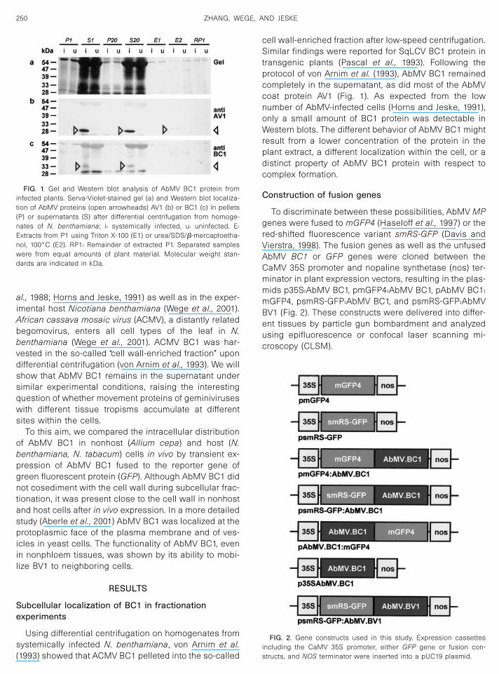
FIG. 3. Patterns of mGFP4, smRS-GFP, mGFP4:AbMV BC1 and smRS-GFP:AbMV BC1 in epidermal cells of onion bulb scales, and of source and sinkleaves of N. tabacum. Cells are shown as composite pictures of CLSM optical sections. False colors represent green fluorescence in different depths of the optical section as indicated by the color code bar at the top left corners. (a) Unfused mGFP4 in an epidermal onion cell at 24 hpb showing fluorescence in the cytoplasm and nucleus. (b–d) mGFP4:AbMV BC1 in onion cells; (b and c) fluorescent flecks at the cell periphery at 18 hpb (b) and at 84 hpb (c). (d) Fluorescent punctate spots surrounding the nucleus (see Fig. 5) at 83 hpb. (e and f) Distribution patterns of smRS-GFP:AbMV BC1 in epidermal cells of tobacco source leaves at 10 hpb (e) and 70 hpb (f) showing the formation of flecks at the cell periphery. (g) Unfused smRS-GFP in epidermal cells of tobacco at 5 dpi showing brighter fluorescence in the central cell and weaker fluorescence in neighboring cells. (h and i) smRS-GFP:AbMV BC1 in epidermal cells of sink leaves in N. tabacum has induced disc-like structures at 76 hpb. Arrowhead indicates stacked discs which were seen throughout several optical sections. (j–k) Distribution of AbMV BC1 after cobombardment with the cognate viral DNAs. Cobombardment of smRS-GFP:AbMV BC1 with AbMV DNA A and B in cells of sink leaves of N. tabacum has induced needle-like structures at 5 dpb (j and j’) and at 8 dpb (k). (j’) Single optical section through the center of the cell in j showing the needle-like structures localized in the cell periphery. Bars represent 50 µm (a–d), 25 µm (e–g), and 10 µm (h–k).

ABUTILON MOSAIC GEMINIVIRUS MOVEMENT PROTEINS 253
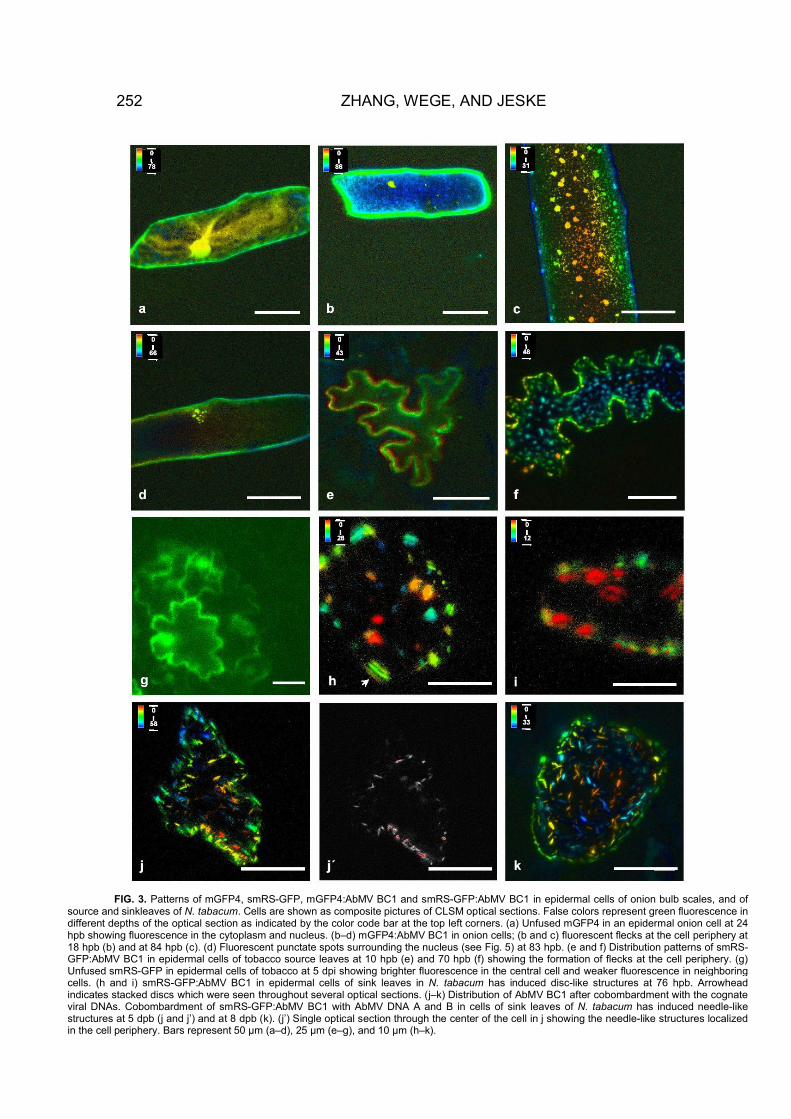
FIG. 5. Distribution of BV1 in relation to BC1 in onion cells (d–f) and sink cells of either N. tabacum (g–i) or N. benthamiana (a–c, j–l). (a–c) Localization of GFP:BC1 surrounding nuclei. (a) One optical section through a nucleus (N). (b) DAPI fluorescence. Note the nucleus (arrow) and the faint green fluorescence at the periphery allowing the identification of the cell borders. (c) The same cell as in (b) with GFP:BC1 fluorescence; arrow indicates the same nucleus as in (b). (d) One nucleus at higher magnification with smRS-GFP:AbMV BV1 accumulating in globular inclusions in the nucleoplasm at 24 hpb. (e, f) and (k, l) The same cells were photographed with different excitation wavelengths to detect mGFP4:BC1, either alone (e, k) or together with smRS-GFP:BV1 (f, l). smRS-GFP:BV1 was either expressed alone (g), or coexpressed with BC1 (h), AbMV DNA A and B (i), or mGFP4:BC1 (j–l). (b, c, e, f) Epifluorescence micrographs using filter cube I (b, e, k) or II (c, f, l). (a, d, g–j) CLSM. Bars represent 10 µm in a–d, g–l, and 50 µm in e, f.
d e f
g h i
j k l
ditions, suggesting that the amount of BC1 is rather low Consistent with these findings, smRS-GFP:AbMVduring natural infection, as expected from Western blot BV1 was predominantly detected in cell nuclei afteranalysis (Fig. 1). biolistic plasmid inoculation and transient expression

in nonhost (Figs. 5 d–5f) as well as in host plants (Figs.5g–5i).
To analyze the relationship between BC1 and BV1, theproteins were fused to two GFPs which can be discrim-inated by their different excitation wavelengths. Whencobombarded into onion cells (Figs. 5e and 5f), BC1 wasdetected either alone (Fig. 5e) or together with BV1 (Fig.5f) in the same cell. BV1 was present in the nucleus,whereas BC1 formed a cap around the nucleus. In ad-dition, peripheral fluorescence close to the cell wall wasobserved during these experiments as shown before.With the chosen constructs it was impossible to deter-mine whether BV1 was present in the peripheral signalstogether with BC1. Therefore, unfused BC1 was coex-pressed with smRS-GFP:AbMV BV1, but the protein didnot redirect GFP:BV1 to the cell periphery in onion cells(data not shown).
The behavior of GFP:BV1 was only altered after bom-bardment of sink cells from host plants (Figs. 5g–5i). Inthis case, GFP:BV1 stayed in the nucleus when ex-pressed alone (Fig. 5g) but moved to the periphery of thecell and to neighboring cells when coexpressed withBC1 (Fig. 5h) or when coinoculated with AbMV DNA Aand B (Fig. 5i). It is improbable that the presence of BV1in adjacent cells is due to two or more hits duringbombardment for the following reasons: First, we rarelydetected such hits during all the other experiments (Ta-
ble 1), only upon coexpression in sink cells. Second, theneighbor cells exhibited a typical feature, namely astrong accumulation of BV1 in the nucleus and only afaint or no signal at the periphery. Third, to provide animpression of the hit frequency after bombardment andto exclude the possibility that two neighboring cells weretargeted just by chance, we performed four additionalexperiments (Table 2) the results of which were consis-tent with the previous findings. Leaves were bombardedwith different construct combinations, and the number oftwo (paired) or more (�two) adjacent fluorescent cellswas compared to the total number of fluorescent cells(Table 2). A low number of paired fluorescent cells (1.3 �0.4%) was found if one of the fusion proteins (GFP:BC1 orGFP:BV1) was expressed alone, which may indicate thebackground probability of hitting two adjacent cells bychance. A significant (�2 � 173; P � �0.0001) increase inthe number of paired cells (7.9 � 1.5%) was observed ifGFP:BV1 plasmids were cobombarded with BC1-contain-ing plasmid constructs (BC1, GFP:BC1, or DNA A andDNA B; Table 2). Moreover, the data underscore the lowprobability of AbMV proteins to be transported to aneighboring cell, which might be one reason for thelimitation of tissue spread during natural infection byAbMV (Wege et al., 2001).
From these results we conclude that GFP:BV1 is mo-bilized by either BC1 or the viral DNA gene products, not
TABLE 1
Distribution Patterns of Fusion Proteins in Allium cepa, Nicotiana tabacum, and N. benthamiana
Plasmid construct PlantTime(pb)
Cells(total)
Distribution patterns of fluorescencea
A B C D E
BC1 alonepmGFP4:AbMV BC1 A. cepa 43 h 63 16 (25) 38 (60) 9 (15)psmRS-GFP:AbMV BC1 45 h 56 11 (20) 39 (70) 6 (10)
Source leaves:psmRS-GFP:AbMV BC1 N. tabacum 48 h 44 9 (20) 25 (57) 10 (23)psmRS-GFP:AbMV BC1 N. benthamiana 60 h 33 3 (9) 27 (82) 3 (9)
Sink leaves:psmRS-GFP:AbMV BC1 N. tabacum 78 h 36 9 (25) 4 (11) 23 (64)psmRS-GFP:AbMV BC1 N. benthamiana 80 h 45 6 (13) 12 (27) 27 (60)
BC1 and cognate viral DNA A and BSource leaves
psmRS-GFP:AbMV BC1 N. tabacum 30 h 42 7 (17) 35 (83)b
5 d 53 5 (9) 48 (91)b
psmRS-GFP:AbMV BC1 N. benthamiana 30 h 38 8 (21) 30 (79)b
5 d 56 7 (12) 49 (88)b
Sink leavespsmRS-GFP:AbMV BC1 N. tabacum 2 d 45 6 (13) 11 (24) 28 (62)
3 d 38 6 (16) 32 (84)psmRS-GFP:AbMV BC1 N. benthamiana 30 h 46 9 (20) 9 (20) 28 (60)
5 d 67 13 (19) 54 (81)
a Number (in parentheses as a percentage) of cells with the respective distribution pattern of fluorescence. (A) evenly distributed fluorescence atcell periphery; (B) punctate flecks at cell periphery; (C) punctate flecks around the nucleus; (D) disc-like structure at cell periphery; (E) needle-likestructures at cell periphery.
b In these experiments the B and C type were not distinguished.
254 ZHANG, WEGE, AND JESKE

only to the cell periphery, but also to the next cell whereit is released and transported to the nucleus. This pro-cess is even more prominent after cobombardment withDNA A and B (Fig. 5i; Table 2), probably because BC1can also be expressed in the neighboring cells underthese conditions. Under the chosen conditions, we neverdetected more than two adjacent fluorescent cells if theviral proteins were ectopically expressed from plasmids,but this occurred reproducibly, although again with a lowfrequency, if full-length viral DNA A and B were cobom-barded (Table 2; �two).
To test whether the GFP moiety in BC1 constructshinders their function, rsGFP:BV1 was coexpressed witheither BC1, GFP:BC1, or BC1:GFP in N. benthamiana sinkleaves. Under these conditions BV1 was mobilized toneighboring cells (Figs. 5j and 5i) and no statisticallysignificant difference between the effects of the BC1constructs with or without GFP fusion was detected(Table 2; 7.3 � 1.5% for BC1 and 7.4 � 1.4% for GFP:BC1).Moreover, GFP:BC1 (Fig. 5k) and BC1:GFP (data notshown) also appeared in the adjacent cells. Therefore,we conclude that the GFP fusion is not the reason for thelack of BC1 movement in all of the other experiments. Onthe contrary, we suggest that complex formation of BV1and BC1 triggers the transport of both proteins as far asthey are expressed in certain competent cells.
DISCUSSION
GFP has been used as a reporter to trace the subcel-lular and intercellular behavior of the MPs of a bipartitegeminivirus in nonhost and host plants. In addition, wehave compared the distribution of MPs in source andsink cells of host plants. In contrast to the injection ofbacterially expressed proteins, biolistic delivery ofGFP:MP fusion gene constructs promises a better fold-ing of the encoded proteins and increases the chance ofobtaining a physiologically significant posttranslationalmodification pattern (Itaya et al., 1997). In particular,phosphorylation might play a crucial role in the regula-tion of viral movement (Sanderfoot and Lazarowitz, 1996;von Arnim et al., 1993; Wege and Jeske, 1998).
The results presented here show that the AbMV BC1accumulated mainly as punctate spots at the cell cortex,similar to the immunolocalized SqLCV BC1 (Sanderfootand Lazarowitz, 1995, 1996; Sanderfoot et al., 1996), andalso close to the nucleus in certain cells. Nevertheless,AbMV BC1 remained soluble after low-speed centrifuga-tion, in the supernatant which contains free proteins as
FIG. 4. Immunological detection of AbMV proteins AV1 and BV1 innuclei. Consecutive sections from a systemically infected Abutilonsellovianum stem node with axillary bud ((a) overview; (b–e) magnifiedconsecutive sections; filled arrowheads indicate the same nucleus)).Proteins were detected using AV1 (a, c, d) and BV1 (b) antiserum, orpreimmune serum control (preBV1) (e). Arrows and arrowheads indi-cate AV1- or BV1-specific violet stain precipitated from NBT/BCIP byalkaline phosphatase. Nuclear localization of the violet signals was
confirmed by DAPI counterstain (not shown). Note that AV1 and BV1were present in the same nucleus in consecutive sections (a, b, and carrowheads). The concentration of BC1 was too low to be detectedunder these conditions. Bars represent 50 �m ((b-e) at the samemagnification)).
255ABUTILON MOSAIC GEMINIVIRUS MOVEMENT PROTEINS

well as microsomes, including plasma membrane vesi-cles (Nagahashi, 1985).
The basic site of accumulation of AbMV BC1, whetherit is close to the cell periphery or the nucleus, wassimilar in host and nonhost cells, suggesting that host-specific factors are not involved in this fundamental pro-cess. Interestingly, we found a plasma membrane local-ization of BC1 even in fission yeast cells (Aberle et al.,2001). Apart from this general feature, the pattern of BC1accumulation within the cells depended considerably onthe developmental stage of the plant in the case ofAbMV. GFP:BC1 induced stacked disc-like structures inepidermal, trichome, and guard cells of sink leaves (Fig.3). This feature was completely absent from cells ofsource leaves. Ward et al. (1997) provided evidence thatBC1-containing tubules might be derived from endoplas-mic reticulum. Localization of BC1 protein close to thenucleus as a second main site of accumulation has notbeen reported so far for other geminiviruses. Very rarely,cells with both sites of accumulation were found, and weassume that the cell physiology governs the distributionof BC1. The change of sites might be regulated by post-translational modifications. Phosphorylation has beenshown for BC1 proteins (von Arnim et al., 1993; Pascal etal., 1994; Wege and Jeske, 1998). Wege and Jeske (1998)showed that AbMV BC1 was phosphorylated in Esche-richia coli, and the pattern of phosphorylation might bedifferent in eukaryotes, thus leading to different targetingof the proteins. Moreover, phosphorylation might be de-
velopmentally regulated, explaining the divergent distri-bution patterns in sink compared to source cells.
In all experiments, whether in host or nonhost plants,in source or sink cells, AbMV GFP:BC1 or BC1:GFP nevermoved to the neighboring cells if expressed alone. Evencobombardment with DNA A and B did not mobilizethese proteins in mature cells. This behavior contrasts tothat of unfused GFP which diffused to the neighboringsink cells (Fig. 3g) as has been reported by Oparka et al.(1999). Presumably the fusion proteins are too large forpassive transport through plasmodesmata. Fluorescentlylabeled, unfused BDMV BC1 was found in neighboringcells after microinjection (Noueiry et al., 1994; Rojas etal., 1998).
The mobilization of GFP:BC1 and GFP:BV1 exclusivelyin cells of sink leaves in host plants suggests that GFPdoes not greatly hinder BC1 and BV1 in this process. BV1proteins of SqLCV and Tomato leaf curl virus (ToLCV)were localized exclusively to the nuclei in infected plants(Pascal et al., 1994) and in transfected protoplasts(Padidam et al., 1999) as confirmed here for AbMV. It hasbeen proposed that BV1 accumulates in nuclei becausenuclear import is faster than export (Lazarowitz andBeachy, 1999; Gerace, 1995). In protoplasts, SqLCV BV1was mobilized to the cell periphery after coexpressionwith the cognate BC1 (Sanderfoot and Lazarowitz, 1995,1996; Sanderfoot et al., 1996). Here we show for the firsttime that BV1 protein is additionally transported to thenucleus of an adjacent cell if coexpressed with BC1.
Evidence from BDMV experiments indicated that bothBC1 and BV1 are nucleic acid-binding proteins and rec-ognize ssDNA and dsDNA in a form- and size-specificmanner (Noueiry et al., 1994; Rojas et al., 1998). Twomodels of the interaction of viral DNA with the movementproteins have been discussed. BV1 might carry viral DNAinto the cytoplasm where it is displaced by BC1, whichthen transports the nucleic acid through the plasmodes-mata (Noueiry et al., 1994; Carrington et al., 1996). Ac-cordingly, BC1 was shown to mediate cell-to-cell move-ment of fluorescently labeled ssDNA and dsDNA in mi-croinjection studies. In contrast, BV1 was only able toexport injected DNA from the nucleus (Noueiry et al.,1994; Rojas et al., 1998).
In the case of SqLCV, BC1 directed BV1 from thenucleus to the cell periphery, suggesting that BC1 inter-acts with BV1 in vivo. SqLCV BC1 did not bind eitherssDNA or dsDNA in vitro, whereas BV1 bound ssDNA.Based on this evidence, a movement model has beenproposed in which BC1 mediates the transport of BV1–ssDNA complexes via tubules transiently induced byvirus infection in developing phloem tissues (Pascal etal., 1994; Ward et al., 1997; Lazarowitz and Beachy, 1999).The apparent discrepancy between the two models hasbeen explained by phloem-limitation of SqLCV, whereasBDMV can also infect nonvascular tissues (Hoefert, 1987;Wang et al., 1996; Lazarowitz and Beachy, 1999).
TABLE 2
Quantification of Transformation Efficiencies
TreatmentExpt.No.
Fluorescent cells Percentage
Total Paired �Two Paired �Two
GFP:BC1 1 525 8 0 1.5 0.02 459 7 0 1.5 0.03 507 9 0 1.8 0.04 392 4 0 1.0 0.0
GFP:BV1 1 465 3 0 0.6 0.02 330 3 0 0.9 0.03 123 2 0 1.6 0.04 396 5 0 1.3 0.0
BC1 � GFP:BV1 1 425 41 0 9.6 0.02 371 20 0 5.4 0.03 684 49 0 7.2 0.04 760 53 0 7.0 0.0
GFP:BC1 � GFP:BV1 1 253 14 0 5.5 0.02 382 33 0 8.6 0.03 419 31 0 7.4 0.04 277 22 0 7.9 0.0
DNA A � DNA B� GFP:BV1 1 225 21 2 9.3 0.9
2 463 39 3 8.4 0.63 624 57 6 9.1 1.04 659 62 8 9.4 1.2
256 ZHANG, WEGE, AND JESKE

The observation of AbMV BC1 protein accumulating atthe outer surface of the nucleus is a new observationand presumably allows further detail of the process to bevisualized. Based on this observation and the detectionof BV1 movement to adjacent cells, we propose that BC1might fetch complexes of viral DNA and BV1 from theouter side of the nuclear envelope and transfer them toplasmodesmata and then to the adjacent cell where theBV1–DNA complex is released for the subsequent nu-clear import. As an extension to previous models ofgeminiviral transport, it seems that, at least for AbMV, thetrigger for cell-to-cell transport of BC1 as well as BV1needs the interaction of both proteins. Moreover, cell-to-cell transport occurs only in certain competent cells ofsink leaves. Here we have shown a limited transport ofGFP:BV1 and GFP:BC1 to adjacent (mostly) epidermalcells which are not normally infected by AbMV. Thesignificance of these observations for the phloem-limitedviral infection has to be established, but the low fre-quency of cell-to-cell movement of GFP:BV1 might con-tribute to an explanation for the low percentage of AbMV-infected cells in a variety of hosts (Wege et al., 2001). Onthe other hand, although AbMV BC1 and/or BV1 may beless efficient for transport than the corresponding pro-teins of other geminiviruses, the results show that theAbMV proteins still possess, in principle, the ability tomove from cell to cell in nonphloem cells.
MATERIALS AND METHODS
Subcellular fractionation
Leaf and upper stem tissue from fully symptomatic,AbMV-infected, or from uninfected N. benthamiana wereused for differential centrifugation analyses according tovon Arnim et al. (1993). The tissue was homogenized inliquid nitrogen, and protein was extracted in 2-ml grind-ing buffer (GB; von Arnim et al., 1993) per gram startingmaterial. The homogenate was centrifuged for 10 min at1000 g using a swing-out rotor to yield pellet P1 andsupernatant S1. The latter was centrifuged at 20,000 g for20 min to produce pellet P20 and supernatant S20. PelletP1 was washed with GB and resuspended in 1–2 mlGB/Triton (1% Triton X-100 in GB) per gram of startingmaterial. Following centrifugation at 1000 g for 10 min,the supernatant was removed and named extract E1; theresidual pellet washed once in GB/Triton, resuspendedin 1–2 ml ESB per gram starting material (von Arnim etal., 1993), boiled for 5 min, and centrifuged again toproduce the supernatant extract E2 and its correspond-ing pellet RP1 (remainder P1). For Western blot analyses,pellets P1, P20, and RP1 were suspended in GB andsuitable aliquots of these suspensions as well as of thedifferent supernatants were mixed with equal amounts of2� SDS sample buffer (Sambrook et al., 1989), boiled for10 min, centrifuged at 20,000 g for 1 min, and run ondiscontinuous SDS polyacrylamide gels (12.5% polyacryl-
amide; Laemmli, 1970). The samples separated in eitherlane were derived from equal amounts of plant material.Protein molecular weight standards were from Pharma-cia or Serva. Gels were either fixed and stained withServa-violet 17 (Serva), or proteins were transferredonto nitrocellulose membrane (Schleicher & Schuell,PROTRAN 0.45 �m) by Western blotting (Towbin et al.,1979; Trans-Blot SD, Bio-Rad, Hercules, CA). Air-driedmembranes were stained for 5 min in 0.2% Ponceau S in3% acetic acid and destained in water.
Immunodetection procedures were modified from deMaio (1994) and Harlow and Lane (1988). Following a 1 hblocking step in 3% BSA in PBS pH 7.2 at room temper-ature, membranes were agitated for 2 � 5 min in PBS-T(0.1% Tween 20 (Sigma) in PBS pH 7.2), 90 min in primaryantibody solution (polyclonal antiAbMV antisera or pre-immune sera diluted 1:1000 in 2.5% BSA, 0.02% NaN3 inPBS), 5 min in 1% NP-40 in PBS, 4 � 5 min in PBS-T, 60min in secondary antibody solution (goat anti-mouseimmunoglobulin G, IgG-alkaline phosphatase conjugate,Biotrend) diluted 1:2000 in AP-dilution buffer (1% BSA in100 mM Tris–Cl pH 7.5, 150 mM MgCl2), 6 � 5 min inTBS-T (0.1% Tween 20 in 25 mM Tris–Cl pH 7.4, 150 mMNaCl), and 5 min in alkaline AP buffer (100 mM Tris–ClpH 9.5, 100 mM NaCl, 50 mM MgCl2). Blots were trans-ferred to detection solution (alkaline AP buffer containing300 �g/ml NBT (Gibco-BRL) and 165 �g/ml BCIP (Gibco-BRL)) and stain precipitation was stopped after 5–15 minby several rinses in 0.5 mM EDTA.
Construction of translational fusion genes andmolecular cloning
Standard molecular cloning procedures were appliedaccording to Sambrook et al. (1989).GFP genes. mGFP4 was the version of GFP (Chalfie et
al., 1994) according to Haseloff et al. (1997), smRS-GFPon plasmid pCD-327 according to Davis and Vierstra(1998), kindly provided by the authors. The plasmidscontain the genes flanked by the CaMV 35S promoterand nos terminator inserted into pBIN19 and pUC119,respectively. mGFP4 and smRS-GFP can be distin-guished by their excitation wavelength during fluores-cence microscopy (see below).Viral fusion constructs. An EcoRI–HindIII fragment
from pBIN35S-mGFP4 (Haseloff et al., 1997) was insertedbetween the corresponding restriction sites of pUC19,giving rise to pmGFP4. pmGFP4 was first digested withEcoRI, filled in with Klenow enzyme, and religated toremove the EcoRI site resulting in pmGFP4�EcoRI. Thisplasmid was amplified with primers 1 and 2 (Table 3).The PCR product was digested with SstI and religatedresulting in pmGFP4(E-S-X). To fuse mGFP4 with AbMVBC1, it was amplified from plasmid AbB (Frischmuth etal., 1990) with primers 3 and 4. The PCR fragment was
257ABUTILON MOSAIC GEMINIVIRUS MOVEMENT PROTEINS

digested with EcoRI and XhoI and inserted intopmGFP4(E-S-X), resulting in pmGFP4:AbMV BC1.
A BamHI–NspV fragment from psmRS-GFP, containingthe smRS-GFP gene (Davis and Vierstra, 1998), wasexchanged with that of pmGFP4(E-S-X), giving rise topsmRS-GFP(E-S-X). The PCR fragment of the AbMV BV1gene, amplified from plasmid AbB (Frischmuth et al.,1990) with primer 5 and 6, was digested with EcoRI andXhoI and inserted into psmRS-GFP(E-S-X), resulting inpsmRS-GFP:AbMV BV1. An EcoRI–XhoI fragment ofAbMV BC1, cut from pmGFP4:AbMV BC1, was also in-serted into psmRS-GFP(E-S-X), resulting in psmRS-GFP:AbMV BC1.
For 5�-terminal fusion of GFP with AbMV BC1, theconstruct was produced by two PCR steps according toHiguchi et al. (1988) as modified by Wurch et al. (1998).Primers 7 and 8 were used to amplify the AbMV BC1gene from plasmid AbB. Primers 9 and 10 were used toamplify mGFP4 and the nos-terminator from pmGFP4.The amplified AbMV BC1 and mGFP4 fragments werefused in frame using an extension polymerization. Theresulting fragment was amplified with primers 7 and 10by a second PCR. It was digested with BamHI and EcoRIand exchanged for GFP in pmGFP4, resulting in pAbMVBC1:mGFP4.E. coli (strain DH5� or JM83) was transformed with the
resulting plasmids. Purified plasmids from isolated col-onies were digested with suitable restriction enzymes tocheck the size of inserts. The constructs were se-quenced with semiautomatic sequencing (Li-Cor DNA-Sequencer 4000L; MWG, Germany). Correct plasmidswere amplified in E. coli and purified for biolistic tran-sient transformation using NUCLEO-BOND cartridges AX500 (Macherey-Nagel, Germany).
Biolistic inoculation of plant tissues
Sixty milligrams of 1.0 �m gold particles (Bio-Rad)were washed twice with 70% ethanol, once in sterilewater with moderate vortexing for 10 min and suspended
in 1 ml 50% (v/v) glycerol. Five microliters of plasmid DNA(1 �g/�l) harboring either GFP alone as a control orfusion constructs were mixed with 25 �l gold suspensionby vortexing for 3 min and kept on ice for 10 min. Tenmicroliters 0.1 M spermidine and 25 �l 2.5 M CaCl2 wereadded and vortexed for 4 min. DNA-coated gold particleswere collected by a brief centrifugation, washed twicewith 70% ethanol, and resuspended in 36 �l 98% ethanol.Six microliters of the suspension was spread on eachplastic carrier disc.
Onion bulb scales were cut into pieces of 1.5 � 1.5 cmand kept in Petri dishes. Completely expanded and smallyoung sink leaves (N. tabacum Samsun nn or N.benthamiana) were cut from 6- to 8-week-old plantswhich were grown in the glasshouse and laid immedi-ately with the lower side up on a sterile wet Whatmanfilter in a plastic Petri dish. The freshly cut ridges werecovered with small pieces of wet napkin paper. Sink orsource status of leaves was determined by monitoring inparallel the translocation of 5(6)-carboxyfluorescein diac-etate (CF) according to Roberts et al. (1997).
Plasmid DNA was delivered into the lower side ofleaves or into the inner peels of bulb scales using aparticle gun (PDS-1000/He; Bio-Rad) using either 900 psirupture discs in the case of leaves (Marc et al., 1998) or1100 psi rupture discs in the case of bulb scales under avacuum of 25 in. Hg. For cobombardment of differentconstructs the plasmids (encoding BC1, BV1, or DNA Aand DNA B) were mixed before gold-coating to deliverthem into the same cell with similar efficiency. Afterbombardment, explants were kept in the dark at 24 or4°C.
Fluorescence microscopy.
For routine observation an Axioskop (Zeiss Corp., Ger-many) was used including filter cube I (365-nm excita-tion; 395-nm beam splitter; 429-nm long-pass emission)for mGFP4 fluorescence and filter cube II (450- to 490-nmexcitation; 510-nm beam splitter; 520-nm band-pass
TABLE 3
Primers Used in Gene Construction
No. Name Sequence (5� to 3�)a Remarks
1 NOS-SstI-XhoI-FOR TAAGAGCTCGAGTTTCCCCGATCGTTCAAACATTTG 5� end of nos. terminator � SstI and XhoI2 mGFP4-SstI-EcoRI-REV GCAGAGCTCGAATTCATCCATGCCATGTGTAATCCC 3� end of gfp � SstI and EcoRI3 AbMVB-EcoRI-BC1-FOR CTTGAATTCATGGATTCTCAGTTAGTAAAT 5� end of AbMV BC1 � EcoRI4 AbMVB-XhoI-BC1-REV AATCTCGAGTTATTTCAATGATTTGGCTTG 3� end of AbMV BC1 � XhoI5 AbBV1-EcoRI-FOR CCCTTGAATTCATGTACCCGTCTAGGAATAAACG 5� end of AbMV BV1 � EcoRI6 AbBV1-XhoI-REV CCCTTCTCGAGTTAACCAATATAGTCAAGGTC 3� end of AbMV BV1 � XhoI7 FOR-AbBC1 CAAACGGATCCAACAATGGATTCTCAGTTAGTAAATCC 5� end of AbMV BC1 � BamHI8 REV-AbBC1-GFP GTTCTTCTCCTTTACTCATTTTCAATGATTTGGCTTG 3� end of AbMV BC1 and 5� end of mgfp4;
complementary to No. 99 FOR-GFP-AbBC1 CAAGCCAAATCATTGAAAATGAGTAAAGGAGAAC 3� end of AbMV BC1 and 5� end of mgfp4
10 REV-GFP-EcoRI CGGCCAGTGAATTCCCGATCTAGTAACA Sequence of pUC19 downstream of gfp
a Added recognition sites for restriction enzymes are underlined.
258 ZHANG, WEGE, AND JESKE

emission) for smRS-GFP. Photographs were taken usingan MC-100 Spot-Camera (Zeiss Corp.) and FujichromeProvia 400 daylight positive film.
Confocal laser scanning microscopy and imageprocessing
Confocal laser scanning microscopy was done using aLSM model 410 invert (Zeiss Corp.). Fluorescent cellswere imaged using a 488-nm argon laser, 670- to 810-nmband filter for red fluorescence of chloroplasts and a 510-to 540-nm emission filter for GFP. Optical sections at 0.5-to 2-�m intervals were made, attenuating the laser in-tensity to the lowest possible to reduce photobleachingof GFP.
The collected optical sections were digitally pro-cessed and assembled by use of the manufacturer’ssoftware (Carl Zeiss LSM Program, Germany). The greenfluorescence was transformed to false colors to indicatethe depth of the optical sections, resulting in pseudo-three-dimensional pictures.
Immunolocalization of AbMV BV1 in paraffin sections
Leaf segments and axillary bud explants from A. sell-ovianum were fixed at room temperature for 1 h in 4%formaldehyde, freshly prepared from paraformaldehyde,in MTSB (microtubule stabilizing buffer: 50 mM PIPES pH6.9 with KOH, 5 mM EGTA, 5 mM MgSO4), and 8 to 14 hat 10°C in 6% formaldehyde in 0.15 M sodium phosphatebuffer pH 7.6. Specimens were agitated at room temper-ature for 2 � 15 min in phosphate buffer, dehydrated ina graded series of ethanol, and infiltrated with Histo-Clear (National Diagnostics):ethanol 1:1 for 1 h, withHisto-Clear 3 � 1 h at room temperature and with Histo-Clear:Paraplast Xtra (Oxford Labware, Sherwood Medi-cal) overnight at 42°C. Infiltration with pure ParaplastXtra was performed at 58°C for 3 days, replacing the waxonce per day. Specimens were solidified on ice, sections(5–10 �m) were collected on silane-coated slides (bind-silane GF31, Wacker), and paraffin was removed by ex-traction with Roticlear (Roth), isopropanol, and ethanol.
Immunodetection procedures were carried out atroom temperature. Sections were rehydrated 2 � 5 minin PBS, incubated for 15 min in 50 mM glycine in PBS,2 � 5 min in PBS, 10 min in blocking solution A (3% BSA,5% normal goat serum (Jackson ImmunoResearch) inPBS), 10 min in blocking solution B (0.2% gelatin (Merck),0.5% BSA in PBS), 2 � 3 min in PBS-T and 2.5 h in primaryantibody solution (polyclonal mouse antiserum or preim-mune serum (Wege and Jeske, 1998), diluted 1:3000 in3% BSA in PBS). Following 3 � 7 min washes with 1%NP-40 (Fluka) in PBS and 2 � 10 min in TBS-T, sectionswere subjected to secondary antibody solution (goatanti-mouse immunoglobulin G, H&L, IgG-alkaline phos-phatase conjugate (Biotrend), diluted 1:250 in AP-dilutionbuffer (1% BSA in 100 mM Tris–HCl pH 7.5, 150 mM
MgCl2) for 1 h). The slides were washed 4 � 5 min inTBS-T, 2 � 5 min in detection buffer 1 (0.1 M maleic acid,0.15 M NaCl pH 7.5), and 5 min in alkaline AP buffer. Thelatter was replaced by detection solution (see above);stain development was observed microscopically andstopped after 5 to 10 min by rinsing in 0.5 mM EDTA. Forvisualization of DNA, sections were counterstained withDAPI (1 �g/ml in water) for 5 min and mounted withCitifluor (Plano). Specimens were analyzed with Zeiss-Axioskop epifluorescence microscopes using differentialinterference contrast (DIC) equipment to enhance con-trast. Pictures were taken on Kodak Ektachrome 64Tfilms with Zeiss-Axiophot photo equipment.
ACKNOWLEDGMENTS
This work was supported by a grant from the Deutsche Forschungs-gemeinschaft (Je 116/5-3). We thank Prof. Dr. R. Ghosh, PD Dr. T.Frischmuth, and Dr. B. Ding for helpful discussions, Dr. Haseloff forpmGFP4, Drs. Davis and Vierstra for pCD-327, Prof. Dr. H.-D. Gortz forproviding the CLSM facilities, and Dr. J. Brenner and Dr. M. Schweikertfor help with the laser scanning microscopy.
REFERENCES
Aberle, H.-J., Rutz, M.-L., Karayavuz, M., Frischmuth, S., Wege, C.,Hulser, D., and Jeske, H. (2001). Localizing the movement proteins ofAbutilon mosaic geminivirus in yeast by subcellular fractionation andfreeze fracture immuno-labeling. Arch. Virology, in press.
Abouzid, A. M., Barth, A., and Jeske, H. (1988). Immunogold labeling ofthe Abutilon mosaic virus in ultrathin sections of epoxy resin embed-ded leaf tissue. J. Ultrastruct. Res. 99, 39–47.
Carrington, J. C., Kasschau, K. D., Mahajan, S. K., and Schaad, M. C.(1996). Cell-to-cell and long-distance transport of viruses in plants.Plant Cell 8, 1669–1681.
Chalfie, M., Tu, Y., Euskirchen, G., Ward, W. W., and Prasher, C. (1994).Green fluorescent protein as a marker for gene expression. Science263, 802–805.
Citovsky, V., Knorr, D., Schuster, G., and Zambryski, P. (1990). The P30movement protein of tobacco mosaic virus is a single-strand nucleicacid binding protein. Cell 60, 637–647.
Davis, S. J., and Vierstra, R. D. (1998). Soluble, highly fluorescentvariants of green fluorescent protein (GFP) for use in higher plants.Plant Mol. Biol. 32, 521–528.
de Maio, A. (1994). Protein blotting and immunoblotting using nitrocel-lulose membranes. In “Protein Blotting. A Practical Approach” (Dun-bar, B. S., Ed.), pp. 11–32. IRL Press, New York.
Ding, B., Itaya, A., and Woo, Y.-M. (1999). Plasmodesmata and cell-to-cell communication in plants. Int. Rev. Cytol. 190, 251–316.
Etessami, P., Callis, R., Ellwood, S., and Stanley, J. (1988). Delimitationof essential genes of cassava latent virus DNA 2. Nucleic Acids Res.16, 4811–4829.
Evans, D., and Jeske, H. (1993). DNA B facilitates, but is not essentialfor, the spread of Abutilon mosaic virus in agroinoculated Nicotianabenthamiana. Virology 194, 752–757.
Frischmuth, T., Zimmat, G., and Jeske, H. (1990). The nucleotide se-quence of abutilon mosaic virus reveals prokaryotic as well aseukaryotic features. Virology 178, 461–468.
Fujiwara, T., Giesman-Cookmeyer, D., Ding, B., Lommel, S. A., andLucas, W. J. (1993). Cell-to-cell trafficking of macromolecules throughplasmodesmata potentiated by the red clover necrotic mosaic virusmovement protein. Plant Cell 5, 1783–1794.
Gerace, L. (1995). Nuclear export signals and the fast track to thecytoplasm. Cell 82, 341–344.
259ABUTILON MOSAIC GEMINIVIRUS MOVEMENT PROTEINS

Harlow, E., and Lane, D. (1988). “Antibodies.” Cold Spring Harbor Lab-oratory Press, Cold Spring Harbor, NY.
Haseloff, J., Siemering, K. R., Prasher, D. C., and Hodge, S. (1997).Removal of a cryptic intron and subcellular localization of greenfluorescent protein are required to mark transgenic Arabidopsisplants brightly. Proc. Natl. Acad. Sci. USA 94, 2122–2127.
Higuchi, R., Krummel, B., and Saiki, R. K. (1988). A general method of invitro preparation and specific mutagenesis of DNA fragments: Studyof protein and DNA interactions. Nucleic Acids Res. 16, 7351–7367.
Hoefert, L. L. (1987). Association of Squash leaf curl virus with nuclei ofsquash vascular cells. Phytopathology 77, 1596–1600.
Horns, T., and Jeske, H. (1991). Localization of Abutilon mosaic virusDNA within leaf tissue by in situ hybridization. Virology 181, 580–588.
Itaya, A., Hickman, H., Bao, Y., Nelson, R. S., and, Ding, B. (1997).Cell-to-cell trafficking of cucumber mosaic virus movement protein:Green fluorescent protein fusion produced by biolistic gene bom-bardment in tobacco. Plant J. 12, 1223–1230.
Jeske, H., Menzel, D., and Werz, G. (1977). Electron microscopic studieson intranuclear virus-like inclusions in mosaic-diseased Abutilonsellowianum Reg. Phytopath. Z. 89, 289–295.
Kasteel, D. T. J., van der Wel, N. N., Jansen, K. A. J., Goldbach, R. W., andvan Lent, J. W. M. (1997). Tubule-forming capacity of the movementproteins of alfalfa mosaic virus and brome mosaic virus. J. Gen. Virol.78, 2089–2093.
Laemmli, U. K. (1970). Cleavage of structural proteins during the as-sembly of the head of bacteriophage T4. Nature 227, 680–685.
Lazarowitz, S. G. (1999). Probing plant cell structure and function withviral movement proteins. Curr. Opin. Plant Biol. 2, 332–338.
Lazarowitz, S. G., and Beachy, R. N. (1999). Viral movement proteins asprobes for intracellular and intercellular trafficking in plants. PlantCell 11, 535–548.
Marc, J., Granger, C. L., Brincat, J., Fisher, D. D., Kao, T., McCubbin,A. G., and Cyr, R. J. (1998). A GFP-MAP4 reporter gene for visualizingcortical microtubule rearrangements in living epidermal cells. PlantCell 10, 1927–1940.
Nagahashi, G. (1985). The marker concept in cell fractionation. In“Modern Methods of Plant Analysis: Cell Components” (Linskens,H. F., and Jackson, J. F., Eds.), pp. 66–84. Springer-Verlag, Berlin/Heidelberg.
Noueiry, A. O., Lucas, W. J., and Gilbertson, R. L. (1994). Two proteins ofa plant DNA virus coordinate nuclear and plasmodesmatal transport.Cell 76, 925–932.
Oparka, K. J., Roberts, A. G., Boevink, P., Santa Cruz, S., Roberts, I.,Pradel, K. S., Imlau, A., Kotlizky, G., Sauer, N., and Epel, B. (1999).Simple, but not branched, plasmodesmata allow the nonspecifictrafficking of proteins in developing tobacco leaves. Cell 97, 743–754.
Osman, T. A., Hayes, R. J., and Buck, K. W. (1992). Cooperative bindingof the red clover necrotic mosaic virus movement protein to single-stranded nucleic acids. J. Gen. Virol. 73, 223–227.
Padidam, M., Beachy, R. N., and Fauquet, C. M. (1999). A phagesingle-stranded DNA (ssDNA) binding protein complements ssDNAaccumulation of a geminivirus and interferes with viral movement.J. Virol. 73, 1609–1616.
Pascal, E., Goodlove, P. E., Wu, L. C., and Lazarowitz, S. G. (1993).Transgenic tobacco plants expressing the geminivirus BL1 proteinexhibit symptoms of viral disease. Plant Cell 5, 795–807.
Pascal, E., Sanderfoot, A. A., Ward, B. M., Medville, R., Turgeon, R., andLazarowitz, S. G. (1994). The geminivirus BR1 movement proteinbinds single-stranded DNA and localizes to the cell nucleus. PlantCell 6, 995–1006.
Perbal, M. C., Thomas, C. L., and Maule, A. J. (1993). Cauliflower mosaicvirus gene I product (P1) forms tubular structures which extend fromthe surface of infected protoplasts. Virology 195, 281–285.
Qin, S. W., Ward, B. M., and Lazarowitz, S. G. (1998). The bipartitegeminivirus coat protein aids BR1 function in viral movement byaffecting the accumulation of viral single-stranded DNA. J. Virol. 72,9247–9256.
Roberts, A. G., Santa Cruz, S., Roberts, I. M., Prior, D. A. M., andTurgeon, R. (1997). Phloem unloading in sink leaves of Nicotianabenthamiana: Comparison of a fluorescent solute with a fluorescentvirus. Plant Cell 9, 1381–1396.
Rojas, M. R., Noueiry, A. O., Lucas, W. J., and Gilbertson, R. L. (1998).Bean dwarf mosaic geminivirus movement proteins recognize DNAin a form- and size-specific manner. Cell 95, 105–113.
Rybicki, E. P., Briddon, R. W., Brown, J. K., Fauquet, C. M., Maxwell, D. P.,Harrison, B. D., Markham, P. G., Bisaro, D. M., Robinson, D., andStanley, J. (2000). Family Geminiviridae. In “Virus Taxonomy” (vanRegenmortel, M. H. V., Fauquet, C. M., and Bishop, D. H. L., Eds.),Classification and nomenclature of viruses. pp. 285–297. AcademicPress, San Diego.
Sambrook, J., Fritsch, E. F., and Maniatis, T. (1989). “Molecular Cloning:A Laboratory Manual.” Cold Spring Laboratory Harbor Press, ColdSpring Harbor, NY.
Sanderfoot, A. A., Ingham, D. J., and Lazarowitz, S. G. (1996). A viralmovement protein as a nuclear shuttle. The geminivirus BR1 move-ment protein contains domains essential for interaction with BL1 andnuclear localization. Plant Physiol. 110, 23–33.
Sanderfoot, A. A., and Lazarowitz, S. G. (1995). Cooperation in viralmovement: The geminivirus BL1 movement protein interacts withBR1 and redirects it from the nucleus to the cell periphery. Plant Cell7, 1185–1194.
Sanderfoot, A. A., and Lazarowitz, S. G. (1996). Getting it together inplant virus movement: Cooperative interactions between bipartitegeminivirus movement proteins. Trends Cell Biol. 6, 353–358.
Scott, A., Wyatt, S., Tsou, P. L., Robertson, D., and Allen, N. S. (1999).Model system for plant cell biology: GFP imaging in living onionepidermal cells. Biotechniques 26, 1125–1132.
Storms, M. M. H., Kormelink, R., Peters, D., Lent, J. W. M. V., andGoldbach, R. W. (1995). The nonstructural NSm protein of tomatospotted wilt virus induces tubular structures in plant and insect cells.Virology 214, 485–493.
Towbin, H., Staehelin, T., and Gordon, J. (1979). Electrophoretic transferof proteins from polyacrylamide gels to nitrocellulose sheets: Proce-dure and some applications. Proc. Natl. Acad. Sci. USA 76, 4350–4354.
von Arnim, A., Frischmuth, T., and Stanley, J. (1993). Detection andpossible functions of African cassava mosaic virus DNA B geneproducts. Virology 192, 264–272.
Wang, H. L., Gilbertson, R. L., and Lucas, W. J. (1996). Spatial andtemporal distribution of bean dwarf mosaic geminivirus in Phaseolusvulgaris and Nicotiana benthamiana. Phytopathology 86, 1204–1214.
Ward, B. M., Medville, R., Lazarowitz, S. G., and Turgeon, R. (1997). Thegeminivirus BL1 movement protein is associated with endoplasmicreticulum-derived tubules in developing phloem cells. J. Virology 71,3726–3733.
Wege, C., and Jeske, H. (1998). Abutilon mosaic geminivirus proteinsexpressed and phosphorylated in Escherichia coli. J. Phytopathol.146, 613–621.
Wege, C., Gotthardt, R.-D., and Jeske, H. (2000). Fulfilling Koch’s pos-tulates for Abutilon mosaic virus. Arch. Virol. 145, 2217–2225.
Wege, C., Saunders, K., Stanley, J., and Jeske, H. (2001). Comparativeanalysis of tissue tropism of bipartite geminiviruses. J. Phytopathol.149, 359–368.
Wellink, J., van Lent, J. W., Verver, J., Sijen, T., Goldbach, R. W., and vanKammen, A. (1993). The cowpea mosaic virus M RNA-encoded 48-kilodalton protein is responsible for induction of tubular structures inprotoplasts. J. Virol. 67, 3660–3664.
Wolf, S., Deom, C. M., Beachy, R. N., and Lucas, W. J. (1989). Movementprotein of tobacco mosaic virus modifies plasmodesmata size ex-clusion limit. Science 246, 377–379.
Wurch, T., Estienne, F., and Pauwels, P. J. (1998). A modified overlapextension PCR method to create chimeric genes in the absence ofrestriction enzymes. Biotechniques 12, 653–657.
260 ZHANG, WEGE, AND JESKE