Tomato yellow leaf curl virus: A whitefly-transmitted geminivirus with a single genomic component
11
VIROLOGY 185, 151.-161 (1991) Tomato Yettow Leaf Curl Virus: A Whitefly-Transmitted Geminivirus with a Single Genomic Component NIR NAVOT,’ ERAN PICHERSKY,* MUHAMMAD ZEIDAN, DANI ZAMIR, AND HENRYK CZOSNEK’ Department of Field and Vegetable Crops and the Otto Warburg Center for Biotechnology in Agriculture, Faculty of Agriculture, The Hebrew University of Jerusalem, P. 0. Box 12, Rehovot, 76 100 Israel; and *Biology Department, Natural Science Building, The University of Michigan, Ann Arbor, Michigan 48 109 Received February 26, 799 1; accepted July 2, 199 1 The genome of the tomato yellow leaf curl virus (TYLCV), a Bemisia taki-transmitted geminivirus, wa8 clon&. All clones obtained were of one genomic mo sequence analysis of the TYLCV genome frames, two on the virion strand and fou geminiviruses. Dimeric copies of the clan Severe yellow leaf curl disease symptoms virus from agroinoculated plants to test p needed for virus replication, systemic infection, and transfer by whitties. Restriction and hybrWkzetW $n viral DNA forms in infected plants and vir genomic component. This is the first report of a whitefly-transmitted geminivirus that possesses a sir&@ ~SWWFYI~C ~OkXU~e. 0 1991 Academic Press, Inc. INTRODUCTION Tomato yellow leaf curl virus (TYLCV) is a whitefly- transmitted geminivirus that causes severe damage to tomato crops in the Middle East (Cohen and Harpaz, 1964; Czosnek et al., 1988a). Symptoms similar to those produced by this disease have also been de- scribed in North and Central Africa, South East Asia, and Taiwan (Makkouk and Laterrot, 1983; Czosnek et a/., 1990). Geminiviruses are characterized by a covalently closed circular single-stranded DNA genome (css- DNA) encapsidated in geminate particles (Goodman, 1977). They have been usually classified into two ma- jor subgroups, based on their insect vector, host range, and genome organization (reviewed by Lazaro- witz, 1987; Davies and Stanley, 1989). One subgroup included leafhopper-transmitted viruses that infect mainly monocotyledonous plants and possess a single genomic component. The other subgroup included vi- ruses that are transmitted by the whitefly Bemisia ta- baci, infect dicotyledonous plants, and possess a bi- partite genome. In infected plants, the two viral geno- mic components (designated as DNA A and DNA B) were found to be equally represented both in the viral ’ Current address: Department of Cell Biology, The Weizmann In- stitute of Science, Rehovot, 76100 Israel. ’ To whom requests for reprints should be addressed. genomic single-stranded DNA (ssDNA) end in its dou- ble-stranded DNA (dsDNA) replicative form (RF) (Ike- gami er al., 1981; Hamilton et a/., 1982; Stanley and Townsend, 1985; Abouzid et a/., 1988; Lazarowitz and Lazdins, 1991). While DNA A can replicate autono- mously, DNA B depends on the presence of A to repli- cate (Rogers et al., 1986; Elmer et al., 1988). DNA B is necessary for systemic spread of the virus (Elmer&a/., 1988; Etessami et al., 1988). Disease symptoms ap- pear in plants only when both viral genomic compo- nents are present simultaneously fStart@y, 1983). Re- cently, Rochester and co-workers f 1990) desicribed the cloning of a TYLCV-like whitefly-transmitted gernini- virus with a bipartite genome from Thailand. The A component of this virus can cause systemic infection in plants following agroinoculation, in the absence of the B component. The disease symptoms presented by these plants are less pronounced than those pro- duced in plants agroinoculated with both components. Since a bipartite genome seemed to be a unifying feature of whitefly-transmitted geminiviruses, it was assumed that TYLCV would possess two genomic components too. Yet we show here that only a single viral genomic component could be found in NLCV-in- fected plants and viruliferous whiteflies. We describe the cloning and sequencing of this molecule and dem- onstrate its infectivity in tomato, by means of agroin- oculation and whitefly-mediated plant-to-plant trans- mission. 151 0042-6822191 $3.00 Copyright @ 1991 by Acsdemlc Press, Inc All rtghts of reproduction in any form reserved
Transcript of Tomato yellow leaf curl virus: A whitefly-transmitted geminivirus with a single genomic component
VIROLOGY 185, 151.-161 (1991)
Tomato Yettow Leaf Curl Virus: A Whitefly-Transmitted Geminivirus with a Single Genomic Component
NIR NAVOT,’ ERAN PICHERSKY,* MUHAMMAD ZEIDAN, DANI ZAMIR, AND HENRYK CZOSNEK’
Department of Field and Vegetable Crops and the Otto Warburg Center for Biotechnology in Agriculture, Faculty of Agriculture, The Hebrew University of Jerusalem, P. 0. Box 12, Rehovot, 76 100 Israel; and *Biology Department,
Natural Science Building, The University of Michigan, Ann Arbor, Michigan 48 109
Received February 26, 799 1; accepted July 2, 199 1
The genome of the tomato yellow leaf curl virus (TYLCV), a Bemisia taki-transmitted geminivirus, wa8 clon&. All clones obtained were of one genomic mo sequence analysis of the TYLCV genome frames, two on the virion strand and fou geminiviruses. Dimeric copies of the clan Severe yellow leaf curl disease symptoms virus from agroinoculated plants to test p needed for virus replication, systemic infection, and transfer by whitties. Restriction and hybrWkzetW $n viral DNA forms in infected plants and vir genomic component. This is the first report of a whitefly-transmitted geminivirus that possesses a sir&@ ~SWWFYI~C ~OkXU~e. 0 1991 Academic Press, Inc.
INTRODUCTION
Tomato yellow leaf curl virus (TYLCV) is a whitefly- transmitted geminivirus that causes severe damage to tomato crops in the Middle East (Cohen and Harpaz, 1964; Czosnek et al., 1988a). Symptoms similar to those produced by this disease have also been de- scribed in North and Central Africa, South East Asia, and Taiwan (Makkouk and Laterrot, 1983; Czosnek et a/., 1990).
Geminiviruses are characterized by a covalently closed circular single-stranded DNA genome (css- DNA) encapsidated in geminate particles (Goodman, 1977). They have been usually classified into two ma- jor subgroups, based on their insect vector, host range, and genome organization (reviewed by Lazaro- witz, 1987; Davies and Stanley, 1989). One subgroup included leafhopper-transmitted viruses that infect mainly monocotyledonous plants and possess a single genomic component. The other subgroup included vi- ruses that are transmitted by the whitefly Bemisia ta- baci, infect dicotyledonous plants, and possess a bi- partite genome. In infected plants, the two viral geno- mic components (designated as DNA A and DNA B) were found to be equally represented both in the viral
’ Current address: Department of Cell Biology, The Weizmann In- stitute of Science, Rehovot, 76100 Israel.
’ To whom requests for reprints should be addressed.
genomic single-stranded DNA (ssDNA) end in its dou- ble-stranded DNA (dsDNA) replicative form (RF) (Ike- gami er al., 1981; Hamilton et a/., 1982; Stanley and Townsend, 1985; Abouzid et a/., 1988; Lazarowitz and Lazdins, 1991). While DNA A can replicate autono- mously, DNA B depends on the presence of A to repli- cate (Rogers et al., 1986; Elmer et al., 1988). DNA B is necessary for systemic spread of the virus (Elmer&a/., 1988; Etessami et al., 1988). Disease symptoms ap- pear in plants only when both viral genomic compo- nents are present simultaneously fStart@y, 1983). Re- cently, Rochester and co-workers f 1990) desicribed the cloning of a TYLCV-like whitefly-transmitted gernini- virus with a bipartite genome from Thailand. The A component of this virus can cause systemic infection in plants following agroinoculation, in the absence of the B component. The disease symptoms presented by these plants are less pronounced than those pro- duced in plants agroinoculated with both components.
Since a bipartite genome seemed to be a unifying feature of whitefly-transmitted geminiviruses, it was assumed that TYLCV would possess two genomic components too. Yet we show here that only a single viral genomic component could be found in NLCV-in- fected plants and viruliferous whiteflies. We describe the cloning and sequencing of this molecule and dem- onstrate its infectivity in tomato, by means of agroin- oculation and whitefly-mediated plant-to-plant trans- mission.
151 0042-6822191 $3.00 Copyright @ 1991 by Acsdemlc Press, Inc All rtghts of reproduction in any form reserved
152 NAVOT ET AL.
MATERIALS AND METHODS
Sources of virus
TYLCV-infected tomato plants (Lycopersicon escu- /en&m Mill.) from several field locations in Israel and both jimsonweed (Datura stramonium L.) and tomato plants from an insect-proof greenhouse in the Agricul- ture Research Organization at Bet Dagan served as sources for viral DNAs. The latter harbor the original TYLCV isolate described by Cohen and Harpaz (1964) and used by Czosnek et al. (1988a) for the isolation and characterization of TYLCV.
Materials and bacterial strains
Enzymes were from Boehringer-Mannheim, United States Biochemical Corp. (USB), and New England Bio- labs. The 1 -kb ladder DNA size marker was from USB. Synthetic oligonucleotides were from Biotechnology General (Rehovot, Israel). GeneScreen Plus hybridiza- tion membranes and radiolabeled nucleotides were from New England Nuclear (NEN). The pTZ18 plasmid was from Pharmacia. Escherichia colistrains XL-l blue [RecA] and NM522 and the pBluescript SK+ vector were from Stratagene. The pCGN 1547 binary vector and Agrobacterium tumefaciens LBA4404 (Hoekema et a/., 1983) were kindly provided by Calgene (Davis, CA). The pBlN 19 binary vector (Bevan, 1984) was from Clonetech Laboratories (Palo Alto, CA).
DNA extraction, electrophoresis, and hybridization
Crude DNA extracts (lysates) from leaves and roots were prepared as described previously (Czosnek et al., 1988b). Total leaf DNA was extracted according to Taylor and Powell (1982). Extracts of nucleic acids from infected plants could be greatly enriched in TYLCV dsDNA by quick precipitation of DNA from ly- sate upon the addition of 0.6 vols of isopropanol. Ly- sates and DNA samples were subjected to electropho- resis in 1.5% agarose gels, either with ethidium bro- mide (lysates) or without it (purified DNA), depurinated in 0.25 N HCI for 15 min, and transferred onto mem- branes in 1.5 M NaCl and 0.5 N NaOH. Probes were radiolabeled by nick-translation (Rigby et a/., 1977) and by random priming with hexanucleotides (Feinberg and Vogelstein, 1984). Hybridizations were done either at 67” (stringent conditions) or at 42’ (nonstringent con- ditions) as described by Sambrook et al. (1989). Blots were washed in 1 X SSC-0.1% SDS for 2 X 30 min, either at 60” (high stringency) or at 42” (low strin- gency). TYLCV DNA forms in infected plants were identified as described previously (Czosnek et a/., 1989) and their identification was confirmed with TYLCV strand-specific ssDNA probes (Zilberstein et a/., 1989). Rapid detection of TYLCV nucleic acids in
plants was achieved by hybridizing leaves squashed on membranes (squash-blot) (Navot et al., 1989).
Isolation and cloning of TYLCV dsDNA
DNA was extracted from TYLCV-infected plants and subjected to agarose gel electrophoresis. The super- coiled covalently closed circular DNA form of TYLCV RF (cccDNA) was isolated from the gel as described previously (Czosnek et al., 1989) digested by /-/pall, and cloned into the Accl site of the pTZ18 plasmid. TYLCV dsDNA was also cloned after DNA extracts from infected plant were digested by EcoRI, Sacl, or Pstl. Linearized viral dsDNA was size selected using either agarose gels or sucrose gradients, purified, and ligated with appropriately digested pTZl8. Ligation mixtures were used to transform E. co/i NM522. Clones containing TYLCV DNA were identified by hy- bridization of colony blots with radiolabeled virion DNA (Czosnek et al., 1988b) and subjected to restriction analysis.
DNA sequence analysis
Sequencing was performed by the chemical degra- dation procedure (Maxam and Gilbert, 1980) and by the chain termination method (Sanger eta/., 1977). The data were compiled and analyzed using the GCG se- quence analysis package (Devereux et al., 1984) and the DNA Strider application for the Macintosh (Marck, 1988). The sequence of cloned TYLCV DNA was com- pared to the published sequences of 10 geminiviruses using the BigGap function of the GCG package.
Infectivity of cloned TYLCV DNA
For infectivity assays in tomato, we constructed tan- dem direct repeats (dimers) of two TYLCV clones and inserted them into the pCGN 1547 and pBlN 19 vectors (see Results). These were introduced into A. tumefa- ciens LBA4404 by direct transformation (An et a/., 1988). For agroinoculation experiments (Grimsley et a/., 1987) a 200-ml culture of LBA4404 was grown at 28” for 48 hr, subjected to centrifugation, and resus- pended in 10 ml of sterile water. The bacteria were injected into plant crowns, stems, and bases of shoot nodes. Twenty-eight tomato plants (L. esculentum cv. Monique) and eight plants of a tomato interspecific Fl hybrid (L. esculentum cv. M82 X L. penneM LA7 16) at their four-leaf stage were agroinoculated. Plants were kept at 25” in a limited-access insect-free growth chamber, with 1800-1~~ illumination for 16 hr/day.
Plant-to-plant whitefly-mediated transmission of TYLCV
The whitefly colony was maintained on TYLCV-free cotton plants (Gossypium hirsutum L.) in insect-proof
THE SINGLE GENOMlC MOLECULE OF TYLCV 153
cages at 30” with a 1 &hr/day illumination regime (Zei- dan and Czosnek, 1991). To transmit the TYLCV dis- ease from agroinoculated to uninfected tomato plants, about 60 whiteflies had access to each agroinoculated plant for an acquisition period of 24 hr; each plant was kept in a separate insect-proof cage. Then, the insects collected from each plant were placed on four unin- fected tomato plants at their four-leaf stage; each group of four plants was kept in a separate cage.
Analysis of TYLCV ssDNA in whiteflies
DNA was extracted from whiteflies following a 24-hr access period to TYLCV-infected tomato plants. The insects were ground in 0.5% SOS-l 00 pg/ml protein- ase K and the mixture was incubated for 2 hr at 55”. Nucleic acids isolated by phenol-chloroform extrac- tions were ethanol-precipitated and dissolved in TE (pH 8.0) (Czosnek eta/., 1989; Zeidan and Czosnek, 1991). To make possible the cleavage of TYLCV ssONA pres- ent in whiteflies, it was rendered partially double- stranded. Complementary oligomers were annealed with the ssONA in a nucleic acid extract from 200 viru- liferous whiteflies, as the mixture was heated to 75” and slowly cooled to 45”. The partially double- stranded DNA was incubated for 2 hr with restriction endonucleases, subjected to electrophoresis, and transferred to a membrane.
RESULTS
Cloning of the single TYLCV genomic molecule
Our cloning strategy was based on two characteris- tics of whitefly-transmitted geminiviruses: (1) Their two genomic components, DNA A and DNA 6, are present in roughly equimolar amounts in infected plants. (2) DNA A and DNA B of each virus show little homology, except for a region of -200 nucleotides (the “com- mon region”-CR). The CR is highly conserved in the two genomic components of each virus, but the CRs of the different geminiviruses are unrelated. Within this region resides a conserved inverted repeat that always contains a single /-/pall site (Lazarowitz, 1987). Thus, Hpall could be used to clone both genomic compo- nents in a single cloning event.
The supercoiled dsONA form of the TYLCV RF iso- lated from field-infected tomato plants was linearized with /-/pall and cloned into an Accl-restricted pTZ18. Colonies containing TYLCV DNA were identified by hy- bridization with virion ssONA. Restriction analysis indi- cated that of the 19 clones obtained, 18 contained in- serts of -2.8 kbp and were very closely related (the Hpall clones were designated pTYH1 to 9 and 11 to 20). One clone (pTYH4) had an insert of -0.3 kbp. Its existence reflected an otherwise undetected polymor-
- -c - -- - + . c-------c -- -- ---
- --- -- -
. -- - I
0 0.5 1 1.5 2 2.5 kb
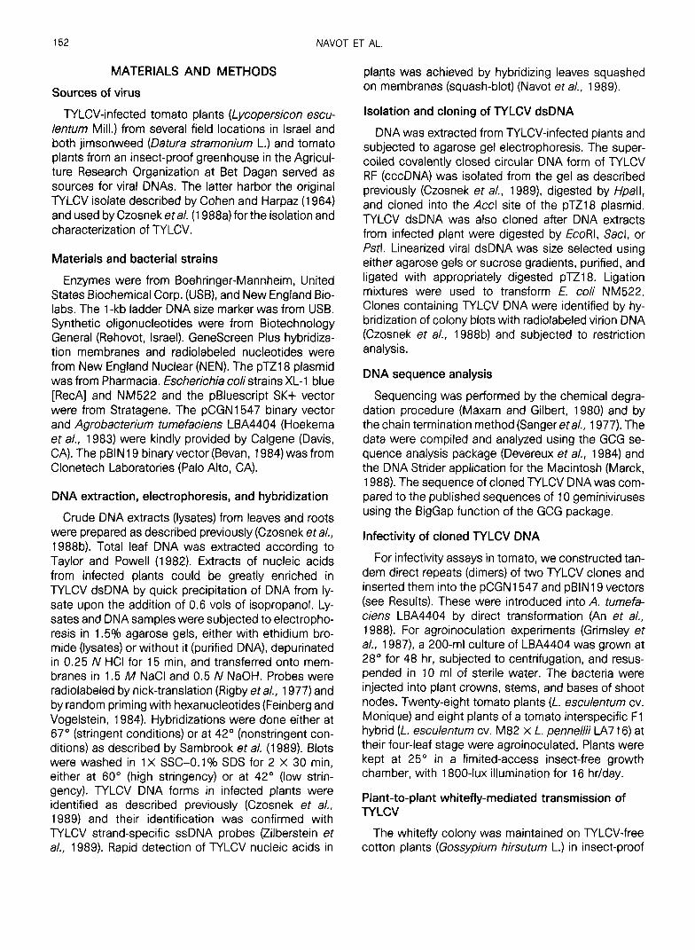
FIG. 1. DNA sequencing strategy of the cloned TYLCV genome. The genome is represented as a thick black tine, with the intergenic region not shaded. Sites of restriction endonucleases used for sub- cloning are shown: Bell (B); EcoRl (E); Haeill (H3); /-!~acjll (HI; Rsal (R); Sacl (Sa); Sau3Al (S3); Sphl (S), and Xbal (X). Arrows below the map indicate the direction and extent of sequencing. Thick arrows are sequences obtained by the dideoxy chain termination method, with the aid of synthetic primers. Thin arrows are sequences obtained by subjecting subclones to the chemical degradation procedure.
phism in the pool of TYLCV RF molecules. The second Hpall site that gave rise to the 0.3-kbp fragment was shown by sequence analysis to have origineted from a single base change at nucleotide 488 (Fig. 2).
TYLCV dsONA was also cloned in other restriction sites. Six EcoRl clones (designated plYE1 to 6) were obtained from individual field-infected tomato plants. Two Sac1 clones (designated pTYS1 and 2) were ob- tained from a greenhouse isolate of TYLCV maintained in tomato and in jimsonweed; 15 Pstl clones (desig- nated pTYP1 to 15) unique for that isolate were also obtained from these plants.
Clones were subjected to restriction analysis. Repre- sentative clones were hybridized with radiufabeled probes from African cassava mosaic virus (ACME) DNA A and B. All of them hybridized with ACMV DNAA but not with DNA B (not shown).
Sequence analysis of the TYLCV genome
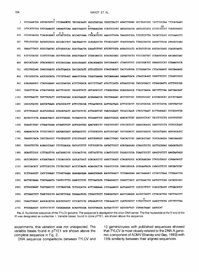
Two independent /-@all clones (pTYH19 and pTYH20), inserted in opposing orientations into the pTZ18 vector, were used for sequence analysis. Sub- clones were created by digestion of pTYH19/20 with EcoRI, Sacl, Sphl, and Xbal, followed by &f-ligation and thereafter by subcloning of Haelil, Sau3Al, and &al fragments of the initial subclones into pUCl8 (Fig. 1). The nucleotide sequence around the single &all site used for cloning was veriiied by partial sequencing of an EcoRl clone (pTYE1). The resulting aeque-r\ce of 2787 nucleotides (27% A, 32% T, 19% C, and 22% G) is shown in Fig. 2 in the virion (sense) strand (EMBL GeneBank accession No. Xl 5658). The 5’ end of the intergenic region (IR) was designated as nucleotide 1. Sequence analysis of an EcoRl clone (pTYE 1) revealed minor variations between it and the two /@all clones. Since these clones were obtained in separate cloning
154 NAVOT ET AL.
1 GTTGAAATGA ATCGGTGTC: CTCAAAQCTC TATGGCAATC GGTGPATCGG TGTCTTACTT ATACTTGGAC ACCTAATGGC TATTTGGTAA TTTCATAMT
101 GTTCATTTCA ATTCAAAATT CMAATTCAA AAATCAAATC ~TTAAAGCGG CCATCCGTAT MTATTACCG GATGGCCGCG CCTTTT%TT TTATGTGGTC
201 CCCACGAGGG TTACACAGA: GTFAkTGTCA ACCAATCMA TT&AT;CTC AAACGTTAGA TAAGTGTTCA TTTGTCTTTA TATACTTGGT CCCCAAGTTT
301 TTTGTCTTGC AATATGTGGG ACCCACTTCT TAATGAATTT CCTGMTCTG TTCACGGATT TCGTTGTATG TTAGCTATTA AATATTTGCA GTCCGTTGAG
401 GAAACTTACG AGCCCAATAC ATTGGGCCAC GATTTAATTA GGGATCTTAT ATCTGTTGTA AGGGCCCGTG ACTATGTCGA AGCGACCAGG CGATATAATC
501 ATTTCCACGC CCGTCTCGAA GGTTCGCCGA AGGCTGMCT TCGACAGCCC ATACAGCAGC CGTGCTGCTG TCCCCATTGT CCMGGCACA AACAAGCGAC
601 GATCATGGAC GTACAGGCCC ATGTACCGAA AGCCCAGAAT ATACAGMTG TATCGAAGCC CTGATGTTCC CCGTGGATGT GAAGGCCCAT GTAAAGTCCA
701 GTCTTATGAG CAACGGGATG ATATTAAGCA TACTGGTATT GTTCGTTGTG TTAGTGATGT TACTCGTGGA TCTGGAATTA CTCACAGAGT GGGTMGAGG
801 TTCTGTGTTA AATCGATATA TTTTTTAGGT AAAGTCTGGA TGGATGAAM TATCMGAAG CAGAATCACA CTAATCAGGT CATGTTCTTC TTGGTCCGTG
901 ATAGAAGGCC CTATGGAAAC AGCCCMTGG ATTTTGGACA GGTTTTTAAT ATGTTCGATA ATGAGCCCAG TACCGCAACC GTGAAGAATG ATTTGCGTGA
1001 TAGGTTTCAA GTGATGAGGA AATTTCATGC TACAGTTATT GGTGGGCCCT CTGGAATGAA GGMCAGGCA TTAGTTAAGA GATTTTTTAA MTTAACAGT
1101 CATGTAACTT TATTTATATT CATTCAGGAG GCAGCAAAGT ACGAGAACCA TACTGAAMC GCCTTGTTAT TGTATATGGC ATGTACGCAT GCCTCTAATC
1201 CAGTGTATGC AACTATGAAA ATACGCATCT ATTTCTATGA TTCAATATCA AATTAATAAA ATTTATATTT TATATCATGA GTTTCTGTTA CATTTATTGT
1301 GTTTTCAAGT ACATCATACA ATACATGATC AACTGCTCTG ATTACATTGT TAATGGAAAT TACACCAAGA CTATCTAAAT ACTTAAGAAC TTCATATCTA
1401 AATACTCTTA AGAAATGACC AGTCTGAGGC TGTAATGTCG TCCAAATTCG GAAGTCGAGA AAACATTTGT GAATCCCCAT TACCTTCCTG ATGTTGTGGT
1501 TGAATCTTAT CTGAATGGAA ATGATGTCGT GGTTCATTAG AAATGGCCTC TGGCTGTGTT CTGTTATCTT GAAATAGAGG GGATTGTTTA TCTCCCAGAT
1601 AAAUCGCCA TTCTCTGCCT GAGGAGCAGT GATGAGTTCC CCTGTGCGTG AATCCATGAT TATTGCAGTT GAGGTGGAGG TAGTATGAGC AGCCACAGTC
1701 TAGGTCTACA CGCTTACGCC TTATTGGTTT CTTCTTGGCT ATCTTGTGTT GGACCTTGAT TGATACTTGC GAACAGTGGC TCGTAGAGGG TGACGMGGT
1801 TGCATTCTTG AGAGCCCMT TTTTCAAGGA TATGTTTTTT TCTTCGTCTA GATATTCCCT ATATGAGGAG GTAGGTCCTG GATTGCAGAG GAAGATAGTG
1901 GGAATTCCCC CTTTAATTTG AATGGGCTTC CCGTACTTTG TGTTGCTTTG CCAGTCCCTC TGGGCCCCCA TGAATTCCTT GAAGTGCTTT AAATMTGCG
2001 GGTCTACGTC ATCMTGACG TTGTACCACG CATCATTACT GTACACCTTT GGGCTTAGGT CTAGATGTCC ACATAAATAA TTATGTGGGC CTAGAGACCT
2101 GGCCCACATT GTTTTGCCTG TTCTGCTATC ACCCTCMTG ACAATACTTA TGGGTCTCCA TGGCCGCGCA GCGGAATACA CGACGTTCTC GGCGACCCAC
2201 TCTTCAAGTT CATCTGGAAC TTGATTAAAA GAAGMGAAA GAAATGGAGA MCATAAACT TCTAAAGGAG GACTAMAAT CCTATCTAAA TTTGAACTTA
2301 AATTATGAAA TTGTAAMTA TAGTCCTTTG GGGCCTTCTC TTTTMTATA TTGAGGGCCT CGGATTTACT GCCTGAATTG AGTGCTTCGG CATATGCGTC
2401 GTTGGCAGAT TGCTGACCTC CTCTAGCTGA TCTGCCATCG ATTTGGGAAA CTCCAAAATC AATGMGTTT CCGTCTTTCT CCACGTAGGT CTTGACATCT
2501 GTTGAGCTCT TAGCTGCCTG AATGTTCGGA TGGAAATGTG CTGACCTGTT TGGGGATACC MGTCGAAGA ACCGTTGGTT CTTACATTGG TATTTGCCTT
2601 CGAATTGGAT MGCACATGG AGATGTGGTT CCCCATTCTC GTGGAGTTCT TTGCAAACTT TGATGTATTT TTTAT:TGTT GGGGTTTCTA GTTTTTTTAA
2701 TTGGGAAAGT GCTTCCTCTT TAGAGAGAGA ACAATTGGGA TATGTTAGGA AATMTTTTT GGCATATATT TTAAATAAAC GAGGCAT
FIG. 2. Nucleotide sequence of the TYLCV genome. The sequence is displayed in the virion DNAsense. The first nucleotide at the 5’end of the IF! was designated as nucleotide 1. Variable bases, found in clone pTYE1, are shown above the sequence.
experiments, this variation was not unexpected. The 10 geminiviruses with published sequences showed variable bases found in pTYE1 are shown above the that TYLCV is most closely related to the DNA A geno- complete sequence in Fig. 2. mic component of ACMV (Stanley and Gay, 1983) with
DNA sequence comparisons between NLCV and 73% similarity between their aligned sequences.

THE SINGLE GENOMIC MOLECULE OF TYLCV 155
TABLE 1
OPEN READING FRAMES IN TYLCV DNA AND HOMOLOGIES WITH COUNTERPARTS IN FOUR GEA~INIVIRUSES
ORF Frame Start stop Amino acids
Protein MW (daltons) ACMV
Amino acid homologys
TGMV BCTV WDV
Vl +2 474 1256 260 30,300 90.7 (80.2) 82.6 (74.1) 48.7 (23.1) 41.5 (18.2) v2 +1 314 664 116 13,450 91.2 (72.6)b 43.1 (17.0) 55.4 (25.7) Cl -1 2787 1714 357 40,653 81.2 (71.7) 79.4 (69.2) 74.9(61.3) 54.6 (34.1)' c2 -2 1805 1400 135 15,601 77.8 (63.7) 69.3 (52.0) 51.2 (29.6) c3 -3 1657 1255 134 15,929 86.6 (73.1) 71.0 (56.5) 66.4 (40.6) -
c4 -2 2636 2343 97 11,091 44.3 (34.0)d 58.8 (49.4) 60.0 (49.4) _-
a Percentage of conserved homology in amino acid sequence is followed in parentheses by the percentage of direct homology. Geminiviruses used for comparisons were: African casava mosaic virus (ACMV; Stanley and Gay, 1983), tomato golden mosaic virus (TGMV; Hamilton et al., 1984), beet curly top virus (BCTV; Stanley et al., 1986), and wheat dwarf virus (WDV; MacDoweil et al., 1985).
b The 13.1 -kDa ORF in the virion (sense) of ACMV DNA A. c Compared to the predicted sequence of the spliced mRNA of WDV (Schalk et al., 1989). d Compared to the amino acid sequence of the protein translated from the second AUG in this ORF.
Potential coding regions and regulatory sequences
To locate potential genes, the DNA sequence was screened on both strands for ORFs with coding poten- tial for proteins of molecular weight 210,000 Da. The ORFs, named according to the terminology of Davies and Stanley (1989), are described in Table 1 and their organization along the genome is depicted in Fig. 3.
FIG. 3. Potential coding regions in TYLCV DNA. Open reading frames that start with an ATG and have the potential to code for proteins with a molecular weight 3 10,000 Da are displayed. The name of each ORF is indicated, corresponding to Table 1. Potential TATA boxes are denoted by open triangles. Polyadenylation signals with the sequence G/AATAAA are denoted by solid triangles. The IR and the position of the conserved stem and loop structure (depicted as a small circle) are also shown.
Two ORFs were located in the Won strand. The pre- dicted amino acid sequence of ORF V2, which pre- cedes Vl (the coat protein gene), is highly homdogous to an ORF of 13.1 kDa, which is found in the same location in DNA A of ACMV, but is abent from the genomes of other whitefly-transmitted *miniviruses (Table 1). On the complementary strand, we found three ORFs analogous to the ACl, AC2, and AC3 ORFs (designated also as AL1 , AL2, and AL3) of the whitefly-transmitted geminiviruses (Davies and Stan- ley, 1989) and a fourth ORF-C4-that is present in the same region in the genomes of all the geminivi- ruses that infect dicotyledonous plants. C4 is seldom shown in the ORF maps of these viruses, possibly be- cause the size of its expected translation product falls often just below IO kDa.
The sequence element with the potential to form a hairpin structure, found in IRS of all geminiviruses (La- zarowitz, 1987), was located between nuck&des 149 and 177 with the conserved loop sequence TAATAT- TAC. Potential promoters conforming to the consen- sus sequence TATAT/AA (Breathnach and Chambon, 198 1) were located in positions similar to those of pro- moters found in ACMV and tomato golden mosaic (TGMV) virus (respectively, Stanley and Gay, 1983; Hamilton er al., 1984) (Fig. 3). The positions of putative polyadenylation signals with the consensus sequence G/AATAAA (Messing et al., 1983) are shown in Fig. 3.
Construction of TYLCV dimers
Two complete tandem direct repeats of the cloned TYLCV genome (dimers) were constructed for agroin- oculation experiments.
One dimer was constructed by combining one /-/pall clone (pTYH20) and one EcoRl clone (pl?‘El) as out-

156 NAVOT ET AL.
BaE Sa H HI SB E
C’L \ D?YEl 5
BaE Ba HI+ S sa HD
I ~l-YH20.4 I ‘) 2
Sa Sa
Ba S Sa S Sa s HD
5 pTYH20.7
FIG. 4. Construction of TYLCV dimer pTYH20.7. (1) Clones pTYH20 and pl?‘El were digested with BamHl and Hincll. The frag- ment containing the IR from pTYE1 was inserted into pTYH20, re- placing its small BarnHI-Hincll fragment, to produce pTYH20.4. (2) This clone was then cut with Sac1 and the 2.8-kbp viral fragment was isolated and self-ligated in a small volume to favor production of multimers. (3) The resulting multimeric DNA was treated with Sphl and separated by electrophoresis in a 1.5% agarose gel. (4) A 2.8- kbp band was eluted and ligated to pTZl8, in a high insert-to-vector ratio. (5) One clone that contained a complete tandem repeat of the viral genome was identified (pTYH20.7) and its integrity was verified by restriction analysis and partial sequencing. Restriction sites indi- cated are: BamHl (Ba), EcoRl (E), Sacl (Sa); Hincll (Hi), Hpall (H), Sphl (S), &/I (B), Hindll (HD).
lined in Fig. 4. This was done because the original /-/pall site of pTYH20 was lost as the result of the clon- ing in the pTZ18 Accl site. The integrity of the resulting cloned dimer (designated pTYH20.7) was verified by restriction analysis and partial sequencing. To deter- mine if pTYH20.7 was functional, it was introduced into tomato protoplasts. Both its replication and the produc- tion of viral ssDNA form were observed (not shown). The 5.6-kbp HindIll-BarnHI fragment of plYH20.7 which contained the complete TYLCV dimer was li- gated into a HindIll- and BarnHI-restricted pBlNl9 bi- nary vector to give pTY60.
To construct the second TYLCV dimer, the 2.8-kbp insert from one Pstl clone (pTYP4) was isolated and ligated with a Pstl-restricted pCGN 1547 binary vector, in a high insert-to-vector ratio. Several clones with a tandem direct repeat of the TYLCV genome were iden- tified by restriction analysis. One of these clones, des- ignated pTY4, was used in our experiments.
The dimers in pBlN 19 and pCGN 1547 binary vectors were introduced into A. tumefaciens LBA4404. The in- tegrity of the dimers in their Agrobecterium host was verified by restriction and hybridization analyses of to- tal DNA isolated from the transformed bacteria.
The cloned TYLCV genome causes systemic infection in agroinoculated plants
Twelve tomato plants were agroinoculated with plY60 (containing the /@all TYLCV dimer) and 24 with plY4 (containing the Pstl TYLCV dimer). Viral DNA was detected by squash-blotting in 34 of the 36 plants 6 days postinoculation (not shown). Severe yellow leaf curl symptoms that were indistinguishable from those associated with natural whitefly-mediated infection ap- peared in all the agroinoculated plants about 15 days postinoculation (not shown). At this time, TYLCV ssDNA and dsDNA were detected in crude DNA prepa- rations (lysates) from leaves of all agroinoculated plants (Fig. 5, lane 3). Identical results were obtained with both dimers. Neither viral DNA nor tomato yellow leaf curl disease symptoms were detected in plants inoculated with Agrobecterium containing binary vec- tors without TYLCV DNA (not shown).
The cloned TYLCV genome is whitefly transmittable
Whiteflies were used to transmit TYLCV from agroin- oculated plants to uninfected tomato test plants. Whiteflies which had access to four plants (L. esculen- turn) agroinoculated 6 weeks earlier with pTY4 were used to inoculate 16 plants. As inoculation controls, whiteflies which had access to a TYLCV-infected to- mato plant from the field were placed on four tomato test plants. Whiteflies from the insect colony kept on
1234567
FIG. 5. TYLCV DNA forms in agroinoculated and whitefly-inocu- lated tomato plants. Crude DNA preparations (lysates) were ex- tracted from roots (lane 1) and leaves (lane 2) of a tomato plant inoculated by whiteflies that had acquired TYLCV from the agroin- oculated plant; from leaves of an agroinoculated plant (lane 3); from roots (lane 4) and leaves (lane 5) of a tomato plant inoculated by whiteflies that had acquired the virus from a TYLCV-infected tomato plant from the field; and from roots (lane 6) and leaves (lane 7) of a tomato plant mock-infected with insects fed on cotton plants. The blot was hybridized with a full-length clone of TYLCV. The positions of the open circular (oc) and supercoiled (SC) TYLCV dsDNA and of the circular single-stranded TYLCV DNA (css) are indicated.

THE SINGLE GENOMIC MOLECULE OF TYLCV 157
cotton plants were also placed on four tomato test plants.
In 15 of the 16 plants inoculated with whiteflies fed on agroinoculated tomato, TYLCV DNA was detected by squash-blotting after 1 week. Typical disease symp- toms appeared in all the plants within 3 weeks postin- oculation (not shown). TYLCV ssDNA and dsDNA spe- cies were seen in lysates prepared from leaves and roots of these plants 3 weeks postinoculation (Fig. 5, lanes 1, 2). At that time, 3 of the 4 plants inoculated by whiteflies fed on a field-infected plant contained viral DNA (Fig. 5, lanes 4, 5). Neither viral DNA nor symp- toms were found in the plants mock-inoculated by whiteflies raised on cotton plants (Fig. 5, lanes 6, 7), confirming that the whitefly colony was TYLCV-free.
The search for the putative TYLCV DNA B component
Our strategy was based on the observation that ex- cept for the CR, the sequences of DNAs A and B in geminiviruses with a bipartite genome share almost no homology; thus outside the CR their restriction maps are different. On the basis of this high level of homol- ogy between the CRs in DNAs A and B, we assumed that the IR of TYLCV (which spans the potential CR) could be used as a probe to detect both putative com- ponents. If TYLCV possessed two genomic compo- nents, cleavage of the viral dsDNA by restriction endo- nucleases would produce fragments whose length to- talled 5.6 kbp. If recognition sites for a certain enzyme existed in only one of the components, fragments that add up to 2.8 kbp and an uncut dsDNA would be seen. A probe containing the CR would detect at least one fragment from each of the components. A probe spe- cific for DNA A would detect either the uncut dsDNA species or some restriction fragments, but not both. If TYLCV had only a single genomic molecule, digesting it with restriction endonucleases would produce frag- ments that add up to 2.8 kbp and agree with the restric- tion map derived from the sequence of the cloned com- ponent. No uncut viral DNA species should be seen.
The probes generated for the detection of TYLCV DNA A and the putative DNA B are shown in Fig. 6. As a probe capable of detecting both DNA A and B, we used the potential common region of TYLCV. This was done by subcloning a 347-bp-long Alul-Alul fragment of the IR from an EcoRl clone of NLCV (pTYE1) into pBluescript SK+ (pTYE1.2). As DNAA-specific probes, we used a Hinfl-Hinfl fragment of 1 146 bp spanning the Cl and C4 ORFs of TYLCV subcloned into pBlue- script SK+ (pTYH20.23); a 170-bp-long Haelll-Haelll fragment of the Cl ORF subcloned into pUCl8 (pTYH20.15); and a Haelll-EcoRI fragment of 357 bp
Xbal
Apal
FIG. 6. Location of probes and of complensntary oiig~nucleotides used for restriction and hybridization analyaea of MCV DNA. The genome is displayed as a circle with the IR es a thick grey region. Probes used for the hybridization an&y& are &.picted inside %he circle as thick black lines. plYEl.2 (a 347-bp AU-A&t s&done) was used to detect DNA A as well as the putative DNA f3. pTYH20.23 (a 1 149-bp Hinfl-Hinfl subclone), pTYHEO. 16 (a 170-bp HaellbHaelll subclone), and pTYH 19.16 (a 357-bp #~til-&~~Rl subclone) were used as DNA A-specific probe&. The locations of complementary oligonucleotides used for sn$&sis of ssDMA in whiteflies are shown as short black lines outside the circb. The rec- ognition sites of restriction endonucleases wed in restiction and hybridization analyses are indicated by black triangles.
spanning the C2 ORF subcloned into pUC18 (pTYH19.16). The ACMV DNA A fuil-J@ny)th clone pJSO92 (Stanley, 1983) was also used m a DNA A-spe- cific probe, because the intergrjnic regions of ACMV and TYLCV are divergent and do not hybridize with each other under stringent conditions.
The putative TYLCV DNA B wm not fwnd in TYLCV dsONA from infected plants
Infected tomato leaf DNA enriched in TYLCV dsDNA was analyzed with eight reatrictiun endonuchees for which no recognition sites were fourad in any of the TYLCV RF-derived clones: B&-#I, EcoRV, Hindill, Kpnl, A&, WI, Smal, and Xhof; with six restriction endonucleases for which one or two sites were found: WI, 001, EcoRI, /-@all, Sacl, and Xbal; and with two endonucleases, Aatll and /WI, for which either orte or no sites were found. Hybridization with the DNA A- specific probes showed that enzymes for which no site exists in the cloned molecub did not cut TYLCV dsDNA. Enzymes for which there a~e.~~o~~~~o~ sites in the cloned molecule of TYLCV (as ~nd~~t#d by se- quence analysis) either linearized the viral dsDNA or
158 NAVOT ET AL.
1234 56
12.8 4 2.0
4 0.8
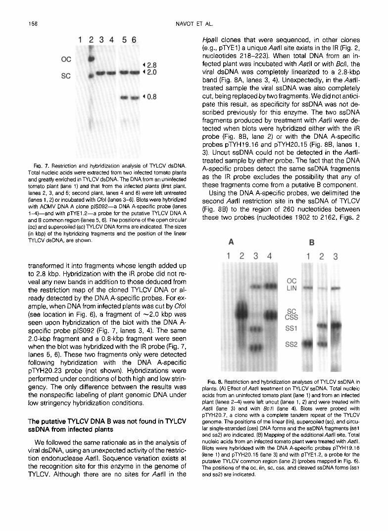
FIG. 7. Restriction and hybridization analysis of TYLCV dsDNA. Total nucleic acids were extracted from two infected tomato plants and greatly enriched in TYLCV dsDNA. The DNA from an uninfected tomato plant (lane 1) and that from the infected plants (first plant, lanes 2, 3, and 5; second plant, lanes 4 and 6) were left untreated (lanes 1.2) or incubated with Cfol (lanes 3-6). Blots were hybridized with ACMV DNA A clone pJS092-a DNA A-specific probe (lanes i-4)-and with pWEl.2-a probe for the putative WLCV DNA A and B common region (lanes 5,6). The positions of the open circular (oc) and supercoiled (SC) TYLCV DNA forms are indicated. The sizes (in kbp) of the hybridizing fragments and the position of the linear TYLCV dsDNA, are shown.
transformed it into fragments whose length added up to 2.8 kbp. Hybridization with the IR probe did not re- veal any new bands in addition to those deduced from the restriction map of the cloned TYLCV DNA or al- ready detected by the DNA A-specific probes. For ex- ample, when DNA from infected plants was cut by Cfol (see location in Fig. 6) a fragment of -2.0 kbp was seen upon hybridization of the blot with the DNA A- specific probe pJS092 (Fig. 7, lanes 3, 4). The same 2.0-kbp fragment and a 0.8-kbp fragment were seen when the blot was hybridized with the IR probe (Fig. 7, lanes 5, 6). These two fragments only were detected following hybridization with the DNA A-specific pTYH20.23 probe (not shown). Hybridizations were performed under conditions of both high and low strin- gency. The only difference between the results was the nonspecific labeling of plant genomic DNA under low stringency hybridization conditions.
The putative TYLCV DNA B was not found in TYLCV ssDNA from infected plants
We followed the same rationale as in the analysis of viral dsDNA, using an unexpected activity of the restric- tion endonuclease Aatll. Sequence variation exists at the recognition site for this enzyme in the genome of TYLCV. Although there are no sites for Aatll in the
/-/pall clones that were sequenced, in other clones (e.g., pTYE1) a unique Aatll site exists in the IR (Fig. 2, nucleotides 218-223). When total DNA from an in- fected plant was incubated with Aatll or with &/I, the viral dsDNA was completely linearized to a 2.8-kbp band (Fig. 8A, lanes 3, 4). Unexpectedly, in the Aatll- treated sample the viral ssDNA was also completely cut, being replaced by two fragments. We did not antici- pate this result, as specificity for ssDNA was not de- scribed previously for this enzyme. The two ssDNA fragments produced by treatment with Aatll were de- tected when blots were hybridized either with the IR probe (Fig. 8B, lane 2) or with the DNA A-specific probes pTYH19.16 and pTYH20.15 (Fig. 8B, lanes 1, 3). Uncut ssDNA could not be detected in the Aatll- treated sample by either probe. The fact that the DNA A-specific probes detect the same ssDNA fragments as the IR probe excludes the possibility that any of these fragments come from a putative B component.
Using the DNA A-specific probes, we delimited the second Aatll restriction site in the ssDNA of TYLCV (Fig. 8B) to the region of 260 nucleotides between these two probes (nucleotides 1902 to 2162, Figs. 2
oc LIN
FIG. 8. Restriction and hybridization analyses of TYLCV ssDNA in plants. (A) Effect of Aafll treatment on TYLCV ssDNA. Total nucleic acids from an uninfected tomato plant (lane 1) and from an infected plant (lanes 2-4) were left uncut (lanes 1, 2) and were treated with Aatll (lane 3) and with &II (lane 4). Blots were probed with pTYH20.7, a clone with a complete tandem repeat of the TYLCV genome. The positions of the linear (lin), supercoiled (SC), and circu- lar single-stranded (css) DNA forms and the ssDNA fragments (ssl and ss2) are indicated. (B) Mapping of the additional Aafll site. Total nucleic acids from an infected tomato plant were treated with Aatll. Blots were hybridized with the DNA A-specific probes pTYH19.16 (lane 1) and plYH20.15 (lane 3) and with pTYE1.2, a probe for the putative TYLCV common region (lane 2) (probes mapped in Fig. 6). The positions of the oc, lin, SC. css, and cleaved ssDNA forms (ssl and ss2) are indicated.

THE SINGLE GENOMIC MOLECULE OF TYLCV 159
and 6). Three imperfect Aatll sites, each with one mis- match, are found in this region. It is possible that an interaction between sequences spanning the proper Aatll site and sequences around one of the three par- tial sites resulted in the formation of a double-stranded region; this allowed the enzyme to cut the ssDNA and produce the two observed fragments.
The putative TYLCV DNA B was not found in viruliferous whiteflies
To discriminate between DNA A and the putative DNA B present in viruliferous whiteflies, we intended to cleave the ssDNA of the A component into several fragments without affecting the B component. If a B component was present, one would expect (following hybridization with the IR probe) to see an uncut DNA species representing the DNA B molecules, in addition to the fragments resulting from the cleavage of DNA A.
On the basis of the sequence of the cloned TYLCV genome, we synthesized three 18-mer oligonucleo- tides complementary to different regions in the ssDNA genome (Fig. 6). Each oligomer contained a single rec- ognition site for one of the following restriction endonu- cleases: Apal (from nucleotide 456 to 473), Xbal (from nucleotide 2054 to 2071), and Sacl (from nucleotide 2498 to 2515) (Fig. 2). After annealing the oligomers with a DNA extract from viruliferous whiteflies, the par- tially dsDNA was incubated with a mixture of Xbal, Sacl, and Apal. The DNA was subjected to electropho- resis and blots were hybridized to the DNA A-specific probe plYH20.23. Three bands were detected (Fig. 9, lane 2) and the circular ssDNA disappeared, as ex- pected from the sequence of the TYLCV DNA A-like genome. When the IR probe was used, only the middle sized band corresponding to the Apal-Sac1 fragment of 745 nucleotides (Fig. 6) was seen (Fig. 9, lane 3). Unrestricted ssDNA was not detected by either probe. DNA from nonviruliferous whiteflies before and after incubation with the oligonucleotides did not hybridize with any TYLCV probe. These results showed that the only TYLCV DNA component detectable in viruliferous whiteflies is of the A type.
DISCUSSION
On the basis of the genome structure of whitefly- transmitted geminiviruses, it was assumed that a bi- partite genome would also be found for TYLCV. How- ever, all the clones obtained from the viral RF were from one DNA A-like component.
We demonstrated that this single component has the capacity to cause systemic infection in tomato, coupled with severe yellow leaf curl disease symp- toms. Still, by using agroinoculation to produce an in- sect-transmitted viral disease, one might miss a sec-
1 2 3
css SSl
FIG. 9. Restriction and hybridization analyses of TYLCV ssDNA present in viruliferous whiteflies. Total nucleic acids extmctk?d from 200 viruliferous whiteflies were hybridized in solution with three sin- gle-stranded 18-mer oligonucleotides, complementary to three re- gions in DNA A of TYLCV. Each oligonucleutide cortt&ed a single restriction site for Apal, Xbal, or Saci (see Fig. 6). The pat?Miy dou- ble-stranded DNA was left untreated (tane 1) or was in~~with a mixture of the three enzymes (lane 2). The blot was hybrkked with the DNA A-specific probe plYH20.23 (lanes 1 1 2). After autoradio- graphic exposure, the probe was stripped by boiling in 1% SDS and 0.1 x SSC, and the blot containing the restricted DNA (lane 3) and the unrestricted DNA (as in lane 1) was r&ybridi& with the probe for the common region pTYE1.2 (probes descrfkd in Fig. 6). The positions of the circular single-stranded (css) and cleaved ssONA forms (~~1-3) are indicated.
ond genomic component or a viral function essential for the plant-to-plant transmission of the virus. There- fore we let whiteflies transmit the TYLCV diw@se from the agroinoculated plants to uninfected teet plants. Typical disease symptoms appeared in et1 Fe& plsnts, and viral nucleic acids were detected in k%v@s and roots of these plants. These resuks proved that the single genomic molecule of TYLCV carries atl the infor- mation needed for its transmission by insects, repfica- tion, and systemic spread.
We initiated the search for the putative 8 component to exclude the possibility that it is present in TYLCV-in- fected tomato but is not required at any stage of the virus infection cycle. Results of cloning experiments and of restriction and hybridization anafyws of viral DNA forms in infected plants and virulifarous white#lies could not corroborate the existence of a second gsno- mic component. In TGMV the presence of the B com- ponent was necessary for virus movement in the in- fected plant (Rogers et al., 1986; Sun&r er al,, 1987). On the other hand, when ACMV DNA A was agroinoc-

160 NAVOT ET AL.
ulated into Nicotiana benthamiana, some systemic spread was observed, but no symptoms appeared (Klinkenberg and Stanley, 1990). DNA A of a TYLCV- like isolate from Thailand seemed even less dependent on DNA B, as it was capable both of systemic move- ment and of mild symptom production in agroinfected tobacco and tomato plants (Rochester et al., 1990). Recently a TYLCV-like isolate from Sardinia was shown, through molecular analysis, agroinoculation, and whitefly-mediated transmission, to possess a sin- gle genomic molecule capable of producing the com- plete disease cycle (Gronenborn, B., personal commu- nication). It is possible that TYLCV and the Sardinian virus represent a time in the evolution of whitefly-trans- mitted geminiviruses, prior to the emergence and spe- cialization of a second genomic component, before DNA A became reliant on the second component for systemic infection and production of the full disease symptoms. The absence of a B component in the ge- nomes of these viruses supports the notion that DNA B has no function in the interaction of a whitefly-transmit- ted geminivirus with its vector (Etessami et a/., 1988).
Sequence analysis of the cloned TYLCV genome re- vealed the presence of a second ORF on the virion strand (designated V2), in addition to the ORF for the coat protein. A highly homologous ORF of 13.1 kDa has been found in ACMV (Townsend et al., 1985) as well as in the TYLCV-like virus from Sardinia (Gronen- born B., personal communication), but not in other whitefly-transmitted geminiviruses. A comparable “precoat” ORF (designated Vl or R2) is present in the genomes of all the leafhopper-transmitted geminivi- ruses, although the homology it shares with V2 of TYLCV is limited (Table 1). Deletion of Vl from the ge- nome of wheat dwarf virus (WDV) had no effect on its ability to replicate in protoplasts of cereals (Laufs et al., 1990; Ugaki et al., 1991). Mutational analysis of this ORF in maize streak virus (MSV) showed that it en- codes an essential function (Mullineaux et a/., 1988), most probably associated with systemic spread of the virus (Boulton et al., 1989; Lazarowitz et al., 1989). In contrast, its complete deletion in ACMV had no effect on the ability of the mutated DNA A to cause systemic infection, following its introduction, together with DNA B, into N. benthamiana (Klinkenberg et a/., 1989). It might be that V2 of TYLCV and the 13.1 -kDa ORF of ACMV have the same function as Vl of leafhopper- transmitted geminiviruses but are needed for systemic spread only in some of their plant hosts. The loss of this gene in other whitefly-transmitted geminiviruses might have occurred during their evolution, as they or their progenitor virus acquired new hosts for which no such function was needed. Mutational analysis of V2 in TYLCV and ACMV is expected to assist in determining the function of this ORF.
ACKNOWLEDGMENTS
Excellent technical assistance was provided by Sara Ovadia and Tzili Pleban. We thank Dr. J. Stanley for probes of ACMV; Drs. Z. Livneh and G. Yagil for helpful discussions; Drs. H. Jeske and B. Gronenborn for sharing with us their results prior to publication and for helpful discussions; Dr. A. Kheyr-Pour for his assistance in agroinoculation experiments; Drs. S. Cohen and H. Antignus for greenhouse TYLCV-infected plants; and the staff of the Center for Micro Computers at the Weizmann Institute of Science for assis- tance and use of their facilities. This research was supported by BARD Project l-l 1 1 O-86 and by Yissum.
REFERENCES
ABOUZID, A. M.. FRISCHMUT, T., and JESKE, H. (1988). A putative repli- cative form of the abutilon mosaic virus in chromatin-like structure. Mol. Gen. Genet. 212, 252-258.
AN, G., EBERT, P. R., MITRA. A., and HA, S. B. (1988). Binary vectors. ln “Plant Molecular Biology Manuel” (S. B. Gelvin, R. A. Schilper- oort, and D. P. S. Verma, Eds.), Section A3, pp. 1-19. Kluwer Academic Pubs. Dordrecht, The Nederlands.
BEVAN, M. (1984). Binary vectors for plant transformation. Nucleic Acids Res. 12, 8711-8721.
BOULTON. M. I., STEINKELLNER, H., DONSON. J., MARKHAM, P. G., KING, D. I., and DAVIES, J. W. (1989). Mutational analysis of the virion- sense genes of maize streak virus. J. Gen. Viral. 70, 2309-2323.
BREATHNACH, R., and CHAMBON, P. (1981). Organization and expres- sion of eucalyotic split genes coding for proteins. Annu. Rev. Bio- them. 50, 349-383.
COHEN, S., and HARPAZ, I. (1964). Periodic, rather than continual, acquisition of a new tomato virus by its vector, the tobacco whitefly (Bemisia fabaci Gennadus). Ent. Exp. Appl, 7, 155-l 66.
CZOSNEK, H., BER. R., ANTIGNUS, Y., COHEN, S., NAVOT, N., and ZAMIR. D. (1988a). Isolation of the tomato yellow leaf curl virus-A gemi- nivirus. Phytopathology 78, 508-5 12.
CZOSNEK, H., BER, R., ANTIGNUS, Y., COHEN, S., NAVOT. N., andZA~i~, D. (1988b). Detection of tomato yellow leaf curl virus in lysates of plants and insects by hybridization with a viral DNA probe. Plant Dis. 72, 949-951.
CZOSNEK, H., BER, R., NAVOT, N., ANTIGNUS, Y., COHEN, S., and ZAMIR.
D. (1989). Tomato yellow leaf curl virus DNA forms in the viral capside, in infected plants and in the insect vector. 1. fhytopathol. 125,47-54.
CZOSNEK, H., NAVOT, N., and LATERROT, H. (1990). Geographical dis- tribution of tomato yellow leaf curl virus: A first survey using a specific DNA probe. Phytopafhol. Me&. 29, l-6.
DAVIES, J. W., and STANLEY, J. (1989). Geminivirus genes and vectors. Trends Genat. 5, 77-81.
DEVEREUX, J., HAERBERLI, P., and SMITHIES, 0. (1984). A comprehen- sive set of sequence analysis programs for the VAX. Nucleic Acids Res. 12, 387-395.
ELMER, J. S., BRAND, L., SUNTER, G., GARDINER, W. E.. BISARO, D. M., and ROGERS, S. G. (1988). Genetic analysis of tomato golden mo- saic virus. II. The product of the AL1 coding sequence is required for replication. Nucleic Acids Res. 16, 7043-7060.
ETESSAMI, P., CALLIS, R., ELLWOOD, S.. and STANLEY, J. (1988). Delimi- tation of essential genes of cassava latent virus DNA 2. Nucleic Acids Res. 16, 7043-7060.
FEINBERG, A. P., and VOGELSTEIN, 8. (1984). A technique for radiola- beling DNA restriction endonuclease fragments to high specific activity. Anal. Biochem. 137, 266-269.
GOODMAN, R. M. (1977). Single-stranded DNA genome in a whitefly- transmitted plant virus. Virology 63, 17 l-l 79.
GRIMSLEY, N., HOHN, T., DAVIES, J. W., and HOHN, B. (1987). Agrobac-

THE SINGLE GENOMIC MOLECULE OF TYLCV 161
tenum-mediated delivery of infectious maize streak virus into maize plants. Nature (London) 325, 177-I 79.
HAMILTON, W. D. O., BISARO, D. M., and BUCK, K. W. (1982). Identifi- cation of novel DNA forms in tomato golden mosaic virus infected tissue: Evidence for a two component genome. Nucleic Acids Res. 10,4901-4912.
HAMILTON, W. D. O., STEIN, V. E., Courts, R. H. A., and BUCK, K. W. (1984). Complete nucleotide sequence of the infectious cloned DNA components of tomato golden mosaic virus: Potential coding regions and regulatory sequences. EM50 1. 3, 2 197-2205.
HOEKEMA, A., HIRSH, P. R., HOOYKAAS, P. J. J., and SCHILPEROORT, R. A. (1983). A binary plant vector strategy based on separation of the vir and T-region of the Agrobacterium tumefaciens Ti-plasmid. Nature (London) 303, 179-l 80.
IKECIAMI, M., HABER, S., and GOODMAN, R. (1981). Isolation and char- acterization of virus-specific double-stranded DNA from tissues infected by bean golden mosaic virus. Proc. Natl. Acad. Sci. USA 78,4102-4106.
KLINKENBERG, F. A., ELLWOOD, S., and STANLEY, J. (1989). Fate of Atrican cassava mosaic virus coat protein deletion mutants after agroinoculation. J. Gen. Viral. 70, 1837-l 844.
KLINKENBERG, F. A., and STANLEY, J. (1990). Encapsidation and spread of African cassava mosaic virus DNA A in the absence of DNA B when agroinoculated to Nicotiana benthamiana. J. Gen. viral. 71, 1409-1412.
L~UFS, I., WIRR, U., KAMMANN, M., MATZEIT, V., SCHAEFER, S., SCHELL, J., CZERNILOWSKY, A. P., BAKER, B., and GRONENBORN, B. (1990). Wheat dwarf virus Ac/Ds vectors: Expression and excision of transposable elements introduced into various cereals by a viral replicon. Proc. Nati. Acad. Sci. USA 87, 7752-7756.
LAZAROWIR, S. G. (1987). The molecular characterization of gemini- viruses. Plant Mol. Biol. Reprod. 4, 177-l 92.
[AZAROWITZ, S. G., and LAZDINS, I. B. (1991). Infectivity and complete nucleotide sequence of the cloned genomic components of a bi- partite squash leaf curl geminivirus with a broad host range pheno- type. Virology 180, 58-69.
LAzARowrrz, S. G., PINDER, A. J., DAMSTEEGT, V. D., and ROGERS, S. G. (1989). Maize streak virus genes essential for systemic spread and symptom development. EMf30 J. 8, 1023-l 032.
MACDOWELL, S. W., MACDONALD, H., HAMILTON, W. D. O., COUTTS, R. H. A., and BUCK, K. W. (1985). The nucleotide sequence of cloned wheat dwarf virus DNA. EMBO 1. 4,2173-2180.
MAKKOUK, K. M., and LATERROT, H. (1983). Epidemiology and control of tomato yetlow leaf curl virus. In “Plant Virus Epidemiology” (R. T. Plumb and Thresh I. M., Eds.), pp. 315-321. Blackwell, Oxford.
MARCK, C. (1988). ‘DNA Strider’: a ‘C’ program for the fast analysis of DNA and protein sequences on the Apple Macintosh family of computers. Nucleic Acids Res. 16, 1829-l 836.
MAXAM, A. M., and GILBERT, W. (1980). Sequencing end-labelled DNAwith base-specific chemical cleavages. In “Methods in Enzy- mology” (L. Grossman and K. Moldave, Eds.), Vol. 65, pp. 499- 560. Academic Press, San Diego, CA.
MESSING, J., GERAGHTY, D., HEIDECKER, G., Hu, N. T., KRIDL, J., and RUBENSTEIN, I. (1983). Plant gene structure. In “Genetic Engineer- ing of Plants” (T. Kasuge, C. P. Meredith, and A. Hollander, Eds.), pp. 2 1 l-227. Plenum, New York.
MULLINEAUX, P. M., BOULTON, M. I., BOWYER, P., VAN DER KLUGT, R., MARKS, M., DONSON, J.. and DAVIES, J. W. (1988). Detection of a non-structural protein of M, 1 1,000 encoded by the virion DNA of maize streak virus. Plant Mol. HoI. 11, 57-66.
NAVOT, N., BER, R., and CZOSNEK, H. (1989). Rapid detection of to- mato yellow leaf curl virus in squashes of plant and insect vectors. Phytopathology 79, 562-568.
RIGBY, P. W. J., DIECKMAN, M., RHODES, C., and BERG, P. (1977). La- belling deoxyribonucleic acid to high specific activity in vitro by nick translation with DNA polymerase I. J. Mol. Biol. 113, 237- 251.
ROCHESTER, D. E., KOSITW\TANA, W., and BEACHY, R. N. (1990). Sys- temic movement and symptom production foIlowing agroinocula- tion with a single DNA of tomato yellow leaf curl geminivirus (Thai- land). virology 178, 520-526.
ROGERS, S. G., BISARO, D. M.. HORSCH, R. B., FRALEY, R. T., HOFFMAN, N. L., BRAND, L., ELMER, J. S.. and LLOYD, A. M. (1986). Tomato golden mosaic virus A component DNA replicates autonomously in transgenic plants. Ce// 45, 593-600.
SAMBROOK, J., FRITSCH, E. F., and MANIATIS, T. (1989). “Molecular Cloning: A Laboratory Manual.” Cold Spring Harbor Laboratory, Cold Spring Harbor, NY.
SANGER, F., NICKLEN, S., and COULSON. A. R. (1977). DNA sequenc- ing with chain-terminating inhibitors. Proc. Nat/, Acad. Sci. USA 74, 5463-5467.
SCHALK, H. J., MATZEIT, V., SCHILLER, B., SCHELL, J., and GRQNENBORN, B. (1989). Wheat dwarf virus, a geminivirus of graminaceous plants needs splicing for replication. EMBO J. 8, 359-364.
STANLEY, J. (1983). Infectivity of the cloned geminivirus genome re- quires sequences from both DNAs. Nature (London) 305, 643- 645.
STANLEY, J.. and GAY, M. R. (1983). Nu&otide sequence of cassava latent virus DNA. Nature (London) 3&f, 260-262.
STANLEY, J., MARKHAM, P. G., CALLIS, R. J., and PINMER, M. S. (1986). The nucleotide sequence of an infectious clone of the geminivirus beet curly top virus. EMBOJ. 8, 1761-1767.
STANLEY, J., and TOWNSEND, R. (1985). Characterization of DNA forms associated with cassava latent virus infection. Nucleic Acids Res. 13, 2 189-2206.
SUNTER, G., GARDINER, W. E.. RUSHING, A. E., ROGERS, S. G., and BISARO, D. M. (1987). Independent encapsidation of tomato golden mosaic virus A component DNA in transgenic plants. Plant Mol. Biol. 8, 477-484.
TAYLOR, B., and POWELL, A. (1982). Isolation of plant RNA and DNA. Focus 4, 4-6.
TOWNSEND, R., STANLEY, J., CURSON, S. J., and SHORT, M. N. (1985). Major polyadenylated transcripts of caasava latent virus and loca- tion of the gene encoding coat protein. &M&O J. 4, 33-37.
UGAKI, M., UEDA, T., TI~AMERMANS. M. C. P.. VIEIRA, J., OLLISTON, K. O., and MESSING, J. (1991). Replication of a geminivirus derived shuttle vector in maize endosperm cells. Nucleic Acids Res. 19, 371-377.
ZEIDAN. M., and CZOSNEK, H. (1991). Acquisition of tomato yellow leaf curl virus by the whitefly Bemisia tabaci. J. Gen. l&o/., in press.
ZILBERSTEIN, A., NAVOT, N., OVADIA, S., REINHARX A., MERZBERG, M., and CZOSNEK. H. (1989). Field-usable assay for diagnosis of the tomato yellow leaf curl virus in squashes of plants and insects by hybridization with a chromogenic DNA probe. Technique 1, 118- 124.