Triterpenoids from Acacia ataxacantha DC - Archive ouverte ...
9
HAL Id: hal-03536465 https://hal.archives-ouvertes.fr/hal-03536465 Submitted on 19 Jan 2022 HAL is a multi-disciplinary open access archive for the deposit and dissemination of sci- entific research documents, whether they are pub- lished or not. The documents may come from teaching and research institutions in France or abroad, or from public or private research centers. L’archive ouverte pluridisciplinaire HAL, est destinée au dépôt et à la diffusion de documents scientifiques de niveau recherche, publiés ou non, émanant des établissements d’enseignement et de recherche français ou étrangers, des laboratoires publics ou privés. Distributed under a Creative Commons Attribution| 4.0 International License Triterpenoids from Acacia ataxacantha DC: antimicrobial and antioxidant activities Abdou Madjid O. Amoussa, Latifou Lagnika, Mélanie Bourjot, Catherine Vonthron-Senecheau, Ambaliou Sanni To cite this version: Abdou Madjid O. Amoussa, Latifou Lagnika, Mélanie Bourjot, Catherine Vonthron-Senecheau, Am- baliou Sanni. Triterpenoids from Acacia ataxacantha DC: antimicrobial and antioxidant activities. BMC Complementary and Alternative Medicine, BioMed Central, 2016, 16 (1), 10.1186/s12906-016- 1266-y. hal-03536465
-
Upload
khangminh22 -
Category
Documents
-
view
0 -
download
0
Transcript of Triterpenoids from Acacia ataxacantha DC - Archive ouverte ...

HAL Id: hal-03536465https://hal.archives-ouvertes.fr/hal-03536465
Submitted on 19 Jan 2022
HAL is a multi-disciplinary open accessarchive for the deposit and dissemination of sci-entific research documents, whether they are pub-lished or not. The documents may come fromteaching and research institutions in France orabroad, or from public or private research centers.
L’archive ouverte pluridisciplinaire HAL, estdestinée au dépôt et à la diffusion de documentsscientifiques de niveau recherche, publiés ou non,émanant des établissements d’enseignement et derecherche français ou étrangers, des laboratoirespublics ou privés.
Distributed under a Creative Commons Attribution| 4.0 International License
Triterpenoids from Acacia ataxacantha DC:antimicrobial and antioxidant activities
Abdou Madjid O. Amoussa, Latifou Lagnika, Mélanie Bourjot, CatherineVonthron-Senecheau, Ambaliou Sanni
To cite this version:Abdou Madjid O. Amoussa, Latifou Lagnika, Mélanie Bourjot, Catherine Vonthron-Senecheau, Am-baliou Sanni. Triterpenoids from Acacia ataxacantha DC: antimicrobial and antioxidant activities.BMC Complementary and Alternative Medicine, BioMed Central, 2016, 16 (1), �10.1186/s12906-016-1266-y�. �hal-03536465�

RESEARCH ARTICLE Open Access
Triterpenoids from Acacia ataxacantha DC:antimicrobial and antioxidant activitiesAbdou Madjid O. Amoussa1,2, Latifou Lagnika1*, Mélanie Bourjot2, Cathérine Vonthron-Senecheau2
and Ambaliou Sanni1
Abstract
Background: Acacia ataxacantha is a medicinal specie used extensively in traditional medicine of Benin republic totreat infectious diseases. Our previous study showed interesting antibacterial and antifungal activities against sixstrains of bacteria and six strains of fungi. The aim of this study was to investigate the antimicrobial and antioxidantactivities of compounds isolated from A. ataxacantha.
Methods: Chromatographic and spectroscopic methods were used to isolate and identify three compounds (1–3)from the bark of A. ataxacantha. Phytochemical investigation of A. ataxacantha (Fabaceae) led to the isolation ofthree triterpenoids (1–3). The structure of isolated compounds was established by differents spectroscopic methodssuch as UV, 1H NMR, 13C NMR, 2D NMR and Mass. All isolated compounds were tested for antimicrobial activityusing agar disc-diffusion and microdilution methods. The radical scavenging activity of isolated compounds wasassessed using 2,2-diphenyl-1-picrylhydrazyl (DPPH) method.
Results: Phytochemical investigation led to the isolation and identification of lupeol (1), betulinic acid (2) andbetulinic acid-3-trans-caffeate (3). Moderate antimicrobial activity was obtained with compound 3 againstmethicillin-resitant Staphylococcus aureus, Enterococcus feacalis and Pseudomonas aeruginosa with MIC value of25 μg/ml and Staphylococcus aureus (MIC of 50 μg/ml). Compounds 3 was more active against Staphylococcusepidermidis and Candida albicans with a MIC value of 12.5 μg/ml in boths cases. Compounds 3 had also interestingantioxidant activity with an IC50 of 3.57 μg/ml compared to quercetin (1.04 μg/ml).
Conclusion: The overall results of this study provide evidence that the compound 3, isolated from A. ataxacantha,exhibit antimicrobial activity against Gram-positive and Gram-negative bacteria and yeast, especially against C.albicans.
Keywords: Acacia ataxacantha, Triterpene, Betulinic acid-3-trans-caffeate, Antibacterial, Antifungal, Antioxidant
BackgroundFabaceae, also known as Leguminosae represented by730 genera like Stylosanthes, Tamarindus, Caesalpinia,Acacia and over 19400 species [1]. Acacia is a cosmopol-itan genus containing in excess 1200 species and thehighest density and the greatest diversity is found intropical and subtropical regions, but also found through-out the world [2]. The aerial parts of different species ofthe genus Acacia are widely used in folk medicine dueto their content of a variety of bioactive components
which are responsible for numerous pharmacologicalproperties such as hypoglycemic, anti-inflammatory, anti-bacterial, antihypertensive, analgesic, anticancer and anti-atherosclerotic [3]. Acacia ataxacantha is widespread inmuch of sub-Saharan Africa. This species is a very thornyshrub with the height of 5 to 8 m. The leaves are alternate,with spine that carries 5 to 12 pairs of pinnae. On twigs,spines are short, clearly pointing down. The fruit pods areflattened, brownish red in the dry state. This specie hasbeen reported in Benin, Nigerian and Kenya for its use intraditional medicine for the treatment of tooth decay,dysentery, bronchitis, cough and joint pain [4–6]. To thebest of our knowledge, little phytochemical work has yetbeen done on A. ataxacantha. In a previous work, we
* Correspondence: [email protected]é de Biochimie et Biologie Moléculaire, Equipe de Biochimie etSubstances Naturelles Bioactives, Faculté des Sciences et Techniques,Université d’Abomey-Calavi, Cotonou 04 BP 0320, BéninFull list of author information is available at the end of the article
© 2016 The Author(s). Open Access This article is distributed under the terms of the Creative Commons Attribution 4.0International License (http://creativecommons.org/licenses/by/4.0/), which permits unrestricted use, distribution, andreproduction in any medium, provided you give appropriate credit to the original author(s) and the source, provide a link tothe Creative Commons license, and indicate if changes were made. The Creative Commons Public Domain Dedication waiver(http://creativecommons.org/publicdomain/zero/1.0/) applies to the data made available in this article, unless otherwise stated.
Amoussa et al. BMC Complementary and Alternative Medicine (2016) 16:284 DOI 10.1186/s12906-016-1266-y

reported the antioxidant, antifungal, antibacterial activitiesand toxicity of the bark extracts of this plant [7–9]. Theseresults suggests that this plant might contain bioactivecompounds that act as antimicrobial and antioxidantagents. It has been reported that in vitro tests do not ne-cessarily confirm that the plant extracts are effective drugsor a suitable candidate for drug development, it provides abasis for understanding the effectiveness of the plant andleads in particular to the search for new active substances[10]. Therefore, the aim of this study was to isolate bio-active compound from A. ataxacantha barks and investi-gate their antimicrobial and antioxidant activities.
MethodsPlant materialAcacia ataxacantha barks were obtained from Ouidah,department of atlantic, South Bénin. Specimens were au-thenticated by Dr. Yedomohan, Botanist from NationalHerbarium of University of Abomey-Calavi. Voucherspecimen (AA 6509/ HNB) have been deposited at thesame Herbarium. The collected material was dried forfour weeks in laboratory (22 °C), grinded into fine powder,and subjected to extraction.
Extraction and isolationDry powdered bark of A. ataxacantha (250 g) was suc-cessively extracted three times (3 × 500 ml) for 72 h withhexane, dichloromethane, ethyl acetate and methanol bymaceration at room temperature. The resulting extractswere filtered, concentrated under reduced pressure, andkept at 4 °C. The dichloromethane extract (2.5 g) waschromatographed by gradient elution on an open col-umn (Silica gel Si 60, 0.063–0.200, mesh) using the mix-ture n-hexane/EtOAc and EtOAc-MeOH in increasingpolarity to yield 36 fractions. These fractions were as-sembled into four fractions (A, B, C and D) according tothe chromatographic profile obtained after thin layerchromatography (TLC) analysis. Fractions A (450 mg)and B (250 mg), soluble in dichloromethane, wererecrystallized with methanol. The white precipitatesobtained were purified by successive washing withmethanol to obtain respectively compound 1 (25 mg)and 2 (32 mg). Purification of fraction C was doneusing preparative HPLC (Gilson VP 250/21, Nucleodur100-5 C18ec, Macherey-Nagel, UV detection 220 and254 nm) with a gradient elution 10:90 to 90:10 (solventA: H2O + 0.1 trifluoroacetic acid (TFA), B: AcN + 0.1TFA) to obtain 7 mg of compound 3.
Chemical elucidation of compoundsStructural determination of the isolated compounds wascarried out by spectrophotometric methods (1D and 2DNMR, mass and UV spectrometry). 1D (1H, 13C) and 2D(COSY, NOESY, HSQC and HMBC) NMR spectrum
were recorded at room temperature with a Bruker NMRspectrometer (400 MHz and 500 MHz), and mass spec-tra were recorded using LC-ESI-MS.
Microbial strainsBacterial cultures used in this study included Staphylococ-cus aureus (ATCC 6538), Staphylococcus epidermidis (CIP8039), Enterococcus faecalis (ATCC 29212), Methicillin-Resistant Staphylococcus aureus and Pseudomonasaeruginosa (CIP 82118), obtained from Laboratoire deBiophotonique et Pharmacologie, University of Strasbourg,France. Candida albicans (CIP 4872) culture used in thepresent study was obtained from national laboratory ofdrug control in Cotonou (Bénin). Bacterial were main-tained on Mueller-Hinton agar (MHA) and yeast onSabouraud Dextrose Agar (SDA) at 4 °C. Sub-culturingwas done weekly. The cells were inoculated in MH brothfor bacteria (37 °C, 18 h) or SD broth for yeast (30 °C,48 h) prior to the test.
Bioautography and identification of antimicrobialcompoundsThis test was performed only on selected bacterial cul-tures which were remarkably inhibited by dichlorometh-ane and ethyl acetate extracts, according to a modifiedversion of the method of Srinivas et al., [11]. 10 μl of theextracts (20 mg/ml) were applied on a chromatographicplate (Pre-coated TLC-sheets ALUGRAM® silica gel 60with fluorescent indicator UV254; layer thickness 0.20 mmfor analytical TLC) followed by elution with a mixture ofdichloromethane/methanol (98:2) and dried in air. Theplates were run in duplicate. The first plate was used asthe reference chromatogram. The spots in the chromato-gram were visualized in UV chamber (wavelength 365 and254 nm) and the plate was sprayed with sulfuric vanillinreagent. Other plates were used for the bioautography.The chromatograms were sprayed with bacterial culture(106 CFU/ml) of S. aureus, Methicillin-resistant S. Aureus,S. epidermidis, E. faecalis, P. aeruginosa and fungi culture(2 × 10⁵ CFU/ml) of C. albicans. Each plate was incubatedat (37 °C, 24 h) for bacteria and (30 °C, 48 h) for yeast.The inhibition zones were visualized by spraying the plateswith p-iodonitrotetrazolium (INT, 2.0 mg/ml).
Disc diffusion assayThe experiment was performed according to themethod described by Qaralleh et al [12] with somemodifications. For the determination of antimicrobialactivity, cultures were adjusted to 106 CFU/ml for bac-teria and 2 × 10⁵ CFU/ml for yeast using 0.5 McFarlandstandards. Subsequently, cultures were inoculated intoMHA for bacteria or SDA for yeast by spreading. Thestock solutions of tested compounds were prepared bysolubilizing 1 mg of compound in 50 μl of dimethyl
Amoussa et al. BMC Complementary and Alternative Medicine (2016) 16:284 Page 2 of 8

sulfoxide 2.5 % (DMSO 2.5 %). Then, these solutionswere diluted in 950 μl of Mueller-Hinton broth forbacteria and Sabouraud broth for yeast strain to obtain1 mg/ml. The sterile discs of 6 mm of diameter wereimpregnated with 100 μg (50 μl, 2 mg/ml) of each com-pound. Discs of gentamicin (30 μg) and fluconazole(25 μg) were used as standard antibacterial and antifun-gal controls respectively. The plates were incubated at37 °C, 24 h for bacteria and 30 °C, 48 h for yeast. Thediameters of inhibitory zones (including the diameter ofthe discs) were measured after the incubation periodand values superior to 7 mm (D 7 mm) were consid-ered as active against microorganisms. All experimentswere performed in triplicate and the antimicrobialactivity was expressed as the mean of inhibition zonediameters.
Minimum inhibitory concentrationThe two-fold serial microdilution method was used todetermine the minimum inhibitory concentration (MIC)values of isolated compounds against microorganisms[9]. 100 μl of isolated compound (100 μg/ml) and 50 μg/ml of antimicrobial standards (Gentamicin, Fluconazol)were serially diluted two-fold in triplicate with Mueller-Hinton broth for antibacterial test and Sabouraud brothfor yeast test in 96-well microplates to make eight con-centrations of isolated compound (0.78–100 μg/ml) andstandards (0.39–50 μg/ml). 100 μl of fresh culture ofbacteria (106 CFU/ml) and yeast (2 × 10⁵ CFU/ml) wereadded to each well. DMSO (2.5 %) was used as negativecontrol while gentamicin and fluconazole were used aspositive controls.
Minimum bactericidal and fungicidal concentrationThe minimum bactericidal (MBC) and minimum fungi-cidal concentration (MFC) of isolated compounds wasdetermined according to the method of Escalona-Arranzet al [13]. To determine the MBC and MFC, aliquots of20 μl from all dilutions not showing any growth of bac-teria and yeast were inoculated on sterile MHA plates (forbacteria) and SDA (for yeast) by spreading using swabsticks. Inoculated plates were incubated at 37 °C for 24 hfor all bacteria, while those with yeast were incubated at30 °C for 48 h. After incubation, the concentration atwhich there is no visible growth on the agar plate was re-corded as the minimal bactericidal concentration (MBC)and minimal fungicidal concentration (MFC). The experi-ment was carried out in triplicate.
Determination of MIC indexThe MIC index (MBC/MIC) was calculated for eachisolated compound and positive control drug to deter-mine whether a compound had bactericidal/fungicidal
(MBC/MIC ≤4) or bacteriostatic/fungistatic (4 <MBC/MIC <32) effect [14].
In vitro antioxidant activityThe isolated compounds (2 mg) and quercetin (control)were dissolved in 1 ml of methanol HPLC-grade.Dilutions were performed to obtain a stock solution at100 μg/ml. The antioxidant activity of isolated compoundson the stable radical 2,2-diphenyl-1-picrylhydrazyl (DPPH)was determined by the method developed by Danielle andLall [15], with slight modifications. In this method 96-wellplates were used. The stock solution (100 μl) of eachisolated compound and quercetin was added separately tothe wells in the top row. A two-fold serial dilutions wasperformed to obtain a concentration range from 1.56 to100 μg/ml. Finally, 200 μl of methanolic solution of2,2-diphenyl-1-picrylhydrazyl (2 %) were introduced ineach well. The plates were allowed to develop in thedark for 30 min before the measurement of the absorb-ance at 517 nm using a Microplate Reader (Rayto-6500).The capability of each compound and the standard toscavenging the free radical was determined as inhibitionpercentage using the following formula:
Inhibition percentage I%ð Þ ¼ ABlank– Asample� �
=ABlank� � � 100
ABlank is the absorbance of the control reaction (con-taining all reagents except the test sample) Asample is theabsorbance of sample/standard.The concentration of compound reducing 50 % of free
radical DPPH (IC50) was determined graphically. Theassay was replicated three times and results are expressedas mean ± standard deviation.
Statiscal analysisAll experiments were conducted in triplicate and the re-sults were expressed as means ± standard deviation. Thegraph was performed using the Graph Pad Prism 6.1software (Microsoft, USA).
Results and discussionPhytochemical investigationThe fractionation of the dichloromethane extract of A.ataxacantha using silica gel column chromatography ledto the isolation of three compounds (1–3) identified aslupeol, betulinic acid and betulinic acid-3-trans-caffeate(Fig. 1).
Compound 1: White powder, 1H NMR (400 MHz,CDCl3): δH 0.72 (3H, s, H-24), 0.78 (3H, s, H-28), 0.81(3H, s, H-25), 0.92 (3H, s, H-27), 0.97 (3H, s, H-23),1.01 (3H, s, H-26), 1.66 (3H, s, H-30), 3.17 (1H, dd,J = 11Hz, 5 Hz, H-3), 4.55 (1H, dd, J = 2.4Hz, 1.4 Hz,H-29 α), 4.67 (1H, d, J = 2.4Hz, H-29 β). The 1H
Amoussa et al. BMC Complementary and Alternative Medicine (2016) 16:284 Page 3 of 8

NMR spectrum of compound 1 revealed the presenceof seven tertiary methyl protons at δ 0.72, 0.78, 0.81,0.92, 0.97, 1.01 and 1.66 (integrated for 3H- each). Asextet of one proton at δ 2.37 assign to 19β-H is char-acteristic of lupeol. The H-3 proton showed a splitdoublet at δ 3.17 with a coupling constant of 11 Hzand 5 Hz while, a pair of doublet at δ 4.55 and δ 4.67(1H, each) was indicative of olefinic protons at (H-29a & b). Compound 1 was identified as lupeol. All spec-tral data were in agreement with literature [16–18].
Compound 2: White powder, 1H NMR (400 MHz,CDCl3): δH 0.65 (3H, s, H-24), 0.76 (3H, s, H-25),0.86 (3H, s, H-26), 0.93 (3H, s, H-23), 0.97 (3H, s,H-27), 1.65 (3H, s, H-30), 3.32 (1H, m, H-3), 4.56(1H, s, H-29 α). The 1H-NMR spectrum of com-pound 2 exhibited signals for methyl groups at δ0.65, 0.76, 0.86, and 0.93, 0.97 and 1.65. The 1HNMR spectrum also displayed signals for olefinichydrogens at δ 4.69, δ 4.56 (2H, s) for H-29 andhydrogen attached to carbon bearing OH (H-3) at δ3.32 (1H, s), respectively. The spectral data ofcompounds 2 were in agreement with previouslypublished data of betulinic acid [19, 20].
Compound 3: White amorphous powder. Themolecular formula of 3 determined to be C39H54O6
by positive mode LC-ESI-MS data at m/z 641.38035[M +Na]+ (calcd for C39H54O6, 641.38035). The UVspectrum exhibited absorption maxima at 245 and320 nm, suggesting the presence of an aromatic ring
Table 1 NMR Spectroscopic Data (500 MHz, CDCl3) of compound3 (betulinic acid-3-trans-caffeate)
Position Compound 3
δH (J in Hz) δC, type
1 1.73 (m), 1.07 (m) 39.2, CH2
2 1.68 (m), 1.68 (m) 24.6, CH2
3 4.57 (t, 5.1) 81.2, CH
4 – 38.3, C
5 0.91 (m) 55.7, CH
6 1.53 (m), 1.46 (m) 19, CH2
7 1.39 (m), 1.51 (m) 35.1, CH2
8 – 42.7, C
9 1.43 (m) 50.7, CH
10 – 34.6, C
11 1.12 (m), 1.46 (m) 21.8, CH2
12 1.26 (m), 1.74 (m) 26.4, CH2
13 2.39 (m) 39.0, CH
14 – 43.3, C
15 1.59 (m), 1.20 (m) 30.5, CH2
16 2.26 (m) 1.44 (m) 32.9, CH2
17 – 56.8, C
18 1.59(m) 49.5, CH
19 3.0(m) 47.1, CH
20 – 150, C
21 1.93 (m), 1.37 (m) 31.4, CH2
22 1.50 (m), 1.92 (m) 37.9, CH2
23 0.86 (s) 28.3, CH3
24 0.89 (s) 16.3, CH3
25 0.93 (s) 16.9, CH3
26 0.97 (s) 16.4, CH3
27 0.97 (s) 14.9, CH3
28 – 177.8, C
29 4.60, 4.57 (brs) 110, CH2
30 1.68 (s) 19.6, CH3
1′ – 128, C
2′ 7.07 (d, 1.2) 122.6, CH
3′ – 115.8, C
4′ – 146.1, C
5′ 6.86 (d, 8.0) 143.9, CH
6′ 6.99 (dd, 8.0, 1.2) 114.6, CH
7′ 7.53 (d, 15.9) 144.3, CH
8′ 6.24 (d, 15.9) 116.9, CH
9′ – 167.7, C
Assignments were based on 2D NMR including HSQC, HMBC and NOESY.Well-resolved couplings are expressed with coupling patterns and couplingconstants in hertz in parentheses
Fig. 1 Isolated triterpenes from Acacia ataxacantha. (1): lupeol; (2):Betulinic acid; (3): Betulinic acid-3-trans-caffeate
Amoussa et al. BMC Complementary and Alternative Medicine (2016) 16:284 Page 4 of 8

in the molecule. 1H and 13C NMR spectral data wereresumed in Table 1. The 2D experiments (COSY,NOESY, HSQC and HMBC) were performed usingstandard Bruker programs.
The compound 3 was inferred to be a triterpene,based on the 1H NMR spectrum with a broad range ofaliphatic signals including six methyl singlets and the13C NMR spectrum with 30 carbons (Table 1). Furtherinvestigation of 13C NMR spectrum revealed thecharacteristic signals for carboxylic acid (δC 177.8),vinyl carbons (quaternary C at δC 150.0 and CH2 at δC110.0) as well as an oxygen-bearing methine (δC 81.2).This information suggested that the compound 3 is aderivative of betulinic acid bearing an additional aro-matic ester moiety at C-3. The aromatic ester wasidentfied as caffeate by 1H NMR displaying trans olefinsignal at δ 6.24 (d, J = 15.9 Hz). The compound 3 wasidentified as betulinic acid-3-trans-caffeate, which wasconfirmed by comparing its data to literature values[21–23].
Antimicrobial activityThe bioautography technique has been used to identifythe bioactive constituents from A. ataxacantha extracts.Inhibition zones of antimicrobial components wereobserved as white spots on a purple red background(Fig. 2). These white areas indicate the presence of anti-microbial compounds which inhibit the growth of mi-croorganisms which did not support the reduction ofINT to the coloured formazan [24]. In a previous study,dichloromethane (DCM) extract demonstrated the low-est MIC against S. aureus, methicillin-resistent S. aureus,
S. epidermidis, E. faecalis and P. aeruginosa [9]. Hence,these bacteria were selected for the bioautography assayto identify antimicrobial compounds. The bioautographyassay exhibited inhibition zones (Rf 0.59) for dichloro-methane extract against both Gram positive (S. aureus,methicillin-resistent S. aureus, S. epidermidis and E.faecalis) and Gram negative (P. aeruginosa) (Fig. 2).Interestingly, the bioautogram with C. albicans showedinhibition zones at Rf 0.59 (Fig. 3) indicating the samecompound was responsible for the antifungal activityagainst C. albicans.Subsequently, an experiment was conducted to isolate
and identify the actives compounds. The isolated com-pounds were investigated for their antimicrobial activityagainst Gram-positive (S. aureus, methicillin-resistent S.aureus, S. epidermidis and E. faecalis), Gram-negative(P. aeruginosa) and yeast (C. albicans). The resultsobtained with disc diffusion assay were presented inTable 2. Only Compound 3 out of the isolated com-pounds was active against all tested microorganisms(Table 2). The diameter of inhibition of compound 3ranged from 15.7 to 23.3 mm. The highest inhibitoryeffect was observed against S. epidermidis with inhib-ition diameter of 23.3 mm. Overall, the Gram-positivebacteria showed a greater susceptibility to compound 3while P. aeruginosa (Gram-negative) showed moderatesensitivity. Compounds 1 and 2 did not inhibit thegrowth of microorganisms at 100 μg/disc. Previous studyshowed that compounds 1 and 2 were active againstEscherichia Coli, Bacillus Subtilis and Staphylococcusaureus with MIC values ranged from 100 to 200 μg/ml[25]. The negligible antimicrobial activity of compounds1 and 2 could be explained by the concentration used.
Fig. 2 Bioautography of A. ataxacantha extracts. DCM: dichloromethane; AcoEt: Ethyle acetate; S.aureus: Staphylococcus aureus; S.am.r: Staphylococcusaureus meticilline resistent; S. epidermidis: Staphylococcus epidermidis; E. feacalis: Enterococcus feacalis; P. aeruginosa: Pseudomonas aeruginosa.White areas indicate where reduction of INT to the coloured formazan did not take place due to the presence of compounds that inhibitedthe bacterial growth
Amoussa et al. BMC Complementary and Alternative Medicine (2016) 16:284 Page 5 of 8
However, it was reported that betulinic acid was not ac-tive against the Gram-positive, Gram-negative bacteriaand yeast [26].The present study has also assessed the quantitative
antimicrobial activity of isolated compounds by determin-ing their MIC, MBC and MFC. The results were reportedin Table 3. Many reports consider the antimicrobial activ-ities of compounds to be significant if the MIC is 10 μg/ml or lower, moderate if 10 ˂ MIC ≤ 100 μg/ml and low ifMIC ˃ 100 μg/ml [27, 28]. Referring to these criteria, thetested compounds had moderate antimicrobial activitywith MICs ranged from 12.5 to 50 μg/ml. MBC and MFCvalues varied between 25 to 50 μg/ml. Only compound 3showed antimicrobial activity against tested microorgan-isms at different level. This compound was activeagainst S. aureus and P. aeruginosa (MIC and MBCwere 25 μg/ml in both cases), methicillin-resitant S.aureus and E. faecalis (MIC or MBC were 50 μg/ml inboth cases). The lowest MIC (12.5 μg/ml) of compound3 was recorded against S. epidermidis with a MBCvalue of 25 μg/ml. Aba et al., also demonstrated theantimicrobial activity of the amyrenol, a triterpene isolatedfrom the roots of A. ataxacantha with MIC value of12 μg/ml and MBC/MFC of 25 μg/ml against Bacillussubtilis, Escherichia coli and Salmonella typhi [29]. Ourresults and those obtained by Aba et al, demonstrated theimportance of triterpenes in the antimicrobial activity ofA. ataxacantha.The mechanism of antibiosis of the compound 3 was
calculated using MIC index as described by Stefanovicand Comic [14], to elucidate whether the observedantibacterial effect was bactericidal or bacteriostatic.Higher values of MBC and MFC than those of MICindicates the bacteriostatic or fungistatic nature of thecompound 3 against methicillin-resistant S. aureus, S.epidermidis, E. faecalis and C. albicans. The samevalues of MBC and MIC observed against S. aureus(50 μg/ml) and P. aeruginosa (25 μg/ml) indicated thebactericidal nature of compound 3. MIC and MBCvalues of compound 3 were most interesting againstP. aeruginosa which is a Gram-negative bacterium.
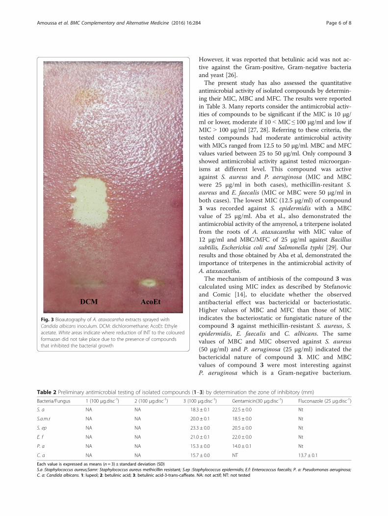
Fig. 3 Bioautography of A. ataxacantha extracts sprayed withCandida albicans inoculum. DCM: dichloromethane; AcoEt: Ethyleacetate. White areas indicate where reduction of INT to the colouredformazan did not take place due to the presence of compoundsthat inhibited the bacterial growth
Table 2 Preliminary antimicrobial testing of isolated compounds (1–3) by determination the zone of inhibitory (mm)
Bacteria/Fungus 1 (100 μg.disc-1) 2 (100 μg.disc-1) 3 (100 μg.disc-1) Gentamicin(30 μg.disc-1) Fluconazole (25 μg.disc-1)
S. a NA NA 18.3 ± 0.1 22.5 ± 0.0 Nt
S.a.m.r NA NA 20.0 ± 0.1 18.5 ± 0.0 Nt
S. ep NA NA 23.3 ± 0.0 20.5 ± 0.0 Nt
E. f NA NA 21.0 ± 0.1 22.0 ± 0.0 Nt
P. a NA NA 15.3 ± 0.0 14.0 ± 0.1 Nt
C. a NA NA 15.7 ± 0.0 NT 13.7 ± 0.1
Each value is expressed as means (n = 3) ± standard deviation (SD)S.a :Staphylococcus aureus;Samr: Staphylococcus aureus methicillin resistant; S.ep :Staphylococcus epidermidis; E.f: Enterococcus faecalis; P. a: Pseudomonas aeruginosa;C. a: Candida albicans. 1: lupeol; 2: betulinic acid; 3: betulinic acid-3-trans-caffeate. NA: not actif; NT: not tested
Amoussa et al. BMC Complementary and Alternative Medicine (2016) 16:284 Page 6 of 8

Several decades ago, the search for antimicrobials wasstill focused on the discovery of natural compoundsable to inhibit Gram-negative bacteria, which are dan-gerous and causing infectious diseases. The Gram-negative cell wall (made up of lipopolysaccharide) iscomplex and multilayered structure, which makes accessto membrane more restricted and barrier to many envir-onmental substances including synthetic and natural anti-biotics [30]. The results of this study indicate that thecompound 3, isolated from the bark of A. ataxacantha,could be an agent able to cross this complex barrier. Inthis study, compound 3 (betulinic acid-3-trans-caffeate) isfound more active than compound 2 (betulinic acid). Thisobservation is in accordance with the structure-activity re-lationship as reported previously [20, 31].
Antioxidant activityThe antioxidant activity of isolated compounds (1–3)were determined using DPPH method and the resultsare reported in Table 4. Compounds 3 (betulinic acid-3-trans-caffeate) had significant antioxidant activity withan IC50 of 3.57 μg/ml compared to quercetin (control)1.04 μg/ml. Compound 1 (lupeol) showed moderate ac-tivity with an IC50 value of 16.77 μg/ml while compound2 (betulinic acid) had weak DPPH scavenging activitywith an IC50 of 25.15 μg/ml. The antioxidant activity oflupeol was previously reported [32, 33]. The interestingantioxidant activity of compound 3 could be attributedto the phenolic nature of the caffeate substituent. Theantioxidant activity of Alkyl caffeates have been alsoreported [34].
ConclusionThe results of the present study showed that thebetulinic acid-3-trans-caffeate isolated from the barkof A. ataxacantha possesses good antimicrobial andantioxidant potency. This compound could be a candi-date for structure-activity study in the case of thedevelopment of novel antimicrobial agents with animproved therapeutic index. This is the first report onthe presence of lupeol, betulinic acid and betulinicacid-3-trans-caffeate in this specie. Further studies arein progress to identify the synergy between the iso-lated compounds and standard antibiotics.
Abbreviations1D, dimensional; 2D, two dimensional; ATCC, American Type Culture Collection;CFU, Colony-forming unit; CIP, collection institute Pasteur; COSY, CorrelationSpectroscopy; DCM, dichloromethane; DMSO, Dimethyl sulfoxide; DPPH, 2,2-diphenyl-1-picrylhydrazyl; EtoAc, ethyl acetate; HMBC, Heteronuclear MultipleBond Correlation; HPLC, High performance liquid chromatography; HSQC,Heteronuclear single quantum coherence spectroscopy; INT, p-iodonitrotetrazo-lium; MBC, minimum bactericidal concentration; MeOH, methanol; MFC,minimum fungicidal concentration; MHA, Mueller-Hinton agar; MIC, minimuminhibitory concentration; NMR, Nuclear magnetic resonance; NOESY, NuclearOverhauser Effect Spectroscopy; SDA Sabouraud Dextrose Agar; TFA,trifluoroacetic acid; TLC, thin layer chromatography
AcknowledgementsWe thank the International Foundation for Science (IFS) (Grant F / 5673-1)for supporting equipment and Wood-Wheland fellowship from InternationalUnion of Biochemistry and Molecular Biology (IUBMB) for the trip to Strasbourg.
FundingThis study was granted from International Foundation for Science (IFS)(Grant F/5673-1).
Availability of data and materialData are all contained within the paper.
Authors’ contributionsLL designed the study, followed the implementation, participated toisolate compounds, wrote the manuscript, AMA isolated compounds,carried out antimicrobial and antioxidant assay, MB and CVS carried outstructural identification of isolated compounds and participated to writethe manuscript, AS coordinate the team and helped to revise themanuscript. All authors read and approved the final manuscript.
Table 3 Minimum inhibitory concentration (MIC) and Minimumbactericidal and fungicidal concentrations (MBC, MFC) ofcompound 3 from A. ataxacantha
Minimum inhibitory concentrations (μg/ml)Microorganismsa Gram (+) bacteria Gram (-) bacteria Yeast
S. a S.a.m.r S. ep E. f P. a C. a
3b 50 25 12.5 25 25 12.5
Gentamicin 0.39 0.39 0.78 0.39 0.78 Nt
Fluconazole Nt Nt Nt Nt Nt 0.78
Minimum bactericidal and fungicidal (μg/ml)
3b 50 50 50 50 25 25
Gentamicin 0.78 1.56 1.56 0.78 1.56 Nt
Fluconazole Nt Nt Nt Nt Nt 1.56
MIC index
3b 1 2 4 2 1 2
Gentamicin 2 4 2 2 2 Nt
Fluconazole Nt Nt Nt Nt Nt 2aS.a : Staphylococcus aureus; S.a.m.r : Staphylococcus aureus methicillin resitant;S.ep : Staphylococcus epidermidis; E.f: Enterococcus faecalis; P.a: Pseudomonasaeruginosa; C.a: Candida albicansb3: betulinic acid-3-trans-caffeateNt: not tested. MIC, MBC or MFC of compounds 1 and 2 were not determined
Table 4 Antioxidant activity of compounds (1–3) isolated fromA. ataxacantha
Samples IC50 (μg/ml)
Compounds
1 16.77 ± 0.18
2 25.15 ± 0.01
3 3.57 ± 0.02
Reference
Qercetin 1.04 ± 0.01
The value of IC50 are expressed as means (n = 3) ± standard deviation (SD)1: lupeol, 2: betulinic acid, 3: betulinic acid-3-trans-caffeate
Amoussa et al. BMC Complementary and Alternative Medicine (2016) 16:284 Page 7 of 8

Competing interestsThe authors declare that they have no competing interests.
Consent for publicationNot applicable.
Ethics approval and consent to participateNot applicable.
Author details1Unité de Biochimie et Biologie Moléculaire, Equipe de Biochimie etSubstances Naturelles Bioactives, Faculté des Sciences et Techniques,Université d’Abomey-Calavi, Cotonou 04 BP 0320, Bénin. 2Laboratoired’Innovation Thérapeutique, Faculté de Pharmacie, UMR CNRS-Unistra 7200,Illkirch, France.
Received: 16 January 2016 Accepted: 5 August 2016
References1. Lalitha P, Sripathi SK, Jayanthi P. Acute toxicity study of extracts of
Eichhornia Crassipes (Mart.) Solms. Asian J Pharm Clin Res. 2012;5(4):59–61.2. Adamu M, Naidoo V, Eloff JN. Some southern African plant species used to
treat helminth infections in ethnoveterinary medicine have excellentantifungal activities. BMC Complement Altern Med. 2012;12:213.
3. Singh BN, Singh BR, Singh RI, Prakesh D, Dhakarey R, Upadhyay G, et al.Oxidative DNA Damage protective activity, antioxidant and antiquorumsensing potentiels of Moringa oleifera. Food Chem Toxicol. 2009;47:1109–16.
4. Adjanohoun I, Ahyi M, Aké A, Akouegninou A, Dalmeida J, Akpovo F, BoukeFK, et al. Contribution aux études ethnobotaniques et floristiques enRépublique Populaire du Bénin. Paris: ACCA; 1989. p. 852.
5. MacDonald I, Joseph OE, Harriet ME. Documentation of medicinal plantssold in markets in Abeokuta, Nigeria. Trop J Pharm Res. 2010;9(2):110–8.
6. Kereru PG, Kenji GM, Gachanga AN, Keriko JM, Mungai G. Traditional medicinesamong EMBU and Mbeere people of Kenya. Afr J CAM. 2007;4(1):75–86.
7. Amoussa AMO, Sanni A, Lagnika L. Antioxidant activity and total phenolic,flavonoid and flavonol contents of the bark extracts of Acacia ataxacantha.J Pharmacogn Phytochem. 2015;4(2):172–8.
8. Amoussa AMO, Tchatchedre M, Laleye A, Sanni A, Lagnika L. Acute toxicityand antifungal effects of Acacia ataxacantha (Bark). Int J PharmacognPhytochem Res. 2015;7(4):661–8.
9. Amoussa AMO, Lagnika L, Sanni A. Acacia ataxacantha (bark): chemicalcomposition and antibacterial activity of the extracts. Int J Pharm Pharm Sci.2014;6(11):138–41.
10. Olajuyigbe OO, Afolayan AJ. In vitro antibacterial and time-kill evaluation ofthe Erythrina caffra Thunb. extract against bacteria associated withdiarrhoea. Sci World J. 2012;2012:8. doi:10.1100/2012/738314.
11. Srinivas P, Rajashekar V, Upender RE, Venkateshwarulu L, Anil KCH.Phytochemical screening and in vitro antimicrobial investigation of themethanolic extract of Xanthium strumarium leaf. Int J Drug Dev Res.2011;3:286–93.
12. Qaralleh HN, Abboud MM, Khleifat KM, Tarawneh KA, Althunibat OY.Antibacterial activity in vitro of Thymus capitatus from Jordan. Pak J PharmSci. 2009;22(3):247–51.
13. Escalona-Arranz JC, Péres-Roses R, Urdaneta-Laffita I, Camacho-Pozo MI,Rodríguez-Amado J, Licea-Jiménez I. Antimicrobial activity of extracts fromTamarindus indica L. leave. Pharmacogn Mag. 2010;6(23):242–7.
14. Stefanovic O, Comic L. Inhibitory effect of Cytisus nigricans L. and Cytisuscapitatus scop on growth of bacteria. Afr J Microbiol Res. 2011;5:4725–30.
15. Danielle B, Lall N. Anticancer activity of certain herbs and spices on thecervical epithelial carcinoma (HeLa) cell Line. Evid Based Complement andAlternat Med. 2012;2012:1–11.
16. Moradkhani S, Kobarfard F, Ayatollahi SAM. Phytochemical investigation onchemical constituents of Achillea tenuifolia Lam. Iran J Pharm Res. 2014;13(3):1049–54.
17. Abdullahi SM, Musa AM, Abdullahi M, Sule MI, Sani YM. Isolation of lupeolfrom the Stem-bark of Lonchocarpus sericeus (Papilionaceae). Scholars AcadJ Biosci. 2013;1(1):18–9.
18. Jain PS, Bari SB. Isolation of lupeol, stigmasterol and campesterol frompetrolium ether extract of woody stem of Whrightia tinctoria. Asian J PlantSci. 2010;9(3):163–7.
19. Getahun T, Reneela P, Aman D. Isolation and characterization of naturalproducts from Helinus mystachnus (Rhamnaceae). J Chem Pharm Res. 2012;4(3):1756–62.
20. Mutai C, Bii C, Vagias C, Abatis D, Roussis V. Antimicrobial activity of Acaciamellifera extracts and lupane triterpenes. J Ethnopharmacol. 2009;123:143–8.
21. Kim KH, Choi SU, Lee KR. Bioactivity-guided isolation of cytotoxictriterpenoids from the trunk of Berberis koreana. Bioorg Med Chem Lett.2010;20(2010):1944–7.
22. Hee KJ, Byun JC, Bandi AKR, Hyun C-G, Lee H. Compounds with elastaseinhibition and free radical scavenging activities from Callistemon lanceolatus.J Med Plants Res. 2009;3(11):914–20.
23. Pan H, Lundgren LN, Anderson R. Triterpene caffeates from bark of Betulapubescens. Phytochemistry. 1994;37:795–9.
24. Eloff JN, Masoko P. The diversity of antifungal compounds of six SouthAfrican Terminalia species (Combretaceae) determined by bioautography.Afr J Biotechnol. 2005;4:1425–31.
25. Ghosh P, Mandal A, Chakraborty P, Rasul MG, Chakraborty M, Saha A.Triterpenoids from Psidium guajava with Biocidal Activity. Indian J PharmSci. 2010;72(4):504–7. doi:10.4103/0250-474X.73936.
26. Braca A, Morelli I, Mendez J, Battinelli L, Braghiroli L, Mazzanti G.Antimicrobial Triterpenoids from Licania heteromorpha. Planta Med. 2000;66(8):768–9. doi:10.1055/s-2000-9601.
27. Kuete V. Potential of Cameroonian plants and derived products againstmicrobial infections: A review. Planta Med. 2010;76:1479–91.
28. Ndjateu FST, Tsafack RBN, Nganou BK, Awouafack MD, Wabo HK, Tene M, etal. Antimicrobial and antioxidant activities of extracts and ten compoundsfrom three Cameroonian medicinal plants: Dissotis perkinsiae(Melastomaceae), Adenocarpus mannii (Fabaceae) and Barteria fistulosa(Passifloraceae). S Afr J Bot. 2014;91:37–42.
29. Aba OY, Ezuruike IT, Ayo RG, Habila JD, Ndukwe GI. Isolation, antibacterialand antifungal evaluation of α-amyrenol from the root extract of Acaciaataxacantha DC. Sch Acad J Pharm. 2015;4(2):124–31.
30. Rakholiya K, Vaghela P, Rathod T, Chanda S. Comparative study ofhydroalcoholic extracts of Momordica charantia L. against foodbornepathogens. Indian J Pharm Sci. 2014;76(2):148–56.
31. Burger MCM, Terezan AP, Cunha GSO, Fernandes JB, da Silva MFGF, VieiraPC, Menezes ACS. Antimicrobial activity of the myrsinoic acid A fromMyrsine coriacea and the semi-synthetic derivatives. Rev Bras Farmacogn.2015;25:451–4.
32. Santiago LA, Mayor ABR. Lupeol: An antioxidant triterpene in Ficuspseudopalma Blanco (Moraceae). Asian Pac J Trop Biomed. 2014;4(2):109–18.doi:10.1016/S2221-1691(14)60218-5.
33. Shirwaikar A, Setty MM, Bommu P, Krishnanand B. Effect of lupeol isolatedfrom Crataeva nurvala Buch.-Ham. stem bark extract against free radicalinduced nephrotoxicity in rats. Indian J Exp Biol. 2004;42:686–90.
34. Wang J, Gu H-S, Pang N, Wang F-Q, Pang F, Cui H-S, et al. Alkyl Caffeatesimprove the antioxidant activity, antitumor property and oxidation stabilityof edible oil. PLoS ONE. 2014;9(4):e95909.
• We accept pre-submission inquiries
• Our selector tool helps you to find the most relevant journal
• We provide round the clock customer support
• Convenient online submission
• Thorough peer review
• Inclusion in PubMed and all major indexing services
• Maximum visibility for your research
Submit your manuscript atwww.biomedcentral.com/submit
Submit your next manuscript to BioMed Central and we will help you at every step:
Amoussa et al. BMC Complementary and Alternative Medicine (2016) 16:284 Page 8 of 8