IN SITU EXTRA CORPOREAL SHOCK-WAVE LITHOTRIPSY AS A PRIMARY TREATMENT FOR URETERAL STONES
Upload
independentCategory
view
2download
0
Kidney International, Vol. 58 (2000), pp. 2301–2313
HORMONES – CYTOKINES – SIGNALING
Antibody to transforming growth factor-b ameliorates tubularapoptosis in unilateral ureteral obstruction
AKIRA MIYAJIMA, JIE CHEN, CATHY LAWRENCE, STEVE LEDBETTER, ROBERT A. SOSLOW,JOSHUA STERN, SHARDA JHA, JOSEPH PIGATO, MATTHEW L. LEMER, DIX P. POPPAS,E. DARRACOTT VAUGHAN, JR., and DIANE FELSEN
Center for Pediatric Urology and Laboratory for Minimally Invasive Urologic Surgery, and Departments of Urology andPathology, Children’s Hospital of New York, and Weill Medical College of Cornell University, New York, New York;and The Genzyme Corporation, Framingham, Massachusetts USA
Conclusion. The present study strongly suggests that an anti-Antibody to transforming growth factor-b ameliorates tubularbody to TGF-b is a promising agent to prevent renal tubularapoptosis in unilateral ureteral obstruction.fibrosis and apoptosis in UUO.Background. Unilateral ureteral obstruction (UUO) is char-
acterized by progressive renal atrophy, renal interstitial fibro-sis, an increase in renal transforming growth factor-b (TGF-b),and renal tubular apoptosis. The present study was undertaken In unilateral ureteral obstruction (UUO), the obstructedto determine the effect of a monoclonal antibody to TGF-b kidney is characterized by interstitial fibrosis and an in-(1D11) in UUO. crease in transforming growth factor-beta (TGF-b). TGF-b
Methods. Mechanical stretch was applied to tubular epithe- is known to be profibrotic in several models of fibrosis,lial cells (NRK-52E) by a computer-assisted system. Three dosesand the use of anti–TGF-b antibody can ameliorate fi-of 1D11 (either 0.5, 2, or 4 mg/rat) were administered to ratsbrosis in glomerulonephritis [1]. Approximately 80% ofone day prior to UUO and every two days thereafter, and kid-the renal volume is occupied by renal tubules [2], and aneys were harvested at day 13. Fibrosis was assessed by measur-
ing tissue hydroxyproline and mRNA for collagen and fibronec- positive correlation was found between the histologictin. Apoptosis was assessed with the terminal deoxy transferase presence of tubular atrophy and a decreased glomerularuridine triphosphate nick end-labeling assay. TGF-b levels were filtration rate in glomerulonephritis [3]. In UUO, it hasdetermined by bioassay. Western blot and immunostaining were been suggested that renal tubular apoptosis is related toused to identify proliferating cell nuclear antigen (PCNA), p53, renal tissue loss and dysfunction [4]. TGF-b has beenbcl-2, and inducible nitric oxide synthase (iNOS).
found to stimulate apoptosis in human gastric cancerResults. Stretch significantly induced apoptosis in NRK-52Ecells [5], uterine epithelial cells [6], and hepatocytes [7],cells, which was accompanied by an increased release of TGF-b;although it has also been shown to inhibit apoptosis1D11 (10 mg/mL) totally inhibited stretch-induced apoptosis.
Control obstructed kidney contained 20-fold higher TGF-b as induced by serum deprivation, Fas, or b-amyloid peptidecompared with its unobstructed kidney; 1D11 neutralized tissue [8–10]. Moreover, TGF-b has been shown to modifyTGF-b of the obstructed kidney. Control obstructed kidney specifically p53 expression in hepatocytes [11], althoughexhibited significantly more fibrosis and tubular apoptosis than TGF-b–induced apoptosis is operative in a p53-indepen-its unobstructed counterpart, which was blunted by 1D11. In dent manner in myeloid leukemia cells [12]. TGF-b iscontrast, 1D11 significantly increased tubular proliferation. p53
also shown to decrease levels of bcl-2–related protein inimmunostaining was localized to renal tubular nuclei of controlB lymphoma cells [13]. However, the effect of TGF-bobstructed kidney and was diminished by 1D11. In contrast,on renal tubular apoptosis is not known.bcl-2 was up-regulated in the 1D11-treated obstructed kidney.
Nitric oxide (NO) is a multifunctional mediator, synthe-Total NOS activity and iNOS activity of the obstructed kidneywere increased by 1D11 treatment. sized by the action of NO synthase (NOS), which has also
been implicated in the fibrosis of UUO. It has been shownthat increasing NO through dietary arginine decreasesfibrosis in UUO [14] and, conversely, that iNOS 2/2Key words: renal tubular obstruction, cell death, fibrosis, progressive
renal atrophy, monoclonal antibody. mice with UUO have increased fibrosis (abstract; Hoch-berg et al, J Urol Suppl 159:131, 1998). NO and inducibleReceived for publication December 23, 1999NOS (iNOS) are shown to increase in the kidney ofand in revised form June 7, 2000
Accepted for publication June 14, 2000 TGF-b 2/2 mice, suggesting that TGF-b is a negativeregulator of NO in the kidney [15]. 2000 by the International Society of Nephrology
2301

Miyajima et al: Antibody to TGF-b in apoptosis2302
In the present experiments, we examined the role of nal deoxy transferase uridine triphosphate nick end-labeling (TUNEL) assay (Intergen Co., Purchase, NY,TGF-b in renal tubular fibrosis and apoptosis by using
1D11, a monoclonal antibody to TGF-b. We examined USA). For flow cytometry, FITC-labeled antidigoxi-genin Ab was applied. Then cells were analyzed on thethe effect of stretch and 1D11 on tubular apoptosis, in
vitro. In UUO, renal tubules may be stretched because flow cytometer after washing. During analysis, cells weregated on forward versus side scatter, and apoptosis-asso-of changes in intrarenal pressure. Therefore, we used
1D11 in vivo to assess its effect on apoptosis in UUO. ciated fluorescence was measured using a log scale.In addition, since TGF-b is a negative regulator of NO,
In vivo unilatereral ureteral obstruction modelthe effects of the antibody on NO expression in UUOwere examined. We utilized 1D11 in rats that were sub- Sprague-Dawley rats (N 5 10 per group) underwent left
unilateral ureteral ligation with 4-0 silk suture throughjected to UUO for 13 days.an abdominal midline incision under sterile conditions.Animals were anesthetized intraperitoneally with 0.35 cc
METHODSpentobarbital (6.5 mg/mL). Animals were housed in a
Materials group of three to five and were fed standard chow andwater ad libitum. Monoclonal antibody to TGF-b (1D11)NRK-52E cells, a clonal line established from normal
rat kidney tubular epithelial cells (CRL1571), were ob- was generously provided by the Genzyme Corp. (Cam-bridge, MA, USA). This antibody has been previouslytained from the American Type Culture Collections
(Rockville, MD, USA). Dulbecco’s modified Eagle’s me- tested, and it was shown that doses as high as 2.5 mg permouse, administered daily for three weeks, showed nodium (DMEM) and fetal calf serum (FCS) were obtained
from GIBCO BRL (Grand Island, NY, USA). histopathology in any organs of the body (unpublishedobservations, B. Pratt, S. Ledbetter, Genzyme Corp.).
Cell culture Three doses of 1D11 (0.5, 2, and 4 mg/rat) were adminis-tered by intraperitoneal injection. An antiverotoxin anti-NRK-52E cells were carried in a monolayer cell cul-
ture and maintained in 75 cm2 culture flask (Corning, body (2 mg/rat) was administered as an isotype control.1D11 was injected to each animal at days 21, 0, 2, 4, 6,Cambridge, MA, USA) with DMEM containing 5%
FCS, 100 U/mL penicillin, and 100 mg/mL streptomycin 8, 10, and 12, and animals were euthanized at day 13.After the administration of intraperitoneal pentobarbi-in a humidified atmosphere of 5% CO2/95% O2 at 378C.tal, both obstructed and unobstructed kidneys were har-
Applying mechanical stretch to cells vested. Animal treatment adhered to approved institu-tional guidelines.Cells were seeded on six-well plates (25 mm diameter)
of flexible silicone or on rigid-bottom plates coated withTGF-b bioassaycollagen type I (Flex I and II culture plates, respectively;
Flexcell Corp., Mckeesport, PA, USA) and DMEM con- Transforming growth factor-b in cell supernatants orin tissue was assayed using a TGF-b bioassay. Tissuetaining 10% FCS. After five hours, the supernatant was
replaced with serum-free medium. NRK-52E cells were TGF-b was extracted as previously described [17]. Minklung epithelial cells (MLECs), which were transfectedsubjected to mechanical stretch using a Flexcell Strain
Unit FX-2000 (Flexcell Corp.). A microprocessor con- with a plasminogen activator inhibitor-1 (PAI-1) pro-moter driving expression of luciferase in a TGF-b–trolled negative pressure to the flexible bottoms, resulting
in reproducible deformation of the silicone rubber and responsive manner, were used to analyze TGF-b activity[18]. These cells were a generous gift from Dr. Danielthe attached cells, providing a maximal elongation of
20%. All experiments were carried out using alternate Rifkin. Transfected MLEC cells (1.6 3 104 per well)were plated in 96-multiwell plate, and they were allowedcycles of five seconds of stretch and five seconds of relax-
ation at a rate of 6 cycles per minute at 378C, 5% CO2 to attach for three hours at 378C in a humidified atmo-sphere of 5% CO2/95% O2. After three hours, the me-in a humidified incubator. All treatments were done in
triplicate, and all experiments were replicated three to five dium was replaced with the test sample (either tissueextract or cell supernatant) and were incubated over-times. Cells were collected for analysis by flow cytometry.
Supernatant was collected for the TGF-b bioassay. night at 378C. For the bioassay, the TGF-b in the testsample was activated as follows: Samples were acidified
Flow cytometric detection of DNA strand breaks to pH 1.5 with 1N HCl (1 hour at 48C), followed byequilibration at neutral pH with 1N NaOH. At the endAfter various treatments, cells were removed from
the bottom with trypsinization. After washing cells with of the incubation, supernatants were discarded, and cellswere washed twice with PBS. Cells were extracted withphosphate-buffered saline (PBS), cells were fixed and
permeabilized with 70% ethanol at 48C for one hour cell lysis buffer (Promega Co., Madison, WI, USA) for30 minutes at room temperature. Cell extracts were[16]. Fragmented DNA was then detected by the termi-

Miyajima et al: Antibody to TGF-b in apoptosis 2303
added to 100 mL of luciferase reagent (Promega). Lucif- trophoresis and visualized by using fusifilm BAS-1500phosphoimager.erase activity was measured in a luminometer. A TGF-b1
standard curve was carried out in the range of 0.01 to Bands on the phosphoimager representing collagen typeIII and fibronectin genes were quantitated using Mac2.0 ng/mL. All assays were performed in triplicate. Re-
sults are normalized to cell number and are expressed BAS Version 2.4 software. In all samples, the expressionof each gene was corrected by dividing probe-specificas the active form of TGF-b in pg/mg tissue.signal by that obtained for the housekeeping gene.
Assessment of tubular atrophy and interstitial fibrosisMeasurement of NO synthase activitySlides were examined by a board-certified pathologist
(R.A.S.). Tubular atrophy was assessed by the presence After harvesting obstructed and contralateral kidneyfrom rats, samples were stored in a 2808C freezer untilof luminal dilation and flattened tubular epithelial cells.
The general presence of interstitial fibrosis was assessed assay. Samples were homogenized with a glass-glassDounce homogenizer (Kontes, Vineland, NJ, USA) inin slides stained with Masson’s trichrome, when they were
examined for tubular atrophy. In addition, interstitial homogenization buffer [1 mmol/L ethylenediaminetetra-acetic acid (EDTA), 1 mmol/L egtazic acid (EGTA), andfibrosis was assessed by measurements of interstitial vol-
ume, using a point-counting method as previously de- protease inhibitor cocktail, pH 7.4]. The samples werecentrifuged for 30 minutes (10,000 3 g) at 48C and werescribed [17]. Changes in the mRNA for collagen III and
fibronectin were assessed by RNAse protection assays. resuspended. Samples were then incubated in buffer con-taining 1 mmol/L nicotinamide adenine dinucleotide phos-
Reverse transcription-polymerase chain reaction phate; Sigma, St. Louis, MO, USA), 10 mmol/L tetrahydro-biopterin, 60 nmol/L calmodulin (Calbiochem, CA, USA),Reverse transcription-polymerase chain reaction was
done on total cellular RNA using synthesized oligonucle- 200 mmol/L CaCl2, 20 mmol/L l-arginine, and tritium-labeled arginine (0.1 mCi/sample; DuPont) at 378C forotide primers that are complementary to the fibronectin
cDNA sequence [19] for EDA domain of fibronectin and one hour in the presence and absence of 1 mmol/LEGTA (Ca chelator) or L-NMA (NOS inhibitor) [21].collagen type III cDNA sequence [20].
Preparation of riboprobe. Linearized cDNA was tran- Reactions were terminated with stop buffer (C2H3O2Na,citrulline, EDTA) and then passed over 1 mL of anscribed in vitro using the Maxiscript kit (Ambion, Austin,
TX, USA) according to the manufacturer’s instructions. exchange resin, DOWEX 50WX8-100 (Sigma). Tritiatedcitrulline was then measured by counting in a scintillationT7 polymerase and p32CTP (3000 Ci/mmol; DuPont-
NEN, Boston, MA, USA) were included in the reaction counter. Measured NOS activity was expressed as pico-mole of citrulline per hour per milligram of protein.mixture to generate P32-labeled riboprobe. The reaction
mixture was incubated for 60 minutes at 378C, and thenWestern blot for iNOS, proliferating cell nuclearthe DNA template was removed by digestion with 0.5antigen, p53, and bcl-2U RNAse-free DNAse. Full-length probes were purified
from the transcription reaction by electrophoresis on 6% Samples were homogenized and centrifuged at 3000r.p.m. for 30 minutes. The supernatant was separated,polyacrylamide/TBE gel (Novax, Novel Experimental
Technology, San Diego, CA, USA), followed by autora- and the protein concentration of the lysate was deter-mined with the Bradford protein assay (Bio-Rad, Her-diography, excision from the gel band, and passive diffu-
sion into probe elution buffer (Maxiscript kit) overnight cules, CA, USA). For Western blot analysis, equal amountsof protein were loaded for sodium dodecyl sulfate-poly-at 378C. The activity of the probe was quantitated by
scintillation counting. acrylamide gel electrophoresis (SDS-PAGE). The pro-tein was then transferred to a nitrocellulose membraneRibonuclease protection assays. Total cellular RNA
from whole kidney tissue was obtained using the RNA- (Bio-Rad). The nitrocellulose membrane was blockedwith PBST (PBS with 0.1% Tween-20) solution containingqueous kit (Ambion). RNAse protection assay was per-
formed using the hybspeed RPA kit (Ambion) according 5% nonfat milk, and incubated overnight at 48C withprimary antibody against iNOS (polyclonal, 1:5000; Trans-to the manufacturer’s instructions. Briefly, radiolabeled
antisense RNA probe for fibronectin and collagen type duction Laboratories, Lexington, KY, USA), proliferatingcell nuclear antigen (PCNA; monoclonal, 1:1000; Dako,III was combined and hybridized with 10 mg of total
cellular RNA from different kidney samples. A probe Carpinteria, CA, USA), p53 (polyclonal, 1:2000; SantaCruz Technology, Santa Cruz, CA, USA), or bcl-2 (mono-for 18S RNA (Ambion) was also included in each hybrid-
ization mixture to normalize total RNA in individual clonal, 1:1000; Santa Cruz Technology). Subsequently, aone-hour incubation with horseradish peroxidase (HRP)-sample. For negative control, yeast RNA alone was com-
bined with probes. Digestion with RnaseA/RnaseT1 mix conjugated secondary antibody (1:5000) was carried out.After washing, streptavidin-HRP was applied to the mem-was performed to degrade unhybridized RNAs. Hybrid-
ized RNA protected from digestion was resolved by elec- brane for 30 minutes, followed by washing and applica-
Miyajima et al: Antibody to TGF-b in apoptosis2304
antibody. Biotinylated rabbit anti-mouse secondary anti-body (Vector) was incubated for 30 minutes. Sectionswere then incubated with avidin-biotin peroxidase com-plex (Vector Laboratories) and developed with diamino-benzedene (DAB). After washing slides, counterstainingwas done with 10% hematoxylin for one to two minutes.PCNA-positive cells of renal tubules, interstitium, andglomerulus were separately counted in 10 high-powerfields (3400) by two different independent investigatorsin a blind fashion.
In situ detection of DNA strand breaks
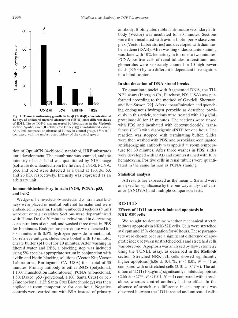
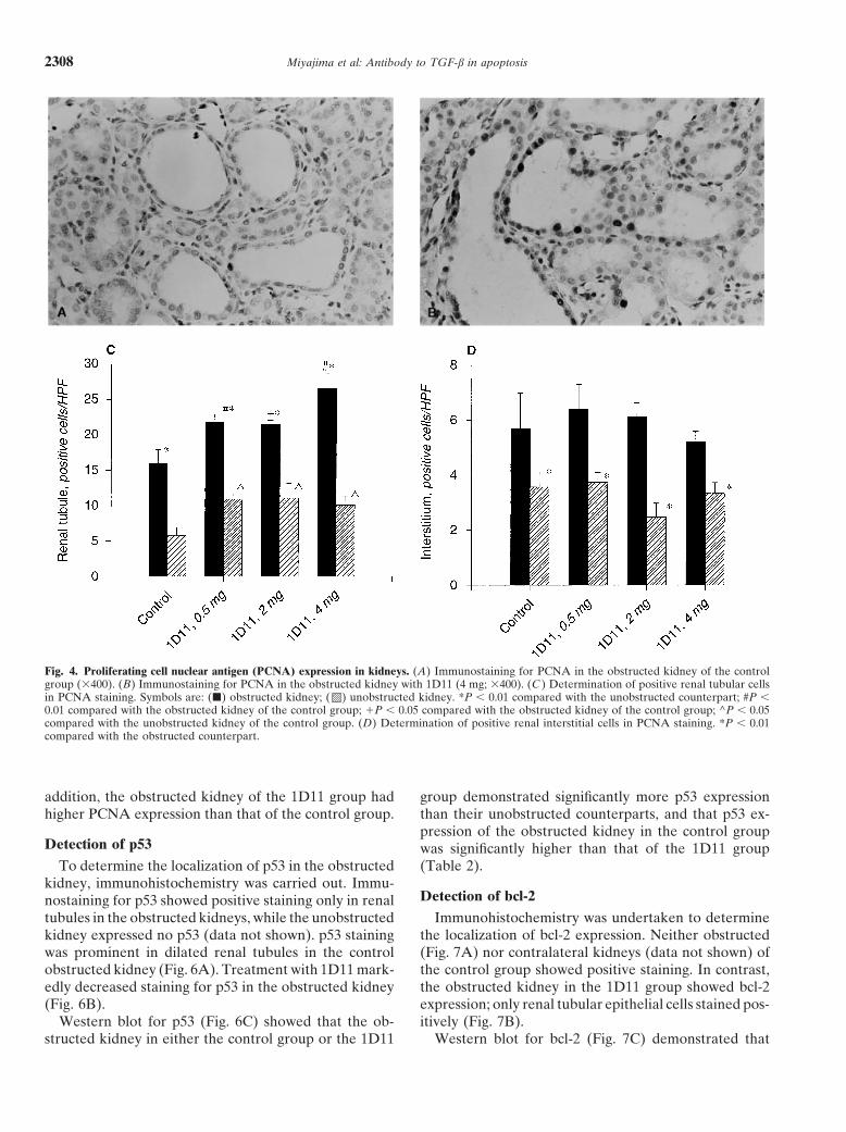
To quantitate nuclei with fragmented DNA, the TU-NEL assay (Intergen Co., Purchase, NY, USA) was per-formed according to the method of Gavrieli, Sherman,and Ben-Sasson [22]. After deparaffinization and quench-ing endogenous hydrogen peroxide as described previ-ously in this article, sections were treated with 10 mg/mLFig. 1. Tissue transforming growth factor-b (TGF-b) concentration at
13 days of unilateral ureteral obstruction (UUO) after different doses proteinase-K for 15 minutes. The sections were rinsedof 1D11. Tissue TGF-b was measured by bioassay as in the Methods with PBS and incubated with deoxynucleotidyl trans-section. Symbols are: (j) obstructed kidney; ( ) unobstructed kidney.
ferase (TdT) with digoxigenin-dNTP for one hour. The*P , 0.01 compared to obstructed kidney in control group. #P , 0.01compared with the unobstructed kidney of the control group. reaction was stopped with terminating buffer. Slides
were then washed with PBS, and peroxidase-conjugatedantidigoxigenin antibody was applied at room tempera-ture for 30 minutes. After three washes in PBS, slidestion of Opti-4CN (4-chloro-1 naphthol, HRP substrate)were developed with DAB and counterstained with 10%until development. The membrane was scanned, and thehematoxylin. Positive cells in renal tubules were quanti-intensity of each band was quantitated by NIH imagetated in the same fashion as PCNA staining.(software downloaded from the Internet). iNOS, PCNA,
p53, and bcl-2 were detected as a band at 130, 36, 53,Statistical analysisand 26 kD, respectively. Intensity was expressed as an
arbitrary unit. All results are expressed as the mean 6 SE and wereanalyzed for significance by the one-way analysis of vari-
Immunohistochemistry to stain iNOS, PCNA, p53, ance (ANOVA) and multiple comparison tests.and bcl-2
Wedges of hemisected obstructed and contralateral kid-RESULTSneys were placed in neutral buffered formalin and were
embedded in paraffin. Paraffin-embedded sections (5 mm) Effects of 1D11 on stretch-induced apoptosis inwere cut onto glass slides. Sections were deparaffinized NRK-52E cellswith Hemo-De for 30 minutes, rehydrated in decreasing We sought to determine whether mechanical stretchconcentrations of ethanol, and washed three times in PBS induces apoptosis in NRK-52E cells. Cells were stretchedfor 10 minutes. Endogenous peroxidase was quenched for
at 6 cpm and 15% elongation for 48 hours. These parame-30 minutes with 0.3% hydrogen peroxide in methanol.
ters were chosen because a significant difference of apo-To retrieve antigen, slides were boiled with 10 mmol/Lptotic index between unstretched cells and stretched cellscitrate buffer (pH 6.8) for 10 minutes. After washing inwas observed. Apoptosis was analyzed by flow cytometryfiltered water and PBS, a blocking step was includedusing the TUNEL assay, as described in the Methodsusing 5% species-appropriate serum in conjunction withsection. Stretched NRK-52E cells showed significantlyavidin and biotin blocking solutions (Vector Kit; Vectorhigher apoptosis (6.06 6 0.41%, P , 0.01, N 5 4) asLaboratories, Burlingame, CA, USA) for a total of 30compared with unstretched cells (3.10 6 0.47%). The ad-minutes. Primary antibody to either iNOS (polyclonal,dition of 1D11 (10 mg/mL) significantly inhibited apoptosis1:100; Transduction Laboratories), PCNA (monoclonal,(2.66 6 0.27%, P , 0.01, N 5 4) compared with stretch1:50; Dako), p53 (polyclonal, 1:100; Santa Cruz) or bcl-alone, whereas control antibody had no effect. In the2 (monoclonal, 1:25; Santa Cruz Biotechnology) was thenabsence of stretch, no difference in an apoptosis wasapplied at room temperature for one hour. Negative
controls were carried out with BSA instead of primary observed between the 1D11 treated and untreated cells.

Miyajima et al: Antibody to TGF-b in apoptosis 2305
Fig. 2. (A) Determination of positive tubular cells in the TUNEL assay.Symbols are (j) obstructed kidney; ( ) unobstructed kidney. *P , 0.01compared with the obstructed kidney of the control group. (B) TUNELassay for the obstructed kidney in the control group (3400). (C) TUNELassay for the obstructed kidney in the 1D11 group (4 mg; 3400).
Effects of 1D11 on TGF-b secretion from NRK-52E ure 1, tissue TGF-b content of the obstructed kidney incells exposed to mechanical stretch the control group (65.1 6 14.3 pg/mg tissue) was signifi-
cantly higher than the unobstructed kidney in the controlTo determine whether TGF-b is secreted by NRK-52Egroup (4.0 6 2.4 pg/mg tissue). However, the tissue TGF-bcells exposed to mechanical stretch, we measured TGF-bcontent of the obstructed kidney in each 1D11 group wasconcentration in the supernatant from NRK-52E cells withnot significantly different from that of its unobstructedthe bioassay. Whereas mechanical stretch significantly in-counterpart. Moreover, tissue TGF-b of the obstructedduces TGF-b (1.60 6 0.25 ng/mL, P , 0.01, N 5 4)kidney in each 1D11 group (1D11-4 mg, 8.9 6 5.2 pg/mgcompared with unstretched cells (1.04 6 0.13 ng/mL), thetissue) was significantly lower than that of the obstructedaddition of 1D11 (10 mg/mL) to stretched cells significantlykidney in the control group. In the unobstructed kidney,reduced the TGF-b concentration (0.88 6 0.09 ng/mL, P ,no significant difference was observed between the con-0.01, N 5 4) compared with cells stretched without 1D11.trol group and any of the 1D11 groups.In UUO, renal tubules may be stretched because of
changes in intrarenal pressure. Therefore, we were inter-In situ TUNELested in using 1D11 in vivo to assess its effect on apopto-
To determine renal tubular apoptosis, the TUNELsis in UUO. 1D11 was administered as described in theassay was performed in paraffin-embedded sections. TheMethods section, and both obstructed and contralateralobstructed kidney of the control group showed significantlykidneys were harvested on day 13 after UUO.higher tubular apoptosis [16.2 6 3.4 nuclei/high power
Measurement of kidney tissue TGF-b concentration field (HPF); Fig. 2A, B), as compared with the unob-in rats with UUO structed kidney of the control group (1.7 6 1.5 nuclei/
HPF). This was readily apparent from apoptotic cellTransforming growth factor-b concentration of kidneytissue was measured with the bioassay. As shown in Fig- counts (Fig. 2A) and the in situ TUNEL assay (Fig. 2B).

Miyajima et al: Antibody to TGF-b in apoptosis2306
Fig. 3. Representative trichrome staining of kidneys with or without 1D11treatment. (A) Control, unobstructed kidney. (B) Control, obstructedkidney. (C ) Obstructed kidney following 1D11 treatment (4 mg).
Fig. 6. Immunohistochemistry for p53. (A) Obstructed kidney, control group. (B) Obstructed kidney with 1D11, 4 mg.
Fig. 7. Immunohistochemistry for bcl-2. (A) Obstructed kidney, control group. (B) Obstructed kidney with 1D11, 4 mg.

Miyajima et al: Antibody to TGF-b in apoptosis 2307
Fig. 6. (C ) Western blot analysis for p53. Abbreviations are: Ob, ob- Fig. 7. (C ) Western blot analysis for bcl-2. Abbreviations are: Ob, ob-structed kidney; Un, contralateral unobstructed kidney; Marker, 51 kD. structed kidney; Un, contralateral unobstructed kidney; Marker, 26 kD.
Table 1. mRNA expression for collagen III and fibronectin with or without 1D11 treatment
Control 1D11 low dose 1D11 high dose
Unobstructed Obstructed Unobstructed Obstructed Unobstructed Obstructed
Collagen III 0.0360.01 0.0560.01a 0.0660.01 0.0660.02a 0.0560.01 0.0460.01Fibronectin 0.1260.02 0.1660.02a 0.0760.01 0.0760.01 0.1260.01 0.1060.01
a P , 0.05, as compared to control unobstructed kidney
Following 1D11 treatment, renal tubular apoptosis mRNA for collagen III and fibronectin in control andwas almost completely eliminated. The obstructed kid- 1D11-treated kidneys. The results of the RNAse protec-ney of all 1D11 groups showed significantly lower tubular tion assay are shown in Table 1. As can be seen, expres-apoptosis (1D11-4 mg: 1.1 6 0.2 nuclei/HPF; Fig. 2A, C) sion of mRNA for both collagen III and fibronectin wasthan that of the control group. In all 1D11 groups, there significantly increased in control obstructed kidneys; thiswas no significant difference between the obstructed and increase was abolished with 1D11 treatment.the unobstructed kidney. There was no significant differ-
Detection of PCNA expressionence in the unobstructed kidneys between 1D11 and thecontrol group. To determine the effect of TGF-b on renal cell prolif-
eration, immunostaining for PCNA was carried out. BothDetection of tubular atrophy and interstitial fibrosisrenal tubules and interstitial cells showed positive PCNA
Tubular atrophy was determined by examination of staining (Fig. 4A, B). To determine whether tubules, inter-Masson’s trichrome slides, as described in the Methods stitium, or glomerulus are affected by 1D11, positive cellssection. Figure 3 contains sections from either control were counted in each compartment. In both the controlkidneys (contralateral and obstructed) or following treat- group and the 1D11 group, renal tubules in the obstructedment with 1D11 (4 mg/rat). In the control contralateral kidneys show significantly more proliferation than in thekidney (Fig. 3A), it can be seen that the tubular lumens unobstructed kidney (Fig. 4C). Renal interstitial prolifer-are not dilated, and the epithelial cells are cuboidal in ation in the obstructed kidney was significantly highershape. The interstitium is not apparent, and the tubules
than in the unobstructed kidney (Fig. 4D). Proliferation ofare back to back. The control obstructed kidney (Fig. 3B)
the glomeruli showed no difference between the obstructedexhibits significant blue staining, signifying interstitialand the unobstructed kidney in all groups (data not shown).fibrosis and resulting in separation of the tubules fromProliferation of renal tubules in both the obstructed andone another. Furthermore, note the significant luminalthe unobstructed kidney treated with 1D11 was signifi-dilation and epithelial flattening. In contrast, the kidneycantly higher than in each counterpart without 1D11treated with a high dose of 1D11 (Fig. 3C) exhibits verytreatments (Fig. 4C). Proliferation of neither interstitiummild epithelial cell flattening and only minimal intersti-nor glomerulus in both obstructed and contralateral kid-tial fibrosis.neys was affected by treatment with 1D11.The change in interstitial volume was assessed by a
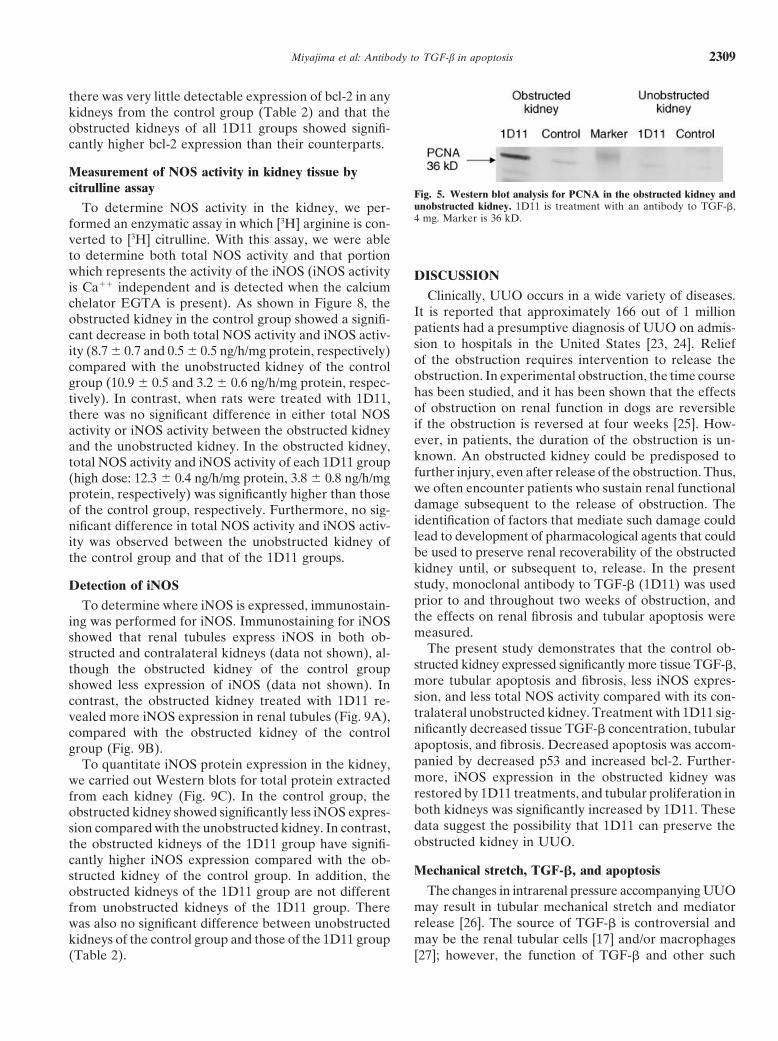
To determine whether total PCNA expression is in-point-counting technique. In the control cortex, intersti-creased in UUO, Western blot was performed for PCNAtial volume was increased from 6.5 6 0.5% to 26.7 6(Fig. 5). In the control group, PCNA expression of the3.9%. In the presence of high-dose 1D11, cortical inter-obstructed kidney was significantly higher than the unob-stitial volume in obstructed kidneys was only 12.1 6structed kidney (Table 2). The obstructed kidney of the1.4%. In the medulla of the obstructed kidney, the in-1D11 group showed significantly higher PCNA expres-crease in interstitial volume was decreased by 46% at the
highest dose of 1D11. We also assessed the expression of sion than unobstructed kidney of the 1D11 group. In
Miyajima et al: Antibody to TGF-b in apoptosis2308
Fig. 4. Proliferating cell nuclear antigen (PCNA) expression in kidneys. (A) Immunostaining for PCNA in the obstructed kidney of the controlgroup (3400). (B) Immunostaining for PCNA in the obstructed kidney with 1D11 (4 mg; 3400). (C ) Determination of positive renal tubular cellsin PCNA staining. Symbols are: (j) obstructed kidney; ( ) unobstructed kidney. *P , 0.01 compared with the unobstructed counterpart; #P ,0.01 compared with the obstructed kidney of the control group; 1P , 0.05 compared with the obstructed kidney of the control group; ^P , 0.05compared with the unobstructed kidney of the control group. (D) Determination of positive renal interstitial cells in PCNA staining. *P , 0.01compared with the obstructed counterpart.
addition, the obstructed kidney of the 1D11 group had group demonstrated significantly more p53 expressionthan their unobstructed counterparts, and that p53 ex-higher PCNA expression than that of the control group.pression of the obstructed kidney in the control group
Detection of p53 was significantly higher than that of the 1D11 group(Table 2).To determine the localization of p53 in the obstructed
kidney, immunohistochemistry was carried out. Immu-Detection of bcl-2nostaining for p53 showed positive staining only in renal
tubules in the obstructed kidneys, while the unobstructed Immunohistochemistry was undertaken to determinethe localization of bcl-2 expression. Neither obstructedkidney expressed no p53 (data not shown). p53 staining
was prominent in dilated renal tubules in the control (Fig. 7A) nor contralateral kidneys (data not shown) ofthe control group showed positive staining. In contrast,obstructed kidney (Fig. 6A). Treatment with 1D11 mark-
edly decreased staining for p53 in the obstructed kidney the obstructed kidney in the 1D11 group showed bcl-2expression; only renal tubular epithelial cells stained pos-(Fig. 6B).
Western blot for p53 (Fig. 6C) showed that the ob- itively (Fig. 7B).Western blot for bcl-2 (Fig. 7C) demonstrated thatstructed kidney in either the control group or the 1D11
Miyajima et al: Antibody to TGF-b in apoptosis 2309
there was very little detectable expression of bcl-2 in anykidneys from the control group (Table 2) and that theobstructed kidneys of all 1D11 groups showed signifi-cantly higher bcl-2 expression than their counterparts.
Measurement of NOS activity in kidney tissue bycitrulline assay Fig. 5. Western blot analysis for PCNA in the obstructed kidney and
unobstructed kidney. 1D11 is treatment with an antibody to TGF-b,To determine NOS activity in the kidney, we per-4 mg. Marker is 36 kD.formed an enzymatic assay in which [3H] arginine is con-
verted to [3H] citrulline. With this assay, we were ableto determine both total NOS activity and that portionwhich represents the activity of the iNOS (iNOS activity DISCUSSIONis Ca11 independent and is detected when the calcium
Clinically, UUO occurs in a wide variety of diseases.chelator EGTA is present). As shown in Figure 8, theIt is reported that approximately 166 out of 1 millionobstructed kidney in the control group showed a signifi-patients had a presumptive diagnosis of UUO on admis-cant decrease in both total NOS activity and iNOS activ-sion to hospitals in the United States [23, 24]. Reliefity (8.7 6 0.7 and 0.5 6 0.5 ng/h/mg protein, respectively)of the obstruction requires intervention to release thecompared with the unobstructed kidney of the controlobstruction. In experimental obstruction, the time coursegroup (10.9 6 0.5 and 3.2 6 0.6 ng/h/mg protein, respec-has been studied, and it has been shown that the effectstively). In contrast, when rats were treated with 1D11,of obstruction on renal function in dogs are reversiblethere was no significant difference in either total NOSif the obstruction is reversed at four weeks [25]. How-activity or iNOS activity between the obstructed kidneyever, in patients, the duration of the obstruction is un-and the unobstructed kidney. In the obstructed kidney,known. An obstructed kidney could be predisposed tototal NOS activity and iNOS activity of each 1D11 groupfurther injury, even after release of the obstruction. Thus,(high dose: 12.3 6 0.4 ng/h/mg protein, 3.8 6 0.8 ng/h/mgwe often encounter patients who sustain renal functionalprotein, respectively) was significantly higher than thosedamage subsequent to the release of obstruction. Theof the control group, respectively. Furthermore, no sig-identification of factors that mediate such damage couldnificant difference in total NOS activity and iNOS activ-lead to development of pharmacological agents that couldity was observed between the unobstructed kidney ofbe used to preserve renal recoverability of the obstructedthe control group and that of the 1D11 groups.kidney until, or subsequent to, release. In the presentstudy, monoclonal antibody to TGF-b (1D11) was usedDetection of iNOSprior to and throughout two weeks of obstruction, andTo determine where iNOS is expressed, immunostain-the effects on renal fibrosis and tubular apoptosis wereing was performed for iNOS. Immunostaining for iNOSmeasured.showed that renal tubules express iNOS in both ob-
The present study demonstrates that the control ob-structed and contralateral kidneys (data not shown), al-structed kidney expressed significantly more tissue TGF-b,though the obstructed kidney of the control groupmore tubular apoptosis and fibrosis, less iNOS expres-showed less expression of iNOS (data not shown). Insion, and less total NOS activity compared with its con-contrast, the obstructed kidney treated with 1D11 re-tralateral unobstructed kidney. Treatment with 1D11 sig-vealed more iNOS expression in renal tubules (Fig. 9A),nificantly decreased tissue TGF-b concentration, tubularcompared with the obstructed kidney of the controlapoptosis, and fibrosis. Decreased apoptosis was accom-group (Fig. 9B).panied by decreased p53 and increased bcl-2. Further-To quantitate iNOS protein expression in the kidney,more, iNOS expression in the obstructed kidney waswe carried out Western blots for total protein extractedrestored by 1D11 treatments, and tubular proliferation infrom each kidney (Fig. 9C). In the control group, theboth kidneys was significantly increased by 1D11. Theseobstructed kidney showed significantly less iNOS expres-data suggest the possibility that 1D11 can preserve thesion compared with the unobstructed kidney. In contrast,obstructed kidney in UUO.the obstructed kidneys of the 1D11 group have signifi-
cantly higher iNOS expression compared with the ob-Mechanical stretch, TGF-b, and apoptosisstructed kidney of the control group. In addition, the
The changes in intrarenal pressure accompanying UUOobstructed kidneys of the 1D11 group are not differentmay result in tubular mechanical stretch and mediatorfrom unobstructed kidneys of the 1D11 group. Thererelease [26]. The source of TGF-b is controversial andwas also no significant difference between unobstructedmay be the renal tubular cells [17] and/or macrophageskidneys of the control group and those of the 1D11 group
(Table 2). [27]; however, the function of TGF-b and other such

Miyajima et al: Antibody to TGF-b in apoptosis2310
Table 2. Quantification of Western blot results in control and 1D11-treated rats
Control 1D11-4 mg 1D11-2 mg 1D11-0.5 mg
Obstructed Unobstructed Obstructed Unobstructed Obstructed Unobstructed Obstructed Unobstructed
PCNA 79436821a 29246390 1569861428ab 57196567c ND ND ND NDp53 111376622a 12426308 19156197b 11686148 25286228b 12796230 40066266b 15526301bcl2 4626243 4086219 85216200ab 16316468 88996554ab 14686566 85816170ab 15916617iNOS 24646302c 53856391 549661201 49426690 51166967 559961813 42356680 53066820
Abbreviation is ND, not determined.a P , 0.01, compared to the unobstructed counterpartb P , 0.01, compared to the obstructed kidney of the control groupc P , 0.05, compared to the unobstructed kidney of the control group
increased TGF-b release and collagen synthesis, whichwere also reversed by an antibody to TGF-b [29]. Theapoptotic effects of stretch were not noted in these studies.
Mechanical stretch and TGF-b appear to be proapo-ptotic in vitro; however, it was not known whetherTGF-b could be implicated in the apoptosis of UUO invivo. Apoptosis in the obstructed kidney has been shownto be increased compared with the unobstructed kidneyin control animals [4]. In the present study, we confirmedthat apoptosis is increased in UUO. The administrationof the antibody to TGF-b strongly decreased renal levelsof TGF-b in the obstructed kidney. Concomitantly, apo-ptosis of tubular cells was dramatically decreased; eventhe lowest dose (0.5 mg) of the antibody to TGF-b de-creased renal tubular apoptosis by greater than 90%.p53 is known to be associated with cell proliferationand regulation of apoptosis [30], and TGF-b–induced
Fig. 8. Nitric oxide synthase (NOS) activity assay. NOS was measuredapoptosis is also reported to involve p53 up-regulationas the conversion of 3H-arginine to citrulline as described in the Methods
section. Abbreviations are: Ob, obstructed kidney; Un, contralateral [11]. In contrast, bcl-2 is known to play an anti-apoptoticunobstructed kidney; iNOS, inducible NOS. Symbols are: (j) control role in tubule damage [31]. We sought to determinegroup; (h) 1D11-0.5 mg; ( ) 1D11-2 mg; ( ) 1D11-4 mg *P , 0.05
whether p53 and bcl-2 are implicated in apoptosis of thiscompared with total NOS activity in the obstructed kidney of the controlgroup; #P , 0.05 compared with iNOS activity in the obstructed kidney model. Immunostaining, in the present study, showedof the control group; 1P , 0.05 compared with the unobstructed kidney that p53 is localized to the renal tubule of the obstructedof the control group (N 5 5).
kidney. Western blot showed that UUO increases p53expression. This is consistent with the previous demon-stration that p53 mRNA and protein were up-regulated
mediators in the sequelae of UUO is still unclear. To at five days after UUO, although p53 localization waselucidate the function of TGF-b in renal tubular apopto- not determined in that study [32]. Treatment with thesis, we stretched tubular epithelial cells and determined antibody to TGF-b decreased renal tubular expressionboth the TGF-b concentration of the supernatant and of p53 in the obstructed kidney, suggesting that p53 ex-apoptosis in those cells. Our results show that mechanical pression in UUO is dependent on TGF-b.stretch induces TGF-b and apoptosis significantly. The On the other hand, preliminary data regarding bcl-2addition of 1D11, in contrast, neutralized TGF-b and re- in UUO had been contradictory. Chevalier reported, induced apoptosis. This suggests that TGF-b is involved the rat UUO model, bcl-2 expression in the contralateralin apoptosis in renal tubular cells exposed to mechanical control kidneys, which was lost in the obstructed kidneysstretch. TGF-b has been previously implicated in stretch [33]. Shappell et al showed that bcl-2 is detectable in theresponses of mesangial and aortic cells. It has been shown human obstructed kidney, especially in dilated tubulesthat stretch of mesangial cells results in increased TGF-b (abstract; Lab Invest 74:169A, 1996). Our immunostain-secretion, as well as collagen synthesis and breakdown. ing showed that bcl-2 is localized in the dilated renalAn antibody to TGF-b was able to reverse these changes, tubule in the obstructed kidney with 1D11, not in thewhen cells were stretched in a high-glucose medium [28]. contralateral unobstructed kidney. However, we could
not detect bcl-2 in both the obstructed and contralateralSimilarly, in aortic smooth muscle cells, stretch induced

Miyajima et al: Antibody to TGF-b in apoptosis 2311
Fig. 9. Inducible nitric oxide synthase (iNOS) expansion in kidney. (A)Immunostaining for iNOS in obstructed kidney with 1D11 (4 mg; 3400).(B) Immunostaining for iNOS in the control obstructed kidney (3400).(C ) Western blot analysis for iNOS. Abbreviations are: Ob, obstructedkidney; Un, contralateral unobstructed kidney; Pos.Ctrl, positive control(purified macrophage iNOS). Marker is 130 kD.
kidneys in the control group, which is partially consistent treatment. These results suggest a beneficial effect ofinhibiting TGF-b in UUO. The effects of 1D11 on fibro-with the results of Chevalier. Bcl-2 in the human kidneys
could be the result of UUO of longer duration than in sis are consistent with previous reports of use of antibodyto TGF-b in models of glomerulonephritis or cornealthe present study or species differences. Furthermore,
Western blot showed that the obstructed kidney of con- fibrosis [1, 34].trol group expresses little bcl-2 and that the treatment
Interaction of NO and TGF-b in UUOwith antibody to TGF-b increases bcl-2 expression inthe obstructed kidney. Of particular interest is the finding Transforming growth factor-b is also known to be a
negative regulator of iNOS [15]. The increased TGF-b,that neutralizing TGF-b by 1D11 resulted in bcl-2 up-regulation in the obstructed kidney, while the control which is found after UUO, could be expected to down-
regulate iNOS in the obstructed kidney of the controlcontralateral kidney expresses little bcl-2. This demon-strates the appearance of bcl-2 in the obstructed kidney, group; 1D11 treatment should be associated with in-
creased iNOS and NO in the obstructed kidney as com-but not in the contralateral unobstructed kidney, subse-quent to TGF-b neutralization and removal of TGF-b’s pared with control. Both total NOS activity and iNOS
activity were determined by citrulline assay, and iNOSinhibition of bcl-2.protein was detected with a Western blot. These studies
Fibrosis and TGF-b demonstrated that the obstructed kidney of controlgroup at day 13 had significantly less NOS expressionThe obstructed kidney is characterized by interstitial
fibrosis and tubular atrophy [24]. In the present experi- compared with the unobstructed kidney. This finding isconsistent with our previous results that mice, at day 14ments, interstitial fibrosis was identified in several ways.
We noted increases in interstitial volume, tissue hydroxy- after UUO, had significantly less NOS activity in theobstructed kidney as compared with the unobstructedproline, and mRNA for collagen III and fibronectin.
These parameters were significantly decreased by 1D11 kidney [abstract; Miyajima et al, J Urol 163(Suppl):83,2000]. In the groups with 1D11, NOS expression andtreatment. Tubular atrophy was also diminished by 1D11
Miyajima et al: Antibody to TGF-b in apoptosis2312
NOS activity of the obstructed kidney were similar to NOTE ADDED IN PROOFthe unobstructed kidney. Therefore, 1D11 contributes The abstract by Hochberg et al on p. 2301 is now an article in press.
The complete citation is: Hochberg D, Johnson CS, Chen J, Cohento the maintenance of NOS expression in the obstructedD, Stern J, Vaughan ED Jr, Poppas DP, Felson D: Interstitial fibrosiskidney by neutralizing TGF-b, suggesting the existenceof unilateral obstruction is exacerbated in kidneys of mice lacking the
of an interaction between TGF-b and NO in UUO. gene for inducible nitric oxide synthase. Lab Invest 80:1721–1728, 2000.
Role of TGF-b and NO in renal tubular proliferation ACKNOWLEDGMENTSIt has been reported that TGF-b inhibits proliferation Akira Miyajima was supported in part by a grant from the Japan
Foundation of Cardiovascular Research. Joshua Stern was in partin tubular cells and glomerular endothelial cells [35].supported by a grant from the Fan Fox and Leslie R. Samuels Founda-NO has also been shown to be antiproliferative in renal tion. We thank Dr. Jo A. Hannafin for her helpful assistance in the
tubules [abstract; Miyajima et al, J Urol 163(Suppl):83, Flexcell system and Dr. Steven S. Gross for generous help in thecitrulline assay.2000]. Proliferation of the obstructed kidney has pre-
viously been studied [4]. An increase in proliferation of Reprint requests to Diane Felsen, Ph.D., Weill Medical College ofboth tubular and interstitial cells, with different time Cornell University, Department of Urology, Box 94, 1300 York Avenue,
New York, New York 10021, USA.courses, was demonstrated. Glomerular proliferationE-mail: [email protected]
was minimal. In the present study, we confirmed thatthere was more proliferation of both tubular and intersti- REFERENCEStial cells in the obstructed kidney compared with the con-
1. Border WA, Okuda S, Languino LR, Sporn MB, Ruoslahtitralateral side. Glomerular proliferation was unchanged.E: Suppression of experimental glomerulonephritis by antiserum
Treatment with 1D11 significantly increased prolifera- against transforming growth factor beta 1. Nature 346:371–374,1990tion in the obstructed and contralateral kidneys. As shown
2. Eddy AA: Experimental insights into the tubulointerstitial diseasehere, 1D11 treatment decreases the antiproliferative cy- accompanying primary glomerular lesions. (editorial) J Am Soctokine TGF-b and increases NO synthesis, which would Nephrol 5:1273–1287, 1994
3. Risdon RA, Sloper JC, De Wardener HE: Relationship betweenincrease tissue NO, the antiproliferative cytokine. Thus,renal function and histological changes found in renal-biopsy speci-
the suppression of TGF-b appears to be more important mens from patients with persistent glomerular nephritis. Lancet2:363–366, 1968to proliferative activity in UUO than the antiprolifera-
4. Truong LD, Petrusevska G, Yang G, Gurpinar T, Shappell S,tive effects of NO.Lechago J, Rouse D, Suki WN: Cell apoptosis and proliferation inexperimental chronic obstructive uropathy. Kidney Int 50:200–207,1996Role of an antibody to TGF-b in UUO
5. Yanagihara K, Tsumoraya M: Transforming growth factor-b1Transforming growth factor-b is a multifunctional cy- induces apoptotic cell death in cultured human gastric carcinoma
cells. Cancer Res 52:4042–4045, 1992tokine with sometimes opposing actions. It is profibrotic6. Rorke EA, Jacobberger JW: Transforming growth factor-beta 1and, in most systems, pro-apoptotic. Conversely, it is con-
(TGF beta 1) enhances apoptosis in human papillomavirus typesidered to be anti-inflammatory because of its effects on 16-immortalized human ectocervical epithelial cells. Exp Cell Res
216:65–72, 1995inflammatory cell migration and mediator release, and7. Sanchez A, Alvarez AM, Benito M, Fabregat I: Apoptosis in-effects on leukocyte adhesion [36, 37]. Thus, use of an duced by transforming growth factor-beta in fetal hepatocyte pri-
antibody to TGF-b may be associated with multiple and, mary cultures: Involvement of reactive oxygen intermediates. J BiolChem 271:7416–7422, 1996possible conflicting, effects. Nevertheless, in the present
8. Herbert JM, Carmeliet P: Involvement of u-PA in the anti-apo-study, we demonstrate that an antibody to TGF-b can ptotic activity of TGF-beta for vascular smooth muscle cells. FEBS
Lett 413:401–404, 1997modify an interaction between TGF-b, NO, p53, and9. Dybedal I, Guan F, Borge OJ, Veiby OP, Ramsfjell V, Nagatabcl-2, all contributing to the prevention of renal tubular S, Jacobsen SE: Transforming growth factor-beta1 abrogates Fas-
apoptosis and the increase in tubular proliferation in induced growth suppression and apoptosis of murine bone marrowprogenitor cells. Blood 90:3395–3403, 1997UUO. It is concluded that an antibody to TGF-b is a
10. Kim ES, Kim RS, Ren RF, Hawver DB, Flanders KC: Trans-promising agent for kidney preservation in UUO. When forming growth factor-beta inhibits apoptosis induced by beta-
amyloid peptide fragment 25-35 in cultured neuronal cells. Brainpatients present with obstructed kidney, a significantRes Mol Brain Res 62:122–130, 1998amount of time can elapse between the diagnosis and
11. Teramoto T, Kiss A, Thorgeirsson SS: Induction of p53 and Baxrelease of obstruction. In this time, the kidney can deteri- during TGF-beta 1 initiated apoptosis in rat liver epithelial cells.
Biochem Biophys Res Commun 251:56–60, 1998orate further. In addition, even if the obstruction is re-12. Selvakumaran M, Lin HK, Miyashita T, Wang HG, Krajewskileased, events occurring in the kidney can predispose it to S, Reed JC, Hoffman B, Liebermann D: Immediate early up-
further damage. Understanding the biochemical events regulation of bax expression by p53 but not TGF beta 1: A para-digm for distinct apoptotic pathways. Oncogene 9:1791–1798, 1994that induce renal apoptosis can lead to development
13. Saltzman A, Munro R, Searfoss G, Franks C, Jaye M, Ivash-of pharmacological agents, which could ameliorate, or chenko Y: Transforming growth factor-beta-mediated apoptosis
in the Ramos B-lymphoma cell line is accompanied by caspasereverse, the damage.

Miyajima et al: Antibody to TGF-b in apoptosis 2313
activation and Bcl-XL downregulation. Exp Cell Res 242:244–254, 25. Vaughan ED Jr, Gillenwater JY: Recovery following completechronic unilateral ureteral occlusion: Functional, radiographic and1998
14. Morrissey JJ, Ishidoya S, McCracken R, Klahr S: Nitric oxide pathologic alterations. J Urol 106:27–35, 197126. Ricardo SD, Ding G, Eufemio M, Diamond JR: Antioxidantgeneration ameliorates the tubulointerstitial fibrosis of obstructive
nephropathy. J Am Soc Nephrol 7:2202–2212, 1996 expression in experimental hydronephrosis: Role of mechanicalstretch and growth factors. Am J Physiol 272:F789–F798, 199715. Vodovotz Y, Geiser AG, Chesler L, Letterio JJ, Campbell
A, Lucia MS, Sporn MB, Roberts AB: Spontaneously increased 27. Diamond JR, Kees-Folts D, Ding G, Frye JE, Restrepo NC:Macrophages, monocyte chemoattractant peptide-1, and TGF-betaproduction of nitric oxide and aberrant expression of the inducible
nitric oxide synthase in vivo in the transforming growth factor beta 1 in experimental hydronephrosis. Am J Physiol 266:F926–F933,19941 null mouse. J Exp Med 183:2337–2342, 1996
16. Telford WG, King LE, Fraker PJ: Rapid quantitation of apopto- 28. Riser BL, Cortes P, Yee J, Sharba AK, Asano K, Rodriguez-Barbero A, Narins RG: Mechanical strain- and high glucose-sis in pure and heterogeneous cell populations using flow cytome-
try. J Immunol Methods 172:1–16, 1994 induced alterations in mesangial cell collagen metabolism: Roleof TGF-beta. J Am Soc Nephrol 9:827–836, 199817. Wright EJ, McCaffrey TA, Robertson AP, Vaughan ED Jr,
Felsen D: Chronic unilateral ureteral obstruction is associated 29. Li Q, Muragaki Y, Hatamura I, Ueno H, Ooshima A: Stretch-induced collagen synthesis in cultured smooth muscle cells fromwith interstitial fibrosis and tubular expression of transforming
growth factor-beta. Lab Invest 74:528–537, 1996 rabbit aortic media and a possible involvement of angiotensin IIand transforming growth factor-beta. J Vasc Res 35:93–103, 199818. Abe M, Harpel JG, Metz CN, Nunes I, Loskutoff DJ, Rifkin
DB: An assay for transforming growth factor-beta using cells trans- 30. Hartwell LH, Kastan MB: Cell cycle control and cancer. Science266:1821–1828, 1994fected with a plasminogen activator inhibitor-1 promoter-luciferase
construct. Anal Biochem 216:276–284, 1994 31. Basile DP, Liapis H, Hammerman MR: Expression of bcl-2 andbax in regenerating rat renal tubules following ischemic injury.19. Glumoff V, Makela JK, Vuorio E: Cloning of cDNA for rat pro
alpha 1 (III) collagen mRNA. Different expression patterns of Am J Physiol 272:F640–F647, 199732. Morrissey JJ, Ishidoya S, McCracken R, Klahr S: Control of p53type I and type III collagen and fibronectin genes in experimental
granulation tissue. Biochim Biophys Acta 1217:41–48, 1994 and p21 (WAF1) expression during unilateral ureteral obstruction.Kidney Int 50(Suppl 57):S84–S92, 199620. Odermatt E, Tamkun JW, Hynes RO: Repeating modular struc-
ture of the fibronectin gene: Relationship to protein structure and 33. Chevalier RL: Growth factors and apoptosis in neonatal ureteralobstruction. J Am Soc Nephrol 7:1098–1105, 1996subunit variation. Proc Natl Acad Sci USA 82:6571–6575, 1985
21. Ross ME, Iadecola C: Nitric oxide synthase expression in cerebral 34. Jester JV, Barry-Lane PA, Petroll WM, Olsen DR, CavanaghHD: Inhibition of corneal fibrosis by topical application of blockingischemia: Neurochemical, immunocytochemical, and molecular ap-
proaches. Methods Enzymol 269:408–426, 1996 antibodies to TGF beta in the rabbit. Cornea 16:177–187, 199735. Sharma K, Ziyadeh FN: The transforming growth factor-beta sys-22. Gavrieli Y, Sherman Y, Ben-Sasson SA: Identification of pro-
grammed cell death in situ via specific labeling of nuclear DNA tem and the kidney. Semin Nephrol 13:116–128, 199336. Smith WB, Noack L, Khew-Goodall Y, Isenmann S, Vadasfragmentation. J Cell Biol 119:493–501, 1992
23. Klahr S: Obstructive nephropathy. (Nephrology Forum) Kidney MA, Gamble JR: Transforming growth factor-beta 1 inhibits theproduction of IL-8 and the transmigration of neutrophils throughInt 54:286–300, 1998
24. Gulmi FA, Felsen D, Vaughan ED Jr: Pathophysiology of Uri- activated endothelium. J Immunol 157:360–368, 199637. Kitamura M, Suto TS: TGF-b and glomerulonephritis: Anti-in-nary Tract Obstruction, in Campbell’s Urology (7th ed), edited by
Walsh PC, Retik AB, Vaughan ED Jr, Wein AJ, Philadelphia, flammatory versus prosclerotic actions. Nephrol Dial Transplant12:669–679, 1997W.B. Saunders, 1998
Copyright © 2022 FDOKUMEN