Antibacterial Properties of the Leaf Extracts of Anthocleista djalonensis A. Chev on Some Pathogenic...
11
____________________________________________________________________________________________ *Corresponding author: Email: [email protected]; European Journal of Medicinal Plants 4(1): 75-85, 2014 SCIENCEDOMAIN international www.sciencedomain.org Antibacterial Properties of the Leaf Extracts of Anthocleista djalonensis A. Chev on Some Pathogenic Organisms A. I. Akinyemi 1* and A. O. Ogundare 1 1 Department of Microbiology, Federal University of Technology, Akure, Nigeria. Authors’ contributions: All the authors have cordially supported to the work and preparation of manuscript. Author AIA have designed the entire study and protocols with interpretations of the results and prepared the first draft of the manuscript. Authors AIA and AOO managed the analyses of the study and computational work respectively. Author AIA guided in the entire research and documented the final draft of the manuscript. All the authors have read and approved the final manuscript. Received 30 th March 2013 Accepted 14 th July 2013 Published 17 th October 2013 ABSTRACT Aims: To determine the antibacterial effect of methanol, petroleum ether and hot water extracts of Anthocleista djalonensis A. Chev on some pathogenic bacteria and to establish the use of the leaf extract in herbal medicine. Study Design: In vitro assay of antibacterial properties. Place and Duration of Study: Department of Microbiology, Federal University of Technology, Akure, Nigeria, between November 2011 and January 2012. Methodology: Collection of bacterial isolates; preparation of plant extracts; phytochemical screening; in vitro susceptibility test (agar well diffusion assay); minimum inhibitory concentration; antibiotics sensitivity test (disc diffusion assay); rate of killing of plant extracts; sodium and potassium ion leakage. Results: The results of the phytochemical screening showed the presence of tannins, saponins, flavonoids, steroid, terpenoid and cardiac glycosides. All the leaf extracts inhibited all the test organisms except Escherichia coli which was not inhibited by petroleum ether extract. The methanol extract had the highest effect on the test organisms. The minimum inhibitory concentrations (MICs) of the extracts ranged between Original Research Article
Transcript of Antibacterial Properties of the Leaf Extracts of Anthocleista djalonensis A. Chev on Some Pathogenic...
____________________________________________________________________________________________
*Corresponding author: Email: [email protected];
European Journal of Medicinal Plants4(1): 75-85, 2014
SCIENCEDOMAIN internationalwww.sciencedomain.org
Antibacterial Properties of the Leaf Extracts ofAnthocleista djalonensis A. Chev on Some
Pathogenic Organisms
A. I. Akinyemi1* and A. O. Ogundare1
1Department of Microbiology, Federal University of Technology, Akure, Nigeria.
Authors’ contributions:
All the authors have cordially supported to the work and preparation of manuscript. AuthorAIA have designed the entire study and protocols with interpretations of the results and
prepared the first draft of the manuscript. Authors AIA and AOO managed the analyses ofthe study and computational work respectively. Author AIA guided in the entire research and
documented the final draft of the manuscript. All the authors have read and approved thefinal manuscript.
Received 30th March 2013Accepted 14th July 2013
Published 17th October 2013
ABSTRACT
Aims: To determine the antibacterial effect of methanol, petroleum ether and hot waterextracts of Anthocleista djalonensis A. Chev on some pathogenic bacteria and toestablish the use of the leaf extract in herbal medicine.Study Design: In vitro assay of antibacterial properties.Place and Duration of Study: Department of Microbiology, Federal University ofTechnology, Akure, Nigeria, between November 2011 and January 2012.Methodology: Collection of bacterial isolates; preparation of plant extracts;phytochemical screening; in vitro susceptibility test (agar well diffusion assay); minimuminhibitory concentration; antibiotics sensitivity test (disc diffusion assay); rate of killing ofplant extracts; sodium and potassium ion leakage.Results: The results of the phytochemical screening showed the presence of tannins,saponins, flavonoids, steroid, terpenoid and cardiac glycosides. All the leaf extractsinhibited all the test organisms except Escherichia coli which was not inhibited bypetroleum ether extract. The methanol extract had the highest effect on the testorganisms. The minimum inhibitory concentrations (MICs) of the extracts ranged between
Original Research Article
European Journal of Medicinal Plants, 4(1): 75-85, 2014
76
10 mg/mL and 40 mg/mL. The result of the antibacterial activity of the leaf extractscompared favourably with the activity of standard antibiotics. It was observed that thenumber of bacterial cells was decreasing with increase in time of interaction between theextracts and the bacterial cells at a concentration 50 mg/mL of the extracts. There wasalso increase in the number of sodium and potassium ion leaked from the bacterial cellsby the leaf extracts.Conclusion: The results of the study indicate the antibacterial potential of Anthocleistadjalonensis A. Chev which may be a source of new bioactive compounds for drugdevelopment. The results obtained also establish the use of the plant in traditionalphytomedicine for the diseases caused by the microorganisms.
Keywords: Antibacterial; phytochemical screening; rate of killing; ion leakage.
1. INTRODUCTION
The increasing rate of infection by antibiotics-resistant microorganisms had causedworldwide concern which has led to a rising interest in the research for natural products fromplants for the discovery of new antimicrobials. Many of these plants have been known tosynthesize active secondary metabolites [1]. Santo et al. remarked that the World HealthOrganization has indeed recognized medicinal plants as the best source for obtaining avariety of synthetic drugs [2]. Therefore, plant materials remain an important resource tocombat serious diseases in the world to cover the basic health needs in the developingcountries.
Infections have increased, within the recent years to a great extent and antibiotics resistanceeffects become an ever-increasing therapeutic problem [3]. Antimicrobial resistance onenteric pathogen is of great public health concern in the developing world. Researchers havealso reported an increasingly wide spread use of antibiotics in food and animals contributingto high dissemination of resistant enteric infections to humans [4].
Anthocleista djalonensis A. Chev belongs to the family Gentianaceae and the Yoruba peopleof South west Nigeria refers to it as “Ewe Shapo” [5]. The tree grows up to 15 m tall; bole upto 40 cm in diameter; twigs sometimes with 2 erect spines or small cushions above the leafaxils [6]. The Genus Anthocleista comprises 14 species [7]; the West Africa species have thesame vernacular names (Cabbage tree) and are used by local practitioners for the samemedicinal purpose. Several members of the genus are used for similar medicinal purposes.It is very difficult to differentiate between the dried bark of the different species [8]. Some ofthe ethno-medical uses of the extract of Anthocleista djalonensis leaves, roots and stembark include treatment of wound, constipation, diarrhoea, dysentery, abdominal pain [9],hepatitis, jaundice, cirrhosis, fungal skin infection, filarial worm infections, acute inflammationand boils on skin [10].
In recent years, infections have increased to a great extent and antibiotics resistance effectsbecome an ever-increasing therapeutic problem [3]. Natural products of higher plants maypossess a new source of antimicrobial agents with possibly novel mechanisms of action[11,12]. Therefore, it is of great interest to carry out a screening of these plants in order tovalidate their use in folk medicine and to reveal the active principle by isolation andcharacterization of their constituents. Systematic screening of them may result in thediscovery of novel active compounds [13].
European Journal of Medicinal Plants, 4(1): 75-85, 2014
77
2. MATERIALS AND METHODS
2.1 Collection and Identification of Bacterial Isolates
The bacterial isolates used were collected from the Microbiology Laboratory, ObafemiAwolowo University Teaching Hospital complex, Ile-Ife, Osun State and they include:Staphylococcus aureus, Klebsiella pnuemoniae, Proteus mirabilis, Pseudomonasaeruginosa, Salmonella typhi and Escherichia coli.
2.2 Standardization of the Test Organisms
A loop full of test organism was inoculated on nutrient broth and incubated for 24 h. Exactly0.2 ml from the 24 h culture of the organisms was dispensed into 20 ml sterile nutrient brothand incubated for 3- 5h to standardize the culture to 0.5 McFarland standards (106cfu/ml)before use according to the method of Oyeleke et al. [14].
2.3 Collection and Extraction of Plant Material
The plant leaf was collected from the forest and wild life reserve of the Federal University ofTechnology, Akure, Nigeria and was authenticated by Prof. Akinyele of the Department ofCrop Science and Pest Management, where a voucher specimen was submitted. Theextraction of the plant material was done according to the method of Harbone, (1998) withslight modification. The leaves were air dried for three weeks and pulverized. Exactly 400 gof the powdered leaf were macerated separately for 72 h using methanol, petroleum etherand hot water followed by sieving with a muslin cloth and filtered with No 1 Whatman filterpaper. The filtrate was collected in a beaker and concentrated en vacuo using rotaryevaporator (Resona, Germany).
2.4 Phytochemical Screening
The extracts were screened for anthraquinone, alkaloid, tannin, saponin, phlobatannin,steroid, flavonoid, terpenoid and cardiac glycoside as described by Trease and Evans [16].
2.5 Experimental Procedure
2.5.1 Phytochemical properties of the leaf extracts of anthocleista djalonensis
The result of the phytochemical screening of the methanol, petroleum ether and hot waterleaf extracts of Anthocleista djalonensis shows the presence of tannins, saponins,flavonoids, steroid, terpenoid and cardiac glycosides
2.5.2 Antibacterial activity
2.5.2.1 Determination of antibacterial activities by leaf extract
The antibacterial activity of the plant extracts was determined using agar dilutionmethod described by Madigan et al. [17]. Exactly one milliliter of each of the standardizedtest organism was transferred into different sterile petri dishes. Mueller Hinton agar was thenpoured on these inocula and the plates swirled for even dispersion of the organism in theagar. After solidification of the agar, a 6 mm diameter cork borer was used to make wells in
European Journal of Medicinal Plants, 4(1): 75-85, 2014
78
to each plate and the prepared extract was introduced. The plates were incubated at 37ºCfor 24 h. Clear zones around the bored holes are indicative of the inhibition of the organismsby the extract. The activity indices (AI) were calculated as the division of zone of inhibition ofthe extract by that of the standard drug- Gentamycin [18].
2.5.2.2 Determination of the minimum inhibitory concentration
This was carried out using the agar dilution method described by Doughari et al. [19].Different concentrations of the extracts (30 mg/mL, 25 mg/mL, 12.5 mg/mL, 10 mg/mL, 6.25mg/mL, and 5mg/mL) were used. Plates were incubated at 37ºC for 24 h, after which theywere observed for clear zones around the wells, indicating inhibition. The concentrationbelow in which there was no zone was noted as minimum inhibitory concentration (MIC).
2.5.2.3 Antibiotics sensitivity test
The Kirby - Bauer test also known as disc diffusion method was used to determine the effectof standard antibiotics on the bacterial isolates as described by Willey et al., [20]. Standardantibiotics disc were placed aseptically on agar plates already seeded with the testorganisms using sterile forceps. The plates were then incubated at 37ºC for 24 hours. Zonesof inhibition around the antibiotics disc were measured in millimeters.
2.5.2.4 Determination of the rate of killing of leaf extract
This was done according to the method of Ogundare and Akinyemi [21] to determine the rateof killing of the test organisms by the extracts. Exactly 5 ml of the standardized broth culturewas introduced into 5mls of 50mg/mL concentration of each of the extracts. The suspensionwas mixed and then plated using pour plate method at 0, 1, 2, 4, 3, 4, 5, 6, 7, 8, 9, 10, 11, 12and 24h. The plates were incubated at 37ºC for 24 h after which observation was made formicrobial growth. The numbers of colonies were counted using the digital colony counter.
2.5.2.5 Determination of sodium and potassium ion leakage
Exactly 0.5 mL each of the standardized organism was added to 4.5 mL of the preparedconcentration of the leaf extracts and then incubated for 18 hours. The solution wascentrifuged at 7 000 revolution per minute (r. p. m.) and the supernatant analyzed using aflame photometer at 589 nm and 766 nm for sodium and potassium ion leakage respectively.The sodium and potassium ion leakage was determined using the method of Hugo andRussel [22].
3. RESULT AND DISCUSSION
Anthocleista djalonensis leaves are extensively used to treat diarrhoea, wound, abdominalpain, boils on skin and fungal skin infection. The result of the phytochemical screening of themethanol, petroleum ether and hot water leaf extracts of Anthocleista djalonensis shows thepresence of tannins, saponins, flavonoids, steroid, terpenoid and cardiac glycosides whichmay be attributed to different solvents used in the extraction as reported by Srinivasan et al.[23,24] and probably account for the antibacterial activity of the extracts may be due to thepresence of metabolites revealed in the phytochemical screening, which possesspharmacological activities responsible for the use of plants in traditional phytomedicine totreat diseases caused by pathogenic microorganisms [9].
European Journal of Medicinal Plants, 4(1): 75-85, 2014
79
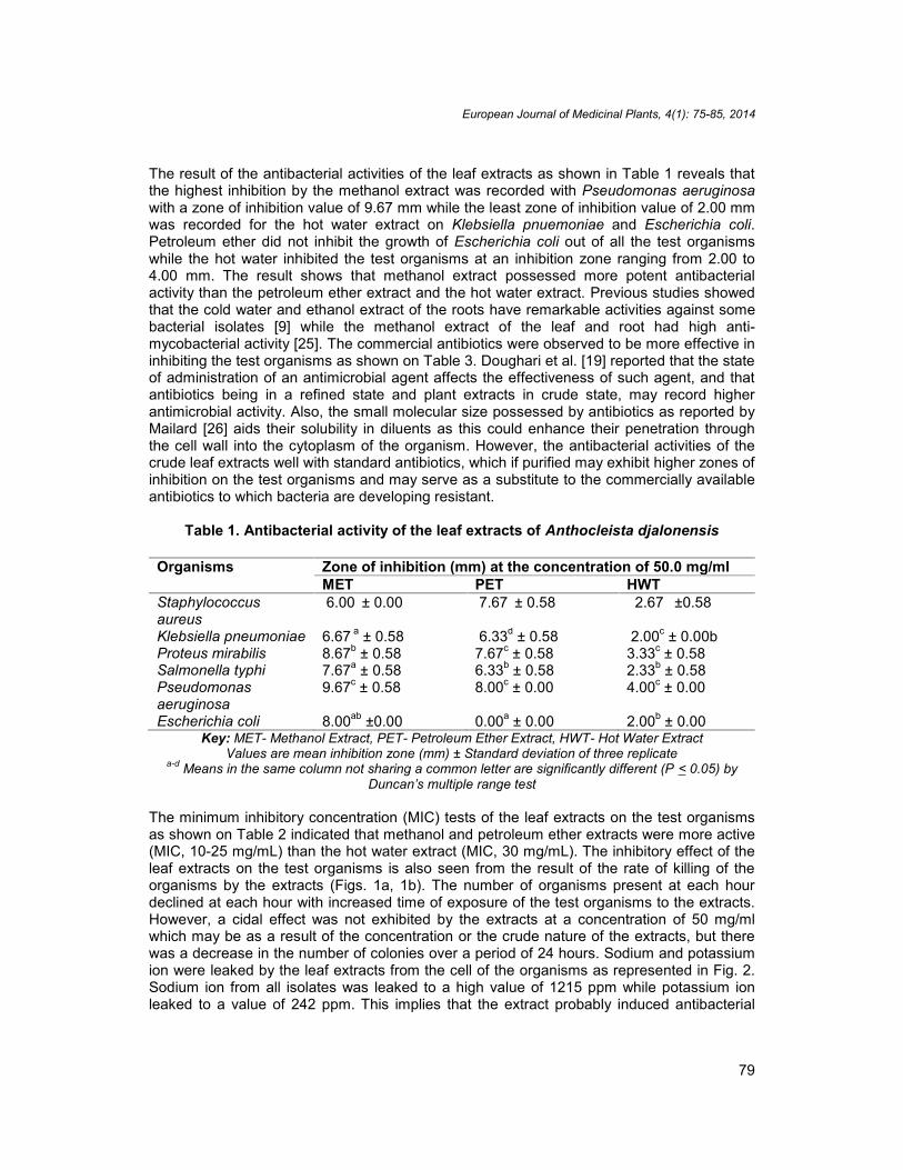
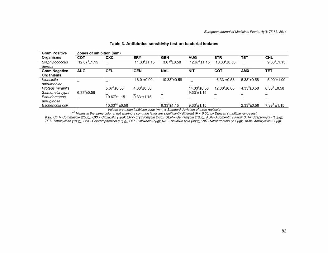
The result of the antibacterial activities of the leaf extracts as shown in Table 1 reveals thatthe highest inhibition by the methanol extract was recorded with Pseudomonas aeruginosawith a zone of inhibition value of 9.67 mm while the least zone of inhibition value of 2.00 mmwas recorded for the hot water extract on Klebsiella pnuemoniae and Escherichia coli.Petroleum ether did not inhibit the growth of Escherichia coli out of all the test organismswhile the hot water inhibited the test organisms at an inhibition zone ranging from 2.00 to4.00 mm. The result shows that methanol extract possessed more potent antibacterialactivity than the petroleum ether extract and the hot water extract. Previous studies showedthat the cold water and ethanol extract of the roots have remarkable activities against somebacterial isolates [9] while the methanol extract of the leaf and root had high anti-mycobacterial activity [25]. The commercial antibiotics were observed to be more effective ininhibiting the test organisms as shown on Table 3. Doughari et al. [19] reported that the stateof administration of an antimicrobial agent affects the effectiveness of such agent, and thatantibiotics being in a refined state and plant extracts in crude state, may record higherantimicrobial activity. Also, the small molecular size possessed by antibiotics as reported byMailard [26] aids their solubility in diluents as this could enhance their penetration throughthe cell wall into the cytoplasm of the organism. However, the antibacterial activities of thecrude leaf extracts well with standard antibiotics, which if purified may exhibit higher zones ofinhibition on the test organisms and may serve as a substitute to the commercially availableantibiotics to which bacteria are developing resistant.
Table 1. Antibacterial activity of the leaf extracts of Anthocleista djalonensis
Organisms Zone of inhibition (mm) at the concentration of 50.0 mg/mlMET PET HWT
Staphylococcusaureus
6.00 ± 0.00 7.67 ± 0.58 2.67 ±0.58
Klebsiella pneumoniae 6.67 a ± 0.58 6.33d ± 0.58 2.00c ± 0.00bProteus mirabilis 8.67b ± 0.58 7.67c ± 0.58 3.33c ± 0.58Salmonella typhi 7.67a ± 0.58 6.33b ± 0.58 2.33b ± 0.58Pseudomonasaeruginosa
9.67c ± 0.58 8.00c ± 0.00 4.00c ± 0.00
Escherichia coli 8.00ab ±0.00 0.00a ± 0.00 2.00b ± 0.00Key: MET- Methanol Extract, PET- Petroleum Ether Extract, HWT- Hot Water Extract
Values are mean inhibition zone (mm) ± Standard deviation of three replicatea-d Means in the same column not sharing a common letter are significantly different (P < 0.05) by
Duncan’s multiple range test
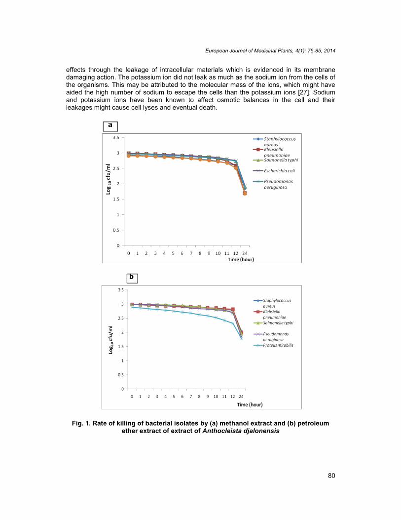
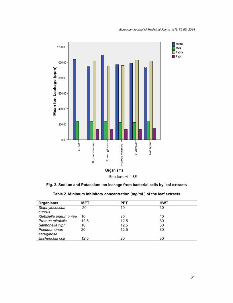
The minimum inhibitory concentration (MIC) tests of the leaf extracts on the test organismsas shown on Table 2 indicated that methanol and petroleum ether extracts were more active(MIC, 10-25 mg/mL) than the hot water extract (MIC, 30 mg/mL). The inhibitory effect of theleaf extracts on the test organisms is also seen from the result of the rate of killing of theorganisms by the extracts (Figs. 1a, 1b). The number of organisms present at each hourdeclined at each hour with increased time of exposure of the test organisms to the extracts.However, a cidal effect was not exhibited by the extracts at a concentration of 50 mg/mlwhich may be as a result of the concentration or the crude nature of the extracts, but therewas a decrease in the number of colonies over a period of 24 hours. Sodium and potassiumion were leaked by the leaf extracts from the cell of the organisms as represented in Fig. 2.Sodium ion from all isolates was leaked to a high value of 1215 ppm while potassium ionleaked to a value of 242 ppm. This implies that the extract probably induced antibacterial
European Journal of Medicinal Plants, 4(1): 75-85, 2014
80
effects through the leakage of intracellular materials which is evidenced in its membranedamaging action. The potassium ion did not leak as much as the sodium ion from the cells ofthe organisms. This may be attributed to the molecular mass of the ions, which might haveaided the high number of sodium to escape the cells than the potassium ions [27]. Sodiumand potassium ions have been known to affect osmotic balances in the cell and theirleakages might cause cell lyses and eventual death.
Fig. 1. Rate of killing of bacterial isolates by (a) methanol extract and (b) petroleumether extract of extract of Anthocleista djalonensis
European Journal of Medicinal Plants, 4(1): 75-85, 2014
81
Fig. 2. Sodium and Potassium ion leakage from bacterial cells by leaf extracts
Table 2. Minimum inhibitory concentration (mg/mL) of the leaf extracts
Organisms MET PET HWTStaphylococcusaureus
20 10 30
Klebsiella pneumoniae 10 25 40Proteus mirabilis 12.5 12.5 30Salmonella typhi 10 12.5 30Pseudomonasaeruginosa
20 12.5 30
Escherichia coli 12.5 20 30
Mea
n Io
n L
eaka
ge
(pp
m)
1200.00
1000.00
800.00
600.00
400.00
200.00
0.00
Organisms
Sal.
typhi
S. aure
us
Pro
teus
mirabili
s
P. aeru
gin
osa
K. pneum
onia
e
E. co
li
Error bars: +/- 1 SE
PetKPetNaMetkMetNa
European Journal of Medicinal Plants, 4(1): 75-85, 2014
82
Table 3. Antibiotics sensitivity test on bacterial isolates
Gram PositiveOrganisms
Zones of inhibition (mm)COT CXC ERY GEN AUG STR TET CHL
Staphylococcusaureus
12.67c±1.15 _ 11.33b±1.15 3.67a±0.58 12.67d±1.15 10.33d±0.58 _ 9.33b±1.15
Gram NegativeOrganisms
AUG OFL GEN NAL NIT COT AMX TET
Klebsiellapneumoniae
_ _ 16.0d±0.00 10.33b±0.58 _ 6.33b±0.58 6.33c±0.58 5.00a±1.00
Proteus mirabilis _ 5.67b±0.58 4.33b±0.58 _ 14.33d±0.58 12.00d±0.00 4.33c±0.58 6.33c ±0.58Salmonella typhi 6.33c±0.58 _ _ _ 9.33c±1.15 _ _ _Pseudomonasaeruginosa
_ 10.67f±1.15 9.33d±1.15 _ _ _ _ _
Escherichia coli _ 10.33de ±0.58 _ 9.33c±1.15 9.33c±1.15 _ 2.33b±0.58 7.33d ±1.15Values are mean inhibition zone (mm) ± Standard deviation of three replicate
a-d Means in the same column not sharing a common letter are significantly different (P < 0.05) by Duncan’s multiple range testKey: COT- Cotrimazole (25μg); CXC- Cloxacillin (5μg); ERY- Erythromycin (5μg); GEN – Gentamycin (10μg); AUG- Augmentin (30μg); STR- Streptomycin (10μg);TET- Tetracycline (10μg); CHL- Chloramphenicol (10μg); OFL- Ofloxacin (5μg); NAL- Nalidixic Acid (30μg); NIT- Nitrofurantoin (200μg); AMX- Amoxycillin (30μg).

European Journal of Medicinal Plants, 4(1): 75-85, 2014
83
4. CONCLUSION
The result showed that methanol and petroleum ether extracts exhibited considerableinhibitory activity against the test organisms as demonstrated by the diameters of the zonesof inhibition. The result from this work established its use traditionally as an antimicrobial.The methanol and petroleum ether extracts of the leaf are potential sources of newantibacterial agents for the treatment of diseases caused by the pathogenic organisms. Theresult of this study suggest that the extracts be purified further and tested for theirantibacterial potential as against the use of crude extracts. Also, further isolation andcharacterization of the active components contained in the leaf extracts be carried out.
The biodiversity of tropical forest plant species, coupled with the chemical diversity foundwithin each plant leads one to the conclusion that tropical plants are perhaps the mostvaluable source of new bioactive chemical entities.
CONSENT
Not applicable.
ETHICAL APPROVAL
Not applicable.
ACKNOWLEDGEMENTS
Authors are thankful to Microbiology Laboratory, Obafemi Awolowo University TeachingHospital complex, Ile-Ife, Osun State, for providing the test organisms.
COMPETING INTEREST
Authors have declared that no competing interests exist.
REFERENCES
1. Kambu K, Di Phanzu N, Coune C, Wauters JN, Angenol L. Contribution a` l`etude desproprietes insecticides et chimiques d` Eucalyptus saligna du Zay’’re. Plantes MedPhytother. 1982;16:34-8.
2. Santos PRV, Oliveira ACX, Tomassini TCB. Controle microbiógico de produtosfitoterápicos. Rev. Farm. Bioquím. 1995;31:35-38.
3. Mahesh B, Satish S. Antimicrobial activity of some important medicinal plants againstplant and human pathogens. World Journal Agricultural Science. 2008;4:839-843.
4. Yah CS, Chineye HU, Eghafona NO. Multi-antibiotics-resistance plasmid profile ofenteric pathogens in pediatric patients from Nigeria. Biokemistri, 2007;19(1):35-42.
5. Onocha PA, Okorie DA, Connolly JD, Krebs HC, Meier B, Habermehl GG. Cytotoxicactivity of the constituents of Anthocleista djalonensis and their derivatives. NigerianJournal of Natural Products and Medicine. 2003;7:58–60.
6. Jensen SR, Schripsema J. Chemotaxonomy and pharmacology of Gentianaceae. In:Struwe L. and Albert V. (Editors). Gentianaceae - Systematics and Natural History.Cambridge University Press, United Kingdom. 2002;573–631
European Journal of Medicinal Plants, 4(1): 75-85, 2014
7. Neuwinger HD. African traditional medicine: a dictionary of plant use and applications.Medpharm Scientific, Stuttgart, Germany. 2000;589
8. De Ruijter A. Anthocleista djalonensis. A. Chev. Record from Protobase. SchmelzerGH. & Gurib- Fakin A. PROTA (Plant Resourses of Tropical Africa); 2007.
9. Okoli AS, Iroegbu CU. Evaluation of Extracts of Anthocleista djalonensis, Nauclealatifolia and Uvaria afzalii for activity against bacterial isolates from cases of non-gonococcal urethritis. Journal of Ethnopharmacology. 2004;92(1):135-144.
10. Aiyeloja AA, Bellow OA. Ethnobotanical potentials of common herbs in Nigeria: ACase study of Enugu State. Educational Research and Review. 2006;1(1):16-22.
11. Ahmad I, Aqil F. In vitro efficacy of bioactive extracts of 15 medicinal plants againstESbL-producing multidrug-resistant enteric bacteria. Microbioliology Research.2007;162:264-275.
12. Barbour EK, Al Sharif M, Sagherian VK, Habre AN, Talhouk RS, Talhouk SN.Screening of selected indigenous plants of Lebanon for antimicrobial activity. Journalof Ethnopharmacology, 2004;93:1-7.
13. Tomoko N, Takashi A, Hiromu T, Yuka I, Hiroko M, Munekazu I, Totshiyuki T, TetsuroI, Fujio A, Iriya I, Tsutomu N, Kazuhito W. Antibacterial activity of extracts preparedfrom tropical and subtropical plants on methicillin-resistant Staphylococcus aureus.Journal of Health Science. 2002;48:273-276.
14. Oyeleke SB, Dauda BEN, Boye OA. Antibacterial activity of Ficus capensis. AfricanJournal of Biotechnology. 2008;7(10):1414-1417
15. Harborne JB. Method of extraction and isolation in Phytochemical Methods. Chapman& Hall, London. 1998;60-66.
16. Trease E, Evans WC. Pharmacognosy William Charles Evans as edited in 15th edition.Saunder Publisher London. 2004;137–440
17. Madigan MT, Martinko MJ, Parker J. Biology of Microorganisms. 9th Edition, PrenticeHall, Inc, Upper Saddle River New Jersey. 2002;983-986.
18. Adegoke AA, Adedayo-Tayo BC. Phytochemical composition and antimicrobial effectsof Corchorus olitorus leaf extracts on four bacterial isolates. Journal of Medicinal PlantResearch. 2009;3(3):155-159.
19. Doughari J, Pukuma M, De N. Antibacterial effects of Balanites aegyptiaca L. Drel.and Moringa oleifera Lam. on Salmonella typhi, African Journal of Biotechnology,2007;6(19):2212-2215.
20. Willey JM, Sherwood LM, Woolverton CJ. Prescott, Harley, and Klein’s Microbiology,NY, McGraw Hill, 7th Ed.; 2008.
21. Ogundare AO, Akinyemi AI. Phytochemical and antibacterial properties of Combretummucronatum (Schumach) leaf extract. African Journal of Microbiology Research.2011;5(18):2632-2637.
22. Hugo WB, Russell AD. Pharmaceutical Microbiology. 1st Ed. Blackwell ScientificPublication Oxford, London. 1997;352.
23. Srinivasan D, Perumalsamy L, Nathan S, Sures T. Antimicrobial activity of certainIndian medicinal plants used in folkloric medicine. Journal of Ethnopharmarmacology.2001;94:217-222.
24. Leke L, Onaji RA, Galadima A, Okoronkwo MU. Phytochemical Screening and Anti-Microbial Activity Studies of the Root Extract of Anthocleista Djalonensis (CabbageTree). International Journal of Chemistry. 2012;4(4):37.
25. Esimone CO, Nworu CS, Onuigbo EB, Omeje JU, Nsirim KL, Ogbu JC, Ngwu MI,Chah KF. Anti-mycobacterial activity of root and leaf extracts of Anthocleistadjalonensis (Longaniaceae) and Diospyrous maspiliformis (Ebanaceae). InternationalJournal of Green Pharmacy. 2009;3:201-205
84
European Journal of Medicinal Plants, 4(1): 75-85, 2014
26. Mailard JY. Bacterial target sites for biocide action. Journal of Applied Microbiology.2002;9:16-27.
27. Ryan KJ, Ray CG. Sherris Medical Microbiology (4th ed.). McGraw Hill; 2004._________________________________________________________________________© 2014 Akinyemi and Ogundare; This is an Open Access article distributed under the terms of the CreativeCommons Attribution License (http://creativecommons.org/licenses/by/3.0), which permits unrestricted use,distribution, and reproduction in any medium, provided the original work is properly cited.
Peer-review history:The peer review history for this paper can be accessed here:
http://www.sciencedomain.org/review-history.php?iid=281&id=13&aid=2278
85