How we classify organisms
18
647 Discovering the Virus Responsible for Hepatitis C You may not be aware that our country is in the midst of an epidemic of this potentially fatal liver disease. Almost 4 million Americans are infected with the hepatitis C virus, most of them without knowing it. Some 9000 peo- ple will die this year in the United States from liver can- cer and chronic liver failure brought on by the virus, and the number is expected to triple in the next decade. In the first years of the new century, the number of annual U.S. deaths caused by hepatitis C is predicted to overtake deaths caused by AIDS. Hepatitis is inflammation of the liver. Researchers in the 1940s identified two distinct forms. One, called infectious hepatitis or hepatitis A, is transmitted by contact with feces from infected individuals. A second form of hepatitis, called serum hepatitis or hepatitis B, is passed only through the blood. Hepatitis B virus was isolated in the mid-1960s, he- patitis A virus a decade later. This led in the 1970s to the development of tests for the two viruses. Disturbingly, a substantial proportion of hepatitis cases did not appear to be caused by either of these two viruses. Clearly another virus was at work. At first, investigators thought it wouldn't be long before it was isolated. How- ever, it was not until 1990 that researchers succeeded in isolating the virus responsible for these "non-A, non-B" cases, a virus that we now call hepatitis C virus (HCV). HCV was difficult to isolate because it cannot be grown reliably in a laboratory culture of cells. Making the prob- lem even more difficult, HCV is a strictly primate virus. It infects only humans and our close relatives—chimpanzees and tamarins. Because it is very expensive to maintain these animals in research laboratories, only small numbers of animals can be employed in any one study. Thus, the virus could not be isolated by the traditional means of pu- rification from extracts of infected cells. What finally suc- ceeded, after 15 years of failed attempts at isolation, was molecular technology. HCV was the first virus isolated en- tirely by cloning the infectious nucleic acid. The successful experiment was carried out by Michael Houghton and fellow researchers at Chiron, a California biotechnology company. What they did was shotgun clone the DNA of infected cells, and then screen for HCV. The genetic material of HCV, like that of many other viruses, is RNA. So the first step was to convert HCV RNA to DNA, so that it could be cloned. There was no need to attempt to achieve entire faithful copies, a touchy and diffi- cult task, because they did not wish to replicate HCV, only identify it. So the researchers took the far easier route of copying the virus RNA as a series of segments, each carry- ing some part of the virus genome. Next, they inserted these DNA copies of HCV genes into a bacteriophage, and allowed the bacteriophage to in- fect Escherichia coli bacteria. In such a "shotgun" experi- ment, millions of bacterial cells are infected with bacterio- phages. The researchers grew individual infected cells to form discrete colonies on plates of solid culture media. The colonies together constituted a "clone library." The prob- lem then is to screen the library for colonies that had suc- cessfully received HCV. To understand how they did this, focus on the quarry, a cell infected with an HCV gene. Once inside a bacterial cell, an HCV gene fragment becomes just so much more DNA, not particularly different from all the rest. The cel- lular machinery of the bacteria reads it just like bacterial genes, manufacturing the virus protein that the inserted HCV gene encodes. The secret is to look for cells with HCV proteins. How to identify an HCV protein from among a back- ground of thousands of bacterial proteins? Houghton and his colleagues tested each colony for its ability to cause a visible immune reaction with serum isolated from HCV- infected chimpanzees. The test is a very simple and powerful one, because its success does not depend on knowing the identity of the genes you seek. The serum of HCV-infected animals con- tains antibodies directed against a broad range of HCV proteins encountered while combating the animal's HCV infection. The serum can thus be used as a probe for the presence of HCV proteins in other cells. Out of a million bacterial clones tested, just one was found that reacted with the chimp HCV serum, but not with serum from the same chimp before infection. Part IX Viruses and Simple Organisms Electron micrograph of hepatitis C virus.
Transcript of How we classify organisms

647
Discovering the Virus Responsiblefor Hepatitis CYou may not be aware that our country is in the midst ofan epidemic of this potentially fatal liver disease. Almost4 million Americans are infected with the hepatitis Cvirus, most of them without knowing it. Some 9000 peo-ple will die this year in the United States from liver can-cer and chronic liver failure brought on by the virus, andthe number is expected to triple in the next decade. Inthe first years of the new century, the number of annualU.S. deaths caused by hepatitis C is predicted to overtakedeaths caused by AIDS.
Hepatitis is inflammation of the liver. Researchers in the1940s identified two distinct forms. One, called infectioushepatitis or hepatitis A, is transmitted by contact with fecesfrom infected individuals. A second form of hepatitis, calledserum hepatitis or hepatitis B, is passed only through theblood. Hepatitis B virus was isolated in the mid-1960s, he-patitis A virus a decade later. This led in the 1970s to thedevelopment of tests for the two viruses. Disturbingly, asubstantial proportion of hepatitis cases did not appear tobe caused by either of these two viruses.
Clearly another virus was at work. At first, investigatorsthought it wouldn't be long before it was isolated. How-ever, it was not until 1990 that researchers succeeded inisolating the virus responsible for these "non-A, non-B"cases, a virus that we now call hepatitis C virus (HCV).
HCV was difficult to isolate because it cannot be grownreliably in a laboratory culture of cells. Making the prob-lem even more difficult, HCV is a strictly primate virus. Itinfects only humans and our close relatives—chimpanzeesand tamarins. Because it is very expensive to maintainthese animals in research laboratories, only small numbersof animals can be employed in any one study. Thus, thevirus could not be isolated by the traditional means of pu-rification from extracts of infected cells. What finally suc-ceeded, after 15 years of failed attempts at isolation, wasmolecular technology. HCV was the first virus isolated en-tirely by cloning the infectious nucleic acid.
The successful experiment was carried out by MichaelHoughton and fellow researchers at Chiron, a California
biotechnology company. What they did was shotgun clonethe DNA of infected cells, and then screen for HCV.
The genetic material of HCV, like that of many otherviruses, is RNA. So the first step was to convert HCV RNAto DNA, so that it could be cloned. There was no need toattempt to achieve entire faithful copies, a touchy and diffi-cult task, because they did not wish to replicate HCV, onlyidentify it. So the researchers took the far easier route ofcopying the virus RNA as a series of segments, each carry-ing some part of the virus genome.
Next, they inserted these DNA copies of HCV genesinto a bacteriophage, and allowed the bacteriophage to in-fect Escherichia coli bacteria. In such a "shotgun" experi-ment, millions of bacterial cells are infected with bacterio-phages. The researchers grew individual infected cells toform discrete colonies on plates of solid culture media. Thecolonies together constituted a "clone library." The prob-lem then is to screen the library for colonies that had suc-cessfully received HCV.
To understand how they did this, focus on the quarry, acell infected with an HCV gene. Once inside a bacterialcell, an HCV gene fragment becomes just so much moreDNA, not particularly different from all the rest. The cel-lular machinery of the bacteria reads it just like bacterialgenes, manufacturing the virus protein that the insertedHCV gene encodes. The secret is to look for cells withHCV proteins.
How to identify an HCV protein from among a back-ground of thousands of bacterial proteins? Houghton andhis colleagues tested each colony for its ability to cause avisible immune reaction with serum isolated from HCV-infected chimpanzees.
The test is a very simple and powerful one, because itssuccess does not depend on knowing the identity of thegenes you seek. The serum of HCV-infected animals con-tains antibodies directed against a broad range of HCVproteins encountered while combating the animal's HCVinfection. The serum can thus be used as a probe for thepresence of HCV proteins in other cells.
Out of a million bacterial clones tested, just one wasfound that reacted with the chimp HCV serum, but notwith serum from the same chimp before infection.
Part IXViruses and Simple
Organisms Electronmicrographof hepatitisC virus.

648 Part IX Viruses and Simple Organisms
Using this clone as a toehold, the researchers were ableto go back and fish out the rest of the virus genome frominfected cells. From the virus genome, it was a straightfor-ward matter to develop a diagnostic antibody test for thepresence of the HCV virus.
Using the diagnostic test, researchers found hepatitis Cto be far more common than had been supposed. This is aproblem of major proportions, because hepatitis C virus isunlike hepatitis A or B in a very important respect: it causeschronic disease. Most viruses cause a brief, intense infec-tion and then are done. Hepatitis A, for example, typicallylasts a few weeks. Ninety percent of people with hepatitis Chave it for years, many of them for decades.
All during these long years of infection, damage is beingdone to the liver. Cells of the immune system called cyto-toxic T cells recognize hepatitis C virus proteins on thesurface of liver cells, and kill the infected cells. Over theyears, many dead liver cells accumulate, and in responsethe cells around them begin to secrete collagen and otherproteins to cover the mess. This eventually produces pro-tein fibers interlacing the liver, fibers which disrupt theflow of materials through the liver's many internal pas-sages. Imagine dropping bricks and rubble on a highway—it gets more and more difficult for traffic to move as therubble accumulates.
If this fibrosis progresses far enough, it results in com-plete blockage, cirrhosis, a serious condition which may in-duce fatal liver failure, and which often induces primaryliver cancer. About 20% of patients develop cirrhosiswithin 20 years of infection.
Luckily, hepatitis C is a very difficult virus to transmit.Direct blood contact is the only known path of direct trans-mission. Sexual transmission does not seem likely, althoughthe possibility is still being investigated. Married partnersof infected individuals rarely get the virus, and its incidenceamong promiscuous gay men is no higher than among thepopulation at large.
Why not move vigorously to produce a vaccine directedagainst hepatitis C? This turns out to be particularly difficultfor this virus, because antibodies directed against it appear tobe largely ineffective. Those few individuals who do succeedin clearing the virus from their bodies gain no immunity tosubsequent infection. They produce antibodies directedagainst the virus, but the antibodies don't protect them. It ap-pears that hepatitis C virus evades our antibody defenses byhigh mutation rates, just as the AIDS virus does. By the timeantibodies are being produced against one version of thevirus, some of the viruses have already mutated to a differentform that the antibody does not recognize. Like chasing aburglar who is constantly changing his disguise, the antibod-ies never learn to recognize the newest version of the virus.
To date, attempts to develop a drug to combat hepatitisC virus focus on the virus itself. This virus carries just onegene, a very big one. When it infects liver cells, this geneis translated into a single immense "polyprotein." Enzymesthen cut the polyprotein into 10 functional pieces. Eachpiece plays a key role in building new viruses in infectedliver cells. Some of these proteins form parts of the virusbody, others are enzymes needed to replicate the virusgene. As you might expect, each of these 10 proteins isbeing investigated as a potential target for a drug to fightthe virus, although no success is reported as yet.
Other attempts to fight hepatitis C focus on the part ofour immune system that attacks infected liver cells. Unlikethe ineffective antibody defense, our bodies' cytotoxic T cellsclearly are able to detect and attack cells carrying hepatitis Cproteins. A vaccine that stimulates these cytotoxic T cellsmight eliminate all infected cells at the start of an infection,stopping the disease in its tracks before it got started. A seri-ous effort is being made to develop such a vaccine.
It doesn't look like an effective remedy is going to beavailable anytime soon. In the meantime, as the death ratesfrom hepatitis C exceed those for AIDS in the next fewyears, we can hope research will further intensify.
How the hepatitis C virus was discovered. Michael Houghton and fellow researchers identified the virus responsible for hepatitis C bymaking DNA copies of RNA from the cells of infected chimpanzees. They then cloned this DNA, using bacteriophages to carry it intobacterial cells. Colonies of the bacteria were then tested with serum from infected chimps. Any colony that produced an immune reactionwould have to contain the virus.

649
32How We Classify
Organisms
Concept Outline
32.1 Biologists name organisms in a systematic way.
The Classification of Organisms. Biologists nameorganisms using a binomial system.Species Names. Every kind of organism is assigned aunique name.The Taxonomic Hierarchy. The higher groups intowhich an organism is placed reveal a great deal about theorganism.What Is a Species? Species are groups of similarorganisms that tend not to interbreed with individuals ofother groups.
32.2 Scientists construct phylogenies to understandthe evolutionary relationships among organisms.
Evolutionary Classifications. Traditional and cladisticinterpretations of evolution differ in the emphasis theyplace on particular traits.
32.3 All living organisms are grouped into one of a fewmajor categories.
The Kingdoms of Life. Living organisms are groupedinto three great groups called domains, and within domainsinto kingdoms.Domain Archaea (Archaebacteria). The oldest domainconsists of primitive bacteria that often live in extremeenvironments.Domain Bacteria (Eubacteria). Too small to see withthe unaided eye, eubacteria are more numerous than anyother organism.Domain Eukarya (Eukaryotes). There are fourkingdoms of eukaryotes, three of them entirely orpredominantly multicellular. Two of the most importantcharacteristics to have evolved among the eukaryotes aremulticellularity and sexuality.Viruses: A Special Case. Viruses are not organisms, andthus do not belong to any kingdom.
All organisms share many biological characteristics.They are composed of one or more cells, carry out
metabolism and transfer energy with ATP, and encodehereditary information in DNA. All species have evolvedfrom simpler forms and continue to evolve. Individuals livein populations. These populations make up communitiesand ecosystems, which provide the overall structure of lifeon earth. So far, we have stressed these common themes,considering the general principles that apply to all organ-isms. Now we will consider the diversity of the biologicalworld and focus on the differences among groups of organ-isms (figure 32.1). For the rest of the text, we will examinethe different kinds of life on earth, from bacteria and amoe-bas to blue whales and sequoia trees.
FIGURE 32.1Biological diversity. All living things are assigned to particularclassifications based on characteristics such as their anatomy,development, mode of nutrition, level of organization, andbiochemical composition. Coral reefs, like the one seen here, arehome to a variety of living things.
books, employed the polynomial system. But as a kind ofshorthand, Linnaeus also included in these books a two-part name for each species. For example, the honeybee be-came Apis mellifera. These two-part names, or binomials(bi, “two”) have become our standard way of designatingspecies.
A Closer Look at Linnaeus
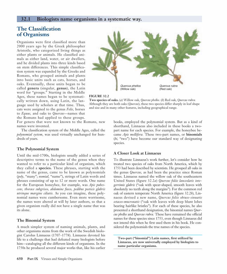
To illustrate Linnaeus’s work further, let’s consider how hetreated two species of oaks from North America, which by1753 had been described by scientists. He grouped all oaks inthe genus Quercus, as had been the practice since Romantimes. Linnaeus named the willow oak of the southeasternUnited States (figure 32.2a) Quercus foliis lanceolatis inte-gerrimis glabris (“oak with spear-shaped, smooth leaves withabsolutely no teeth along the margins”). For the common redoak of eastern temperate North America (figure 32.2b), Lin-naeus devised a new name, Quercus foliis obtuse-sinuatis setaceo-mucronatis (“oak with leaves with deep blunt lobesbearing hairlike bristles”). For each of these species, he alsopresented a shorthand designation, the binomial names Quer-cus phellos and Quercus rubra. These have remained the officialnames for these species since 1753, even though Linnaeus didnot intend this when he first used them in his book. He con-sidered the polynomials the true names of the species.
Two-part (“binomial”) Latin names, first utilized byLinnaeus, are now universally employed by biologists toname particular organisms.
650 Part IX Viruses and Simple Organisms
The Classification of OrganismsOrganisms were first classified more than2000 years ago by the Greek philosopherAristotle, who categorized living things aseither plants or animals. He classified ani-mals as either land, water, or air dwellers,and he divided plants into three kinds basedon stem differences. This simple classifica-tion system was expanded by the Greeks andRomans, who grouped animals and plantsinto basic units such as cats, horses, andoaks. Eventually, these units began to becalled genera (singular, genus), the Latinword for “groups.” Starting in the MiddleAges, these names began to be systemati-cally written down, using Latin, the lan-guage used by scholars at that time. Thus,cats were assigned to the genus Felis, horsesto Equus, and oaks to Quercus—names thatthe Romans had applied to these groups.For genera that were not known to the Romans, newnames were invented.
The classification system of the Middle Ages, called thepolynomial system, was used virtually unchanged for hun-dreds of years.
The Polynomial System
Until the mid-1700s, biologists usually added a series ofdescriptive terms to the name of the genus when theywanted to refer to a particular kind of organism, whichthey called a species. These phrases, starting with thename of the genus, came to be known as polynomials(poly, “many”; nomial, “name”), strings of Latin words andphrases consisting of up to 12 or more words. One namefor the European honeybee, for example, was Apis pubes-cens, thorace subgriseo, abdomine fusco, pedibus posticis glabrisutrinque margine ciliatis. As you can imagine, these poly-nomial names were cumbersome. Even more worrisome,the names were altered at will by later authors, so that agiven organism really did not have a single name that wasits alone.
The Binomial System
A much simpler system of naming animals, plants, andother organisms stems from the work of the Swedish biolo-gist Carolus Linnaeus (1707–1778). Linnaeus devoted hislife to a challenge that had defeated many biologists beforehim—cataloging all the different kinds of organisms. In the1750s he produced several major works that, like his earlier
32.1 Biologists name organisms in a systematic way.
Quercus phellos(Willow oak)
Quercus rubra(Red oak)
FIGURE 32.2Two species of oaks. (a) Willow oak, Quercus phellos. (b) Red oak, Quercus rubra.Although they are both oaks (Quercus), these two species differ sharply in leaf shapeand size and in many other features, including geographical range.

Species NamesTaxonomy is the science of classify-ing living things, and a group of or-ganisms at a particular level in a clas-sification system is called a taxon(plural, taxa). By agreement amongtaxonomists throughout the world, notwo organisms can have the samename. So that no one country is fa-vored, a language spoken by no coun-try—Latin—is used for the names.Because the scientific name of an or-ganism is the same anywhere in theworld, this system provides a standardand precise way of communicating,whether the language of a particularbiologist is Chinese, Arabic, Spanish,or English. This is a great improve-ment over the use of common names,which often vary from one place tothe next. As you can see in figure32.3, corn in Europe refers to theplant Americans call wheat; a bear is alarge placental omnivore in theUnited States but a koala (a vegetar-ian marsupial) in Australia; and arobin is a very different bird in Eu-rope and North America.
Also by agreement, the first wordof the binomial name is the genus towhich the organism belongs. Thisword is always capitalized. The sec-ond word refers to the particularspecies and is not capitalized. The twowords together are called the scien-tific name and are written in italics ordistinctive print: for example, Homosapiens. Once a genus has been used inthe body of a text, it is often abbrevi-ated in later uses. For example, the di-nosaur Tyrannosaurus rex becomes T.rex, and the potentially dangerousbacterium Escherichia coli is known asE. coli. The system of naming animals,plants, and other organisms estab-lished by Linnaeus has served the sci-ence of biology well for nearly 230years.
By convention, the first part of abinomial species name identifiesthe genus to which the speciesbelongs, and the second partdistinguishes that particularspecies from other species in thegenus.
Chapter 32 How We Classify Organisms 651
(a)
(b)
(c)
FIGURE 32.3Common names make poor labels. The common names corn (a), bear (b), and robin (c)bring clear images to our minds (photos on left), but the images are very different tosomeone living in Europe or Australia (photos on right). There, the same common namesare used to label very different species.

The Taxonomic HierarchyIn the decades following Linnaeus, taxonomists began togroup organisms into larger, more inclusive categories.Genera with similar properties were grouped into a clustercalled a family, and similar families were placed into thesame order (figure 32.4). Orders with common propertieswere placed into the same class, and classes with similarcharacteristics into the same phylum (plural, phyla). Forhistorical reasons, phyla may also be called divisions amongplants, fungi, and algae. Finally, the phyla were assigned toone of several great groups, the kingdoms. Biologists cur-rently recognize six kingdoms: two kinds of bacteria (Ar-chaebacteria and Eubacteria), a largely unicellular group ofeukaryotes (Protista), and three multicellular groups(Fungi, Plantae, and Animalia). In order to remember theseven categories of the taxonomic hierarchy in their properorder, it may prove useful to memorize a phrase such as“kindly pay cash or furnish good security” (kingdom–phy-lum–class–order–family–genus–species).
In addition, an eighth level of classification, called do-mains, is sometimes used. Biologists recognize three do-mains, which will be discussed later in this chapter. Thescientific names of the taxonomic units higher than thegenus level are capitalized but not printed distinctively,italicized, or underlined.
The categories at the different levels may include many,a few, or only one taxon. For example, there is only one liv-ing genus of the family Hominidae, but several living gen-era of Fagaceae. To someone familiar with classification orwith access to the appropriate reference books, each taxonimplies both a set of characteristics and a group of organ-isms belonging to the taxon. For example, a honeybee hasthe species (level 1) name Apis mellifera. Its genus name(level 2) Apis is a member of the family Apidae (level 3). Allmembers of this family are bees, some solitary, others liv-ing in hives as A. mellifera does. Knowledge of its order(level 4), Hymenoptera, tells you that A. mellifera is likelyable to sting and may live in colonies. Its class (level 5) In-secta indicates that A. mellifera has three major body seg-ments, with wings and three pairs of legs attached to themiddle segment. Its phylum (level 6), Arthropoda, tells usthat the honeybee has a hard cuticle of chitin and jointedappendages. Its kingdom (level 7), Animalia, tells us that A.mellifera is a multicellular heterotroph whose cells lack cellwalls.
Species are grouped into genera, genera into families,families into orders, orders into classes, and classes intophyla. Phyla are the basic units within kingdoms; such asystem is hierarchical.
652 Part IX Viruses and Simple Organisms
Eastern gray squirrelSciurus carolinensis
FIGURE 32.4The hierarchical systemused in classifying anorganism. The organism isfirst recognized as a eukaryote(domain: Eukarya). Second,within this domain, it is ananimal (kingdom: Animalia).Among the different phyla ofanimals, it is a vertebrate(phylum: Chordata,subphylum: Vertebrata). Theorganism’s fur characterizes itas a mammal (class:Mammalia). Within this class,it is distinguished by itsgnawing teeth (order:Rodentia). Next, because ithas four front toes and fiveback toes, it is a squirrel(family: Sciuridae). Withinthis family, it is a tree squirrel(genus: Sciurus), with gray furand white-tipped hairs on thetail (species: Sciuruscarolinensis, the eastern graysquirrel).

What Is a Species?In the previous section we discussed how species are namedand grouped, but how do biologists decide when one or-ganism is distinct enough from another to be called its ownspecies? In chapter 22, we reviewed the nature of speciesand saw there are no absolute criteria for the definition ofthis category. Looking different, for example, is not a use-ful criterion: different individuals that belong to the samespecies (for example, dogs) may look very unlike one an-other, as different as a Chihuahua and a St. Bernard. Thesevery different-appearing individuals are fully capable of hy-bridizing with one another.
The biological species concept (figure 32.5) essentiallysays that two organisms that cannot interbreed and producefertile offspring are different species. This definition of aspecies can be useful in describing sexually reproducingspecies that regularly outcross—interbreed with individu-als other than themselves. However, in many groups of or-ganisms, including bacteria, fungi, and many plants and an-imals, asexual reproduction—reproduction withoutsex—predominates. Among them, hybridization cannot beused as a criterion for species recognition.
Defining Species
Despite such difficulties, biologists generally agree on theorganisms they classify as species based on the similarity ofmorphological features and ecology. As a practical defini-tion, we can say that species are groups of organisms thatremain relatively constant in their characteristics, can bedistinguished from other species, and do not normally in-terbreed with other species in nature.
Evolutionary Species Concept
This simple definition of species leaves many problems un-solved. How, for instance, are we to compare living specieswith seemingly similar ones now extinct? Much of the dis-agreement among alternative species concepts relates tosolving this problem. When do we assign fossil specimens aunique species name, and when do we assign them tospecies living today? If we trace the lineage of two sisterspecies backwards through time, how far must we go beforethe two species converge on their common ancestor? It isoften very hard to know where to draw a sharp line be-tween two closely related species.
To address this problem, biologists have added an evo-lutionary time dimension to the biological species concept.A current definition of an evolutionary species is a singlelineage of populations that maintains its distinctive identity fromother such lineages. Unlike the biological species concept, theevolutionary species concept applies to both asexual andsexually reproducing forms. Abrupt changes in diagnosticfeatures mark the boundaries of different species in evolu-tionary time.
How Many Species Are There?
Scientists have described and named a total of 1.5 millionspecies, but doubtless many more actually exist. Somegroups of organisms, such as flowering plants, vertebrateanimals, and butterflies, are relatively well known with anestimated 90% of the total number of species that actuallyexist in these groups having already been described. Manyother groups, however, are very poorly known. It is gener-ally accepted that only about 5% of all species have beenrecognized for bacteria, nematodes (roundworms), fungi,and mites (a group of organisms related to spiders).
By taking representative samples of organisms from dif-ferent environments, such as the upper branches of tropicaltrees or the deep ocean, scientists have estimated the totalnumbers of species that may actually exist to be about 10million, about 15% of them marine organisms.
Most Species Live in the Tropics
Most species, perhaps 6 or 7 million, are tropical. Presentlyonly 400,000 species have been named in tropical Asia,Africa, and Latin America combined, well under 10% of allspecies that occur in the tropics. This is an incredible gapin our knowledge concerning biological diversity in a worldthat depends on biodiversity for its sustainability.
These estimates apply to the number of eukaryotic or-ganisms only. There is no functional way of estimating thenumbers of species of prokaryotic organisms, although it isclear that only a very small fraction of all species have beendiscovered and characterized so far.
Species are groups of organisms that differ from oneanother in recognizable ways and generally do notinterbreed with one another in nature.
Chapter 32 How We Classify Organisms 653
(a) (b)
(c)
FIGURE 32.5The biological speciesconcept. Horses (a) anddonkeys (b) are not thesame species, because theoffspring they producewhen they interbreed,mules (c), are sterile.

Evolutionary ClassificationsAfter naming and classifying some 1.5 million organisms,what have biologists learned? One very important advan-tage of being able to classify particular species of plants, an-imals, and other organisms is that individuals of speciesthat are useful to humans as sources of food and medicinecan be identified. For example, if you cannot tell the fungusPenicillium from Aspergillus, you have little chance of pro-ducing the antibiotic penicillin. In a thousand ways, justhaving names for organisms is of immense importance inour modern world.
Taxonomy also enables us to glimpse the evolutionaryhistory of life on earth. The more similar two taxa are,the more closely related they are likely to be. By lookingat the differences and similarities between organisms, bi-ologists can construct an evolutionary tree, or phy-logeny, inferring which organisms evolved from whichother ones, in what order, and when. The reconstructionand study of phylogenies is called systematics. Within aphylogeny, a grouping can be either monophyletic, para-phyletic, or polyphyletic. A monophyletic group in-cludes the most recent common ancestor of the groupand all of its descendants. A paraphyletic group includesthe most recent common ancestor of the group but notall of its descendants. And, a polyphyletic group doesnot include the most recent common ancestor of all themembers of the group. Monophyletic groups are com-monly assigned names, but systematists will not assign ataxonomic classification to a polyphyletic group. Para-phyletic groups may be considered taxa by some scien-tists, although they do not accurately represent the evo-lutionary relationships among the members of the group(figure 32.6).
Cladistics
A simple and objective way to construct a phylogenetictree is to focus on key characters that a group of organ-isms share because they have inherited them from a com-mon ancestor. A clade is a group of organisms related bydescent, and this approach to constructing a phylogeny iscalled cladistics. Cladistics infers phylogeny (that is,builds family trees) according to similarities derived froma common ancestor, so-called derived characters. A de-rived character that is unique to a particular clade issometimes called a synapomorphy. The key to the ap-proach is being able to identify morphological, physio-logical, or behavioral traits that differ among the organ-isms being studied and can be attributed to a commonancestor. By examining the distribution of these traitsamong the organisms, it is possible to construct a clado-
654 Part IX Viruses and Simple Organisms
32.2 Scientists construct phylogenies to understand the evolutionaryrelationships among organisms.
Ray Shark
Wha
le
Cow Orang
utan
Gorilla
Chimpa
nzee
Human
Monophyletic group
Ray Shark
Wha
le
Cow Orang
utan
Gorilla
Chimpa
nzee
Human
Paraphyletic group
Ray Shark
Wha
le
Cow Orang
utan
Gorilla
Chimpa
nzee
Human
Polyphyletic group
(a)
(b)
(c)
FIGURE 32.6(a) A monophyletic group consists of the most recent commonancestor and all of its descendants. All taxonomists acceptmonophyletic groups in their classifications and in the aboveexample would give the name “Apes” to the orangutans, gorillas,chimpanzees, and humans. (b) A paraphyletic group consists of themost recent common ancestor and some of its descendants.Taxonomists differ in their acceptance of paraphyletic groups. Forexample, some taxonomists arbitrarily group orangutans, gorillas,and chimpanzees into the paraphyletic family Pongidae, separatefrom humans. Other taxonomists do not use the family Pongidaein their classifications because gorillas and chimpanzees are moreclosely related to humans than to orangutans. (c) A polyphyleticgroup does not contain the most recent common ancestor of thegroup, and taxonomists do not assign taxa to polyphyletic groups.For example, sharks and whales could be classified in the samegroup because they have similar shapes, anatomical features, andhabitats. However, their similarities reflect convergent evolution,not common ancestry.

gram (figure 32.7), a branching diagram that representsthe phylogeny.
In traditional phylogenies, proposed ancestors willoften be indicated at the nodes between branches, and thelengths of branches correspond to evolutionary time, withextinct groups having shorter branches. In contrast, clado-grams are not true family trees in that they do not identifyancestors, and the branch lengths do not reflect evolution-ary time (see figure 32.6). Instead, they convey compara-tive information about relative relationships. Organismsthat are closer together on a cladogram simply share amore recent common ancestor than those that are fartherapart. Because the analysis is comparative, it is necessary tohave something to anchor the comparison to, some solidground against which the comparisons can be made. Toachieve this, each cladogram must contain an outgroup, arather different organism (but not too different) to serve asa baseline for comparisons among the other organismsbeing evaluated, the ingroup. For example, in figure 32.7,the lamprey is the outgroup to the clade of animals thathave jaws.
Cladistics is a relatively new approach in biology and hasbecome popular among students of evolution. This is be-cause it does a very good job of portraying the order inwhich a series of evolutionary events have occurred. Thegreat strength of a cladogram is that it can be completelyobjective. In fact, most cladistic analyses involve manycharacters, and computers are required to make the com-parisons.
Sometime it is necessary to “weight” characters, or takeinto account the variation in the “strength” of a character,such as the size or location of a fin or the effectiveness ofa lung. To reduce a systematist’s bias even more, manyanalyses will be run through the computer with the traitsweighted differently each time. Under this procedure,several different cladograms will be constructed, the goalbeing to choose the one that is the most parsimonious,or simplest and thus most likely. Reflecting the impor-tance of evolutionary processes to all fields of biology,most taxonomy today includes at least some element ofcladistic analysis.
Chapter 32 How We Classify Organisms 655
Lamprey Tiger Gorilla Human
Jaws
Lungs
Amnioticmembrane
Hair
No tail
Bipedal
LizardSalamanderShark
Traits:Organism
Jaws Lungs Amnioticmembrane
Hair No tail Bipedal
Lamprey
Shark
Salamander
Lizard
Tiger
Gorilla
Human
0 0 0 0 0 0
1 0 0 0 0 0
1 1 0 0 0 0
1 1 1 0 0 0
1 1 1 1 0 0
1 1 1 1 1 0
1 1 1 1 1 1
FIGURE 32.7A cladogram. Morphological data for a groupof seven vertebrates is tabulated. A “1”indicates the presence of a trait, or derivedcharacter, and a “0” indicates the absence ofthe trait. A tree, or cladogram, diagrams theproposed evolutionary relationships amongthe organisms based on the presence ofderived characters. The derived charactersbetween the cladogram branch points areshared by all organisms above the branchpoint and are not present in any below it. Theoutgroup, in this case the lamprey, does notpossess any of the derived characters.

Traditional Taxonomy
Weighting characters lies at the core of traditional taxon-omy. In this approach, taxa are assigned based on a vastamount of information about the morphology and biologyof the organism gathered over a long period of time. Tradi-tional taxonomists consider both the common descent andamount of adaptive evolutionary change when grouping or-ganisms. The large amount of information used by tradi-tional taxonomists permits a knowledgeable weighting ofcharacters according to their biological significance. In tra-ditional taxonomy, the full observational power and judg-ment of the biologist is brought to bear—and also any bi-ases he or she may have. For example, in classifying theterrestrial vertebrates, traditional taxonomists place birds intheir own class (Aves), giving great weight to the charactersthat made powered flight possible, such as feathers. How-ever, cladists (figure 32.8) lumps birds in among the rep-tiles with crocodiles. This accurately reflects their true an-
cestry but ignores the immense evolutionary impact of aderived character such as feathers.
Overall, classifications based on traditional taxonomyare information-rich, while classifications based on clado-grams need not be. Traditional taxonomy is often usedwhen a great deal of information is available to guide char-acter weighting, while cladistics is a good approach whenlittle information is available about how the character af-fects the life of the organism. DNA sequence comparisons,for example, lend themselves well to cladistics—you have agreat many derived characters (DNA sequence differences)but little or no idea of what impact the sequence differ-ences have on the organism.
A phylogeny may be represented as a cladogram basedon the order in which groups evolved. Traditionaltaxonomists weight characters according to assumedimportance.
656 Part IX Viruses and Simple Organisms
MammalsMammals
TurtlesTurtles
CrocodiliansCrocodilians
BirdsBirds
Dinosaurs
DinosaursLizards and
snakesLizards and
snakes
Early reptiles
ClassMammalia
Class Reptilia
Class Aves
Mammalia Reptilia
Archosaurs
(a) Traditional phylogeny and taxonomic classification (b) Cladogram and cladistic classification
FIGURE 32.8Traditional and cladistic interpretations of vertebrate classification. Traditional and cladistic taxonomic analyses of the same set ofdata often produce different results: in these two classifications of vertebrates, notice particularly the placement of the birds. (a) In thetraditional analysis, key characteristics such as feathers and hollow bones are weighted more heavily than others, placing the birds in theirown group and the reptiles in a paraphyletic group. (b) Cladistic analysis gives equal weight to these and many other characters and placesbirds in the same grouping with crocodiles, reflecting the close evolutionary relationship between the two. Also, in the traditionalphylogeny, the branch leading to the dinosaurs is shorter because the length corresponds to evolutionary time. In cladograms, branchlengths do not correspond to evolutionary time.

The Kingdoms of LifeThe earliest classification systems recognized only twokingdoms of living things: animals and plants (figure32.9a). But as biologists discovered microorganisms andlearned more about other organisms, they added kingdomsin recognition of fundamental differences discoveredamong organisms (figure 32.9b). Most biologists now use asix-kingdom system first proposed by Carl Woese of theUniversity of Illinois (figure 32.9c).
In this system, four kingdoms consist of eukaryotic or-ganisms. The two most familiar kingdoms, Animalia andPlantae, contain only organisms that are multicellular dur-ing most of their life cycle. The kingdom Fungi containsmulticellular forms and single-celled yeasts, which arethought to have multicellular ancestors. Fundamental dif-ferences divide these three kingdoms. Plants are mainly sta-tionary, but some have motile sperm; fungi have no motilecells; animals are mainly motile. Animals ingest their food,plants manufacture it, and fungi digest it by means of se-creted extracellular enzymes. Each of these kingdoms prob-ably evolved from a different single-celled ancestor.
The large number of unicellular eukaryotes are arbitrar-ily grouped into a single kingdom called Protista (seechapter 35). This kingdom includes the algae, all of whichare unicellular during parts of their life cycle.
The remaining two kingdoms, Archaebacteria and Eu-bacteria, consist of prokaryotic organisms, which are vastlydifferent from all other living things (see chapter 34). Ar-chaebacteria are a diverse group including themethanogens and extreme thermophiles, and differ fromthe other bacteria, members of the kingdom Eubacteria.
Domains
As biologists have learned more about the archaebacteria, ithas become increasingly clear that this ancient group isvery different from all other organisms. When the full ge-nomic DNA sequences of an archaebacterium and a eubac-terium were first compared in 1996, the differences provedstriking. Archaebacteria are as different from eubacteria aseubacteria are from eukaryotes. Recognizing this, biologistsare increasingly adopting a classification of living organ-isms that recognizes three domains, a taxonomic levelhigher than kingdom (figure 32.9d). Archaebacteria are inone domain, eubacteria in a second, and eukaryotes in thethird.
Living organisms are grouped into three generalcategories called domains. One of the domains, theeukaryotes, is subdivided into four kingdoms: protists,fungi, plants, and animals.
Chapter 32 How We Classify Organisms 657
32.3 All living organisms are grouped into one of a few major categories.
Animalia
AnimaliaPlantae
Plantae
FungiProtistaArchaebacteria
AnimaliaPlantaeFungiProtistaMonera
(a) A two-kingdom system—Linnaeus
(b) A five-kingdom system—Whittaker
(c) A six-kingdom system—Woese
(d) A three-domain system—Woese
EukaryaArchaeaBacteria
Eubacteria
FIGURE 32.9Different approaches to classifying living organisms. (a) Linnaeus popularized a two-kingdom approach, in which the fungi and thephotosynthetic protists were classified as plants, and the nonphotosynthetic protists as animals; when bacteria were described, they toowere considered plants. (b) Whittaker in 1969 proposed a five-kingdom system that soon became widely accepted. (c) Woese haschampioned splitting the bacteria into two kingdoms for a total of six kingdoms, or even assigning them separate domains (d).

Domain Archaea (Archaebacteria)The term archaebacteria (Greek, archaio, ancient) refers tothe ancient origin of this group of bacteria, which seem tohave diverged very early from the eubacteria (figure32.10). This conclusion comes largely from comparisonsof genes that encode ribosomal RNAs. The last severalyears have seen an explosion of DNA sequence informa-tion from microorganisms, information which paints amore complex picture. It had been thought that by se-quencing numerous microbes we could eventually come upwith an accurate picture of the phylogeny of the earliestorganisms on earth. The new whole-genome DNA se-quence data described in chapter 19 tells us that it will notbe that simple. Comparing whole-genome sequences leadsevolutionary biologists to a variety of trees, some of whichcontradict each other. It appears that during their earlyevolution microorganisms have swapped genetic informa-tion, making constructing phylogenetic trees very difficult.
As an example of the problem, we can look at Thermo-toga, a thermophile found on Volcano Island off Italy. Thesequence of one of its RNAs places it squarely within theeubacteria near an ancient microbe called Aquifex. RecentDNA sequencing, however, fails to support any consistentrelationship between the two microbes. There is disagree-ment as to the serious effect of gene swapping on the abil-ity of evolutionary biologists to provide accurate phyloge-nies from molecular data. For now, we will provisionallyaccept the tree presented in figure 32.10. Over the next fewyears we can expect to see considerable change in acceptedviewpoints as more and more data is brought to bear.
Today, archaebacteria inhabit some of the most extremeenvironments on earth. Though a diverse group, all archae-bacteria share certain key characteristics (table 32.1). Theircell walls lack peptidoglycan (an important component ofthe cell walls of eubacteria), the lipids in the cell mem-branes of archaebacteria have a different structure thanthose in all other organisms, and archaebacteria have dis-tinctive ribosomal RNA sequences. Some of their genespossess introns, unlike those of other bacteria.
The archaebacteria are grouped into three general cate-gories, methanogens, extremophiles, and nonextreme ar-chaebacteria, based primarily on the environments in whichthey live or their specialized metabolic pathways.
Methanogens obtain their energy by using hydrogengas (H2) to reduce carbon dioxide (CO2) to methane gas(CH4). They are strict anaerobes, poisoned by even tracesof oxygen. They live in swamps, marshes, and the intestinesof mammals. Methanogens release about 2 billion tons ofmethane gas into the atmosphere each year.
Extremophiles are able to grow under conditions thatseem extreme to us.
Thermophiles (“heat lovers”) live in very hot places, typi-cally from 60º to 80ºC. Many thermophiles are au-totrophs and have metabolisms based on sulfur. Some
thermophilic archaebacteria form the basis of food websaround deep-sea thermal vents where they must with-stand extreme temperatures and pressures. Other types,like Sulfolobus, inhabit the hot sulfur springs of Yellow-stone National Park at 70º to 75ºC. The recently de-scribed Pyrolobus fumarii holds the current record forheat stability, with a 106ºC temperature optimum and113ºC maximum—it is so heat tolerant that it is notkilled by a one-hour treatment in an autoclave (121ºC)!Halophiles (“salt lovers”) live in very salty places like theGreat Salt Lake in Utah, Mono Lake in California, andthe Dead Sea in Israel. Whereas the salinity of seawateris around 3%, these bacteria thrive in, and indeed re-quire, water with a salinity of 15 to 20%.
pH-tolerant archaebacteria grow in highly acidic (pH =0.7) and very basic (pH = 11) environments.
Pressure-tolerant archaebacteria have been isolated fromocean depths that require at least 300 atmospheres ofpressure to survive, and tolerate up to 800 atmospheres!
Nonextreme archaebacteria grow in the same envi-ronments eubacteria do. As the genomes of archaebacteriahave become better known, microbiologists have been ableto identify signature sequences of DNA present in all ar-chaebacteria and in no other organisms. When samplesfrom soil or seawater are tested for genes matching thesesignal sequences, many of the bacteria living there prove tobe archaebacteria. Clearly, archaebacteria are not restrictedto extreme habitats, as microbiologists used to think.
Archaebacteria are poorly understood bacteria thatinhabit diverse environments, some of them extreme.
658 Part IX Viruses and Simple Organisms
DomainBacteria
(Eubacteria)
DomainArchaea
(Archaebacteria)
Common ancestor
DomainEukarya
(Eukaryotes)
FIGURE 32.10An evolutionary relationship among the three domains.Eubacteria are thought to have diverged early from theevolutionary line that gave rise to the archaebacteria andeukaryotes.

Domain Bacteria (Eubacteria)The eubacteria are the most abundant organisms on earth.There are more living eubacteria in your mouth than thereare mammals living on earth. Although too tiny to see withthe unaided eye, eubacteria play critical roles throughoutthe biosphere. They extract from the air all the nitrogenused by organisms, and play key roles in cycling carbon andsulfur. Much of the world’s photosynthesis is carried out byeubacteria. However, certain groups of eubacteria are alsoresponsible for many forms of disease. Understanding theirmetabolism and genetics is a critical part of modern medi-cine.
There are many different kinds of eubacteria, and theevolutionary links between them are not well understood.While there is considerable disagreement among taxono-mists about the details of bacterial classification, most rec-ognize 12 to 15 major groups of eubacteria. Comparisonsof the nucleotide sequences of ribosomal RNA (rRNA)molecules are beginning to reveal how these groups are re-lated to one another and to the other two domains. Oneview of our current understanding of the “Tree of Life” ispresented in figure 32.11. The oldest divergences representthe deepest rooted branches in the tree. The root of thetree is within the eubacterial domain. The archaebacteriaand eukaryotes are more closely related to each other thanto eubacteria and are on a separate evolutionary branch ofthe tree, even though archaebacteria and eubacteria areboth prokaryotes.
Eubacteria are as different from archaebacteria as fromeukaryotes.
Chapter 32 How We Classify Organisms 659
BACTERIA
Purple bacteria
Common ancestor
Cyanobacteria
Flavobacteria
Thermotoga
Pyrodictium
ThermoproteusMethanobacterium
Methanopyrus
Thermoplasma
Methano-coccus
Thermo-coccus
Halobacterium
Aquifex
Gram-positivebacteria
Entamoebae
Slime moldsAnimals
Fungi
Plants
Ciliates
Flagellates
Diplomonads
Microsporidia
ARCHAEA EUKARYA
FIGURE 32.11A tree of life. This phylogeny, prepared from rRNA analyses, shows the evolutionary relationships among the three domains. The base ofthe tree was determined by examining genes that are duplicated in all three domains, the duplication presumably having occurred in thecommon ancestor. When one of the duplicates is used to construct the tree, the other can be used to root it. This approach clearlyindicates that the root of the tree is within the eubacterial domain. Archaebacteria and eukaryotes diverged later and are more closelyrelated to each other than either is to eubacteria.
Table 32.1 Features of the Domains of Life
Domain
Feature Archaea Bacteria Eukarya
Amino acid Methionine Formyl- Methioninethat initiates methionineprotein synthesisIntrons Present in Absent Present
some genesMembrane- Absent Absent Presentbounded organellesMembrane Branched Unbranched Unbranchedlipid structureNuclear Absent Absent PresentenvelopeNumber of Several One Severaldifferent RNA polymerasesPeptidoglycan Absent Present Absentin cell wallResponse Growth Growth Growth notto the not inhibited inhibited inhibitedantibiotics streptomycin and chloram-phenicol

Domain Eukarya (Eukaryotes)For at least 2 billion years, bacteria ruled the earth. Noother organisms existed to eat them or compete withthem, and their tiny cells formed the world’s oldest fossils.The third great domain of life, the eukaryotes, appear inthe fossil record much later, only about 1.5 billion yearsago. Metabolically, eukaryotes are more uniform thanbacteria. Each of the two domains of prokaryotic organ-isms has far more metabolic diversity than all eukaryoticorganisms taken together. However, despite the metabolicsimilarity of eukaryotic cells, their structure and functionallowed larger cell sizes and, eventually, multicellular lifeto evolve.
Four Kingdoms of Eukaryotes
The first eukaryotes were unicellular organisms. A widevariety of unicellular eukaryotes exist today, grouped to-gether in the kingdom Protista on the basis that they donot fit into any of the other three kingdoms of eukaryotes.Protists are a fascinating group containing many organ-isms of intense interest and great biological significance.They vary from the relatively simple, single-celledamoeba to multicellular organisms like kelp that can be 20meters long.
Fungi, plants, and animals are largely multicellular king-doms, each a distinct evolutionary line from a single-celledancestor that would be classified in the kingdom Protista.
Because of the size and ecological dominance of plants, ani-mals, and fungi, and because they are predominantly multi-cellular, we recognize them as kingdoms distinct from Pro-tista, even though the amount of diversity among theprotists is much greater than that within or between thefungi, plants, and animals.
Symbiosis and the Origin of Eukaryotes
The hallmark of eukaryotes is complex cellular organiza-tion, highlighted by an extensive endomembrane systemthat subdivides the eukaryotic cell into functional compart-ments. Not all of these compartments, however, are de-rived from the endomembrane system. With few excep-tions, all modern eukaryotic cells possess energy-producingorganelles, the mitochondria, and some eukaryotic cellspossess chloroplasts, which are energy-harvesting or-ganelles. Mitochondria and chloroplasts are both believedto have entered early eukaryotic cells by a process calledendosymbiosis (endo, inside). We discussed the theory ofthe endosymbiotic origin of mitochondria and chloroplastsin chapter 5; also see figure 32.12. Both organelles containtheir own ribosomes, which are more similar to bacterial ri-bosomes than to eukaryotic cytoplasmic ribosomes. Theymanufacture their own inner membranes. They divide in-dependently of the cell and contain chromosomes similarto those in bacteria. Mitochondria are about the size ofbacteria and contain DNA. Comparison of the nucleotidesequence of this DNA with that of a variety of organisms
660 Part IX Viruses and Simple Organisms
Thermophiles
Halophiles
Methanogens
Purple bacteria
Photosynthetic bacteria
Photosyntheticprotists
Nonphotosynthetic protists
Brown algae
Animalia
Fungi
Protista
Plantae
Eubacteria
Red algae
Green algae
Other bacteria
Archaebacteria
Ancestraleukaryoticcell
Originalcell
Mitochondria
Chloroplasts
FIGURE 32.12Diagram of the evolutionary relationships among the six kingdoms of organisms. The colored lines indicate symbiotic events.

indicates clearly that mitochondria arethe descendants of purple bacteria thatwere incorporated into eukaryotic cellsearly in the history of the group. Chloro-plasts are derived from cyanobacteriathat became symbiotic in several groupsof protists early in their history.
Some biologists suggest that basalbodies, centrioles, flagella, and ciliamay have arisen from endosymbioticspirochaete-like bacteria. Even today,so many bacteria and unicellular pro-tists form symbiotic alliances that theincorporation of smaller organisms withdesirable features into eukaryotic cellsappears to be a relatively commonprocess.
Key Characteristics of Eukaryotes
Multicellularity. The unicellularbody plan has been tremendously suc-cessful, with unicellular prokaryotesand eukaryotes constituting about half of the biomass onearth. Yet a single cell has limits. The evolution of multi-cellularity allowed organisms to deal with their environ-ments in novel ways. Distinct types of cells, tissues, andorgans can be differentiated within the complex bodies ofmulticellular organisms. With such a functional divisionwithin its body, a multicellular organism can do manythings, like protect itself, resist drought efficiently, regu-late its internal conditions, move about, seek mates andprey, and carry out other activities on a scale and with acomplexity that would be impossible for its unicellularancestors. With all these advantages, it is not surprisingthat multicellularity has arisen independently so manytimes.
True multicellularity, in which the activities of individ-ual cells are coordinated and the cells themselves are incontact, occurs only in eukaryotes and is one of theirmajor characteristics. The cell walls of bacteria occasion-ally adhere to one another, and bacterial cells may also beheld together within a common sheath. Some bacteriaform filaments, sheets, or three-dimensional aggregates(figure 32.13), but the individual cells remain independentof each other, reproducing and carrying on their meta-bolic functions and without coordinating with the othercells. Such bacteria are considered colonial, but none aretruly multicellular. Many protists also form similar colo-nial aggregates of many cells with little differentiation orintegration.
Other protists—the red, brown, and green algae, for ex-ample—have independently attained multicellularity. Cer-
tain forms of multicellular green algaewere ancestors of the plants (see chapters35 and 37), and, like the other photosyn-thetic protists, are considered plants insome classification schemes. In the sys-tem adopted here, the plant kingdom in-cludes only multicellular land plants, agroup that arose from a single ancestorin terrestrial habitats and that has aunique set of characteristics. Aquaticplants are recent derivatives.
Fungi and animals arose from unicel-lular protist ancestors with differentcharacteristics. As we will see in subse-quent chapters, the groups that seem tohave given rise to each of these king-doms are still in existence.
Sexuality. Another major characteris-tic of eukaryotic organisms as a group issexuality. Although some interchange ofgenetic material occurs in bacteria (seechapter 34), it is certainly not a regular,predictable mechanism in the samesense that sex is in eukaryotes. The sex-
ual cycle characteristic of eukaryotes alternates betweensyngamy, the union of male and female gametes producinga cell with two sets of chromosomes, and meiosis, cell divi-sion producing daughter cells with one set of chromo-somes. This cycle differs sharply from any exchange of ge-netic material found in bacteria.
Except for gametes, the cells of most animals and plantsare diploid, containing two sets of chromosomes, duringsome part of their life cycle. A few eukaryotes completetheir life cycle in the haploid condition, with only one setof chromosomes in each cell. As we have seen, in diploidcells, one set of chromosomes comes from the male parentand one from the female parent. These chromosomes seg-regate during meiosis. Because crossing over frequently oc-curs during meiosis (see chapter 12), no two products of asingle meiotic event are ever identical. As a result, the off-spring of sexual, eukaryotic organisms vary widely, thusproviding the raw material for evolution.
Sexual reproduction, with its regular alternation be-tween syngamy and meiosis, produces genetic variation.Sexual organisms can adapt to the demands of their envi-ronments because they produce a variety of progeny.
In many of the unicellular phyla of protists, sexual re-production occurs only occasionally. Meiosis may haveoriginally evolved as a means of repairing damage to DNA,producing an organism better adapted to survive changingenvironmental conditions. The first eukaryotes were prob-ably haploid. Diploids seem to have arisen on a number ofseparate occasions by the fusion of haploid cells, whichthen eventually divided by meiosis.
Chapter 32 How We Classify Organisms 661
FIGURE 32.13Colonial bacteria. No bacteria aretruly multicellular. These glidingbacteria, Stigmatella aurantiaca, haveaggregated into a structure called afruiting body; within, some cellstransform into spores.

Eukaryotic Life Cycles
Eukaryotes are characterized by three major types of lifecycles (figure 32.14):
1. In the simplest cycle, found in algae, the zygote is theonly diploid cell. Such a life cycle is said to be charac-terized by zygotic meiosis, because the zygote im-mediately undergoes meiosis.
2. In most animals, the gametes are the only haploidcells. Animals exhibit gametic meiosis, meiosis pro-ducing gametes which fuse, giving rise to a zygote.
3. Plants show a regular alternation of generations be-tween a multicellular haploid phase and a multicel-lular diploid phase. The diploid phase undergoesmeiosis producing haploid spores that give rise tothe haploid phase, and the haploid phase produces
gametes that fuse to form the zygote. The zygote isthe first cell of the multicellular diploid phase. Thiskind of life cycle is characterized by alternation ofgenerations and has sporic meiosis.
The characteristics of the six kingdoms are outlined intable 32.2.
Eukaryotic cells acquired mitochondria and chloroplastsby endosymbiosis, mitochondria being derived frompurple bacteria and chloroplasts from cyanobacteria.The complex differentiation that we associate withadvanced life-forms depends on multicellularity andsexuality, which must have been highly advantageous tohave evolved independently so often.
662 Part IX Viruses and Simple Organisms
Table 32.2 Characteristics of the Six Kingdoms
NuclearKingdom Cell Type Envelope Mitochondria Chloroplasts Cell Wall
Archaebacteria and Eubacteria
Protista
Fungi
Plantae
Animalia
Prokaryotic
Eukaryotic
Eukaryotic
Eukaryotic
Eukaryotic
Absent
Present
Present
Present
Present
Absent
Present or absent
Present or absent
Present
Present
None (photosyntheticmembranes in some types)
Present (some forms)
Absent
Present
Absent
Noncellulose (polysaccharideplus amino acids)
Present in some forms,various types
Chitin and other noncellulosepolysaccharidesCellulose and otherpolysaccharides
Absent
2n
Zygote
n
++––
Haploidindividuals
–
+
Meiosis
Syngamy
Haploidcells
Gametes
(a) Zygotic meiosis
Haploid DiploidKey:
+––
–
+ Syngamy
Gametes
Gametes
(b) Gametic meiosis (c) Sporic meiosis
n
2n
Reproductivecell2n
2n
Diploidindividual
Zygote
Meiosis
n
++––
–
+
Meiosis
Syngamy
Spores
Gametes
n
Gametophytes(haploid)
2n
Spore-formingcell2n
2n
Sporophyte(diploid)
Zygote
+
FIGURE 32.14Diagrams of the three major kinds of life cycles in eukaryotes. (a) Zygotic meiosis, (b) gametic meiosis, and (c) sporic meiosis.

Viruses: A Special CaseViruses pose a challenge to biologists as they do not possessthe fundamental characteristics of living organisms. Virusesappear to be fragments of nucleic acids originally derivedfrom the genome of a living cell. Unlike all living organ-isms, viruses are acellular—that is, they are not cells and donot consist of cells. They do not have a metabolism; inother words, viruses do not carry out photosynthesis, cellu-lar respiration, or fermentation. The one characteristic oflife that they do display is reproduction, which they do byhijacking the metabolism of living cells.
Viruses thus present a special classification problem. Be-cause they are not organisms, we cannot logically placethem in any of the kingdoms. Viruses are really just com-plicated associations of molecules, bits of nucleic acids usu-ally surrounded by a protein coat. But, despite their sim-plicity, viruses are able to invade cells and direct the geneticmachinery of these cells to manufacture more of the mole-cules that make up the virus (figure 32.15). Viruses can in-fect organisms at all taxonomic levels.
Viruses are not organisms and are not classified in thekingdoms of life.
Chapter 32 How We Classify Organisms 663
Table 32.2 Characteristics of the Six Kingdoms
Means of Genetic Recombination, Mode of Nervous if Present Nutrition Motility Multicellularity System
Conjugation, transduction, transformation
Fertilization and meiosis
Fertilization and meiosis
Fertilization and meiosis
Fertilization and meiosis
Autotrophic (chemo-synthetic, photosyn-thetic) or heterotrophicPhotosynthetic or het-erotrophic, or combina-tion of bothAbsorption
Photosyntheticchlorophylls a and b
Digestion
Bacterial flagella, gliding or nonmotile
9 + 2 cilia and flagella;amoeboid, contractile fibrils
Nonmotile
None in most forms, 9 + 2 cilia and flagella ingametes of some forms9 + 2 cilia and flagella,contractile fibrils
Absent
Absent in most forms
Present in most forms
Present in all forms
Present in all forms
None
Primitive mechanismsfor conducting stimuliin some formsNone
None
Present, often complex
FIGURE 32.15Viruses are cell parasites. In this micrograph, several T4bacteriophages (viruses) are attacking an Escherichia coli bacterium.Some of the viruses have already entered the cell and arereproducing within it.

664 Part IX Viruses and Simple Organisms
• A fundamental division among organisms is betweenprokaryotes, which lack a true nucleus, andeukaryotes, which have a true nucleus and severalmembrane-bound organelles.
• Prokaryotes, or bacteria, are assigned to two quitedifferent kingdoms, Archaebacteria and Eubacteria.
• The eukaryotic kingdoms are more closely relatedthan are the two kingdoms of prokaryotes. Manydistinctive evolutionary lines of unicellular eukaryotesexist, most are in the Protista kingdom.
• Three of the major evolutionary lines of eukaryoticorganisms that consist principally or entirely ofmulticellular organisms are recognized as separatekingdoms: Plantae, Animalia, and Fungi.
• True multicellularity and sexuality are found onlyamong eukaryotes. Multicellularity confers theadvantages of functional specialization. Sexualitypermits genetic variation among descendants.
• Viruses are not organisms and are not included in theclassification of organisms. They are self-replicatingportions of the genomes of organisms.
5. Is there a greater fundamentaldifference between plants andanimals or between prokaryotesand eukaryotes? Explain.6. From which of the foureukaryotic kingdoms have theother three evolved?7. What is the apparent origin ofthe organelles found in almostall eukaryotes? 8. What defines if a collection ofcells is truly multicellular? Didmulticellularity arise once ormany times in the evolutionaryprocess? What advantages domulticellular organisms haveover unicellular ones?9. What are the three majortypes of life cycles in eukaryotes?Describe the major events ofeach.
32.3 All living organisms are grouped into one of a few major categories.
Chapter 32 Summary Questions Media Resources
32.1 Biologists name organisms in a systematic way.
• Biologists give every species a two-part (binomial)name that consists of the name of its genus plus adistinctive specific epithet.
• In the hierarchical system of classification used inbiology, genera are grouped into families, familiesinto orders, orders into classes, classes into phyla, andphyla into kingdoms.
• There are perhaps 10 million species of plants,animals, fungi, and eukaryotic microorganisms, butonly about 1.5 million of them have been assignednames. About 15% of the total number of species aremarine; the remainder are mostly terrestrial.
1. What was the polynomialsystem? Why didn’t this systembecome the standard for namingparticular species?2. From the most specific to themost general, what are thenames of the groups in thehierarchical taxonomic system?Which two are given specialconsideration in the way inwhich they are printed? Whatare these distinctions?
• Taxonomists may use different approaches to classifyorganisms.
• Cladistic systems of classification arrange organismsaccording to evolutionary relatedness based on thepresence of shared, derived traits.
• Traditional taxonomy classifies organisms based onlarge amounts of information, giving due weight tothe evolutionary significance of certain characters.
3. What types of features areemphasized in a cladisticclassification system? What isthe resulting relationship oforganisms that are classified inthis manner? 4. What does it mean whencharacters are weighted?
32.2 Scientists construct phylogenies to understand the evolutionary relationships among organisms.
www.mhhe.com/raven6e www.biocourse.com
• Hierarchies
• Book Reviews:Ship Fever by Barrett
• Art Activity:OrganismClassification
• Kingdoms• Three Domains• Phylogeny
• Book Review:Thowim Way Legby Flannery


![1] Material classification: There are different ways of classify](https://static.fdokumen.com/doc/165x107/6335324ed2b728420307cfa1/1-material-classification-there-are-different-ways-of-classify.jpg)