Anti-peptide antibodies: A tool for detecting IL-8 in salmonids
10
Electronic Journal of Biotechnology ISSN: 0717-3458 http://www.ejbiotechnology.info DOI: 10.2225/vol15-issue5-fulltext-15 RESEARCH ARTICLE Anti-peptide antibodies: A tool for detecting IL-8 in salmonids Paula Santana 1 · Claudia Palacios 1 · Edgar Narváez 1 · Fanny Guzmán 1,3 · José Gallardo 2 · Luis Mercado 1,3 1 Pontificia Universidad Católica de Valparaíso, Instituto de Biología, Laboratorio de Genética e Inmunología Molecular, Grupo de Marcadores Inmunológicos en Organismos Acuáticos, Valparaíso, Chile 2 Pontificia Universidad Católica de Valparaíso, Facultad de Recursos Naturales, Escuela de Ciencias del Mar, Laboratorio de Genética Aplicada, Valparaíso, Chile 3 Pontificia Universidad Católica de Valparaíso, Núcleo Biotecnológico de Curauma, Valparaíso, Chile Corresponding author: [email protected] Received August 13, 2012 / Invited Article Published online: September 15, 2012 © 2012 by Pontificia Universidad Católica de Valparaíso, Chile Abstract Background: Interleukin 8 is a chemokine that is produced by several types of cells, like macrophages and has chemotactic activity in particular on neutrophils, playing a key role during the inflammatory process. It has been demonstrated at the molecular level that this molecule is present and conserved in several vertebrate groups, pointing its importance. Analysis of the amino acid sequence of IL-8, projected from cDNA of Salmo salar, presents homology with the sequences of mammals, poultry and lamprey, indicating the presence of a homologous molecule in higher fish. However, there is no information at protein level, which allows characterizing the regulatory role of this molecule during the immune response in fish. Results: In this work, we designed and synthesized an epitope peptide of 10 residues with a purity of 95% and mass of 1158.7 kDa, which showed a random coil structure. From this peptide it was able to generate a polyclonal mono-specific antibody which was capable of detecting the whole molecule of IL-8 in tissue and cellular model of salmonids. Conclusions: The resulting antibody is a versatile tool for detecting IL-8 by different immune techniques such as ELISA, dot blot, western blotting and immunocytofluorescence. Analysis of IL-8 at proteomic level is a useful method for characterizing immune properties of this molecule in fish. Keywords: anti-IL-8, fish antibodies, fish chemokine, rainbow trout, synthetic peptide epitope INTRODUCTION ARTICLE The family Salmonidae (including salmon, trout, charr, whitefish and grayling), are of economic and environmental importance, leading to a high level of interest in many different areas of biology. Atlantic salmon (Salmo salar), and the closely related rainbow trout (Oncorhynchus mykiss), are commonly used as important pivotal species to monitor the health of aquatic environments (Davison et al. 2010). Basic immunological knowledge of both species serves as a foundation for improving fish health, conserving wild stocks, and increasing the commercial sustainability of aquaculture. The inflammatory response is the reaction of all Metazoan organisms to pathogen invasion that initiates when pathogen-derived molecules are recognized by specific pattern recognition receptors (PRRs), expressed mainly on cells of the innate immune system (Constantini et al. 2010). The successive expression of pro-inflammatory cytokines and chemokines limits pathogen spread, attracts and activates immune cells to help in the elimination of the invaders.
Transcript of Anti-peptide antibodies: A tool for detecting IL-8 in salmonids
Electronic Journal of Biotechnology ISSN: 0717-3458 http://www.ejbiotechnology.info DOI: 10.2225/vol15-issue5-fulltext-15 RESEARCH ARTICLE
Anti-peptide antibodies: A tool for detecting IL-8 in salmonids
Paula Santana1 · Claudia Palacios1 · Edgar Narváez1 · Fanny Guzmán1,3 · José Gallardo2 · Luis Mercado1,3
1 Pontificia Universidad Católica de Valparaíso, Instituto de Biología, Laboratorio de Genética e Inmunología Molecular, Grupo de
Marcadores Inmunológicos en Organismos Acuáticos, Valparaíso, Chile
2 Pontificia Universidad Católica de Valparaíso, Facultad de Recursos Naturales, Escuela de Ciencias del Mar, Laboratorio de
Genética Aplicada, Valparaíso, Chile
3 Pontificia Universidad Católica de Valparaíso, Núcleo Biotecnológico de Curauma, Valparaíso, Chile
Corresponding author: [email protected] Received August 13, 2012 / Invited Article Published online: September 15, 2012 © 2012 by Pontificia Universidad Católica de Valparaíso, Chile
Abstract
Background: Interleukin 8 is a chemokine that is produced by several types of cells, like macrophages and has chemotactic activity in particular on neutrophils, playing a key role during the inflammatory process. It has been demonstrated at the molecular level that this molecule is present and conserved in several vertebrate groups, pointing its importance. Analysis of the amino acid sequence of IL-8, projected from cDNA of Salmo salar, presents homology with the sequences of mammals, poultry and lamprey, indicating the presence of a homologous molecule in higher fish. However, there is no information at protein level, which allows characterizing the regulatory role of this molecule during the immune response in fish. Results: In this work, we designed and synthesized an epitope peptide of 10 residues with a purity of 95% and mass of 1158.7 kDa, which showed a random coil structure. From this peptide it was able to generate a polyclonal mono-specific antibody which was capable of detecting the whole molecule of IL-8 in tissue and cellular model of salmonids. Conclusions: The resulting antibody is a versatile tool for detecting IL-8 by different immune techniques such as ELISA, dot blot, western blotting and immunocytofluorescence. Analysis of IL-8 at proteomic level is a useful method for characterizing immune properties of this molecule in fish.
Keywords: anti-IL-8, fish antibodies, fish chemokine, rainbow trout, synthetic peptide epitope INTRODUCTION ARTICLE
The family Salmonidae (including salmon, trout, charr, whitefish and grayling), are of economic and environmental importance, leading to a high level of interest in many different areas of biology. Atlantic salmon (Salmo salar), and the closely related rainbow trout (Oncorhynchus mykiss), are commonly used as important pivotal species to monitor the health of aquatic environments (Davison et al. 2010). Basic immunological knowledge of both species serves as a foundation for improving fish health, conserving wild stocks, and increasing the commercial sustainability of aquaculture.
The inflammatory response is the reaction of all Metazoan organisms to pathogen invasion that initiates when pathogen-derived molecules are recognized by specific pattern recognition receptors (PRRs), expressed mainly on cells of the innate immune system (Constantini et al. 2010). The successive expression of pro-inflammatory cytokines and chemokines limits pathogen spread, attracts and activates immune cells to help in the elimination of the invaders.

Santana et al.
DOI: 10.2225/vol15-issue5-fulltext-15 2
The interleukin-8 (IL-8) of fish, also known as CXCL8, is a member of a multifunctional chemokine family comprised of around 40 small cytokines with the ability to induce the migration of cells to sites of infection or injury (Qiu et al. 2009; Alejo and Tafalla, 2011).
Initiation of immune responses depends on the recognition of pathogen-associated molecular patterns (PAMPs), present on the pathogen by PRRs on phagocytic cells such as macrophages (Medzhitov, 2007). Many cell-types, including macrophages, produce IL-8 in response to a variety of stimuli (Laing et al. 2002). One of the most remarkable properties of IL-8 is the dynamic change of its expression level. In healthy tissues, IL-8 is barely detectable, but its expression can be rapidly induced by 10 to 100-fold by various elicitors, such as tumour necrosis factor-alpha (TNF-α), and IL-1β, bacteria, viral products or cellular stress (Abdelkhalek et al. 2009; Alejo and Tafalla, 2011).
IL-8 contains four cysteine residues, the first two of which are separated by one amino acid, that form two intra-molecular disulphide bonds (Qiu et al. 2009; Constantini et al. 2010). Cysteine residues have an important role in the tertiary structure of proteins (Qiu et al. 2009). Many of the CXC chemokines, which includes IL-8, contain a three amino acid residue motif, ELR, prior to the first cysteine (Qiu et al. 2009; Constantini et al. 2010). It is known that the ELR motif plays a critical role in the neutrophil-attracting ability of IL-8 in higher vertebrates (Constantini et al. 2010). Both trout and salmon CXC chemokines lack ELR motif, however salmonid IL-8 has a similar DLR motif (Asp-Leu-Arg). It seems that this motif does not play an essential role for the attraction of neutrophils, at least in black seabream, Acanthopagrus schlegeli (Cai et al. 2009). The IL-8 gene has been cloned in several fish species, including lamprey, banded dogfish, flounder, rainbow trout, and various teleosts (Constantini et al. 2010). But to identify the functional role and tissue localization of IL-8 secreted in fish, specific antibodies against IL-8 of salmonids are required. The literature is very scarce and is known among researchers in fish immunology, the complexity of the generation of antibodies against recombinant cytokines. In the present study, we have synthesized a 10 amino acid candidate peptide epitope corresponding to amino acids 44-54 of the IL-8 of rainbow trout. This peptide was used to produce a polyclonal antibody from mouse ascitic fluid and we describe its usefulness in the detection of IL-8 in biological samples.
MATERIALS AND METHODS
Peptide design
Peptides were designed taking the available sequence of O. mykiss IL-8 (Q98TQ2). To define the best antigenic epitopes, the method of Kolaskar and Tongaonkar (1990) was used in the bioinformatics server from the immunomedicine group of the Universidad Complutense de Madrid [http://imed.med.ucm.es/Tools/antigenic.pl]. Protscale (Gasteiger et al. 2005) by the Expasy server (Gasteiger et al. 2003) was used to analyze the physicochemical behaviour of the antigenic sequences and identify regions of high hydrophilicity and mean flexibility. The antigenic regions in the whole molecule were located using a human homology model (pdb: 1 mgs) by the automatic modelling mode in the workspace of Swiss model server (Arnold et al. 2006). Also, ClustalW alignment (Larkin et al. 2007) was performed with trout and Salmo salar (B5X6N7) IL-8 sequences to determine conserved regions. The region between amino acids 44-54 (IGKLIKKVEM) was chosen as candidate epitope to be chemically synthesized.
Peptide synthesis, purification and characterization
The peptide was synthesized by the solid phase multiple peptide system (Houghten, 1985) using Fmoc amino acids (Iris and Rink resin 0.65 meq/g). Then, it was cleaved by TFA/TIS/H20 (95/2.5/2.5) [Novabiochem] and 100 μg were purified in a RP-HPLC with 0-70% acetronitrile in 30 min with a flow of 1 ml/min. The peptide was lyophilized an analyzed by MALDI-TOF mass spectrometry to confirm its molecular mass. Also, circular dichroism (CD) spectroscopy of synthesized peptide was carried out on JASCO J-815 CD Spectrometer coupled to a Peltier JASCO CDF-426S/15 system for temperature control (Jasco Corp., Tokyo, Japan). In the far ultra-violet (UV) range (190-250 nm) using quartz cuvettes of 0.1 cm path length and 1 nm bandwidth at 0.1 nm resolution. Each spectrum was recorded as an average of three repeated scans in a continuous scanning mode with 50 nm/min scanning speed with response time of 1 sec. The contribution blank was subtracted from each spectrum. Molar ellipticity was calculated using 0,017 mol/L of the peptide. CD spectra of the peptides were recorded in
Anti-peptide antibodies: A tool for detecting IL-8 in salmonids
DOI: 10.2225/vol15-issue5-fulltext-15 3
different solvents, such as pure HPLC grade water and trifluoroethanol (TFE, 30% v/v in water). The spectrum was recorded at 20ºC and then was seen with a variation in temperature between -5ºC and 50ºC.
Immunization and antibody purification
Six week-old female CF-1 mice were injected intraperitoneally at 0, 7, 14, 21 days with 0.6 µg of peptide diluted 1:1 in FIS (Bethke et al. 2012), as a T helper cell activator and 500 μl of Freund’s adjuvant (Thermo). At day 6 Pristane (2,6,10,14-tetramethylpentadecane) (SIGMA), was injected to induce the tumour formation. At day 30 mouse were anesthetized with a small dose of chloroform and drained with a hypodermic needle 18G*1½ (NIPRO). Ascitic fluid was centrifuged at 6500 g for 20 min and the supernatant stored at -80ºC. Antibodies were purified by immunoaffinity chromatography using cyanogen bromide-activated sepharose 4B (CNBr-4B), according to the instructions of manufacturer (Sigma). Briefly, 1 mg of the synthetic epitope was coupled to CNBr-4B in coupling buffer (0.1 m NaHCO3, 0.5 m NaCl, pH 8.4), and the unreacted active groups were blocked with 0.1 M Tris, pH 8.0. Then, the ascitic fluid diluted ten times with phosphate-buffered saline (PBS, pH 7.4), was loaded onto the column, and the IgG’s were eluted with 100 mM glycine (pH 2.5). The purified antibody, designated as anti-epitope of IL-8, was collected in 1 M Tris (pH 8.0) and stored at -20ºC.
Antibody characterization
Dot blot. Synthetic IL-8 epitope was placed onto a nitrocellulose membrane (Thermo 0.45 µm), seeding 2, 1, 0.5, 0.25, 0.125 µg, and other peptide with similar pI was used as negative control. The membrane was blocked with 3% BSA in PBS 1X (PBSA), for 90 min at 37ºC, and washed 2 times with PBS and once with PBS containing 0.05% Tween-20 (PBST). Thereafter, the membrane was incubated for 90 min at 37ºC with purified anti-epitope of IL-8 at 300 ng/μl. After 3 washes with PBST, anti-IgG mouse-HRP (Thermo), diluted 1:7000 was added and incubated for 1 hr at 37ºC. Following 3 washes with PBST, membrane was revealed with ECL western blotting substrate (Pierce).
ELISA. Calibration curve for evaluating anti-peptide of IL-8 antibody was performed. Briefly, 100 µl of the samples with concentrations up to 25 µM were incubated ON at 4ºC, and then washed with PBST 0.05% in Mindray Microplate washer. Samples were blocked with 1% BSA for 90 min at 37ºC and incubated with 40 ng x µl-1 of first antibody (mouse IgG anti-epitope of IL-8), for 90 min at 37ºC. After couple washes, anti-mouse IgG-HRP (Thermo), 1:7000 was incubated for 60 min at 37ºC. 100 µl per well of TMB single solution (Invitrogen), was added and incubated for 30 min at RT, the reaction was stopped with 50 µl of sulphuric acid 1 N and read at 450 nm with a VERSA max microplate reader. All assays were performed in triplicate.
Western blotting. To demonstrate the ability of anti-epitope of IL-8 to recognize the whole molecule gut of trout fed with beta-glucans was used as model samples. 40 µg gut total protein, was heated to 90ºC for 10 min in sample buffer and then electrophoresed on an 18% Tris-glycin polyacrylamide gel at 100 V for 2 hrs. After electrophoresis, the gel was transblotted onto a 0.45 µm PVDF membrane at 100 V for 1 hr. The membrane was then incubated in 5% Bovine serum albumin (BSA), in PBS 1X on a shaker for 1 hr at room temperature. The membrane was incubated with pre-absorbed first antibody anti-epitope of IL-8, at 160 ng/μl ON at room temperature, and then washed five times. Second antibody anti-IgG mouse-HRP (Thermo), diluted 1:7000 was added and incubated at room temperature for 60 min. The membranes were revealed with ECL western blotting substrate (Pierce). The pre-absorbed procedure was carried out using 40 µg total protein of brain from trout and one concentration of 160 ng/μl of the first antibody. This was incubated for 2 hrs at room temperature on a shaker. Finally centrifuged at 14,000 x g for 15 min at 4ºC. The supernatant was recollected and used immediately.
Immunocytofluorescence
To detect the location and relative abundance of IL-8 with the anti-epitope antibody, immunofluorescence was used. The presence of IL-8 in Atlantic salmon kidney (ASK) cell line by indirect immunocytofluorescence was carried out on Leica TCS SP5 II Spectral Confocal Microscope (Leica Microsystems Inc.). The cells were grown on poly-lysine-coated glass coverslips to 60-70% confluence in a 1:1 mixture of Minimum Essential Medium Eagle (MEM), supplemented with 15% fetal bovine serum (FBS, Gibco). Subsequently, the cells were stimulated for 4 hrs with attenuated Aeromonas salmonicida. The cells were fixed with methanol:acetone (1:1), for 90 sec at room

Santana et al.
DOI: 10.2225/vol15-issue5-fulltext-15 4
temperature (RT). Afterwards the cells were first pre-incubated with PBS containing 5% bovine serum albumin (BSA), for 15 min at room temperature and then exposed to affinity-purified anti-epitope of IL-8 antibody (1:50 in PBS + 1% BSA), overnight at 4ºC, then rinsed once with PBS containing 0.05% Tween-20 (PBST), and twice with PBS 1X and incubated with fluorescein isothiocyanate conjugated goat anti-mouse IgG (diluted 1:200 in PBS/BSA 1%), for 1 hr at RT, rinsed once with PBS containing 0.05% Tween-20 (PBST), and twice with PBS 1X. Nuclei were stained with propidium iodide 20 µg/ml in PBS 1X for 5 min at RT. Then, rinsed once with PBS containing 0.05% Tween-20 (PBST), and twice with PBS 1X and mounted in Vecta shield (Vector Laboratories Inc.).
RESULTS
Structural and in silico analysis
The IL-8 sequence of rainbow trout has 97 amino acid residues, which consists of a signal peptide of 22 residues and a mature peptide of 75 residues. Of this sequence a candidate peptide consisting of ten residues (IGKLIKKVEM), was synthesized and used to generate a polyclonal antibody against IL-8. Figure 1a shows the alignment of IL-8 sequence of Salmo salar and Oncorhynchus mykiss. The alignment between the two species showed a 94.8% identity.
Regarding the result obtained by bioinformatics tools, the candidate epitope (Figure 1b), has low hydrophobicity and chain flexibility properties as described for antibody forming epitopes. This epitope is in the region of the mature peptide and has an identity of 90% in rainbow trout and Atlantic salmon (Figure 1a). Human IL-8 structure was useful to see the localization of the synthesized peptide-epitope. Figure 1b shows in red colour, the target epitope which is exposed to recognition by the generated antibody.
Fig. 1 (a) Alignment of IL-8 sequences of Salmo salar and Oncorhynchus mykiss. The salmonid IL-8 antibody used in this study is raised against the amino acid sequence indicated in gray box (b) Secondary structure using homology model of IL-8 (pdb: 1 mgs), red indicates epitope.

Anti-peptide antibodies: A tool for detecting IL-8 in salmonids
DOI: 10.2225/vol15-issue5-fulltext-15 5
The peptide was synthesized using Fmoc strategy and its purity and molecular mass confirmed by RP-HPLC (95%) and MALDI-TOF MS (1158,7 kDa) (data not shown). Circular dichroism confirmed that the peptide tends to have a flexible lineal random coil like structure (Figure 2a). By subjecting this peptide at different temperatures ranging from -5ºC to 50ºC, it was observed that this flexible structure does not change significantly by the effect of temperature (Figure 2b).
Antibody production
Immunorecognition of anti-epitope of IL-8 was demonstrated via dot blot, using the IgGs generated in CF-1 mouse ascites tumors as antibodies (Figure 3a). The minimal concentration is around 0.125 ng/µl giving the antibody a high titer use. The recognition of the synthetic peptide was confirmed by ELISA (Figure 3b), whose linear regression (anti epitope r2 = 0.95), is according to previous established parameters and having no cross-reaction with other epitope sequences (Figure 3a and Figure 3b).
IL-8 detection in rainbow trout
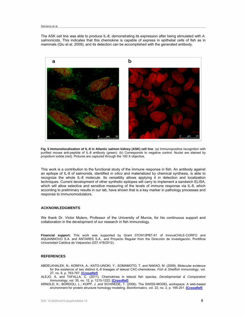
To demonstrate the utility of this antibody to recognize the whole molecule of IL-8 on biological samples of rainbow trout, immunoblotting analysis of gut tissue stimulated with beta-glucans was performed. 18% SDS-PAGE shows a large number of proteins with apparent molecular masses ranging from 9-170 kDa in gut trout protein extracts (Figure 4a). Pre-absorbed anti-peptide antibody used for western blot showed one defined band near the 10 kDa that corresponds to the whole IL-8 sequence molecular mass. Using a different model, the localization and protein expression of IL-8 in Atlantic salmon kidney (ASK) cell line was observed by immunofluorescence analysis. The cells stimulated with attenuated Aeromonas salmonicida show immuno-positive recognition of anti-IL-8 (Figure 5a). Unstimulated cells were used as negative control, which showed negative immunoreactivity (Figure 5b).
DISCUSSION
Tegumentary barriers in fish are the first line of defence of the organism against potential pathogens such as viruses, bacteria and fungi. However, when these barriers are overcome, the innate and adaptive immune system are crucial in protecting the organism against pathogens, which have different PAMPs that are recognized by pattern recognition receptor (PRR), initiating a cascade of events such as the production and release of cytokines and chemokines (Chettri et al. 2011). In vertebrates, a key chemokine involved in the initiation of the early inflammatory response is IL-8, a molecule capable of re-directing neutrophils to the site of inflammation. Innate immunity in teleost holds the major immune
Fig. 2 (a) Far UV Circular dichroism spectrum of synthetic peptide at 20ºC. (b) Temperature-dependent Far UV Circular dichroism spectra of synthetic peptide, between -5 and 50ºC.

Santana et al.
DOI: 10.2225/vol15-issue5-fulltext-15 6
response capacity, so it is thought that innate mechanisms are essential for survival, and probably more robust (Qiu et al. 2009).
The presence of a homologous molecule to mammalian IL-8 has been described in O. mykiss (Laing et al. 2002; Alejo and Tafalla, 2011), which has a neutrophil attractant effect on trout (Zhang et al. 2002; Jimenez et al. 2006), and is capable of expressing its gene level in response to different PAMPs in some tissues and cells like spleen, head kidney, gill, blood cells and macrophages in Oncorhynchus mykiss (Laing et al. 2002; Castro et al. 2011; Pérez-Sánchez et al. 2011; Raida et al. 2011). Because of the importance of this molecule in the early innate immune response, it is required to have molecular markers at protein level for further characterization. In literature, there is no data on the production of antibodies against fish IL-8, also it is known that the use of generated antibodies from recombinant molecules have not resulted useful for the detection of native molecules (Zou et al. 2003).
A proven strategy for the generation of antibodies is using synthetic epitopes (Houghten, 1985), which additionally is a useful technique for immunity molecules of fish, that are known for its complexity to generate antibodies when immunized as recombinants (Bethke et al. 2012). In this case the anti-epitope of IL-8, demonstrates ability to recognize the whole IL-8 molecule of salmonids, as is anti-epitope of salmonid NKEF, which detects the entire molecule in various biological samples (Bethke et al. 2012).
The peptide epitope presented a designed linear structure with flexible random coil, which facilitates its recognition by antibodies and together with the presence of hydrophobic residues such as valine at the exposed surface, promotes immunogenicity (Kolaskar and Tongaonkar, 1990). In our immunization protocol the synthetic peptide FIS was used, which has been described as a Th cell determinant,
Fig. 3 Positive immunoblotting assays for anti-peptide antibodies. (a) Dot blot detection of synthetic peptide with 0.3 µg/µl of mouse anti-peptide of IL-8 antibody. This assay shows higher immunorecognition for the antigen with concentrations up to 25 µM. The other peptide is used like negative control with a pI similar to synthetic peptide (b) Correlation between the antibody and synthetic peptide by ELISA.

Anti-peptide antibodies: A tool for detecting IL-8 in salmonids
DOI: 10.2225/vol15-issue5-fulltext-15 7
improving humoral adaptive immune responses (Prieto et al. 1995). This prevents the use of carrier molecules allowing the exclusive immunization of the synthetic peptide epitope. This immunization strategy allows generating antibodies with high level of specific recognition without requiring the development of monoclonal antibodies for detection of molecules associated with the immune system of teleost fish (Narvaez et al. 2010, Bethke et al. 2012).
Looking at the few existing developed antibodies against recombinant cytokines, they show the existence of cross-reactivity in the detection by western blotting (Roher et al. 2011). The developed mono-specific antibodies against one epitope can also generate cross reaction, thus using a good system of pre-absorption independent of the immunoaffinity purification with the epitope, is essential.
For the pre-absorption, tissues that do not express the protein of interest to be detected by the antibody are required. In higher vertebrates, expression of IL-8 in brain is undetectable, which has also been proven in fish under different stimuli, PAMPs or pathogens (Laing et al. 2002). With such background, pre-absorption of trout total brain protein was used, reaching an optimal blocking which enhanced the detection of the band of IL-8 in the expected molecular weight with no significant cross-reactivity (Figure 4). There is no information in the literature on pre-absorption of antibodies against fish cytokines, which is an interesting contribution and with a huge projection of starting a more extended use of teleosts cytokine antibodies.
After verifying that the developed antibody recognizes the synthetic epitope, the ability to recognize the whole molecule in two model systems was demonstrated. Pérez-Sánchez, 2011 indicated an increase of IL-8 at transcriptional level in small beta-glucans immunostimulated fish. Our laboratory has confirmed these results at the protein level. In this work identification of IL-8 in the expected molecular weight was confirmed, as a single band.
Fig. 4 IL-8 detection with purified mouse anti-peptide antibody in gut sample. (a) 18% SDS-PAGE. (b) Western blot analysis.
Santana et al.
DOI: 10.2225/vol15-issue5-fulltext-15 8
The ASK cell line was able to produce IL-8, demonstrating its expression after being stimulated with A. salmonicida. This indicates that this chemokine is capable of express in epithelial cells of fish as in mammals (Qiu et al. 2009), and its detection can be accomplished with the generated antibody.
This work is a contribution to the functional study of the immune response in fish. An antibody against an epitope of IL-8 of salmonids, identified in silico and materialized by chemical synthesis, is able to recognize the whole IL-8 molecule. Its versatility allows applying it in detection and localization techniques. Current development of other synthetic epitopes will carry to implement a sandwich ELISA, which will allow selective and sensitive measuring of the levels of immune response via IL-8, which according to preliminary results in our lab, have shown that is a key marker in pathology processes and response to immunomodulators.
ACKNOWLEDGMENTS
We thank Dr. Victor Mulero, Professor of the University of Murcia, for his continuous support and collaboration in the development of our research in fish immunology.
Financial support: This work was supported by Grant 07CN13PBT-61 of InnovaCHILE-CORFO and AQUAINNOVO S.A. and ANTARES S.A., and Proyecto Regular from the Dirección de Investigación, Pontificia Universidad Católica de Valparaíso (037.418/2012).
REFERENCES
ABDELKHALEK, N.; KOMIYA, A.; KATO-UNOKI, Y.; SOMAMOTO, T. and NAKAO, M. (2009). Molecular evidence for the existence of two distinct IL-8 lineages of teleost CXC-chemokines. Fish & Shellfish Immunology, vol. 27, no. 6, p. 763-767. [CrossRef]
ALEJO, A. and TAFALLA, C. (2011). Chemokines in teleost fish species. Developmental & Comparative Immunology, vol. 35, no. 12, p. 1215-1222. [CrossRef]
ARNOLD, K.; BORDOLI, L.; KOPP, J. and SCHWEDE, T. (2006). The SWISS-MODEL workspace: A web-based environment for protein structure homology modeling. Bioinformatics, vol. 22, no. 2, p. 195-201. [CrossRef]
Fig. 5 Immunolocalization of IL-8 in Atlantic salmon kidney (ASK) cell line. (a) Immunopositive recognition with purified mouse anti-peptide of IL-8 antibody (green). (b) Corresponds to negative control. Nuclei are stained by propidium iodide (red). Pictures are captured through the 100 X objective.
Anti-peptide antibodies: A tool for detecting IL-8 in salmonids
DOI: 10.2225/vol15-issue5-fulltext-15 9
BETHKE, J.; ROJAS, V.; BERENDSEN, J.; CÁRDENAS, C.; GUZMÁN, F.; GALLARDO, J.A. and MERCADO, L. (2012). Development of a new antibody for detecting natural killer enhancing factor (NKEF)-like protein in infected salmonids. Journal of Fish Diseases, vol. 35, no. 5, p. 379-388.[CrossRef]
CAI, Z.; GAO, C.; ZHANG, Y. and XING, K. (2009). Functional characterization of the ELR motif in piscine ELR+CXC-like chemokine. Marine Biotechnology, vol. 11, no. 4, p. 505-512. [CrossRef]
CASTRO, R.; ZOU, J.; SECOMBES, C.J. and MARTIN, S.A.M. (2011). Cortisol modulates the induction of inflammatory gene expression in a rainbow trout macrophage cell line. Fish & Shellfish Immunology, vol. 30, no. 1, p. 215-223. [CrossRef]
CHETTRI, J.; RAIDA, M.; HOLTEN-ANDERSEN, L.; KANIA, P.W. and BUCHMANN, K. (2011). PAMP induced expression of immune relevant genes in head kidney leukocytes of rainbow trout (Oncorhynchus mykiss). Developmental & Comparative Immunology, vol. 35, no. 4, p. 476-482. [CrossRef]
CONSTANTINI, S.; FACCHIANO, A.; RANDELLI, E.; CASANI, D.; SCAPIGLIATI, I.G. and BUONOCORE, F. (2010). 3D Modelling of three pro-inflammatory molecules in selected fish species. Current Pharmaceutical Design, vol. 16, no. 38, p. 4203-4212. [CrossRef]
DAVISON, W.S.; KOOP, B.F.; JONES, S.J.M.; ITURRA, P.; VIDAL, R.; MAASS, A.; JONASSEN, I.; LIEN, S. and OMHOLT, S.W. (2010). Sequencing the genome of the Atlantic salmon (Salmo salar). Genome Biology, vol. 11, Issue 9, no. 403.
GASTEIGER, E.; GATTIKER, A.; HOOGLAND, C.; IVANYI, I.; APPEL, R.D. and BAIROCH, A. (2003). ExPASy: The proteomics server for in-depth protein knowledge and analysis. Nucleic Acids Research, vol. 31, no. 13, p. 3784-3788. [CrossRef]
GASTEIGER, E.; HOOGLAND, C.; GATTIKER, A.; DUVAUD, S.; WILKINS, M.R.; APPEL, R.D. and BAIROCH, A. (2005). Protein identification and analysis tools on the ExPASy Server. In: WALKER, J.M. ed. The Proteomics Protocols Handbook. Humana Press, 607 p. ISBN 1-58829-343-2.
HOUGHTEN, R. (1985). General method for the rapid solid-phase synthesis of large numbers of peptides: Specificity of antigen-antibody interaction at the level of individual amino acids. Proceedings of the National Academy of Sciences of the United States of America, vol. 82, no. 15, p. 5131-5135. [CrossRef]
JIMENEZ, N.; COLL, J.; SALGUERO, F.J. and TAFALLA, C. (2006). Co-injection of interleukin 8 with the glycoprotein gene from viral hemorrhagic septicemia virus (VHSV) modulates the cytokine response in rainbow trout (Oncorhynchus mykiss). Vaccine, vol. 24, no. 27-28, p. 5615-5626.[CrossRef]
KOLASKAR, A.S. and TONGAONKAR, P.C. (1990). A semi-empirical method for prediction of antigenic determinants on protein antigens. FEBS Letters, vol. 276, no. 1-2, p. 172-174. [CrossRef]
LAING, K.J.; ZOU, J.J.; WANG, T.; BOLS, N.; HIRONO, I.; AOKI, I. and SECOMBES, C.J. (2002). Identification and analysis of an interleukin-8 like molecule in rainbow trout Oncorhynchus mykiss. Developmental & Comparative Immunology, vol. 23, no. 5, p. 433-444.[CrossRef]
LARKIN, M.; BLACKSHIELDS, G.; BROWN, N.P.; CHENNA, R.; MCGETTIGAN, P.A.; MCWILLIAM, H., VALENTIN, F.; WALLACE, I.M.; WILM, A.; LOPEZ, R.; THOMPSON, J.D.; GIBSON, T.J. and HIGGINS, D.G. (2007). Clustal W and Clustal X version 2.0. Bioinformatics, vol. 23, no. 21, p. 2947-2948. [CrossRef]
MEDZHITOV, R. (2007). Recognition of microorganisms and activation of the immune response. Nature, vol. 449, no. 7164, p. 819-826. [CrossRef]
NARVAEZ, E.; BERENDSEN, J.; GUZMÁN, F.; GALLARDO, J.A. and MERCADO, L. (2010). An immunological method for quantifying antibacterial activity in Salmo salar (Linnaeus, 1758) skin mucus. Fish & Shellfish Immunology, vol. 28, no. 1, p. 235-239. [CrossRef]
PÉREZ-SÁNCHEZ, T.; BALCÁZAR, J.; MERRIFIELD, D.; CARNEVALI, O.; GIOACCHINI, G.; BLAS, I. and RUIZ-ZARZUELA, I. (2011). Expression of immune-related genes in rainbow trout (Oncorhynchus mykiss) induced by probiotic bacteria during Lactococcus garvieae infection. Fish & Shellfish Immunology, vol. 31, no. 2, p. 196-201. [CrossRef]
PRIETO, I.; HERVÁS-STUBBS, S.; GARCÍA-GRANERO, M.; BERASAIN, C.; RIEZU-BOJ, J.-I.; LASARTE, J.-J.; SAROBE, P.; PRIETO, J. and BORRÁS-CUESTA, F. (1995). Simple strategy to induce antibodies of distinct specificity: Application to the mapping of gp120 and inhibition of HIV-1 infectivity. European Journal of Immunology, vol. 25, no. 4, p. 877-883. [CrossRef]
QIU, L.; ZHANG, H.; YANG, J. and JIANG, S. (2009). Molecular cloning and mRNA expression analysis of interleukin-8 gene in Japanese sea perch (Lateolabrax japonicus). Molecular Biology Report, vol. 36, no. 5, p. 1099-1105. [CrossRef]
RAIDA, M.; HOLTEN-ANDERSEN, L. and BUCHMANN, K. (2011). Association between Yersinia ruckeri infection, cytokine expression and survival in rainbow trout (Oncorhynchus mykiss). Fish & Shellfish Immunology, vol. 30, no. 6, p. 1257-1264. [CrossRef]
ROHER, N.; CALLOL, A.; PLANAS, J.V.; GOETZ, F.W. and MACKENZIE, S.A. (2011). Endotoxin recognition in fish results in inflammatory cytokine secretion not gene expression. Innate Immunity, vol. 17, no. 1, p. 16-28. [CrossRef]
ZHANG, H.; THORGAARD, G. and RISTOW, S. (2002). Molecular cloning and genomic structure of an interleukin-8 receptor-like gene from homozygous clones of rainbow trout (Oncorhynchus mykiss). Fish & Shellfish Immunology, vol. 13, no. 3, p. 251-258. [CrossRef]
ZOU, J.; PEDDIE, S.; SCAPIGLIATI, G.; ZHANG, Y.; BOLS, N.C.; ELLIS, A.E. and SECOMBES, C.J. (2003). Functional characterization of the recombinant tumor necrosis factors in rainbow trout, Oncorhynchus mykiss. Development & Comparative Immunology, vol. 27, no. 9, p. 813-822. [CrossRef]
Santana et al.
DOI: 10.2225/vol15-issue5-fulltext-15 10
How to reference this article:
SANTANA, P.; PALACIOS, C.; NARVÁEZ, E.; GUZMÁN, F.; GALLARDO, J. and MERCADO, L. (2012). Anti-peptide antibodies: A tool for detecting IL-8 in salmonids. Electronic Journal of Biotechnology, vol. 15, no. 5. http://dx.doi.org/10.2225/vol15-issue5-fulltext-15
Note: Electronic Journal of Biotechnology is not responsible if on-line references cited on manuscripts are not available any more after the date of publication. Supported by UNESCO / MIRCEN network.