Annual Report - CiteSeerX
226
20 40 60 80 100 Intensity (a.u.) 2q (degrees) Co 0.3 Zn 0.7 Fe 2 O 4 size:72(Å) Temp:2K Annual Report 2 2 0 0 1 1 0 0 – – 2 2 0 0 1 1 1 1 0 2 4 6 8 10 12 0 4 8 12 R A R N Film thickness ( nm ) R (KW/ ) UGC-DAE CONSORTIUM FOR SCIENTIFIC RESEARCH University Campus, Khandwa Road, Indore 452001
-
Upload
khangminh22 -
Category
Documents
-
view
0 -
download
0
Transcript of Annual Report - CiteSeerX
20 40 60 80 100
Inte
nsi
ty (
a.u
.)
2θ (degrees)
Co0.3Zn0.7Fe2O4
size:72(Å)Temp:2K
Annual Report 22001100 –– 22001111
0 2 4 6 8 10 12
0
4
8
12
RA
RN
Film thickness (nm)
R (K
Ω/
)
UGC-DAE CONSORTIUM FOR SCIENTIFIC RESEARCH University Campus, Khandwa Road, Indore 452001


UGC-DAE CONSORTIUM FOR SCIENTIFIC RESEARCH (An Autonomous Institution of UGC)
Annual Report 22001100 –– 22001111
University Campus, Khandwa Road, Indore 452001
www.csr.res.in
UGC – DAE CONSORTIUM FOR SCIENTIFIC RESEARCH
Head Office
Director: Dr. Praveen Chaddah UGC-DAE CSR
University campus, Khandwa Road Indore (M. P.) 452 001
Tel: 0731 2463945, 2463913, 2762267 Fax: 0731 2462294
E-mail: [email protected]
Indore Centre
Centre-Director: Prof. Ajay Gupta UGC-DAE CSR, Indore Centre
University campus, Khandwa Road Indore (M. P.) 452 001
Tel: 0731 2472200, 2463913, 2762267 Fax: 0731 2465437, 2462294
E-mail: [email protected]
Kolkata Centre
Centre-Director: Dr.Ajit Kumar Sinha UGC-DAE CSR, Kolkata Centre
3/LB8, plot 8, Bidhan Nagar Kolkata 700 091
Tel: 033 23351866, 23358035, 23356542 Fax: 033 23356543, 23357008
E-mail: [email protected]
Mumbai Centre
Centre-Director: Dr. Ashok V. Pimpale* UGC-DAE CSR, Mumbai Centre
R-5 Shed, Bhabha Atomic Research Centre Trombay, Mumbai 400085
Tel: 022 25505327, 25594930 Fax: 022 25505402
E-mail: [email protected], [email protected]
*Dr. A.V. Pimpale superannuated on May 31, 2011. Dr. V. Siruguri ([email protected]) is Centre-Director since then.
C O N T E N T S
1 Director’s Report 01
2 Collaborative Research using DAE and CSR facilities 07
2.1 Collaborative Research at Dhruva Reactor, BARC 2.2 Collaborative Research at VECC 2.3 Photoelectron spectroscopy on INDUS-1, RRCAT 2.4 Collaborative Research at Indore Centre 2.5 Collaborative Research at Kolkata Centre 2.6 Collaborative Research at Mumbai Centre
07 25 31 32 46 69
3 In-house Research activities 70
3.1 Research activity at Indore Centre 70 (Bulk magnetic materials and oxides; Thin films and multilayers; Nanomaterials; Other studies) 3.2 Research activity at Kolkata Centre 96
(Trace element studies; Condensed matter studies; Chemical sciences; Nuclear structure; Biological studies)
3.3 Research activity at Mumbai Centre 108 (Neutron scattering studies; Magnetic oxides; Dielectric studies; Other studies)
4 New Facilities Acquired / Developed 123
5 Publications in Journals 128
6 Presentations in Conferences/Symposia 139
7 Workshops and Seminars organized by UGC-DAE CSR 146
8 Theses and Student Projects 153
9 Seminar/Workshop/Lectures delivered by UGC-DAE CSR Scientists 155
10 Other Activities 160
11 List of Collaborative Research Schemes 166
12 Utilisation of in-house facilities of UGC-DAE CSR : User List 172
13 General information on staff position 201
14 Specializations and research facilities of our Scientists/Engineers 202
15 List of staff 205
16 Committees 208

1
1. Director’s Report
The UGC-DAE Consortium for Scientific Research provides cutting-edge facilities for experimental research, in condensed matter physics and in accelerator-based sciences, to the university system. We provide access to big-science facilities in various laboratories of DAE that are unique in the country. We have set up many state-of-art facilities in our own laboratories that are arguably the best in India, and some high-end research facilities in physical and engineering sciences have also been commissioned at our Node at Kalpakkam. Four major new experimental facilities have been added at the Indore Centre this past year. We hope that many university users will benefit from these additions. Two beamlines are being built on the X-ray synchrotron source Indus-2. One hundred and twenty-four collaborative research programmes were running this year on these advanced experimental facilities. We are happy that the LTHM diffraction beamline we built on the Dhruva reactor is now being utilized; we had seven research programmes running on this. During this year we have also initiated utilization of the TIFR Pelletron for nuclear physics experiments, where five collaborative research programmes were initiated. The number of DAE institutes whose high-tech experimental facilities are made available to university researchers has now gone up to six.
We continue the tradition of providing statistical indicators in the next few pages as a reminder and check on our sustained commitment and conscious efforts to reach out across the country. The report also describes research work of our users from the value-added access to the big-science facilities of DAE, including the two beamlines we have built on Dhruva Reactor (for magnetic diffraction at low temperature and high magnetic fields) and on INDUS-1 Synchrotron (for photoemission spectroscopy). These two beamlines are unique in the country and have led to publications in high- impact journals, from researchers in the educational system. These also enable them to submit research proposals at the best such facilities internationally. We have also contributed to the setting up of the Indian National Gamma Array and co-host its utilization at VECC; this has also led to high- impact research output. The report then describes work done utilizing the research facilities (many of these are at the cutting-edge) in our own laboratories.
Our scientists are internationally competitive and this is brought out by the reports on our in-house research. Our scientists further strive to compete at the forefront of international research. This research effort ensures that they can help the university researchers optimally exploit the capabilities of state-of-art instruments; it also provides an invigorating ambience to the university academics and students during their stay in

2
our labs. This report describes briefly our activities during the last year. The first benchmark of research is journal publications, and these are listed as a testimony to our effectiveness.
We are conscious about citations and impact factors, but look beyond numbers to see whether the research output makes an impact on the work of other well-established research groups internationally. We wish to impact the work of research groups nationally, by creating synergy through focused utilization of state-of-art experimental facilities. One area where we are focusing is on measurements at low temperatures and high magnetic fields (LTHM). This is an area in which experimental work is spreading to more educational institutes, and we have had researchers from sixty cities across the country come and utilize these LTHM facilities, building a core group that will lead to generation of impact making ideas by researchers from our university system. We welcome your suggestions.
I thank Dr. T. Shripathi for compiling and editing this report.
Praveen Chaddah
3
Distribution of Universities/Institutions’ users for in-house facilities and Collaborative Research Schemes of CSR, Indore
4
0
20
40
60
80
100
120
140
Nu
mb
er o
f Un
iver
siti
es/C
olle
ges
/Inst
itu
tio
ns
1991
-92
1992
-93
1993
-94
1994
-95
1995
-96
1996
-97
1997
-98
1998
-99
1999
-200
0
2000
-200
1
2001
-200
2
2002
-200
3
2003
-200
4
2004
-200
5
2005
-200
6
2006
-200
7
2007
-200
8
2008
-200
9
2009
-201
0
2010
-201
1
Years
Number of Universities/Institutions participating in UGC-DAE CSR, Indore Programmes
Indore
Mumbai
Kolkata
Utilisation of UGC-DAE CSR Facilities
0
100
200
300
400
500
600
700
800
1991
-92
1992
-93
1993
-94
1994
-95
1995
-96
1996
-97
1997
-98
1998
-99
1999
-200
0
2000
-200
1
2001
-200
2
2002
-200
3
2003
-200
4
2004
-200
5
2005
-200
6
2006
-200
7
2007
-200
8
2008
-200
9
2009
-201
0
2010
-201
1
Years
Nu
mb
er o
f ext
ern
al u
sers
IndoreMumbaiKolkata
5
0
20
40
60
80
100
120
140
160
180
Num
ber
1991
-92
1992
-93
1993
-94
1994
-95
1995
-96
1996
-97
1997
-98
1998
-99
1999
-200
0
2000
-200
1
2001
-200
2
2002
-200
3
2003
-200
4
2004
-200
5
2005
-200
6
2006
-200
7
2007
-200
8
2008
-200
9
2009
-201
0
2010
-201
1
Years
In-house and Collaborative Research Publications of UGC-DAE CSR, Indore
IndoreMumbaiKolkata
0
10
20
30
40
50
60
70N
um
ber
1991
-92
1992
-93
1993
-94
1994
-95
1995
-96
1996
-97
1997
-98
1998
-99
1999
-200
0
2000
-200
1
2001
-200
2
2002
-200
3
2003
-200
4
2004
-200
5
2005
-200
6
2006
-200
7
2007
-200
8
2008
-200
9
2009
-201
0
2010
-201
1
Years
Collaborative Research Schemes sponsored by UGC-DAE CSR, Indore
IndoreMumbaiKolkata

6
7
2. Collaborative Research using DAE and CSR facilities
The DAE facilities namely - Dhruva Reactor, VECC, Indus Synchrotron source as well as the accelerators at IOP and Kalpakkam have been extensively used by the university researchers. In addition to this, the in-house research facilities established by CSR at its three laboratories have also attracted large number of University researchers. Most of the activities at DAE and few at in-house facilities are supported by long term collaborative research programmes of CSR, while most of the research work carried out by the university users at our laboratories is facilitated through its short term support.
Some of the research activities of the current year through these programmes are reported below. Though the publications from these research activities may take some time to appear in journals, a large number of publications resulting from earlier collaborative research have appeared in refereed journals in the current year. These are listed in a separate section.
2.1. Collaborative Research at Dhruva Reactor, BARC
At the beginning of this year 24 collaborative research schemes (CRS) were operating. The distribution of the projects was as follows: ten on small angle neutron scattering, nine on neutron powder diffraction, three on study of glassy and alloy systems and two on applications of neutron activation. During the year seven projects were closed after completion. The annual project review meeting was held on October 10, 2010 and besides reviewing the progress of various ongoing projects, 8 new projects were sanctioned after peer-review and presentations by the principal investigators. However, work on new project could not be started due to some problems at the institution of the PI. Thus on 31 March 2011 a total of 24 CRS were operating.. Out of these, eleven projects are on soft matter using small angle neutron scattering (SANS) – nine of these using SANS and two using high resolution ultra SANS facilities; nine are on powder diffraction (including magnetic diffraction) – seven on the new CSR diffractometer and two on the older diffractometer on beam line T1013, two are on glassy systems using high Q diffractometer, and, two projects are based on neutron activation analysis.
The new projects have started around Jan-Feb 2010. The results obtained from some of the ongoing CRS are described below.
2.1.1. Structural Studies of Coated and Compacted Micro-spheres of Glass
Coating with Zinc Oxide: ZnO thin films were grown on microspheres by the Chemical Bath Deposition technique. Before the deposition, the glass microspheres were cleaned using acetone followed by alcohol, then deionized water, each for 15 minutes. The CBD bath contained zinc sulfate as the zinc source, ammonium sulfate as the buffer agent, ammonia as a chelating agent for controlling the release of Zn 2+ ions during the reaction.
The glass microspheres were poured into the chemical bath for thin film growth. During CBD, the solution was maintained at a temperature between 80-85°C and its pH was at 9.5–11, while the bath was continuously stirred
8
with a magnetic stirrer for 20 min. After completion of film deposition, the glass microspheres were taken out of the solution and rinsed with de-ionized water. These coated microspheres were then compacted to form a cylinder of 8 mm diameter and 4 cm length which was then sintered at 640°C.
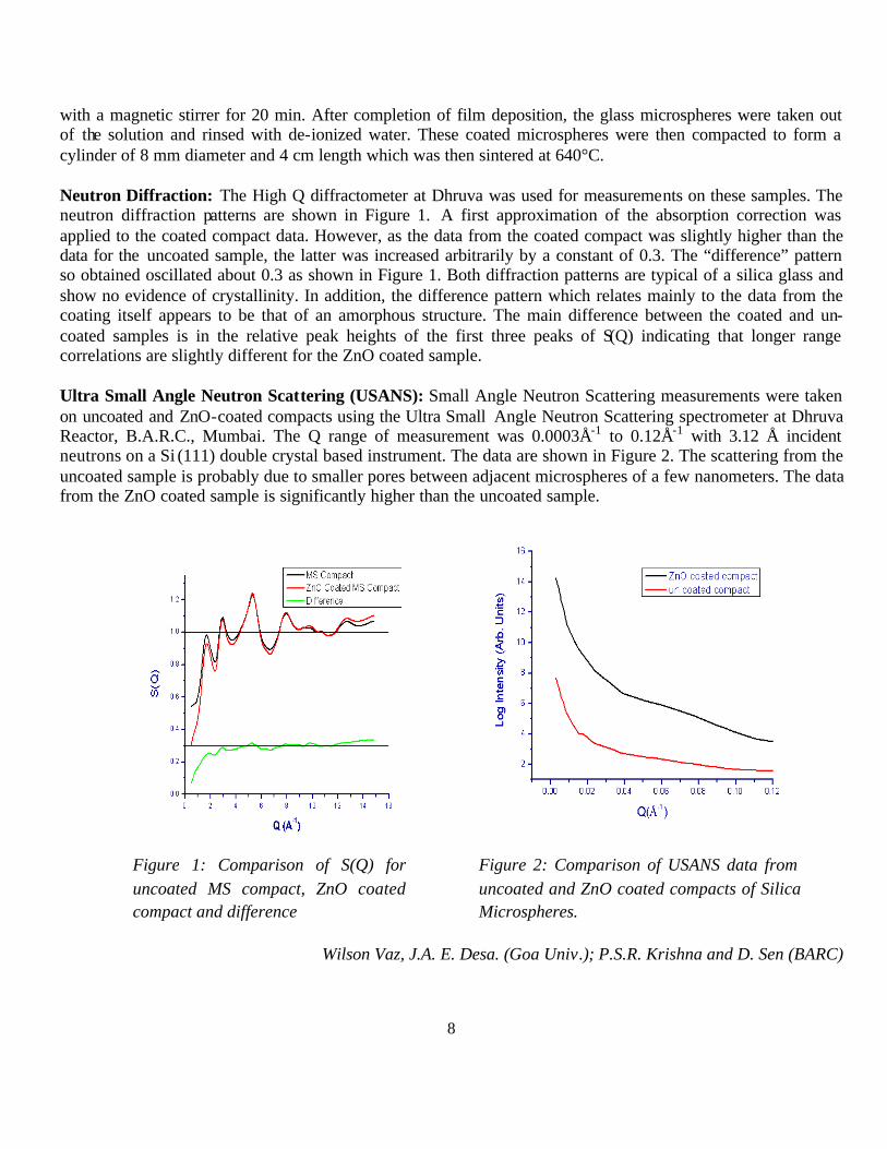
Neutron Diffraction: The High Q diffractometer at Dhruva was used for measurements on these samples. The neutron diffraction patterns are shown in Figure 1. A first approximation of the absorption correction was applied to the coated compact data. However, as the data from the coated compact was slightly higher than the data for the uncoated sample, the latter was increased arbitrarily by a constant of 0.3. The “difference” pattern so obtained oscillated about 0.3 as shown in Figure 1. Both diffraction patterns are typical of a silica glass and show no evidence of crystallinity. In addition, the difference pattern which relates mainly to the data from the coating itself appears to be that of an amorphous structure. The main difference between the coated and un-coated samples is in the relative peak heights of the first three peaks of S(Q) indicating that longer range correlations are slightly different for the ZnO coated sample.
Ultra Small Angle Neutron Scattering (USANS): Small Angle Neutron Scattering measurements were taken on uncoated and ZnO-coated compacts using the Ultra Small Angle Neutron Scattering spectrometer at Dhruva Reactor, B.A.R.C., Mumbai. The Q range of measurement was 0.0003Å-1 to 0.12Å-1 with 3.12 Å incident neutrons on a Si (111) double crystal based instrument. The data are shown in Figure 2. The scattering from the uncoated sample is probably due to smaller pores between adjacent microspheres of a few nanometers. The data from the ZnO coated sample is significantly higher than the uncoated sample.
Figure 1: Comparison of S(Q) for uncoated MS compact, ZnO coated compact and difference
Figure 2: Comparison of USANS data from uncoated and ZnO coated compacts of Silica Microspheres.
Wilson Vaz, J.A. E. Desa. (Goa Univ.); P.S.R. Krishna and D. Sen (BARC)
9
2.1.2. Studies of Porous Materials by SANS
Silica microspheres in the size range 5-20 µm, and 106-110µm and have been used to make porous glass. Compacts were prepared according to the schedule described by Carrasco et al. (2005).
Density measurements of the compacts show a trend with sintering time which may be related to the process of densification of the compacts in which the microspheres move relative to each other. This gradual process leads to agglomeration of nearest-neighbour microspheres, causing the existing pores to become larger and possibly more interconnected. This view is supported by SEM micrographs of the compacts sintered for 3, 6, 24 hours.
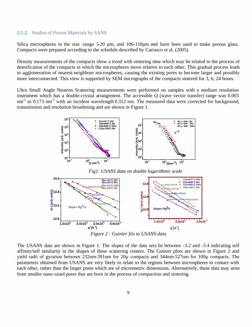
Ultra Small Angle Neutron Scattering measurements were performed on samples with a medium resolution instrument which has a double-crystal arrangement. The accessible Q (wave vector transfer) range was 0.003 nm-1 to 0.173 nm-1 with an incident wavelength 0.312 nm. The measured data were corrected for background, transmission and resolution broadening and are shown in Figure 1.
101
102
103
104
105
106
10-3 10-2 10-1
Inte
nsity
(ar
b. u
nits
)
110µ-610°C-24hr
Q (nm-1)
110µ-640°C-3hr 110µ-640°C-24hr 110µ-740°C-3hr
101
102
103
104
105
10-3 10-2 10-1 Q(nm-1)
Inte
nsit
y (A
rb. U
nits
)
q≈-3
20 µ - 640° - 24hr
20 µ - 640°- 3hr20 µ - 640° - 6hr
Fig1: USANS data on double logarithmic scale
1.0x10⁻⁵ 2.0x10⁻⁵ 3.0x10⁻⁵ 4.0x10 -5
12.6
13.2
13.8
14.4
ln I
(arb
.un
its)
q2(A-2)
Rg=391 nm
Rg=313 nm
Slope= Rg2/3
20µ−840°C-1hr20µ−640°C-24hr20µ−640°C-6hr20µ−640°C-3hr
Rg=329 nm
Rg=232 nm
12.0
12.8
13.6
1.0x10⁻⁵ 2.0x10 -5 3.0x10 -5
Rg=510nm
Rg=491nm
Rg=527nm
Rg=344nm
Slope= Rg2/3
110µ-640°C-24hr
ln I
(arb
.uni
ts)
110µ-740°C-3hr
q2(A-2)
110µ-610°C-24hr 110µ-640°C-3hr
Figure 2 : Guinier fits to USANS data
The USANS data are shown in Figure 1. The slopes of the data sets lie between -3.2 and -3.4 indicating self affinity/self similarity in the shapes of these scattering centers. The Guinier plots are shown in Figure 2 and yield radii of gyration between 232nm-391nm for 20µ compacts and 344nm-527nm for 100µ compacts. The parameters obtained from USANS are very likely to relate to the regions between microspheres in contact with each other, rather than the larger pores which are of micrometric dimensions. Alternatively, these data may arise from smaller nano-sized pores that are born in the process of compaction and sintering.
10
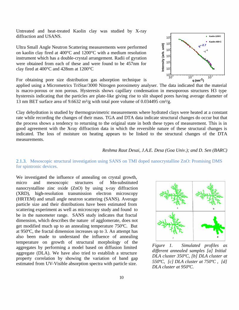
Untreated and heat-treated Kaolin clay was studied by X-ray diffraction and USANS.
Ultra Small Angle Neutron Scattering measurements were performed on kaolin clay fired at 400°C and 1200°C with a medium resolution instrument which has a double-crystal arrangement. Radii of gyration were obtained from each of these and were found to be 457nm for clay fired at 400°C and 428nm at 1200°C
For obtaining pore size distribution gas adsorption technique is applied using a Micrometrics TriStar/3000 Nitrogen porosimetry analyser. The data indicated that the material is macro-porous or non porous. Hysteresis shows capillary condensation in mesoporous structures H3 type hysteresis indicating that the particles are plate-like giving rise to slit shaped pores having average diameter of 13 nm BET surface area of 9.6632 m²/g with total pore volume of 0.034495 cm³/g.
Clay dehydration is studied by thermogravimetric measurements where hydrated clays were heated at a constant rate while recording the changes of their mass. TGA and DTA data indicate structural changes do occur but that the process shows a tendency to returning to the original state in both these types of measurement. This is in good agreement with the X-ray diffraction data in which the reversible nature of these structural changes is indicated. The loss of moisture on heating appears to be linked to the structural changes of the DTA measurements.
Reshma Raut Desai, J.A.E. Desa (Goa Univ.); and D. Sen (BARC)
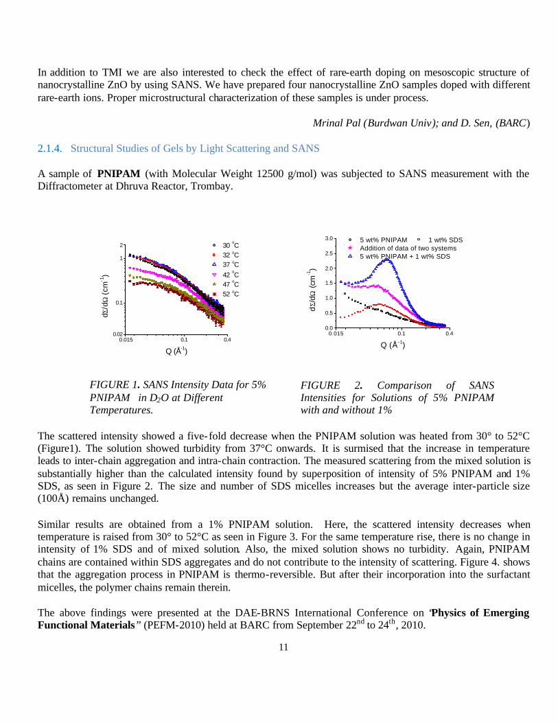
2.1.3. Mesoscopic structural investigation using SANS on TMI doped nanocrystalline ZnO: Promising DMS for spintronic devices. We investigated the influence of annealing on crystal growth, micro and mesoscopic structures of Mn-substituted nanocrystalline zinc oxide (ZnO) by using x-ray diffraction (XRD), high-resolution transmission electron microscopy (HRTEM) and small angle neutron scattering (SANS). Average particle size and their distributions have been estimated from scattering experiment as well as microscopy study and found to be in the nanometer range. SANS study indicates that fractal dimension, which describes the nature of agglomerate, does not get modified much up to an annealing temperature 750ºC. But at 950ºC, the fractal dimension increases up to 3. An attempt has also been made to understand the influence of annealing temperature on growth of structural morphology of the aggregates by performing a model based on diffusion limited aggregate (DLA). We have also tried to establish a structure property correlation by showing the variation of band gap estimated from UV-Visible absorption spectra with particle size.
10-3 10-2 10-1100
101
102
103
104
105
106
q (nm-1)
Kaolin-400°C
Inte
nsi
ty (
arb
. un
it)
~q-4
q≈-2.7
Kaolin-1200°C
Figure 1. Simulated profiles as different annealed samples [a] Initial DLA cluster 350ºC, [b] DLA cluster at 550ºC, [c] DLA cluster at 750ºC , [d] DLA cluster at 950ºC.
11
In addition to TMI we are also interested to check the effect of rare-earth doping on mesoscopic structure of nanocrystalline ZnO by using SANS. We have prepared four nanocrystalline ZnO samples doped with different rare-earth ions. Proper microstructural characterization of these samples is under process.
Mrinal Pal (Burdwan Univ); and D. Sen, (BARC)
2.1.4. Structural Studies of Gels by Light Scattering and SANS
A sample of PNIPAM (with Molecular Weight 12500 g/mol) was subjected to SANS measurement with the Diffractometer at Dhruva Reactor, Trombay.
0.015 0.1 0.40.02
0.1
1
2 30 oC
32 oC 37 oC
42 oC
47 oC
52 oC
dΣ/d
Ω (
cm-1
)
Q (Å-1)
FIGURE 1. SANS Intensity Data for 5% PNIPAM in D2O at Different Temperatures.
0.015 0.1 0.40.0
0.5
1.0
1.5
2.0
2.5
3.0 5 wt% PNIPAM 1 wt% SDS Addition of data of two systems 5 wt% PNIPAM + 1 wt% SDS
dΣ/
dΩ (
cm-1)
Q (Å-1)
FIGURE 2. Comparison of SANS Intensities for Solutions of 5% PNIPAM with and without 1%
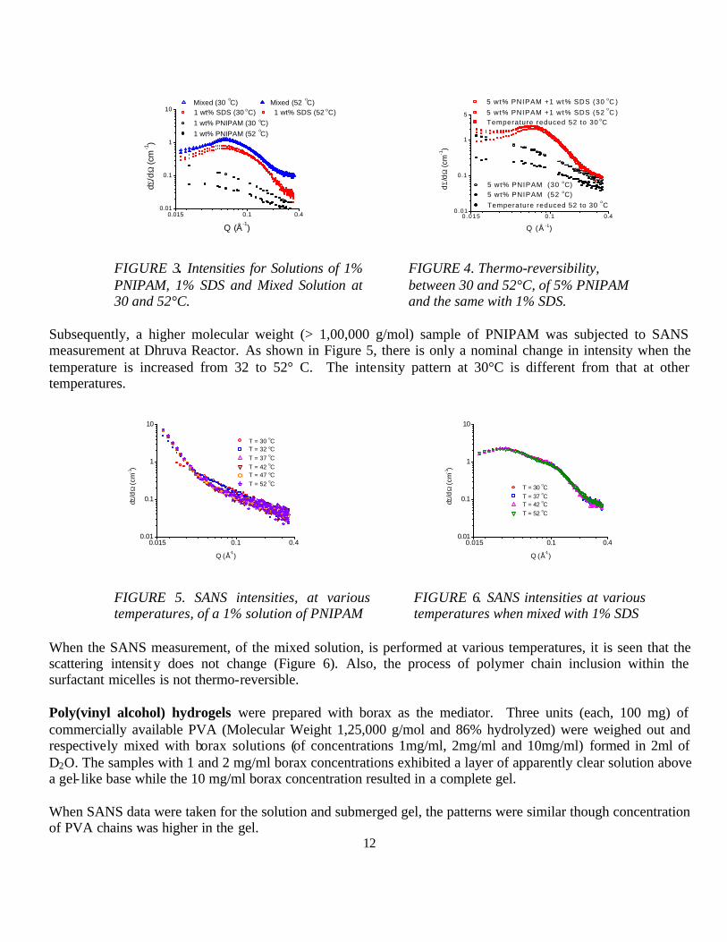
The scattered intensity showed a five-fold decrease when the PNIPAM solution was heated from 30° to 52°C (Figure1). The solution showed turbidity from 37°C onwards. It is surmised that the increase in temperature leads to inter-chain aggregation and intra-chain contraction. The measured scattering from the mixed solution is substantially higher than the calculated intensity found by superposition of intensity of 5% PNIPAM and 1% SDS, as seen in Figure 2. The size and number of SDS micelles increases but the average inter-particle size (100Å) remains unchanged.
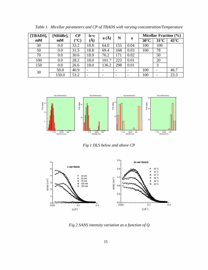
Similar results are obtained from a 1% PNIPAM solution. Here, the scattered intensity decreases when temperature is raised from 30° to 52°C as seen in Figure 3. For the same temperature rise, there is no change in intensity of 1% SDS and of mixed solution. Also, the mixed solution shows no turbidity. Again, PNIPAM chains are contained within SDS aggregates and do not contribute to the intensity of scattering. Figure 4. shows that the aggregation process in PNIPAM is thermo-reversible. But after their incorporation into the surfactant micelles, the polymer chains remain therein.
The above findings were presented at the DAE-BRNS International Conference on “Physics of Emerging Functional Materials” (PEFM-2010) held at BARC from September 22nd to 24th, 2010.
12
0.015 0.1 0.40.01
0.1
1
10 Mixed (30
oC) Mixed (52
oC)
1 wt% SDS (30 oC) 1 wt% SDS (52 oC) 1 wt% PNIPAM (30 oC) 1 wt% PNIPAM (52
oC)
dΣ/d
Ω (
cm-1)
Q (Å-1)
FIGURE 3. Intensities for Solutions of 1% PNIPAM, 1% SDS and Mixed Solution at 30 and 52°C.
0.015 0.1 0.40.01
0.1
1
5
5 wt% PNIPAM (30 oC) 5 wt% PNIPAM (52 oC)
Temperature reduced 52 to 30 oC
5 wt% PNIPAM +1 wt% SDS (30 oC )
5 wt% PNIPAM +1 wt% SDS (52 oC )
Temperature reduced 52 to 30 oC
dΣ/d
Ω (c
m-1
)
Q (Å -1)
FIGURE 4. Thermo-reversibility, between 30 and 52°C, of 5% PNIPAM and the same with 1% SDS.
Subsequently, a higher molecular weight (> 1,00,000 g/mol) sample of PNIPAM was subjected to SANS measurement at Dhruva Reactor. As shown in Figure 5, there is only a nominal change in intensity when the temperature is increased from 32 to 52° C. The intensity pattern at 30°C is different from that at other temperatures.
0.015 0.1 0.40.01
0.1
1
10
T = 30 oC T = 32 oC T = 37 oC T = 42 oC T = 47 oC T = 52 oC
dΣ/d
Ω (
cm-1)
Q (Å-1)
FIGURE 5. SANS intensities, at various temperatures, of a 1% solution of PNIPAM
0.015 0.1 0.40.01
0.1
1
10
T = 30 oC T = 37 oC T = 42 oC T = 52 oC
dΣ/d
Ω (
cm-1)
Q (Å-1)
FIGURE 6. SANS intensities at various temperatures when mixed with 1% SDS
When the SANS measurement, of the mixed solution, is performed at various temperatures, it is seen that the scattering intensity does not change (Figure 6). Also, the process of polymer chain inclusion within the surfactant micelles is not thermo-reversible.
Poly(vinyl alcohol) hydrogels were prepared with borax as the mediator. Three units (each, 100 mg) of commercially available PVA (Molecular Weight 1,25,000 g/mol and 86% hydrolyzed) were weighed out and respectively mixed with borax solutions (of concentrations 1mg/ml, 2mg/ml and 10mg/ml) formed in 2ml of D2O. The samples with 1 and 2 mg/ml borax concentrations exhibited a layer of apparently clear solution above a gel- like base while the 10 mg/ml borax concentration resulted in a complete gel.
When SANS data were taken for the solution and submerged gel, the patterns were similar though concentration of PVA chains was higher in the gel.

13
SANS intensity data from the gel- like layers (1 and 2 mg/ml borax solutions) and the full gel (10 mg/ml borax) are shown in Figure 8. As the borax concentration increases from 1 to 2 mg/ml, it is observed that the scattered intensity from the PVA cross- linked regions increases due to the larger number of such scattering centers in the 2mg/ml sample. In the case of the higher concentration of borax (i.e. 10mg/ml), the intensity is lower than the 1mg/ml in the lower Q region, but rises above the 2mg/ml concentration in the higher Q region. A greater degree of cross- linking in this sample leads to formation of larger agglomerates, whose sizes cannot be detected by the lowest Q of the instrument. This results in a lowering of intensity in the lower Q region. Simultaneously, the smaller-sized cross-linked regions also increase in number, resulting in an increase in intensity at the higher Q end of the measured range.
0.015 0.1 0.40.05
0.1
1
2
dΣ/d
Ω (c
m-1
)
Gel Solution
Q (Å-1)
FIGURE 7. SANS intensities of the solution and gel layers
0.015 0.1 0.40.05
0.1
1
5 borax conc. = 1 mg/ml borax conc. = 2 mg/ml borax conc. = 10 mg/ml
dΣ/d
Ω (
cm-1
)
Q (Å-1)
FIGURE 8. SANS intensities of the gels with varying borax concentrations
The above findings have been presented at the 55th DAE Solid State Physics Symposium that was held at Manipal University, Manipal from December 26th to 30th, 2010.
Rheology measurements were also performed of the various PVA gels. Some of the resultant plo ts are as shown below (figures 9, 10):
1 10 10010
100
1000
Pa
ω/s
G'(Storage) G"(Loss)
Sample-E
FIGURE 9. Rheology plot for gel with 1mg/ml borax concentration at 30°C
0.1 1 10 1001
10
100
1000
G'(30) G"(30) G'(50) G"(50)
Pa
ω/s
F
FIGURE 9. Rheology plot for gel with 1mg/ml borax concentration at 30°C

14
PVA-borax hydrogels incorporated with varying proportions of rare earth (Neodymium and Praseodymium) ions have been prepared and will be soon subjected to DLS and rheology measurements at BARC.
L. Basco, J. A. E Desa, (Goa Univ.); and V.K. Aswal (BARC)
2.1.5. Structural and interactional behavior of mixed micelles of twin tail surfactants with triblock copolymers.
The aggregation behavior of surfactant-triblock polymer systems carries enormous interest on account of their technical importance as well as the variety observed in their aggregation phenomena which can be studied theoretically as well as experimentally. The hydrophobic interaction among surfactant and triblock polymer (TBP) molecules leads to the formation of mixed micelles. Mixed systems of twin tail surfactants and triblock polymers are widely used in cosmetics and pharmaceutical industries due to their antibiotic activity. These are also used in food industry, synthesis of nano particles, as catalysts, as surfactant-supramolecular assemblies and as drag reducing agents. We have carried out surface tension and cloud point measurements of mixed micelles of twin tail surfactants such as Lecithin and Didodecyldimethyl ammonium bromide (DDAB) with triblock copolymer L35. In addition to this we have also carried out small angle neutron scattering (SANS), surface tension, conductivity and fluorescence measurements of mixed micelles of twin tail surfactants of varying hydrophobicity such as Didodecyldimethyl ammonium bromide, Dimethylditetradecyl ammonium bromide, Dihexadecyldimethyl ammonium bromide and Dimethyldioctadecyl ammonium chloride with varying concentration of L64 (1wt %, 4wt %, 8wt %, 12wt %). SANS data shows prolate ellipsoidal behavior. The synergism between twin tail surfactants and TBP have been observed
Rajwinder Kaur, R.K.Mahajan (GND Univ., Amritsar); V.K.Aswal (BARC)
2.1.6. SANS Study of Clouding Phenomenon in charged micellar solutions
The research work carried out in the scheme is related to Clouding Phenomenon in charged micellar solutions in presence of various stimuli (temperature, [salt], pH, etc). In this connection synthesis of various anionic surfactants (tetra butyl ammonium dodecylsulfate (TBADS), tetra butyl ammonium sulfonato myristic acid methyl ester (TBAMES) and tetra butyl ammonium sulfonato palmitic acid methyl ester (TBAPES) has been carried out. The purity of these surfactants was ensured by NMR and absence of minima in Surface Tension vs. [surfactant] plot. The preliminary micellization parameters (critical micelle concentration (cmc), degree of counter ion dissociation, area per head group, etc) were obtained by conductivity and surface tension measurements. However, good results were not obtained with TBAPES and not studied further. It has been found that all above surfactants have shown clouding behavior though they were ionic in nature.
The combination of visual observation, Cloud Point, DLS and SANS measurements is adopted for collecting information. The data demonstrate the gradual variation of micellar fraction in the system with temperature below CP and even beyond CP; furthermore, it has been shown that the micellar fraction can be tuned with the help of concentration of tetra-n-butylammonium dodecyl sulphate [TBADS], Temperature, [Salt] and nature of salt. (Table 1)
15
Table 1. Micellar parameters and CP of TBADS with varying concentration/Temperature
[TBADS], mM
[NH4Br], mM
CP (°C)
b=c (Å) a (Å) N a
Micellar Fraction (%) 30°C 31°C 45°C
30 0.0 33.2 18.8 64.0 155 0.04 100 100 50 0.0 31.5 18.8 69.4 168 0.03 100 78 70 0.0 30.6 18.9 76.2 171 0.02
50
100 0.0 28.2 18.0 101.7 223 0.01 20 150 0.0 26.6 18.0 136.2 298 0.01 3
30 50.0 40.9 - - - - 100 - 46.7 150.0 53.2 - - - - 100 - 23.3
Fig 1 DLS below and above CP
0.015 0.1 0.40
1
2
3
4
5
6
C mM TBADS
30 mM 50 mM 70 mM 100 mM 150 mM
dΣ/d
Ω (
cm-1)
Q (Å-1)
0.015 0.1 0.40.0
0.4
0.8
1.2
1.6
2.020 mM TBADS
30 oC 32 oC 34 oC 36 oC 38 oC 40 oC
dΣ/d
Ω (
cm-1)
Q (Å-1)
Fig 2 SANS intensity variation as a function of Q
Size distribution(s)
5 10 50100 500Diameter (nm)
5
10
15
% in
cla
ss
Size distribution(s)
5 10 50100 500Diameter (nm)
10
20
% in
cla
ss
Size distribution(s)
5 10 50100 500Diameter (nm)
10
20
% in
cla
ss
Size distribution(s)
5 10 50100 500Diameter (nm)
20
40
60
% in
cla
ss

16
It is found that a very small portion of the total micellar concentration converts into micellar clusters and causes turbidity in the solution. However, at CP most of the surfactant is present in micellar form which decreases on heating beyond CP (Table 1). The presence of NH4Br delays the CP. Similarly, micellar fraction also varies with [NH4Br].
DLS measurements, below and above the CP, have also shown the development of bigger morphologies in addition to smaller ones even below CP (Fig1).
Two micellar morphologies near CP can have promising applications as a smart nano container loading amphiphilic compounds (e.g charged drugs, dyes, proteins etc) as well as bearing a thermo responsive surface that is useful for physical affinity control.
Arti Bhadoria, Sanjeev Kumar Sanjeev Kumar (M.S. Univ. Baroda); and V.K. Aswal (BARC)
2.1.7. Interaction of Serum Albumins and Drugs
Table 1 Binding parameters, i.e, Stern-Volmer quenching constant, Ksv, number of binding sites, n, and binding constant, K, for BSA with AMT / PMT at 280 and 295nm excitation wavelengths.
Ksv 10–4 (L mol-1) R2 n K 10–4 (mol-1) BSA 280nm AMT 2.55 0.9912 0.865 0.45 PMT 3.15 0.9941 1.186 32.5 295nm AMT 2.51 0.9951 0.916 0.87 PMT 3.17 0.9954 1.129 16.7
The binding study of drugs with serum albumins is of imperative and fundamental importance. Binding studies of two amphiphilic drugs, i.e., Amitriptyline Hydrochloride and Promethazine Hydrochloride, with Bovine Serum Albumin (BSA) were made by using different techniques.
240 250 260 270 280 290 3000.0
0.1
0.2
0.3
0.4
0.5
0.6
0.7
(a)6
1
Abs
orba
nce
Wavelegth (nm)
1- Native BSA2- 6 µM AMT3- 28µM AMT4- 52µM AMT5- 76µM AMT6- 100µM AMT
240 250 260 270 280 290 300
0.0
0.1
0.2
0.3
0.4
0.5
(b) 6
1
Abs
orba
nce
Wavelenght (nm)
1- Native BSA2- 6 µM PMT3- 28µM PMT4- 52µM PMT5- 76µM PMT6- 100µM PMT
Figure 1. Ultraviolet absorbance spectra of native BSA and BSA-AMT (a) / BSA-PMT (b) complexes.

17
Fluorescence: Informations about molecular environment in the vicinity of fluorophore molecules were obtained by fluorescence experiments (Table 1).
UV-Vis spectroscopy: The changes in BSA-PMT complexes were found to be more prominent than in the BSA-AMT complexes (Fig. 1).
Circular Dichroism: The conformational changes in the secondary structure of the serum albumin, monitored by the far UV-CD in the range of 200-250nm, also favour UV-visible results.
205 210 215 220 225 230 235 240 245
-25000
-20000
-15000
-10000
-5000
0
(a)
5
4 3
21
MR
E (d
eg cm
2 dm
ol-1)
Wavelength (nm)
1-Native BSA2-10 µM AMT3-40 µM AMT4-100µM AMT5-250µM AMT
205 210 215 220 225 230 235 240 245
-25000
-20000
-15000
-10000
-5000
0
(b)
5
4 3
2
1MR
E (d
eg cm
2 dm
ol-1)
Wavelength (nm)
1-Native BSA2-10 µM PMT3-40 µM PMT4-100µM PMT5-250µM PMT
Figure 2. CD spectra of native BSA and BSA-AMT (a) / BSA-PMT (b) complexes.
Kabir-ud-Din (AMU); V.K.Aswal(BARC)
2.1.8. Neutron Diffraction Studies of LaSrCoRuO6 type Double Perovskites
Neutron diffraction experiments have been carried out to study the effect of thermally induced and substitutional disorder on the magnetic properties of LaSrCoRuO6 double perovskite. While the ordered sample is antiferromagnetic, the disordered sample exhibits negative values of magnetization measured in low applied fields. Isothermal magnetization on this sample shows hysteresis due to the presence of ferromagnetic interactions.
Based on neutron diffraction and X-ray absorption fine structure (XAFS) studies, these results have been interpreted to be due to disorder in site occupancy of Co and Ru leading to octahedral distortions and formation of Ru–O–Ru ferromagnetic linkages. Below 150K these ferromagnetic Ru spins polarize the Co spins in a direction opposite to that of the applied field resulting in observed negative magnetization. This work has resulted in three publications.
P.S. Rama Murthy (Goa University) and A. Das (BARC)
18
2.1.9. Structure of 0.4Sb2Se3-0.6CuI (Chalcohalide) glass using Neutron Diffraction
The chalcogenide-halide glasses are a new class of glasses, which have infrared transmission properties, superior to the simple chalcogenide glasses. Chalcohalide glasses do possess interesting optical properties, which make them candidates for CO2 laser fibers and infrared windows because of high transmission of IR. It would be interesting to understand their structure as they are known to be network glasses having both short and intermediate range orders.
The starting materials used were 4N pure Sb metal, 5N pure Se granules and analytical grade anhydrous Copper halide flakes. These materials were weighed, mixed and placed into a quartz glass tube sealed ampoules of outer diameter 12 mm and inner diameter 10 mm with one end sealed, which was then evacuated and sealed under a vacuum of about 10-6 Torr. The ampoule was heated at 825 0C for 12 hrs in an electric furnace. The ampoule was shaken for homogeneity. The ampoule was then quenched in chilled water.
The glass formation was determined by x-ray diffraction for the sample 0.4Sb2Se3-0.6CuI. IR transmission measurements show that this composition has high transmission in the range from 10µm to 30µm (=75%). The microhardness of glass is found to be 108.8 kg/mm2 with standard deviation of 4.8kg/mm2. Glass transition temprature is found to be 1670C.
We have done neutron diffraction studies on the sample 0.4Sb2Se3-0.6CuI on the High-Q diffractometer at the Dhruva reactor, BARC, Trombay, to understand the short range order and network connectivity. The experimentally obtained S(Q) vs. Q function is given in Fig. 1. The T(r)=4πρrg(r) obtained by Fourier transformation using MCGR method is plotted as a function of r in Fig. 2 from where we have obtained the distances and co-ordinations of various pair cor relations.
Fig.1. S(Q) vs. Q
Fig.2. T(r)=4πrρg(r) vs. R
We have assumed continuously ordered chemically ordered network model (COCRN). The short range order mainly consists of Sb-Se bonds as well as Cu-Se bonds. Se-Se bonds are ruled out as the first 2 peaks in T(r) are larger and match with Sb-Se and Cu-Se (also Cu-I) bonds. The distances are 2.48(1)Å and 2.66(1)Å. From bond energy considerations these are more preferable bonds and the co-ordinations obtained are 2.9(2) and 3.7(2). As Cu-I bonds also are preferred and come around the same distances, if we assume one Cu-I bond then there are 3.0(2) Cu-Se bonds. Similarly Sb-I bonds also can come around the ident ified Sb-Se distance. So if we assume one Sb-I bond there then we will have 2.2(2) Sb-Se bonds. From these results we can say that Sb-
0 2 4 6 8 10 12 14 16
-0.5
-0.4
-0.3
-0.2
-0.1
0.0
0.1
0.2
S(Q
)-1
Q(A-1) 0 1 2 3 4 5 6
0
1
2
3
T(r) =
4 π
ρ r
g(r)
r (A)
19
(Se,I) pyramids are connected to Cu-(Se,I) bonds with co-ordinations of 3 and 4. Se-Se (non-bonding), Sb-Se (2nd neighbor) and Cu-Cu distances are identified to come in the r-range of 3.5Å to 4.2Å. Our results are in good agreement with the known distances in these chalcohalide glasses. From these experiments it is clear that Cu is actively participating in the network formation.
M. S. Jogad, Rashmi M Jogad, Rakesh Kumar (Sharanabasaveshwar college of science, Gulburga Univ., Gulburga); P S R Krishna and G P Kothiyal, (BARC)
2.1.10. Probing the 4d and 5d magnetism in prototypical Ba3M1+xM’2-xO9 (M = 3d metal and M’ = 4d/5d metal) system, using neutron diffraction
Doping in BaRuO3 by 3d transition metal oxide like Cu, Ti, Co, Ni etc, or rare earth elements like La, Eu, Lu, Sm etc or even the alkali metals Na, K, Li stabilize the structure in 6 layer hexagonal structure similar to the hexagonal BaTiO 3 structure with space group P63/mmc having the chemical formulae Ba3MRu2O9 where M is the dopant element. This structure consists of a couple of face-shared octahedra connected via a single corner shared octahedron. In all most every case, M ions occupy the corner shared octahedra (2a site), whereas Ru sits in the face shared position (4f site). However, Fe is the exception from this trend where in Ba3FeRu2O9 (BFRO), both Fe and Ru are able to occupy both face shared and corner shared positions making this member special in this 6 layered hexagonal ruthenates family. Now, as a result of this unusual disorder effect, many possible magnetic interactions become realizable. Our study involving several experimental methods on this compound established that the magnetic structure of Ba3FeRu2O9 is indeed very different from all other 6H ruthenates. Detailed magnetization and powder neutron diffraction (ND) experiments proved that at low temperature, the system takes up a global spin-glass like order. The structural data from the diffraction studies definitely indicated presence of large disorder, but the local structural study revealed that this Fe/Ru site disorder could also extend beyond a unit cell and create local chemical inhomogeneity, affecting the high-temperature magnetism of this material. There is a gradual decrease of 57Fe Mössbauer spectral intensity with decreasing temperature (below 100 K), which reveals that there is a large spread in the magnetic ordering temperatures, corresponding to many spatially inhomogeneous regions. However, finally at about 25 K, the whole compound is found to take up a global glasslike magnetic ordering. These results have been published in Physical Review B.
On the other hand, Ba3ZnRu2O9 has the similar 6H hexagonal structure like Ba3FeRu2O9, but it has no site disorder i.e. Zn sits in the corner shared octahedra and Ru sits in the face shared octahedral forming Ru2O9 dimer. As expected, the magnetic behavior of this system is similar to that of a spin dimer. However, below 100 K, a clear magnetic transition takes place and the simple dimer picture fails to describe the magnetic property. To check whether any structural transition is taking place across the transition, ND measurements were performed at 300K and 2K. Both the patterns were refined with space group P63/mmc. The lattice constants at 300 K (5K) are a=b=5.7705Å (5.7546 Å) and c= 14.1703 Å (14.12399 Å). An unusual compression of the Ru2O9 dimer is observed across the magnetic transition temperature (the Ru-Ru distance in the Ru2O9 dimer is 2.7284 Å and 2.6790 Å at the 300K and 2K respective ly), which must be associated with the unusual magnetic phase. Most importantly, unusual changes in magnetic structures are observed when the 4d Ru ions are progressively replaced by 5d Ir ions. In order to study this, two more compounds in the series were synthesized, namely, Ba3ZnRuIrO9 and Ba3ZnIr2O9. In the following figure (left panel), magnetization data from these three compounds is shown, where a progressive change can be observed. Room temperature ND patterns for the samples are shown below (right panel).
20
Srimanta Middey, Sugata Ray, K. Mukherjee (IACS); P. L. Paulose, E. V. Sampathkumaran (TIFR); C. Meneghini (Elettra, Italy); D. D. Sarma (IISc); S. D. Kaushik, V. Siruguri
2.1.11. Neutron diffraction studies on (Ba,Sc)3YIr2O9 and Ba3YRu2O9 compounds
Single phase samples of Ba3YIr2O9, Sc3YIr2O9, and Ba3YRu2O9 have been prepared and characterized by magnetic susceptibility and heat capacity measurements. Neutron diffraction measurements on the above samples are currently ongoing at the UGC-DAE CSR beamline at BARC. In particular, low-temperature measurements are planned very soon to examine the onset of magnetic order with a decrease in temperature. It is also planned to investigate the effect of a magnetic field (if any) on the transition temperature. Room temperature neutron diffraction profiles for a couple of samples are shown below.
20 40 60 80 100 120-2000
0
2000
4000
6000
8000
10000
12000
14000 Obs Cal Dif Bragg
Inte
nsity
(co
unts
)
2 θ (degrees)
Ba3YRu2O9
|
20 40 60 80 100 1200
1000
2000
3000
4000
5000
Obs Cal Diff Bragg
Ba3YIr2O9
2θ
Inte
nsity
A.V. Mahajan (IIT Bombay); V Siruguri
21
2.1.12. Size dependent magnetic properties of Co-Zn spinel ferrite nanoparticles using neutron diffraction technique.
The aim of the study is to understand the type of the magnetic ordering and find out the magnetic properties of two compositions of CoZnFe2O4 system one is above percolation threshold [Co0.5Zn0.5Fe2O4] and the other is below percolation threshold [Co0.3Zn0.7Fe2O4] using neutron diffraction at the low temperature and high field. Both compositions have three different sizes. The size varies from 6nm to bulk. Following is the brief detail about the samples:
1) Co0.5Zn0.5Fe2O4: Particles in three different sizes i.e. 9nm, 40nm and bulk 2) Co0.3Zn0.7Fe2O4: Particles in three different sizes i.e. 7nm, 40nm and bulk
The above six samples are characterized using X-ray and neutron diffraction at room temperature (figure:1), without magnetic field.
Figure:1 Neutron diffraction pattern for both the compositions Co0.5Zn0.5Fe2O4 and Co0.3Zn0.7Fe2O4 having three different sizes.
The results indicate that all the samples are having single phase FCC spinel ferrite system. The extensive analysis has been carried out using TOPAS and Fullprof programs. The parameter obtained from the neutron diffraction data analysis are shown in table :1.
As shown in the table, magnetic moment of both the composition initially increases but in case of bulk it shows different behavior. Consistent behavior is observed in both the composition. It is to be noted that structural strain & stress obtained from the analysis is negligible.
20 40 60 80 100
Inte
nsi
ty (
a.u
.)
2θ (degrees)
size:92(Å)
Inte
nsi
ty(a
.u.) size: 400(Å)
Inte
nsi
ty(a
.u.) size: BulkCo0.5Zn0.5Fe2O4
20 40 60 80 100 120
Inte
nsi
ty(a
rb.u
)
2θ (degrees)
size:72(Å)
In
ten
sity
(arb
.u)
size:400(Å)
Inte
nsi
ty(a
rb.u
)
size:Bulk(Å)Co0.3Zn0.7Fe2O4
22
Table:1 Structural and magnetic parameters like lattice constant, oxygen parameter, cation distribution, and magnetic moments are determined from Neutron diffraction pattern analysis for Co0.3Zn0.7Fe2O4 and Co0.5Zn0.5Fe2O4 systems.
Composition Particles size (Å)
Lattice
parameter
(Å) ±0.001
Cation distribution
Magnetic moment
A-
site
B-
site
A-site
(µB) ±0.1
B-site
(µB) ±0.1
Net
(µB) ±0.1
Co0.5Zn0.5Fe2O4 92 ±5 8.352 Zn+2- A site
Co+2-
B site
1.42 -2.78 1.36 400±20 8.406 1.35 -3.31 1.96
Bulk 8.413 0.75 -2.26 1.51 Co0.3Zn0.7Fe2O4 72 ±5 8.360 Zn+2-
A site Co+2-
B site
1.12 -1.35 0.23 400±20 8.414 1.11 -1.98 0.87
Bulk 8.424 0.53 -1.02 0.49
As shown in the neutron diffraction data, in magnetization data also the Co0.3Zn0.7Fe2O4 and Co0.5Zn0.5Fe2O4 spinel oxide with average particle size 400(Å) shows an enhancement in both magnetization (i.e., more ferromagnetic) and ordering temperature comparing the bulk sample. The experimental data also suggest that the lowering of B site spin canting play an important role in controlling the magnetic order of the sample. Importantly, the grain boundary spins of the 400(Å) size sample do not show to be magnetically inactive, rather they give rise to a preferential orientation. The room temperature magnetization loops are shown in figure 2. Initially with size the magnetization is increases but after that the magnetization is decrease for bulk sample. It shows the “inverse spin canting effect”.
Figure:2 M-H loop for both the compositions for three different sizes at 300K
-20000 0 20000 40000 60000 80000-60
-45
-30
-15
0
15
30
45
60
75
72(Å)
Bulk
M(e
mu
/gm
)
Magnetic Field (Oe)
400(Å)
Co0.3Zn0.7Fe2O4
72(Å):-35.9emu/gm
400(Å):-64.3emu/gm
Bulk:-50.6emu/gm
T=300K
-20000 0 20000 40000 60000 80000-80
-60
-40
-20
0
20
40
60
80
92(Å)
Co0.5Zn0.5Fe2O4
92(Å):-50.0emu/gm
400(Å):-77.9emu/gm
Bulk:-76.2emu/gm
Bulk
M(e
mu
/gm
)
Magnetic Field(Oe)
400(Å)T=300K
23
20 40 60 80 100
Inte
nsi
ty (
a.u
.)
2θ (degrees)
Co0.3Zn0.7Fe2O4
size:72(Å)Temp:2K
For sample Co0.3Zn0.7Fe2O4 nano size (72 Å), low temperature (2K) neutron diffraction data has been collected (figure:3), at low temperature (2K) magnetic moment for A and B site is 2.01 µB and -1.50 µB respectively (net magnetic moment 0.5 µB) which is higher than the room temperature magnetic moment .
From figure: 3 it’s clear at low temperature (2K) also it is not showing any canted structure peak in neutron diffraction data, while this system is the below percolation threshold.
Figure:3 Neutron diffraction of Co0.3Zn0.7Fe2O4 nano size (72 Å) sample at 2K low temperature.
R.V. Upadhyay (Charotar Inst. Technology, Changa); V. Siruguri
2.1.13. Neutron diffraction studies of collapse of charge ordering in narrow band half-doped manganite Y0.5Ca0.5MnO3 nanoparticles
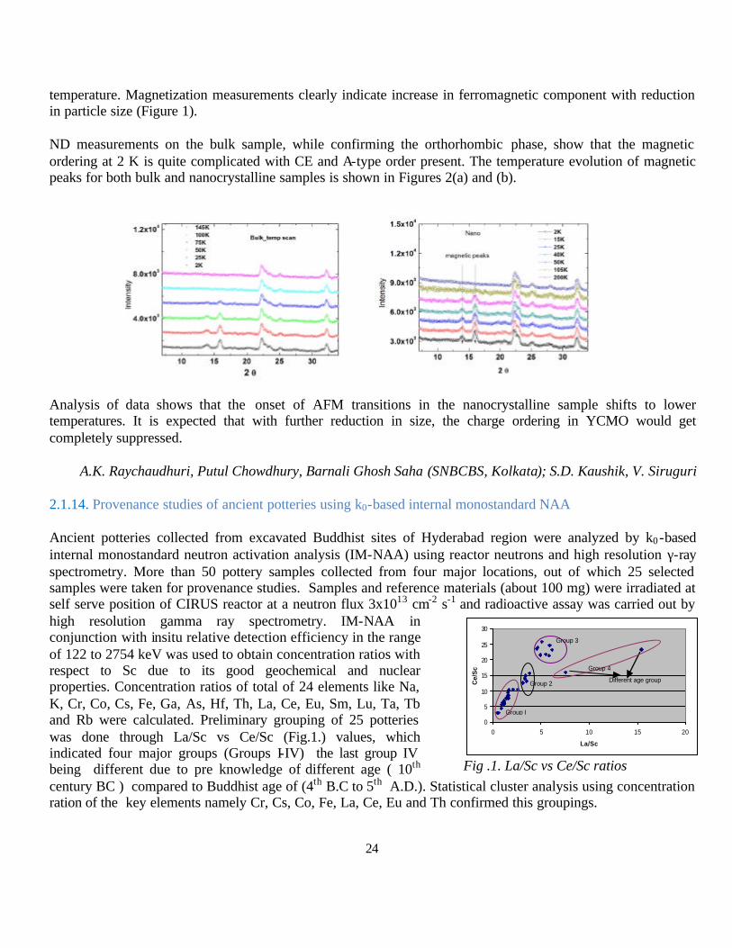
The half-doped manganite Y0.5Ca0.5MnO3 (YCMO) is a robust charge ordered (CO) insulator. It has a TCO ~ 260-290K and antiferromagnetic (AFM) spin ordering takes place at ~ 135 K. YCMO has the smallest band width among materials showing charge and orbital ordering (COO) due to the small size of Y (rA~1.128A°) and COO does not melt even in a field of 50T The present study investigates whether the charge ordered insulating state gets destabilized by size reduction to nanoscopic dimensions, since it is known that charge and orbital ordering is susceptible to destabilization when a number of physical parameters are changed. Neutron diffraction (ND) studies were carried out on YCMO of three different sizes ranging from bulk (~1µ) to nano (~80nm).
Nano and bulk YCMO were synthesized by chemical solution deposition technique (CSD), where metal acetates are taken as precursor materials in stoichiometric amount ; acetic acid, water and appropriate amount of ethylene glycol used as solvent were mixed and heated. The gel was prepared and dried over night and then annealed at higher temperature (~800oC) for the nano sized sample and annealing at much higher temperature (1350oC) for longer time gives the bulk sample (~1micron). Samples of two different sizes, 80 nm and 1 micron, were prepared. Phase formation and phase purity was characterized using XRD and particle size was determined using SEM and TEM measurements. Rietveld refinement of the XRD data shows that the structure of bulk YCMO is orthorhombic at room
24
temperature. Magnetization measurements clearly indicate increase in ferromagnetic component with reduction in particle size (Figure 1).
ND measurements on the bulk sample, while confirming the orthorhombic phase, show that the magnetic ordering at 2 K is quite complicated with CE and A-type order present. The temperature evolution of magnetic peaks for both bulk and nanocrystalline samples is shown in Figures 2(a) and (b).
Analysis of data shows that the onset of AFM transitions in the nanocrystalline sample shifts to lower temperatures. It is expected that with further reduction in size, the charge ordering in YCMO would get completely suppressed.
A.K. Raychaudhuri, Putul Chowdhury, Barnali Ghosh Saha (SNBCBS, Kolkata); S.D. Kaushik, V. Siruguri
2.1.14. Provenance studies of ancient potteries using k0-based internal monostandard NAA Ancient potteries collected from excavated Buddhist sites of Hyderabad region were analyzed by k0-based internal monostandard neutron activation analysis (IM-NAA) using reactor neutrons and high resolution γ-ray spectrometry. More than 50 pottery samples collected from four major locations, out of which 25 selected samples were taken for provenance studies. Samples and reference materials (about 100 mg) were irradiated at self serve position of CIRUS reactor at a neutron flux 3x1013 cm-2 s-1 and radioactive assay was carried out by high resolution gamma ray spectrometry. IM-NAA in conjunction with insitu relative detection efficiency in the range of 122 to 2754 keV was used to obtain concentration ratios with respect to Sc due to its good geochemical and nuclear properties. Concentration ratios of total of 24 elements like Na, K, Cr, Co, Cs, Fe, Ga, As, Hf, Th, La, Ce, Eu, Sm, Lu, Ta, Tb and Rb were calculated. Preliminary grouping of 25 potteries was done through La/Sc vs Ce/Sc (Fig.1.) values, which indicated four major groups (Groups I-IV) the last group IV being different due to pre knowledge of different age ( 10th century BC ) compared to Buddhist age of (4th B.C to 5th A.D.). Statistical cluster analysis using concentration ration of the key elements namely Cr, Cs, Co, Fe, La, Ce, Eu and Th confirmed this groupings.
Fig .1. La/Sc vs Ce/Sc ratios
0
5
10
15
20
25
30
0 5 10 15 20
La/Sc
Ce/
Sc
Group I
Group 2
Group 3
Different age group
Group 4

25
Instead of absolute concentrations, grouping could be done using concentration ratios obtained using an internal monostandard and the method becomes standard less and simple. The work carried out resulted in two journal and three conference publications. Further work on a large number of potteries and bricks are being analyzed for provenance study.
N. Lakshmana Da, (GITAM Univ.); A. Acharya, (BARC)
2.1.15. Study of Selenium and Arsenic Toxicity Using Neutron Activation Analysis
The study on metalloid toxicity was undertaken by estimating the levels of selenium and arsenic along with other elements using neutron activation analysis. The work carried out in the last phase focused on the mobilization of arsenic in sediments and selenium in plant. 39 sediment samples, 12 soil samples and 32 plant samples were irradiated in CIRUS reactor for seven hours in lots of 8-10 samples in the last twelve months. The samples were assayed using gamma ray spectrometry in the laboratories of Analytical Chemistry Division and Radiochemistry Division after giving sufficient cooling time. The following table gives the latest data in the form of concentration of Selenium in mg/kg (ppm) in roots, shoots, leaves and seeds of Desi Chick Pea plant grown in non seleniferous soil besides in soil spiked with 4 mg/kg of selenium.
It was observed that the chickpea plant had a good potential not only for phytoremediation but also for bio-fortification of selenium.
Alok Srivastava (Punjab Univ); R. Acharya and A.V.R.Reddy (BARC)
2.2. Collaborative Research at VECC
2.2.1. Lifetime measurements for ∆I = 1 and ∆J=2 bands in 83Kr, 111In & 113Sb nuclei.
Nuclei in the atomic mass region ~ 80 – 110 contain rich structural information. The interesting feature for these nuclei is the observation of weakly deformed ∆I =1 bands consisting of intense dipole transitions that involve perpendicular coupling of holes in high-Ω proton orbitals with neutrons occupying the low-Ω shells. Such structures are termed as “magnetic bands”. Lifetime measurements provide the crucial information on the underlying structure of these states.
2.2.1.1. Excited states of 83Kr, populated in the 76Ge(11B, 3npγ) reaction at a beam energy of 50 MeV, have been studied with Gamma Detector Array at IUAC, New Delhi. This array comprised of twelve Compton suppressed HPGe detectors along with fourteen element BGO multiplicity filter. The ∆I = 1 band has been observed up to 5639.4 keV with spin (27/2-). Mean lifetimes have been measured for the four excited states up to spin 23/2- belonging to the ∆I = 1 band using the Doppler Shift Attenuation Method (DSAM). The B(M1)
Sample Concentration mg/kg (ppm)
Root Control 0.79 ± 0.07
Stem Control 0.72± 0.05 Leaves Control 0.58± 0.07
Seed Control 0.64± 0.04
Root - Se 67.95± 0.36 Stem - Se 66.73± 0.35
Leaves - Se 66.42± 0.41 Seed - Se 64.63± 0.31

26
rates derived from the present lifetime results decrease smoothly with increase in spin indicating that the angular momentum belonging to this band are generated by shears mechanism.
S. Ganguly (Chandannagore College); A Dey (VECC), P. Banerjee, S Bhattacharya (SINP); R. P. Singh, S. Muralithar, R Kumar, and R. K. Bhowmik (IUAC)
2.2.1.2. Excited states of 113Sb were populated in the 100Mo(20Ne, p6n) reaction at a beam energy of 136 MeV using the INGA array at VECC, Kolkata. States only up to 59/2- were observed in the ∆J = 2 band. Mean lifetimes for the five states (from 4460 keV to 7998 keV) have been measured for the first time using Doppler Shift Attenuation Method. An upper limit of the lifetime 0.14 ps has been estimated for the 9061 keV, 47/2-
state. The B(E2) values, derived from the present lifetime results, correspond to a large quadrupole deformation of β2= 0.32. The observed reduction in the experimental B(E2) values for the 918.4 keVand 985.0 keV transitions may be interpreted as due to the proton alignment in the g7/2 orbital. The dynamic moment of inertia has been observed to be about half of the rigid body value at the highest observed frequency.
S. Ganguly (Chandernagore College); P. Banerjee (SINP); A. Dey and S. Bhattacharya (VECC)
2.2.1.3. The two ∆I = 1 bands in 111In, built upon the 3461.0 and 4931.8 keV states, have been studied using the INGA array at IUAC, New Delhi. The bands were populated in the reaction 100Mo(19F, a4n? ) at a beam energy of 105 MeV. Mean lifetimes of nine states, four in the first and five in the second band, have been determined for the first time from Doppler shift attenuation data. The deduced B(M1) rates and their behavior as a function of level spin support the interpretation of these bands within the framework of the shears mechanism. The geometrical model of Machiavelli et al. has been used to derive the effective gyromagnetic ratios for the two bands
P. Banerjee, M. K. Pradhan (SINP); S. Ganguly (Chandannagore College); H. P. Sharma (BHU); S. Muralithar, R. P. Singh, and R. K. Bhowmik (IUAC)
2.2.2. Proton radioactivity with a finite range Yukawa interaction.
In order to investigate the ability of the finite range interaction having a single Yukawa term in the finite range
( ) )()()(1
)()1(61)()1()( 3300 rfPMPHPBPWr
Rb
RPxtrPxtrveff τστσ
γ
σσ δρ
ρδ −−++
++++=
→
→
→→→
(1)
part that has been successful in the investigation of the nuclear matter properties at extreme conditions for its application to finite nucleus, we have calculated the prediction of the Wood-Saxon (WS) density distribution of the interaction under the semi-classical approximation. With the predicted WS density distribution the binding energies are of the nuclei over the periodic table are reproduced with in 1/2 % and rms charge radii with in 2 % of their experimentally measured values. The densities thus obtained for the proton radioactive nuclei are used to calculate the proton-nucleus interaction potential and in the calculation of decay half- lives of the proton.
The Nucleon- Nucleus interaction provides a wide source of information to determine the nuclear structure including spin, isospin, momenta and densities. It is also gives a picture towards the formation of exotic nuclei
27
in the laboratory. In this context the study of elastic scattering of Nucleon-Nucleus is more interesting than that of Nucleus-Nucleus at different energy. One of the theoretical method to study such types of reaction is ”Relativistic Impulse Approximation” (RIA). The basic ingredients in this approach are the nucleon-nucleon (NN) scattering amplitude and the nuclear scalar and vector densities of the target nucleus.
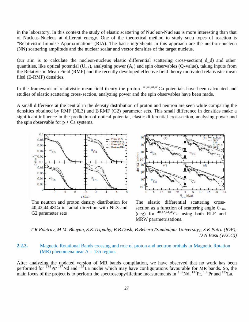
Our aim is to calculate the nucleon-nucleus elastic differential scattering cross-section( d_d) and other quantities, like optical potential (Uopt), analysing power (Ay) and spin observables (Q-value), taking inputs from the Relativistic Mean Field (RMF) and the recently developed effective field theory motivated relativistic mean filed (E-RMF) densities.
In the framework of relativistic mean field theory the proton- 40,42,44,48Ca potentials have been calculated and studies of elastic scattering cross-section, analyzing power and the spin observables have been made.
A small difference at the central in the density distribution of proton and neutron are seen while comparing the densities obtained by RMF (NL3) and E-RMF (G2) parameter sets. This small difference in densities make a significant influence in the prediction of optical potential, elastic differential crosssection, analysing power and the spin observable for p + Ca systems.
T R Routray, M M. Bhuyan, S.K.Tripathy, B.B.Dash, B.Behera (Sambalpur University); S K Patra (IOP); D N Basu (VECC))
2.2.3. Magnetic Rotational Bands crossing and role of proton and neutron orbitals in Magnetic Rotation (MR) phenomena near A = 135 region.
After analyzing the updated version of MR bands compilation, we have observed that no work has been performed for 135Pr/ 137Nd and 135La nuclei which may have configurations favourable for MR bands. So, the main focus of the project is to perform the spectroscopy/lifetime measurements in 137Nd, 137Pr, 135Pr and 135La.
The neutron and proton density distribution for 40,42,44,48Ca in radial direction with NL3 and G2 parameter sets
The elastic differential scattering cross-section as a function of scattering angle θc.m. (deg) for 40,42,44,48Ca using both RLF and MRW parametrisations.

28
The high spin states in 135Pr were populated by the reaction 123Sb (16O, 4n) 135Pr using a 16O beam of 82 MeV from the pelletron accelerator of Inter University Accelerator Centre (IUAC), New Delhi. This experiment was performed in the second campaign of INGA. The analysis of the experiment is in progress.
The high spin states in 135La were populated by the reaction 128Te (11B, 4n) 135La using a 11B beam of 50.5 MeV from the pelletron accelerator of Tata Institute of Fundamental Research (TIFR), Mumbai. This experiment was performed recently during the current INGA campaign at TIFR, Mumbai.
Partial level scheme for negative parity band in
135Pr
Ritika Garg, S. Kumar, Mansi Saxena, Savi Goyal, Davinder Siwal, Sunil Kalkal, S. Verma, S. Mandal, R. Singh, S. C. Pancholi (University of Delhi); R. Palit(TIFR); Deepika Choudhury, A. K. Jain (IIT Roorkee);
G. Mukherjee (VECC); R. Kumar, S. Muralithar, R. K. Bhowmik, and R. P. Singh (IUAC)); S. S. Ghugre
29
2.2.4. Study of shape coexistence in 153Ho and few-valence particle nuclei around 146Gd Core.
The low energy excitation spectra of few – valence - particle nuclei around doubly magic 146Gd nucleus show wide variations in their excitation spectra. Spectra exhibiting characteristics of extreme single particle, multiparticle hole excitation, magnetic bands to strong collectivity manifested through superdeformation, and triaxial superdeformation have been widely studied.
Another distinguishing feature of mass A~150 region is the existence of an island of high spin isomers which are excited in heavy ion reactions. The isomers can indicate a sharp change of structural configurations within the same nucleus.
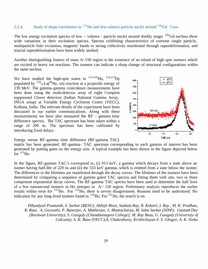
We have studied the high-spin states in 151-154Ho, 152,153Dy populated by 139
57La(20Ne, xn) reaction at a projectile energy of 139 MeV. The gamma-gamma coincidence measurements have been done using the multi-detector array of eight Compton suppressed Clover detectors (Indian National Gamma Array, INGA setup) at Variable Energy Cyclotron Centre (VECC), Kolkata, India. The relevant details of the experiment have been discussed in our earlier communications. Along with these measurements we have also measured the RF - gamma time difference spectra. The TAC spectrum has been taken within a range of 200 ns. The spectrum has been calibrated by introducing fixed delays
Energy versus RF-gamma time difference (RF-gamma TAC) matrix has been generated. RF-gamma- TAC spectrum corresponding to each gamma of interest has been generated by putting gates on the energy axis. A typical example has been shown in the figure depicted below for 153Ho.
In the figure, RF-gamma TAC’s correspond to, (i) 913 keV, a gamma which decays from a state above an isomer having half- life of 229 ns and (ii) the 533 keV gamma, which is emitted from a state below the isomer. The differences in the lifetimes are manifested through the decay curves. The lifetimes of the isomers have been determined by comparing a sequence of gamma gated TAC spectra and fitting them with one, two or three component exponential decay curves. The RF-gamma TAC spectra have been used to determine the half lives of a few nanosecond isomers in Ho isotopes in A~ 150 region. Preliminary analysis reproduces the earlier results within error for 153Ho. For 152Ho, there is severe disagreement. Reasons need to be understood. No indication for any long- lived isomers found in 154Ho. For 151Ho, the search is on.
Dibyadyuti Pramanik, S. Sarkar (BESU); Abhijit Bisoi, Sudatta Ray, R. Kshetri, I. Ray , M. K. Pradhan, R. Raut, A. Goswami, P. Banerjee, A. Mukherjee, S. Bhattacharya, M. Saha Sarkar (SINP); Gautam Dey
(Burdwan University); S. Ganguly (Chandannagore College); M. Ray Basu, G. Ganguly (University of Calcutta); S. K. Basu (VECC);A. Chakraborty, Krishichayan S. S. Ghugre, A. K. Sinha
30
2.2.5. Collaborative Research Scheme using ECR based Low Energy Heavy Ion beam Facility at VECC
2.2.5.1. Iron ion implantation in ZnO films.
It is well known that as-synthesized ZnO is always a n-type semiconductor, possibly due to the inherent oxygen vacancies. Synthesizing p-type ZnO fims has remained a challenge by conventional chemical techniques and earlier attempts have been unsuccessful. As a result realization of p-n junctions using ZnO as the active material has not been possible. However metal ion implantation in ZnO is known to produce significant hole concentration due to substitution of the Zn by the implanted metal ions or also due to the presence of the metal ion as an interstitial impurity. If this hole concentration can be increased beyond the intrinsic electron concentration then p-type conductivity can be realized. With this objective ZnO films were implanted with 90 keV Fe ions using the high current ECR based ion implantation facility at VECC. 250nm thick ZnO films were prepared by spincoating technique on glass substrates. Fe ions were chosen so that in addition to the desired p-type conductivity one could also get a dilute magnetic semiconductor. The Fe implanted samples were characterized by GIXRD and FTIR. Magnetoresistance (MR) and Hall measurements on these samples confirm the formation of p-type carriers in the implanted samples, though their origin seems to be from very deep acceptor levels. The MR results indicate that the conductivity in the implanted samples is predominantly p-type below 200K and n-type above 200K. This is also corroborated by the Hall measurements. Photoluminescence studies on these samples show emission bands that could be ascribed to sub-bandgap states. The experimental results are being analyzed and will be communicated very soon.
S.Keshri (BIT Mesra); G.S.Taki (VECC); J.B.M.Krishna
2.2.5.2. Ion Beam Assisted Synthesis and Characterization of Novel Optically Active Glass/Polymer
Nanoparticles (NPs) embedded in glass matrix would impart optically active properties in the resulting nanocomposites, which can be used for optical and optoelectronic devices. Ion beam assisted synthesis of nanocrystals in glass has attracted a lot of scientific attraction due to controlled implantation of cluster of atoms which can be precipitated in to nanocrystals. The ion beam method is also effective in dispersing these nanomaterials in the host, thereby giving room for tailoring the spatial distribution and size of the nanocrystals. However, defects created during the ion implantation also contribute to the observed optical properties. So it is important to investigate the radiation damage in the host material during the ion implantation. With this objective PMMA samples were implanted with oxygen ions having different charge states but same kinetic energy using the VECC ECR facility. The purpose of this study was to investigate the dependence of charge state of the incident ions, if any, on the defects produced. The beam energy was 63 keV and oxygen ions with +2 and +7 charge states were implanted into the PMMA samples. Optical reflectivity studies on the implanted samples showed higher reflectivity in the NIR region for the implanted samples. The change in reflectivity was significantly higher in the case of O7+ implanted samples. Whereas Dielectric measurements revealed that the samples implanted with O2+ ions have higher dielectric constant at low frequencies than the ones implanted with O7+ ions. The results indicate that the modified optical properties of PMMA samples have a strong dependence on the charge state of the implanted ions. The results have been presented in Nuclear and Radiochemistry Symposium, February 22-26, 2011, GITAM Univ. Visakhapatnam.
R.K.Dutta (IIT Roorkee); G.S.Taki (VECC); J.B.M.Krishna
31
2.2.5.3. N ion implantation in ZnSe bulk and nanocrytalline films
ZnSe is a wide bandgap semiconducting material with a band gap of about 2.7eV. This is inherently n-type semiconductor and has very good luminescent properties. In order to use this material for electroluminescent display applications it is required to fabricate p-n junctions. Nitrogen ion implantation is known to produce p-type conductivity in ZnSe. In this project CVT grown ZnSe single crystal samples were implanted with 45 keV N-ions at VECC ECR facility. Photoluminescence and optical absorption studies were carried out on the implanted samples. The implanted samples showed a red shift in the photoluminescence bands indicating a decrease in the bandgap of the samples. The results of this study have been accepted for publication in the International Conference on Materials for Advanced Technologies (ICMAT)-2011, June 26 – July 1, 2011, Suntec, Singapore.
R.Dhanasekaran (Crystal Growth Centre, Anna Univ.); G.S.Taki (VECC); J.B.M.Krishna
2.3. Photoelectron spectroscopy on INDUS-1, RRCAT
The research work described below utilized the beamline for photoelectron spectroscopy set up by CSR on Indus-1.
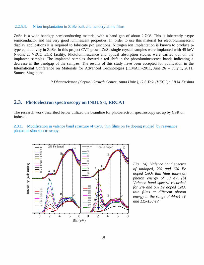
2.3.1. Modification in valence band structure of CeO2 thin films on Fe doping studied by resonance photoemission spectroscopy.
0 2 4 6 80 2 4 6 8
B
A
113 115 118 120 121 122 124 125 127 130
C
Inte
nsity
(arb
uni
ts)
BE (eV)
115 118 121 122 124 125 127 130
D
B
B
A
A
2% Fe doped 44 47 50 52 54 55 56 57 58 60 62 64
6% Fe doped
D
C
C C
B
A
44 eV 47 50 52 53 54 55 56 57 58 60 62 64
Fig. (a): Valence band spectra of undoped, 2% and 6% Fe doped CeO2 thin films taken at photon energy of 50 eV, (b) Valence band spectra recorded for 2% and 6% Fe doped CeO2
thin films at different photon energy in the range of 44-64 eV and 115-130 eV.
32
Studied the modification in electronic properties of pulsed laser deposited of CeO2 thin films due to Fe doping (2 and 6 at %), with the help of x-ray photoemission spectroscopy (XPS) and resonance photoemission spectroscopy (RPES) measurements. XPS results indicate the ionic state of Fe in the Fe doped films, ruling out the possibility of Fe metallic clusters. Valence band spectra (VBS) of CeO2 show an additional feature after Fe doping, suggesting its incorporation in the CeO2 matrix. RPES studies of these films reveal the hybridization between oxygen vacancy induced Ce localized states and Fe derived states.
Amit Khare,. Sanyal (Barkatulla Univ., Bhopal); R.J.Choudhary, D.M.Phase
2.4. Collaborative Research Schemes using in-house facilities at Indore Centre
Large number of university users utilised Indore Centre's in-house facilities for their research acitivities. In the following section some of these activies are reported.
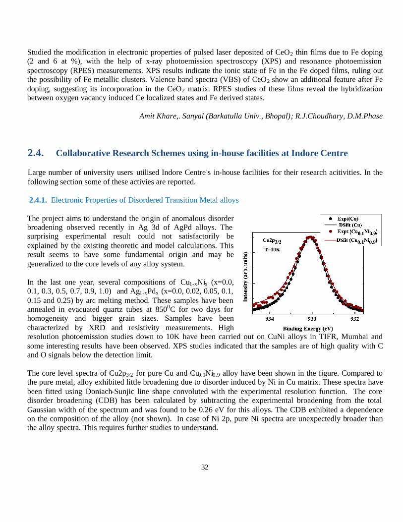
2.4.1. Electronic Properties of Disordered Transition Metal alloys
The project aims to understand the origin of anomalous disorder broadening observed recently in Ag 3d of AgPd alloys. The surprising experimental result could not satisfactorily be explained by the existing theoretic and model calculations. This result seems to have some fundamental origin and may be generalized to the core levels of any alloy system.
In the last one year, several compositions of Cu1-xNix (x=0.0, 0.1, 0.3, 0.5, 0.7, 0.9, 1.0) and Ag1-xPdx (x=0.0, 0.02, 0.05, 0.1, 0.15 and 0.25) by arc melting method. These samples have been annealed in evacuated quartz tubes at 8500C for two days for homogeneity and bigger grain sizes. Samples have been characterized by XRD and resistivity measurements. High resolution photoemission studies down to 10K have been carried out on CuNi alloys in TIFR, Mumbai and some interesting results have been observed. XPS studies indicated that the samples are of high quality with C and O signals below the detection limit.
The core level spectra of Cu2p3/2 for pure Cu and Cu0.1Ni0.9 alloy have been shown in the figure. Compared to the pure metal, alloy exhibited little broadening due to disorder induced by Ni in Cu matrix. These spectra have been fitted using Doniach-Sunjic line shape convoluted with the experimental resolution function. The core disorder broadening (CDB) has been calculated by subtracting the experimental broadening from the total Gaussian width of the spectrum and was found to be 0.26 eV for this alloys. The CDB exhibited a dependence on the composition of the alloy (not shown). In case of Ni 2p, pure Ni spectra are unexpectedly broader than the alloy spectra. This requires further studies to understand.
33
At low temperatures, thermal disorder is almost suppressed and the possible disorders causing the broadening are chemical and local structural distortions. CDB can be used as a measure of disorder in the system. The transport properties can have direct relationship to the CDB as the disorder influences the transport properties.
V. Rama Rao Medicherla (Siksha ‘O’ Anusandhan University, Bhubaneswar)
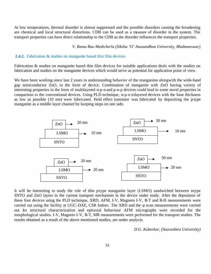
2.4.2. Fabrication & studies on manganite based thin film devices
Fabrication & studies on manganite based thin film devices for suitable applications deals with the studies on fabrication and studies on the manganite devices which would serve as potential for application point of view.
We have been working since last 2 years in understanding behavior of the manganites alongwith the wide-band gap semiconductor ZnO, in the form of device. Combination of manganite with ZnO having variety of interesting properties in the form of multilayered n-p-n and p-n-p devices could lead to some novel properties in comparison to the conventional devices. Using PLD technique, n-p-n trilayered devices with the base thickness as low as possible (10 nm) were fabricated. Field effect transistor was fabricated by depositing the p-type manganite as a middle layer channel by keeping steps on one side.
It will be interesting to study the role of thin p-type manganite layer (LSMO) sandwiched between n-type SNTO and ZnO layers in the current transport mechanism in the device under study. After the deposition of these four devices using the PLD technique, XRD, AFM, I-V, Magneto I-V, R-T and R-H measurements were carried out using the facility at UGC-DAE, CSR Indore. The XRD and the φ scan measurements were carried out for structural characterization and epitaxial behaviour AFM micrographs were recorded for the morphological studies. I-V, Magneto I-V, R-T, MR measurements were performed for the transport studies. The results obtained as a result of the above mentioned studies, are under analysis.
D.G. Kuberkar, (Saurashtra University)
ZnO
LSMO
SNTO
10 nm
20 nm ZnO
LSMO
SNTO
10 nm
50 nm
ZnO
LSMO SNTO
20 nm
20 nm ZnO
LSMO
SNTO
20 nm
50 nm

34
40 42 44 46 48 50
2θ
0%
5%
10%
20%
30%
50%
75%
100%
Inte
nsity
(ar
b. u
nits
)
Co (111)
0.1 0.2 0.3 0.4 0.510-4
10-2
1
102
104
106
108
1010
1012
75%
50%
30%
20%
10%
5%
Spin Up Spin Down Fit
Pol
ariz
ed N
eutr
on R
efle
ctiv
ity
qz(nm-1)
0%
2.4.3. Microstructure of thin films prepared by reactive sputtering
In this work thin films of various elements using a gas mixture of nitrogen and argon were studied. The nitride thin films of many elements are used in different applications. Recently, reactive sputtering has been used to tailor the microstructure of the thin films. It has been found that roughness of the thin films can be minimized with reactive sputtering and the structural properties can be changed from crystalline to nanocrystalline to amorphous or coarse grained structures. Our aim therefore is to understand the growth of the thin films with reactive sputtering and the mechanism affecting the microstructure of thin films. Thin films of Co, Cu, Ti, Al, Si and C by varying the amount of reactive nitrogen have been prepared using magnetron sputtering.
FIG.1: XRD (left) and PNR (right) pattern of cobalt nitride thin films prepared using different nitrogen partial pressure.
In Co thin films prepared using reactive nitrogen to argon ratio of 0, 10, 20, 30, 50, 75 and 100%. X-ray diffraction and polarized neutron reflectivity measurements have been done and are shown in fig. 1. Thin films with 0% and 5% nitrogen partial pressure show the crystalline behavior corresponding to pure cobalt phase. At higher nitrogen partial pressures, it shows the amorphous type behavior. Above 50% nitrogen partial pressure, cobalt nitride phases evolve and at 75% nitrogen partial pressure nanocrystalline cobalt nitride phase is formed. With 100% nitrogen partial pressure, this nanocrystalline phase shows a broad diffuse maximum in the x-ray diffraction pattern indicating formation of amorphous cobalt nitride phase. From PNR measurements it was found that pattern show a separation between spin up and spin down reflectivities indicating the ferromagnetic structure of the deposited thin films. The magnetic moment in these films was found to decrease as the nitrogen partial pressure was increased. Above 50% nitrogen partial pressure, both spin up and spin down reflectivities appear together indicating that the nature of the cobalt nitride thin films is not ferromagnetic.
Rachana Gupta (IET, DAVV, Indore)
35
2.4.4. Exploration of CuInSe2 type diluted magnetic semiconductors for spintronics application. Considering the well-established CuInSe2 as a prototype system, a new compound of the chalcopyrite type, by replacing In with Zn, had been attempted by metallurgical method. The X-ray diffraction pattern of the ordered defect compound Cu1-xZn1-ySe2-d with the characteristic peaks of a chalcopyrite phase, such as (112), (204) and (312) confirm the tetragonal structure. On doping with Cr the band gap blue shifted to 4.09 from 3.4 eV indicating possibility of band gap tailoring. Cu1-xZn1-ySe2-d when magnetically doped with Cr shows ferromagnetic ordering at room temperature [1]. Thin films were deposited by the thermal evaporation technique using the stabilized polycrystalline compounds as charge. Structural, compositional, morphological, and optical properties of the films were analyzed. Use of these films as electrodes in dye sensitized solar cell (DSSC) had been demonstrated [2].
The preparation of this non-oxide compound by mixing metal powders at ambient condition is difficult. Hence, efforts had been taken also through the mechanical alloying method to prepare Pure CuZnSe2 (upto 20 h of milling) and Cr(2%) doped CuZnSe2 (upto 25 h) . XRD patterns of the milled and Nitrogen annealed samples indicate the formation of hexagonal CuZnSe2 along with the intermediate hexagonal phase of ZnSe. Band gap, as estimated from the reflectance spectrum, of 20 h milled CuZnSe2 is 2.9 eV. Magnetization study of Cr(2%) doped CuZnSe2 indicates that, as the milling hour increases the MMax value also increases. Efforts continue to optimize the best suitable condition for preparing single phase CuZnSe2 and Cr doped CuZnSe2.
C. Venkateswaran, (University of Madras)
2.4.5. Evolution of Magnetism with Oxygen Deficiency in Cobalt doped TiO 2 Diluted Magnetic Semiconductor
After the discovery of room temperature ferromagnetism in Co doped TiO 2 it has attracted the interest of experimentalists as well as theoreticians. Some explained the ferromagnetism as the intrinsic behavior of the material, while others have found Co clustering showing the magnetic behavior. While some other workers have found the change of oxidation state of Ti from +4 state to +3 state gives rise to the magnetic phenomena. In all the above mentioned cases the role of oxygen vacancy is vital. Recently people have been able to see magnetism in undoped TiO 2 somehow related to oxygen vacancies. So, whether doped or undoped TiO2, the role of oxygen concentration plays an important role. But how the oxygen concentration can be tuned? A possible attempt is done in this work by changing the O2 partial pressure during the thin film deposition of TiO 2 and 5% Co doped TiO 2 on Si substrate (p-type, 100 oriented) and quartz using the pulsed laser deposition method. A sintered polycrystalline Ti0.95Co0.05O2 target (2.5cm diameter) was prepared by chemical route. The target was ablated by a KrF excimer laser (Lambda Physik COMPex 201 model, Germany: wavelength, 248nm) with laser energy 240mJ at 10 Hz and voltage 21 kV. Four samples were prepared by varying partial pressures 4.5X10⁻⁵ Torr (vacuum) [A1], 1X10⁻⁴ Torr (O2) [A2], 1mTorr(O2)[A3], 300mT(O2)[A4]. The as deposited samples were characterized by glancing angle XRD, Raman Spectroscopy and FE-SEM. The XRD reveals that films deposited at vacuum has phase neither matched with anatase nor with rutile. With minimum oxygen partial pressure rutile phase has formed. With further increasing oxygen partial pressure, the anatase phase appears. The XRD results are in broad agreement with Raman results. FE-SEM results reveal a clear
36
morphological change with increasing oxygen partial pressure. At maximum oxygen partial pressure (i.e. 300 mT) droplets of material are formed on the film surface because of the collision between the ablated material in the plume and the oxygen environment. Oxygen slows down the plume resulting agglomerated large particulates on the surface.
Our result confirms the role of oxygen partial pressure on the phase formation of the TiO 2 thin films. In our future work will focus on the oxygen partial pressure dependent magnetic properties of both pure and Co doped TiO2 thin films. Swift Heavy Ion Irradiation of these films and its consequences on the magnetic properties of these films will be undertaken.
Chandana Rath, Pankaj Mohanty (Banaras Hindu University); R. J. Choudhary
2.4.6. Structural and Magnetic studies of some nanostructured mixed metal oxides synthesized via spray pyrolysis of polymer precursor.
We have carried out ,during the first quarter of the year, the work related to the optimization of the annealing temperature involved in the synthesis of Zinc nanoferrites (ZNF) particles, which were characterized as crystalline nanoparticles of particle size 20nm using the various techniques like XRD,TEM,EDS,FTIR,VSM, Mossbauer etc. To understand the effect of annealing temperature on particle size and crystalline nature, we have synthesized four fractions of samples via spray pyrolyisis and annealed at temperatures at 250,350,450 and 550. The XRD, FTIR and Mossbauer studies were carried out for exploring the effect of annealing temperature on crystalline nature , particle size, cation distributions and state of Mossbauer active nuclear sites. The particle sizes, estimated from XRD patterns , is found to be varying in the range 10nm-35nm when the annealing temperature increases from 250C to 550C.The results suggest that annealing temperature involved in the synthesis process does have an effect on the particle sizes and low temperature annealing process is preferred for synthesis of nanoferrites with smaller particle sizes. We have further synthesized a type of nanoferrites –Magnesium nanoferrites (MgNF) using the same technique. Three fractions of Magnesium nanoferrites (MgFe2O4) with varying molar ratios been prepared using our technique and characterized using XRD,EDS,FTIR and Mossbauer . We found that the fraction coded as M5 ( Mg:Fe=1:2)is highly crystalline and monophasic. The estimated particle size is found to 14nm. FTIR studies also support the formation of magnesium nanoparticles. Mossbauer spectrum recorded at room temperature shows a doublet and a weak sextect. The average internal hyperfine field is calculated and is found to be 22T. A temperature dependent Mossbauer study is required for elucidating ferromagnetic transition temperature, inversion parameter and canting angle etc. This work is in progress. It has been reported that mixed nanoparticles, especially magnesium doped zinc ferrite in nanofrom shows magnetoresistance property. Keeping this in mind, we have successfully prepared mixed ferrite nanoparticles of Mgx Zn(1-x) Fe2O4, during the last quarter of the year. Nine fractions of the samples, by varying values of x from x= 0.1 to x= 0.9, have been prepared . The XRD, Mossbauer ,VSM, FTIR and EDS of all the fractions were recorded and analysis of the data are in progress. The findings of the few studies have reported in the various seminars and conferences.
Jacob Mathew
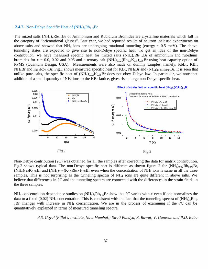
37
2.4.7. Non-Debye Specific Heat of (NH4)xRb1-xBr
The mixed salts (NH4)xRb1-xBr of Ammonium and Rubidium Bromides are crystalline materials which fall in the category of “orientational glasses”. Last year, we had reported results of neutron inelastic experiments on above salts and showed that NH4 ions are undergoing rotational tunneling (energy ~ 0.5 meV). The above tunneling states are expected to give rise to non-Debye specific heat. To get an idea of the non-Debye contribution, we have measured specific heat for mixed salts (NH4)xRb1-xBr of ammonium and rubidium bromides for x = 0.0, 0.02 and 0.05 and a ternary salt (NH4)0.02(Rb0.5K0.5)0.98Br using heat capacity option of PPMS (Quantum Design, USA). Measurements were also made on dummy samples, namely, RbBr, KBr, NH4Br and K0.5Rb0.5Br. Fig.1 shows measured specific heat for KBr, NH4Br and (NH4)0.02K0.98Br. It is seen that unlike pure salts, the specific heat of (NH4)0.02K0.98Br does not obey Debye law. In particular, we note that addition of a small quantity of NH4 ions to the KBr lattice, gives rise a large non-Debye specific heat.
5 10 15 20 25 30
0.005
0.01
0.015
0.02
0.025
0.030.035
C/T
3(m
J/g-
K4)
T(K)
(NH4)Br KBr (NH4)0.02K0.98Br
Fig.1
2 4 6 8 10 12 14
0.00
0.01
0.02
C/T
3 (m
J/g
m-K
4 )
T (K)
(NH4)0.02K0.98Br (NH4)0.02Rb0.98Br (NH4)0.02(K0.5Rb0.5)0.98Br
Effect of strain field on specific heat (NH4)x(K,Rb)1-xBr
Measured Specific HeatCorrected for matrix (KBr/RbBr/KRbBr) contribution
Fig.2
Non-Debye contribution (?C) was obtained for all the samples after correcting the data for matrix contribution. Fig.2 shows typical data. The non-Debye specific heat is different as shown figure 2 for (NH4)0.02Rb0.98Br, (NH4)0.02K0.98Br and (NH4)0.02(K0.5Rb0.5)0.98Br even when the concentration of NH4 ions is same in all the three samples. This is not surprising as the tunneling spectra of NH4 ions are quite different in above salts. We believe that differences in ?C and the tunneling spectra are connected with the differences in the strain fields in the three samples.
NH4 concentration dependence studies on (NH4)xRb1-xBr show that ?C varies with x even if one normalizes the data to a fixed (0.02) NH4 concentration. This is consistent with the fact that the tunneling spectra of (NH4)xRb1-
xBr changes with increase in NH4 concentration. We are in the process of examining if the ?C can be quantitatively explained in terms of measured tunneling spectra.
P.S. Goyal (Pillai’s Institute, Navi Mumbai); Swati Pandya, R. Rawat, V. Ganesan and P.D. Babu

38
-8 -6 -4 -2 0 2 4 6 8
-1
0
1
Magnetic field H (Gauss)
Ker
r In
tens
ity
at 0 deg at 45 deg at 90 deg at 135 deg at 180 deg
-200 -100 0 100 200
-1.0
-0.5
0.0
0.5
1.0
Ker
r Int
ensi
ty
Magnetic field H (Gauss)
at 0 deg at 45 deg at 90 deg at 135 deg at 180 deg
2.4.8. Room temperature Magnetism in Fe doped SbSe Alloys thin film
Thin films of Fe0.008Sb1-xSex for x=0.01, 0.05, 0.10 were prepared using Physical Vapor Deposition (PVD) technique. For study of these thin films X-ray diffraction, R-T measurements were done. For surface morphology Atomic force Microscopy (AFM) and for magnetic interaction Magnetic Force Microscopy (MFM) was done.
The surface morphologies of the alloys were observed by the AFM and magnetic interaction by MFM techniques. The AFM images show particle size distribution from 47 to 63nm respectively & the surface smoothness with a root mean square roughness was appreciable (1.78 - 3.01 nm). The MFM images shown below are not similar in contrast with the AFM image indicating the absence of any surface effects and the magnetic signals is due to intrinsic magnetic properties of the alloy. However the brightness in MFM images being almost uniform with small variation supports the absence of any magnetic clusters. The magnetic clusters were also not observed in XRD pattern. It seems that the Ferromagnetic interactions could be due to long range ordering of Fe local moments mediated by electron charge carriers introduced via Se doping.
Mitesh Sarkar (M S University, Vadodara)
2.4.9. Magnetic properties in Co-based multilayer system
A comparison of two independent nano systems consisting of Fe-Co-Al pertaining to a nominal stoichiometry of Fe2CoAl (one a multilayer system of total thickness of 1100 Å and the other high energy ball milled nanopowders of Fe2CoAl) shows that through a proper choice of process parameters, magnetic properties like directional anisotropy and coercivity and curie temperature can be controlled without sacrificing the fairly large saturation magnetization values in these samples.
A. Fe-Co-Al multilayer system: The gain in high saturation magnetization with soft magnetic properties in thin Fe-Co films is offset by reduction of anisotropy fields restricting their applications. Interfacial interactions or exchange coupling for e.g. using spacer layers gives good anisotropy fields. Here we have used Al spacer layers to get trilayer stack of [57Fe 25 Å/Co 11 Å /Al 17 Å] on Si (100), using ion beam sputtering (IBS). Magneto- optical Kerr effect (MOKE) measurement to get angular dependent hysteresis loops have been used to study the anisotropy in the MLS.
AFM Image (LHS) for X=0.05 sample and corresponding MFM image in RHS
Fig1.Angular dependent MOKE hysteresis loops of (a) as deposited & (b) annealed MLS

39
30 m 60 m 90 m 120 m 150 m 180 m16
17
18
19
20
21
22
Terminal sizeattaitned
Par
ticl
e S
ize
(nm
)
Milling Time20 40 60 80 100 120 140 160 180 200
45
50
55
60
65
70
75
80
85
Coe
rciv
ity H
C (G
auss
)
Milling Time (minutes)
40 80 120 160 200100
110
120
130
140
150
160
satu
ratio
n m
agne
tizat
ion
(em
u/g)
Milling time (minutes)
The excellent soft magnetic properties coupled with in plane uniaxial magnetic anisotropy in as-deposited MLS is destroyed on annealing due to further mixing at the interfaces and also nucleation of new phases.
B. Fe-Co- Al mechanically alloyed system:Part of argon arc melted Fe2CoAl, was high energy ball milled for various periods using WC milling medium (BPR = 20:1). Initially the particle size decreases linearly as a function of increase in milling time and attains terminal size at 150 minutes (Fig 2). Correspondingly coercivity increases by nearly 40 times with only a ~12% reduction in saturation magnetization (Fig 2).
Fig.2 variation of various parameters with milling time
N Lakshmi (Mohanlal Sukhadia University, Udaipur); Dr. V. R. Reddy
2.4.10. Magnetic and electrical behavior of Al doped La0.7Ca0.3MnO3 manganites
The effects of doping on magnetic and electrical transport mechanism of polycrystalline samples La0.7Ca0.3Mn1- xAlxO3 _x=0, 0.02, 0.04, 0.06, 0.08, 0.1_ have been investigated. Magnetization data reveal that long-range ferromagnetic ordering persists in all samples and the saturation moment decreases linearly as x increases. Resistivity data have been fitted with the variable range hopping model to estimate the density of state at Fermi level. It was observed that the substitution of Al in the series leads to a decrease in conductivity of the doped manganites samples, with conduction being controlled by the disorder induced localization of charge carriers.
S.Tiwari, B.L.Ahuja (M.LS.Univ., Udaipur); D.M.Phase and, R.J.Choudhary
Fig. The temperature dependence of MR% of La0.7Ca0.3Mn1-xAlxO3 (x=0, 0.02, 0.04, 0.06, 0.08, 0.1) samples.
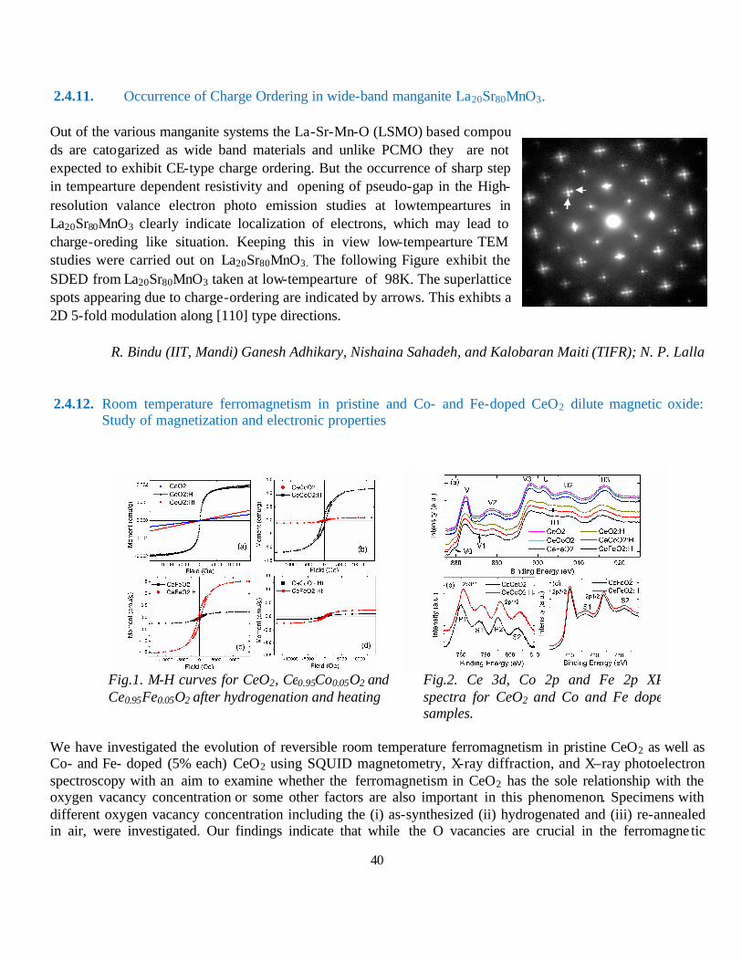
40
2.4.11. Occurrence of Charge Ordering in wide-band manganite La20Sr80MnO3. Out of the various manganite systems the La-Sr-Mn-O (LSMO) based compou ds are catogarized as wide band materials and unlike PCMO they are not expected to exhibit CE-type charge ordering. But the occurrence of sharp step in tempearture dependent resistivity and opening of pseudo-gap in the High-resolution valance electron photo emission studies at lowtempeartures in La20Sr80MnO3 clearly indicate localization of electrons, which may lead to charge-oreding like situation. Keeping this in view low-tempearture TEM studies were carried out on La20Sr80MnO3. The following Figure exhibit the SDED from La20Sr80MnO3 taken at low-tempearture of 98K. The superlattice spots appearing due to charge-ordering are indicated by arrows. This exhibts a 2D 5-fold modulation along [110] type directions.
R. Bindu (IIT, Mandi) Ganesh Adhikary, Nishaina Sahadeh, and Kalobaran Maiti (TIFR); N. P. Lalla
2.4.12. Room temperature ferromagnetism in pristine and Co- and Fe-doped CeO2 dilute magnetic oxide: Study of magnetization and electronic properties
Fig.1. M-H curves for CeO2, Ce0.95Co0.05O2 and Ce0.95Fe0.05O2 after hydrogenation and heating
Fig.2. Ce 3d, Co 2p and Fe 2p XPS spectra for CeO2 and Co and Fe doped samples.
We have investigated the evolution of reversible room temperature ferromagnetism in pristine CeO2 as well as Co- and Fe- doped (5% each) CeO2 using SQUID magnetometry, X-ray diffraction, and X–ray photoelectron spectroscopy with an aim to examine whether the ferromagnetism in CeO2 has the sole relationship with the oxygen vacancy concentration or some other factors are also important in this phenomenon. Specimens with different oxygen vacancy concentration including the (i) as-synthesized (ii) hydrogenated and (iii) re-annealed in air, were investigated. Our findings indicate that while the O vacancies are crucial in the ferromagne tic
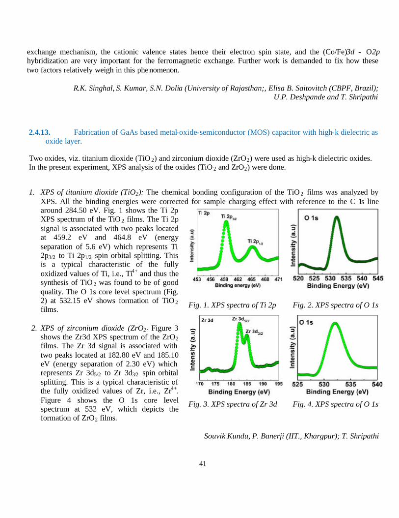
41
exchange mechanism, the cationic valence states hence their electron spin state, and the (Co/Fe)3d - O2p hybridization are very important for the ferromagnetic exchange. Further work is demanded to fix how these two factors relatively weigh in this phenomenon.
R.K. Singhal, S. Kumar, S.N. Dolia (University of Rajasthan;, Elisa B. Saitovitch (CBPF, Brazil); U.P. Deshpande and T. Shripathi
2.4.13. Fabrication of GaAs based metal-oxide-semiconductor (MOS) capacitor with high-k dielectric as oxide layer.
Two oxides, viz. titanium dioxide (TiO 2) and zirconium dioxide (ZrO2) were used as high-k dielectric oxides. In the present experiment, XPS analysis of the oxides (TiO 2 and ZrO2) were done.
1. XPS of titanium dioxide (TiO2): The chemical bonding configuration of the TiO 2 films was analyzed by
XPS. All the binding energies were corrected for sample charging effect with reference to the C 1s line around 284.50 eV. Fig. 1 shows the Ti 2p XPS spectrum of the TiO 2 films. The Ti 2p signal is associated with two peaks located at 459.2 eV and 464.8 eV (energy separation of 5.6 eV) which represents Ti 2p3/2 to Ti 2p1/2 spin orbital splitting. This is a typical characteristic of the fully oxidized values of Ti, i.e., Ti4+ and thus the synthesis of TiO 2 was found to be of good quality. The O 1s core level spectrum (Fig. 2) at 532.15 eV shows formation of TiO 2 films.
2. XPS of zirconium dioxide (ZrO2: Figure 3 shows the Zr3d XPS spectrum of the ZrO2 films. The Zr 3d signal is associated with two peaks located at 182.80 eV and 185.10 eV (energy separation of 2.30 eV) which represents Zr 3d5/2 to Zr 3d3/2 spin orbital splitting. This is a typical characteristic of the fully oxidized values of Zr, i.e., Zr4+. Figure 4 shows the O 1s core level spectrum at 532 eV, which depicts the formation of ZrO2 films.
Souvik Kundu, P. Banerji (IIT., Khargpur); T. Shripathi
Fig. 1. XPS spectra of Ti 2p Fig. 2. XPS spectra of O 1s
Fig. 3. XPS spectra of Zr 3d Fig. 4. XPS spectra of O 1s
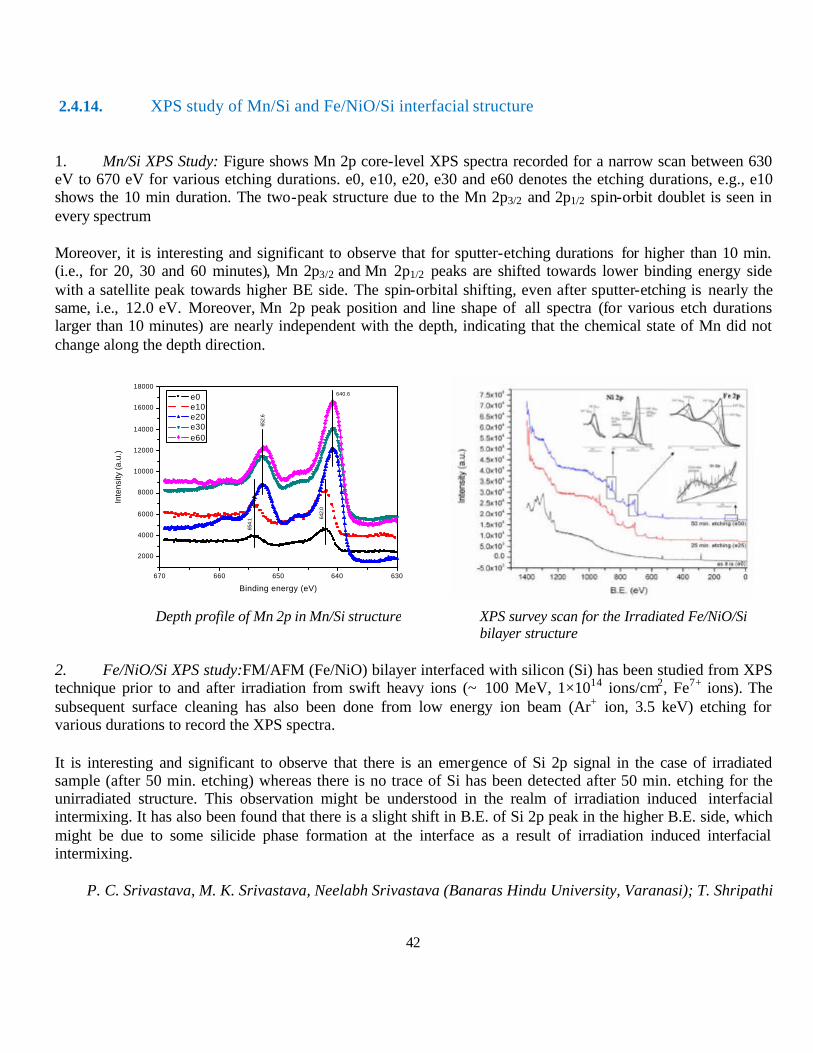
42
2.4.14. XPS study of Mn/Si and Fe/NiO/Si interfacial structure
1. Mn/Si XPS Study: Figure shows Mn 2p core-level XPS spectra recorded for a narrow scan between 630 eV to 670 eV for various etching durations. e0, e10, e20, e30 and e60 denotes the etching durations, e.g., e10 shows the 10 min duration. The two-peak structure due to the Mn 2p3/2 and 2p1/2 spin-orbit doublet is seen in every spectrum
Moreover, it is interesting and significant to observe that for sputter-etching durations for higher than 10 min. (i.e., for 20, 30 and 60 minutes), Mn 2p3/2 and Mn 2p1/2 peaks are shifted towards lower binding energy side with a satellite peak towards higher BE side. The spin-orbital shifting, even after sputter-etching is nearly the same, i.e., 12.0 eV. Moreover, Mn 2p peak position and line shape of all spectra (for various etch durations larger than 10 minutes) are nearly independent with the depth, indicating that the chemical state of Mn did not change along the depth direction.
Depth profile of Mn 2p in Mn/Si structure XPS survey scan for the Irradiated Fe/NiO/Si bilayer structure
2. Fe/NiO/Si XPS study:FM/AFM (Fe/NiO) bilayer interfaced with silicon (Si) has been studied from XPS technique prior to and after irradiation from swift heavy ions (~ 100 MeV, 1×10¹⁴ ions/cm2, Fe7+ ions). The subsequent surface cleaning has also been done from low energy ion beam (Ar+ ion, 3.5 keV) etching for various durations to record the XPS spectra.
It is interesting and significant to observe that there is an emergence of Si 2p signal in the case of irradiated sample (after 50 min. etching) whereas there is no trace of Si has been detected after 50 min. etching for the unirradiated structure. This observation might be understood in the realm of irradiation induced interfacial intermixing. It has also been found that there is a slight shift in B.E. of Si 2p peak in the higher B.E. side, which might be due to some silicide phase formation at the interface as a result of irradiation induced interfacial intermixing.
P. C. Srivastava, M. K. Srivastava, Neelabh Srivastava (Banaras Hindu University, Varanasi); T. Shripathi
670 660 650 640 630
2000
4000
6000
8000
10000
12000
14000
16000
18000
Inte
nsity
(a.u
.)
Binding energy (eV)
e0 e10 e20 e30 e60
640.6
652.
6
642.
0
654.
1
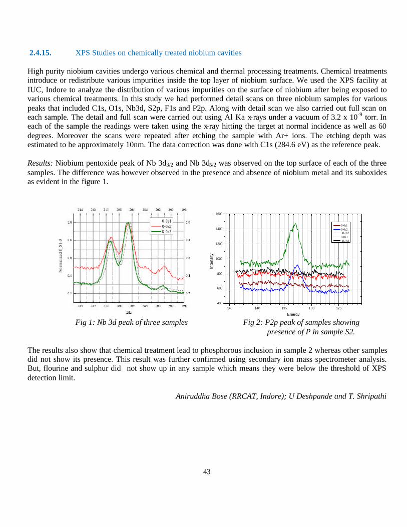
43
145 140 135 130 125400
600
800
1000
1200
1400
1600 In
tens
ity
Energy
0-0s1 0-0s2 38-0s2 0-0s3 38-0s3
2.4.15. XPS Studies on chemically treated niobium cavities
High purity niobium cavities undergo various chemical and thermal processing treatments. Chemical treatments introduce or redistribute various impurities inside the top layer of niobium surface. We used the XPS facility at IUC, Indore to analyze the distribution of various impurities on the surface of niobium after being exposed to various chemical treatments. In this study we had performed detail scans on three niobium samples for various peaks that included C1s, O1s, Nb3d, S2p, F1s and P2p. Along with detail scan we also carried out full scan on each sample. The detail and full scan were carried out using Al Ka x-rays under a vacuum of 3.2 x 10⁻⁹ torr. In each of the sample the readings were taken using the x-ray hitting the target at normal incidence as well as 60 degrees. Moreover the scans were repeated after etching the sample with Ar+ ions. The etching depth was estimated to be approximately 10nm. The data correction was done with C1s (284.6 eV) as the reference peak.
Results: Niobium pentoxide peak of Nb 3d3/2 and Nb 3d5/2 was observed on the top surface of each of the three samples. The difference was however observed in the presence and absence of niobium metal and its suboxides as evident in the figure 1.
Fig 1: Nb 3d peak of three samples Fig 2: P2p peak of samples showing presence of P in sample S2.
The results also show that chemical treatment lead to phosphorous inclusion in sample 2 whereas other samples did not show its presence. This result was further confirmed using secondary ion mass spectrometer analysis. But, flourine and sulphur did not show up in any sample which means they were below the threshold of XPS detection limit.
Aniruddha Bose (RRCAT, Indore); U Deshpande and T. Shripathi
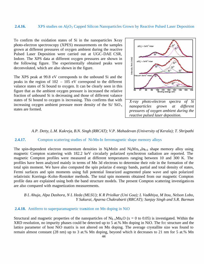
44
2.4.16. XPS studies on Al2O3 Capped Silicon Nanoparticles Grown by Reactive Pulsed Laser Deposition To confirm the oxidation states of Si in the nanoparticles X-ray photo-electron spectroscopy (XPES) measurements on the samples grown at different pressures of oxygen ambient during the reactive Pulsed Laser Deposition were carried out at UGC-DAE CSR, Indore. The XPS data at different oxygen pressures are shown in the following figure. The experimentally obtained peaks were deconvoluted, which are also shown in the figure. The XPS peak at 99.8 eV corresponds to the unbound Si and the peaks in the region of 102 – 105 eV correspond to the different valance states of Si bound to oxygen. It can be clearly seen in this figure that as the ambient oxygen pressure is increased the relative fraction of unbound Si is decreasing and those of different valance states of Si bound to oxygen is increasing. This confirms that with increasing oxygen ambient pressure more density of the Si/ SiO x states are formed.
A.P. Detty, L.M. Kukreja, B.N. Singh (RRCAT); V.P. Mahadevan (University of Kerala); T. Shripathi
2.4.17. Compton scattering studies of Ni-Mn-In ferromagnetic shape memory alloys
The spin-dependent electron momentum densities in Ni2MnIn and Ni2Mn1.4In0.6 shape memory alloy using magnetic Compton scattering with 182.2 keV circularly polarized synchrotron radiation are reported. The magnetic Compton profiles were measured at different temperatures ranging between 10 and 300 K. The profiles have been analyzed mainly in terms of Mn 3d electrons to determine their role in the formation of the total spin moment. We have also computed the spin polarize d energy bands, partial and total density of states, Fermi surfaces and spin moments using full potential linearized augmented plane wave and spin polarized relativistic Korringa–Kohn–Rostoker methods. The total spin moments obtained from our magnetic Compton profile data are explained using both the band structure models. The present Compton scattering investigations are also compared with magnetization measurements.
B L Ahuja, Alpa Dashora, N L Heda (MLSU); K R Priolkar (Uni Goa); L Vadkhiya, M Itou, Nelson Lobo, Y Sakurai, Aparna Chakrabarti (RRCAT); Sanjay Singh and S.R. Barman
2.4.18. Antiferro to superparamagnetic transition on Mn doping in NiO
Structural and magnetic properties of the nanoparticles of Ni1-xMnxO (x = 0 to 0.05) is investigated. Within the XRD resolution, no impurity phases could be detected up to 5 at.% Mn doping in NiO. The fcc structure and the lattice parameter of host NiO matrix is not altered on Mn doping. The average crystallite size was found to remain almost constant (28 nm) up to 3 at.% Mn doping, beyond which it decreases to 21 nm for 5 at.% Mn
X-ray photo-electron spectra of Si nanoparticles grown at different pressures of oxygen ambient during the reactive pulsed laser deposition.
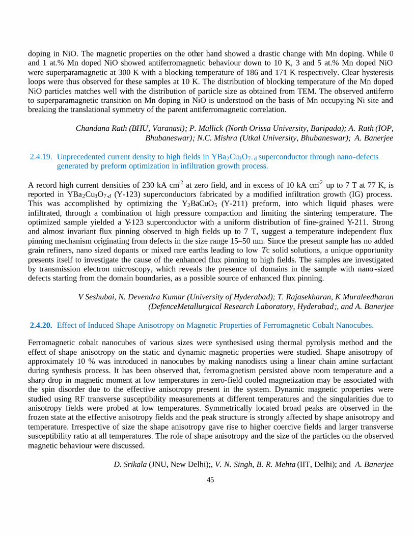
45
doping in NiO. The magnetic properties on the other hand showed a drastic change with Mn doping. While 0 and 1 at.% Mn doped NiO showed antiferromagnetic behaviour down to 10 K, 3 and 5 at.% Mn doped NiO were superparamagnetic at 300 K with a blocking temperature of 186 and 171 K respectively. Clear hysteresis loops were thus observed for these samples at 10 K. The distribution of blocking temperature of the Mn doped NiO particles matches well with the distribution of particle size as obtained from TEM. The observed antiferro to superparamagnetic transition on Mn doping in NiO is understood on the basis of Mn occupying Ni site and breaking the translational symmetry of the parent antiferromagnetic correlation.
Chandana Rath (BHU, Varanasi); P. Mallick (North Orissa University, Baripada); A. Rath (IOP, Bhubaneswar); N.C. Mishra (Utkal University, Bhubaneswar); A. Banerjee
2.4.19. Unprecedented current density to high fields in YBa2Cu3O7- d superconductor through nano-defects generated by preform optimization in infiltration growth process.
A record high current densities of 230 kA cm-2 at zero field, and in excess of 10 kA cm-2 up to 7 T at 77 K, is reported in YBa2Cu3O7-d (Y-123) superconductors fabricated by a modified infiltration growth (IG) process. This was accomplished by optimizing the Y2BaCuO5 (Y-211) preform, into which liquid phases were infiltrated, through a combination of high pressure compaction and limiting the sintering temperature. The optimized sample yielded a Y-123 superconductor with a uniform distribution of fine-grained Y-211. Strong and almost invariant flux pinning observed to high fields up to 7 T, suggest a temperature independent flux pinning mechanism originating from defects in the size range 15–50 nm. Since the present sample has no added grain refiners, nano sized dopants or mixed rare earths leading to low Tc solid solutions, a unique opportunity presents itself to investigate the cause of the enhanced flux pinning to high fields. The samples are investigated by transmission electron microscopy, which reveals the presence of domains in the sample with nano-sized defects starting from the domain boundaries, as a possible source of enhanced flux pinning.
V Seshubai, N. Devendra Kumar (University of Hyderabad); T. Rajasekharan, K Muraleedharan (DefenceMetallurgical Research Laboratory, Hyderabad;, and A. Banerjee
2.4.20. Effect of Induced Shape Anisotropy on Magnetic Properties of Ferromagnetic Cobalt Nanocubes.
Ferromagnetic cobalt nanocubes of various sizes were synthesised using thermal pyrolysis method and the effect of shape anisotropy on the static and dynamic magnetic properties were studied. Shape anisotropy of approximately 10 % was introduced in nanocubes by making nanodiscs using a linear chain amine surfactant during synthesis process. It has been observed that, ferromagnetism persisted above room temperature and a sharp drop in magnetic moment at low temperatures in zero-field cooled magnetization may be associated with the spin disorder due to the effective anisotropy present in the system. Dynamic magnetic properties were studied using RF transverse susceptibility measurements at different temperatures and the singularities due to anisotropy fields were probed at low temperatures. Symmetrically located broad peaks are observed in the frozen state at the effective anisotropy fields and the peak structure is strongly affected by shape anisotropy and temperature. Irrespective of size the shape anisotropy gave rise to higher coercive fields and larger transverse susceptibility ratio at all temperatures. The role of shape anisotropy and the size of the particles on the observed magnetic behaviour were discussed.
D. Srikala (JNU, New Delhi);, V. N. Singh, B. R. Mehta (IIT, Delhi); and A. Banerjee
46
2.4.21. Ac-Susceptibility Study In Rare Earth Substituted Magnetite Ferrofluids.
It is shown that variation of the third order ac-susceptibility as a function of measuring field and frequency lead to distinguish between superparamagnetic and spin glass like ordering in the rare earth substituted magnetic ferrofluids. The observed divergence of the peak values of the third order susceptibility as a function of measuring field and frequency tending to zero is consistent with theoretical prediction for the ground state for a spin glass like system. This behavior is further substantiated by a linear dependence of log- log plots of peak of the third order susceptibility as a function of the measuring field and frequency.
R. V. Upadhyaya (Charotar University of Science & Technology, Changa); Kinnari Parekh (IIT-Gandhinagar, Ahmedabad); Kranti Kumar and A. Banerjee
2.4.22. Magnetic properties of nanoparticles of cobalt chromite.
Magnetic properties of cobalt chromite nanoparticles of size 8-12 nm synthesized through conventional coprecipitation route are reported. Magnetization versus temperature measurement plot reveals a transition from paramagnetic to superparamagnetic (SPM) phase in contrast with the transition from paramagnetic to long-range ferrimagnetic phase at Curie temperature, Tc, reported in bulk. The blocking temperature, TB, of SPM phase is found to be 50-60 K. On cooling in the presence of 10 kOe field these nanoparticles show an enhancement in coercivity and shifting of loop at 10 K, which is absent at 50 K. While the later observation supports the blocking temperature of the SPM phase, the former one is attributed to a disordered spin configuration at the surfaces and the distribution of nanoparticle sizes.
Chandana Rath, P. Mohanty (BHU); and A. Banerjee
2.5. Collaborative Research Schemes using in-house facilities at Kolkata Centre
2.5.1. Facile Room temperature Synthesis of Lanthanum Oxalate nanorods using reverse micelle and their interaction with antioxidative Naphthalimide derivative
Polycrystalline lanthanum oxalate (LaOX) nanorods (NRs) were succesfully synthesized by using reverse micellar method. The study espouses the versatility of the reverse micellar method forming monodisperse, stable nanorods at room temperature. The as-synthesized LaOX nanorods were characterized by X-ray diffraction (XRD), scanning electron microscopy (SEM), transmission electron microscopy (TEM), dynamic light scattering (DLS) and fourier transformed infrared (FTIR) techniques. Result shows that the nanorods of LaOX with aspect ratio 10.2:1 have preferred growth direction of [020]. These fluorescent nanorods have an emission maximum at 329 nm. In this report, a fluorescence resonance energy transfer (FRET) phenomenon has been observed between LaOX nanorods (energy donor) and naphthalimide (NAP) derivative (energy acceptor) and this mechanism can be helpful for determination of LaOX in biological samples.
S. Chall, S. C. Bhattacharya (Jadavpur Univ) and A. Saha.
47
2.5.2. Radiation Induced Synthesis of Metal Clusters: Effect of Dose Rate and LET
Metallic nanomaterials have tremendous scope because of their potential application in various important fields due to their unique catalytic, electrocatalytic, biosensing, and magnetic properties. Swollen hexagonal liquid crystals (SLC) using the cationic surfactant (CTAB) were synthesized by dissolving CTAB (1.03 g) water containing 0.1 M Pt (NH3)4Cl2 which gives viscous micellar solution. By adding cyclohexane, unstable emulsion is formed which when vortexed with 1-pentanol, the co-surfactant, leads to the formation of the transparent liquid crystal. The reductions of mesophases were carried out with 60Co gamma source with dose rate 5kGy/hrs for 8 hours. A homogenous black gel was obtained. After reduction, the mesophases were destabilized using isopropyl alcohol and Pt nanostructures were extracted by centrifugation. The sol was deposited on a carbon coated copper grid for the transmission electron microscopy (TEM). The catalytic efficiency of these nanostructures were investigated in the reduction of p-nitro phenol by sodium borohydride. This reaction can be easily studied by monitoring the disappearance of the peak at 401 nm, corresponding to the disappearance of the nitrophenolate ion and appearance of peaks at 230 nm and 300 nm corresponding to the formation of p-Aminophenol.
A. Kalekar and G. Sharma (Pune University); A. Saha
2.5.3. Radiation Induced Modification of DNA and Some Pyrimidine Nucleosides and Nucleotides
Free radical induced modification of DNA and its nucleotides and nucleosides is a topic of utmost importance due to the deleterious effects of high energy radiation on biological systems. In order to standardize the effect of gamma radiation on a series of pyrimidine nucleotides, reaction of hydroxyl radical with deoxy cytidine and deoxy thymidine was carried out in aqueous medium. Both HPLC and LC-MS/MS analyses were carried out. The products identified from the reaction of hydroxyl radicals with deoxythymidine and deoxycytidine (gamma radiolysis) are given below. The results are very encouraging. The exact mechanism is yet to be formulated.
S. K. Mathews, C.T. Aravindkumar (Mahatma Gandhi University) and A. Saha)
2.5.4. Study of Effect of Radiation on the Metalloid/ Oxide Incorporated Polymeric high Temperature Resistive Flexible Nano-Composite Thin Films for Microelectronics Application
Films of polymers Polycarbonate (PC) and Polysulfone (PS) (Virgin or pure) and Composites films of these incorporating metalloid (Fe) and Oxide (TiO 2) have been prepared. Solvent Cast technique has been adopted to prepare these polymer films. The circular films of thickness 0.020-0.050 mm and diameter 9cm. have been obtained and are being used for various investigations. PC and PS based Composite films have been prepared containing Fe and TiO 2 in 1%, 2%, 3% and 4% by weight percentage and are being utilized in various investigations visualized. Since the Composites under investigation are hygroscopic in nature, only a limited no. of samples have been prepared for present use. The samples prepared were carefully stored in incubator/desicator. Films of Pure PC and PS and Composites have been subjected to investigations. FT-IR spectra of Pure PC and TiO 2 incorporated PC have been obtained and being analyzed. FT-IR spectra of Pure PC and Fe incorporated PC have been obtained and being analyzed. FT-IR spectra of Pure PS and TiO 2 incorporated PS have been obtained and being analyzed. FT-IR spectra of Pure PS and Fe incorporated PS have been obtained and being analyzed. DSC measurement have been undertaken in limited number of samples of

48
Pure PC and Fe incorporated PC the variation in Tg and thermal properties due to palm leaf incorporation in different amount are being analyzed.
Arunendra Kumar Patel, J.M.Keller (Rani Durgavati University) and A. Saha).
2.5.5. Studies on ion – beam induced modification and gamma–radiation induced graft copolymerization of N–vinyl pyrrolidone and glycidyl methacrylate on polyurethane
Segmented polyurethanes are block copolymers of (AB)n type which are a unique class of thermoplastic material. By changing the composition and chemical structure of the hard and soft segments, the physical properties and morphology of the resultant polyurethane can be radically altered. It is this potential for tailoring the physical properties of polyurethanes to suit a specific application that has generated the high level of interest in it. The polyurethanes were synthesized by a two step process. A pre-polymer based on 4,4′- diphenyl methane diisocyenate ( MDI ) and polypropylene glycol ( PPG ) was first prepared followed by chain extension with short- chain diols, 1,4 – butane diol. The polymerization reaction was followed by FTIR and the disappearance of the –NCO peak at 2260 cm-1 indicated the completion of the reaction.
The graft copolymerization of N-vinyl pyrollidone and glycidyl methacrylate separately onto polyurethane were initiated by gamma – irradiation. Irradiated the weighed polyurethane sample to a fixed dose under atmospheric pressure. After irradiation the sample was immediately put into a stoppered conical flask and added the monomer solution in methanol. Bubbled nitrogen gas and then put the stopper. Heated at a fixed temperature for a fixed period of time. At the end of the reaction, taken out the PU-sample. Removed the homopolymer by acetone . Dried the sample in a vacuum oven at 50-60oC for several hours. The grafting was attempted by irradiation at a dose of 100KGy.However it was observed that after polymerization with glycidyl me thacrylate in methanol the preirradiated polyurethane samples showed a tendency to break up. The grafting of N-vinyl pyrrolidone was attempted in water. The polyurethane samples were irradiated to a dose of 100KGy. The preirradiated samples were then treated with 10 vol% solution of N-vinyl pyrrolidone in water at 60 0 C . Positive results were observed. PU-samples were soaked in 30% methanol soln. of GMA for 24h. It was then irradiated at two different doses. At the end of irradiation the grafted samples were taken out and washed with acetone to remove the homopolymer.
FTIR – spectroscopy is a useful technique for identification of various functional groups present in a polyurethane. The peak at 3306.40 cm-1 is due to N-H absorption. The urethane amide, c=o peak appears at 1722.68 cm-1. the peak at 1594.92cm-1 is due to c=c of aromatic ring. The strong peak at 1020cm-1 is due to bending vibration of C-H in plane aromatic ring and c-o-c asymmetric stretching. Experimental results indicates that the polyurethane tends to break down in solvents like methanol, acetone etc. after gamma-irradiation. This prevented the graft copolymerization of glycidyl methacrylate by pre- irradiation technique. However the graft copolymerization can be achieved by the pre-irradiation technique in water. But due to insolubility of glycidylmethacrylate in water, the percentage of grafting was not very high. We need to solve this problem of choosing a suitable solvent before proceeding further with the pre- irradiation technique of grafting.Further the radiation dose was 100KGy. Probably we need to reduce the radiation dose also. For that first we have to find out the molecular weight of polyurethane as well as to use a bulky chain extender diol to enhance the phase segregation. Enhancement of molecular weight of polyurethane and increased phase segregation may increase its mechanical strength, stopping the disintegration process in solvents like methanol, acetone etc. Further we

49
may try to introduce certain percentage of cross- linking by introducing allophanate linkages by reacting the polyurethane with diisocyanate. On the other hand direct gamma irradiation in presence of the monomer, gives relatively good result. This methodology needs to be studied further in detail.
S. K. Pathak, D. Kakati (Gauhati University) and A. Saha
2.5.6. Study of the effects of radiation dose and ion beam on the structure and ion-exchange property of polyoxometallates
Polyoxometallates, in addition to their importance in catalysis, biochemical separation, medicinal chemistry and in the design of new materials with novel electronic, magnetic and topological properties have received significant attention in the field of radioanalytical chemistry as synthetic inorganic ion exchangers for trace level separations of cations due to the excellent stability of these materials towards thermal and radiation dose. The equilibrium mixtures, 90Sr-90Y and 137Cs-137mBa were obtained from Board of Radiation and Isotope Technology (BRIT), India. To measure the stability, the material was irradiated by γ- dose of a 60Co radiation source.
R. P. Chattopadhyay, S. Basu (Burdwan University) and A. Saha
2.5.7. Spectroscopic properties of ?- irradiated rare earth oxide based ferrofluids
Ferrofluids are being considered as potential candidates both in basic and applied research owing to their novel optical and magneto-optical properties. We have synthesized surfactant (CTAB) coated nanoscale gadolinium oxide (Gd2O3) based ferrofluid and irradiated by ?-rays with doses in the range of 32 Gy-2.635 kGy. HRTEM analysis shows that the particles have developed intragranular defects owing to gamma ray irradiation. (FT-IR) and photoluminescence (PL) study also support the formation defect ordering upon irradiation. Further, PL study indicates abrupt change of the symmetry factor with increase in ?-dose. By viewing the nature of variation between relative intensity of the defect related emission and symmetry factor with dose, one can predict the tunability of PL response. Understanding photoluminescence response of irradiated nanoscale rare earth oxides would find new avenues for lasing and optoelectronics devices.
M. Devi, N. Paul, D. Mohanta (Tezpur University); A. Saha
2.5.8. Process Development of Radiation Resistant Optical Fiber
In the process of developing the technology for radiation resistant optical fiber, the irradiation experiments of both bulk as well as fiber samples have been carried out. The PLC based gamma chamber with 60Co as the source was used as the source for ? ray. Irradiation was carried out at dose of 4.66 kGy/hr (100% Dose rate) and 2.33 kGy/hr (50% Dose rate). UV-Vis absorption measurements have been done in order to identify the absorption zones for different color centers in both un-irradiated and irradiated silica. FTIR measurement of bulk glass samples have also been done, in order to check the absorption due to different OH concentrations in the samples both before and after irradiation. On-line radiation induced attenuation measurement using synthesized fibers under dose of 4.66 kGy/hr (100% Dose rate) and 2.33 kGy/hr (50% Dose rate) have also been carried out. Wavelength range was 200-1100 nm. The measurements were carried up to a cumulative dose of
50
1Mrad both in case of 100% and 50% irradiation dose rate. The detailed analysis of the observed results is in progress.
A. Bhattacharjee, S. Pal, R. Sen, K. Dasgupta (CGCRI, Kolkata); G. Bhoumik (BARC); A. Datta, A. Saha
2.5.9. Studies of gamma radiation resistance of polyolefin composites
The stabilization of polyolefins (viz. polyethylene and polypropylene) is very important from processing as well as service life aspects. However the commonly used stabilization package does not provide sufficient stability required while exposed to Gamma irradiation typically in excess of 10 kGy. This shortcoming is manifested in terms of yellowing of the exposed sample coupled with deteriorated mechanical properties. A total of nine of experimental formulations (six for polyethylene and three for polypropylene) were subjected to Gamma irradiation upto 35 kGy and the change in color and mechanical properties of the formulations were collected. An improvement in color and mechanical property retention was observed in few experimental formulations. More work for further development is planned.
R. Datta, S. Ganguly, S. Paul (Haldia Petrochemicals); A. Datta, A. Saha
2.5.10. Emission of nano-sized organic carbon aerosols from the gasoline engine exhaust
Formation of nano organic carbon (NOC) particulates of diameter less than 5 nm has been reported in hydrocarbon flames. The presence of these ultra- fine nanoparticles in the atmosphere can have severe adversities on the environment and human health. In the present work, water soluble NOC particles have been collected from the gasoline engine exhaust as hydrosol samples at different engine loads. UV-vis absorption and fluorescence tests on the hydrosol samples show resemblance with the particulates collected from the flame. The absorption spectra and fluorescence peaks at different engine loads show similarities with the respective results of aqueous extract of aerosol samples as well as the rainwater samples. These indicate the presence of NOC particles in the atmospheric aerosol for which engine emission possibly contribute as a major source. The size distribution of the particles in the engine exhaust has been analyzed using dynamic light scattering. It has been found that with the increase in load the mean particle size increases. The increase in particle size with engine load may be attributed to the increase in engine temperature considering the low coagulation rate of the particulates in the collected size range. Coagulation of particulates in the exhaust line may occur when the particulates are sufficiently increased in size beyond a certain engine load. The number concentration of the particulates in the collected sample evaluated from the absorption results shows a decreasing trend with the increase in load.
B. Paul, A. Datta (Jadavpur University); A. Datta and A. Saha
2.5.11. Mössbauer studies of titaniferous magnetite ore.
Vanadium bearing titaniferous magnetite ore collected from western part of west Bengal were studied by room temperature 57Fe Mössbauer spectroscopy to investigate the Fe2+/ Fe3+ ratio in the samples. The samples exhibited three clear sextets and one doublet. The presence of hematite, magnetite and ilmenite were confirmed from the Mössbauer spectra. Mössbauer spectra taken at low temperature indicates the Verwey transition temperature to be below 120K. The obtained Fe2+/ Fe3+ ratios are being correlated with the geochemical
51
history of the ore which will be beneficial for tackling the problem of separation of titanium enriched non-magnetic fraction from the vanadium bearing magnetic fraction.
S. Mukherjee (Jadavpur University; S.P.Pati, D.Das
2.5.12. 119Sn Mössbauer studies on Ni-Mn-Sn Heusler alloy.
Room temperature 119Sn Mössbauer measurements was carried out on Ni-Mn-Sn alloy bulk samples as well as on ribbons prepared by melt spinning. Mossbauer data are being correlated with thermomagnetic , magnetic and microstructural data to investigate the effect of rapid solidification on the degree of disorder in the alloys.
Babita D. Ingale (Pune University); A. Roychowdhury, S.P. Pati, D.Das
2.5.13. Mössbauer studies on nickel-zinc ferrite nanoparticles annealed at different temperature.
Room temperature 57Fe Mössbauer studies were carried out on Ni-Zn Ferrite nanoparticles prepared by a chemical route. Samples were annealed at different temperatures e.g. 200, 400, 600 and 800 0C. Superparamagnetic doublet was obtained for the sample heat treated at 2000C . The samples heat treated at higher temperature showed doublets superimposed on a relaxed sextet whereas a clear sextet was observed for 8000C annealed sample . The spectra were fitted with the distribution of hyperfine fields . The average hyperfine field was found to increase with increase in annealing temperature. The results will be communicated soon.
G.C. Das (Jadavpur University); S.P. Pati, D.Das
2.5.14. Mössbauer studies of mechanically milled α-Fe2O3 samples
Mössbauer studies were carried out on mechanically milled bulk α-Fe2O3 samples. All the spectra showed a clear sextet having hyperfine field ∼ 51 Tesla. Broadening of line width was observed for prolonged duration milled samples and hence were fitted with two sextets. The addition component was assigned to the iron atoms residing at grain boundaries. Low temperature Mössbauer experiment showed absence of Morin transition in the samples up to 20K.
Pradip Brahma (Gurudas College, Kolkata); S.P. Pati, D.Das
2.5.15. 57Fe and 119Sn Mössbauer studies of some organometallic samples
Fe and Sn based organnomaetallic complexes prepared by a novel technique have been characterized by Mössbauer spectroscopy. The novel polyaza macrocyclic moieties functionalised with carbonyl backbone have been prepared employing the cyclocondensation reactions of the appropriate polyamines with specific polyaldehydes in a multi step procedure. The room temperature Mössbauer experiments were carried out to confirm the valence state of Fe and Sn in the complexes and local symmetry around the probe atoms.
Zafar A. Siddiqi (Aligarh Muslim university); A. Roychowdhury, S. P. Pati, D. Das

52
2.5.16. Mössbauer studies Sn doped α-Fe2O3 nanoparticles
Room temperature Mössbauer studies were carried out on nanoparticles of undoped and Sn doped α-Fe2O3 nanoparticles (Sn doping of 2%, 4%, 6%, 10%, 15%, 20% and 25%) prepared by a chemical coprecipitation method. All the spectra consisting of a single sextet confirming the α-Fe2O3 phase. But in the case of 25% doped sample the line width was reasonably high and hence fitted with two discrete sextets. The appearance of an additional sextet may be due to the formation of a secondary phase. The analysis of the Mossbauer spectra is being carried out to confirm the inverted magnetic core-shell structure as concluded from SQUID measurements of the doped samples.
Amal Kumar Das (IIT,KGP); S. P. Pati, D. Das
2.5.17. Positron annihilation lifetime studies on Mn- doped ZnO nanomaterials:
Positron lifetime measurements were done on Mn- doped ZnO nanomaterials prepared by a sol-gel method. Samples were annealed at 500 ºC with different annealing durations. The observed data was deconvoluted with two lifetime components. The variation of positron lifetime parameters with the annealing durations gives idea about the defects present in the samples and their growth. An attempt is being made to correlate the defect present in the samples with magnetic properties as measured by SQUID/VSM technique.
S. Bandyopadhyay (Univ. of Calcutta); A. K. Mishra and D. Das
2.5.18. Free volume study in polymer samples by PALS:
Positron annihilation lifetime measurements (PALS) were done on polymer samples to probe the free volume. The data was deconvoluted with three- lifetime parameters. The largest lifetime component in the samples (~2ns) indicates the presence of free volume and the corresponding intensity measure its concentration. The obtained free volume concentrations in the samples by positron lifetime are being compared with its conductivity results.
K. S. Usha Devi (N.S.S. College Pandalam, Kerala); A. K. Mishra, A.Roychowdhury and D. Das
2.5.19. Air Pollution Biomonitoring by Lichens In And Around Kolkata
Various environmental studies have shown that lichens can be used to assess environmental contamination. Pollution intensity is measured by geographical distribution, morphological characters and physiological response in these indicator species. In continuation to the study involving passive monitoring of air pollution in and around Kolkata city using epiphytic lichen Parmelia caperata as bioindicator, the main focus in this year was to assess the seasonal variation in elemental composition in this lichen species and to asseess the impact of
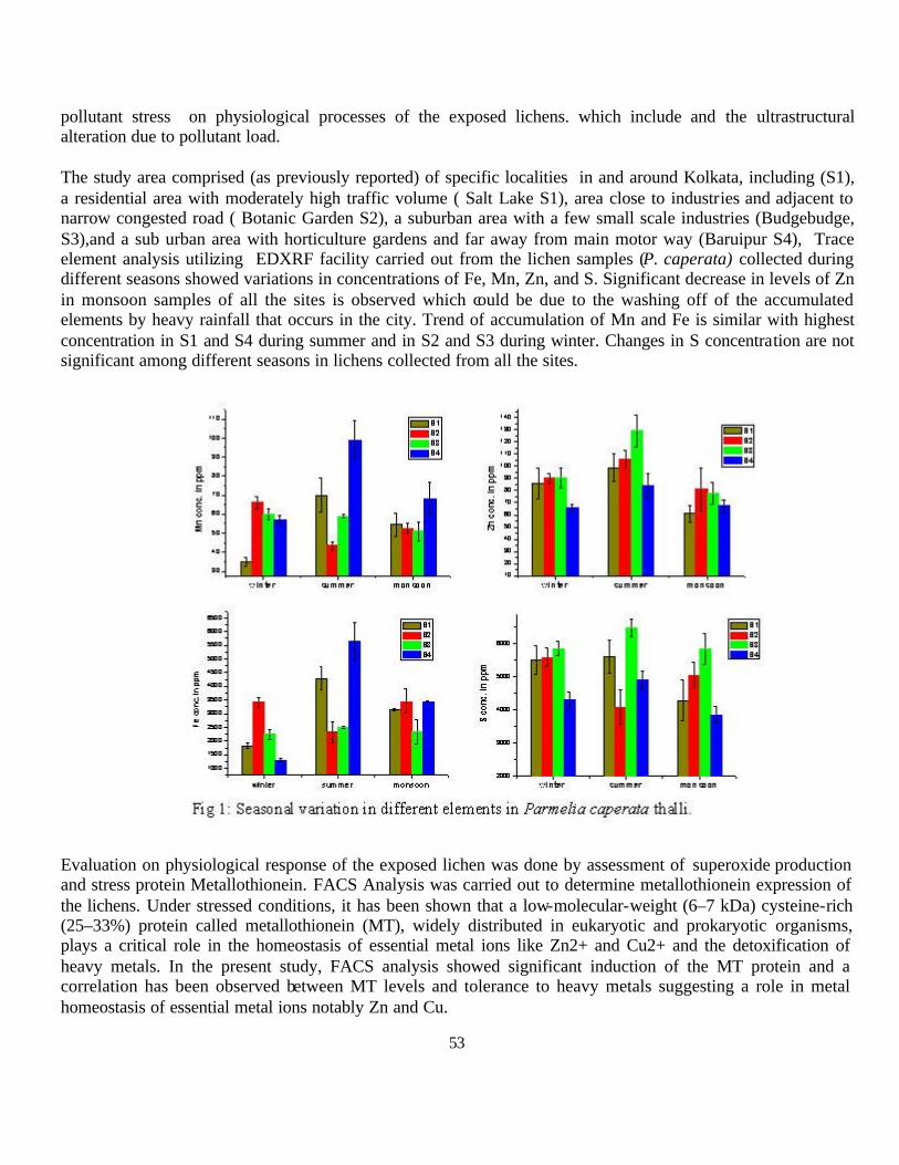
53
pollutant stress on physiological processes of the exposed lichens. which include and the ultrastructural alteration due to pollutant load.
The study area comprised (as previously reported) of specific localities in and around Kolkata, including (S1), a residential area with moderately high traffic volume ( Salt Lake S1), area close to industries and adjacent to narrow congested road ( Botanic Garden S2), a suburban area with a few small scale industries (Budgebudge, S3),and a sub urban area with horticulture gardens and far away from main motor way (Baruipur S4), Trace element analysis utilizing EDXRF facility carried out from the lichen samples (P. caperata) collected during different seasons showed variations in concentrations of Fe, Mn, Zn, and S. Significant decrease in levels of Zn in monsoon samples of all the sites is observed which could be due to the washing off of the accumulated elements by heavy rainfall that occurs in the city. Trend of accumulation of Mn and Fe is similar with highest concentration in S1 and S4 during summer and in S2 and S3 during winter. Changes in S concentration are not significant among different seasons in lichens collected from all the sites.
Evaluation on physiological response of the exposed lichen was done by assessment of superoxide production and stress protein Metallothionein. FACS Analysis was carried out to determine metallothionein expression of the lichens. Under stressed conditions, it has been shown that a low-molecular-weight (6–7 kDa) cysteine-rich (25–33%) protein called metallothionein (MT), widely distributed in eukaryotic and prokaryotic organisms, plays a critical role in the homeostasis of essential metal ions like Zn2+ and Cu2+ and the detoxification of heavy metals. In the present study, FACS analysis showed significant induction of the MT protein and a correlation has been observed between MT levels and tolerance to heavy metals suggesting a role in metal homeostasis of essential metal ions notably Zn and Cu.

54
Fig 2: Histogram of MT content of P. caperata thalli by FACS analysis: X-axis represents relative fluorescence intensity of the cells and Y-axis represents cell counts. M1 represents the binding region. (a) thalli without the FITC tag ( negative control), (b) thalli with the FITC tag.
Fig3: Autofluorescense and Diclorofluorescein fluorescence of the algal component of lichen Parmelia caperata collected from S1, (a) autofluorescence only (b) both autofluorescence and DCF fluorescence.
Reactive Oxygen Species production in lichens exposed to pollutants was also analysed using flow cytometric technique. Analysis of the algal component of dissociated unstained lichens produced a consistent fluorescence, primarily owing to differences in chlorophyll content. Samples stained with DCFDA produced a consistent fluorescence pattern, with only a portion of the cells stained with DCFDA.
Fluorescent Microscopy Observation of ROS: In addition to physiological characterization, we have also performed Scanning Electron Microscopic study of lichen samples from all the four sites. Particulate

55
depositions are found on the surface of lichen thalli at all the sites, however, their occurrence is more in samples from urban polluted site Fig 1. It is interesting to mention that some reproductive structures are found in lichen thalli collected from Site 3 during monsoon season. The reproductive bodies of lichen are very much pollution sensitive and generally do not grow in an atmosphere burdened with pollutant load. Therefore, their presence at samples from site 3 indicates a comparatively cleaner area at this site.
Fig 1: Large particle detected on the thallus surface of an in situ Parmelia caperata from Botanic Garden (S2) and (b) Salt Lake (S1), Kolkata during winter.
Fig 2: Reproductive bodies observed on the thallus surface of an in situ Parmelia caperata from Baruipur (S4), Kolkata during winter.
S.Majumder, N.K.Jana (Charuchandra College); S.Santra (Kalyani University); A.Chakraborty, M.Sudarshan
2.5.20. Bioremediation of hexavalent chromium and associated heavy metals using heavy metal stress tolerant microbes isolated from chromite mine environment of Orissa.
Hexavalent chromium Cr (VI) is a major pollutant in chromite mine area of Sukinda, Odisha, the richest chromiferous field mass in Indian sub-continent. Cr (VI) is known to be toxic to living organisms as a mutagenic carcinogenic, cytotoxic and genotoxic agent. Thus, chromium remediation is extremely essential to keep the environment free of pollutant for safety of living beings. Keeping these in view, the present study is aimed to isolate chromium tolerant microbes from heavy metal contaminated chromite mine environment of

56
Sukinda and evaluate their bioremediation potential with a view to remediate the toxic hexavalent chromium from the environment.
Soil and water samples were collected from different location of Kaliapani, one of the chromite mines of Sukinda (operated by Orissa Mining Corporation Ltd, Bhubaneswar) and brought to the Department of Biotechnology, College of Engineering and Technology, Bhubaneswar during February, 2010. The soils samples were analysed for their physico-chemical and heavy metal content and microbial population. Physico-chemical properties (pH, salinity, colour, texture, nutrient content etc.) were assessed by method of Jackson (1973) and trace elements along with heavy metal contents (Mg, Ti, Ni, Cr, Zn, Pb etc.) of the soil and water samples were analysed using EDXRF and AAS system respectively. Generally, chromite mine soils samples contain various heavy metals such as Cr, Mn ,Cu, Ni, CO, Fe etc. at very high concentrations (Fig: 1). Besides the physico-chemical and heavy metal contents, the soil and water samples were also analysed for their microbial populations such as bacteria (general bacteria, spore forming bacteria, free living nitrogen fixing, cellulose degrading and phosphate solibulizing bacteria etc.), fungi and actinomycetes. Assessment of microbial population revealed that, the mine soils harbour very low microbial population of all kinds in comparison to that of fallow land. Apart from these, attempt has also been made to isolate chromium tolerant microorganisms from mine soil and water samples. In total, 47 chromium tolerant bacteria were isolated from mine soil and water samples using chromium enriched nutrient agar media.
Fig: 1
Fig: 2
Fig: 3 Fig: 4
0
10000
20000
30000
40000
50000
60000
Fe Ti Cr Mn Ni Zn Sr Pb
Series1
Conc
entr
atio
n
Different
44%
26%17%
13%
Range of Cr (VI) (400-900) ppm tolerance bacteria, soil
samples, chromite mine Sukinda400 ppm 500 ppm
0% 4% 11%
15%35%
35%
Range of other heavy metals (400-500) ppm tolerance Cr (VI)
resistance bacteria, soil samples, chromite mine Sukinda
CuSO4 CoCl2
57
These bacterial isolates were further screened for their increased hexavalent chromium tolerance in presence and finally twenty bacteria were selected as highly chromium tolerant or resistant bacteria (having Cr (VI) tolerance of > 500 ppm). The strains were then evaluated for their Minimum Inhibitory Concentration (MIC) towards hexavalent chromium following Disc Diffusion Method. The MIC values of the bacteria ranges between 500 ppm to 2450 ppm (Fig: 2). However, all these 20 CRB bacterial strains were further screened for their tolerance towards other heavy metals such as Co2+, Ni2+, Pb2+, Cu2+, Mn2+ and Se3- (Fig: 4), Further bioreduction of hexavalent chromium using highly Cr(VI) tolerant baceria is under progress.
The selected 20 highly heavy metal tolerant bacterial strains were then subjected to phenotypic characterization with a view to identify the strains. Phenotypic characterization of the bacterial isolates included morphological, microscopic (shape, size, Gram stains etc.) and biochemical (catalase, methyl red test, protease test etc.) tests which were carried out as per methods of Holt (1984). The bacteria l strains were then identified following Bergey’s Manual of Systematic Bacteriology. Some of the bacterial genera so identified include Bacillus, Kurthia, Planococcus, Pseudomonas, Clostridium etc.
References: Holt, J.G. (1984). Bergey’s Manual of Systematic Bacteriology, Vol. I and II. Williams Wilkins, Baltimore, USA., 1:1-964. 2:965-1599. Jackson, M.L. (1973). Soil chemical analysis Prentice Hall, New Delhi.
H.N Thatoi, Sasmita Das, (CET, Bhubaneswar); Anindita Chakraborty, M.Sudarshan
2.5.21. Studies on trace element distribution and their role in salt stress adaptation in halophytic plants of mangrove vegetation of West Bengal
Mangroves are trees and shrubs that grow in saline coastal habitats in the tropics and subtropics. They play an important role as a filter and natural pollution treatment centre. In the salt stress condition, cations performs the buffering activities specially potassium and calcium. Generally, accumulation does occur at the root level, with restricted transport to aerial portion of the plants. Many trace elements played a crucial role in metabolism as well as salt stress adaptation of mangrove vegetation. It also known that trace elements mostly sediment in the mixture zone of fresh water and salt water, which further accumulated in plants and animals through usual uptake and assimilation processes. This study relating to distribution and localization of trace elements in plants of salt stress environment has special significance. It was designed
Ø To study the trace element distribution in soil and halophytic plants of mangrove ecosystem in West Bengal
Ø Biochemical characterization of salt tolerant species through analysis of characteristics of metabolites Ø To study the metal binding with cellular protein in salt stress condition in selected mangrove plant
Five sampling stations, were selected as the study area , namely Taki (S1), Hasnabad (S2), Hemnagar (S3), Jharkhali (S4) and Henry Island (S5), were chosen considering the metal deposition patterns along the drainage network systems with luxurient mangrove vegetation and inhabited by many endangered species of flora and fauna representing genetically diverse ecosystem. of water sample was measured by the Refractometer and
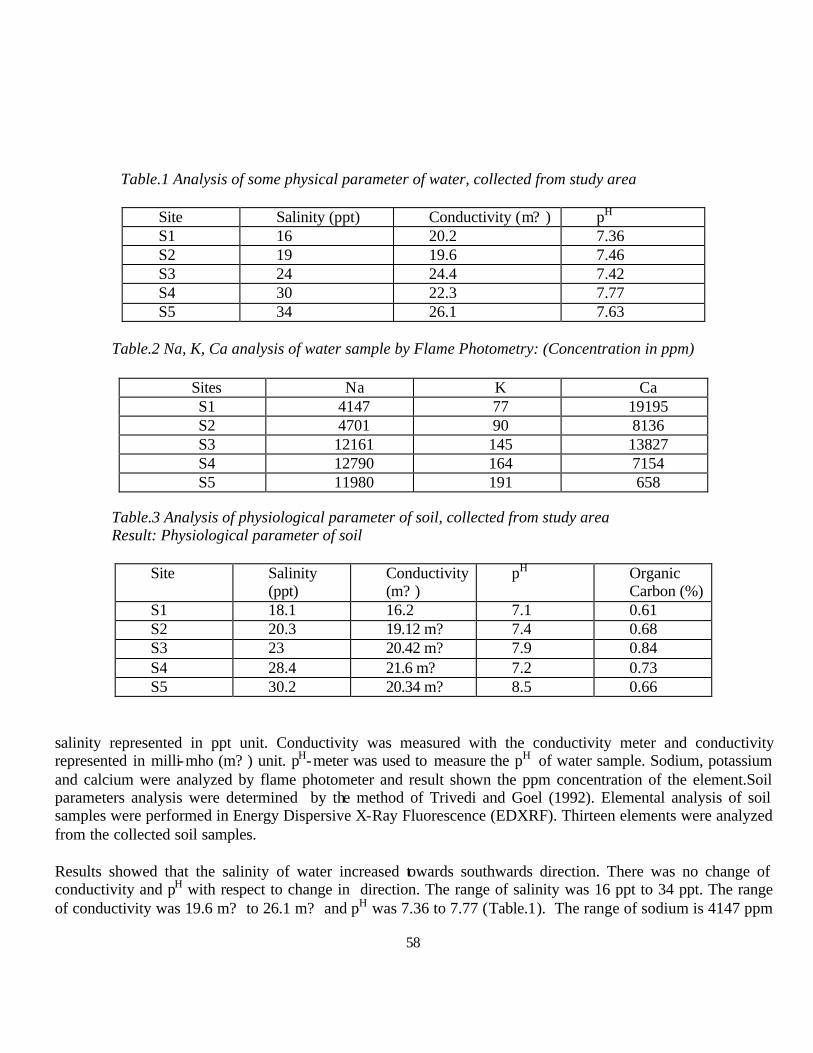
58
Table.1 Analysis of some physical parameter of water, collected from study area
Site Salinity (ppt) Conductivity (m? ) pH S1 16 20.2 7.36 S2 19 19.6 7.46 S3 24 24.4 7.42 S4 30 22.3 7.77 S5 34 26.1 7.63
Table.2 Na, K, Ca analysis of water sample by Flame Photometry: (Concentration in ppm)
Sites Na K Ca S1 4147 77 19195 S2 4701 90 8136 S3 12161 145 13827 S4 12790 164 7154 S5 11980 191 658
Table.3 Analysis of physiological parameter of soil, collected from study area Result: Physiological parameter of soil
Site Salinity
(ppt) Conductivity (m? )
pH Organic Carbon (%)
S1 18.1 16.2 7.1 0.61 S2 20.3 19.12 m? 7.4 0.68 S3 23 20.42 m? 7.9 0.84 S4 28.4 21.6 m? 7.2 0.73 S5 30.2 20.34 m? 8.5 0.66
salinity represented in ppt unit. Conductivity was measured with the conductivity meter and conductivity represented in milli-mho (m? ) unit. pH-meter was used to measure the pH of water sample. Sodium, potassium and calcium were analyzed by flame photometer and result shown the ppm concentration of the element.Soil parameters analysis were determined by the method of Trivedi and Goel (1992). Elemental analysis of soil samples were performed in Energy Dispersive X-Ray Fluorescence (EDXRF). Thirteen elements were analyzed from the collected soil samples.
Results showed that the salinity of water increased towards southwards direction. There was no change of conductivity and pH with respect to change in direction. The range of salinity was 16 ppt to 34 ppt. The range of conductivity was 19.6 m? to 26.1 m? and pH was 7.36 to 7.77 (Table.1). The range of sodium is 4147 ppm

59
to 12790 ppm, pottasium is 77 ppm to 191ppm and calsium is 658 ppm to 19195 ppm (Table.2). Soil salinity towards north to south directionincreased. The range of salinity is 18.1ppt to 30.2 ppt. Conductivity, pH, Organic Carbon does not show any relationship with direction. The ranges of Conductivity, pH, Organic Carbon are 16.2 m? to 20.42 m? , 7.1 to 8.5, 0.61 % to 0.84 % respectively (Table.3).
Threshold limit value in soil of Cu, Fe, Mn, Ni, Pb, Zn are 14 to29 ppm, 50 to 1000 ppm, 260 to 840 ppm, 13 to 30ppm, 17 to 26 ppm and 34 to 84 ppm respectively. In Henry island (S5) and Hasnabad(S2) , Pb and Fe was higher than threshold limit value. Fe is also higher than the threshold limit value in Taki (S1) (Table.2).
Table.4 Analysis of trace element distribution in soil of Mangrove ecosystem Result: Elemental Status of Soil sample in five sampling site (Concentration in ppm)
Element S1 S2 S5 Mg 15699±5752 18357±1507 23022±1433 Al 63106±25714 82637±5550 101603±2762 Si 247367±105828 256134±16923 300176±3144 K 26635±6900 127994±1654 36267±587 Ca 20799±5992 13994±504 7126±262 Ti 3865±914 4949±193 5628±24 Mn 280±43 324±17 367±30 Fe 16017±2564 22676±1496 21395±59 Ni 10.1±3 21.2±2 18.3±0.7 Cu 15.8±2 21±2 19.3±0.4 Zn 23.59±3 39.8±4 29.6±1 Sr 66.3±12 65±6 61±1.2 Pb 22.8±4 32±2 29±0.01
Soil and water of S5 site are more saline than other sampling sites since this site is in close proximity to the sea. Concentration of sodium and potassium ion are also observed to be higher in this site because of their high salt concentration in seawater. The pH also ranged from neutral to basic because of higher salt concentration. Pb concentration is higher in Henry Island and Hasnabad which could be due to the use of leaded petrol in the local vahicles. . Using of iron fertiliser may be the cause of high iron value in the Taki & Hasnabad region.
S. Bhar, S.C. Santra, (Kalyani University); Anindita Chakraborty and M. Sudarshan
2.5.22. Rational and Productive use of Protein wastes from the Tannery Industries”
Introduction:Tannery effluent with a host of elements with, either toxic or biologically active but in very high concentrations, pose serious toxic effects through impairment of cellular respiration by inhibition of various mitochondrial enzymes. Metal accumulation in different tissue of fish cultured in ECW has been studied in detail and the findings have been already published in reputed journal (Aquaculture Research 2011). Whereas impact of composite effluent in the aminotransferase activity in a fish biosystems has also been studied, exposing guppy fish in different sub-lethal (3%, 6% and 9%) concentration of tannery effluent for three
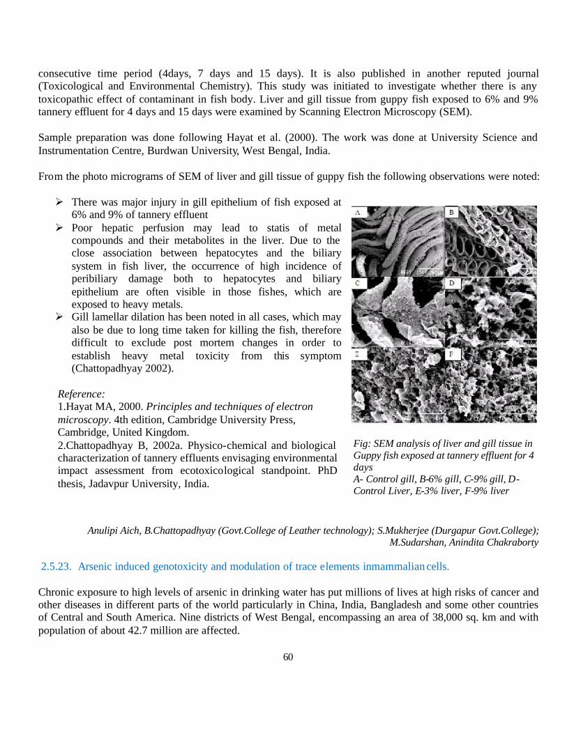
60
consecutive time period (4days, 7 days and 15 days). It is also published in another reputed journal (Toxicological and Environmental Chemistry). This study was initiated to investigate whether there is any toxicopathic effect of contaminant in fish body. Liver and gill tissue from guppy fish exposed to 6% and 9% tannery effluent for 4 days and 15 days were examined by Scanning Electron Microscopy (SEM).
Sample preparation was done following Hayat et al. (2000). The work was done at University Science and Instrumentation Centre, Burdwan University, West Bengal, India.
From the photo micrograms of SEM of liver and gill tissue of guppy fish the following observations were noted:
Ø There was major injury in gill epithelium of fish exposed at 6% and 9% of tannery effluent
Ø Poor hepatic perfusion may lead to statis of metal compounds and their metabolites in the liver. Due to the close association between hepatocytes and the biliary system in fish liver, the occurrence of high incidence of peribiliary damage both to hepatocytes and biliary epithelium are often visible in those fishes, which are exposed to heavy metals.
Ø Gill lamellar dilation has been noted in all cases, which may also be due to long time taken for killing the fish, therefore difficult to exclude post mortem changes in order to establish heavy metal toxicity from this symptom (Chattopadhyay 2002).
Reference: 1.Hayat MA, 2000. Principles and techniques of electron microscopy. 4th edition, Cambridge University Press, Cambridge, United Kingdom. 2.Chattopadhyay B, 2002a. Physico-chemical and biological characterization of tannery effluents envisaging environmental impact assessment from ecotoxicological standpoint. PhD thesis, Jadavpur University, India.
Anulipi Aich, B.Chattopadhyay (Govt.College of Leather technology); S.Mukherjee (Durgapur Govt.College); M.Sudarshan, Anindita Chakraborty
2.5.23. Arsenic induced genotoxicity and modulation of trace elements inmammalian cells.
Chronic exposure to high levels of arsenic in drinking water has put millions of lives at high risks of cancer and other diseases in different parts of the world particularly in China, India, Bangladesh and some other countries of Central and South America. Nine districts of West Bengal, encompassing an area of 38,000 sq. km and with population of about 42.7 million are affected.
Fig: SEM analysis of liver and gill tissue in Guppy fish exposed at tannery effluent for 4 days A- Control gill, B-6% gill, C-9% gill, D-Control Liver, E-3% liver, F-9% liver
61
Trace element profile is one of the valuable markers of the healthy status of the body. Any alteration in this profile signifies malfunctioning of the normal metabolism of the body. The modulation in trace element profile can act as biomarker for the study of Arsenic-toxicity. It is already been known through literature survey that the LD50 dose of arsenic trioxide in mice through oral administration is 33-39 mg/kg. Since arsenic is ubiquitous in nature and is mostly present in the range as high as up to 4ppm in drinking water so we selected several doses within this range for our in vivo experiment in mice. The Swiss albino mice were treated with different concentrations of Arsenic trioxide (As2O3) orally (adlibitum) and the doses used, were 0.4mg/L(ppm), 0.8 ppm, 2ppm and 4ppm for different time durations i.e., for 1 month and 15 days. At the end of the treatment period, tissues (liver, kidney, spleen and skin) were dissected out, blotted properly, lyophilized and grounded in an agate mortar. The tissue samples were analyzed by Jordan Vally EX-3600 ED-XRF system. The modulation of the trace element profile was then checked by comparing with the control (Gr. I)
The elements in liver which showed the significant decrease with respect to control after As2O3 treatment were P, K, Ca, Mn, and Cu in the treatment Gr. IV (As2O3 0.4 ppm for 1 Month), Gr.V (As2O3 0.8 ppm for 1 Month), Gr.VI (As2O3 2 ppm for 1Month), Gr.VII (As2O3 4 ppm for 1 Month) and Gr.VIII (As2O3.4ppm for 15 days) though the changes were not uniform in different treatment groups. In the kidney there was a significant decrease in the element Mn in the Gr. IV and VIII and the element Fe in the Gr.IV. The decrease was significant in comparison to the control. The spleen seems to be most severely affected organ as nearly all the element showed a significant depletion in their concentration with respect to control in the treatment Gr. VI, VII and VII. Mg and K concentration depleted in the skin significantly in Gr. VIII and VI respectively, where as a significant decrease was observed for the P and S in the Gr. VI, VII, and VIII.
Ritu Srivastav, Anshuman Chattopadhya (Visva Bharati); M.Sudarshan, Anindita Chakraborty
2.5.24. Regulation and expression of antioxidant enzymes and comparative study on metal detection of Withania somnifera and Abutilon indicum L in response to various metal stress.
Withania somnifera and Abutilon indicum L are two important medicinal plants which can naturally grow in soil enriched with stress inducing methods like Cu, Zn, Mn and Fe. These elements sometimes act as macro/micro nutrients or sometimes also act as soil pollutants. The plants sustain themselves in this stressed conditions through their natural adaptive defence mechanisms. Defence include involvements of anti oxidants, metalloprotiens and trace element interactions. This stude was designe to probe into regulation and expression of these factors to unravel natural variability and further to use these variations for development of new variants through tissue culture techniques.
The plant and seed materials of W. somnifera and A. indicum used for this experiment were collected from different places like:
1. Ramakrishna Mission Ashrama, Narendrapur, Kolkata. 2. Patanjali Yogapitha, Bahadarabad, Haridwar. 3. Govt. Nursery, Ghatkia, Khandagiri, Bhubaneswar. 4. Botanical Garden of P.G.Department of Botany, Utkal University, Bhubaneswar. 5. Kalyani Nursury, Patia, Bhubaneswar. 6. Forest Nursury, CRP square, Bhubaneswar.
62
Only mature and healthy seeds were washed thoroughly under running tap water for 30 min followed by treatment with an aqueous solution of 5 % (cm3 m-3) teepol (Reckitt's Colman, Kolkata, India) for 10 min and rinsed 5 times with double distilled water. The seeds were then surface disinfected with an aqueous solution of 0.1 % (g dm-3) HgCl2 (Hi-Media, Mumbai, India) for 5 min and rinsed five times with autoclaved double distilled water. The disinfected seeds were inoculated in 150 cm3 Erlenmeyer flasks (Borosil, Bangalore, India) containing germinating media i.e. ½ MS (Murashige & Skoog 1962) medium. The pH of the medium was adjusted to 5.8 ± 0.5 before autoclaving at 121 0C and 104 kPa for 15 min. The seeds were allowed to germinate at 25 ± 20C, 60 % relative humidity and 35 µmol m-2 s-1 photon flux density provided by cool white fluorescent tubes (Philips, Bangalore, India). For in vivo study, the seeds were sterilized as mentioned for in vitro study and then placed in a plastic pot supplied with sterilized soil, sand and vermicompost.
The freshly collected seeds did not germinate satisfactorily in the soil condition. Out of above mentioned varieties seeds collected from Ramakrishna Mission Ashrama, Narendrapur and Patanjali Yogapitha, Bahadarabad, Haridwar gave highest % of seed germination. These two varieties seeds produced good and healthy plants.
Table 1: Percentage of seed germination of W. somnifera under in vivo and in vitro condition from different collected seeds
Collection sites of seeds % seed germination in in vivo % seed germination in in vitro Ramakrishna Mission Ashrama
66 83
Patanjali Yogapitha 43 52 Govt. Nursery 37 61 Botanical garden 69 87 Kalyani Nursury 30 34 Forest Nursury 35 47
From Table 1, it clears that the percentage of seed germination is high in case of in vitro seeds in all varieties of seeds. The seeds collected from Ramakrishna Mission Ashrama and Botanical garden of Utkal University gave highest percentage of seed germination so further study proceeded by these two types of seeds.
Table 2: Percentage of seed germination of A. indicum under in vivo and in vitro condition from different collected seeds
Collection sites of seeds % seed germination in in vivo % seed germination in in vitro Patanjali Yogapitha, Bahadarabad, Haridwar
53 50
Basudevpur, Bhadrak 92 45 Govt. Nursery 68 40 Botanical garden 88 47 Kalyani Nursury 61 33 Forest Nursury 59 31
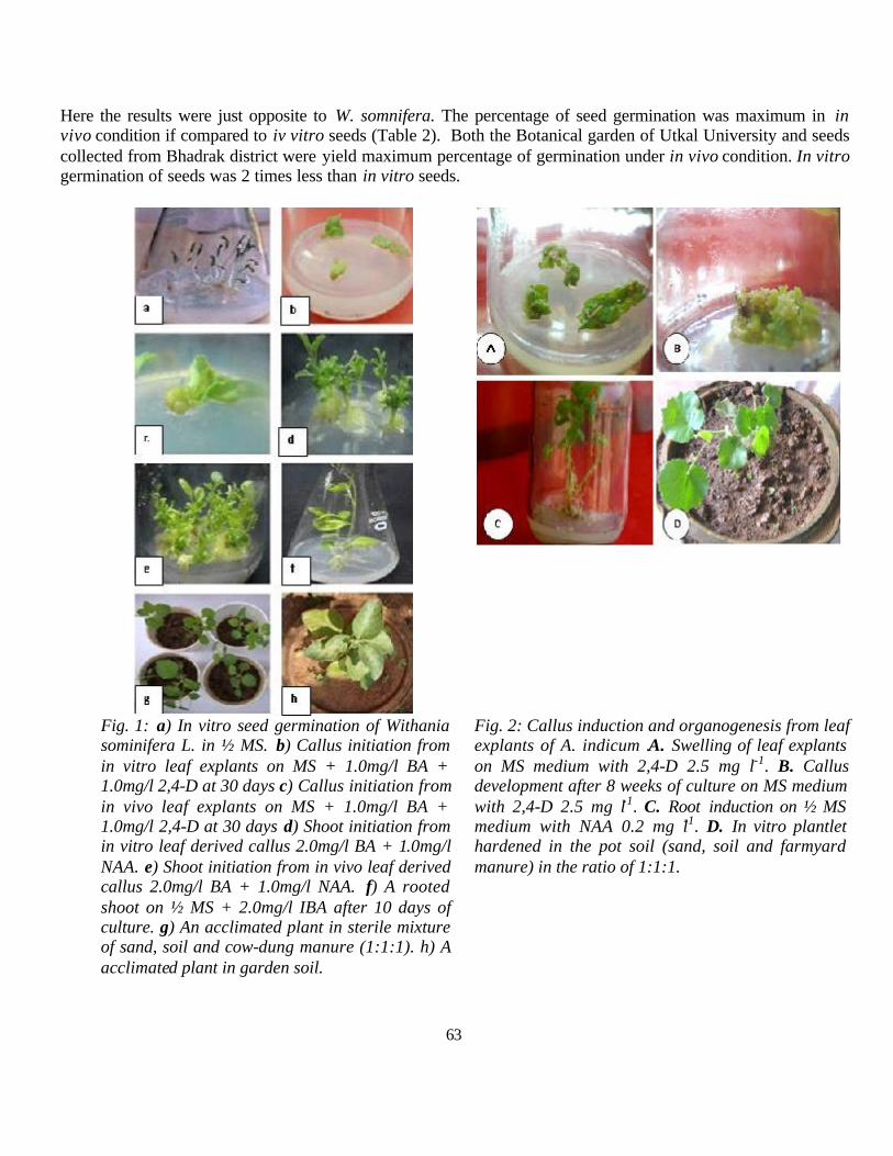
63
Here the results were just opposite to W. somnifera. The percentage of seed germination was maximum in in vivo condition if compared to iv vitro seeds (Table 2). Both the Botanical garden of Utkal University and seeds collected from Bhadrak district were yield maximum percentage of germination under in vivo condition. In vitro germination of seeds was 2 times less than in vitro seeds.
Fig. 1: a) In vitro seed germination of Withania sominifera L. in ½ MS. b) Callus initiation from in vitro leaf explants on MS + 1.0mg/l BA + 1.0mg/l 2,4-D at 30 days c) Callus initiation from in vivo leaf explants on MS + 1.0mg/l BA + 1.0mg/l 2,4-D at 30 days d) Shoot initiation from in vitro leaf derived callus 2.0mg/l BA + 1.0mg/l NAA. e) Shoot initiation from in vivo leaf derived callus 2.0mg/l BA + 1.0mg/l NAA. f) A rooted shoot on ½ MS + 2.0mg/l IBA after 10 days of culture. g) An acclimated plant in sterile mixture of sand, soil and cow-dung manure (1:1:1). h) A acclimated plant in garden soil.
Fig. 2: Callus induction and organogenesis from leaf explants of A. indicum .A. Swelling of leaf explants on MS medium with 2,4-D 2.5 mg l-1. B. Callus development after 8 weeks of culture on MS medium with 2,4-D 2.5 mg l-1. C. Root induction on ½ MS medium with NAA 0.2 mg l-1. D. In vitro plantlet hardened in the pot soil (sand, soil and farmyard manure) in the ratio of 1:1:1.
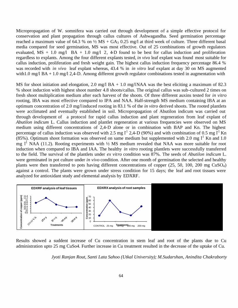
64
Micropropagation of W. somnifera was carried out through development of a simple effective protocol for conservation and plant propagation through callus cultures of Ashwagandha. Seed germination percentage reached a maximum value of 64.3 % on ½ MS + GA3 0.25 mg/l at third week of culture. Three different basal media compared for seed germination, MS was most effective. Out of 25 combinations of growth regulators evaluated, MS + 1.0 mg/l BA + 1.0 mg/l 2, 4-D found to be best for callus induction and proliferation regardless to explants. Among the four different explants tested, in vivo leaf explant was found most suitable for callus induction, proliferation and fresh weight gain. The highest callus induction frequency percentage 86.4 % was recorded with in vivo leaf explant whereas, 43.4 % in in vitro leaf explant at day 30 on MS augmented with1.0 mg/l BA + 1.0 mg/l 2,4-D. Among different growth regulator combinations tested in augmentation with
MS for shoot initiation and elongation, 2.0 mg/l BA + 1.0 mg/l NAA was the best eliciting a maximum of 82.3 % shoot induction with highest shoot number 4.8 shoots/callus. The original callus was sub-cultured 2 times on fresh shoot multiplication medium after each harvest of the shoots. Of three different auxins tested for in vitro rooting, IBA was most effective compared to IPA and NAA. Half-strength MS medium containing IBA at an optimum concentration of 2.0 mg/l induced rooting in 83.1 % of the in vitro derived shoots. The rooted plantlets were acclimated and eventually established in soil. Micropropagation of Abutilon indicum was carried out through development of a protocol for rapid callus induction and plant regeneration from leaf explant of Abutilon indicum L. Callus induction and plantlet regeneration at various frequencies were observed on MS medium using different concentrations of 2,4-D alone or in combination with BAP and Kn. The highest percentage of callus induction was observed with 2.5 mg l-1 2,4-D (90%) and with combination of 0.5 mg l-1 Kn (85%). Optimum shoot formation was observed on same medium but supplemented with 2.0 mg l-1 Kn and 1.0 mg l-1 NAA (11.2). Rooting experiments with ½ MS medium revealed that NAA was more suitable for root induction when compared to IBA and IAA. The healthy in vitro rooting plantlets were successfully transferred to the field. The survival of the plantlets under ex vitro condition was 87%. The seeds of Abutilon indicum L. were germinated in pot culture under in vivo condition. After one month of germination the selected and healthy plants were then transferred to pots having different concentrations of copper (25, 50, 100, 200 mg CuSO4) against a control. The plants were grown under stress condition for 15 days; the leaf and root tissues were analyzed for antioxidant study and elemental analysis by EDXRF.
Results showed a suddent increase of Cu concentration in stem leaf and root of the plants due to Cu administration upto 25 mg CuSo4. Further increase in Cu treatment resulted in the decrease of the uptake of Cu.
Jyoti Ranjan Rout, Santi Lata Sahoo (Utkal University); M.Sudarshan, Anindita Chakraborty
9.810
10.210.410.610.8
1111.211.4
Con
cent
ratio
n of
cop
per
(ppm
)
Treatments
EDXRF analysis of leaf tissues
0102030405060
70
CONTROL 25 mg 50 mg 100 mg 200 mg
Co
nce
ntr
atio
n (p
pm
_
Treatments
EDXRX analysis of root samples
0
5
10
15
20
25
CONTROL 25 mg 50 mg 100 mg 200 mg
Concentration (ppm)
Treatments
EDXRF analysis of stem samples
65
2.5.25. Influences of trace elements in immune functions in Visceral Leishmaniasis and Post Kala-azar Dermal Leishmaniasis of India
This program was designed to probe into the possible involvement of different trace elements in different clinical forms of Indian leishmaniasis (prevalent in eastern regions of the country) with special reference to in the immune response towards the parasite. This. Blood collected from Kala-azar (VL) and post kalazar dermal leishmaniasis (PKDL) patients as well as from controls are made into targets for trace element analysis utilizing EDXRF. Blood of BALB/c mice infected with Leishmania parasites were also made into targets to be analysed. The purpose of selecting this model (the model animal system for experimenting visceral leishmaniasis) is to establish the correlation between the blood profile for trace elements in the infected mice with the Kala-azar patients, if any. Out of 7 human blood samples six were from VL in which two samples did not show and LD bodies in the bone marrow smear
No LD was detected in the PKDL sample also. Trace elemental analysis shows lower Mg conc in the infected groups while it is higher in patients’ blood where no LD bodies are detected as well as control female. Comparable with the blood profile of infected patients infected mice also showed lower conc. of Mg in blood. In case of S, No significant variation was noticed In S concentration in any of the samples. Contrary to Mn, Cu was higher in conc. in patients’ blood compared to that of the control except for one VL & one PKDL patient that did not show LD bodies. Infected mice showed highest P conc.There is reduction in Zn conc. in the patients as well as infected mice blood compared to the control group. Only the suspected PKDL patient showed an exorbitant high value.. Infected mice showed lower Ca value and higher K valuecompared to the rest. The present study if done with large sample size might throw some light on role of specific elements in progression of the disease and might help in developing strategies for therapy as well as immunoprophylactic measures against Indian forms of Leishmaniasis
Priyanka Chakraborty, Madhumita Manna (Bethuene College); M.Sudarshan, Anindita Chakraborty
2.5.26. A Comprehensive Study on Radiation Induced Metal Tolerance in Fungal Strain
In this program a few strains of fungi viz. Aspergillus, Penicillium are being considered as effective alternatives for remediation of metal pollution by utilizing ionizing radiation in potentiating the metal accumulating property of the microbes. Low doses of gamma radiation are observed to induce mutagenic effects in fungi like other eukaryotic system producing mainly random mutations in DNA. It is already reported that gamma is the best mutagenic agent than UV radiation or other chemical mutagens in fungi. Gamma irradiated fungal mutants have been reported to have different metabolic profile when compared to their normal counterparts like higher extra cellular enzyme activities, more lignin degrading capacities etc. During this period fungal strains were isolated from waste disposal sites of urban Kolkata that are found to tolerate high concentrations of metals viz. Cd, Pb, Zn, Hg. The survival, colony forming ability of these fungi were assessed as a function of increasing concentration of these metals . Minimum inhibitory concentration (MIC) of the four metals with respect to each strain in laboratory controlled conditions was also determined. The maximum tolerant (from each respective metal exposed) groups of these isolated strains were then exposed to different doses of gamma rays (20 -100 Gy absorbed dose) and compared with the unirradiated sets for their survival, CFA in response to the same concentrations of the metals. Gamma irradiation was found to increase in cadmium tolerance in case of Aspergillus niger and Penicillium cyclopium. Penicillium cyc lopium showed max. metal tolerance reflecting highest CFA (colony forming ability) when exposed to 40Gy of gamma irradiation

66
while in case of Aspergillus niger an absorbed dose of 20 Gy produced similar effect. Potentiating effect of gamma radiation was also noted in case tolerating Zn.
Results indicated strain-specific dose-dependant sensitivity and response in modulating metal tolerance of the selected fungal strains.
Dippanita Das, S.Santra, (Kalyani University; Anindita Chakraborty
2.5.27. Radiation and Molecular Immunology: Understanding the Mechanism of Protection in Leishmania Infection using Radio-attenuated Leishmania parasites
In this program efficacy of radio-attenuated Leishmania parasite is being evaluated as prophylactic and therapeutic agent against experimental visceral Leishmaniasis in murine model. In our previous study, three different doses of radiation – 50Gy, 100Gy, 150Gy respectively was standardized for preparation of live attenuated vaccine candidate for immunization through intracardiac, intraperitoneal and intramuscular routes. In this study the efficacy of subcutaneous (s.c) route was assesse both in therapeutic and prophylactic protocols. In prophylactic protocol 4-6 weeks old BALB/c mice were immunized with three different doses of attenuated parasites in two subsequent doses in fifteen days interval. Fifteen days after the last immunization, mice were
cont 20 GY 40GY 60GY 80GY 100GY-1
0
1
2
3
4
5
6
7
8
9
10
11
12Aspergillus terreusMetal- Zinc(10000ppm)Time- 9th day of incubation
No.
of c
olon
ies
Dose(Gray)
c o n t 2 0 G y 4 0 G y 6 0 G y 8 0 G y 1 0 0 G y- 1
0
1
2
3
4
5
6
7
P e n i c i l l i u m c y c l o p i u mM e t a l - 6 0 0 p p m C d
Num
ber o
f col
onie
s
D o s econt 20 gy 60 gy 80 gy 100gy
0
20
40
60
80
100
Aspergillus terreusMetal- zinc(9500ppm)Time-9th day of incubation
No
of c
olon
ies
Dose(Gray)
cont 20 Gy 40 Gy 60 Gy 80 Gy 100 Gy0
5
10
15
20
25
30
35
40Aspergillus nigerMetal- Cd(84 ppm))
No
of c
olon
ies
Dose
9th Day 14th Day
con t 2 0 G y 40 Gy 6 0 G y 8 0 G y 1 0 0 G y
0
2
4
6
8
1 0
1 2
1 4
1 6
1 8
2 0Aspergil lus nigerMetal -Cd(100ppm)
No
of c
olon
ies
Dose
9th Day 14th Day
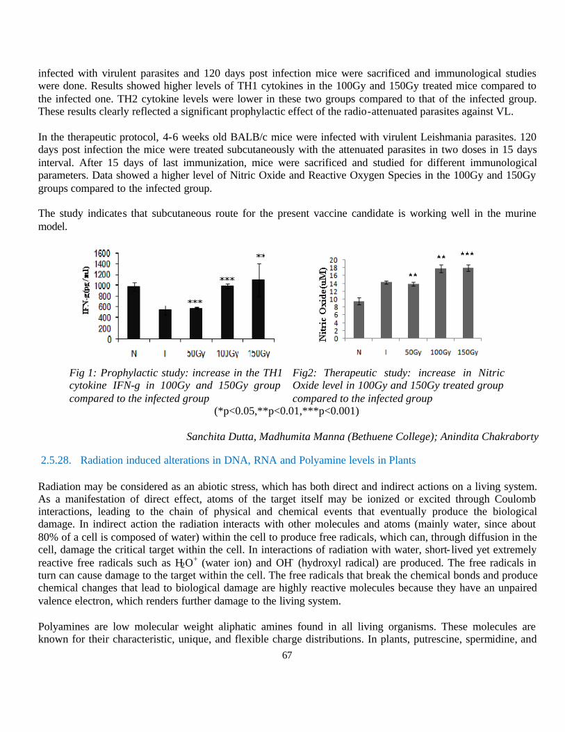
67
infected with virulent parasites and 120 days post infection mice were sacrificed and immunological studies were done. Results showed higher levels of TH1 cytokines in the 100Gy and 150Gy treated mice compared to the infected one. TH2 cytokine levels were lower in these two groups compared to that of the infected group. These results clearly reflected a significant prophylactic effect of the radio-attenuated parasites against VL. In the therapeutic protocol, 4-6 weeks old BALB/c mice were infected with virulent Leishmania parasites. 120 days post infection the mice were treated subcutaneously with the attenuated parasites in two doses in 15 days interval. After 15 days of last immunization, mice were sacrificed and studied for different immunological parameters. Data showed a higher level of Nitric Oxide and Reactive Oxygen Species in the 100Gy and 150Gy groups compared to the infected group. The study indicates that subcutaneous route for the present vaccine candidate is working well in the murine model.
Fig 1: Prophylactic study: increase in the TH1 cytokine IFN-g in 100Gy and 150Gy group compared to the infected group
Fig2: Therapeutic study: increase in Nitric Oxide level in 100Gy and 150Gy treated group compared to the infected group
(*p<0.05,**p<0.01,***p<0.001)
Sanchita Dutta, Madhumita Manna (Bethuene College); Anindita Chakraborty
2.5.28. Radiation induced alterations in DNA, RNA and Polyamine levels in Plants
Radiation may be considered as an abiotic stress, which has both direct and indirect actions on a living system. As a manifestation of direct effect, atoms of the target itself may be ionized or excited through Coulomb interactions, leading to the chain of physical and chemical events that eventually produce the biological damage. In indirect action the radiation interacts with other molecules and atoms (mainly water, since about 80% of a cell is composed of water) within the cell to produce free radicals, which can, through diffusion in the cell, damage the critical target within the cell. In interactions of radiation with water, short- lived yet extremely reactive free radicals such as H2O+ (water ion) and OH- (hydroxyl radical) are produced. The free radicals in turn can cause damage to the target within the cell. The free radicals that break the chemical bonds and produce chemical changes that lead to biological damage are highly reactive molecules because they have an unpaired valence electron, which renders further damage to the living system.
Polyamines are low molecular weight aliphatic amines found in all living organisms. These molecules are known for their characteristic, unique, and flexible charge distributions. In plants, putrescine, spermidine, and
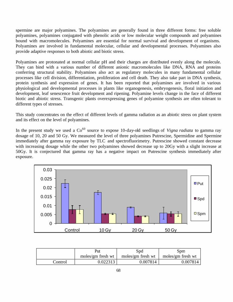
68
spermine are major polyamines. The polyamines are generally found in three different forms: free soluble polyamines, polyamines conjugated with phenolic acids or low molecular weight compounds and polyamines bound with macromolecules. Polyamines are essential for normal survival and development of organisms. Polyamines are involved in fundamental molecular, cellular and developmental processes. Polyamines also provide adaptive responses to both abiotic and biotic stress.
Polyamines are protonated at normal cellular pH and their charges are distributed evenly along the molecule. They can bind with a various number of different anionic macromolecules like DNA, RNA and proteins conferring structural stability. Polyamines also act as regulatory molecules in many fundamental cellular processes like cell division, differentiation, proliferation and cell death. They also take part in DNA synthesis, protein synthesis and expression of genes. It has been reported that polyamines are involved in various physiological and developmental processes in plants like organogenesis, embryogenesis, floral initiation and development, leaf senescence fruit development and ripening. Polyamine levels change in the face of different biotic and abiotic stress. Transgenic plants overexpressing genes of polyamine synthesis are often tolerant to different types of stresses.
This study concentrates on the effect of different levels of gamma radiation as an abiotic stress on plant system and its effect on the level of polyamines.
In the present study we used a Co60 source to expose 10-day-old seedlings of Vigna radiata to gamma ray dosage of 10, 20 and 50 Gy. We measured the level of three polyamines Putrescine, Spermidine and Spermine immediately after gamma ray exposure by TLC and spectrofluorimetry. Putrescine showed constant decrease with increasing dosage while the other two polyamines showed decrease up to 20Gy with a slight increase at 50Gy. It is conjectured that gamma ray has a negative impact on Putrescine synthesis immediately after exposure.
Put moles/gm fresh wt
Spd moles/gm fresh wt
Spm moles/gm fresh wt
Control 0.022313 0.007814 0.007814
0
0.005
0.01
0.015
0.02
0.025
0.03
Control 10 Gy 20 Gy 50 Gy
Put
Spd
Spm

69
10 Gy 0.017347 0.00549 0.00549 20 Gy 0.009044 0.004134 0.004134 50 Gy 0.005827 0.005399 0.005399
Sarmistha Raychaudhuri (CU); Nirmal Anindita Chakraborty
2.6. Collaborative Research Schemes using in-house facilities at Mumbai Centre
2.6.1. Synthesis and adsorption study of gold nanoparticles in chitosan latex particles
Gold nanoparticles coated with glutathione were synthesized at 80oC. These coating gives positive surface charge on nanoparticles, therefore, well dispersing. Part of glutathione coated Au nanoparticle dispersion was further modified with tri- sodium citrate to induce negative surface charge on nanoparticles. We have carried out dynamic light scattering measurements of gold (Au) nanoparticles with both positive and negative surface charges. DLS measurement showed the mean hydrodynamic diameter around 18 nm. DLS measurement on chitosan dispersion showed the chitosan particle size around 1000 nm (1 µm) with broad distribution of size between 300 – 2000 nm (FWHM). When gold nanoparticles were added to the chitosan latex dispersion and measured the change in mean hydrodynamic diameter of the chitosan particle with time using DLS, we observed that initially the size compresses and then increases. SEM images of pristine chitosan latexes (Figure 3) and Au nanoparticles adsorbed chitosan latexes (Figure 4) show a clear change in structural morphology of chitosan latexes.
We are carrying out further measurements like TEM and UV-vis spectroscopy to study the distribution of Au nanoparticles inside chitosan latexes. We are also trying to study the nature of interaction between nanoparticles and chitosan molecules using appropriate potential model.
A. Nimrodh Ananth, S. Umapathy (Madurai Kamraj University); S. Bhattacharya (BARC); G. Ghosh
SEM image of a chitosan latex particle
SEM image of a Au-chitosan latex particle

70
3. In-house Research activities
3.1. Research activity at Indore Centre
3.1.1. Bulk magnetic materials and oxides
3.1.1.1. CCoorrrree llaatteedd MMaaggnneett iicc aanndd EElleeccttrr iiccaa ll OOrrrrddeerrss iinn HHeexxaaggoonnaa ll YYMMnn11--xxFFeexxOO33 MMuulltt iiffee rrrroo iicc
Fe-doped hexagonal Y-Mn-Fe-O samples were synthesized by conventional solid-state reaction route and characterized structurally, electrically, and magnetically. Room temperature XRD confirmed formation of hexagonal structure with space group P63cm. With Fe-doping, the distortion of MnO5 polyhedra increases and the Mn-O-Mn bonds in a-b plane become longer, resulting in slight reduction of AFM Neèl temperature from 71K to 68K. A concurrent T-shift in the dielectric permittivity together with its non-monotonic magnitude observed indicates somewhat uncommon magnetolectric coupling in this compound. Also Fe-doping suppresses the value of M, effective moment µeff, and Weiss temperature ?p, suggesting that most probably the Fe has +2 valance state. With increasing Fe-content, ferroelectric P-E loops in YMFO become lossier (more conducting), without saturation up to 12 keV.
Sonu Namdeo, A.M. Awasthi
0 50 100 150 200 250 300
4
6
8
10
12
14
M (
emu/
mol
e)
Temperature (K)
YMnO 3 YMn0.95Fe0.05O3
YMn0.90Fe0.10O3
40 60 80 1008.0
8.5
9.0
TN
YMn1-xFexO3
104 Hz
Perm
itivi
ty ε
'
Temperature (K)
x = 0.00 x = 0.05 x = 0.10
-4 -2 0 2 4
-0.12
-0.08
-0.04
0.00
0.04
0.08
0.12
P (µ
C/c
m2 )
E (kV/cm)
YMnO3 YMn0.95Fe0.05O3
YMn0.90Fe0.10O3
32.8 33.0 33.20
2k
4k
6k
20 40 60 80 100 120-2k
0
2k
4k
6k
0 5 10
6.146
6.148
11.372
11.376
11.380
11.384
YMnO 3 YMn0.95Fe0.05 O3
YMn0.90Fe0.10 O3
x (Fe At.%)
YMnO3
Inte
nsity
(arb
. uni
ts)
2θ (Deg.)
Observed Calculated IObs-ICal
Bragg Position
a, b (Å)
c (Å)
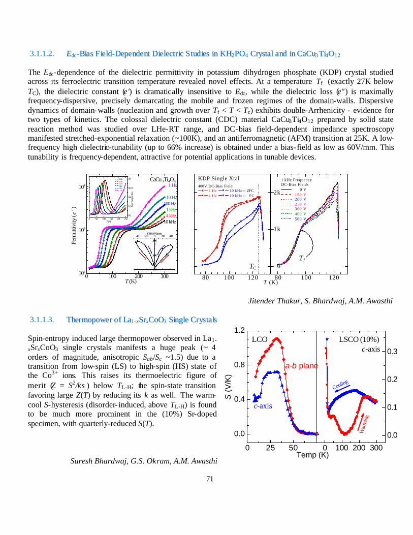
71
3.1.1.2. EEddcc--BBiiaass FF iiee lldd--DDeeppeennddeenntt DDiiee lleeccttrr iicc SS ttuudd iieess iinn KKHH22PPOO44 CCrryyssttaa ll aanndd iinn CCaaCCuu33 TTii44OO1122
The Edc-dependence of the dielectric permittivity in potassium dihydrogen phosphate (KDP) crystal studied across its ferroelectric transition temperature revealed novel effects. At a temperature Tf (exactly 27K below TC), the dielectric constant (ε') is dramatically insensitive to Edc, while the dielectric loss (ε") is maximally frequency-dispersive, precisely demarcating the mobile and frozen regimes of the domain-walls. Dispersive dynamics of domain-walls (nucleation and growth over Tf < T < Tc) exhibits double-Arrhenicity - evidence for two types of kinetics. The colossal dielectric constant (CDC) material CaCu3Ti4O12 prepared by solid state reaction method was studied over LHe-RT range, and DC-bias field-dependent impedance spectroscopy manifested stretched-exponential relaxation (~100K), and an antiferromagnetic (AFM) transition at 25K. A low-frequency high dielectric-tunability (up to 66% increase) is obtained under a bias- field as low as 60V/mm. This tunability is frequency-dependent, attractive for potential applications in tunable devices.
Jitender Thakur, S. Bhardwaj, A.M. Awasthi
3.1.1.3. TThheerrmmooppoowweerr oo ff LLaa11--xxSSrrxxCCooOO33 SSiinnggllee CCrryyss ttaa llss
Spin-entropy induced large thermopower observed in La1-
xSrxCoO3 single crystals manifests a huge peak (~ 4 orders of magnitude, anisotropic Sab/Sc ~1.5) due to a transition from low-spin (LS) to high-spin (HS) state of the Co3+ ions. This raises its thermoelectric figure of merit (Z = S2/κσ) below TL-H; the spin-state transition favoring large Z(T) by reducing its κ as well. The warm-cool S-hysteresis (disorder- induced, above TL-H) is found to be much more prominent in the (10%) Sr-doped specimen, with quarterly-reduced S(T).
Suresh Bhardwaj, G.S. Okram, A.M. Awasthi
0 100 200 300102
103
104
1500
2000
2500
-400 -200 0 200 400E-Field (V/4.2mm)
ε'
0 50 100 150 200 2500.0
0.2
0.4
0.6
0.8
Loss
Tan
gent
(tan
δ)
T (K)
10 kHz 4 kHz 1 kHz 100 Hz 10 Hz 1Hz
1 kHz 4 kHz10 kHz
CaCu3Ti4O12
Perm
ittiv
ity (ε
' )
T (K)
1 Hz
10 Hz100 Hz
80 100 120 80 100 120
0
1k
2k
TC
400V DC-Bias Field 1 Hz 10 kHz -- ZFC 1 Hz 10 kHz -- FC
KDP Single Xtal
T (K)
T f
1 kHz FrequencyDC-Bias Fields
0 V 150 V 200 V 250 V 300 V 400 V 500 V
0 25 50
0.0
0.4
0.8
1.2
0 100 200 300
0.0
0.1
0.2
0.3
c-axis
LCO
a-b plane
S (
V/K
)
Temp (K)
War
min
g
Cooling
LSCO (10%) c-axis

72
3.1.1.4. Study of Spin Dynamics in Magnetic Glass Phase of Pr0.5Ca0.5Mn0.975Al0.025O3
Under certain experimental conditions involving low temperatures and high magnetic fields, Pr0.5Ca0.5Mn0.975Al0.025O3 (PCMAO), exists in magnetic glass phase which is different from spin glass, re-entrant spin glass or cluster glass. It is observed that the specific heat of PCMAO while warming in H = 0 after cooling the sample in 0, 5 and 8T [Banerjee et al Phys. Rev. B 79, 312403 (2009)] shows strong deviation from the Debye T3 dependence and also its value at any given temperature depends on the protocol followed. The excess Cp varies linearly with T, which is characteristic of tunneling states in structural glasses.
A proposal was submitted at PSI, Switze rland with the objective to study the low energy magnetic excitations in above sample using FOCUS spectrometer at SINQ, PSI Switzerland. We have studied neutron inelastic scattering from above sample under several different experimental conditions. Sample was cooled from room temperature to T = 5 K in H = Hc, warming it to T = T0 in H = Hw and then recording the neutron spectra. This measurement protocol is referred as (Hc, Hw). Three sets of neutron experiments were carried corresponding to incident neutron wavelengths of 4.8 ? , 3.2 ? and 2.0 ?.
In the first set Incident neutron wave- length used was 4.8 ? (Ei ~ 3.55 meV, ?E ~ 90 µeV). Measurements were taken at 5K,10K, 20K and 30K corresponding to protocols (0,0), (8,0), (4,0) and (4,4). Fig.1 shows the measured distributions at 5K and 30K for (0, 0), (8, 0) and (0, 4) set of measurements. It is seen that there is a broad distribution beneath the elastic peak and this distribution broadens with an increase in temperature. However, it is not clear if the observed distribution arises because of the sample or the sample environment (magnet). It was seen that the measured distributions are independent of the protocol.
Figure:1 Neutron elastic spectra for (0,0), (8,0) and (0,4) protocols at 5 and 30K.
Figure:2 Neutron inelastic spectra for (0,0), (4,4), (8,0) (8,4) at 10K along with empty sample holder. Insert shows the enlarged view of 12 meV peak.
Another set of experiment was performed using the Incident neutron wavelength = 2.0 ? (20.43 meV, ?E ~ 1 meV ). This set of measurements were carried out at T = 10 K corresponding to protocols (0,0), (8,0), (8,4), (4,4) and (0,4) as shown in Fig.2. All spectra show an inelastic peak at about 12 meV. It is interesting to note that the intensity of the peak depends on the protocol. It may be mentioned that the empty sample holder also showed a small peak at 12 meV.
Archana Lakhani, A. Banerjee, P. Chaddah; P.S. Goyal (Pillai College, Navi Mumbai); Jan P. Embs (PSI, Switzerland)
-1.0 -0.5 0.0 0.5 1.0
0
200
400
600
800
1000 (0,4) 5K (0,4) 30K (8,0) 5K (8,0) 30K (0,0) 5K (0,0) 30K
PCMAO23/10/10
λ = 4.8 A∆Ε = 90µeV
Inst
ensi
ty (A
U)
Energy transfer (meV) - 5 0 5 10 15
0
3000
6000
9000
12000
10 11 12 13 14 15
0
500
1000
1500
2000
2500 (0,0)10K (4,4)10K (8,0)10K (8,4)10K ESH
PCMAO25-26/10/10
λ = 2.0A∆Ε = 1 meV
Inte
snity
(a.u
)
Energy transfer(meV)
Inte
snity
(a.u
)
Energy transfer(meV)
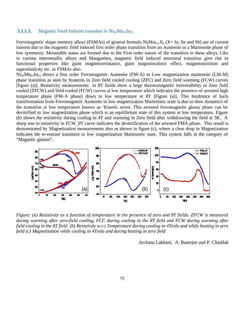
73
3.1.1.5. Magnetic Field Induced transition in Ni50Mn35In15 Ferromagnetic shape memory alloys (FSMAs) of general formula Ni2Mn2-yXy (X= In, Sn and Sb) are of current interest due to the magnetic field induced first order phase transition from an Austenite to a Martensite phase of low symmetry. Metastable states are formed due to the First order nature of the transition in these alloys. Like in various intermetallic alloys and Manganites, magnetic field induced structural transition give rise to functional properties like giant magnetoresistance, giant magnetocaloric effect, magnetostriction and superelasticity etc. in FSMAs also. Ni50Mn35In15 shows a first order Ferromagnetic Austenite (FM-A) to Low magnetization martensite (LM-M) phase transition as seen by hystersis in Zero field cooled cooling (ZFC) and Zero field warming (FCW) curves [figure (a)]. Resistivity measurements in 8T fields show a large thermomagnetic irreversibility in Zero field cooled (ZFCW) and field cooled (FCW) curves at low temperature which indicates the presence of arrested high temperature phase (FM-A phase) down to low temperature in 8T [Figure (a)]. This hindrance of back transformation from Ferromagnetic Austenite to low magnetization Martenistic state is due to slow dynamics of the transition at low temperature known as ‘Kinetic arrest. This arrested ferromagnetic glassy phase can be devitrified to low magnetization phase which is an equilibrium state of this system at low temperature. Figure (b) shows the resistivity during cooling in 4T and warming in Zero field after withdrawing the field at 5K. A sharp rise in resistivity in FCW_0T curve indicates the devitrification of the arrested FMA phase. This result is demonstrated by Magnetization measurements also as shown in figure (c), where a clear drop in Magnetization indicates the re-entrant transition to low magnetization Martensitic state. This system falls in the category of “Magnetic glasses”.
Figure: (a) Resistivity as a function of temperature in the presence of zero and 8T fields. ZFCW is measured during warming after zero-field cooling, FCC during cooling in the 8T field and FCW during warming after field cooling in the 8T field. (b) Resistivity w.r.t Temperature during cooling in 4Tesla and while heating in zero field (c) Magnetization while cooling in 4Tesla and during heating in zero field
Archana Lakhani, A. Banerjee and P. Chaddah
0 50 100 150 200 250 30020
30
40
50
60
70
80
90
FCW
FCC
ZFCW
ZFC
ZFW
H=8T
H=8T
H=0
ρ(µO
hm-c
m)
T(K)
(a)0 50 100 150 200 250 300
40
50
60
70
80
90
100 FCW in 0T FCC in 4T
ρ (µ
Ohm
-cm
)
T(K)
0 50 100 150 200 250 300 350
0
20
40
60
80
100
120 FCC_4T FCW_0T
M (e
mu/
gm)
T(K)
(b) (c)0 50 100 150 200 250 300
20
30
40
50
60
70
80
90
FCW
FCC
ZFCW
ZFC
ZFW
H=8T
H=8T
H=0
ρ(µO
hm-c
m)
T(K)0 50 100 150 200 250 300
40
50
60
70
80
90
100 FCW in 0T FCC in 4T
ρ (µ
Ohm
-cm
)
T(K)
0 50 100 150 200 250 300 350
0
20
40
60
80
100
120 FCC_4T FCW_0T
M (e
mu/
gm)
T(K)
0 50 100 150 200 250 30020
30
40
50
60
70
80
90
FCW
FCC
ZFCW
ZFC
ZFW
H=8T
H=8T
H=0
ρ(µO
hm-c
m)
T(K)0 50 100 150 200 250 300
40
50
60
70
80
90
100 FCW in 0T FCC in 4T
ρ (µ
Ohm
-cm
)
T(K)
0 50 100 150 200 250 300 350
0
20
40
60
80
100
120 FCC_4T FCW_0T
M (e
mu/
gm)
T(K)
0 50 100 150 200 250 30020
30
40
50
60
70
80
90
FCW
FCC
ZFCW
ZFC
ZFW
H=8T
H=8T
H=0
ρ(µO
hm-c
m)
T(K)0 50 100 150 200 250 300
40
50
60
70
80
90
100 FCW in 0T FCC in 4T
ρ (µ
Ohm
-cm
)
T(K)
0 50 100 150 200 250 300 350
0
20
40
60
80
100
120 FCC_4T FCW_0T
M (e
mu/
gm)
T(K)
0 50 100 150 200 250 30020
30
40
50
60
70
80
90
FCW
FCC
ZFCW
ZFC
ZFW
H=8T
H=8T
H=0
ρ(µO
hm-c
m)
T(K)0 50 100 150 200 250 300
40
50
60
70
80
90
100 FCW in 0T FCC in 4T
ρ (µ
Ohm
-cm
)
T(K)
0 50 100 150 200 250 300 350
0
20
40
60
80
100
120 FCC_4T FCW_0T
M (e
mu/
gm)
T(K)
0 50 100 150 200 250 30020
30
40
50
60
70
80
90
FCW
FCC
ZFCW
ZFC
ZFW
H=8T
H=8T
H=0
ρ(µO
hm-c
m)
T(K)0 50 100 150 200 250 300
40
50
60
70
80
90
100 FCW in 0T FCC in 4T
ρ (µ
Ohm
-cm
)
T(K)
0 50 100 150 200 250 300 350
0
20
40
60
80
100
120 FCC_4T FCW_0T
M (e
mu/
gm)
T(K)
0 50 100 150 200 250 30020
30
40
50
60
70
80
90
FCW
FCC
ZFCW
ZFC
ZFW
H=8T
H=8T
H=0
ρ(µO
hm-c
m)
T(K)0 50 100 150 200 250 300
40
50
60
70
80
90
100 FCW in 0T FCC in 4T
ρ (µ
Ohm
-cm
)
T(K)
0 50 100 150 200 250 300 350
0
20
40
60
80
100
120 FCC_4T FCW_0T
M (e
mu/
gm)
T(K)
0 50 100 150 200 250 30020
30
40
50
60
70
80
90
FCW
FCC
ZFCW
ZFC
ZFW
H=8T
H=8T
H=0
ρ(µO
hm-c
m)
T(K)0 50 100 150 200 250 300 350
0
20
40
60
80
100
120 FCC_4T FCW_0T
M (e
mu/
gm)
T(K)
0 50 100 150 200 250 30020
30
40
50
60
70
80
90
FCW
FCC
ZFCW
ZFC
ZFW
H=8T
H=8T
H=0
ρ(µO
hm-c
m)
T(K)0 50 100 150 200 250 300
40
50
60
70
80
90
100 FCW in 0T FCC in 4T
ρ (µ
Ohm
-cm
)
T(K)
0 50 100 150 200 250 300 350
0
20
40
60
80
100
120 FCC_4T FCW_0T
M (e
mu/
gm)
T(K)
0 50 100 150 200 250 30020
30
40
50
60
70
80
90
FCW
FCC
ZFCW
ZFC
ZFW
H=8T
H=8T
H=0
ρ(µO
hm-c
m)
T(K)0 50 100 150 200 250 300
40
50
60
70
80
90
100 FCW in 0T FCC in 4T
ρ (µ
Ohm
-cm
)
T(K)
0 50 100 150 200 250 300 350
0
20
40
60
80
100
120 FCC_4T FCW_0T
M (e
mu/
gm)
T(K)
0 50 100 150 200 250 30020
30
40
50
60
70
80
90
FCW
FCC
ZFCW
ZFC
ZFW
H=8T
H=8T
H=0
ρ(µO
hm-c
m)
T(K)0 50 100 150 200 250 300
40
50
60
70
80
90
100 FCW in 0T FCC in 4T
ρ (µ
Ohm
-cm
)
T(K)
0 50 100 150 200 250 300 350
0
20
40
60
80
100
120 FCC_4T FCW_0T
M (e
mu/
gm)
T(K)
0 50 100 150 200 250 30020
30
40
50
60
70
80
90
FCW
FCC
ZFCW
ZFC
ZFW
H=8T
H=8T
H=0
ρ(µO
hm-c
m)
T(K)0 50 100 150 200 250 300
40
50
60
70
80
90
100 FCW in 0T FCC in 4T
ρ (µ
Ohm
-cm
)
T(K)
0 50 100 150 200 250 30020
30
40
50
60
70
80
90
FCW
FCC
ZFCW
ZFC
ZFW
H=8T
H=8T
H=0
ρ(µO
hm-c
m)
T(K)
0 50 100 150 200 250 30020
30
40
50
60
70
80
90
FCW
FCC
ZFCW
ZFC
ZFW
H=8T
H=8T
H=0
ρ(µO
hm-c
m)
T(K)

74
3.1.1.6. Study of Pd doped FeRh under high pressure
The first order transition in Pd doped FeRh has been studied under high pressure. The transition temperature increases with the application of external pressure. It is consistent with length contraction across the first order ferromagnetic to antiferromagnetic transition with lowering temperature. The transition temperature is shifted above 300K for pressure higher than 9 kbar. Whereas, the application of magnetic field favors ferromagnetic state and transition is shifted to lower temperature with applied magnetic field. Following figure shows the resistivity variation under various constant magnetic field for 9 kbar applied pressure.
R. Rawat, Pallavi Kushwaha, Pallab Bag and Sachin Kumar
3.1.1.7. Study of magnetic transition in RTiSi (R = Heavy rare earth) system
The magnetic transition in RTiSi (R = Heavy rare earth) system has been studied by magnetoresistance and heat capacity measurements. These systems are known to order antiferromagnetically and have one of the largest magnetic ordering temperatures of rare earth sub-lattice in ternary rare earth compounds. Our isothermal magnetoresistance study indicates magnetic field induced metamagnetic transition below magnetic ordering temperature for Ho and Dy compounds. Magnetic entropy change has been estimated from heat capacity data which suggest strong crystal field effect. Some of these results are presented at DAE Solid State Physics Symposium 2010 held at Manipal Institute of Technology, Manipal.
Pallab Bag, Pallavi Kushwaha and R. Rawat
3.1.1.8. Study of magnetoresistance behavior of doped Mn2Sb
Magnetoresistance behavior is studied in Mn2Sb with a new dopant Ga at Mn site. Resistivity shows a first order magnetic transition even though the lattice parameter is found to remain unchanged with substitution. The transition temperature, obtained from resistivity vs temperature curve under various constant magnetic field, remains more or less constant with varying magnetic field. However resistivity for high field shows larger high temperature phase at 5K. Magnetoresistance shows thermomagnetic irreversibility and depends on the history of the sample. These results are explained by considering coexisting metastable high temperature and low temperature phase.
Pallavi Kushwaha, Pallab Bag, R. Rawat and P Chaddah; M. Gurram (IIT, Gauhati)
0 100 200 3000
10
20
30
9 kbar
0 kOe 48 kOe 80 kOe
Pd doped FeRh
Res
ista
nce
(Ω)
Temperature (K)

75
3.1.1.9. Interparticle interaction and crossover in critical lines on field-temperature plane in Pr0.5Sr0.5MnO3 nanoparticles.
The magnetic properties and the effects of interparticle interaction on them have been studied in nanoparticles of half-doped Pr0.5Sr0.5MnO3. Three samples consisting of nanoparticles of different average particle sizes are synthesized to render the variation in interparticle interaction. Though all the samples crystallize in the same structure to that of their bulk compound, the low-temperature ferromagnetic-antiferromagnetic transition, which is present in bulk compound, is not evident in the nanoparticles (Fig.-1)
0 50 100 150 200 250 300 3500.0
0.5
1.0
1.5
2.0
2.5
N650
N600
N700ZFC
FCW
M (1
03 em
u m
ole-1
)
T (K)
H = 100 Oe
Fig. - 1 Fig.-2
Linear as well as nonlinear ac susceptibility coupled with dc magnetic measurements have shown the superparamagnetic behavior of these nanoparticles where the blocking temperature increases with the increasing particle size. Presence of interparticle interaction is confirmed from the temperature variation in coercive field and the analysis of frequency dependent ac susceptibility. We have identified the nature of this interaction to be of dipolar type and show that its strength decreases with the increasing particle size. The effect of this dipolar interaction on magnetic properties is intriguing as the compounds exhibit crossover from de Almeida-Thouless- to Gabay-Toulouse type critical lines on field-temperature plane above their respective interaction field (Fig. 2). In agreement with theoretical prediction, we infer that this crossover is induced by the unidirectional anisotropy arising from interparticle interaction, and this is confirmed from the presence of exchange bias phenomenon.
A. K. Pramanik and A. Banerjee
3.1.1.10. Effect of simultaneous application of magnetic field and pressure on magnetic transitions in La0.5Ca0.5MnO3
Simultaneous effect of magnetic field and hydrostatic pressure on temperature dependence of magnetization of half-doped LCMO is studied here. No significant effect on the second-order PM to FM transition could be observed in the pressure range of <10 kbar, however, the lower temperature first-order FM-AFM transition is seriously affected, especially at the higher fields. The temperatures related to supercooling (T*) and superheating (T**) decrease and the magnetization value at 5 K increases with the increase in pressure for all fields. However, decrease in T* and increase in M(5 K) show sharp changes at the lowest nonzero pressure, sharpness of which increase with the field (Fig.-1).
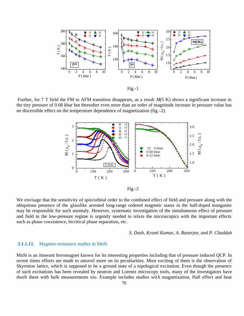
76
0 2 4 6 8 10140
160
180
200
0 2 4 6 8 10
120
140
160
0 2 4 6 8 10
1.0
1.5
2.0
2.5
3.0
P ( kbar ) P ( kbar )
T**
T (
K )
1T 3T 2T 4T
1T 3T 2T 4T
T*
T (
K )
1T 3T 2T 4T
P ( kbar )
M (
µB /
f.u.
) M(5K)
Fig.-1
Further, for 7 T field the FM to AFM transition disappears, as a result M(5 K) shows a significant increase in the tiny pressure of 0.68 kbar but thereafter even more than an order of magnitude increase in pressure value has no discernible effect on the temperature dependence of magnetization (fig.-2).
0 100 200 3000
1
2
3
0 100 200 300
1.0
1.5
2.0
2.5
3.0
0 kbar
M (
µB /
f.u. )
T ( K )
1T 3T 4T 5T 7T
M (
µB /
f.u. )
T ( K )
0 kbar 0.68 kbar 9.12 kbar
Fig.-2
We envisage that the sensitivity of spin/orbital order to the combined effect of field and pressure along with the ubiquitous presence of the glasslike arrested long-range ordered magnetic states in the half-doped manganite may be responsible for such anomaly. However, systematic investigation of the simultaneous effect of pressure and field in the low-pressure regime is urgently needed to rela te the microscopics with the important effects such as phase coexistence, bicritical phase separation, etc.
S. Dash, Kranti Kumar, A. Banerjee, and P. Chaddah
3.1.1.11. Magneto-resistance studies in MnSi
MnSi is an itinerant ferromagnet known for its interesting properties including that of pressure induced QCP. In recent times efforts are made to unravel more on its peculiarities. Most exciting of them is the observation of Skyrmion lattice, which is supposed to be a ground state of a topological excitation. Even though the presence of such excitations has been revealed by neutron and Lorentz microcopy tools, many of the investigators have dwelt them with bulk measurements too. Example includes studies with magnetization, Hall effect and heat
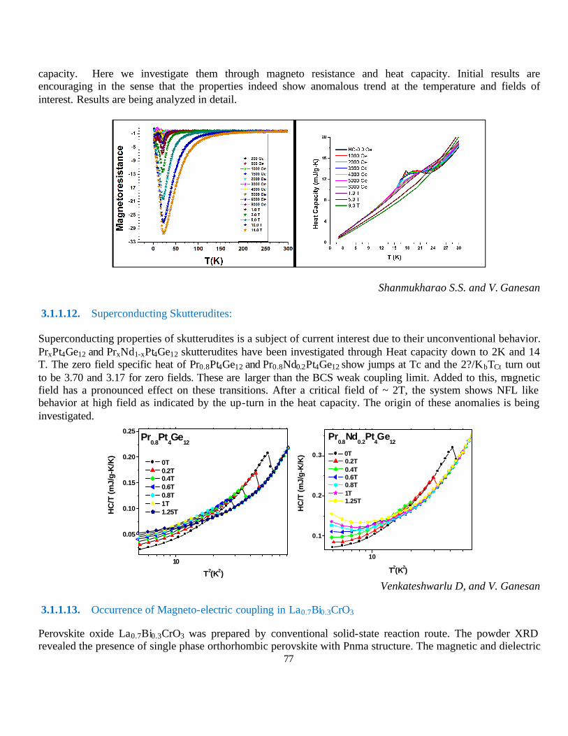
77
capacity. Here we investigate them through magneto resistance and heat capacity. Initial results are encouraging in the sense that the properties indeed show anomalous trend at the temperature and fields of interest. Results are being analyzed in detail.
Shanmukharao S.S. and V. Ganesan
3.1.1.12. Superconducting Skutterudites: Superconducting properties of skutterudites is a subject of current interest due to their unconventional behavior. PrxPt4Ge12 and PrxNd1-xPt4Ge12 skutterudites have been investigated through Heat capacity down to 2K and 14 T. The zero field specific heat of Pr0.8Pt4Ge12 and Pr0.8Nd0.2Pt4Ge12 show jumps at Tc and the 2?/KbTCt turn out to be 3.70 and 3.17 for zero fields. These are larger than the BCS weak coupling limit. Added to this, magnetic field has a pronounced effect on these transitions. After a critical field of ~ 2T, the system shows NFL like behavior at high field as indicated by the up-turn in the heat capacity. The origin of these anomalies is being investigated.
10
0.05
0.10
0.15
0.20
0.25
Pr0.8
Pt4Ge
12
HC
/T (m
J/g-
K/K
)
T2(K2)
0T 0.2T 0.4T 0.6T 0.8T 1T 1.25T
10
0.1
0.2
0.3
Pr0.8
Nd0.2
Pt4Ge
12
HC
/T (m
J/g-
K/K
)
T2(K2)
0T 0.2T 0.4T 0.6T 0.8T 1T 1.25T
Venkateshwarlu D, and V. Ganesan
3.1.1.13. Occurrence of Magneto-electric coupling in La0.7Bi0.3CrO3
Perovskite oxide La0.7Bi0.3CrO3 was prepared by conventional solid-state reaction route. The powder XRD revealed the presence of single phase orthorhombic perovskite with Pnma structure. The magnetic and dielectric

78
studies were carried out at low temperatures using MPMS-7 SQUID magnetometer and Hioki-LCR impedance analyzer respectively. The following Fig.(a) shows 100Oe field-cool (FC) magnetization vs temperature (M-T) data from 10K-300K. It can be seen that with decreasing temperature the magnetization increases at around 230K and then it saturates. The undoped LaCrO3 is known to be antiferromagnetic. The observed M-T data indicate that La0.7Bi0.3CrO3 under goes a paramagnetic to a canted antiferromagnetic (CAF) type spin ordering at 230K. The occurrence of finite magnetization in La0.7Bi0.3CrO3 can result from several possibilities.
The ferroelectric nature of La0.7Bi0.3CrO3 even in a centrosymmetric structure non-zero magnetic moment may appear due to the presence of anisotropy introduced by lone pair electron of doped Bi. The Reitveld refinement results clearly indicate anisotropic structural distortion as a function of Bi doping. Such distortions give possibility of non-zero ferromagnetic moment arising due to Dzyaloshinsky–Moriya (D–M) interaction. The D–M interaction arises due to asymmetric exchange interaction between the moments. The D-M interaction causes canting of the moments, giving rise to improper cancellation of moments in an otherwise antiferromagnetically ordered system. Fig.(b) represents the e'-T curves from 150K-275K at frequencies 1KHz, 10KHz , 100KHz and 1MHz. From fig.(b) it can be seen that e'-T variation shows a pronounced frequency dispersion , The present observation shows that La0.7Bi0.3CrO3 under goes a relaxor ferroelectric transition. We observe an interesting feature i.e a sudden jump in the e'-T data at 230K without any frequency dispersion. The jump in e'-T coincides with the antiferromagnetic transition temperature TN. The coincidence of jump in e'-T variation and TN clearly indicate the presence of magneto-electric (ME) coupling in La0.7Bi0.3CrO3
Aga Shahee, Dhirendra kumar, N. P. Lalla
3.1.1.14. Study of GaFeO3 based magneto-electric materials
GaFeO3 (GFO) that exhibits piezoelectricity and ferrimagnetism is considered to be a very promising multiferroic material for the following reasons: (i) its TC is of about 200 K and may be increased to values close to the room temperature by increasing the Fe content (x) of Ga2-xFexO3 (TC ~ 350 K for x=1.4), controlling the site disorder between Ga and Fe with different preparation methods etc., (ii) it is relatively easy to prepare single phase GFO samples with the conventional solid state route and (iii) is considered to be environmental and social friendly when compared to most of the other lead and bismuth based magnetoelectric materials. Temperature dependent 57Fe Mossbauer spectroscopy (5K–723K) measurements are carried out on polycrystalline multiferroic GaFeO3. The observed Lamb-Mossbauer factor as a function of temperature f(T), which is proportional to integral over the first Brillouin zone of the phonon spectrum, shows a unequivocal variation at magnetic Curie temperature (TC). The observations are discussed in terms of spin-phonon coupling and multiferroic nature. The observed variation of hyperfine field (T<TC) match with the bulk magnetization data. Critical exponent (β) is estimated as 0.38 ± 0.02 from the data.
Kavita Sharma, V.Raghavendra Reddy and Ajay Gupta
Fig.: Temperature variations of (a) magnetization and (b) e' of La0.7Bi0.3CrO3
150 175 200 225 250150
200
250
230K
(b)
ε'
Temp. (K)
1KHz 10KHz 100KHz 1MHz
0 50 100 150 200 250 300
0.00
0.05
0.10
0.15
0.20(a)
Field cooled
M (e
mu/
gm)
Temp. (K)
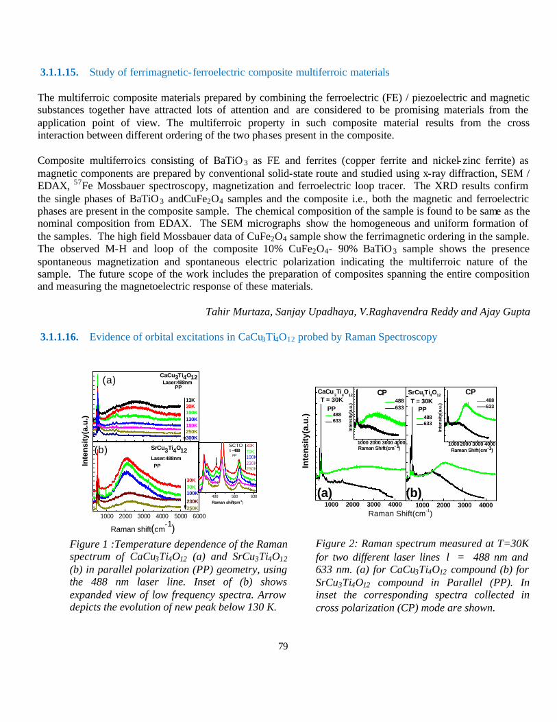
79
3.1.1.15. Study of ferrimagnetic- ferroelectric composite multiferroic materials
The multiferroic composite materials prepared by combining the ferroelectric (FE) / piezoelectric and magnetic substances together have attracted lots of attention and are considered to be promising materials from the application point of view. The multiferroic property in such composite material results from the cross interaction between different ordering of the two phases present in the composite.
Composite multiferroics consisting of BaTiO 3 as FE and ferrites (copper ferrite and nickel-zinc ferrite) as magnetic components are prepared by conventional solid-state route and studied using x-ray diffraction, SEM / EDAX, 57Fe Mossbauer spectroscopy, magnetization and ferroelectric loop tracer. The XRD results confirm the single phases of BaTiO 3 andCuFe2O4 samples and the composite i.e., both the magnetic and ferroelectric phases are present in the composite sample. The chemical composition of the sample is found to be same as the nominal composition from EDAX. The SEM micrographs show the homogeneous and uniform formation of the samples. The high field Mossbauer data of CuFe2O4 sample show the ferrimagnetic ordering in the sample. The observed M-H and loop of the composite 10% CuFe2O4- 90% BaTiO3 sample shows the presence spontaneous magnetization and spontaneous electric polarization indicating the multiferroic nature of the sample. The future scope of the work includes the preparation of composites spanning the entire composition and measuring the magnetoelectric response of these materials.
Tahir Murtaza, Sanjay Upadhaya, V.Raghavendra Reddy and Ajay Gupta
3.1.1.16. Evidence of orbital excitations in CaCu3Ti4O12 probed by Raman Spectroscopy
1000 2000 3000 4000 5000 6000
CaCu3Ti4O12
13K30K100K130K180K250K300K
Raman shift(cm-1)
Laser:488nmPP
490 560 630
30K70K100K230K250K
SCTO
Raman shift(cm-1)
λ=488PP
(b)
(a)
30K70K100K
230K250K
SrCu3Ti4O12Laser:488nm PPIn
ten
sity
(a.u
.)
1000 2000 3000 4000
10002000 3000 40001000 2000 3000 4000
1000 2000 3000 4000
Inte
nsity
(a.u
.)
(a)
CaCu3Ti
4O
12T = 30K
PP
Raman Shift (cm-1)
488 633
CP
Raman Shift(cm-1)
488 633
488 633
488 633
CP
Raman Shift(cm-1)
PP
Inte
nsity
(a.u
.)T = 30KSrCu3Ti4O12
Inte
nsi
ty(a
.u.)
(b)
Figure 1 :Temperature dependence of the Raman spectrum of CaCu3Ti4O12 (a) and SrCu3Ti4O12 (b) in parallel polarization (PP) geometry, using the 488 nm laser line. Inset of (b) shows expanded view of low frequency spectra. Arrow depicts the evolution of new peak below 130 K.
Figure 2: Raman spectrum measured at T=30K for two different laser lines λ = 488 nm and 633 nm. (a) for CaCu3Ti4O12 compound (b) for SrCu3Ti4O12 compound in Parallel (PP). In inset the corresponding spectra collected in cross polarization (CP) mode are shown.
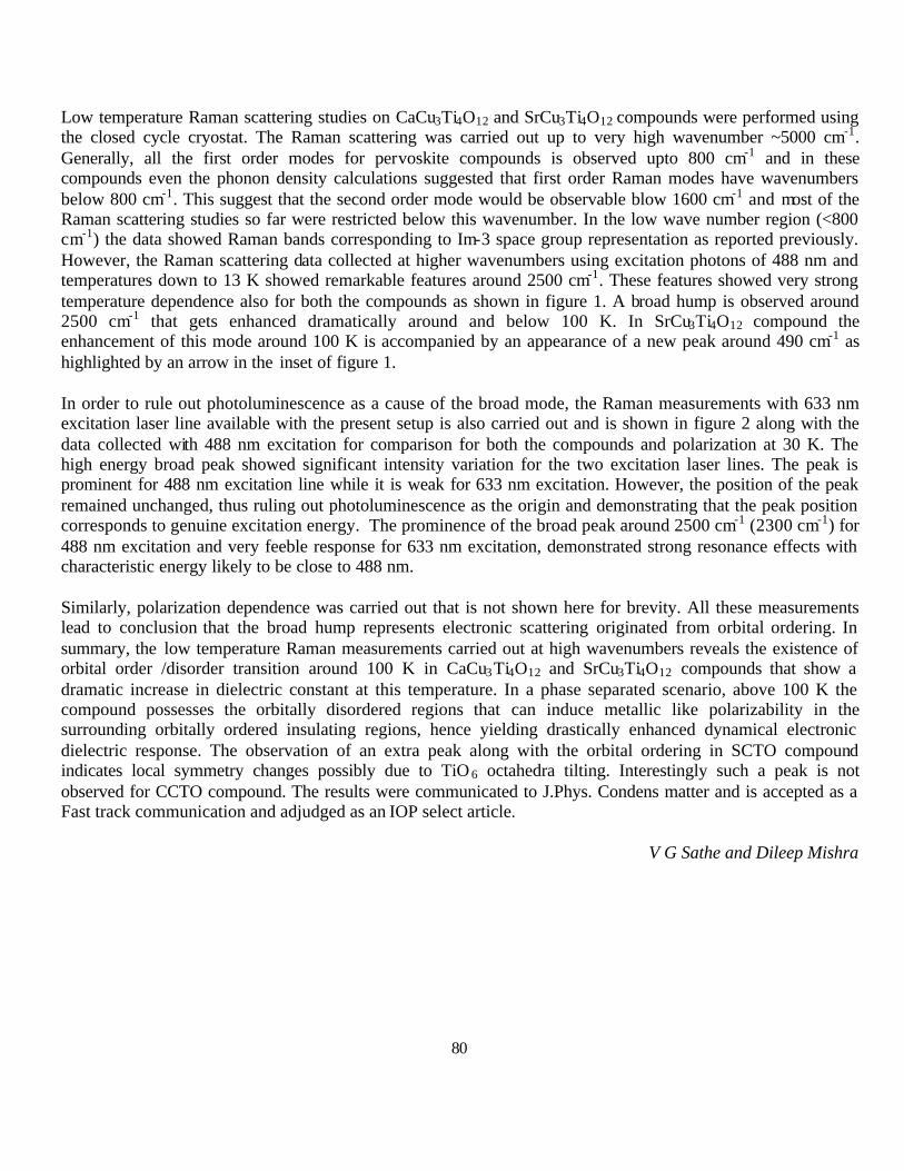
80
Low temperature Raman scattering studies on CaCu3Ti4O12 and SrCu3Ti4O12 compounds were performed using the closed cycle cryostat. The Raman scattering was carried out up to very high wavenumber ~5000 cm-1. Generally, all the first order modes for pervoskite compounds is observed upto 800 cm-1 and in these compounds even the phonon density calculations suggested that first order Raman modes have wavenumbers below 800 cm-1. This suggest that the second order mode would be observable blow 1600 cm-1 and most of the Raman scattering studies so far were restricted below this wavenumber. In the low wave number region (<800 cm-1) the data showed Raman bands corresponding to Im-3 space group representation as reported previously. However, the Raman scattering data collected at higher wavenumbers using excitation photons of 488 nm and temperatures down to 13 K showed remarkable features around 2500 cm-1. These features showed very strong temperature dependence also for both the compounds as shown in figure 1. A broad hump is observed around 2500 cm-1 that gets enhanced dramatically around and below 100 K. In SrCu3Ti4O12 compound the enhancement of this mode around 100 K is accompanied by an appearance of a new peak around 490 cm-1 as highlighted by an arrow in the inset of figure 1.
In order to rule out photoluminescence as a cause of the broad mode, the Raman measurements with 633 nm excitation laser line available with the present setup is also carried out and is shown in figure 2 along with the data collected with 488 nm excitation for comparison for both the compounds and polarization at 30 K. The high energy broad peak showed significant intensity variation for the two excitation laser lines. The peak is prominent for 488 nm excitation line while it is weak for 633 nm excitation. However, the position of the peak remained unchanged, thus ruling out photoluminescence as the origin and demonstrating that the peak position corresponds to genuine excitation energy. The prominence of the broad peak around 2500 cm-1 (2300 cm-1) for 488 nm excitation and very feeble response for 633 nm excitation, demonstrated strong resonance effects with characteristic energy likely to be close to 488 nm.
Similarly, polarization dependence was carried out that is not shown here for brevity. All these measurements lead to conclusion that the broad hump represents electronic scattering originated from orbital ordering. In summary, the low temperature Raman measurements carried out at high wavenumbers reveals the existence of orbital order /disorder transition around 100 K in CaCu3Ti4O12 and SrCu3Ti4O12 compounds that show a dramatic increase in dielectric constant at this temperature. In a phase separated scenario, above 100 K the compound possesses the orbitally disordered regions that can induce metallic like polarizability in the surrounding orbitally ordered insulating regions, hence yielding drastically enhanced dynamical electronic dielectric response. The observation of an extra peak along with the orbital ordering in SCTO compound indicates local symmetry changes possibly due to TiO 6 octahedra tilting. Interestingly such a peak is not observed for CCTO compound. The results were communicated to J.Phys. Condens matter and is accepted as a Fast track communication and adjudged as an IOP select article.
V G Sathe and Dileep Mishra
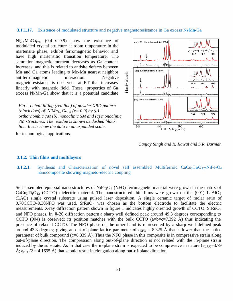
81
3.1.1.17. Existence of modulated structure and negative magnetoresistance in Ga excess Ni-Mn-Ga
Ni2-xMnGa1+x (0.4=x=0.9) show the existence of modulated crystal structure at room temperature in the martensite phase, exhibit ferromagnetic behavior and have high martensitic transition temperature. The saturation magnetic moment decreases as Ga content increases, and this is related to antisite defects between Mn and Ga atoms leading to Mn-Mn nearest neighbor antiferromagnetic interaction. Negative magnetoresistance is observed at RT that increases linearly with magnetic field. These properties of Ga excess Ni-Mn-Ga show that it is a potential candidate
for technological applications.
Sanjay Singh and R. Rawat and S.R. Barman
3.1.2. Thin films and multilayers
3.1.2.1. Synthesis and Characterization of novel self assembled Multiferroic CaCu3Ti4O12-NiFe2O4 nanocomposite showing magneto-electric coupling
Self assembled epitaxial nano structures of NiFe2O4 (NFO) ferrimagnetic material were grown in the matrix of CaCu3Ti4O12 (CCTO) dielectric material. The nanostructured thin films were grown on the (001) LaAlO 3 (LAO) single crystal substrate using pulsed laser deposition. A single ceramic target of molar ratio of 0.70CCTO-0.30NFO was used. SrRuO3 was chosen as the bottom electrode to facilitate the electric measurements. X-ray diffraction pattern shown in figure 1 indicates highly oriented growth of CCTO, SrRuO3 and NFO phases. In θ-2θ diffraction pattern a sharp well defined peak around 49.3 degrees corresponding to CCTO (004) is observed; its position matches with the bulk CCTO (a=b=c=7.392 Å) thus indicating the presence of relaxed CCTO. The NFO phase on the other hand is represented by a sharp well defined peak around 43.3 degrees; giving an out-of-plane lattice parameter of cNFO = 8.325 Å that is lower than the lattice parameter of bulk compound (c=8.339 Å). Thus the NFO phase in this composite is in compressive strain along out-of-plane direction. The compression along out-of-plane direction is not related with the in-plane strain induced by the substrate. As in that case the in-plane strain is expected to be compressive in nature (aLAO=3.79 Å; aNFO/2 = 4.1695 Å) that should result in elongation along out-of-plane direction.
Fig.: Lebail fitting (red line) of powder XRD pattern (black dots) of NiMn1.1Ga1.9 (x= 0.9) by (a) orthorhombic 7M (b) monoclinic 5M and (c) monoclinic 7M structures. The residue is shown as dashed black line. Insets show the data in an expanded scale.

82
The phase separation and rod like growth of the NFO phase is further confirmed by SEM studies shown in figure 1. In the figure the bright portion represents NFO phase in the matrix of dark portion representing CCTO phase. These observations were confirmed by taking energy dispersive spectroscopy (EDAX) on bright and dark portions. In summary, the self assembled oriented (1-x)CaCu3Ti4O12-(x)NiFe2O4 (x=0.3) nanocomposite film was grown by pulsed laser deposition. The x-ray diffraction and Atomic force microscopy shows separation of two phases and presence of vertical strain. The vertical strain switches the NiFe2O4 from tension to compression state. The composite film shows both dielectric as well ferromagnetic properties. The magnetization measurements showed clear signature of magnetoelectric coupling around 100 K where CaCu3Ti4O12 shows sudden change in dielectric constant. The magnetoelectric coup ling can not be explained by simply invoking elastic strain or mechanical coupling and demands new understanding and consideration of direct coupling mechanism in these types of composite structures.
Anju Ahlawat, V.G. Sathe
3.1.2.2. Surfactant Controlled interdiffusion in Cu/Co multilayers
Control over the roughness and thermal stability is an important issue in thin film multilayers. Intermixing at the interfaces and asymmetry of roughness strongly influence the structural, electrical and magnetic properties of the multilayer. Magnetic multilayers with smooth and symmetric interfaces are always required for practical applications. Cu/Co multilayers are known to exhibit the giant magneto resistance (GMR) effect due to spin dependent scattering at the interfaces. Although many aspects of its magnetic properties have been studied, the investigation of how to obtain smooth interfaces with better performance is still necessary. Due to the difference in the surface
free energy (?) of Cu (?Co= 1.8 J/m2) and Co (?Cu=2.55 J/m2), Co agglomerates over the Cu layer whereas Cu spread and make smooth film on Co. As a result, asymmetric Co-on-Cu and Cu-on-Co interface occurs. One promising way to obtain atomically smooth and
20 30 40 50 60 70 80
1
10
100
1000
10000
Inte
nsity
(arb
.uni
ts)
2θ
CCTO(004)
LAO(002)
SRO(002)
NFO(004)
Figure 1 (a). X-ray diffraction pattern recorded in ? -2? geometry of the CCTO-NFO composite film grown on LAO substrate with a layer of SrRuO3, (b) SEM image of composite film.
FIG 1: X ray reflectivity pattern of samples prepared without surfactant (a) and with Ag surfactant (b) in as deposited and annealed state. The inset compares the normalized reflectivities after multiplying with qz
4.
0.08 0.12 0.16 0.2010
-5
10-3
10-1
10
103
105
with surfactant
(b)(a)
X-ra
y R
efle
ctivi
ty
qz (Å-1)
no surfactant
0.12 0.13
673 K
573 K473 K
as-deposited
673 K573 K
473 K
As-depo. 473 K 573 K 673 K
R·q
4 z
as-deposited
0.08 0.12 0.16 0.2010
- 5
10- 3
10- 1
10
103
105
0.12 0.13
As-depo. 473 K 573 K 673 K
R·q
z4
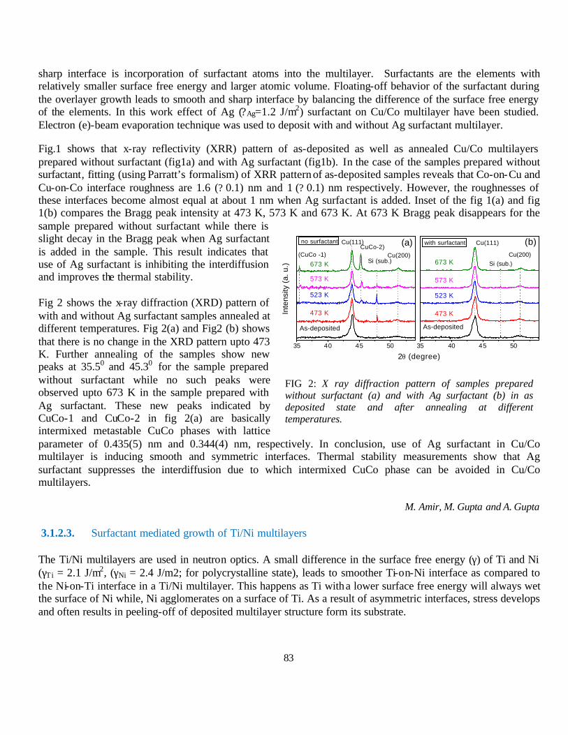
83
573 K
Cu(111)
Si (sub.)Cu(200)
no surfactant (a)CuCo-2)Cu(111)
673 K
As-deposited
473 K
523 K
573 K
with surfactant
Si (sub.)Cu(200)
(b)
673 K
(CuCo -1)
Inte
nsity
(a. u
.)523 K
473 K
35 40 45 50
2θ (degree)
35 40 45 50
As-deposited
sharp interface is incorporation of surfactant atoms into the multilayer. Surfactants are the elements with relatively smaller surface free energy and larger atomic volume. Floating-off behavior of the surfactant during the overlayer growth leads to smooth and sharp interface by balancing the difference of the surface free energy of the elements. In this work effect of Ag (?Ag=1.2 J/m2) surfactant on Cu/Co multilayer have been studied. Electron (e)-beam evaporation technique was used to deposit with and without Ag surfactant multilayer.
Fig.1 shows that x-ray reflectivity (XRR) pattern of as-deposited as well as annealed Cu/Co multilayers prepared without surfactant (fig1a) and with Ag surfactant (fig1b). In the case of the samples prepared without surfactant, fitting (using Parratt’s formalism) of XRR pattern of as-deposited samples reveals that Co-on-Cu and Cu-on-Co interface roughness are 1.6 (? 0.1) nm and 1 (? 0.1) nm respectively. However, the roughnesses of these interfaces become almost equal at about 1 nm when Ag surfactant is added. Inset of the fig 1(a) and fig 1(b) compares the Bragg peak intensity at 473 K, 573 K and 673 K. At 673 K Bragg peak disappears for the sample prepared without surfactant while there is slight decay in the Bragg peak when Ag surfactant is added in the sample. This result indicates that use of Ag surfactant is inhibiting the interdiffusion and improves the thermal stability.
Fig 2 shows the x-ray diffraction (XRD) pattern of with and without Ag surfactant samples annealed at different temperatures. Fig 2(a) and Fig2 (b) shows that there is no change in the XRD pattern upto 473 K. Further annealing of the samples show new peaks at 35.50 and 45.30 for the sample prepared without surfactant while no such peaks were observed upto 673 K in the sample prepared with Ag surfactant. These new peaks indicated by CuCo-1 and CuCo-2 in fig 2(a) are basically intermixed metastable CuCo phases with lattice parameter of 0.435(5) nm and 0.344(4) nm, respectively. In conclusion, use of Ag surfactant in Cu/Co multilayer is inducing smooth and symmetric interfaces. Thermal stability measurements show that Ag surfactant suppresses the interdiffusion due to which intermixed CuCo phase can be avoided in Cu/Co multilayers.
M. Amir, M. Gupta and A. Gupta
3.1.2.3. Surfactant mediated growth of Ti/Ni multilayers
The Ti/Ni multilayers are used in neutron optics. A small difference in the surface free energy (γ) of Ti and Ni (γTi = 2.1 J/m2, (γNi = 2.4 J/m2; for polycrystalline state), leads to smoother Ti-on-Ni interface as compared to the Ni-on-Ti interface in a Ti/Ni multilayer. This happens as Ti with a lower surface free energy will always wet the surface of Ni while, Ni agglomerates on a surface of Ti. As a result of asymmetric interfaces, stress develops and often results in peeling-off of deposited multilayer structure form its substrate.
FIG 2: X ray diffraction pattern of samples prepared without surfactant (a) and with Ag surfactant (b) in as deposited state and after annealing at different temperatures.
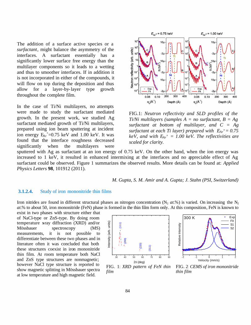
84
The addition of a surface active species or a surfactant, might balance the asymmetry of the interfaces. A surfactant essentially has a significantly lower surface free energy than the multilayer components so it leads to a wetting and thus to smoother interfaces. If in addition it is not incorporated in either of the compounds, it will flow on top during the deposition and thus allow for a layer-by- layer type growth throughout the complete film.
In the case of Ti/Ni multilayers, no attempts were made to study the surfactant mediated growth. In the present work, we studied Ag surfactant mediated growth of Ti/Ni multilayers, prepared using ion beam sputtering at incident ion energy EAr
+=0.75 keV and 1.00 keV. It was found that the interface roughness decreased significantly when the multilayers were sputtered with Ag as surfactant at an ion energy of 0.75 keV. On the other hand, when the ion energy was increased to 1 keV, it resulted in enhanced intermixing at the interfaces and no appreciable effect of Ag surfactant could be observed. Figure 1 summarizes the observed results. More details can be found at: Applied Physics Letters 98, 101912 (2011).
M. Gupta, S. M. Amir and A. Gupta; J. Stahn (PSI, Switzerland)
3.1.2.4. Study of iron mononitride thin films
Iron nitrides are found in different structural phases as nitrogen concentration (N2 at.%) is varied. On increasing the N2 at.% to about 50, iron mononitride (FeN) phase is formed in the thin film form only. At this composition, FeN is known to exist in two phases with structure either that of NaCl-type or ZnS-type. By doing room temperature x-ray diffraction (XRD) and/or Mössbauer spectroscopy (MS) measurements, it is not possible to differentiate between these two phases and in literature often it was concluded that both these structures coexist in iron mononitride thin film. At room temperature both NaCl and ZnS type structures are nonmagnetic; however NaCl type structure is reported to show magnetic splitting in Mössbauer spectra at low temperature and high magnetic field.
FIG.1: Neutron reflectivity and SLD profiles of the Ti/Ni multilayers (samples A = no surfactant, B = Ag surfactant at bottom of multilayer, and C = Ag surfactant at each Ti layer) prepared with EAr+= 0.75 keV, and with EAr+ = 1.00 keV. The reflectivities are scaled for clarity.
FIG. 1: XRD pattern of FeN thin film
FIG. 2: CEMS of iron mononitride thin film
30 40 50 60 70 80 90
(311
)
(220
)(200
)
(111
)
Inte
nsity
(ar
b. u
nits
)
2θ (deg)
-2 -1 0 1 2Velocity (mm/s)
R
elat
ive
Inte
nsity
Exp Fit S1 S2
300 K
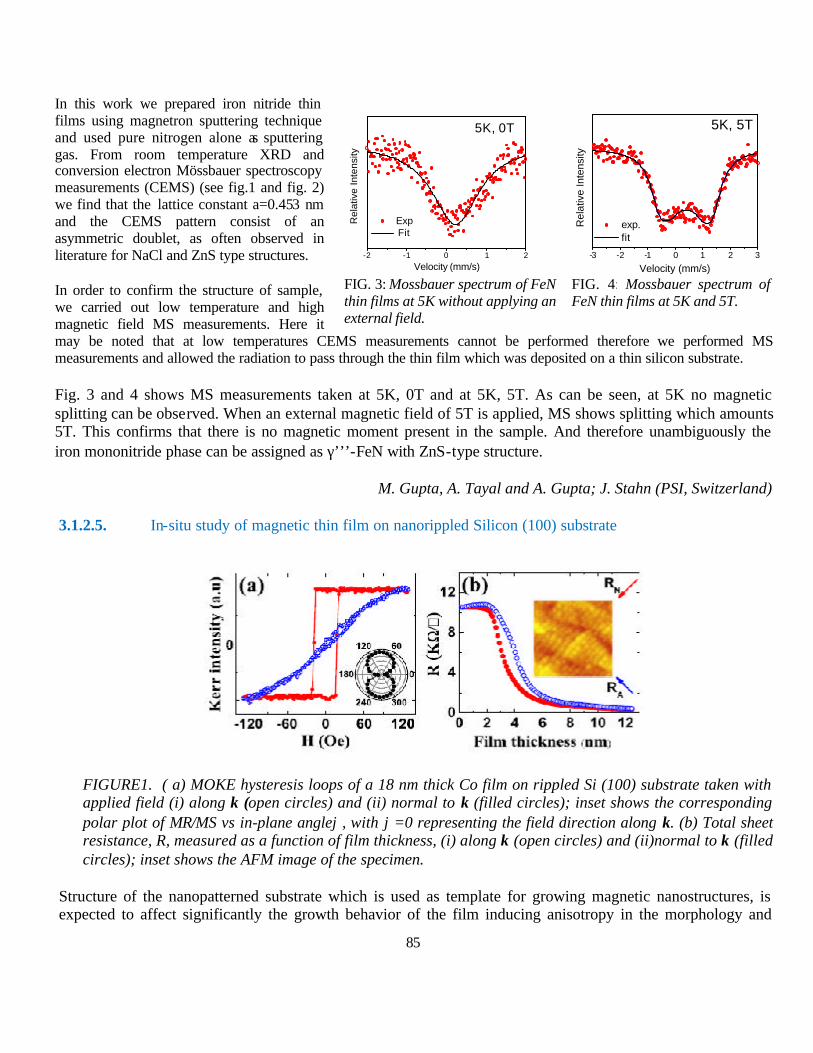
85
In this work we prepared iron nitride thin films using magnetron sputtering technique and used pure nitrogen alone as sputtering gas. From room temperature XRD and conversion electron Mössbauer spectroscopy measurements (CEMS) (see fig.1 and fig. 2) we find that the lattice constant a=0.453 nm and the CEMS pattern consist of an asymmetric doublet, as often observed in literature for NaCl and ZnS type structures.
In order to confirm the structure of sample, we carried out low temperature and high magnetic field MS measurements. Here it may be noted that at low temperatures CEMS measurements cannot be performed therefore we performed MS measurements and allowed the radiation to pass through the thin film which was deposited on a thin silicon substrate.
Fig. 3 and 4 shows MS measurements taken at 5K, 0T and at 5K, 5T. As can be seen, at 5K no magnetic splitting can be observed. When an external magnetic field of 5T is applied, MS shows splitting which amounts 5T. This confirms that there is no magnetic moment present in the sample. And therefore unambiguously the iron mononitride phase can be assigned as γ’’’-FeN with ZnS-type structure.
M. Gupta, A. Tayal and A. Gupta; J. Stahn (PSI, Switzerland)
3.1.2.5. In-situ study of magnetic thin film on nanorippled Silicon (100) substrate
FIGURE1. ( a) MOKE hysteresis loops of a 18 nm thick Co film on rippled Si (100) substrate taken with applied field (i) along k (open circles) and (ii) normal to k (filled circles); inset shows the corresponding polar plot of MR/MS vs in-plane angleϕ, with ϕ=0 representing the field direction along k. (b) Total sheet resistance, R, measured as a function of film thickness, (i) along k (open circles) and (ii)normal to k (filled circles); inset shows the AFM image of the specimen.
Structure of the nanopatterned substrate which is used as template for growing magnetic nanostructures, is expected to affect significantly the growth behavior of the film inducing anisotropy in the morphology and
FIG. 3: Mossbauer spectrum of FeN thin films at 5K without applying an external field.
FIG. 4: Mossbauer spectrum of FeN thin films at 5K and 5T.
-2 -1 0 1 2
5K, 0T
Velocity (mm/s)
Rel
ativ
e In
tens
ity
Exp Fit
-3 -2 -1 0 1 2 3
Rel
ativ
e In
tens
ity
Velocity (mm/s)
exp. fit
5K, 5T

86
magnetic properties. Therefore, in the present work growth behavior of ultrathin Co film deposited on nanorippled Si substrates has been studied in order to elucidate the anisotropy in the growth process and the subsequent anisotropy in the film morphology. Magnetic and transport measurements using in-situ MOKE and four probe resistivity method have been done simultaneously with an aim to correlate morphology and electron transport with magnetic properties.
A clear anisotropy in the growth behavior along and normal (Fig.1b) to the ripples has been observed, resulting in preferential coalescence of islands normal to the ripple wave vector. This results in generation of tensile stress in a direction normal to the ripple wave vector, which coupled with the magnetoelastic and magnetocrystalline energies results in preferent ial orientation of c-axis along the ripple wave vector. Hence origin of the strong magnetic anisotropy (Figure 1a) in the film plane (easy axis perpendicular to the ripple wave vector) is attributed to a possible anisotropy of both nucleation and growth rates, which result in some preferred orientation of the grains. With increasing film thickness, as the contribution of the bulk of the film to the magnetization increases, therefore anisotropy is found to exhibits a monotonous decrease with increasing film thickness.
Sarathlal K. V., Dileep K. Gupta and Ajay Gupta,
3.1.2.6. Structural, electrical and magneto transport properties of pulsed laser deposited Ag doped Fe3O4 thin films.
Ag doped Fe3O4 films used in present work were prepared by pulsed laser deposition (PLD) on single crystal Si(111) substrate from Ag doped Fe2O3 target. In this work, x-ray diffraction, resistivity, magnetoresistance X-ray photoelectron spectroscopy (Xps) and vibration sample magnetometer (VSM) measurements were made to reveal the effect of Ag doping on the structural, electrical and magnetic properties of Ag doped Fe3O4 film. Xps study confirms that silver is present in metallic form. The resistivity measured by four probe method shows that above 250 K, the resistance drops rapidly because of the formation of low resistance path due to silver granules along with SiO 2 inversion layer. Direct tunneling between silver grain and Fe3O4 dominates at low temperature. Magnetization measurements reveal that Ag granules reduce the saturation magnetization of Fe3O4.
D.M.Phase, R.J.Choudhary and Ridhi Master
3.1.2.7. Rectifying and magneto-transport behavior of epitaxial bilayers of TiO 2 / La0.7Sr0.3MnO3 on LaAlO3 and SrTiO3 substrates
Recently, hybrid structure of semiconductor – ferromagnetic (FM) oxide materials have fascinated huge curiosity for their efficacy in ever growing demand for spintronics based devices. The character of interface and strain inculcated in the film due to substrate mismatch play a decisive role in formatting the spin injection from ferromagnetic layer into semiconducting oxide layer. In the present study, we report the structural, electrical and magneto-transport properties of epitaxial bilayer of TiO 2/La0.7Sr0.3MnO3 (LSMO) on c-LaAlO3 (LAO) and c-SrTiO3 (STO) substrates deposited by pulsed laser deposition. In plane epitaxy of the bilayer was confirmed by performing in plane phi scan x-ray diffraction. Four probe resistivity measurements revealed metal- insulator transition temperature (TMI) at ~ 290 K for bilayer deposited on LAO substrate Fig. (a), whereas, bilayer deposited on STO substrate showed TMI higher than room temperature Fig. (b).

87
1E-3
0.01
0.1
1
10
100
1000
0 50 100 150 200 250 300
0.010
0.012
0.014
0.016
0.018
0.020
0.022
TiO
2
ρ (Ω
-cm
)
ρ (Ω
-cm
)
T (K)
Ti0.98
Fe0.02
O2
Ti0.96
Fe0.04
O2
1x10⁻⁵Torr
Fig. Four probe resistivity measurements at different magnetic fields of TiO2/La0.7Sr0.3MnO3 (LSMO) on (a) c-LaAlO3 (LAO) and (b) c-SrTiO3 (STO) substrates.
These bilayers on LAO and STO substrates also reveal low temperature resistivity minima at 67 K and 47 K respectively, which do not vary even on application of 8 Tesla magnetic field. Interestingly the resistivity behavior of these bilayers reveals hysteresis behavior depending on the protocol used for cooling and heating in unequal field, though hysteresis effect being lower for film on LAO than on STO substrate. These bilayers demonstrate a rectifying behavior, consistent with p- type carriers in LSMO and n- type carriers in TiO 2 layers. Strikingly, the rectifying behavior of the bilayers shows a negative magneto-resistance effect on application of magnetic field in the studied temperature range of 5 K to 300 K, in contrast to positive magneto-resistance in previously reported similar bilayer structures.
R. J. Choudhary, Komal Bapna, and D. M. Phase
3.1.2.8. Effect of oxygen partial pressure on the structural and electrical properties of Fe doped and undoped TiO2 films on LaAlO3 substrate
We have studied the influence of oxygen partial pressures (OPP) and Fe doping (2 and 4 at. %) on structural and electrical properties of TiO2 thin films deposited on (001) LaAlO3 substrates using pulsed laser deposition. OPP is varied in the range of 250 mTorr to 1x10⁻⁵ Torr. X-ray photoelectron spectroscopy suggests that Fe is not in metal cluster form. It is found that the evolution of the three phases; anatase, rutile and brookite of TiO 2 as well as the magneli phase (TinO2n-1) strongly depends on the OPP and Fe doping concentration. It turns out that Fe doped film grown at a particular OPP does not follow the structural trend of either the corresponding undoped film or the doped
0 60 120 180 240 300
1000
1200
1400
1600TiO2/LSMO/LAO
Cooling-0Tesla Warming-8Tesla Cooling-8Tesla Warming-4Tesla Cooling-0Tesla Warming-4Tesla
R
(Ω)
T(K)
(a)
0 70 140 210 280
400
600
800
1000
1200
R(Ω
)
T(K)
Cooling-0Tesla Warming-8Tesla Cooling-8Tesla Warming-4Tesla Cooling-0Tesla Warming-4Tesla
TiO2/LSMO/STO
(b)
Fig.: Resistivity dependence on temperature for undoped and doped TiO2 films grown at 1x10 -5 Torr.

88
film but with different Fe concentration. These films reveal different electrical transport properties. Undoped and Fe doped films grown at 250 mTorr show insulating behavior, whereas films grown at 1x10⁻² and 1x10⁻⁴ Torr reveal high temperature metallic state to low temperature semiconducting transition. Interestingly, films deposited at 1x10⁻⁵ Torr reveal charge ordering, which is contributed to the magneli phase of TiO 2. The present study suggests that functionality of TiO 2 thin film based devices can be tuned by properly selecting the OPP and dopant concentration.
R. J. Choudhary, Komal Bapna, and D. M. Phase
3.1.2.9. Magneto-electric behavior of ferrimagnetic BixCo2-xMnO4 (x = 0, 0.1 and 0.3) thin films.
Polycrystalline films of BixCo2-xMnO4 (x = 0, 0.1, and 0.3) with preferred orientation were grown by PLD technique on amorphous quartz, LAO and YBCO buffer layer coated LAO substrates. Single phased growth of the films was confirmed by XRD and Raman spectrum. Dielectric data of the BixCo2-xMnO4 films reveals the weak ferroelectric behavior in the Bi- substituted films. These films are found to possess lower dielectric constant compared to that of the bulk samples and exhibited better ferroelectric nature at higher temperature. DC magnetization studies show that the thin films of BixCo2-xMnO4 deposited on LAO exhibit well defined hysteresis loop at 150 K, which reflects the FM behavior. Both MS and FM-TC increase as Bi-content increases in the films owing to the Bi- induced redistribution of cations among the octahedral and tetrahedral sites. The BixCo2-xMnO4 films with higher Bi-content demonstrated magnetoelectric coupling, through the variation of dielectric constant in response to the applied magnetic field and exhibited maxima just below FM-TC, similar to bulk samples, which confirms the magnetic origin of ferroelectricity. Magnetoelectric coupling present in these films is due to the interplay between structural distortion and magnetic exchange interaction. Finally, the BixCo2-xMnO4 thin films exhibit ferrimagnetic properties along with a weak ferroelectricity, and are suitable for multiferroic device applications.
R. J. Choudhary; N. E. Rajeevan, (Calicut University, Calicut).
3.1.2.10. Evidence of quantum correction to conductivity in strained epitaxial LaNiO 3 films
Swift heavy ion irradiation induced modification on structural, electrical, and magnetotransport properties of epitaxial thin films of LaNiO 3 (LNO) on LaAlO 3 (001) single crystals has been studied. Deposited films were irradiated at varying fluence 1x10¹¹, 1x10¹², and 5x10¹² ions/cm2 using 200 MeV Ag15+ beam. X-ray diffraction results reveal c-axis oriented epitaxial growth of the LNO film which is maintained even up to the highest fluence. All the films, except the one irradiated with highest fluence, show metallic behavior along with a resistivity upturn at lower temperatures. Film irradiated with the highest fluence value exhibits semiconducting behavior in the studied temperature range. Low temperature resistivity of the metallic films has been explained by quantum corrections to conductivity and it is observed that localization increases with the disorder. Weak localization and renormalization of electron–electron interactions are the contributions to the quantum corrections. Presence of weak localization in metallic films is also supported by our magnetotransport data. At high temperatures, variable range hopping shown by the film irradiated with the highest fluence confirms the semiconducting behavior, which may be due to the disorder induced localization of charge carriers.
R. J. Choudhary;Yogesh Kumar and Ravi Kumar (NIT Hamirpur)

89
3.1.2.11. Effect of oxygen partial pressure on the structural and electrical properties of undoped and Fe doped Molybdenum oxide films.
We have studied the effect of oxygen partial pressure (OPP) on the growth properties of pulsed laser deposited undoped and 5% Fe doped MoOx thin films on c-axis Al2O3 substrates. OPP is kept at 350 mTorr, 1x10⁻² Torr and 1x10⁻⁶ Torr. It turns out that OPP and Fe doping strongly control the phase and orientation of growth of Molybdenum oxide films. XRD patterns of undoped samples show that MoO3 phase is formed at 350 m Torr [in 0k0 direction and 1x10⁻⁶ Torr [in hk0 direction], whereas at 10 m Tor the MoO2 phase exists. At 10 mTorr, the growth direction of undoped MoO2 changes from b-axis oriented growth to a-axis oriented growth after Fe doping. While there is phase transformation of MoO3 film grown at 1x10⁻⁶ Torr to a-axis oriented MoO2 after Fe doping. Structure and phases of these films are also confirmed by Raman spectroscopy. Resistivity data show the metallic character of MoO2 phase.
Fig: XRD patterns of (a) MoOx and (b) 5% Fe doped MoOx films grown at different OPP values.
R. J. Choudhary and D. M. Phase;. Shailja Tiwari and B. L. Ahuja, (MLS University, Udaipur)
3.1.2.12. Interface study of Ion beam sputtered Fe/Al interface as a function of annealing temperature
Ion beam sputtered Fe/Al multilayer samples (MLS), with an overall atomic concentration ratio of Fe:Al=3:1, have been studied. Phase formation and microstructural evolution with thermal annealing have been studied by x-ray diffraction, x-ray reflectivity, cross-sectional transmission electron microscopy, MOKE, and dc magnetization. After each stage of in-situ annealing, Grazing incidence x-ray reflectivity (XRR) and x-ray diffraction (XRD) measurements were carried out at synchrotron source using P08, High Resolution XRD beamline, Desy, Hamburg, Germany,.The corresponding TEM measurements were done at TASC laboratory, Trieste, Italy. The reflectivity pattern of the as-deposited multilayer sample clearly shows Bragg peaks up to the third order, which indicates that the deposited multilayer structure is of good quality and the deposited layers are flat and parallel to the substrate. The structural studies show that adjacent Fe and Al layers are partially intermixed during deposition; forming FeAl transition layers a few nanometers thick at the interfaces.
24 32 40 48 56 64 72 80
MoO
3(010
0)
MoO
3(080
)
MoO
3(060
)
MoO
3(040
)
Al 2O
3(006
)
350 m Torr
MoO
2(040
)
MoO
2(020
)
Al 2O
3(006
)
10 m Torr
Inte
nsity
(Arb
. Uni
t)
MoO
3(440
)
Al 2O
3(006
)
MoO
3(220
)
2θο
1x10⁻⁶ Torr
24 32 40 48 56 64 72 80
MoO
2(400
)
Al 2O
3(006
)
MoO
2(200
)
5% Fe Vacuum (1x10⁻⁶ Torr)
MoO
2(400
)
Al 2O
3(006
)
MoO
2(200
)
5 % Fe 10 m Torr
2θο
Inte
nsit
y (A
rb. U
nit) A
l 2O3(0
06)
MoO
3(010
0)
MoO
3(080
)
MoO
3(060
)
MoO
3(040
) 5% Fe 350 m Torr

90
However, at 2000C, a large degree of atomic mixing causes a progressive loss of periodicity due to the formation of disordered intermetallic FeAl layer at the interfaces, which on further annealing at 4000C
transforms to Fe3Al. It has been reported; at higher temperature the mobility of Al atoms is much higher than that of Fe atoms and therefore, it is expected that the highly mobile Al atoms would start diffusing into the relatively immobile lattice of Fe, leading to the formation of a disordered Fe–Al layer at the interfaces. Further, results indicate that after nucleation, the growth of intermetallic FeAl phase mainly occurs through solid-state reaction of Al atoms at the FeAl/a-Fe interface. These results were further supported by TEM measurements. For direct observation and better understanding of the interfacial changes occurring during annealing, comparative XTEM investigations on as-deposited and annealed samples were carried out employing imaging and selected area diffraction (SAD) modes. The layer contrast in the TEM micrograph of as-deposited Fe/Al MLS is mainly due to the difference in atomic number (Z), since Fe has a much larger electron scattering form factor than Al. While the interfaces between Fe and Al are well defined, there is waviness along the interfaces due to intermixing of Al and Fe. However, after annealing considerable mixing has occurred at interfaces in these samples, a layered structure is still present, consisting mostly of intermetallic Fe–Al layers between Al rich regions. The corresponding magnetic measurement shows that magnetization decreases with increase in temperature and Curie temperature (Tc) is found to be 4600C and is much less than that of bulk bcc Fe. The decrease in the magnetization with temperature is due to increase in FeAl coordination, leading to the formation of intermetallic FeAl alloy layer at the interface. The increase of FeAl coordination means a decreasing coordination of its own kind for Fe, which results in two important effects. The ferromagnetic coupling between Fe atoms is weakened due to the reduction of the coordination number of like neighbours as well as increase in inter-atomic distance between them. Secondly, an increase of the Al coordination keeps the anti- ferromagnetic order of FeAl, while gradually quenching the moment of Fe. Therefore, the observed magnetic behaviour is mainly attributed to the formation of nonmagnetic FeAl phase and increase in anti- ferromagnetic interlayer coupling at the interface.
Shreeja Pillai, T. Shripathi; Ranjeet Brajpuriya (Amity University, Haryana)
Fig. 1 XRD patterns of Fe/Al MLS as a function of annealing temperature.
Fig. 2 Micrograph image of as-deposited and Fe/Al MLS annealed at 4000C.
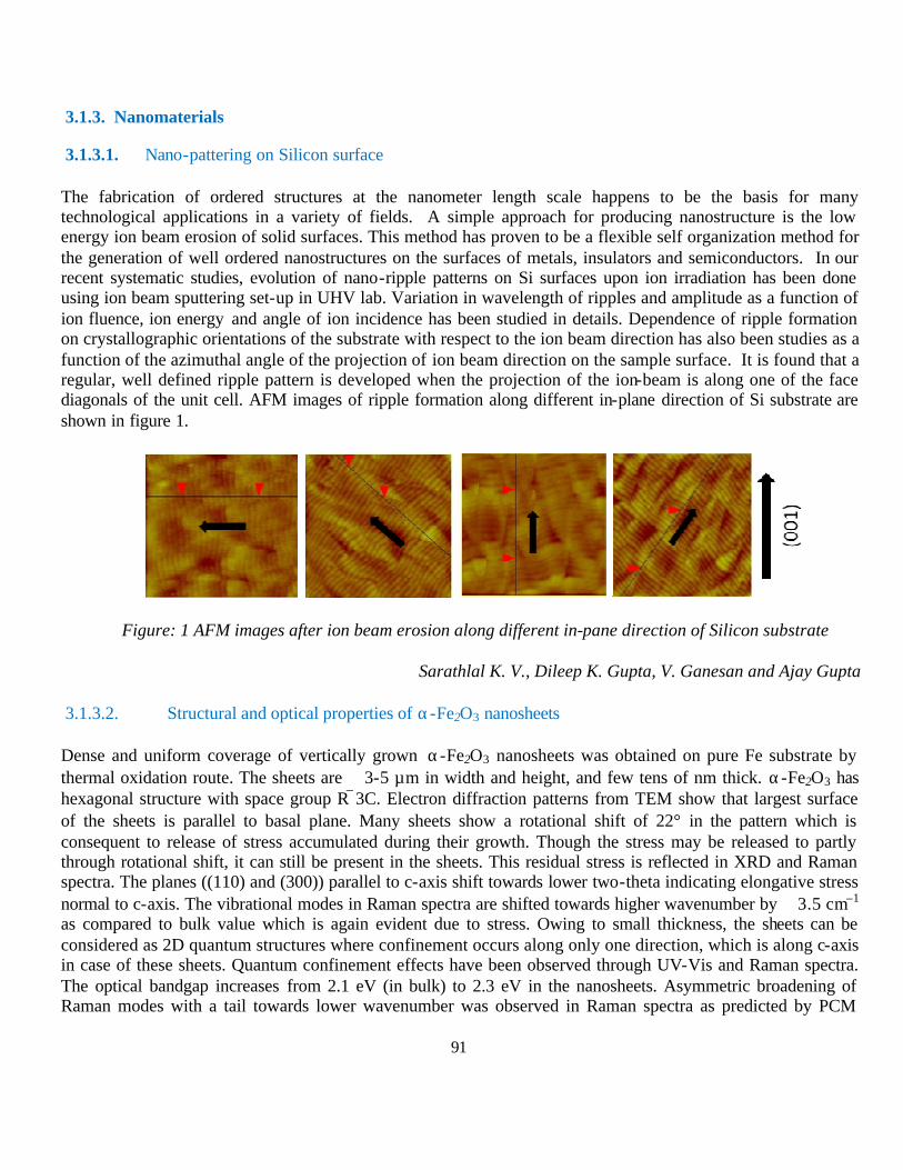
91
3.1.3. Nanomaterials
3.1.3.1. Nano-pattering on Silicon surface
The fabrication of ordered structures at the nanometer length scale happens to be the basis for many technological applications in a variety of fields. A simple approach for producing nanostructure is the low energy ion beam erosion of solid surfaces. This method has proven to be a flexible self organization method for the generation of well ordered nanostructures on the surfaces of metals, insulators and semiconductors. In our recent systematic studies, evolution of nano-ripple patterns on Si surfaces upon ion irradiation has been done using ion beam sputtering set-up in UHV lab. Variation in wavelength of ripples and amplitude as a function of ion fluence, ion energy and angle of ion incidence has been studied in details. Dependence of ripple formation on crystallographic orientations of the substrate with respect to the ion beam direction has also been studies as a function of the azimuthal angle of the projection of ion beam direction on the sample surface. It is found that a regular, well defined ripple pattern is developed when the projection of the ion-beam is along one of the face diagonals of the unit cell. AFM images of ripple formation along different in-plane direction of Si substrate are shown in figure 1.
Figure: 1 AFM images after ion beam erosion along different in-pane direction of Silicon substrate
Sarathlal K. V., Dileep K. Gupta, V. Ganesan and Ajay Gupta
3.1.3.2. Structural and optical properties of α-Fe2O3 nanosheets
Dense and uniform coverage of vertically grown α-Fe2O3 nanosheets was obtained on pure Fe substrate by thermal oxidation route. The sheets are ∼ 3-5 µm in width and height, and few tens of nm thick. α-Fe2O3 has hexagonal structure with space group R3C. Electron diffraction patterns from TEM show that largest surface of the sheets is parallel to basal plane. Many sheets show a rotational shift of 22° in the pattern which is consequent to release of stress accumulated during their growth. Though the stress may be released to partly through rotational shift, it can still be present in the sheets. This residual stress is reflected in XRD and Raman spectra. The planes ((110) and (300)) parallel to c-axis shift towards lower two-theta indicating elongative stress normal to c-axis. The vibrational modes in Raman spectra are shifted towards higher wavenumber by ∼ 3.5 cm–1 as compared to bulk value which is again evident due to stress. Owing to small thickness, the sheets can be considered as 2D quantum structures where confinement occurs along only one direction, which is along c-axis in case of these sheets. Quantum confinement effects have been observed through UV-Vis and Raman spectra. The optical bandgap increases from 2.1 eV (in bulk) to 2.3 eV in the nanosheets. Asymmetric broadening of Raman modes with a tail towards lower wavenumber was observed in Raman spectra as predicted by PCM
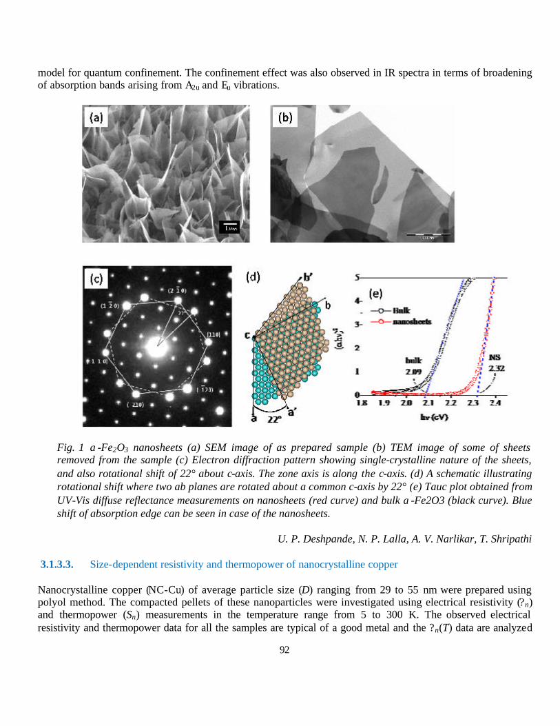
92
model for quantum confinement. The confinement effect was also observed in IR spectra in terms of broadening of absorption bands arising from A2u and Eu vibrations.
Fig. 1 α-Fe2O3 nanosheets (a) SEM image of as prepared sample (b) TEM image of some of sheets removed from the sample (c) Electron diffraction pattern showing single-crystalline nature of the sheets, and also rotational shift of 22° about c-axis. The zone axis is along the c-axis. (d) A schematic illustrating rotational shift where two ab planes are rotated about a common c-axis by 22° (e) Tauc plot obtained from UV-Vis diffuse reflectance measurements on nanosheets (red curve) and bulk α-Fe2O3 (black curve). Blue shift of absorption edge can be seen in case of the nanosheets.
U. P. Deshpande, N. P. Lalla, A. V. Narlikar, T. Shripathi
3.1.3.3. Size-dependent resistivity and thermopower of nanocrystalline copper Nanocrystalline copper (NC-Cu) of average particle size (D) ranging from 29 to 55 nm were prepared using polyol method. The compacted pellets of these nanoparticles were investigated using electrical resistivity (?n) and thermopower (Sn) measurements in the temperature range from 5 to 300 K. The observed electrical resistivity and thermopower data for all the samples are typical of a good metal and the ?n(T) data are analyzed

93
in the framework of the Bloch-Grüneisen theory. Further, the validity of the Nordheim-Gorter rule has also discussed. Our analysis indicates the systematic departure from the bulk property for NC-Cu samples, decreasing effective Debye temperature, exponential decay of both the residual resistivity ratio and the temperature coefficient of resistivity [a = (1/ρ)dρ/dT] as D decreases, yet the Boltzmann theory of electron transport still holds true (kFl >>1). This scenario is intriguing. The temperature dependence of Sn abruptly changes almost oppositely to bulk thermopower SBulk behavior, thereby revealing the negative sign in higher temperature than ~10K on making nanocrystlline. As the particle size decreases, there is systematic evolution of Sn and exhibition of a significant enhancement of phonon drag peak. The present findings turn out to be a case of the significant influences of the grain boundaries, surface atoms and phonon confinement.
G. S. Okram; N. Kaurav, (Holkar Sci. College, Indore)
3.1.3.4. Influence of cappant and reductant on resistivity and thermopower of nanocrystalline nickel We demonstrate here combined role of reductants and cappants on average particle size of several nanocrystalline nickel (n-Ni) samples and consequent influence on resistivity (?) and thermoelectric power (TEP). These results strongly consolidate earlier findings of anomalous properties of n-Ni samples and suggest that the cappants in conjunction with the uncompensated surface metallic bonds should not hinder the future interconnect of the electrical circuits to semiconducting level. The additional cappants have enhanced dominance over size reduction and hence physical properties. They are also dependent on the reductant type.
Figure Resistivity ? curves of n-Ni prepared by using different surfactants. Legend: A- acid, EG- ethylene glycol, Ur-urea, PEG-polyethylene glycol, TPAH- tetrapropyl ammonium hydroxide ; Samples have been numbered according to decreasing order of resistivity ?. Inset: ?300K (?5K) vs cappant type.
G. S. Okram
3.1.3.5. Unusual luminescence property in Eu3+ doped CaMoO4 nanophosphors
Tetragonal body centered CaMoO4 has inversion symmetry. When Eu3+ ions occupy Ca3+ sites of CaMoO4, intensity of magnetic dipole allowed transition (5D0→7F1) should be more than that of electric dipole transition (5D0→7F2) according to Judd-Ofelt theory. Opposite effects are found in our study. We have prepared nanocrystals of these materials at 150 °C using urea hydrolysis in ethylene glycol. Solubility limit of Eu3+ is found to be up to 3 at.%. Above this, other extra phases could be observed due to difference in charges of Eu3+ and Ca2+. Particle size measured by transmission electron microscopy (TEM) is found to be 20 nm. Lattice fringes in high resolution TEM image and selected area electron diffraction establish clearly the tetragonal phase, which is same as found in x-ray diffraction study. From luminescence study, the intensity of the electric dipole allowed transition is more than that of magnetic dipole transition and this is opposite to site symmetry study (CaO8). Possible way to answer is from polarizability effect from MoO4 tetrahedron. Interestingly, the contribution of energy transfer process from Mo-O charge transfer band to Eu3+ and the role of Eu3+ over surface of particle could be able to distinguish when luminescence decay processes are performed at two
0 50 100 150 200 250 3000.0
0.1
1.0
2 4 61E-51E-41E-3
0.010.1
1
Cappants1. OleicA+EG (31nm)2. Ur+PEG+EG (18nm)3. OleicA+TPAH+EG (31nm)4. TPAH+Diol (23nm)5. Diol (21nm)6. OleicA+Diol (20nm)
6453
1
2
ρ (m
Ωcm
)
T(K)
300K
Cappants
ρ(m
Ωcm
)
5K

94
different excitations (250 and 398 nm). Luminescence intensity and lifetimes increase significantly with increasing heat-treatment temperature. This is attributed to the reduction of non-radiative process from surface and H2O present in particles after heat treatment.
A.K. Parchur, R. K. Vatsa, S. B. Rai, (BHU, Varanasi); R. A. Singh (HSGU, Sagar); R. S.
Ningthoujam, M. Tyagi, R. Tewari (BARC, Mumbai) and G. S. Okram
3.1.4. Other studies
3.1.4.1. High energy resolution bandpass photon detector for inverse photoemission spectroscopy
We report a bandpass ultraviolet photon detector for inverse photoemission spectroscopy with energy resolution of 82 meV. The detector (Sr0.7Ca0.3F2/acetone) consists of Sr0.7Ca0.3F2 entrance window with energy transmission cutoff of 9.85 eV and acetone as detection gas with 9.7 eV photoionization threshold. The response function of the detector, measured using synchrotron radiation, has a nearly Gaussian shape. The n=1 image potential state of Cu(100) and Fermi edge of silver have been measured to demonstrate the improvement in resolution compared to the CaF2/acetone detector. To show the advantage of improved resolution of the Sr0.7Ca0.3F2/acetone detector, the metal to semiconductor transition in Sn has been studied. The pseudogap in the semiconducting phase of Sn could be identified, which is not possible with the CaF2/acetone detector because of its worse resolution.
Fig.: Response functions of CaF2, MgF2/Kr/CaF2/acetone, and Sr0.7Ca0.3F2 detectors normalized to the same height for comparing their shape. The fitted curve for the Sr0.7Ca0.3F2 detector is shown as a continuous line.
M. Maniraj, S. W. D'Souza, J. Nayak, Abhishek Rai, Sanjay Singh and S.R. Barman; B. N. Raja Sekhar (BARC)
3.1.4.2. Orbital ordering in the geometrically frustrated MgV2O4
The orbital degree of freedom is an important entity in the condensed matter physics which plays a crucial role in stabilizing many exotic phases observed in the strongly correlated electron systems. When the degenerate d orbitals of the transition metals are partially filled then occupation of a particular orbital at a particular site is expected to be dictated by the occupation of another orbital at another site, which can lead to various kind of orbital ordering (OO) similar to the spin ordering. Such OO often reduces the crystal symmetry as it expands the lattice along the direction of the ordering. Thus in the geometrically frustrated system OO is expected to relax the frustration leading to the formation of novel magnetic phases earlier forbidden by the frustration.
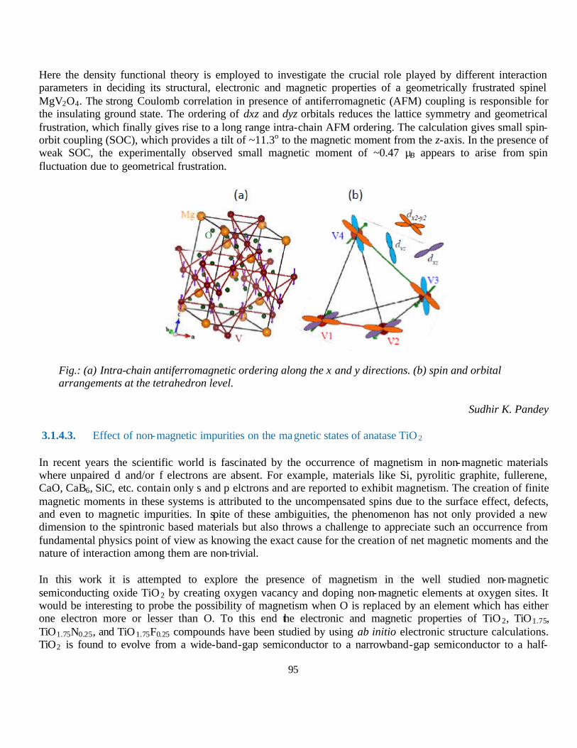
95
Here the density functional theory is employed to investigate the crucial role played by different interaction parameters in deciding its structural, electronic and magnetic properties of a geometrically frustrated spinel MgV2O4. The strong Coulomb correlation in presence of antiferromagnetic (AFM) coupling is responsible for the insulating ground state. The ordering of dxz and dyz orbitals reduces the lattice symmetry and geometrical frustration, which finally gives rise to a long range intra-chain AFM ordering. The calculation gives small spin-orbit coupling (SOC), which provides a tilt of ~11.3o to the magnetic moment from the z-axis. In the presence of weak SOC, the experimentally observed small magnetic moment of ~0.47 µB appears to arise from spin fluctuation due to geometrical frustration.
Fig.: (a) Intra-chain antiferromagnetic ordering along the x and y directions. (b) spin and orbital arrangements at the tetrahedron level.
Sudhir K. Pandey
3.1.4.3. Effect of non-magnetic impurities on the magnetic states of anatase TiO 2
In recent years the scientific world is fascinated by the occurrence of magnetism in non-magnetic materials where unpaired d and/or f electrons are absent. For example, materials like Si, pyrolitic graphite, fullerene, CaO, CaB6, SiC, etc. contain only s and p elctrons and are reported to exhibit magnetism. The creation of finite magnetic moments in these systems is attributed to the uncompensated spins due to the surface effect, defects, and even to magnetic impurities. In spite of these ambiguities, the phenomenon has not only provided a new dimension to the spintronic based materials but also throws a challenge to appreciate such an occurrence from fundamental physics point of view as knowing the exact cause for the creation of net magnetic moments and the nature of interaction among them are non-trivial.
In this work it is attempted to explore the presence of magnetism in the well studied non-magnetic semiconducting oxide TiO 2 by creating oxygen vacancy and doping non-magnetic elements at oxygen sites. It would be interesting to probe the possibility of magnetism when O is replaced by an element which has either one electron more or lesser than O. To this end the electronic and magnetic properties of TiO 2, TiO1.75, TiO1.75N0.25, and TiO1.75F0.25 compounds have been studied by using ab initio electronic structure calculations. TiO2 is found to evolve from a wide-band-gap semiconductor to a narrowband-gap semiconductor to a half-

96
metallic state and finally to a metallic state with oxygen vacancy, N-doping and F-doping, respectively. Present work clearly shows the robust magnetic ground state for N- and F-doped TiO2. The N-doping gives rise to magnetic moment of ~0.4 µB at N-site and ~0.1 µB each at two neighboring O-sites, whereas F-doping creates a magnetic moment of ~0.3 µB at the nearest Ti atom.
Sudhir K. Pandey and R. J. Choudhary
3.2. Research Activity at Kolkata Centre
3.2.1. Trace Elemental Studies using in-house facilities.
3.2.1.1. Effects of traffic generated dusts and its elemental constituents on the phyloplane microflora
Phylloplane or plant leaf surface is a habitat for many microorganisms. They are known to fix atmospheric nitrogen, produce plant growth regulators and can control plant parasites either by stimulating plants to synthesize phytoalexins or by producing antibacterial and antifungal compounds. Short-term and long-term changes in the abiotic conditions under which plants are grown may affect not only the growth and productivity of the plants, but also affect the populations of microorganisms living on plant surfaces. Changes in phyllosphere microbial populations may, in turn, affect plant growth and the plant’s ability to withstand aggressive attacks by pathogens. The microflora of road-side plants are exposed to traffic generated air pollutants that not only include particulate matter but also gaseous pollutants like SOx and NOx. In our earlier study we have reported that the kerbside canopies adsorb the resuspended roadside dusts contaminated with heavy metals and the microflora of these plants are challenged with the thrash condition of dust load. These particulate pollutants containing heavy metals get settled on leaf surfaces of roadside plants and come in contact with phylloplane microorganisms. Varying degrees of growth inhibition by trace metals have been shown in vitro for different species of pathogenic and saprophytic phylloplane fungi.
During this period a comparative study was carried out between phylloplane microflora, dust load and elemental profile of dusts deposited over leaf surfaces of P. longifolia from roadside (Karunamoyee) and non-roadside area (Calcutta University Campus, Raza Bazar) in Kolkata, India. More dusts were found to be adsorbed on the leaves collected from road-side area (0.314 ± 0.068 mg/cm2) in comparison to that of non-roadside area (0.058 ± 0.012 mg/cm2).Scanning Electron Microscopic observation of leaves of Polyalthia longifolia from each site and the dusts collected by surface washing of the leaves revealed that unlike the non-polluted site, the stomatal pores in the leaves from the polluted site are blocked by the dust particulates and no micro-organisms especially fungi was observed. This may be due to blockage by dusts as the growth of fungal hyphae around or into the stomatal pore may be one of the mechanisms to draw water and nutrient from the leaf cells and so in these cases unavailable for the fungal hyphae. Elemental profiling by SEM-EDAX analysis of leaf surfaces has shown the presence of trace elements in general and heavy metals in particular. Presence of toxic elements like As and Hg as well as other trace elements like Al, Si, Cl, Ti, Cr, Mn, Fe, Ni, Cu and Zn may be attributed for microflora retardation over the leaf surfaces exposed to traffic generated dust particulates. Results suggested deleterious effects of particulate pollutants on the phylloplane microflora of Polyalthia longifolia plants living in an urban environment which may sequentially give the e=way to more polluted condition of the environment by altering ntrgen fixation and / or parasitic growths on the concerned plants
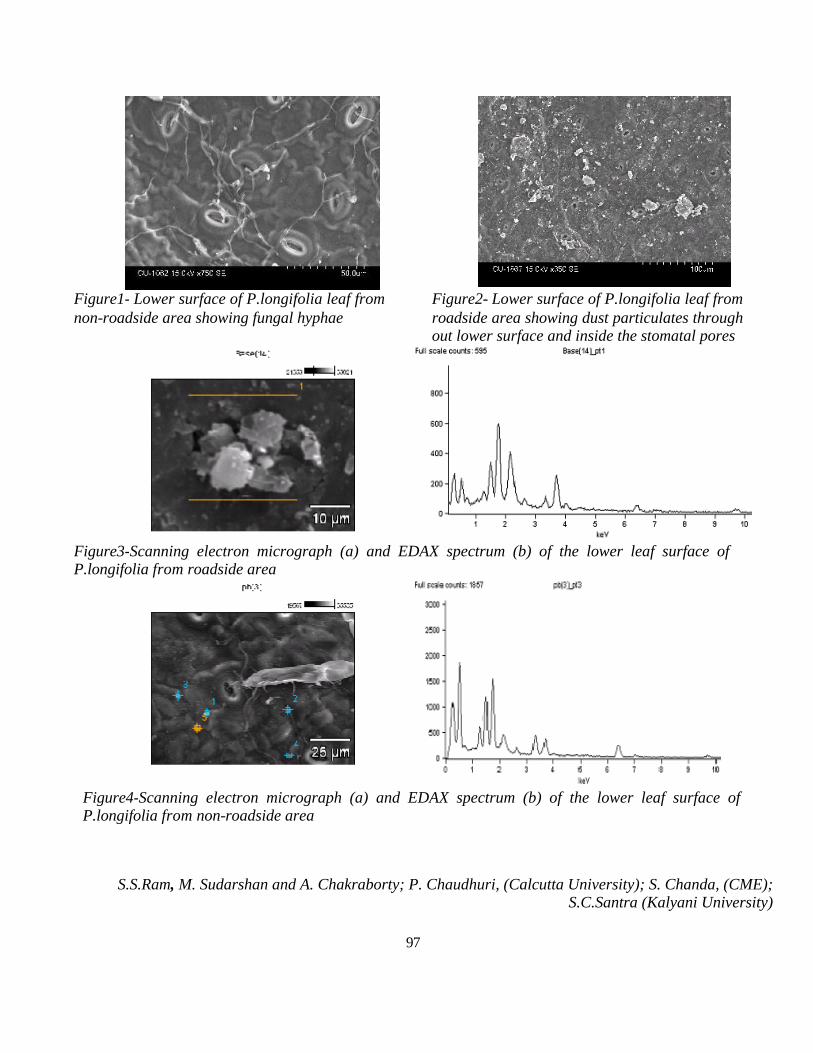
97
Figure1- Lower surface of P.longifolia leaf from non-roadside area showing fungal hyphae
Figure2- Lower surface of P.longifolia leaf from roadside area showing dust particulates through out lower surface and inside the stomatal pores
Figure3-Scanning electron micrograph (a) and EDAX spectrum (b) of the lower leaf surface of P.longifolia from roadside area
Figure4-Scanning electron micrograph (a) and EDAX spectrum (b) of the lower leaf surface of P.longifolia from non-roadside area
S.S.Ram, M. Sudarshan and A. Chakraborty; P. Chaudhuri, (Calcutta University); S. Chanda, (CME); S.C.Santra (Kalyani University)
98
3.2.2. Nuclear Technique based condensed matter studies.
3.2.2.1. Exchange bias and suppression of superparamagnetism of α-Fe nanoparticles in NiO matrix
Nanocomposites of ferromagnetic α-Fe (10 wt. %) and antiferromagnetic NiO were prepared by the ball milling method ( available at Bengal Engineering and Science University, Shhibpur, Howrah) with different milling durations. These samples were characterized by XRD, TEM, Mössbauer spectroscopy, positron annihilation spectroscopy and SQUID. Strong exchange bias coupling has been shown to be the reason for suppression of superparamagnetism in the finer particles. (Revised manuscript submitted to Material Science and Engineering:B)
S. P. Pati, B. Bhushan, D. Das; A. Basumallick (BESU, Howrah)
3.2.2.2. Structural, hyperfine and magnetic characterization of thermo-chemically prepared Fe-Co nanoalloy
Nanosized Fe-Co alloy of average crystallite size of 35 nm was synthesized by thermal decomposition of chemically prepared cobalt ferrite in hydrogen atmosphere. The nanosized alloy was characterized by XRD, EDX, TG-DTA, Mössbauer spectroscopy and SQUID magnetometer. Fe-Co nanoalloy exhibited high saturation magnetization (~ 75 emu/g) and extremely low coercive field (~ 6.6 Oe). Hyperfine magnetic field of Fe was found to increase after alloying it with Co.
S. P. Pati, B. Bhushan, D. Das
3.2.2.3. Enhanced magnetic properties in BiFeO3 nanoparticles co-doped with Ba and Ca
Bi0.95CaxBa0.05-xFeO3 nanoparticles were prepared by a sol-gel route with x = 0.01, 0.025 and 0.04. XRD and TEM measurements revealed the pure-phase formation of the co-doped BFO nanoparticles. 57Fe Mössbauer spectroscopy confirmed that iron is present only in 3+ valence state in all co-doped samples. Significant enhancement in the magnetic properties was observed in Ba and Ca co-doped BFO nanoparticles.
B. Bhushan, D. Das; S. Kumar (Jadavpur Univ., Kolkata)
3.2.3. Chemical sciences & radiochemistry research work
3.2.3.1. Biomimetic Synthesis of BSA capped CdS QDs
In recent years, semiconductor nanoparticles have attracted much attention in both fundamental research and technical applications, owing to their unique size-dependent optical and electronic properties. Biological macromolecules are capable of controlling inorganic crystals nucleation and growth to a remarkable degree through biomineralization. Thereupon, it is logical approach to use biomolecules to grow monodisperse nanocrystals via biomimetic method. In the present work a simple and convenient method for the synthesis of CdS nanoparticles in a foam matrix using the protein bovine serum albumin (BSA) is reported. BSA is one of the most widely studied proteins and is the most abundant in plasma with a typical concentration of 5mg/100ml. BSA shows excellent foaming behavior and these aqueous foams behave as excellent template for the growth of Nanoparticles. The BSA foam procedure also results in the nanoparticles being coated/stabilized by the BSA
99
molecules. Many interesting reports have appeared in literature where polypeptides/proteins have been used to assemble nanoparticles or the nanoparticle surfaces have been modified by protein/polymer coatings owing to their interest in the field of biomedical applications. However in most of these cases the assembly or modification of the surface is done on preformed nanoparticles. In this experiment the protein is used as a template to synthesize the nanoparticles. This could probably lead to a better coverage of the nanoparticle surface with the protein making them a better candidate for biological applications. The properties of these as-prepared products are investigated by UV–vis spectroscopy, photo luminescence spectroscopy (PL), transmission electron microscopy (TEM), selected area diffraction (SEAD), high-resolution microscopy (HRTEM), XRD and circular dichroism spectroscopy.
D.Ghosh and A. Saha; P. K. Bag (Calcutta University)
3.2.3.2. Room Temperature Aqueous Synthesis of Bipyramidal Silver Nanostructures
The proposed strategy demonstrates the formation of pentagonal right bipyramidal silver nanostructures in aqueous phase at room temperature. We have explored a combination of a cationic surfactant, namely, cetyl tri-methyl ammonium bromide (CTAB) and a polymer, such as polyvinyl pyrrolidone (PVP) for capping preferential crystallographic facets of silver nanosurface. And structural transformation of these anisotropic nanostructures was studied by various following factors such as effect of Ag seed concentration, the ratio of concentration of CTAB with the concentration of silver nitrate, the effect of PVP and its concentration dependence, the ascorbic acid concentration dependence and the effect of pH. CTAB functions as template directing agent while PVP acts as shape directing agent. Zeta potential measurements and Fourier Transform Infra-red (FTIR) data analysis reveal that PVP substitutes CTAB over the facets of silver nanobypiramids.
Y. N. Rao and A. Saha; S. K. Das (VECC)
3.2.3.3. Detection of Vasodialator Bradykinin by Biofunctionalized Quantum Dots: A Detailed Study of Quantum Dot-Bradykinin Interaction
Studies investigating the role of a physiologically important peptide hormone, vasodialator bradykinin in disease states such as hypertension, sepsis, and asthma have been confounded by difficulties in measuring the concentration of this peptide. The purpose of this study was to determine a simple, fast and easy method for detection of bradykinin by CdTe QDs in the limit 0.6µM to 5 µM and also to study the interaction of QD-BK in details. Micromolar concentrations of tyrosine-Bradykinin enhanced the fluorescence of cysteine capped CdTe QDs. The enhancement followed Langmuir binding isotherm, nature of the enhancement was studied by varying QD size, solvent polarity, salt effect and also by measuring fluorescence lifetime. Further we studied the interaction in ground state by FTIR and CD spectra, and also by iso thermal calorimetry(ITC), ITC of QD-protein has been hardly reported so far. In conclusion, we can say that our method can be successfully used for the detection of human salivery bradykinin, BK(1-5) (which is the stable metabolite of Bradykinin in human plasma) and Hyp3-BK in human urine. Such a method will allow for further elucidation of the role of bradykinin in the pathophysiology of important human diseases, such as hypertension, diabetes, asthma and sepsis and can be used as part of an ongoing clinical development programme for pain diagonesis and treatment strategies.
S. Mondal, S. Ghosh and A. Saha

100
3.2.3.4. Gamma irradiation route to photoluminescent selenium-based QDs under ambient conditions
Semiconductor nanocrystallites have continued to be an active field of research for more than a decade primarily for two reasons. First, it gives a unique opportunity to study the material properties at the nanometer level and consequently to understand the underlying physics at reduced dimensions. The second reason is based on their immense potential for application in the area of optoelectronic devices. Literatures are replete with several reports of synthesis of semiconductor nanoparticles of different materials like CdS, CdTe, CdSe and HgTe, ZnSe with varying functionality. In general, high temperature or use of toxic gas, like H2S, is requisite conditions for most of the above mentioned methods. In contrast to this, γ-radiation assisted synthesis offers a simplified approach to prepare size-controlled nanoparticles at room temperature. As the precipitation of Cd2+ with Se2- is quicker than their homogeneous mixing, the inhomogeneity at early stages leads to broadening of the size distribution. A distinct advantage of radiolytic synthesis against the other methods is in-situ generation of Se2-, which results in homogenous mixing. Work demonstrates a distinct advantage of radiolytic method over other chemical methods. In this work, we successfully report to synthesize CdSe, ZnSe, Ag2Se, QDs by γ-irradiating aqueous solution Cd2+, Zn2+, Ag2+,ions, using different capping agents and sodium selenosulphate solution under ambient pressure and at room temperature. Optical characterization has been conducted using techniques like Uv-vis spectrophotometer, luminescence spectrophotometer and dynamic light scattering.
A. Datta, Y. N. Rao, and A. Saha
3.2.3.5. Radiation- induced Self organization of Functionalized Inorganic-Organic Hybrid Nanocomposite Assemblies
We demonstrate a unique solvated electron mediated single-pot synthesis of self-assembly of CdS/Dendrimer nanocomposites having long-range correlation. Interestingly, the present method has enabled to produce chain-like assembly of CdS nanoparticles of high stability within the dendrimer matrix with particle size distribution as low as 6%. The long-range network structures of composite particles are visualized with transmission electron microscopy and atomic force microscopy. Further, the structures were investigated with FESEM. Particles were characterized by optical spectroscopy and dynamic light scattering. A possible mechanism of the formation of a long-range self-organization from nanometer scale to micrometer is discussed. It is illustrated that the directed self-assembly is controlled by the surface functionality of dendrimer molecule, solvent and crystal phase of CdS nanocrystals. The present investigation opens up new potential routes to manipulation of semiconductor nanocomposites for optical diagnostics and applications that require ordered arrangements of dendrimer nanocomposites
S. Ghosh, A. Datta and A. Saha; N. Biswas, A. Datta (SINP)
3.2.3.6. Biofunctionalized Quantum dots as Fluorescence Probes for the detection of Vitamin B12
In the present work, we have attempted to study the interaction of highly fluorescent semiconductor nanocrystal (or Quantum Dots, QDs) with bioactive molecule, Vitamin B12 (VB12).The detection of Vitamin B12 is of great importance in pharmaceutical, food industry and also marker for different diseases in clinical chemistry. VB12 significantly quenched the photoluminescence (PL) of the both functionalized semiconductor nanocrystal such as CdS and CdTe in the concentration range of 0.6-33.3 µM. The quenching followed a linear Stern-Volmer equation with an excellent 0.999 correlation coefficient.Detailed studies by spectroscopy, show that binding of
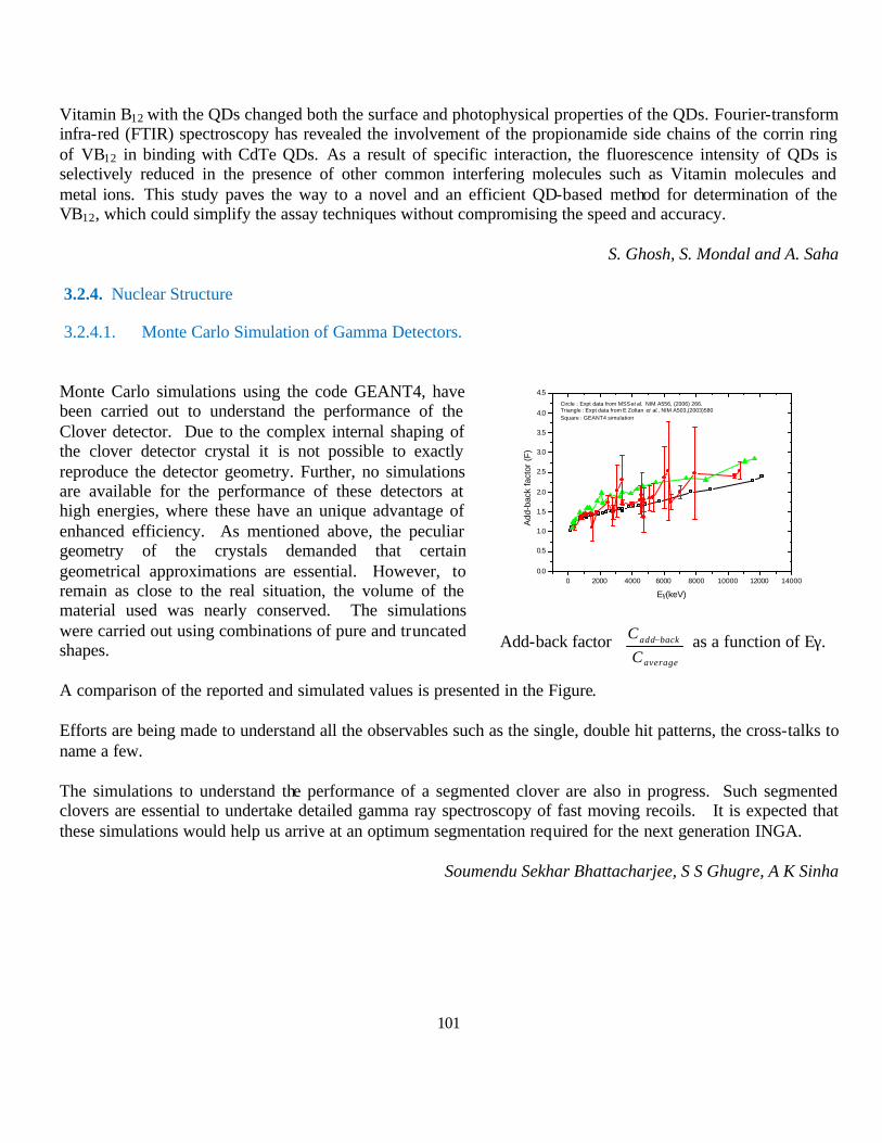
101
Vitamin B12 with the QDs changed both the surface and photophysical properties of the QDs. Fourier-transform infra-red (FTIR) spectroscopy has revealed the involvement of the propionamide side chains of the corrin ring of VB12 in binding with CdTe QDs. As a result of specific interaction, the fluorescence intensity of QDs is selectively reduced in the presence of other common interfering molecules such as Vitamin molecules and metal ions. This study paves the way to a novel and an efficient QD-based method for determination of the VB12, which could simplify the assay techniques without compromising the speed and accuracy.
S. Ghosh, S. Mondal and A. Saha
3.2.4. Nuclear Structure
3.2.4.1. Monte Carlo Simulation of Gamma Detectors.
Monte Carlo simulations using the code GEANT4, have been carried out to understand the performance of the Clover detector. Due to the complex internal shaping of the clover detector crystal it is not possible to exactly reproduce the detector geometry. Further, no simulations are available for the performance of these detectors at high energies, where these have an unique advantage of enhanced efficiency. As mentioned above, the peculiar geometry of the crystals demanded that certain geometrical approximations are essential. However, to remain as close to the real situation, the volume of the material used was nearly conserved. The simulations were carried out using combinations of pure and truncated shapes.
A comparison of the reported and simulated values is presented in the Figure.
Efforts are being made to understand all the observables such as the single, double hit patterns, the cross-talks to name a few.
The simulations to understand the performance of a segmented clover are also in progress. Such segmented clovers are essential to undertake detailed gamma ray spectroscopy of fast moving recoils. It is expected that these simulations would help us arrive at an optimum segmentation required for the next generation INGA.
Soumendu Sekhar Bhattacharjee, S S Ghugre, A K Sinha
0 2000 4000 6000 8000 10000 12000 140000.0
0.5
1.0
1.5
2.0
2.5
3.0
3.5
4.0
4.5Circle : Expt data from MSS et al. NIM A556, (2006) 266.Triangle : Expt data from E Zoltan et al., NIM A503,(2003)580Square : GEANT4 simulation
Add
-bac
k fa
ctor
(F
)
Eγ(keV)
Add-back factor
−
average
backadd
CC as a function of Eγ.
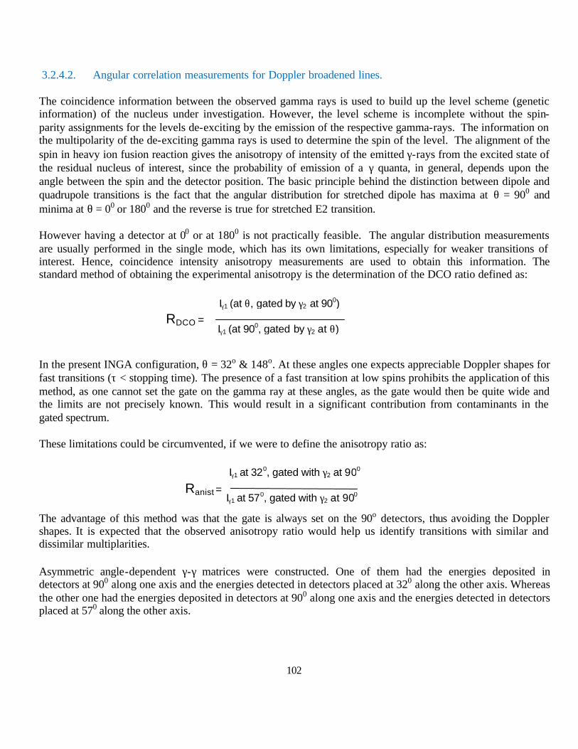
102
3.2.4.2. Angular correlation measurements for Doppler broadened lines.
The coincidence information between the observed gamma rays is used to build up the level scheme (genetic information) of the nucleus under investigation. However, the level scheme is incomplete without the spin-parity assignments for the levels de-exciting by the emission of the respective gamma-rays. The information on the multipolarity of the de-exciting gamma rays is used to determine the spin of the level. The alignment of the spin in heavy ion fusion reaction gives the anisotropy of intensity of the emitted γ-rays from the excited state of the residual nucleus of interest, since the probability of emission of a γ quanta, in general, depends upon the angle between the spin and the detector position. The basic principle behind the distinction between dipole and quadrupole transitions is the fact that the angular distribution for stretched dipole has maxima at θ = 900 and minima at θ = 00 or 1800 and the reverse is true for stretched E2 transition.
However having a detector at 00 or at 1800 is not practically feasible. The angular distribution measurements are usually performed in the single mode, which has its own limitations, especially for weaker transitions of interest. Hence, coincidence intensity anisotropy measurements are used to obtain this information. The standard method of obtaining the experimental anisotropy is the determination of the DCO ratio defined as:
In the present INGA configuration, θ = 32o & 148o. At these angles one expects appreciable Doppler shapes for fast transitions (τ < stopping time). The presence of a fast transition at low spins prohibits the application of this method, as one cannot set the gate on the gamma ray at these angles, as the gate would then be quite wide and the limits are not precisely known. This would result in a significant contribution from contaminants in the gated spectrum.
These limitations could be circumvented, if we were to define the anisotropy ratio as:
The advantage of this method was that the gate is always set on the 90o detectors, thus avoiding the Doppler shapes. It is expected that the observed anisotropy ratio would help us identify transitions with similar and dissimilar multiplarities.
Asymmetric angle-dependent γ-γ matrices were constructed. One of them had the energies deposited in detectors at 900 along one axis and the energies detected in detectors placed at 320 along the other axis. Whereas the other one had the energies deposited in detectors at 900 along one axis and the energies detected in detectors placed at 570 along the other axis.
RDCO = Iγ1 (at 900, gated by γ2 at θ)
Iγ1 (at θ, gated by γ2 at 900)
Iγ1 at 570, gated with γ2 at 900
Iγ1 at 320, gated with γ2 at 900
Ranist =
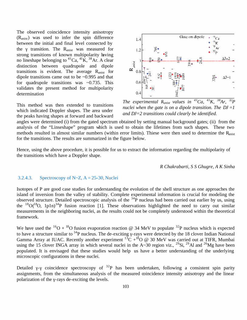
103
The observed coincidence intensity anisotropy (Ranist) was used to infer the spin difference between the initial and final level connected by the γ transition. The Ranist was measured for strong transitions of known multipolarity having no lineshape belonging to 41Ca, 41K, 38Ar. A clear distinction between quadrupole and dipole transitions is evident. The average Ranist for dipole transitions came out to be ~0.995 and that for quadrupole transitions was ~0.735. This validates the present method for multipolarity determination
This method was then extended to transitions which indicated Doppler shapes. The area under the peaks having shapes at forward and backward angles were determined (i) from the gated spectrum obtained by setting manual background gates; (ii) from the analysis of the “Linseshape” program which is used to obtain the lifetimes from such shapes. These two methods resulted in almost similar numbers (within error limits). Thiese were then used to determine the Ranist for the transitions. The results are summarized in the figure below.
Hence, using the above procedure, it is possible for us to extract the information regarding the multipolarity of the transitions which have a Doppler shape.
R Chakrabarti, S S Ghugre, A K Sinha
3.2.4.3. Spectroscopy of N~Z, A = 25-30, Nuclei
Isotopes of P are good case studies for understanding the evolution of the shell structure as one approaches the island of inversion from the valley of stability. Complete experimental information is crucial for modeling the observed structure. Detailed spectroscopic analysis of the 34P nucleus had been carried out earlier by us, using the 18O(18O, 1p1n)34P fusion reaction [1]. These observations highlighted the need to carry out similar measurements in the neighboring nuclei, as the results could not be completely understood within the theoretical framework.
We have used the 16O + 18O fusion evaporation reaction @ 34 MeV to populate 32P nucleus which is expected to have a structure similar to 34P nucleus. The de-exciting γ-rays were detected by the 18 clover Indian National Gamma Array at IUAC. Recently another experiment 13C +18O @ 30 MeV was carried out at TIFR, Mumbai using the 15 clover INGA array in which several nuclei in the A~30 region viz., 29Si, 29Al and 26Mg have been populated. It is envisaged that these studies would help us have a better understanding of the underlying microscopic configurations in these nuclei.
Detailed γ-γ coincidence spectroscopy of 32P has been undertaken, following a consistent spin parity assignments, from the simultaneous analysis of the measured coincidence intensity anisotropy and the linear polarization of the γ-rays de-exciting the levels.
The experimental Ranist values in 41Ca, 41K, 38Ar, 32P nuclei when the gate is on a dipole transition. The ∆J =1 and ∆J=2 transitions could clearly be identified.
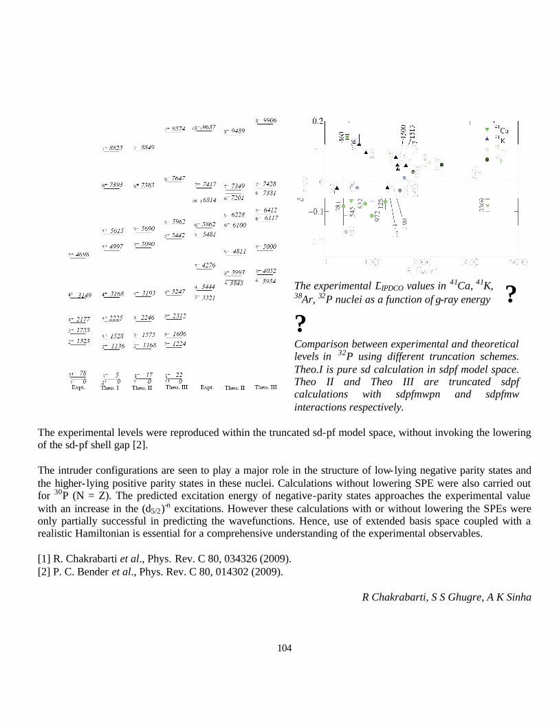
104
The experimental ∆IPDCO values in 41Ca, 41K, 38Ar, 32P nuclei as a function of γ-ray energy ? ? Comparison between experimental and theoretical levels in 32P using different truncation schemes. Theo.I is pure sd calculation in sdpf model space. Theo II and Theo III are truncated sdpf calculations with sdpfmwpn and sdpfmw interactions respectively.
The experimental levels were reproduced within the truncated sd-pf model space, without invoking the lowering of the sd-pf shell gap [2].
The intruder configurations are seen to play a major role in the structure of low-lying negative parity states and the higher- lying positive parity states in these nuclei. Calculations without lowering SPE were also carried out for 30P (N = Z). The predicted excitation energy of negative-parity states approaches the experimental value with an increase in the (d5/2)-n excitations. However these calculations with or without lowering the SPEs were only partially successful in predicting the wavefunctions. Hence, use of extended basis space coupled with a realistic Hamiltonian is essential for a comprehensive understanding of the experimental observables.
[1] R. Chakrabarti et al., Phys. Rev. C 80, 034326 (2009). [2] P. C. Bender et al., Phys. Rev. C 80, 014302 (2009).
R Chakrabarti, S S Ghugre, A K Sinha
105
3.2.5. Biological Sciences using in-house facilities
3.2.5.1. Radiation induced stress DNA damage and genotoxicity : sensitization /protection by herbal components
In this program effect of ionizing radiation on plasmid DNA was assessed (in vitro). Role of phytochemicals purified from the plant Seabuckthron (Hippophae rhamnoides) was also evaluated to probe into its potential to modulate radiation induced damage in in vitro condition.
Radiation exposure of plasmid DNA was carried out in different radiation doses. (5 Gy,10 Gy,15 Gy, 20 Gy), in presence and absence of the phytochemicals followed by agarose gel electrophoresis of the exposed DNA with respect to the control (unirradiated one). Gamma irradiation resulted in cleavage of the DNA and phytochemicals like quercetin, gallic acid, ellagic acid, isorhamnetin have radioprotective activity on plasmid DNA in 100 µM dose. Among these gallic acid has most potent radioprotective activity
Fig 1: Gel electrophoresis showing the cleavage of supercoiled pET28a plasmid DNA (300 ng in each lane) with different doses of ? radiation.
Fig. 2: Gel electrophoresis showing the cleavage of supercoiled pET28a plasmid DNA (300 ng in each lane). Lane 1: Control (C), Lane 2: Irradiated by 10Gy (R). Lane3: 0.1% Ethanol (ET), Lane 4: Quercetin(Q) (100µM), Lane 5: Gallic acid(GA) (100µM), Lane 6: Ellagic acid(EA)(100µM), Lane 7: Isorhamnetin (ISR)(100µM). All phytochemicals were incubated with DNA at 37°C for 45 minutes before radiation.
S. Selvaraj and A. Chakraborty; S Dey, A Khan and K Manna (Calcutta University)
3.2.5.2. Effect of environmental carcinogens on some aspects of cytoskeletal integrity
During development of cancer the first phase or initiation phase of the multistep process is a very critical phase as this is the phase of onset of metabolic alterations.Cytoskeleton play major role in this multistep–multihit process of carcinogenesis. Disorganization of cytoskeleton has been associated with malignancy as the endpoint. Hence understanding of the mechanisms of cytoskeletal integrity and /or its perturbation along with involvement of some specific metabolic pathways during initiation of chemically induced carcinogenesis in mice has been taken up. This program is designed determine the differential signature of the two environmental chemical carcinogens p-DAB and DENA if any, during initiation phase of hepatocarcinogenesis induction of the exposed mice by probing cytoskeleton and further to correlate such changes with activity of relevant enzymes and proteins. As intracellular calcium, [Ca2+]i, is known to be responsible for many molecular processes involved in cell movement and a change in [Ca2+]i can trigger the cell to respond by altering some
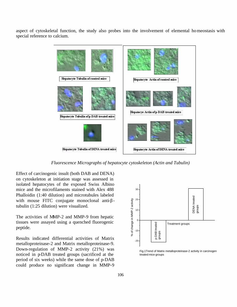
106
aspect of cytoskeletal function, the study also probes into the involvement of elemental homeostasis with special reference to calcium.
Fluorescence Micrographs of hepatocyte cytoskeleton (Actin and Tubulin)
Effect of carcinogenic insult (both DAB and DENA) on cytoskeleton at initiation stage was assessed in isolated hepatocytes of the exposed Swiss Albino mice and the microfilaments stained with Alex 488 Phalloidin (1:40 dilution) and microtubules labeled with mouse FITC conjugate monoclonal anti-β-tubulin (1:25 dilution) were visualized.
The activities of MMP-2 and MMP-9 from hepatic tissues were assayed using a quenched fluorogenic peptide.
Results indicated differential activities of Matrix metalloproteinase-2 and Matrix metalloproteinase-9. Down-regulation of MMP-2 activity (21%) was noticed in p-DAB treated groups (sacrificed at the period of six weeks) while the same dose of p-DAB could produce no significant change in MMP-9
-20
-10
0
10
20
30
Fig.1Trend of Matrix metalloproteinase-2 activity in carcinogen treated mice groups
p-D
AB
trea
ted
grou
ps
DE
NA
trea
ted
grou
ps
% o
f cha
nge
in M
MP
-2 a
ctiv
ity
Treatment groups

107
activity when compared to that of the control group. Upregulation of both MMP-2 (31%) and MMP-9 (10%) was found in DENA treated groups when compared to their normal counterparts.
Although Matrix metalloproteinase are upregulated in almost every type of cancer it is hypothesized that MMP-2 could not be activated in a long trip. Both MMP-2 and MMP-9 exhibits dynamic changes in their activities at different stages. The interplay between TIMP and MMP might be responsible for this aberrant behavior.
Quantitative evaluation of the total content of cytoskeletal proteins and intracellular calcium was standardized using flowcytometric technique
Flowcytometric analysis of cytoskeletal protein (Actin and Tubulin) in control mice revealed the expression of these two proteins in hepatocyte.
Anindita Chakraborty; Dipanwita Mukherjee, Aniruddha Mukherjee (University of Calcutta),)

108
3.3. Research activity at Mumbai Centre
3.3.1. Neutron scattering studies
3.3.1.1. Microscopic origin of multiferroicity in Bi2Fe4O9
Bi2Fe4O9 is a very common by-product/undesired impurity phase in synthesis of multiferroic BiFeO3 by solid state reaction route. Bi2Fe4O9 crystallizes in the orthorhombic phase with space group Pbam and crystal structure of this compound was reported earlier using X-Ray and neutron diffraction (ND). Iron atoms occupy two crystallographic positions namely Fe1 (tetrahedrally coordinated by four oxygen atoms) and Fe2 (octahedrally coordinated by six oxygen atoms). In the structure, columns of edge-sharing Fe2 octahedra form chains along the c axis, and these chains are linked together by corner-sharing Fe1 tetrahedra and Bi atoms. As a result, the different Fe3+ magnetic atoms form a distorted pentagonal Cairo lattice. We have previously reported the substantial magnetoelectric coupling near room temperature in polycrystalline Bi2Fe4O9 based on magnetization and in-field dielectric study. Recent single crystal ND study indicate an imbricated, non-collinear spin structure conforming to a centrosymmetric magnetic space group thus ruling out ferroelectricity and showed the presence of high geometrical frustration. Thus, the diverse nature of observations indicates that the origin of ferroelectricity in this interesting compound is not fully understood and careful study of local spin pair correlation close to transition temperature is needed to understand the origin of ferrolectricity in the system.
Polycrystalline Bi2Fe4O9 sample was prepared by conventional solid state route and phase purity was confirmed by XRD (Fig. 1). Inset of Fig 2 shows the temperature dependent DC susceptibility (?) measured in magnetic field of 0.5 T. The magnetization behaviour and magnitude is similar to the reported value of Bi2Fe4O9 single crystal. ND pattern at 300 K carried out at Focussing Crystal based Diffractometer at UGC-DAE CSR beam line in Dhruva at a wave- length of 1.48 Å is shown in Fig. 2. Black circles represent ND data, red line is the calculated pattern and blue circles show the empty vanadium can background. The pattern clearly shows a diffuse scattering hump centred around Q ~ 1.3 Å-1, antiferromagnetic reflection evolves as we go below TN.
Fig.1 Fig.2
Persistence of spin correlations well above TN is rather unusual for this compound and seems to be a result of the high geometrical frustration coupled with the unique magnetic structure. A detailed study of the spin-pair
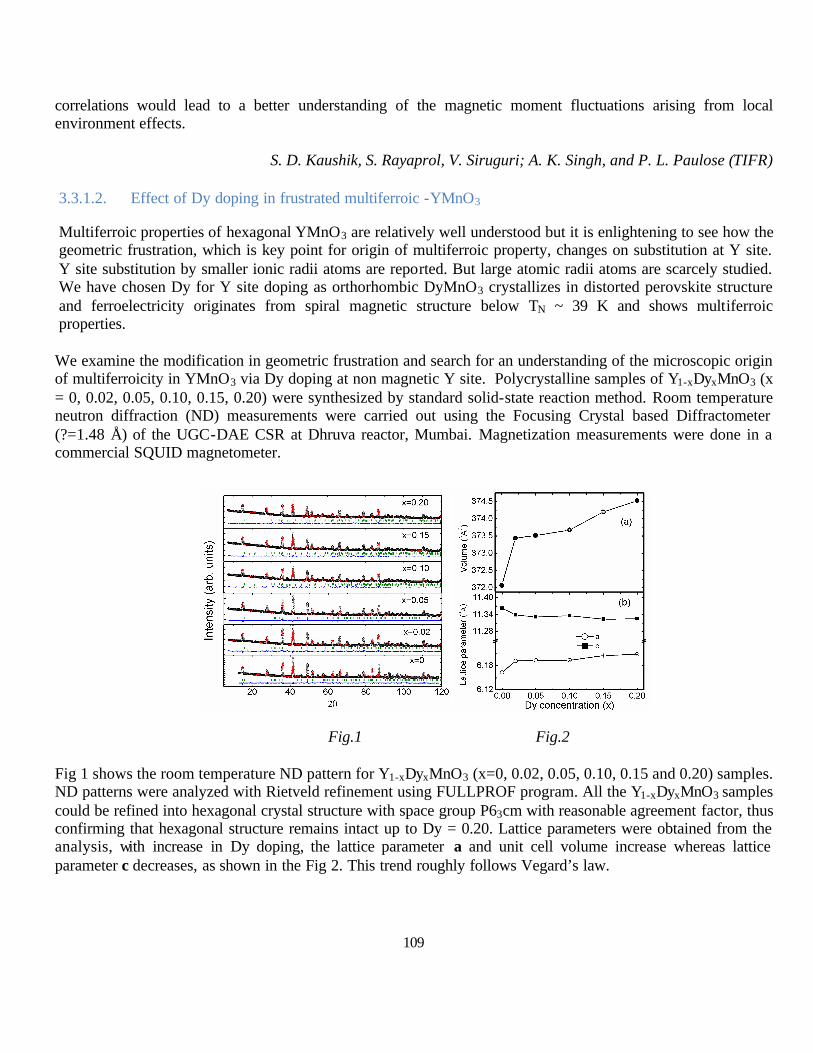
109
correlations would lead to a better understanding of the magnetic moment fluctuations arising from local environment effects.
S. D. Kaushik, S. Rayaprol, V. Siruguri; A. K. Singh, and P. L. Paulose (TIFR)
3.3.1.2. Effect of Dy doping in frustrated multiferroic -YMnO3
Multiferroic properties of hexagonal YMnO3 are relatively well understood but it is enlightening to see how the geometric frustration, which is key point for origin of multiferroic property, changes on substitution at Y site. Y site substitution by smaller ionic radii atoms are reported. But large atomic radii atoms are scarcely studied. We have chosen Dy for Y site doping as orthorhombic DyMnO3 crystallizes in distorted perovskite structure and ferroelectricity originates from spiral magnetic structure below TN ~ 39 K and shows multiferroic properties.
We examine the modification in geometric frustration and search for an understanding of the microscopic origin of multiferroicity in YMnO3 via Dy doping at non magnetic Y site. Polycrystalline samples of Y1-xDyxMnO3 (x = 0, 0.02, 0.05, 0.10, 0.15, 0.20) were synthesized by standard solid-state reaction method. Room temperature neutron diffraction (ND) measurements were carried out using the Focusing Crystal based Diffractometer (?=1.48 Å) of the UGC-DAE CSR at Dhruva reactor, Mumbai. Magnetization measurements were done in a commercial SQUID magnetometer.
Fig 1 shows the room temperature ND pattern for Y1-xDyxMnO3 (x=0, 0.02, 0.05, 0.10, 0.15 and 0.20) samples. ND patterns were analyzed with Rietveld refinement using FULLPROF program. All the Y1-xDyxMnO3 samples could be refined into hexagonal crystal structure with space group P63cm with reasonable agreement factor, thus confirming that hexagonal structure remains intact up to Dy = 0.20. Lattice parameters were obtained from the analysis, with increase in Dy doping, the lattice parameter a and unit cell volume increase whereas lattice parameter c decreases, as shown in the Fig 2. This trend roughly follows Vegard’s law.
Fig.1 Fig.2

110
To understand the implications of Dy doping on magnetic properties, we have further studied the temperature dependent DC susceptibility. Fig 3 shows inverse of the DC susceptibility as a function for some of the samples. By fitting the Curie-Weiss law, θCW and TN are determined and frustration factor f = |θCW|/TN is calculated. Inset of Fig 3 shows the decrease in the frustration factor as Dy doping increases. This decrease in the frustration factor is indicative of the release in the buckling in the Y plane due to Dy doping. Partial Dy doping (0 <x< 0.20) at Y site of YMnO3 does not change the crystal structure. The lattice parameters a and unit cell volume increase whereas lattice parameter c decreases. These changes lead to a decrease in the frustration factor which is expected to modify the buckling in the Y plane and, in turn, vary the MnO5 polyhedron tilt.
These findings were presented in 55th Solid State Physics Symposium 2010, Dec 26 -30, 2010 held at Manipal.
S. D. Kaushik, V. Siruguri; A. K. Singh, S. Patnaik, (JNU)
3.3.2. Magnetic Oxides
3.3.2.1. Magnetodielectric Coupling in the Manganocuprate, Gd3Ba2Mn2Cu2O12:
Figure: Capacitance and tand measured at various probe frequencies is plotted as function of temperature for Gd-3222. The frequency dependence of both C and tanδ brings out the analogy to ferroelectric relaxor, where the relaxation of dipoles can be described in terms of Maxwell-Wagner relaxation.
Fig.3
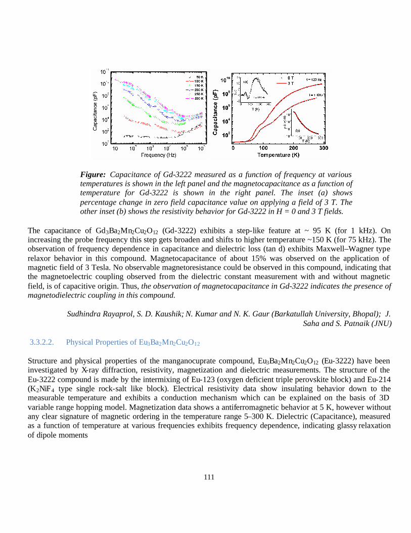
111
Figure: Capacitance of Gd-3222 measured as a function of frequency at various temperatures is shown in the left panel and the magnetocapacitance as a function of temperature for Gd-3222 is shown in the right panel. The inset (a) shows percentage change in zero field capacitance value on applying a field of 3 T. The other inset (b) shows the resistivity behavior for Gd-3222 in H = 0 and 3 T fields.
The capacitance of Gd3Ba2Mn2Cu2O12 (Gd-3222) exhibits a step-like feature at ~ 95 K (for 1 kHz). On increasing the probe frequency this step gets broaden and shifts to higher temperature ~150 K (for 75 kHz). The observation of frequency dependence in capacitance and dielectric loss (tan d) exhibits Maxwell–Wagner type relaxor behavior in this compound. Magnetocapacitance of about 15% was observed on the application of magnetic field of 3 Tesla. No observable magnetoresistance could be observed in this compound, indicating that the magnetoelectric coupling observed from the dielectric constant measurement with and without magnetic field, is of capacitive origin. Thus, the observation of magnetocapacitance in Gd-3222 indicates the presence of magnetodielectric coupling in this compound.
Sudhindra Rayaprol, S. D. Kaushik; N. Kumar and N. K. Gaur (Barkatullah University, Bhopal); J. Saha and S. Patnaik (JNU)
3.3.2.2. Physical Properties of Eu3Ba2Mn2Cu2O12
Structure and physical properties of the manganocuprate compound, Eu3Ba2Mn2Cu2O12 (Eu-3222) have been investigated by X-ray diffraction, resistivity, magnetization and dielectric measurements. The structure of the Eu-3222 compound is made by the intermixing of Eu-123 (oxygen deficient triple perovskite block) and Eu-214 (K2NiF4 type single rock-salt like block). Electrical resistivity data show insulating behavior down to the measurable temperature and exhibits a conduction mechanism which can be explained on the basis of 3D variable range hopping model. Magnetization data shows a antiferromagnetic behavior at 5 K, however without any clear signature of magnetic ordering in the temperature range 5–300 K. Dielectric (Capacitance), measured as a function of temperature at various frequencies exhibits frequency dependence, indicating glassy relaxation of dipole moments
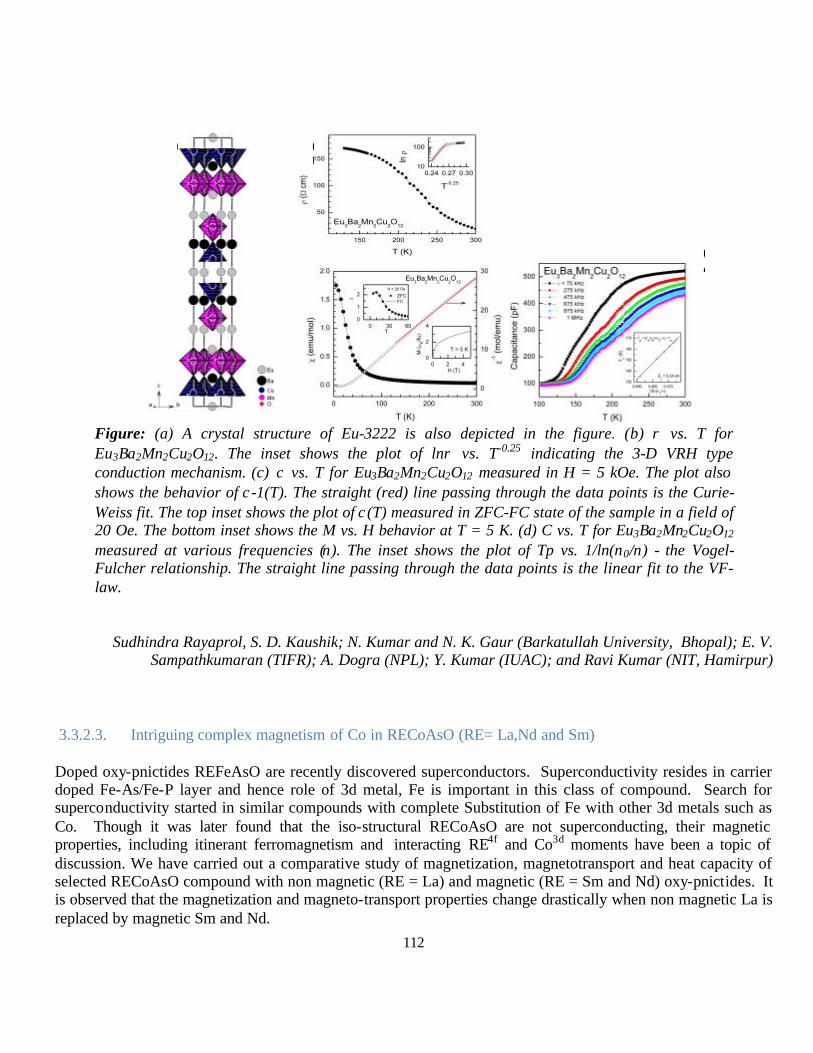
112
Figure: (a) A crystal structure of Eu-3222 is also depicted in the figure. (b) ρ vs. T for Eu3Ba2Mn2Cu2O12. The inset shows the plot of lnρ vs. T-0.25 indicating the 3-D VRH type conduction mechanism. (c) χ vs. T for Eu3Ba2Mn2Cu2O12 measured in H = 5 kOe. The plot also shows the behavior of χ-1(T). The straight (red) line passing through the data points is the Curie-Weiss fit. The top inset shows the plot of χ(T) measured in ZFC-FC state of the sample in a field of 20 Oe. The bottom inset shows the M vs. H behavior at T = 5 K. (d) C vs. T for Eu3Ba2Mn2Cu2O12 measured at various frequencies (ν). The inset shows the plot of Tp vs. 1/ln(ν0/ν) - the Vogel-Fulcher relationship. The straight line passing through the data points is the linear fit to the VF-law.
Sudhindra Rayaprol, S. D. Kaushik; N. Kumar and N. K. Gaur (Barkatullah University, Bhopal); E. V. Sampathkumaran (TIFR); A. Dogra (NPL); Y. Kumar (IUAC); and Ravi Kumar (NIT, Hamirpur)
3.3.2.3. Intriguing complex magnetism of Co in RECoAsO (RE= La,Nd and Sm)
Doped oxy-pnictides REFeAsO are recently discovered superconductors. Superconductivity resides in carrier doped Fe-As/Fe-P layer and hence role of 3d metal, Fe is important in this class of compound. Search for superconductivity started in similar compounds with complete Substitution of Fe with other 3d metals such as Co. Though it was later found that the iso-structural RECoAsO are not superconducting, their magnetic properties, including itinerant ferromagnetism and interacting RE4f and Co3d moments have been a topic of discussion. We have carried out a comparative study of magnetization, magnetotransport and heat capacity of selected RECoAsO compound with non magnetic (RE = La) and magnetic (RE = Sm and Nd) oxy-pnictides. It is observed that the magnetization and magneto-transport properties change drastically when non magnetic La is replaced by magnetic Sm and Nd.
(b
(c
(d
(a

113
Polycrystalline samples LaCoAsO, NdCoAsO and SmCoAsO were prepared by solid state reaction. XRD analysis indicates that these compounds crystallized in P4/nmm space group with lattice parameters a and c as 4.086(3)Å, 8.358(2)Å for La, 3.965(1)Å, 8.277(4)Å for Nd and 3.957(3)Å, 8.242(2)Å for Sm, the lattice parameters clearly follow the ionic size variation of trivalent La, Nd, and Sm.
LaCoAsO exhibits simple PM-FM transition at ~80K. Compound is paramagnetic with linear MH plots and at lower temperatures (5, 50K), remains ferromagnetic with nearly no coercive field (Hc). NdCoAsO is PM above 70K, FM between 70K down to 20K and AFM below 20K at low field at higher field FM & AFM not clearly seen. The SmCoAsO show successive PM-FM-AFM transitions at very low field. The MH at 10 and 20K shows meta-magnetic like shallow. Small hysteresis is observed in MH at 20K and 20kOe field at the meta-magnetic transition shoulder. LaCoAsO is metallic with reasonable magnetoresistance (MR) appears below Currie temperature (Tc) and is maximum at 60K. NdCoAsO shows a step like up turn at around 18K in zero field and shifts to lower temperature (18K to 4K) with an applied field of 140kOe.
The unusual increase of MR at lower temperatures is due to the ordering of Nd4f. SmCoAsO does not show any step like transition in zero field, but shows this step like feature under field. It may infer that Sm4f spins comes to similar ordered state under field as that of Nd in zero field.

114
The CP(T) plots are smooth and as such various Co magnetic orderings are not seen. The expected TN
Sm (AFM) is seen as a peak in CP(T) at 5.4K, and for Nd below 2K, in SmCoAsO and NdCoAsO respectively. In CP/T vs T, slope changes first near 80K, which roughly coincides with the FM ordering of Co spins.
At around 20K and 15K, the change in CP/T vs T plots are coincide with the complex AFM ordering of Sm4f-Co3d and Nd4f-Co3d interplayed matrix. On the other hand in LaCoAsO, the only ordering seen is for Co spins with Tc
Co at around 80K.
All the studies sample are in single phase. The Co, in these compounds is in itinerant ferromagnetic state with its paramagnetic moment above 1.5µB and it orders ferro-magnetically (FM) with small saturation moment of around 0.20µB below ~80K. This bulk intrinsic magnetism of Co changes dramatically when nonmagnetic La is changed by magnetic Sm and Nd. The transition of Co spins from FM to AFM, for magnetic Sm and Nd in RECoAsO is both field and temperature dependent. It is clear that Sm/Nd magnetic moments interact with the ordered Co spins in adjacent layer and thus transforms the FM ordering to AFM. All the studied compounds are metallic in nature, and their magnetotransport R(T)H follows the temperature and field dependent FM-AFM transition of ordered Co spins up, the magnetization results of RECoAsO (RE = La, Nd and Sm), though all the three are itinerant ferromagnets, the Nd and Sm undergoes successive FM-AFM transformation at low temperatures. The nature of transformation (FM-AFM) appears qualitatively same for both Nd and Sm, yet some intrinsic differences between two have been observed.
These findings were presented in 55th Conference on Magnetism and Magnetic Material 2010, Nov 14- 18, 2010 held at Atlanta USA.
S. D. Kaushik; Anand Pal, V.P.S. Awana and H. Kishan (NPL); Mushahid Hussain (Jamia Millia Islamia); M.Tropeano (CNR-SPIN, Universita de Genova, Itali.
3.3.3. Dielectric Studies
3.3.3.1. Low temperature dielectric measurements on FeTiMO6 (M=Ta,Nb,Sb):
Low temperature dielectric measurements on FeTiMO6 (M=Ta,Nb,Sb) rutile-type oxides at frequencies from 0.1Hz to 1MHz revealed anomalous dielectric relaxations with frequency dispersion. Unlike the high-temperature relaxor response of these materials, these low temperature relaxations are polaronic in nature. The relationship between frequency and temperature of dielectric loss peak follows T-1/4 behaviour. The frequency dependence of a.c.conductivity shows the well-known universal die lectric response (UDR), while the d.c. conductivity follows Mott variable range hopping (VRH) behaviour, confirming the polaronic origin of the
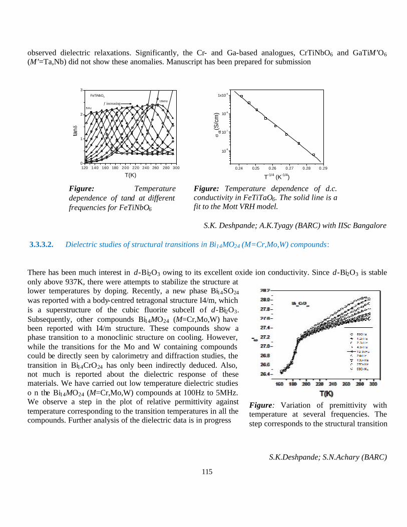
115
observed dielectric relaxations. Significantly, the Cr- and Ga-based analogues, CrTiNbO6 and GaTiM’O6 (M’=Ta,Nb) did not show these anomalies. Manuscript has been prepared for submission
Figure: Temperature dependence of tanδ at different frequencies for FeTiNbO6
Figure: Temperature dependence of d.c. conductivity in FeTiTaO6. The solid line is a fit to the Mott VRH model.
S.K. Deshpande; A.K.Tyagy (BARC) with IISc Bangalore
3.3.3.2. Dielectric studies of structural transitions in Bi14MO24 (M=Cr,Mo,W) compounds:
There has been much interest in δ-Bi2O3 owing to its excellent oxide ion conductivity. Since δ-Bi2O3 is stable only above 937K, there were attempts to stabilize the structure at lower temperatures by doping. Recently, a new phase Bi14SO24 was reported with a body-centred tetragonal structure I4/m, which is a superstructure of the cubic fluorite subcell of δ-Bi2O3. Subsequently, other compounds Bi14MO24 (M=Cr,Mo,W) have been reported with I4/m structure. These compounds show a phase transition to a monoclinic structure on cooling. However, while the transitions for the Mo and W containing compounds could be directly seen by calorimetry and diffraction studies, the transition in Bi14CrO24 has only been indirectly deduced. Also, not much is reported about the dielectric response of these materials. We have carried out low temperature dielectric studies o n the Bi14MO24 (M=Cr,Mo,W) compounds at 100Hz to 5MHz. We observe a step in the plot of relative permittivity against temperature corresponding to the transition temperatures in all the compounds. Further analysis of the dielectric data is in progress
S.K.Deshpande; S.N.Achary (BARC)
120 140 160 180 200 220 240 260 280 3000
1
2
3
16kHz
5Hz
f increasing
FeTiNbO6
tanδ
T(K)0.24 0.25 0.26 0.27 0.28 0.29
10-8
10-7
10-6
1x10⁻⁵
σdc
(S/c
m)
T-1/4 (K-1/4)
Figure: Variation of premittivity with temperature at several frequencies. The step corresponds to the structural transition

116
3.3.3.3. Dielectric response of La0.5Ca0.5-xSrxMnO3 (0.1 = x = 0.4) manganites:
The dielectric behavior of half-doped manganites La0.5Ca0.5-
xSrxMnO3 (0.1 = x = 0.4) with varying magnetic ground states was studied at low temperatures (293K to 123K) over a frequency range of 100Hz to 5MHz. The real part of relative permittivity as a function of temperature, e′(T), exhibits a maximum around the ferromagnetic (TC) and charge ordering transition (TCO) temperatures accompanied with high dielectric losses. In contrast to samples having x = 0.3, for x = 0.4, the permittivity exhibits a strong temperature dependence in the vicinity of magnetic phase transitions. This behavior may be correlated with the presence of competing magnetic interactions (magnetic polarons) close to the magnetic transitions. This work has been published in the Journal of Applied Physics
S.K.Deshpande ; Amitabh Das (BARC).
3.3.3.4. Low temperature dielectric measurements on La2CoMnO6:
Low temperature dielectric measurements were carried out on the double-perovskite La2CoMnO6, annealed under different ambient conditions, to understand the role of preparation conditions on structure and physical properties. The plot of dielectric loss factor tanδ against temperature shows dielectric relaxations arising from polaronic conduction. The relaxations appear to be dependent on the synthesis conditions. Further studies on similar double-perovskites are being carried out
S.K.Deshpande ; S.N.Achary, (BARC)
Figure:. Relaxation time as a function of 1000/T for La0.5Ca0.5-xSrxMnO3(x=0.1). Solid line is the Arrhenius fit. Inset shows the temperature dependence of dielectric constant at selected frequencies
Temperature dependence of tanδ at different frequencies for La2CoMnO6
-160 -140 -120 -100 -80 -60 -40 -20 0 200.0
0.5
1.0
1.5
2.0
2.5
3.0
3.5
4.0
4.5
5.0
5.5
6.0
La2CoMnO6 13kHz 58kHz 120kHz 500kHz 1MHz 5MHz
tanδ
T(C)
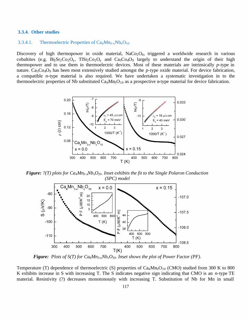
117
3.3.4. Other studies
3.3.4.1. Thermoelectric Properties of Ca4Mn3-xNbxO10
Discovery of high thermopower in oxide material, NaCo2O4, triggered a worldwide research in various cobaltites (e.g. Bi2Sr2Co2Oy, TlSr2Co2Oy and Ca3Co4O9 largely to understand the origin of their high thermopower and to use them in thermoelectric devices. Most of these materials are intrinsically p-type in nature. Ca3Co4O9 has been most extensively studied amongst the p-type oxide material. For device fabrication, a compatible n-type material is also required. We have undertaken a systematic investigation in to the thermoelectric properties of Nb substituted Ca4Mn3O10 as a prospective n-type material for device fabrication.
Figure: ?(T) plots for Ca4Mn3-xNbxO10. Inset exhibits the fit to the Single Polaron Conduction
(SPC) model
Figure: Plots of S(T) for Ca4Mn3-xNbxO10. Inset shows the plot of Power Factor (PF).
Temperature (T) dependence of thermoelectric (S) properties of Ca4Mn3O10 (CMO) studied from 300 K to 800 K exhibits increase in S with increasing T. The S indicates negative sign indicating that CMO is an n-type TE material. Resistivity (?) decreases monotonously with increasing T. Substitution of Nb for Mn in small

118
percentage (~ 5%) drastically alters the S and ? properties of CMO. We also find that the power factor (PF = S2/?) of CMO can be enhanced by one order of magnitude, without altering the crystal structure.
Sudhindra Rayaprol; and Shovit Bhattacharya (BARC)
3.3.4.2. Synthesis, Structure and Physical Properties of a New Germanide: Eu2AuGe3
Fig. 1: View of the Eu2AuGe3 structure approximately along the a axis. Europium, gold, and germanium atoms are drawn as medium gray, black filled, and open circles, respectively. The two-dimensional [AuGe3] networks, atom labels, and relevant interatomic distances.
Fig. 2: Magnetic susceptibility measured for a single crystal of Eu2AuGe3 in two different orientations, magnetic field along the c-axis (H || c) and magnetic field perpendicular to the c-axis (H ⊥ c). The measurements were done in both zero field cooled (ZFC) and field cooled (FC) states of the crystal. The ZFC data are shown as open symbols, where as the FC data is shown as continuous line. The inset shows the low temperature antiferromagnetic peak around 11 K seen in both orientations. The inset also highlights the magnetic anisotropy between the two orientations.
The germanide Eu2AuGe3 was obtained as large single crystals in high yield from a reaction of the elements in liquid indium. At room temperature Eu2AuGe3 crystallizes with the Ca2AgSi3 type ordered variant of the AlB2 type structure in space group Fmmm. The gold and germanium atoms build up slightly distorted graphite-like layers which consist of Ge6 and Au2Ge4 hexagons, leading to two different hexagonal-prismatic coordination environments for the europium atoms. Magnetic susceptibility data showed Curie-Weiss law behavior above 50 K and antiferromagnetic ordering at 11 K. The experimentally measured magnetic moment indicates divalent europium. The compound exhibits a distinct magnetic anisotropy based on single crystal measurements and at 5 K it shows a metamagnetic transition at ~ 10 kOe. Electrical conductivity measurements show metallic behavior. The structural transition at 130 K observed in the single crystal data was very well supported by the conductivity measurements. 151Eu Mössbauer spectroscopic data show an isomer shift of -11.24 mm/s at 77 K, supporting the divalent character of europium. In the magnetically ordered regime one observes superposition of two signals with hyperfine fields of 26.0 (89%) and 3.5 (11%) T, respectively indicating differently ordered domains.
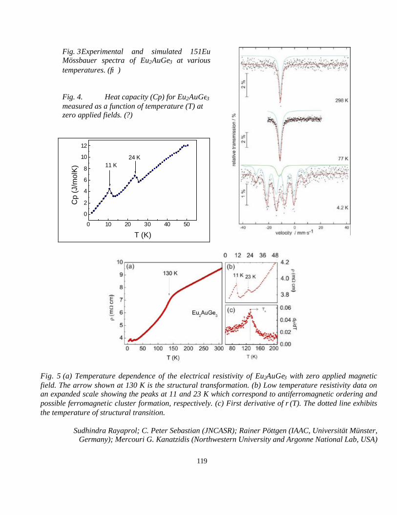
119
Fig. 3 Experimental and simulated 151Eu Mössbauer spectra of Eu2AuGe3 at various temperatures. (→)
Fig. 4. Heat capacity (Cp) for Eu2AuGe3 measured as a function of temperature (T) at zero applied fields. (?)
0 10 20 30 40 50
0
2
4
6
8
10
12
Cp
(J/m
olK
)
T (K)
11 K24 K
Fig. 5 (a) Temperature dependence of the electrical resistivity of Eu2AuGe3 with zero applied magnetic field. The arrow shown at 130 K is the structural transformation. (b) Low temperature resistivity data on an expanded scale showing the peaks at 11 and 23 K which correspond to antiferromagnetic ordering and possible ferromagnetic cluster formation, respectively. (c) First derivative of ρ(T). The dotted line exhibits the temperature of structural transition.
Sudhindra Rayaprol; C. Peter Sebastian (JNCASR); Rainer Pöttgen (IAAC, Universität Münster, Germany); Mercouri G. Kanatzidis (Northwestern University and Argonne National Lab, USA)

120
3.3.4.3. Anomalous heat capacity and X-ray photoelectron spectroscopy of Superconducting FeSe1/2Te1/2
Superconductivity has been observed in FeSe1-x system at ~8 K in its tetragonal form. It has been observed that superconductivity in this system is significantly affected by externally applied pressure and chalcogenide substitutions. In particular, an applied pressure up to 4.15 GPa enhances its Tc to ~37 K with (dTc/dP) of around 9K/GPa. The effect of chemical pressure has been studied in these systems by means of Se-site substitution. It is found that superconducting trans ition temperature increases with Te doping at Se site. In FeSe1-xTex system, Tc reaches a maximum at about 50% substitution, and then decreases with more Te doping. Interestingly, FeTe is no longer superconducting. We have studied the synthesis, structure, magnetization, heat capacity and X-ray photo electron spectroscopy (XPS) of FeSe1/2Te1/2 compound, with highest Tc value of ~14.5 K
Sample studied was prepared by solid state reaction and XRD was fitted in FULLPROF with P4/nmm space group, lattice parameters a = 3.793(3) Å and c = 6.015(2) Å. Refinement with better goodness of fitting parameters was obtained by taking Fe ions at two different sites (2a and 2c). The occupation of Fe at interstitial 2c site was found ~8%.
Fig. 1 Fig.2
The Fig (1) insets show the ? – T and ? -T plots demonstrating bulk superconductivity at Tc ~12K. Fig (2) shows the temperature dependent specific heat measurement. The room temperature CP is around 55 J/mol-K which is in general agreement with the reported values. The data was fitted using Cp= γT+BT3+CT5 above Tc from 13 K to 16 K. Where, γT and BT3+CT5 are electronic and phononic specific heat contributions respectively. Fitting of the curves reveals that γ ~57.73 mJ/mol-K2, however Debye temperature ~171 K. From this fitting we estimated corresponding normal state behavior below Tc. As far as the entropy contribution due to superconducting condensate is concerned, the superconducting state – normal state is plotted in upper inset of Fig (2). At Tc the discontinuity in electronic Cp vs. T plot is seen and marked. The shape of Lambda transition for studied FeSe1/2Te1/2 does not exhibit sharp discontinuity at Tc as seen for other superconductors, rather a broad hump like structure.

121
XPS, revealed well defined positions for Fe, Se and Te but with sufficient hybridization. The Fe (2p3/2) spectra could be resolved into three spin-orbit components at 706.65 eV, 708.50 eV and 712.00 eV after the carbon correction. A Fe (2p1/2) core level spectrum was also deconvoluted three peaks in similar way. The 3d core level spectrum [Fig 3(b)] of Te is deconvoluted into three peaks with binding energy 572.00 eV, 572.65 eV and 573.90 eV. The peak at energy 572.65 eV corresponds to pure Te metal while peak at lower binding energy 572.00 eV may have appeared due to the hybridization. In case of Se, the core level spectra [Fig 3(c)] are deconvoluted into two peaks with binding energies 53.52 eV and 55.28 eV. The peak for higher BE corresponds to pure Se while peak at lower BE appeared due to the hybridization. In Fig 3(a) the peak at binding energy 706.65 eV corresponds to pure Fe metal. The peak observed at 708.50 eV corresponds to FeO (Fe2+), whereas peak at 712.00 eV corresponds to a satellite transition that is characteristic feature of Fe XPS spectra. The absence of satellite peak (712.00 eV) and resemblance of 706.65 eV Fe metal peak was correlated with an itinerant character of Fe 3d electrons. However, the presence of satellite peak with Fe2+ peak and simultaneous appearance of Fe metal peak (706.65 eV) may be caused by charge-carrier localization induced by excess Fe at 2c site. The existence of itinerant character of Fe 3d electronic state caused by hybridization of 3d states of Fe ions at 2a site and Se/Te 4p/5p states.
We found that in FeSe1/2Te1/2 about 8% per mole of Fe occupies the interstitial 2c site. These Fe ions have localized magnetic moments which lead to a broad cusp like anomaly in electronic specific heat rather than well defined sharp lambda transition. This observation is further supported by our XPS measurements that show that Fe ions have two type of behavior. One, arising due to itinerant nature of Fe 3d electrons due to hybridization of Fe 3d and Se/Te 4p/5p states. Another, results from charge-carrier localization induced by excess Fe at 2c site.
These findings were presented in 55th Conference on Magnetism and Magnetic Material 2010, Nov 14- 18, 2010 held at Atlanta USA.
S. D. Kaushik; V.P.S. Awana, Govind, Anand Pal, Bhasker Gahtori, A. Vajpayee, Jagdish Kumar and H. Kishan (NPL)
3.3.4.4. Heat capacity from Tunneling spectra of ammonium alkali bromides
In the earlier work, inelastic neutron scattering measurements were carried out on (NH4)x(K,Rb)1-xBr compounds (with x = 0, 0.02, 0.05 and 0.10) to measure the tunneling spectra at low temperatures (2K). The main interest in these compounds is because their unusual specific heat behavior at low temperatures (< 20K),
3(a) 3(b) 3(c)

122
which does not follow Debye T3 law. The measured tunneling spectra (-0.2 meV to 0.7 meV) showed a tunneling peak at around 0.4 – 0.5 meV when NH4 is introduced into KBr and RbBr matrix. For the mixed salt of (NH4)0.02(K0.5Rb0.5)0.98Br, the tunneling peak disappears and a broad distribution around the elastic peak appears indicating a shift in the tunneling energies low values and also distribution of tunneling states. The tunneling spectra so measured are used as the density of states g(E) to calculate the specific contribution from the tunneling states. The g(E), measured for pure matrix, i.e., KBr and RbBr, was subtracted from g(E) of the samples to remove, phonic contribution, if present. The g(E = hω) so obtained is used to calculate the specific heat for different compounds using the relation
C = R
where R is the gas constant, g0, g1 are population of energy levels. The specific heat so obtained shows a peak around ~2K and decreases as the temperature increases and becomes negligibly small by 20K or so. Such kind of peak in specific heat arising from tunneling excitations is known as Schottky specific heat in the literature. Efforts are being made to correlate the calculated tunneling specific heat with the actual measured specific heat. For this comparison, one needs to identify correctly and subtract the phononic specific heat from the measured values. This process is in progress.
P. D. Babu; P S. Goyal, (Pillai College, Navi Mumbai); F. Jurayani (PSI, Switzerland)
Cryogen Production at Indore Centre
LHe Production Status LN2 Production Status
P. Saravanan
0
10
20
30
40
2005 2006 2007 2008 2009 2010
Litr
es in
Tho
usan
ds
0
40
80
120
160
2005 2006 2007 2008 2009 2010
Lite
rs in
Tho
usan
ds

123
4. New Facilities Acquired / Developed
4.1 At Indore Centre
4.1.1 Ferroelectric Loop Tracer
One commercial ferroelectric loop tracer (P-E loop tracer) supplied by M/s Radiant Instruments, USA capable of measuring ferroelectric hysteresis loops, leakage current, fatigue etc., on ceramics and thin films samples is installed. The following are the specifications of the system
Voltage: ± 100V & ± 10 KV (with external amplifier)
Frequency range ~ 100kHz - 0.03Hz
Minimum leakage current 2 pAmp
In-house fabricated test fixtures for measuring at low temperature (down to 100K), room temperature and high temperatures (~473K) are available . Poling of the ceramic samples is possible.
VR Reddy ([email protected])
4.1.2 High Pressure cell for resistivity/magnetoresistance
High Pressure cell for resistivity/magnetoresistance from easyLab Technologies, UK has been installed. With this addition resistivity and magnetoresistance measurements (1.5 K – 300 K and up to 8 Tesla magnetic field) are now possible in the presence of pressure up to 25 k bar. Figure shows giant magnetoresistance in Pd doped FeRh at room temperature under 19.9 kbar pressure. At ambient pressure it shows spontaneous AFM-FM transition around (TN) 200 K. The application of pressure shifts TN to higher temperature resulting in AFM state at room temperature. Therefore no transition is observed in the
0 50 100 150 200 250 300
10
20
30
40
19.9 kbar
0 Tesla cooling 8 Tesla (ZFCW) 8 Tesla (FCC) 8 Tesla (FCW)
Pd doped FeRh
Res
ista
nce
(mΩ
)
Temperature (K)
Photograph of P-E measurement from M/s Radiant Tech, USA along with in-house made sample holders
Photograph of P-E measurement from M/s Radiant Tech, USA along with in-house made sample holders
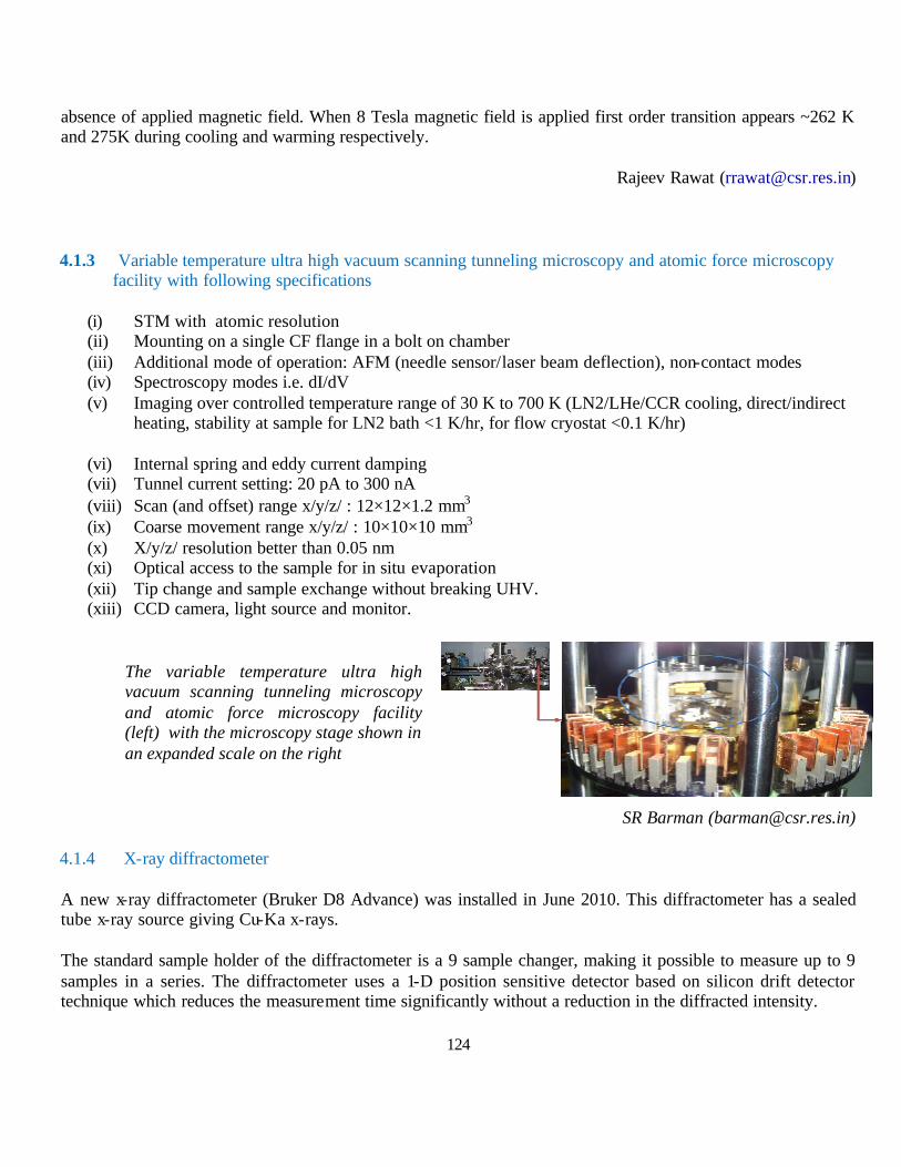
124
absence of applied magnetic field. When 8 Tesla magnetic field is applied first order transition appears ~262 K and 275K during cooling and warming respectively.
Rajeev Rawat ([email protected])
4.1.3 Variable temperature ultra high vacuum scanning tunneling microscopy and atomic force microscopy facility with following specifications
(i) STM with atomic resolution (ii) Mounting on a single CF flange in a bolt on chamber (iii) Additional mode of operation: AFM (needle sensor/laser beam deflection), non-contact modes (iv) Spectroscopy modes i.e. dI/dV (v) Imaging over controlled temperature range of 30 K to 700 K (LN2/LHe/CCR cooling, direct/indirect
heating, stability at sample for LN2 bath <1 K/hr, for flow cryostat <0.1 K/hr)
(vi) Internal spring and eddy current damping (vii) Tunnel current setting: 20 pA to 300 nA (viii) Scan (and offset) range x/y/z/ : 12×12×1.2 mm3 (ix) Coarse movement range x/y/z/ : 10×10×10 mm3 (x) X/y/z/ resolution better than 0.05 nm (xi) Optical access to the sample for in situ evaporation (xii) Tip change and sample exchange without breaking UHV. (xiii) CCD camera, light source and monitor.
SR Barman ([email protected])
4.1.4 X-ray diffractometer
A new x-ray diffractometer (Bruker D8 Advance) was installed in June 2010. This diffractometer has a sealed tube x-ray source giving Cu-Ka x-rays.
The standard sample holder of the diffractometer is a 9 sample changer, making it possible to measure up to 9 samples in a series. The diffractometer uses a 1-D position sensitive detector based on silicon drift detector technique which reduces the measurement time significantly without a reduction in the diffracted intensity.
The variable temperature ultra high vacuum scanning tunneling microscopy and atomic force microscopy facility (left) with the microscopy stage shown in an expanded scale on the right
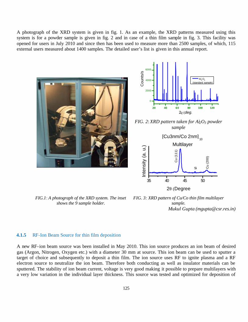
125
A photograph of the XRD system is given in fig. 1. As an example, the XRD patterns measured using this system is for a powder sample is given in fig. 2 and in case of a thin film sample in fig. 3. This facility was opened for users in July 2010 and since then has been used to measure more than 2500 samples, of which, 115 external users measured about 1400 samples. The detailed user’s list is given in this annual report.
FIG. 2: XRD pattern taken for Al2O3 powder
sample
FIG.1: A photograph of the XRD system. The inset
shows the 9 sample holder. FIG. 3: XRD pattern of Cu/Co thin film multilayer
sample. Mukul Gupta ([email protected])
4.1.5 RF-Ion Beam Source for thin film deposition
A new RF-ion beam source was been installed in May 2010. This ion source produces an ion beam of desired gas (Argon, Nitrogen, Oxygen etc.) with a diameter 30 mm at source. This ion beam can be used to sputter a target of choice and subsequently to deposit a thin film. The ion source uses RF to ignite plasma and a RF electron source to neutralize the ion beam. Therefore both conducting as well as insulator materials can be sputtered. The stability of ion beam current, voltage is very good making it possible to prepare multilayers with a very low variation in the individual layer thickness. This source was tested and optimized for deposition of
20 40 60 80 100 120
0
2000
4000
6000
Cou
nts/
s
2θ (deg.)
Al2 O3 (standard sample)
35 40 45 50
Cu
(200
)
Inte
nsity
(a.
u.)
2θ (Degree)
[Cu3nm/Co 2nm]10
Multilayer
Cu
(111
)
Si
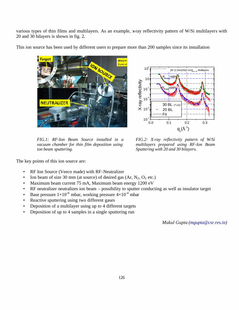
126
various types of thin films and multilayers. As an example, x-ray reflectivity pattern of W/Si multilayers with 20 and 30 bilayers is shown in fig. 2.
This ion source has been used by different users to prepare more than 200 samples since its installation
FIG.1: RF-Ion Beam Source installed in a vacuum chamber for thin film deposition using ion beam sputtering.
FIG.2: X-ray reflectivity pattern of W/Si multilayers prepared using RF-Ion Beam Sputtering with 20 and 30 bilayers.
The key points of this ion source are:
• RF Ion Source (Veeco made) with RF-Neutralizer • Ion beam of size 30 mm (at source) of desired gas (Ar, N2, O2 etc.) • Maximum beam current 75 mA, Maximum beam energy 1200 eV • RF neutralizer neutralizes ion beam – possibility to sputter conducting as well as insulator target • Base pressure 1×10⁻⁸ mbar, working pressure 4×10⁻⁴ mbar • Reactive sputtering using two different gases • Deposition of a multilayer using up to 4 different targets • Deposition of up to 4 samples in a single sputtering run
Mukul Gupta ([email protected])
0.0 0.1 0.2 0.310- 7
10- 5
10- 3
10- 1
10
103
RBragg
=68%
X-r
ay re
flect
ivity
qz(Å-1)
30 BL (×100)
20 BL Fit
[W (1.5nm)/Si(3 nm)]20 or 30
Multilayers
RBragg
=64%

127
4.2 Kalpakkam Node
Building construction was completed in April 2011. The building was inaugurated by Dr. S. Banerjee, Chairman, AEC & Secretary, DAE on April 28, 2011. Dr. Baldev Raj, Director,IGCAR and Prof. Ajay Gupta, Centre Director, UGC-DAE CSR, Indore Centre and other senior scientists from IGCAR as well as research students participated in the inaugural function.
Dr. S. Banerjee, Chairman, AEC & Secretary, DAE, Dr. Baldev Raj, Director, IGCAR, Prof. Ajay Gupta, Centre Director, UGC-DAE CSR, Indore, Dr. G. Amarendra, Scientist In-Charge, UGC-DAE CSR, Kalpakkam Node during the inaugural function on April 28, 2011.
Advanced experimental facilities such as Infrared float zone single Crystal Growth Furnace, SEM, TEM, Ball Milling, Ball Indentation and Small Punch Creep apparatus have been installed in Physical and Engineering science wings.
Physical Sciences wing UGC-DAE CSR, Kalpakkam Node Engineering Sciences wing
G. Amarendra ([email protected])

128
5. Publications in Journals
Names of authors from Universities and colleges are shown in bold face, names of authors from UGC-DAE CSR are underlined and those from DAE marked by #
5.1 Publications from Collaborative research: 1. Room temperature ferromagnetism in Cr doped chalcopyrite type Cu–Zn–Se compound, D. Paul Joseph and C.
Venkateswaran, Phys. Status Solidi A, 207, No. 11, 2549–2552 (2010).
2. Fabrication of dye sensitized solar cell using Cr doped Cu–Zn–Se type chalcopyrite thin film, D. Paul Joseph, S. Ganesan, M. Kovendhan, S. Austin Suthanthiraraj, P. Maruthamuthu, and C. Venkateswaran, Phys. Status Solidi A, 1–6 (2011) / DOI 10.1002/pssa.201026368.
3. A study of microstructural and optical properties of nanocrysialline ceria thin films prepared by pulsed laser ablation, G. Balakrishnan, S. Tripura Sundari, P. Kuppusami#, P. Chandra Mohan, M.P. Srinivasan, E. Mohandas, V. Ganesan, D. Sastikumar, Thin Solid Films 519 2520 (2011)
4. Ac-Susceptibility Study In Rare Earth Substituted Magnetite Ferrofluids, R. V. Upadhyaya, Kinnari Parekh#, A. Banerjee and Kranti Kumar, Physics Procedia., 9, 32 (2010).
5. Ag15+ and O7+ ion irradiation induced improvement in dielectric properties of the Ba(Co1/3Nb2/3)O3 thin films Bhagwati Bishnoi, P.K. Mehta, C.J. Panchal, M.S. Desai, Ravi Kumar, V. Ganesan Journal of Alloys and Compounds 509 2745 (2011)
6. Antiferro to superparamagnetic transition on Mn doping in NiO P. Mallick, Chandana Rath, A. Rath, A. Banerjee and N.C. Mishra. Solid State Commun., 150, 1342 (2010).
7. Study of electronic structure and magnetization correlations in hydrogenated and vacuum annealed Ni doped ZnO, R. K. Singhal, S. C. Sharma, P. Kumari, Sudhish Kumar, Y. T. Xing, U. P. Deshpande, T. Shripathi, and Elisa Saitovitch, J. Appl. Phys. 109, (2011) 063907-14 doi:10.1063/1.3556458.
8. Switch ‘on’ and ‘off’ ferromagnetic ordering through the induction and removal of oxygen vacancies and carriers in doped ZnO: A magnetization and electronic structure study, R. K. Singhal, A. Samariya, S. Kumar , Y. T. Xing, U. P. Deshpande, T. Shripathi, S. N. Dolia, and Elisa B. Saitovitch, physica status solidi (a) 207 (2010) 2373–2386, DOI:10.1002/pssa.200925637.
9. A close correlation between induced ferromagnetism and oxygen deficiency in Fe doped In2O3, R.K. Singhal, A. Samariya, Sudhish Kumar, S.C. Shar ma, Y.T. Xing, U.P. Deshpande,T. Shripathi, E. Saitovitch, Applied Surface Science 257 (2010) 1053–1057, doi:10.1016/j.apsusc.2010.07.106.
10. Defect-induced reversible ferromagnetism in Fe-doped ZnO semiconductor: An electronic structure and magnetization study, Arvind Samariya, R.K.Singhal, Sudhish Kumar, Y.T.Xing, Mariella Alzamora, S.N.Dolia, U.P. Deshpande, T.Shripathi, Elisa B.Saitovitch, Materials Chemistry and Physics 123 (2010) 678 – 85
11. Structural, optical, and electrical characteristics of 70 Mev Si5+ ion irradiation-induced nanoclusters of gallium Nitride, S. Suresh, V. Ganesh, U. P. Deshpande, T. Shripathi, K. Asokan, D. Kanjilal, K. Baskar , J Mater Sci, 46 (2011) 1015-21, DOI 10.1007/s10853-010-4866-9.
12. Effect of hydrogenation vs. re-heating on intrinsic magnetization of Co doped In2O3, A. Samariya, R.K. Singhal, Sudhish Kumar, Y.T. Xing, S.C. Sharma, P. Kumari, D.C. Jain, S.N. Dolia, U.P. Deshpande, T. Shripathi, E. Saitovitch, Applied Surface Science 257 (2010) 585–590
13. Correlation between microstructure, magnetic and electronic properties of Fe1- xAlx (0.2=x=0.6) alloys produced by arc melting, R. Brajpuriya, P. Sharma, S. Jani , S. Kaimal, T. Shripathi, N. Lakshmi and K. Venugopalan, Applied Surface Science 257, (2010), 10-16.
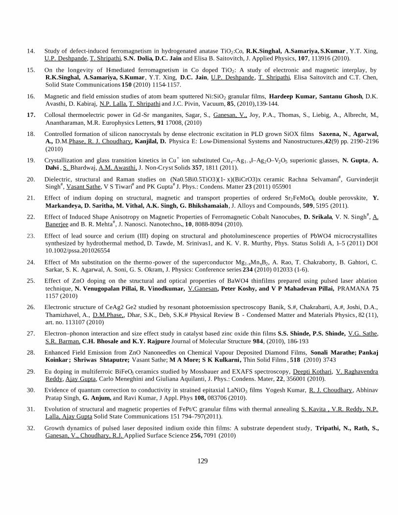
129
14. Study of defect-induced ferromagnetism in hydrogenated anatase TiO2:Co, R.K.Singhal, A.Samariya, S.Kumar , Y.T. Xing, U.P. Deshpande, T. Shripathi, S.N. Dolia, D.C. Jain and Elisa B. Saitovitch, J. Applied Physics, 107, 113916 (2010).
15. On the longevity of H-mediated ferromagnetism in Co doped TiO2: A study of electronic and magnetic interplay, by R.K.Singhal, A.Samariya, S.Kumar , Y.T. Xing, D.C. Jain, U.P. Deshpande, T. Shripathi, Elisa Saitovitch and C.T. Chen, Solid State Communications 150 (2010) 1154-1157.
16. Magnetic and field emission studies of atom beam sputtered Ni:SiO2 granular films, Hardeep Kumar, Santanu Ghosh, D.K. Avasthi, D. Kabiraj, N.P. Lalla, T. Shripathi and J.C. Pivin, Vacuum, 85, (2010),139-144.
17. Collosal thermoelectric power in Gd -Sr manganites, Sagar, S., Ganesan, V., Joy, P.A., Thomas, S., Liebig, A., Albrecht, M., Anantharaman, M.R. Europhysics Letters, 91 17008, (2010)
18. Controlled formation of silicon nanocrystals by dense electronic excitation in PLD grown SiOX films Saxena, N., Agarwal, A., D.M.Phase, R. J. Choudhary, Kanjilal, D. Physica E: Low-Dimensional Systems and Nanostructures,42(9) pp. 2190-2196 (2010)
19. Crystallization and glass transition kinetics in Cu+ ion substituted Cux–Ag1- xI–Ag2O–V2O5 superionic glasses, N. Gupta, A. Dalvi , S. Bhardwaj, A.M. Awasthi, J. Non-Cryst Solids 357, 1811 (2011).
20. Dielectric, structural and Raman studies on (Na0.5Bi0.5TiO3)(1- x)(BiCrO3)x ceramic Rachna Selvamani#, Gurvinderjit Singh#, Vasant Sathe, V S Tiwari# and PK Gupta# J. Phys.: Condens. Matter 23 (2011) 055901
21. Effect of indium doping on structural, magnetic and transport properties of ordered Sr2FeMoO6 double perovskite, Y. Markandeya, D. Saritha, M. Vithal, A.K. Singh, G. Bhikshamaiah , J. Alloys and Compounds, 509, 5195 (2011).
22. Effect of Induced Shape Anisotropy on Magnetic Properties of Ferromagnetic Cobalt Nanocubes, D. Srikala, V. N. Singh#, A. Banerjee and B. R. Mehta#, J. Nanosci. Nanotechno., 10, 8088-8094 (2010).
23. Effect of lead source and cerium (III) doping on structural and photoluminescence properties of PbWO4 microcrystallites synthesized by hydrothermal method, D. Tawde, M. Srinivas1, and K. V. R. Murthy, Phys. Status Solidi A, 1–5 (2011) DOI 10.1002/pssa.201026554
24. Effect of Mn substitution on the thermo -power of the superconductor Mg1-xMnxB2, A. Rao, T. Chakraborty, B. Gahtori, C. Sarkar, S. K. Agarwal, A. Soni, G. S. Okram, J. Physics: Conference series 234 (2010) 012033 (1-6).
25. Effect of ZnO doping on the structural and optical properties of BaWO4 thinfilms prepared using pulsed laser ablation technique, N. Venugopalan Pillai, R. Vinodkumar, V.Ganesan, Peter Koshy, and V P Mahadevan Pillai, PRAMANA 75 1157 (2010)
26. Electronic structure of CeAg2 Ge2 studied by resonant photoemission spectroscopy Banik, S.#, Chakrabarti, A.#, Joshi, D.A., Thamizhavel, A., D.M.Phase,, Dhar, S.K., Deb, S.K.# Physical Review B - Condensed Matter and Materials Physics, 82 (11), art. no. 113107 (2010)
27. Electron–phonon interaction and size effect study in catalyst based zinc oxide thin films S.S. Shinde, P.S. Shinde, V.G. Sathe, S.R. Barman, C.H. Bhosale and K.Y. Rajpure Journal of Molecular Structure 984, (2010), 186-193
28. Enhanced Field Emission from ZnO Nanoneedles on Chemical Vapour Deposited Diamond Films, Sonali Marathe; Pankaj Koinkar ; Shriwas Shtaputre; Vasant Sathe; M A More; S K Kulkarni, Thin Solid Films , 518 (2010) 3743
29. Eu doping in multiferroic BiFeO3 ceramics studied by Mossbauer and EXAFS spectroscopy, Deepti Kothari, V. Raghavendra Reddy, Ajay Gupta, Carlo Meneghini and Giuliana Aquilanti, J. Phys.: Condens. Mater, 22, 356001 (2010).
30. Evidence of quantum correction to conductivity in strained epitaxial LaNiO3 films Yogesh Kumar, R. J. Choudhary, Abhinav Pratap Singh, G. Anjum, and Ravi Kumar, J Appl. Phys 108, 083706 (2010).
31. Evolution of structural and magnetic properties of FePt/C granular films with thermal annealing S. Kavita , V.R. Reddy, N.P. Lalla, Ajay Gupta Solid State Communications 151 794–797(2011).
32. Growth dynamics of pulsed laser deposited indium oxide thin films: A substrate dependent study, Tripathi, N., Rath, S., Ganesan, V., Choudhary, R.J. Applied Surface Science 256, 7091 (2010)

130
33. Highly conductive and transparent laser ablated nanostructured Al: ZnO thin films Vinodkumar, R., Navas, I., Chalana, S.R., Gopchandran, K.G., Ganesan, V., Philip, R., Sudheer, S.K., Mahadevan Pillai, V.P. Applied Surface Science 257, 708 (2010)
34. High-TC phase transition in K2Ti6O 13 lead-free ceramic synthesised using solid-state reaction Vikram, S.V., D.M.Phase, Chandel, V.S. Journal of Materials Science: Materials in Electronics,21 (9) pp. 902-905 (2010).
35. Influence Europium oxide on the Structural and Optical properties of Pulsed Laser Ablated Barium Tungstate ThinFilms, N.Venugopalan Pillai, P.Mahadevanpillai, R.Vinodkumar, I.Navas, V.Ganesan, Peter Koshy Journal of Alloys and Compounds 509 2745 (2011)
36. Influence of Bi substitution on microwave dielectric properties of BaO–La2O3–Sm2O3–TiO2 ceramics, S. Bindra Narang, Shalini Bahel, S. Dash, J Mater Sci: Mater Electron, 21, 1186 (2010).
37. Influence of silver doping on the electrical and magnetic behavior of La0.7Ca0.3MnO3 manganites, Y. Kalyana Lakshmi, P. Venugopal Reddy, Solid State Sciences, 12, 1731 (2010).
38. Inspection of Multiferroicity in BiMn2-xTixO5 ceramics through specific heat and Raman spectroscopic studies D.K.Shukla,Ravi kumar, S.Mollahl, R.J.Choudary , P.N.Vishwakarma , V.G.Sathe and V.Ganesan Journal of Physics: Condensed Matter ,22,485901(2010)
39. Intrinsic ferromagnetism and magnetic anisotropy in Gd -doped ZnO thin films synthesized by pulsed spray pyrolysis method M. Subramanian, P. Thakur, M. Tanemura, T. Hihara, V. Ganesan, T. Soga,K. H. Chae, R. Jayavel, and T. Jimbo, Journal of Applied Physics 108, 053904 (2010)
40. Investigation of ground state in sodium doped neodymium manganites, Y. Kalyana Lakshmi, P. Venugopal Reddy , Phys. Lett. A, 375, 1543 (2011).
41. Investigation of mixed spinel structure of nanostructured nickel ferrite, John Jacob and M. Abdul Khadar, J. Appl. Phys., 107, 114310 (2010).
42. Magnetic and electrical behavior of Al doped La0.7Ca0.3MnO3 manganites, Shailja Tiwari, D. M. Phase, R. J. Choudhary, H. S. Mund, and B. L. Ahuja, J. Appl. Phys. 109, 033911 (2011).
43. Magnetic field dependence of the pinning effect in BaZrO3-doped YBa2Cu3O7-δ ceramic superconductor, A. Mohanta and D. Behera, Solid State Commun., 150, 1325 (2010).
44. Magnetic properties of nanoparticles of cobalt chromite, Chandana Rath, P. Mohanty and A. Banerjee, J. Magn. Magn. Mater., 323, 1698 (2011).
45. Magneto Transport of La0.70Ca0.3-xSrxMnO3(Ag):A Potential room temperature bolometer and magnetic sensor, V.P.S.Awana,Rahul Tripathi,Neeraj Kumar, H.Kishan, G.L.Bhalla, R.Zeng, L.S,Sharath Chandra, V.Ganesan, and H.U.Habermeier, J.Appl.Phys. 107, 09D723 (2010)
46. Magnetocaloric effect and magnetoresistance of Ni–Fe–Ga alloys D Pal# and K Mandal#, J. Phys. D: Appl. Phys. 43 (2010) 455002.
47. Magnetocaloric effect in HoMnO3 crystal A.Midya, P. Mandal, S. Das, S. Banerjee, L. S. Sharath Chandra, V. Ganesan, and S. Roy Barman Applied Physics Letters 96, 142514 (2010)
48. Magneto-electric behavior of ferrimagnetic BixCo2-xMnO4 (x = 0, 0.1 and 0.3) thin films. N.E. Rajeevan, Ravi Kumar, D. K. Shukla, R. J. Choudhary, P. P. Pradyumnan, P. Thakur, A. K. Singh, S. Patnaik, S. K. Arora, I. V. Shvets, J. Mag. Mag Mat. 323 (2011) 1760.
49. Modification in field sensitivity of manganite based multilayered device using different geometry. P.S. Vachhani, J.H. Markna, D.G. Kuberkar, Materials Science and Engineering B 172 (2010) 183.
50. Mossbauer and magnetic studies in nickel ferrite nanoparticles: Effect of size distribution, Rakesh Malik, S. Annapoorni, Subhalakshmi Lamba , V.Raghavendra Reddy, Ajay Gupta, Parmanand Sharma, Akihisa Inoue., J. Magn. Magn. Materials 322 (2010) 3742
51. Mössbauer studies and magnetic studies of Ni doped orthoferrites PrFe1-xNixO3 (x ≤ 0.3), Abida Bashir Makhdoomi, M. Ikram, Ravi Kumar, J. Magn. Magn. Mater., 322, 2581 (2010).
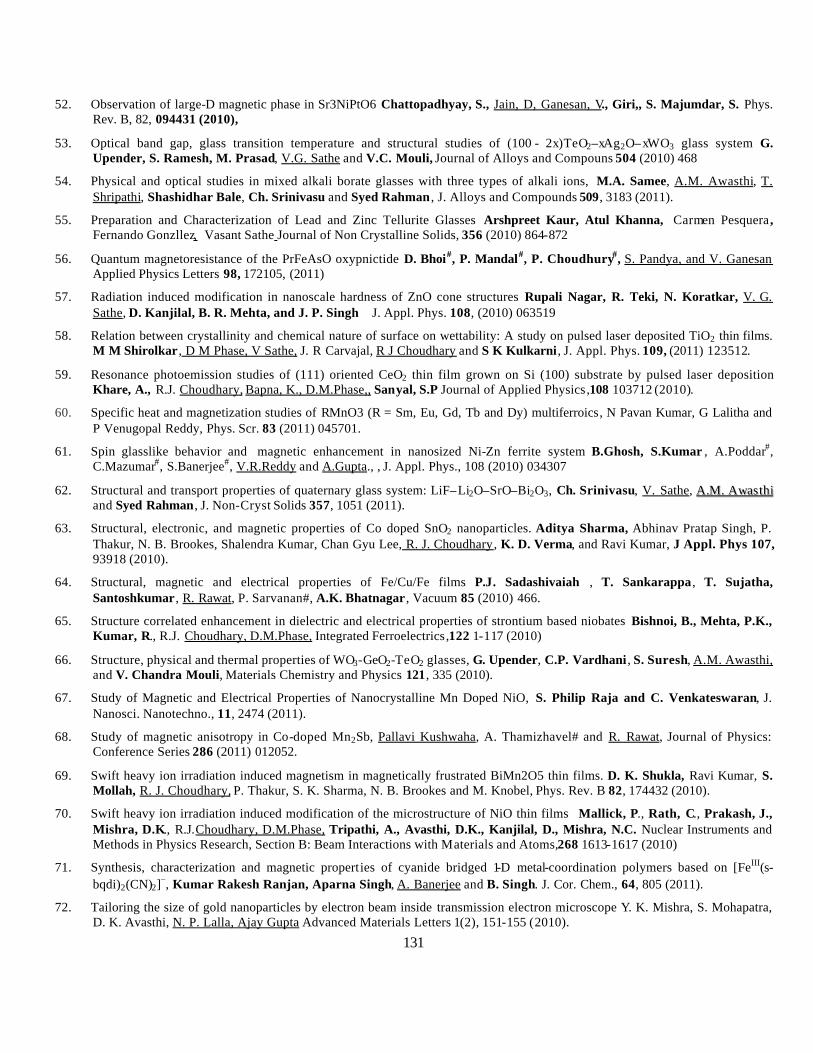
131
52. Observation of large-D magnetic phase in Sr3NiPtO6 Chattopadhyay, S., Jain, D, Ganesan, V., Giri,, S. Majumdar, S. Phys. Rev. B, 82, 094431 (2010),
53. Optical band gap, glass transition temperature and structural studies of (100 - 2x)TeO2–xAg2O–xWO3 glass system G. Upender, S. Ramesh, M. Prasad, V.G. Sathe and V.C. Mouli, Journal of Alloys and Compouns 504 (2010) 468
54. Physical and optical studies in mixed alkali borate glasses with three types of alkali ions, M.A. Samee, A.M. Awasthi, T. Shripathi, Shashidhar Bale, Ch. Srinivasu and Syed Rahman , J. Alloys and Compounds 509, 3183 (2011).
55. Preparation and Characterization of Lead and Zinc Tellurite Glasses Arshpreet Kaur, Atul Khanna, Carmen Pesquera , Fernando Gonzllez, Vasant Sathe Journal of Non Crystalline Solids, 356 (2010) 864-872
56. Quantum magnetoresistance of the PrFeAsO oxypnictide D. Bhoi#, P. Mandal#, P. Choudhury#, S. Pandya, and V. Ganesan Applied Physics Letters 98, 172105, (2011)
57. Radiation induced modification in nanoscale hardness of ZnO cone structures Rupali Nagar, R. Teki, N. Koratkar, V. G. Sathe, D. Kanjilal, B. R. Mehta, and J. P. Singh J. Appl. Phys. 108, (2010) 063519
58. Relation between crystallinity and chemical nature of surface on wettability: A study on pulsed laser deposited TiO2 thin films. M M Shirolkar , D M Phase, V Sathe, J. R Carvajal, R J Choudhary and S K Kulkarni, J. Appl. Phys. 109, (2011) 123512.
59. Resonance photoemission studies of (111) oriented CeO2 thin film grown on Si (100) substrate by pulsed laser deposition Khare, A., R.J. Choudhary, Bapna, K., D.M.Phase,, Sanyal, S.P Journal of Applied Physics,108 103712 (2010).
60. Specific heat and magnetization studies of RMnO3 (R = Sm, Eu, Gd, Tb and Dy) multiferroics, N Pavan Kumar, G Lalitha and P Venugopal Reddy, Phys. Scr. 83 (2011) 045701.
61. Spin glasslike behavior and magnetic enhancement in nanosized Ni-Zn ferrite system B.Ghosh, S.Kumar , A.Poddar#, C.Mazumar#, S.Banerjee#, V.R.Reddy and A.Gupta., , J. Appl. Phys., 108 (2010) 034307
62. Structural and transport properties of quaternary glass system: LiF–Li2O–SrO–Bi2O3, Ch. Srinivasu, V. Sathe, AA..MM.. AAwwaass tthh ii and Syed Rahman, J. Non-Cryst Solids 357, 1051 (2011).
63. Structural, electronic, and magnetic properties of Co doped SnO2 nanoparticles. Aditya Sharma, Abhinav Pratap Singh, P. Thakur, N. B. Brookes, Shalendra Kumar, Chan Gyu Lee, R. J. Choudhary, K. D. Verma, and Ravi Kumar, J Appl. Phys 107, 93918 (2010).
64. Structural, magnetic and electrical properties of Fe/Cu/Fe films P.J. Sadashivaiah , T. Sankarappa , T. Sujatha, Santoshkumar , R. Rawat, P. Sarvanan#, A.K. Bhatnagar , Vacuum 85 (2010) 466.
65. Structure correlated enhancement in dielectric and electrical properties of strontium based niobates Bishnoi, B., Mehta, P.K., Kumar, R., R.J. Choudhary, D.M.Phase, Integrated Ferroelectrics,122 1-117 (2010)
66. Structure, physical and thermal properties of WO3-GeO2-TeO2 glasses, G. Upender, C.P. Vardhani , S. Suresh, A.M. Awasthi, and V. Chandra Mouli, Materials Chemistry and Physics 121, 335 (2010).
67. Study of Magnetic and Electrical Properties of Nanocrystalline Mn Doped NiO, S. Philip Raja and C. Venkateswaran, J. Nanosci. Nanotechno., 11, 2474 (2011).
68. Study of magnetic anisotropy in Co-doped Mn2Sb, Pallavi Kushwaha, A. Thamizhavel# and R. Rawat, Journal of Physics: Conference Series 286 (2011) 012052.
69. Swift heavy ion irradiation induced magnetism in magnetically frustrated BiMn2O5 thin films. D. K. Shukla, Ravi Kumar, S. Mollah, R. J. Choudhary, P. Thakur, S. K. Sharma, N. B. Brookes and M. Knobel, Phys. Rev. B 82, 174432 (2010).
70. Swift heavy ion irradiation induced modification of the microstructure of NiO thin films Mallick, P., Rath, C., Prakash, J., Mishra, D.K., R.J.Choudhary, D.M.Phase, Tripathi, A., Avasthi, D.K., Kanjilal, D., Mishra, N.C. Nuclear Instruments and Methods in Physics Research, Section B: Beam Interactions with Materials and Atoms,268 1613-1617 (2010)
71. Synthesis, characterization and magnetic properties of cyanide bridged 1-D metal-coordination polymers based on [FeIII(s-bqdi)2(CN)2]−, Kumar Rakesh Ranjan, Aparna Singh, A. Banerjee and B. Singh. J. Cor. Chem., 64, 805 (2011).
72. Tailoring the size of gold nanoparticles by electron beam inside transmission electron microscope Y. K. Mishra, S. Mohapatra, D. K. Avasthi, N. P. Lalla, Ajay Gupta Advanced Materials Letters 1(2), 151-155 (2010).
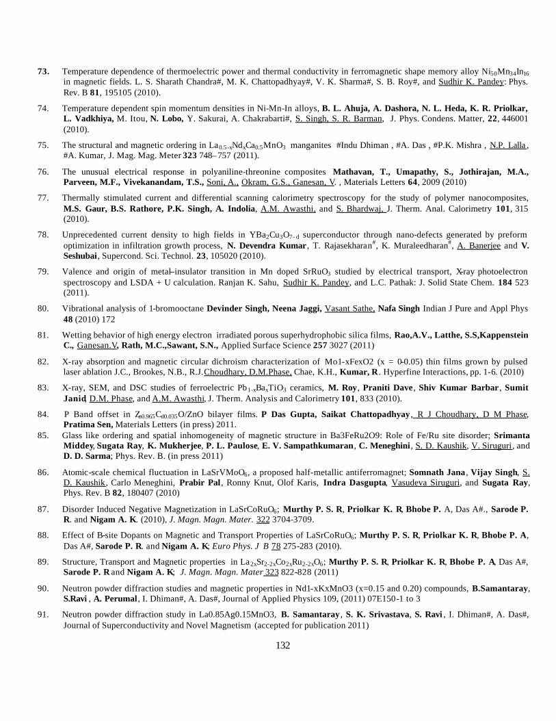
132
73. Temperature dependence of thermoelectric power and thermal conductivity in ferromagnetic shape memory alloy Ni50Mn34In16 in magnetic fields. L. S. Sharath Chandra#, M. K. Chattopadhyay#, V. K. Sharma#, S. B. Roy#, and Sudhir K. Pandey: Phys. Rev. B 81, 195105 (2010).
74. Temperature dependent spin momentum densities in Ni-Mn-In alloys, B. L. Ahuja, A. Dashora, N. L. Heda, K. R. Priolkar, L. Vadkhiya, M. Itou, N. Lobo, Y. Sakurai, A. Chakrabarti#, S. Singh, S. R. Barman, J. Phys. Condens. Matter, 22, 446001 (2010).
75. The structural and magnetic ordering in La0.5-xNdxCa0.5MnO3 manganites #Indu Dhiman , #A. Das , #P.K. Mishra , N.P. Lalla , #A. Kumar, J. Mag. Mag. Meter 323 748–757 (2011).
76. The unusual electrical response in polyaniline-threonine composites Mathavan, T., Umapathy, S., Jothirajan, M.A., Parveen, M.F., Vivekanandam, T.S., Soni, A., Okram, G.S., Ganesan, V. , Materials Letters 64, 2009 (2010)
77. Thermally stimulated current and differential scanning calorimetry spectroscopy for the study of polymer nanocomposites, M.S. Gaur, B.S. Rathore, P.K. Singh, A. Indolia, A.M. Awasthi, and S. Bhardwaj, J. Therm. Anal. Calorimetry 101, 315 (2010).
78. Unprecedented current density to high fields in YBa2Cu3O7- d superconductor through nano-defects generated by preform optimization in infiltration growth process, N. Devendra Kumar, T. Rajasekharan#, K. Muraleedharan#, A. Banerjee and V. Seshubai, Supercond. Sci. Technol. 23, 105020 (2010).
79. Valence and origin of metal–insulator transition in Mn doped SrRuO3 studied by electrical transport, X-ray photoelectron spectroscopy and LSDA + U calculation. Ranjan K. Sahu, Sudhir K. Pandey, and L.C. Pathak: J. Solid State Chem. 184 523 (2011).
80. Vibrational analysis of 1-bromooctane Devinder Singh, Neena Jaggi, Vasant Sathe, Nafa Singh Indian J Pure and Appl Phys 48 (2010) 172
81. Wetting behavior of high energy electron irradiated porous superhydrophobic silica films, Rao,A.V., Latthe, S.S,Kappenstein C., Ganesan.V, Rath, M.C.,Sawant, S.N., Applied Surface Science 257 3027 (2011)
82. X-ray absorption and magnetic circular dichroism characterization of Mo1-xFexO2 (x = 0-0.05) thin films grown by pulsed laser ablation J.C., Brookes, N.B., R.J.Choudhary, D.M.Phase, Chae, K.H., Kumar, R . Hyperfine Interactions, pp. 1-6. (2010)
83. X-ray, SEM, and DSC studies of ferroelectric Pb1-xBaxTiO3 ceramics, M. Roy, Praniti Dave , Shiv Kumar Barbar , Sumit Janid, D.M. Phase, and A.M. Awasthi, J. Therm. Analysis and Calorimetry 101, 833 (2010).
84. P Band offset in Zn0.965Cd0.035O/ZnO bilayer films. P Das Gupta, Saikat Chattopadhyay , R J Choudhary, D M Phase, Pratima Sen, Materials Letters (in press) 2011.
85. Glass like ordering and spatial inhomogeneity of magnetic structure in Ba3FeRu2O9: Role of Fe/Ru site disorder; Srimanta Middey, Sugata Ray, K. Mukherjee, P. L. Paulose, E. V. Sampathkumaran , C. Meneghini, S. D. Kaushik, V. Siruguri, and D. D. Sarma; Phys. Rev. B. (in press 2011)
86. Atomic-scale chemical fluctuation in LaSrVMoO6, a proposed half-metallic antiferromagnet; Somnath Jana , Vijay Singh, S. D. Kaushik, Carlo Meneghini, Prabir Pal , Ronny Knut, Olof Karis, Indra Dasgupta, Vasudeva Siruguri, and Sugata Ray, Phys. Rev. B 82, 180407 (2010)
87. Disorder Induced Negative Magnetization in LaSrCoRuO6; Murthy P. S. R, Priolkar K. R, Bhobe P. A, Das A#., Sarode P. R. and Nigam A. K. (2010), J. Magn. Magn. Mater. 322 3704-3709.
88. Effect of B-site Dopants on Magnetic and Transport Properties of LaSrCoRuO6; Murthy P. S. R, Priolkar K. R, Bhobe P. A, Das A#, Sarode P. R. and Nigam A. K; Euro Phys. J B 78 275-283 (2010).
89. Structure, Transport and Magnetic properties in La2xSr2-2xCo2xRu2-2xO6; Murthy P. S. R, Priolkar K. R, Bhobe P. A, Das A#, Sarode P. R and Nigam A. K; J. Magn. Magn. Mater 323 822-828 (2011)
90. Neutron powder diffraction studies and magnetic properties in Nd1-xKxMnO3 (x=0.15 and 0.20) compounds, B.Samantaray, S.Ravi , A. Perumal , I. Dhiman#, A. Das#, Journal of Applied Physics 109, (2011) 07E150-1 to 3
91. Neutron powder diffraction study in La0.85Ag0.15MnO3, B. Samantaray , S. K. Srivastava, S. Ravi , I. Dhiman#, A. Das#, Journal of Superconductivity and Novel Magnetism (accepted for publication 2011)
133
92. Neutron Powder Diffraction Studies in LaMn1/xCuxO3 (x=0.05, 0.10 and 0.15), B. Samantaray, S. K. Srivastava, S. Mohanty, S. Ravi , I. Dhiman# and A. Das#, Journal of Applied Physics 107 (2010) 09D719 -1 to 3
93. Structural characterization of microwave synthesized zinc substituted cobalt ferrite nanoparticles, Harshida Parmar , Rucha Desai and R V Upadhyay; Applied Physics A (Material Science & Processing); DOI 10.1007/s00339-010-6115-0
94. Surface active and aggregation behavior of methylimidazolium-based ionic liquids in water; N.M. Vaghela, N.V. Sastry and V.K. Aswal#; Colloid Polym. Sci. 289, 309 (2011)
95. Macroscopic and microscopic structural integrity in magnetic colloids—cationic micellar solution: Rheology, SANS and magneto-optical study; R. Patel, R.V. Upadhyay, V.K. Aswal#, J.V. Joshi and P.S. Goyal; J. Mag. Mag. Mat. 323, 849 (2011)
96. Effect of additives on the surface active and morphological features of 1-octyl-3-methylimidazolium halide aggregates in aqueous media; N.M. Vaghela, N.V. Sastry and V.K. Aswal#, Colloid Surf. A 373, 101 (2011)
97. SANS study to probe nanoparticle dispersion in nanocomposite membranes of aromatic polyamide and functionalized silica nanoparticles; G.L. Jadav, V.K. Aswal and P.S. Singh; J. Colloid Interface Sci. 351, 304 (2010)
98. Small Angle Neutron Scattering Study of Structural Aspects of Nonionic Surfactants (Tween 20 and Tween 80) in the Presence of Polyethylene Glycols and Triblock Polymers; R.K. Mahajan, J. Chawla, K.K. Vohra and V.K. Aswal#; J. Appl. Polym Sci. 117, 3038 (2010)
99. Effect of an amphiphilic diol (Surfynol®) on the micellar characteristics of PEO–PPO–PEO block copolymers in aqueous solutions; Y. Kadam, B. Bharatiya , P.A. Hassan, G. Verma, V.K. Aswal# and P. Bahadur; Colloids Surfaces A 363, 110 (2010).
100. Effect of n-Hexanol and n-Hexylamine on the Micellar Solutions of Pluronic F127 and P123 in Water and 1M NaCl; B. Bharatiya, G. Ghosh, V. K. Aswal# and P. Bahadur; J. Dispersion Sci. Techno. 31 660 (2010)
101. Multi-technique approach on the effect of surfactant concentrations on the thermal unfolding of rabbit serum albumin: Formation and solubilization of the protein aggregates; Mohd. S. Ali, J. M. Khan, V. K. Aswal#, R. H. Khan and Kabir-ud-Din; Colloids Surfaces B 80, 169 (2010) .
102. Induced micellization and micellar transitions in aqueous solutions of non-linear block copolymer Tetronic® T904; Y. Kadam, K. Singh, D.G. Marangoni , J.H. Ma, V.K. Aswal#, P. Bahadur; J. Colloid Interface Sci. 351, 449 (2010)
103. Unfolding of rabbit serum albumin by cationic surfactants: Surface tensiometry, small-angle neutron scattering, intrinsic fluorescence, resonance Rayleigh scattering and circular dichroism studies; Mohd. Sajid Ali, Nuzhat Gull, Javed M. Khan , V.K. Aswal#, Rizwan H. Khan and Kabir-ud-Din; J. Colloid Interface Sci. 352, 436 (2010)
104. Analysis of large and non-standard geometry samples of ancient potteries by internal monostandard neutron activation analysis using in situ detection efficiency, K.B. Dasari, R. Acharya#, K.K. Swain, N. Lakshmana Das , A.V.R. Reddy#; J. Radianal. Nucl. Chem. 286 (2010) 525.
105. Effect of CuO addition on the optical and electrical properties of sodium zinc borophosphate glasses; U. B. Chanshetti, V. Sudarsan, M. S. Jogad and T. K.Chondhekar, Physica B (Accepted) 2011.
106. Magnetic and Mössbauer effect study of (Co0.5Zn0.4Cu0.1Fe2O4)(1-x) (Al2O3/PVA)x (x = 0 and 0.30 synthesised by sonchemical route S.Mukherjee, D.Das, S.Mukherjee and P.K.Chakrabarti, J. Phys. Chem. C, 2010, 114 (35), pp 14763–14771
107. Exchange interaction at the interface of Fe-NiO nanocomposites, S. P. Pati, B. Bhushan and D. Das, J. Solid State Chem., 2010, 183, 2903.
108. Sr induced modification of structural, optical and magnetic properties in Bi1-xSrxFeO3 (x = 0, 0.01, 0.03, 0.05 and 0.07) multiferroic nanoparticles, B.Bhushan, A. Basumallick, N. Y. Vasanthacharya, S. Kumar and D. Das, Solid State Sciences, 2010, 12, 1063
109. Probing defects in chemically synthesized ZnO nanostructures by positron annihilation & photoluminescence spectroscopy. S.K.Chaudhuri, Manoranjan Ghosh, D.Das, A.K.Raychaudhuri, Journal of Applied Physics. 108 ,064319 (2010)
110. Evidence of formation of tetravacancies in uniformly oxygen irradiated n-type silicon. S.K.Chaudhuri, K.Goswami, S.S Ghugre, D.Das , Physica B, 406 (2011) 693-698

134
111. Energy dependence of incomplete fusion processes in the 16O + 181Ta system: Measurement and analysis of forward -recoil-range distributions at Elab = 7 MeV/nucleon, Devendra P.Singh, Unnati, P.P.Singh, A.Yadav, M.K.Sharma, B.P.Singh, K.S.Golda, R.Kumar, A.K.Sinha, R.Prasad, Phys.Rev. C 81, 054607 (2010).
112. Application of relativistic mean field and effective field density to calculate scattering observables for Ca isotopes, M. Bhuyan , R. N. Panda, T. R. Routray and S. K. Patra#, Phys. Rev. C 82, 064602 (2010).
113. Spectroscopic properties of ?-irradiated rare earth oxide based ferrofluids, M. Devi , N. Paul, D. Mohanta and A. Saha, Journal of Experimental Nanoscience, 2011 (In press).
114. Paramagnetic susceptibilities, crystal field Stark energies and hyperfine properties of Eu3+ in europium trifluoromethane sulfonate nonahydrate, D. Bisui K. N. Chattopadhyay , M.Ghosh, P.K.Chakrabarti , Journal of Physics and Chemistry of Solids 71, 1278 (2010).
115. Growth and characterization of Cd1-xZnxTe thin films prepared from elemental multilayer deposition, R. Ganguly, S. Hajra, T. Mandal, P.Banerjee, B. Ghosh, Applied Surface Science 214, 4879 (2010).
116. Sodium titanium silicate as ion exchanger: synthesis, characterization and application in separation of 90Y from 90Sr; R. Chakraborty and P. Chattopadhyay, Journal of Radioanalytical and Nuclear Chemistry, 2011 (in press).
117. Quinolinephosphomolybdate as ion exchanger: synthesis, characterization and application in separation of 90Y from 90Sr; R. Chakraborty, S. Dhara, S. Basu and P. Chattopadhyay, Journal Radioanalytical and. Nuclear Chemistry (In press),
118. Effect of gamma irradiation on a polymer electrolyte: Variationin crystallinity, viscosity and ion-conductivity with dose, P. Nanda, S.K. De , S. Manna, U. De#, S. Tarafdar , Nuclear Instruments and Methods in Physics Research B 268 73 (2010).
119. Elemental profile of agricultural soil by the EDXRF technique and use of the Principal Component Analysis (PCA) method to interpret the complex data, Virendra Singh,H.M. Agrawal, G.C. Joshi,M. Sudarshan and A.K. Sinha, Applied Radiation and Isotopes (In Press)
120. Protective role of zinc in ameliorating arsenic-induced oxidative stress and histological changes in rat liver, A. Kumar, A. Malhotra, P. Nair, Garg M.L., Dhawan DK, J Environ Pathol Toxicol Oncol. 2010;29(2):91-100
121. Influence of extraneous supplementation of zinc on trace elemental profile leading to prevention of dimethylhydrazine-induced colon carcinogenesis, V.D. Chadha, M.L. Garg, D. Dhawan, Toxicol Mech Methods. 2010 Oct;20(8):493-7.
122. Altered uptake and retention of 65Zn following arsenic exposure-modulation by zinc treatment. Ashok Kumar, Praveen Nair, Anshoo Malhotra, Shaoli Majumdar, M.L Garg and D.K Dhawan. BTER (in press).
123. Pixe Analysis of Blood Samples of Orthodontic Patients to Detect Ni Poisoning, International Journal of PIXE, P. Balouria, M. OswaL, S. Kumar, I.M. Govil, B.P. Mohanty, S.P. Singh and M.L. Garg, (In Press)
124. Impact of composite tannery effluent on the amino-transferase activities in a fish biosystem, using Guppy fish (Poecilia reticulata) as an experimental model Anulipi Aich, Buddhadeb Chattopadhyay, Siddhartha Datta and Subhra K. Mukhopadhyay, , Toxicological & Environmental Chemistry, 93(1): 85, (2011).
125. Study of trace metals in Indian major carp species from wastewater-fed ?shponds of East Calcutta Wetlands Anulipi Aich1, Anindita Chakraborty, Mathumal Sudarshan, Buddhadeb Chattopadhyay & Subhra Kumar Mukhopadhyay, Aquaculture Research, doi:10.1111/j.1365-2109.2011.02800.x.
126. pH dependant separation of uranium by chelation chromatography using pyridine 2,6-dimethanol as a chelator:Single crystal X-ray structural confirmation of the chelated uranium complex, Raja Saha, Sudipta Das, Arnab Banerjee, Animesh Sahana, M.Sudarshan, A.M.Z.Slawin, Yang Li, Debashis Das Journal of Hazadrous Materials 181, 154, 2010.
127. Electronic structure studies of Fe doped CeO2 thin films by resonance photoemission spectroscopy. Amit Khare, R. J. Choudhary, D. M. Phase and Sankar P. Sanyal, J. Appl. Phys. 109, (2011) 123706.
128. Growth kinetics of intermetallic alloy phase at the interfaces of a Ni/Al multilayer using polarized neutron and x-ray reflectometry, S. Singh#, S. Basu#, Mukul Gupta, C. F. Majkrzak, and P. A. Kienzle, Physical Review B 81, 235413 (2010).
129. Indium Flux-Growth of Eu2AuGe 3: A New Germanide with an AlB2 Superstructure and Low Temperature Phase Transitions; C. Peter Sebastian, Christos D. Malliakas, Maria Chondroudi, Inga Schellenberg, Sudhindra Rayaprol, Rolf-Dieter Hoffmann, Rainer Pöttgen, Mercouri G. Kanatzidis, Inorg. Chem. 49 9574 (2010).
135
130. Observation of Giant Electroresistance in Sm0.55Sr0.45MnO3; Rajneesh Mohan, Naresh Kumar, Bharat Singh, N. K. Gaur, Shovit Bhattacharya #, S. Rayaprol, A. Dogra, S. K. Gupta# and R. K. Singh, J. Alloys Compd. 508 L32 (2010)
131. Non-collinear magnetic order in the S = ½ magnet: Sr3ZnRhO6; A. D. Hiller, D. T. Adroja, W. Kockelmann, L. Chapon, S. Rayaprol, P. Manuel, H. Milchor and E. V. Sampathkumaran, Phys. Rev. B 83 024414 (2011)
132. Magnetic and Dielectric Properties of R2CuTiO6 (R = Y, La, Pr and Nd); Kiran Singh, Naresh Kumar , Bharat Singh, S. D. Kaushik, N. K. Gaur, Shovit Bhattacharya#, S. Rayaprol and Charles Simon, J. Supercond. Novel Magn, Article in Press (2011)
133. Low field magnetoresistance, specific heat and magnetocaloric effect in Sr substituted Pr0.7Ca0.3MnO3, Anjana Dogra, S. Rayaprol, Shovit Bhattacharya#, Matthias Eul, Wilfried Hermes, and Rainer Pöttgen, J. Supercond. Novel Magn, Article in Press (2011) DOI: 10.1007/s10948-010-0846-1
134. Dielectric properties of some MM'O4 and MTiM'O6 (M=Cr, Fe, Ga; M'=Nb, Ta, Sb) rutile-type oxides, R.Mani#, S.N. Achary#, Keka R.Chakraborty#, S.K.Deshpande, Joby E.Joy#, J.Gopalakrishnan# and A.K. Tyagi#, J.Solid State Chem.183, 1380 (2010).
135. The dielectric response of La0.5Ca0.5-xSrxMnO3 (0.1 ≤ x ≤ 0.4) manganites with different magnetic ground states, Indu Dhiman#, S.K.Deshpande and A.Das#, J.Appl.Phys. 108, 083915 (2010).
136. Growth of SnO2/W18O49 nanowire hierarchical heterostructure and their application as chemical sensor, S.Sen#, Prajakta Kanitkar, Ankit Sharma#, K.P.Muthe#, A.Rath#, S.K.Deshpande, Manmeet Kaur#, R.C.Aiyer, S.K.Gupta# and J.V.Yakhmi#, Sensors and Actuators B147, 453 (2010).
137. Role of structural disorder in charge transport properties of cobalt phthalocyanine thin films grown by molecular-beam epitaxy, Ajay Singh#, Soumen Samanta#, Arvind Kumar#, A.K.Debnath#, D.K.Aswal#, S.K.Gupta#, J.V.Yakhmi#, Y.Hayakawa# and S.K.Deshpande, Organic Electronics.11, 1835 (2010).
138. Role of annealing conditions on the ferromagnetic and dielectric properties of La2NiMnO6, F.N.Sayed#, S.N.Achary#, O.D.Jayakumar#, S.K.Deshpande, P.S.R.Krishna#, S. Chatterjee#, P.Ayyub# and A.K.Tyagi#, J.Mater.Res.26, 567 (2011).
139. Solution behavior of aqueous mixtures of low and high molecular weight hydrophobic amphiphiles, C.Rodríguez-Abreu M. Sanchez-Domínguez, B. Šarac, M. B. Rogac, R. G. Shrestha, L. K. Shrestha, D. Varade, G. Ghosh and V. K. Aswal#, Colloid Polym. Sci. 288 (2010) 739.
140. Buckling driven morphological transformation of droplets of a mixed colloidal suspension during evaporation induced self assembly by spray drying, D. Sen#, J. S. Melo#, J. Bahadur#, S. Mazumder#, S. Bhattacharya#, G. Ghosh, D. Dutta# and S.F. D’ Souza#
, Eur. Phys. J.- E 31 (2010) 393.
141. Evaporation-induced self assembly of nanoparticles in non-buckling regime: Volume fraction dependent packing, J. Bahadur#, D. Sen#, S. Mazumder#, Bhaskar Paul#, Arshad Khan#, G. Ghosh, J. Colloid Intef. Sci., 351 (2010) 357.
142. Solution behaviour of poly(N-isopropylacrylamide) in water: Effect of additives, T. Patel, G. Ghosh, S. -I. Yusa, P. Bahadur , J. Disp. Sci. Tech. (2011) In press .
143. Intriguing complex magnetism of Co in RECoAsO (RE= La, Nd and Sm); Anand Pal, V. P. S. Awana, M.Tropeano, S. D. Kaushik, Mushahid Hussain and H. Kishan, J. Appl. Phys. 109, 07E121 (2011).
144. Anomalous heat capacity and X-ray photoelectron spectroscopy of Superconducting FeSe1/2Te1/2; V. P. S. Awana, Govind, Anand Pal, Bhasker Gahtori, S. D. Kaushik, A.Vajpayee, Jagdish Kumar and H. Kishan, J. Appl. Phys. 109, 07E122 (2011).
145. Neutron diffraction and magnetization study of La0.7Ca0.3FeO3; Anjana Dogra, Neeraj Kumar, V. P. S. Awana, S. Rayaprol, S. D. Kaushik, V. Siruguri and H. Kishan, J. Appl. Phys. 109, 07E132 (2011).
146. Angular momentum distribution for the formation of evaporation residues in fusion of 19F with 184W near the Coulomb barrier, S.Nath, J.Gehlot, E.Prasad, J.Sadhukhan#, P.D.Shidling, N.Madhavan, S.Muralithar, K.S.Golda, A.Jhingan, T.Varughese, P.V.Madhusudhana Rao , A.K.Sinha, S.Pal#, Nucl.Phys. A850, 22 (2011).
147. Evaporation residue excitation function from complete fusion of 19F with 184W, S.Nath, P.V.Madhusudhana Rao, S.Pal#, J.Gehlot, E.Prasad, G.Mohanto, S.Kalkal, J.Sadhukhan, P.D.Shidling , K.S.Golda, A.Jhingan, N.Madhavan, S.Muralithar, A.K.Sinha, Phys.Rev. C 81, 064601 (2010).
136
148. Conclusive evidence of quasifission in reactions forming the 210Rn compound nucleus, E.Prasad, K.M.Varier, R.G.Thomas#, P.Sugathan, A.Jhingan, N.Madhavan, B.R.S.Babu, R.Sandal, S.Kalkal, S.Appannababu, J.Gehlot, K.S.Golda, S.Nath, A.M.Vinodkumar , B.P.A.Kumar, B.V.John#, G.Mohanto, M.M.Musthafa, R.Singh, A.K.Sinha, S.Kailas#, Phys.Rev. C 81, 054608 (2010).
149. Study of fission fragment mass distribution for 16O + 194Pt reaction, E.Prasad, K.M.Varier, B.R.S.Babu, N.Madhavan, K.S.Golda, S.Nath, B.P.A.Kumar, J.J.Das, J.Gehlot, P.Sugathan, A.Jhingan, A.K.Sinha, B.R.Behera, R.Sandal, H.Singh, R.Singh, R.G.Thomas#, S.Kailas#, Nucl.Phys. A834, 208c (2010).
150. Correlation of Trace elemental profiles in Blood Samples of Indian Patients with Leukoplakia and Oral Submucous Fibrosis. J G Ray, Ranjan Ghosh, Debarati Mallick, Niharika Swain, Premdeep Gandhi, S S Ram, S Selvaraj, A Rathore, M Sudarshan and Anindita Chakraborty, 2011. Biological Trace Element Research (In Press)
151. Radiation induced effects on viability and antioxidant enzymes of crustaceans from different habitats. D. Mukherjee, M. Manna, S. Selvaraj, S. Bhattacharya, S. Homechoudhury and A. Chakraborty. J. Environ. Biol.; 31, 251-254 (2010
152. Fluoride-induced genotoxicity in mouse bone marrow cells: e? ect of buthionine sulfoximine and N-acetyl-L-cysteine, S.Podder, A. Chattopadhyay, S. Bhattacharya, M.R.Ray and A. Chakraborty, J.Appl.Toxicol DOI 10.1002/jat.1605 (2010)
153. Comparative study on Passivation of GaAs0.86P0.14/Al0.6Ga0.4As near-surface quantum well S. Pal#, S. D. Singh#, S. Porwal#, S. W. D’Souza, S. R. Barman, and S. M. Oak#, J. Vac. Sci. Technol. A, 28, 1319 (2010).
154. Physical properties of ZnO thin films deposited at various substrate temperatures using spray pyrolysis, T. Prasada Rao, M.C. Santhosh Kumar, A. Safarulla, V. Ganesan, S. R. Barman and C. Sanjeeviraja, Physica B, 405, 2226 (2010).
155. Optical absorption and photoluminiscent studies of Ce doped Co tungstate nano-materials, S. J. Naik, U. Subramanian, R. B. Tangsali and A. V. Salker, J. Phys. D 44, 11504 (2011).
156. Highly conducting zinc oxide thin films achieved without post growth annealing, B. Singh, Z. A. Khan, I. Khan, and S. Ghosh, Appl. Phys. Lett. 97, 241903 (2010).
157. XPS characterization of corrosion films formed on the crystalline, amorphous and nanocrystalline states of the alloy Ti60Ni40, Shubhra Mathur, Rishi Vyas, K. Sachdev, S.K. Sharma, J. Non-Crys. Sol., 357, 1632 (2011).
158. Solid state studies on cobalt and copper tungstates nano materials, S.J. Naik, A.V. Salker, Solid State Sciences 12 (2010) 2065.
159. Symmetry breaking in Ni-doped PrFeO3 thin films established by Raman study, Feroz Ahmad Mir, M. Ikram and Ravi Kumar, Phase Transitions 84 167–178 (2011,).
160. Enhancement of stiffness of vertically standing Si nanosprings by energetic ions, Rupali Nagar, B. R. Mehta, J. P. Singh, C. Patzig, B. Rauschenbach, and D. Kanjilal, Journal of Applied Physics 107, 094315 (2010)
161. Self - Diffusion in Magnetron Sputtered Nanocrystalline Fe Films, Sujay Chakravarty, U. Tietze, D. Lott, M. Horisberger, J. Stahn, N. P. Lalla, H. Schmidt , Journal of Nano Research,11, 13 – 18 ( 2010).
162. Microwave assisted synthesis of tetragonal nanocrystalline zirconia nanoparticles, Reena Dwivedi, Anjali Maurya, R. Prasad and K.S. Bartwal#, J. Of Alloys and compounds, 509 6848-51 (2011)
163. Kinetics studies and mechanism evolution of the epoxidation of styrene over nanoporous Au doped TS-I, Samidha Saxena, Reena Dwivedi, Sheenu Bhadaurial, and Rajendra Prasad, Polish Journal of Chemical Technology, 12 72-78 (2010).
164. Lattice expansion in ZnSe quantum dots, Saikat Chattopadhyay, Naveen V. Kulkarni, R. Prasad, Agha Shahee , B.N. Raja Sekhar# and P. Sen, Materials Letters 65 1626-27, (2011).
165. Nanosize effects in Eu doped La0.67Ca0.33MnO3 perovskite, D.Roja Sree, Shravan Kumar Cholleti, S.G.Fard, Ch.Gopal Reddy, P.Y.Reddy, K.Rama Reddy and T.Goverdhan Reddy, J. Appl. Phys., 108 113917 (2010).

137
5.2 Publications from in-house research:
166. Dominance of magnetic scattering in Al70Pd20+XMn10-X (X=0,1&2), Al70Pd20Mn8(TM)2(TM=Fe, Cr, Co and Ni) and Al70-
XBxPd20Mn10 (X = 0, 0.5,1, 2 and 4) stable Icosahedral Quasicrystals. #Archna Sagdeo and N.P.Lalla , Quasicrystal: Types Systems and Techniques ISBN 978-1-61761-123-0 Editor: Beth E. Puckermann, Nova scientific Publishers,Inc (2010).
167. Effect of simultaneous application of magnetic field and pressure on magnetic transitions in La0.5Ca0.5MnO3 S. Dash, Kranti Kumar, A. Banerjee and P. Chaddah Phys. Rev. B, 82, 172412 (2010).
168. Effect of strain on the phase separation and devitrification of the magnetic glass state in thin films of La5/8 - y Pry Ca3/8MnO3 (y = 0.45) V G Sathe, Anju Ahlawat, R Rawat and P Chaddah J. Phys.: Condens. Matter 22 (2010) 176002
169. Evidence of orbital excitations in CaCu3Ti4O12 probed by Raman spectroscopy Dileep K Mishra and V G Sathe J. Phys.: Condens. Matter FTC 23 (2011) 072203
170. Growth study of Co thin film on nanoripled Si(100) substrate, Sarathlal K.V., Dileep Kumar and Ajay Gupta., Appl. Phys. Lett., 98 (2011) 123111
171. Infrared spectroscopic study of pulsed laser deposited Fe3O4 thin film onSi (111) substrate across Verwey transition temperature, Ridhi Master, Shailja Tiwari, R. J. Choudhary, U. P. Deshpande, T. Shripathi, J.of Appl. Phys 109, 043502 (2011).
172. Low temperature Raman and high field 57Fe Mossbauer study of polycrystalline GaFeO3, Kavita Sharma, V Raghavendra Reddy, Deepti Kothari, Ajay Gupta, A Banerjee and V G Sathe J. Phys.: Condens. Matter 22 (2010) 146005
173. Magneto-transport studies of FeSe0.9 - xMx (M = Si, Sb), Swati Pandya, Siya Sherif, L S Sharath Chandra# and V Ganesan Supercond. Sci. Technol. 24 045011, 201
174. Mossbauer, Raman and x-ray diffraction studies of superparamagnetic NiFe2O4 nanoparticles prepared by sol-gel auto-combustion method, Anju Ahlawat, V.G.Sathe, V.R.Reddy and Ajay Gupta., J. Magn. Magn. Mater., 323 (2011) 2049
175. Raman study of NiFe2O4 nanoparticles, bulk and films: effect of laser power Anju Ahlawat and V. G. Sathe, Journal of Raman Spectrosc. 42, (2011) 1087–1094.
176. Resistive Broadening in Sulfur doped FeTe Swati Pandya , Siya Sherif, L.S. Sharath Chandra# and V. Ganesan Supercond. Sci. Technol. 23, 075015 (2010)
177. Structural and magnetic properties of epitaxial Fe3O4/ZnO and ZnO/Fe3O4 bilayers grown on c-Al2O3 substrate. Ridhi Master, R. J. Choudhary, and D. M. Phase, J. Appl. Phys. 108, 103909 (2010).
178. Structural and magnetic study of swift heavy ion irradiated Fe/W multilayer structure, Sharmistha Bagchi, S. Jani, Shahid Anwar, N. Lakshmi and N. P. Lalla : J. Mag. Mag. Materials 322, 3851-3856 (2010).
179. Surfactant mediated growth of Ti/Ni multilayers, Mukul Gupta, S. M. Amir, A. Gupta and J. Stahn, Applied Physics Letters 98, 101912 (2011).
180. The interparticle interaction and crossover in critical lines on field-temperature plane in Pr0.5Sr0.5MnO3 nanoparticles. A. K. Pramanik and A. Banerjee. Phys. Rev. B, 82, 094402 (2010).
181. The pH-controlled particle size tuning of nanocrystalline Ni in polyol synthesis method without additional cappant, G. S. Okram, A. Soni and R. Prasad, Adv. Sci. Lett. 4, 132–135 (2011).
182. Thermally Stimulated Depolarization Current Studies of Relaxation in L-Asparagine Monohydrate, Deepti Jain, L.S. Sharath Chandra, S. Bharadwaj, S. Anwar, V. Ganesan, N.P. Lalla, A.M. Awasthi and R. Nath, IEEE Transactions on Dielectric and Electrical Insulation 17(4), 1128 (2010).
183. Tuning the magneic propoerties of the multiferroic LuFe2O4, S.Patankar, S.K.Pandey, V.R.Reddy, A.Gupta, A.Banerjee and P.Chaddah., by moderate thermal treatment., Euro Phys. Lett., 90 (2010) 57007
184. Magneto-electric coupling in Ca3CoMnO6; S. D. Kaushik, S. Rayaprol, J. Saha , N. Mohapatra, V. Siruguri, P. D. Babu, S. Patnaik and E. V. Sampathkumaran, J. Appl. Phys. 108 084106 (2010)
185. Magnetic anomalies and electronic structure of Ce2Cu2Mg and Ce2Pd2Mg; Wilfried Hermes, Stefan Linsinger, Sudhindra Rayaprol, Selcan Tuncel, Rolf-Dieter Hoffmann, Reinhard K Kremer, Ove Jepsen, and Rainer Pöttgen, J. Supercond. Novel Magn, Article in Press (2011) DOI: 10.1007/s10948-010-1061-9

138
186. Preparation of porous magnetic nanocomposite materials using highly concentrate emulsions as templates, G. Ghosh, A. Vilchez, J. Esquena, C. Solans, C. Rodriguez-Abreu, Prog. Col. Polym. Sci. (2011) In press.
187. Nuclei in the vicinity of 'island of inversion' through the fusion reaction, S.S.Ghugre, Pramana 75, 13 (2010).
188. Aqueous Synthesis of ZnTe/Dendrimer Nanocomposites and its Antimicrobial Activity: Implications in Therapeutics , S. Ghosh, D. Ghosh, P. K. Bag, S. C. Bhattacharya and A. Saha, Nanoscale, 2011 (In Press) (Feature Article).
189. Probing of Ascorbic Acid by CdS/Dendrimer nanocomposites: A Spectroscopic Investigation, S. Ghosh, S. C. Bhattacharya and A. Saha, Analytical Bioanalytical Chemistry, 397,1573 (2010).
190. Y Gamma Irradiation Route to Synthesis of Highly Re-dispersible Natural Polymer Capped Silver Nanoparticles ,. N. Rao, D. Banerjee#, A. Datta, S. K. Das#, R. Guin# and A. Saha, Radiation Physics and Chemistry,79, 1240 (2010).
191. Quantum dot based probing of mannitol: An implication in clinical diagnostics, D. Ghosh, S. Ghosh and A. Saha, Analytica Chimica Acta, 675, 165 (2010).
192. Trace Elements in Nungsham, the red edible algae of Manipur. Ch. Bino Devi, N.K. Sharat Singh, N. Rajmuhon Singh, N. Rajendro Singh, M. Sudarshan, A. Chakraborty, S.S. Ram. International Journal of Applied Biology and Pharmaceutical Technology Vol.2(1) (2011) 198.
193. Energy-dispersive X-ray fluorescence – A tool for interdisciplinary research. M Sudarshan,S S Ram, S Majumdar, J P Maity, J G Ray and A Chakraborty. Pramana – J. Phys., Vol. 76(2) 2011.
194. Physiological responses of water hyacinth to water pollution in and around Kolkata S. S. Ram, A. Chakraborty and S Sahoo, International Journal of Environmental Science 1(2), 183 (2010).
195. Characterization of dust particulates deposited on plant leaf surfaces using EDXRF: An approach for pollution monitoring, S.S.Ram, S.Majumder, P.Chaudhuri, S.Chanda, S.Santra, A. Chakraborty and M.Sudarshan,: International Journal of Environmental Science 1(2), 233 (2010).
196. Elemental alteration, Iron overloading and metallothionein induction in experimental hepatocarcinogenesis: a free radical-mediated process? D . Mishra, M Sudarshan and A. Chakraborty, Toxicol. Lett. 203, 40 (2011).

139
6. Presentations in Conferences/Symposia:
1. Swift heavy ion irradiation effect on nanometer range W/Fe multilayers. Sharmistha Bagchi, N. P. Lalla , S. Jani and N. Lakshmi : AIP- proceedings 1276, 107-113 (2010).
2. A Small-Angle Neutron Scattering Study of Poly(N-isopropyl acrylamide) in Water; M.B. Lawrence , J.A.E. Desa, V.K. Aswal# and M.V. Badiger; AIP Conference Proceedings 1313, 373 (2010)
3. Structural characterization of PLD-grown nanometer size ferromagnetic NiFeMo films Banerjee, M., Majumdar, A.K., R.J.Choudhary,D.M. Phase, Rai, S.#, Tiwari, P.#, Lodha, G.S# Materials Research Society Symposium Proceedings,1200 pp. 44-48 (2010)
4. Preparation and characterization of nanocrystalline soft magnetic FeXN thin films, Rachana Gupta, Ranjeeta Gupta, Mukul Gupta, and Ajay Gupta, 55th DAE Solid State Physics symposium 2010, Manipal, India.
5. Modifications of defects concentrations induced by ammonia flow rate and its effects on gallium nitride grown by MOCVD, Suresh S., Ganesan.V, Prem kumar T, Balaji .M, Ganesan .V, Basker.K. Materials Research Society Symposium Proceedings, 1195, 109 (2010)
6. Study of Complex Magnetic Phases in LuFe2O4 S. Patankar and A. Banerjee DAE Solid State Physics Symposium, Manipal (2010)
7. Correlated Magnetic and Electrical Orders in YMn1- xFexO3 Multiferroics, Sonu Namdeo and A.M. Awasthi, International Conference on Physics of Emerging Functional Materials, AIP Conf. Proc. 1313 (2010) 239-240.
8. Electrical transport in superionic thin films prepared by pulsed laser deposition, Neha Gupta, Anshuman Dalvi , D.M. Phase, A.M. Awasthi, DAE-SSPS (2010).
9. Novel Studies of Ferroelectric Domain-Wall Dynamics, Jitender Kumar, S. Bhardwaj, and A.M. Awasthi, DAE-SSPS (2010).
10. Spin-State Transition in La1-xSrxCoO3 Single Crystals, S. Bhardwaj, D. Prabhakaran, and A.M. Awasthi, DAE-SSPS (2010).
11. Magnetoresistance Studies on Ga excess Ni-Mn-Ga ferromagnetic shape memory alloy, Sanjay Singh, Rajeev Rawat and S. R. Barman, AIP Conference Proceedings 1349, 2010 .
12. Modulation on Ni2MnGa(001) surface, S. W. D'Souza, Abhishek Rai, J. Nayak, M. Maniraj, R. S. Dhaka, S. R. Barman, D. L. Schalgel, T. A. Lograsso, AIP Conference Proceedings 1349 2010.
13. Photon detector for inverse photoemission spectroscopy with improved energy resolution, M. Maniraj, S. W. D’Souza and S. R. Barman, AIP Conference Proceedings 1349, 2010 (in print).
14. Valence band alignment between nitrided GaPN/GaP(111) surface using x-ray photoelectron spectroscopy, S. K. Khamari#, V. K. Dixit#, A. K. Sinha#, S. Banik, S. R. Barman, S. M. Oak#, AIP Conference Proceedings 1349, 2010 .
15. Magnetic Phase Transitions in Cobalt Chromite Nanoparticles, Chandana Rath, L Kumar , and T. Shripathi, Oral presentation at International Conference on Superconductivity and Magnetism 2010, Antalya, Turkey April 25 – 30, 2010.
16. Large Relative Cooling Power in Dy5Si4:Dy5Si3 composite S.Shanmukharao Samatham, Venkateshwarlu D, Swati Pandya , Mohan Gangrade, L.S.Sharath Chandra#,Deepti Jain, and V.Ganesan DAE SSPS-2010
17. Heat Capacity and Magnetocaloric Study on Dy5Ge3Si S.Shanmukharao Samatham, Venkateshwarlu D, Swati Pandya, Mohan Gangrade, L.S.Sharath Chandra#, Deepti Jain and V.Ganesan DAE SSPS-2010
18. Possibility of Non Fermi Liquid Like States Co-exists With Superconductivity In Doubly Filled Skutterudites Venkateshwarlu D , Shanmukhrao S , Swati Pandya , L.S.Sharath Chandra# , P.N.Vishwakarma, Deepti Jain, MohanGangrade and V.Ganesan DAE SSPS-2010
19. Dimensionality study on Fe based Superconductors Swati Pandya, Siya Sherif, L.S. Sharath Chandra# and V. GanesanDAE SSPS-2010
20. 2D Lowest Landau Level Scaling in FeTe0.5Se0.5 Swati Pandya, Siya Sherif, L.S. Sharath Chandra# and V. Ganesan DAE SSPS-2010
140
21. Effect of Cu-doping on Nanocrystalline NiO Thin Films Shweta Moghe, A.D. Acharya, Richa Panda, S.B. Shrivastava, Mohan Gangrade, V. Ganesan DAE SSPS-2010
22. Temperature Dependent Photosensitivity of Cu Doped CdS Thin Film Richa Panda , Swati Pandya, Vandana Rathore, Manoj Rathore, Vilas Shelke, Nitu Badera, Deepti Jain, L. S. Sharathchandra#, M. Gangrade and V. Ganesan DAE SSPS-2010
23. Bacterial Spring Constant In Log-Phase Growth Deepti Jain, H.Nanda, R.Nath, D.S.Chitnis and V.Ganesan DAE SSPS-2010
24. Investigation Of Cd-Doped And Undoped ZnO Thin Films A.D. Acharya, Shweta Moghe, Richa Panda, S.B. Shrivastava, Mohan Gangrade, V. Ganesan DAE SSPS-2010
25. Shape And Size Effects On Magnetic Behavior Of Nano-Crystalline (La,Sr) (Mn,Fe)O Perovskite Systems.Usha Chandra,Pooja Sharma, Deepti Jain and V.Ganesan DAE SSPS-2010
26. Structural & Transport Properties Of Nano-Structured Nd0.7ca0.3Mno3 Jessica R.Chocha, Pooja A.Chhelavda,V.Ganesan and J.A.bhalodia DAE SSPS-2010
27. Thermally Stimulated Dehydration Studies in L-Asparagine Monohydrate Deepti Jain, S. Bharadwaj, V. Ganesan, A. M. Awasthi and R. Nath DAE SSPS-2010
28. Multiferroic Studies on La0.7Bi0.3CrO3 Perovskite, Aga Shahee, Dhirendra kumar and N. P. Lalla 55th DAESSP 2010.
29. Structural studies on Multiferroic La1-xBixCrO3 Perovskites Aga Shahee, Dhirendra Kumar and N. P. Lalla 55th DAESSP 2010.
30. Electrical studies of nanometer range W/Co multilayer structures. Sharmistha Bagchi and N. P. Lalla : International conference ICONSAT-2010
31. Effect of Ag surfactant on Cu/Co multilayers deposited by RF-ion beam sputtering, S. M. Amir, Mukul Gupta, Ajay Gupta, and A. Wildes, 55th DAE Solid State Physics symposium 2010, Manipal, India.
32. Surfactant mediated growth of Ti/Ni multilayers, Mukul Gupta, S. M. Amir, Ajay Gupta, S. P. Tiwari, and J. Stahn, 55th DAE Solid State Physics symposium 2010, Manipal, India.
33. Self diffusion of Fe in CoFeB thin films, Ranjeeta Gupta, Mukul Gupta, Ajay Gupta, and A. Wildes, 55th DAE Solid State Physics symposium 2010, Manipal, India.
34. Interdiffusion in W/Si multilayers with boron carbide interlayers, Satish Potdar, Mukul Gupta, Ajay Gupta, M. Schneider and J. Stahn, 55th DAE Solid State Physics symposium 2010, Manipal, India.
35. Structural and magnetic properties of Cu/Co multilayers prepared using Ag surfactant, S. M. Amir, M. Gupta, A. Gupta, and J. Stahn, Oral presentation at ICMM, Kolkata, Oct. 25-29, 2010.
36. Effect of Composition on L10 Ordering and Self- Diffusion of Fe in FePt Alloy, V.Phatak, Ajay Gupta, Mukul Gupta and A.Wildes, Oral presentation at ICMM, Kolkata, Oct. 25-29, 2010.
37. Fe/Au Multilayers: Structure and Magnetoresistance, S. Singh#, S. Basu#, C. L. Prajapat#, M. Gupta, D. Bhattacharya#, 55th DAE Solid State Physics symposium 2010, Manipal, India.
38. Dielectric properties of BaxSr1-x(NO3)2 system, P. M. Kumar, G. S. Okram, G. Sathaiah and K.A. Hussain, Natl. Sem. Ferroelectrics and Dielectrics- XVI, Dec 2-4, 2010, Bilaspur (CG), accepted (Oral presentation).
39. Influence of cappant and reductant on resistivity and thermopower of nanocrystalline nickel, G. S. Okram, NANO2010: Intl. Conf. Nanomater. Nanotechnology, Coimbatore, Oral presentation.
40. Effect of EDTA on Luminescence Property of Eu+3 doped YPO4 Nanoparticles. A. K. Parchur, G. S. Okram, R. A. Singh, R. Tewari, L. Pradhan, R. K. Vatsa and R. S. Ningthoujam. AIP proc. Intl. Conf. Phys. Emerging Funct. Mater. (PEFM-2010), BARC, Mumbai, CP1313, 391 (2010).
41. Superconductivity in heavily Nd-doped La2CaBa2Cu5Oz System, S. R. Mankadia, S. M. Dalsaniya, G. S. Okram, P. Igalwar, M. R. Gonal and J. A. Bhalodia. DAE-SSPS, accepted (2010).
42. Anomalous Seebeck coefficient of the NaxCoO2 system, N. Kaurav, K. K. Choudary, G. S. Okram and Y. K. Kuo. DAE-SSPS, accepted (2010).

141
43. Thermal and electrical properties of polyaniline-glycine composites, T. Mathavan, S. Umapathy, M. A. Jothirajan, T. S. Vivekanandam and G. S. Okram, DAE-SSPS, accepted (2010).
44. Transport properties of Fe3-xTixO4 epitaxial thin films D.Varshney, A.Yogi, K.Verma and D.M.Phase Proc. of DAE Solid State Symp. (2010).
45. Electronic structure of EuCu2Ge2 studied by Resonant Photoemission Spectroscopy. S.Banik, S.K.Deb, D.M.Phase and A.Thamizhavel Proc. of DAE Solid State Symp. (2010).
46. Temperature dependent switching of magnetoresistance in trilayered ZnO/LSMO/SrNTO device U.D.Khacher, P.S.Solanki, R.J.Choudhary, D.M.Phase and D.G.Kuberkar Proc. of DAE Solid State Symp. (2010).
47. Anisotropic behavior of electron phonon coupling in Fe3O4 thin films Ridhi Master, D.M.Phase and R.J.Choudhary Proc. of DAE Solid State Symp. (2010).
48. Growth and characterization of ZnO/Fe3O4 Bilayer Structure On c-Al2O3 substrate D.M.Phase, Ridhi Master, A.D.Wadikar and R.J.Choudhary Proc. of DAE Solid State Symp. (2010).
49. The effect of oxygen partial pressure on the growth of Fe doped and undoped TiO2 films on LAO substrate. R.J.Choudhary, Komal Bapna, D.M.Phase Proc. of DAE Solid State Symp. (2010).
50. Raman and Photoluminescence Studies of Pulsed Laser Deposited CeO2 Thin Films. Amit Khare, R. J. Choudhary , Prashant Sharma, A. C. Pandey and S. P. Sanyal , Presented at 55th DAE-Solid State Symposium at Manipal University, Manipal.
51. Resonant Photoemission Of Fe Doped TiO2 Thin Films Using Indus-1 Synchrotron Source. Komal Bapna, D. M. Phase and R. J. Choudhary. Presented at 55th DAE-Solid State Symposium at Manipal University, Manipal.
52. Raman Spectroscopic Studies of Co-based Spinel Multiferroic Thin Films. N.E.Rajeevan, Ravi Kumar, D.K.Shukla, R.J. Choudhary and P.P.Pradyumnan. Presented at 55th DAE-Solid State Symposium at Manipal University, Manipal.
53. Effect of Site Selective Doping of Co on the Physical and Structural/Micro structural Properties of Bi-Based High Temperature Superconductors , Indu Verma, R. Rawat, V. Ganesan, D. M. Phase, A. Banergee, and B. Das , AIP Conf. Proc. 1313, 195 (2010).
54. Non-Debye Specific Heat of (NH4)xRb1-xBr. P.S. Goyal#, Swati Pandya, P.D. Babu, R. Rawat and V. Ganesan, 55th DAE-Solid state Physics Symposium, 26-30th Dec. 2010 held at Manipal Institute of Technology, Manipal.
55. Magnetoresistance studies on Ga excess Ni-Mn-Ga ferromagnetic shape memory alloy Sanjay Singh, Rajeev Rawat and S.R. Barman, 55th DAE-Solid state Physics Symposium, 26-30th Dec. 2010 held at Manipal Institute of Technology, Manipal.
56. Heat Capacity Study of Magnetic Transition in TbTiSi and DyTiSi Compounds Pallab Bag, Pallavi Kushwaha and R. Rawat, poster at 55th DAE-Solid state Physics Symposium, 26-30th Dec. 2010 held at Manipal Institute of Technology, Manipal.
57. Influence of oxygen content in oriented LaCoO3-d thin films: Probed by X-ray diffraction and Raman spectroscopy Dileep Mishra, Anju Ahalawat, and V.G. Sathe Proceedings of the DAE Solid State Physics Symposium (2010)in press.
58. Physical Properties of Eu3Ba2Mn2Cu2O12; N. Kumar, S. Rayaprol, A. Dogra, S. D. Kaushik, K. Singh, N. K. Gaur, G. Anjum, Y. Kumar, Ravi Kumar, K. K. Iyer and E. V. Sampathkumaran, AIP Conference Proceedings, 1313 355 (2010)
59. Dielectric Properties of Gd3Ba2Mn2Cu2O12 Manganocuprate; S. Rayaprol, S. D. Kaushik, N. Kumar , N. K. Gaur, J. Saha, S. Patnaik, J. Appl. Phys. 109 07D709 (2011), Presented at 12th Magnetism and Magnetic Materials Conference held at Atlanta (USA) from 12th – 14th November 2010
60. Neutron diffraction and magnetization study of La0.7Ca0.3FeO3; A. Dogra, N. Kumar, S. Rayaprol, S. D. Kaushik, V. Siruguri, V. P. S. Awana and H. Kishan, J. Appl. Phys. 109 07E132 (2011), Presented at 12th Magnetism and Magnetic Materials Conference held at Atlanta (USA) from 12th – 14th November 2010
61. Magnetocapacitance in Ca3CoMnO6; S. D. Kaushik, S. Rayaprol, J. Saha, N. Mohapatra, V. Siruguri, P. D. Babu, S. Patnaik, and E. V. Sampathkumaran, J. Appl. Phys. 109 07D734 (2011), Presented at 12th Magnetism and Magnetic Materials Conference held at Atlanta (USA) from 12th – 14th November 2010

142
62. Thermoelectric properties of Ca4Mn3-xNbxO10; S. Bhattacharya#, Bharat Singh, S. Rayaprol, N. K. Gaur, M. R. Gonal#, and S. K. Gupta#, AIP Conference Proceedings, 1349 Article in press (2011), Presented at the 55th DAE-Solid State Physics Symposium held at Manipal University, Manipal during Dec 26 – 30, 2010
63. Effect of Dy doping in frustrated multiferroic h -YMnO3; S. D. Kaushik, A. K. Singh, V. Siruguri, and S. Patnaik, presented in 55th Solid State Physics Symposium 2010 held at Manipal to appear in AIP Conf Proc 1349 (in press 2011)
64. Doping effects of spin frustrated hexagonal Y0.95Dy0.05MnO3; A. K. Singh, S. D. Kaushik, V. Siruguri, and S. Patnaik, presented in 55th Solid State Physics Symposium 2010 held at Manipal to appear in AIP Conf Proc 1349 ( in press 2011)
65. Low Temperature Neutron Diffraction Studies on Ca3CoMnO6; S. Rayaprol, S. D. Kaushik, N. Mohapatra, V. Siruguri, P. D. Babu and E. V. Sampathkumaran presented in 55th Solid State Physics Symposium 2010 held at Manipal to appear in AIP Conf Proc 1349 ( in press 2011)
66. Preparation of porous magnetic nanocomposite materials using highly concentrate emulsions as templates; G. Ghosh, A. Vilchez, J. Esquena, C. Solans and C. Rodriguez-Abreu at the European Colloids and Interface Science (ECIS) conference, held at Prague, Czech Republic, during 6-10 September, 2010.
67. Neutron powder diffraction study in La0.85Ag0.15MnO3 Compound, B. Samantaray, S.K. Srivastava , S.Ravi , I. Dhiman# and A. Das#, 2011 International Magnetics Conference (INTERMAG 2011), Taipei, April 24/29, 2011 (accepted for presentation)
68. Neutron powder diffraction studies and magnetic properties in Nd1-xKxMnO3, (x = 0.15 and 0.20) compounds, B. Samantaray , S. Ravi , A. Perumal, I.Dhiman# and A.Das#,; 55th Annual Conference on Magnetism and Magnetic Materials (MMM 2010), Atlanta, USA, Nov.14/18, 2010
69. A paper entitled “Structural Studies of Coated and Compacted Glass Microspheres”, J.A.E Desa, was presented at the Poster Presentations of the XXII International Congress on Glass held in Salvador da Bahia, Brazil from September 20-25, 2010. The contents of the poster represented the last two years work of this project.
70. Abstract entitled “Structural and Porosity Studies of Compacted Glass Microspheres”, J.A.E Desa, was submitted to the International Conference on the Chemistry of Glasses and Glass-Forming Melts, September 4–8, 2011.
71. A Small-Angle Neutron Scattering Study of Poly(N-isopropyl acrylamide) In Water L. Basco, J A E Desa and V.K. Aswal, was presented at the International for DAE-BRNS International Conference on Physics of Emerging Functional Materials (September 22-24, 2010). The corresponding Paper has appeared in AIP Conference Proceedings (Vol. 1313 pp 373-375).
72. A Study of Cross-linked Regions of Poly(Vinyl Alcohol) Gels by Small-Angle Neutron Scattering; L. Basco, J A E Desa and V.K. Aswal, was presented at the 55th DAE-SSPS held at Manipal University, Manipal from December 26-30, 2010.
73. K. B. Dasari, R. Acharya, N. Lakshmana Das, A. V. R. Reddy : 4th International symposium on Nuclear Analytical Chemistry (NAC-IV) held at BARC, Mumbai during Nov. 15th to 19th, 2010, page no294.
74. K. B. Dasari, R. Acharya, N. Lakshmana Das, A. V. R. Reddy: 16th Radiochemical conference (Radchem 2010) held at Prague, during April 18th to 23rd, 2010. J. Chemical listy 104 (2010) 114.
75. N. Lakshmana Das, K. B. Dasari, R. Acharya, A. V. R. Reddy International symposium on archaeometry (ISA 2010) held at USA, during May 10th - 14th, 2010.
76. Special Bulletin: K. B. Dasari, R. Acharya, N. Lakshmana Das, A. V. R. Reddy: Special Bull. Peaceful uses of atomic energy, BARC, 2011.
77. Neutron Diffraction Investigation of CoxZn1-xFe2O4 Nanoparticles, Harshida Parmar, V.Siruguri and R V Upadhyay; Paper presented in 55th DAE Solid State Physics Symposium (AIP) 2011
78. Mechanical & optical properties of 0.4Sb2Se3-0.6CuI (Chalcohalide) glass; Rashmi M Jogada, Rakesh Kumarb P S R Krishnac
,M. S. Jogad, R D Mathada and G P Kothiyal; International Workshop and Symposium on the Synthesis and Characterisation of Glass/Glass-Ceramics(IWSSCGGC-2010) PP-12, p-68
79. Structure of 0.4Sb2Se3-0.6CuI (Chalcohalide) glass using Neutron Diffraction; Rashmi M Jogada, Rakesh Kumarb P S R Krishnac ,M. S. Jogad, R D Mathada and G P Kothiyal; International Workshop and Symposium on the Synthesis and Characterisation of Glass/Glass-Ceramics(IWSSCGGC-2010),PP-13, p69
143
80. Preparation Techniques of Glasses(0.4Sb2Se3-0.6CuI (Chacohalide) glass); Rashmi M Jogada, Rakesh Kumarb P S R Krishnac
,M. S. Jogadd, R D Mathada and G P Kothiyal; Proceedings of National Conference on Chemistry for Advanced Materials -NCCAM-2010, Naldurga, Osmanabad, Mahratstra, 1-3, March 2010, p 58-60
81. Effect of Swift Heavy Ion irradiation on lithium zinc silicate glasses: a Photoluminescence study; Mahantappa. S. Jogada*, P S R Krishnab, Rashmi M Jogadc, V Sudarsand, and G P Kothiyal; Solid State Physics Symposium, Manipal 2010
82. Enhanced magnetic properties in BiFeO3 nanoparticles co-doped with Ba and Ca, B. Bhushan, S.P. Pati , N. Y. Vasanthacharya, S. Kumar and D. Das accepted in National Conference on Magnetic materials and Applications (MagMA-2011) held at S.N.Bose National Centre for Basic Science during January 24-25,2011.
83. Exchange bias and room temperature coercivity enhancement in mechanically milled Fe-NiO nanocomposites , S. P. Pati , B. Bhushan , A. Basumallick and D. Das presented in International Conference on Fundamental and Application of Nano science and technology held at Jadavpur University on December 9 to 11, 2010.(Oral Presentation)
84. Structural, hyperfine and magnetic characterization of thermo -chemically prepared Fe-Co nanoalloy, S. P. Pati, B. Bhushan and D. Das, presented in International Conference on Magnetic Materials 2010 (ICMM 2010), held at SINP, Kolkata from October 25 to 29, 2010.
85. Ce concentration dependent EPR study of BiFeO3 nanoparticles, B. Bhushan, S. Kumar and D. Das , presented in International Conference on Magnetic Materials 2010 (ICMM 2010), held at SINP, Kolkata from October 25 to 29, 2010.
86. Study of nano - second isomers near 146Gd, Dibyadyuti Pramanik, Abhijit Bisoi#, Sudatta Ray#, A. Chakraborty, Gautam Dey, Krishichayan , R. Kshetri#, I. Ray#, S. Ganguly, M. K. Pradhan#, R. Raut#, M. Ray Basu, G. Ganguly, S. S. Ghugre, A. K. Sinha, S. K. Basu#, A. Goswami#, P. Banerjee#, A. Mukherjee#, S. Bhattacharya#, M. Saha Sarkar#, S. Sarkar , Proceedings of the DAE Symp. on Nucl. Phys. 55, 2010.
87. Spectroscopic studies of sd and fp shell N ~ Z nuclei, R. Bhattacherjee, R. Chakrabarti, S. S. Ghugre, A. K. Sinha, S. Mukhopadhyay , L. Chaturvedi , M. Kumar Raju, A. Dhal, N. Madhavan, R. P. Singh, S. Muralithar, B. K. Yogi, U. Garg, Proceedings of the DAE Symp. on Nucl. Phys. 55, 2010.
88. Nuclear Structure in 98Tc:Linear Polarization and DCO ratio measurements, R.K. Sinha, A. Dhal, D. Negi, D. Choudhury, G. Mohanto, M. Patial, N. Gupta, S. Kumar, S. Agarwal , R.P. Singh, S. Muralithar, N. Madhavan, S.S. Ghugre, J.B. Gupta, A.K. Sinha, A.K. Jain, I.M. Govil, R.K. Bhowmik, S.C. Pancholi, L.C. Chaturvedi , Proceedings of the DAE Symp. on Nucl. Phys. 55, 2010.
89. In beam spectroscopy of negative parity states in 135Pr, Ritika Garg, S. Kumar , Mansi Saxena, Savi Goyal, Davinder Siwal, Sunil Kalkal, S. Verma, S. Mandal, R. Singh, S. C. Pancholi, R. Palit#, Deepika Choudhury, A. K. Jain, S. S. Ghugre, G. Mukherjee#, R. Kumar, S. Muralithar, R. K. Bhowmik, R. P. Singh, Proceedings of the DAE Symp. on Nucl. Phys. 55, 2010.
90. Evolution of nuclear structure in 151-154Ho isotopes, Dibyadyuti Pramanik, Abhijit Bisoi#, S. Ray#, A. Chakraborty, G. Dey, Krishichayan, R. Kshetri#, I. Ray#, S. Ganguly, M. K. Pradhan#, R. Raut#, M. Ray Basu, G. Ganguly, S. S. Ghugre, A. K. Sinha, S. K. Basu#, A. Goswami#, P. Banerjee#, A. Mukherjee#, S. Bhattacharya#, M. Saha Sarkar#, S. Sarkar , Proceedings of the DAE Symp. on Nucl. Phys. 55, 2010.
91. A comprehensive study of in-complete fusion reaction dynamics in 16O + 181Ta system at 4-7 MeV/nucleon, Devendra P. Singh, Vijay R. Sharma, Abhishek Yadav, Pushpendra P. Singh, Unnati, M. K. Sharma, Golda K. S., R. Kumar, A. K. Sinha, B. P. Singh, R. Prasad, Proceedings of the DAE Symp. on Nucl. Phys. 55, 2010.
92. Angular momentum distribution for the formation of ERs in the reaction 19F +184W near the Coulomb barrier, S. Nath, J. Gehlot, E. Prasad, Jhilam Sadhukhan#, P. D. Shidling, Madhavan Narayanasamy, Muralithar Sivaramakrishnan, Golda K. S., A. Jhingan, T. Varughese, P. V. Madhusudhana Rao , A. K. Sinha, Santanu Pal#, Proceedings of the DAE Symp. on Nucl. Phys. 55, 2010.
93. Spin distribution in fusion reaction 30Si+170Er, Gayatri Mohanto, N. Madhavan, S. Nath, J. Gehlot, M. B. Naik, Prasad E., Ish Mukul, T. Varughese, A. Jhingan, R. K. Bhowmik, A. K. Sinha, I. Mazumdar#, D. A. Gothe#, P. B. Chavan#, Santanu Pal#, V. S. Ramamurthy#, Proceedings of the DAE Symp. on Nucl. Phys. 55, 2010.
94. ER excitation function for 16O + 194Pt reaction, E. Prasad, K. M. Varier, N. Madhavan, S. Nath, J. Gehlot, Kalkal Sunil, Jhilam Sadhukhan#, Gayatri Mohanto, P. Sugathan, A. Jhingan, B. R. S. Babu, T. Varughese, Golda K. S., B. P. Ajith Kumar, B. Satheesh, S. Pal#, R. Singh, A. K. Sinha, S. Kailas, Proceedings of the DAE Symp. on Nucl. Phys. 55, 2010.

144
95. Fission hindrance studies in 224Th: Spin distribution measurements, M. B. Naik, N. M. Badiger, N. Madhavan, S. Nath, J. Gehlot, T. Varughese, G. Mohanto, A. K. Sinha, P. Sugathan, Ish Mukul, A. Jhingan, I. Mazumdar#, D. A. Gothe#, P. B. Chavan#, S. Kailas#, Proceedings of the DAE Symp. on Nucl. Phys. 55, 2010.
96. Mass and isotopic yield distributions of fission-like residues in 16O + 181Ta system at ELab ~ 6 MeV/A, Vijay R. Sharma, Abhishek Yadav, Devendra P. Singh, Pushpendra P. Singh, Unnati, Manoj K. Sharma, R. Kumar, K. S. Gold, B. P. Singh, A. K. Sinha, R. Prasad, Proceedings of the DAE Symp. on Nucl. Phys. 55, 2010.
97. Excitation functions for some Ne induced reactions with holmium: Incomplete fusion Vs complete fusion, Avinash Agarwal, Munish Kumar, I. A. Rizvi, Anjali Sharma, Tauseef Ahmad, S. S. Ghugre, A. K. Sinha, A. K. Chaubey, Proceedings of the DAE Symp. on Nucl. Phys. 55, 2010.
98. HYbrid Recoil mass Analyzer (HYRA) coupled to TIFR 4? Spin spectrometer facility at IUAC, N. Madhavan, I. Mazumdar#, T. Varughese, J. Gehlot, S. Nath, D. A. Gothe#, P. B. Chavan#, G. Mohanto, M. B. Naik, I. Mukul, A. K. Sinha, Proceedings of the DAE Symp. on Nucl. Phys. 55, 2010.
99. Neutron-proton effective mass splitting and thermal evolution of nuclear matter, T.R. Routray, S.K. Tripathy, B. Behera, Proceedings of the DAE Symp. on Nucl. Phys. 55, 2010.
100. Composition and EOS of ? -stable charge neutral n+ p+ e+ ? matter at finite temperature, S. K. Tripathy, M. Bhuyan, T. R. Routray, B. Behera, Proceedings of the DAE Symp. on Nucl. Phys. 55, 2010.
101. The elastic scattering of 40,42,44,48Ca at 1.0 GeV in the framework of relativistic impulse approximation, M. Bhuyan, R. N. Panda, T. R. Routray , S. K. Patra#, Proceedings of the DAE Symp. on Nucl. Phys. 55, 2010.
102. Structure of 32P at high spins, R Chakrabarti, Annual Fall Meeting of the APS Division of Nuclear Physics at Santa Fe, New Mexico, U.S.A November 2010.
103. Preparation and characterization of palm leaf incorporated polyvinyl alcohol bio composites , A. Patel, R. Bajpei, J. M. Keller, A. Saha, International Conferences on Advances in Condensed & Nanomaterials held at Punjab University on Feb 22-26, 2011.
104. Quantum dot based probing of mannitol: An implication in clinical diagnostics , D. Ghosh, S. Ghosh and A. Saha, in 3rd Asia-Pacific Conference on Radiation Chemistry, organized by BARC, held at Lonavala on September 14-17, 2010.
105. Biomimetic synthesis of BSA capped CdS QDs, D. Ghosh, S. Ghosh and A. Saha, International Conference on Fundamental and Applications of Nanoscience and Technology, held at Jadavpur University, December 9-11, 2010,
106. Synthesis of Nanostructures in Swollen Liquid Crystalline Mesophases , A. Kalekar, G. Sharma and A. Saha, in 3rd Asia-Pacific Conference on Radiation Chemistry, organized by BARC, held at Lonavala on September 14-17, 2010.
107. Biofunctionalized Quantum dots as Fluorescence Probes for the detection of Vitamin B12 in aqueous solution, S. Ghosh, S. Mondal, and A. Saha, International Conference on Nanoscience, Nanotechnology and Advanced Material, held at Gitam University, Visakhapatnam on December 17-19, 2010.
108. Radiation induced self-organization of functionalized inorganic-organic hybrid nanocomposites, S. Ghosh, A. Datta, N. Biswas#, A. Datta# and A. Saha, in 3rd Asia-Pacific Conference on Radiation Chemistry, organized by BARC, held at Lonavala on September 14-17, 2010.
109. Room temperature synthesis of bipyramidal silver nanostructures in aqueous phase, Y. N. Rao, S. K. Das# and A. Saha. in 3rd Asia-Pacific Conference on Radiation Chemistry, organized by BARC, held at Lonavala on September 14-17, 2010.
110. γ-Irradiation route to photoluminescent Selenium-based QDs under ambient conditions, A. Datta, Y. N. Rao, S. Ghosh and A. Saha, International Conference on Fundamental and Applications of Nanoscience and Technology, held at Jadavpur University, December 9-11, 2010.
111. Biofunctionalized quantum dots as fluorescence probes for the detection of bradykinin, S. Mondal, S. Ghosh and A. Saha, National Symposium on Radiation and Photochemistry, held at Jodhpur University, March 10-12, 2011.
112. Sodium titanium silicate as ion exchanger: synthesis, characterization and application in separation of 90Y from 90Sr; R. Chakraborty and P. Chattopadhyay, Fourth International Symposium on Nuclear Analytical Chemistry (NAC-IV), Bhabha Atomic Research Centre, Trombay, Mumbai - 400 085, November 15-19, 2010.
145
113. Physical stress (Gamma ray) induced alterations in free polyamine content in Mung Bean Vigna radiata (L.) Wilczek.Mandar Sengupta, Nirmalya Ghoshal, and Sarmistha Raychaudhuri.National Conference on Plant Physiology, BHU, November, 2010.(Poster)
114. Modulation of Matrix metalloproteinase Activity By Chemical Carcinogens” D.Mukherjee, S.S. Ram, S. Majumdar, A. Mukhopadhyay, and A. Chakraborty. National symposium on ‘Recent Trends in Frontiers Research’, March 24,2011,WBUT, Kolkata, West Bengal.
115. Elemental Profiling & Physiological alteration in Epiphytic Lichen: A tool for assessing Air Quality in Kolkata City” S. Majumder, S.S.Ram, S. Santra, M. Sudarshan, A. Chakraborty, N.K.Jana. 18th West Bengal State Science and Technology Congress 2011.
116. Dust adsorption efficiency and elemental profiling of traffic generated dusts on the leaves of Ficus benghalensis exposed to roadside particulate pollution in Kolkata” S.S.Ram, S.Majumder, P.Chaudhuri, S.Chanda, S.Santra, P.K.Maity, U.K. Mukhopadhyay, A. Chakraborty, M.Sudarshan at national conference on frontiers in modern biology, 26-27 February 2011, IISERK, Mohanpur, Nadia, West Bengal.
117. Assessment of dust interception potentiality and ultra-microscopic study of the phylloplane structure of Polyalthia longifolia exposed to roadside particulate pollution” S.S.Ram, S.Majumder, S.Chanda, S.Santra, P.K.Maity, A. Chakraborty, M.Sudarshan, P.Chaudhuri at international conference on Frontiers in Bio-Sciences, October 1-3, 2010, NIT,Rourkela.
118. Dust interception potential of leaves of trees in kolkata: Some preliminary observations under scanning electron microscope” S.S.Ram, S.Majumder, P.Chaudhuri, S.Chanda, S.Santra, P.K.Maity, A. Chakraborty, M.Sudarshan at international conference on Electron Microscopy and Related Techniques, March 8-10, 2010, BARC,Mumbai.
119. Metallo-Resiatance in fungi exposed to ionizing radiation” D. Das , S. Majumdar, S.S.Ram, A. Chakraborty, S.C.Santra, National symposium on ‘Recent Trends in Frontiers Research’, March 24,2011,WBUT, Kolkata, West Bengal
120. Characterization and evaluation of hexavalent chromium tolerance of some selected bacterial isolates from Sukinda chromite mine, Odisha. H.N Thatoi, Sasmita Das, Anindita Chakraborty, M.Sudarshan National Conferences on New Frontiers in Life Sciences held on the occasion of the 13th Orissa Bigyan Congress during December 09-11, 2010, Bhubaneswar, Orissa.
121. Impact of composite tannery effluent on the amino-transferase activities in a fish biosystem, using Guppy fish (Poecilia reticulata) as an experimental model,Anulipi Aich, Buddhadeb Chattopadhyay, Siddhartha Datta and Subhra K. Mukhopadhyay, 2010, Golden Jubilee International Seminar: Researches in Zoology – Basic and Applied, Burdwan University
122. Toxicity Study of Tannery Effluents Using a Fish Model (Poecilia reticulata), Anulipi Aich, Buddhadeb Chattopadhyay, Siddhartha Datta and Subhra K. Mukhopadhyay, 2010, VIII Asia International Conference of Leather Science and Technology (AICLST), Hotel Stadel, Kolkata
123. Copper Stress induced alteration in protein profile and antioxidant enzymes activities in in vitro grown Withania sominfera L. Rout JR, Keshari N, Ram SS, Samanta L, Chakraborty A, Sudarshan M and Sahoo L at International Seminar on Frontiers in Biological Sciences, NIT Rourkela
124. Structural and transport studies of Co thin film on nano-rippled substrates, Sarathlal K.V, Dileep Kumar and Ajay Gupta, Proceedings of the 56th DAE Solid State Physics Symposium (2010)
125. Effect of roughness on growth of ultra thin nano-crysllaine Cobalt film; In -situ magnetic and transport studies, Dileep Kumar and Ajay Gupta, Proceedings of the 56th DAE Solid State Physics Symposium (2010)
126. In-situ magneto-optical Kerr effect for interface magnetism, Dileep Kumar, Sarath K.V, Oliva Saldanha and Ajay Gupta, Proceedings of the 56th DAE Solid State Physics Symposium (2010).

146
7. Workshops and Seminars organized by UGC-DAE CSR
7.1 Awareness Workshop on “The Facilities of UGC-DAE Consortium for Scientific Research”
An awareness workshop on “The facilities of UGC-DAE Consortium for Scientific Research (UGC-DAE CSR)” was held at the Department of Physics, Manipal Institute of Technology (MIT), Manipal – 576104, Karnataka during September 6-7, 2010. The workshop started with a brief inaugural function. Dr G K Prabhu, Registrar, Manipal University was the chief guest. Dr Ajay Gupta, Centre-Director, UGC-DAE-CSR, Indore was the guest of honour. Dr R S Aithal, joint director, MIT, Manipal presided over the inaugural function of the workshop. Prof. Ashok Rao, head of the physics department, MIT welcomed the gathering. Dr Y Raviprakash of MIT, physics department was was the master of ceremony.
Prof. Ajay Gupta delivered his inaugural address highlighting the motive of the workshop and invited researchers from universities and colleges to actively participate in the research programs of CSR that will be a mutually beneficial one. About 40 participants attended the workshop from all the major universities of region close to Manipal, apart from the local participants. Lectures were delivered by the Scientists of Consortium, Dr.S.K.Deb, Dr.A.K.sinha and Dr.S.S.Jha of RRCAT and Prof.Ashok Rao of MIT. The participants raised questions about various current scientific issues that were answered by the speakers and senior scientists in the gathering. Prof.P.R.Ramesh of MIT, Physics department proposed the vote of thanks.
Prof. Ajay Gupta delivered two talks covering “Overview of facilities at UGC-DAE CSR, Indore Centre” and “Thin film preparation and characterization techniques viz., XRR, GIXRD and MOKE”. Dr. A.V.Pimplae delivered a talk covering “Overview of facilities at UGC-DAE CSR, Mumbai Centre”. Dr.S.K.Deb delivered talk covering “Synchrotron Radiation facility at RRCAT, Indore”. Dr.V.Ganesan delivered two talks covering “Low temperature and high magnetic field facilities & Scanning Probe microscopy”. Dr. N.P. Lalla delivered a talk on “Transmission Electron and Scanning Electron Microscopy”. Dr.T.Shripathi delivered a talk on “Photoelectron microscopy”. Dr.M.Sudarshan delivered a talk on “Overview of facilities at UGC-DAE CSR, Kolkata Centre”. Dr.V.R.Reddy delivered a talk on “Mossbauer and Raman Spectroscopy” and Prof.Ashok Rao delivered a talk on “Overview of facilities at Manipal Institute of Technology”.
7.2 Awareness Workshop at IIT Madras
The Mumbai Centre of the UGC-DAE CSR organized an awareness workshop at IIT Madras in collaboration with the Department of Physics of the IIT during September 27-29, 2010. The theme of the workshop was on condensed matter research facilities at the different centres of the CSR – Mumbai, Indore and Kolkata with special emphasis on neutron scattering facilities at the Dhruva reactor, BARC available through the Mumbai Centre. The workshop was meant primarily for the young faculty and research students working in the different areas of condensed matter research at the colleges and institutions in and around Chennai. About forty such participants plus about ten research students of IIT Madras attended the workshop. One day of the workshop

147
was devoted to a visit to IGCAR, Kalpakkam to acquaint the participants with the upcoming node of UGC-DAE CSR and research facilities being established there. The speakers at the workshop were drawn from the faculty of all the three centres of UGC-DAE CSR, DAE laboratories BARC, Mumbai, RRCAT, Indore, IGCAR, Kalpakkam and IIT Madras.
The workshop began with Dr P. Chaddah, Director, UGC-DAE CSR, Indore stressing the mandate of the CSR – to improve the quality of research at universities and colleges by making available mega research faculties of DAE such as the neutron scattering facilities at the Dhruva reactor at BARC, Mumbai, synchrotron radiation sources INDUS1,2 at RRCAT, Indore, high energy accelerated ions from Variable Energy Cyclotron Centre, Kolkata as well as various accelerators and other experimental facilities at IGCAR, Kalpakkam and IOP, Bhubaneshwar. These facilities are supplemented by characterization and sample preparation facilities as well as other middle level but unique experimental research facilities available at the laboratories of CSR, particularly at Indore centre. He highlighted the sample environment of low temperatures available from in house liquid helium plant and high magnetic fields up to 14 Tesla for doing various measurements – electrical, magnetic and thermal properties as well as spectroscopic, structural and microscopic studies. He invited the participants to visit the CSR laboratories and interact with the various scientists to enlarge the scope of their research work.
Dr. R. Mukhopadhyay, BARC described in detail the various neutron scattering instruments installed and commissioned at the Dhruva reactor. These are collectively termed as NFNBR or National Facility for Neutron Beam Research and include three powder diffractometers for different types of crystal structure studies, two small angle neutron scattering set ups for studying structures of large molecules over distances of several nanometers, a high q-diffractometer for studying alloys and glasses, a spectrometer and a reflectometer both using polarized neutrons, a quasi elastic spectrometer, a triple axis spectrometer, neutron radiography facility and a host of other instruments. He also described some specific physics applications of these instruments such as study of voids in zeolites by using quasi elastic scattering of neutrons, magnetic structures in manganites and other novel compounds etc. All these instruments are available to the university academic community and CSR-Mumbai Centre provides a mechanism for doing collaborative research projects using these facilities. Dr. V. K. Aswal, BARC described the SANS – small angle neutron scattering facilities at the Dhruva reactor, BARC. There are two such instruments, both use neutrons from guide tubes. One set up utilizes a BeO filter as monochromator and a set of slits for defining incident neutron direction. Typical problems studied include protein crystallization, gelation studies and studies of surfactants etc. The other set up utilizes two Si (111) crystals in anti parallel mode for getting monochromatic neutrons and it has a some what higher resolution in comparison to the former set up.

148
Dr S. K. Deb, RRCAT, Indore described the synchrotron radiation sources INDUS-1,2 and the various beam lines and experimental stations installed and under development on these for doing research work. INDUS-1 is a 450 MeV electron storage ring giving light mostly in the xuv region of the electromagnetic spectrum with a critical wavelength of about 60 Å. A number of beam lines, including one by CSR, Indore centre, are installed and commissioned at bending magnets on INDUS-1. These include beam lines for doing angle integrated and angle resolved photoelectron spectroscopy studies in the photon energy range 10 to 200 eV, a photo physics beam line in the vuv range for studying atoms and molecules in the gas phase. INDUS-2 is a storage ring with electron energy of 2.5 GeV. INDUS-2 can thus give radiation in the hard X-ray region of the electromagnetic spectrum. A number of beam lines on INDUS-2 are at various stages of development. Three beam lines are almost complete and undergoing various evaluation tests – EXAFS beam line using an elliptically bent single crystal to focus a polychromatic beam just in front of a sample and a linear position sensitive detector put a little distance behind the sample, so that absorption at different wavelengths could be monitored as a spectrum on the linear detector, energy dispersive x-ray diffraction beam line and angle dispersive multipurpose diffraction beam line. CSR Indore Centre is involved in developing two beam lines on INDUS-2: a magnetic circular dichroism beam line for studying absorption of polarized x-radiation and an imaging beam line.
Professor A. Gupta gave a bird’s eye view of the large number of sophisticated experimental facilities available at the Indore centre of the CSR. These include sample preparation techniques – a number of furnaces including arc furnace, a planetary ball mill, thin film deposition systems using magnetron sputtering, pulsed laser deposition, thermal evaporation, electron beam and ion beam assisted evaporation, X-ray diffractometer for sample characterization and structural studies, scanning electron microscope and transmission electron microscope, various atomic force microscopes and a confocal microscope, spectroscopic techniques of laser Raman, UV-vis_NIR, Auger and photo electron as well as inverse photo electron spectroscopies, Mossbauer and EXAFS, using Kerr effect to do magnetic studies, and bulk condensed matter studies of electrical, magnetic and thermal properties including VSM and squid magnetometer . The centre has well equipped low temperature facilities –liquid helium and liquid nitrogen plants, a dilution refrigerator and a host of dedicated CCR’s so that almost all studies can carried out at temperatures down to liquid helium. High magnetic field (up to 14 Tesla) sample environment coupled with these low temperatures is also available for measurement of some electric and magnetic properties.
Dr A. V. Pimpale gave a talk on basic interaction between neutrons and condensed matter. Before coming to this main topic of his presentation, he described in brief the Mumbai centre of the CSR and mentioned about their regular programs of neutron school for fresh researchers entering the field of neutron studies of condensed matter and collaborative research projects in this area supported by CSR. He then discussed the nuclear and magnetic interactions of low energy thermal neutron with a nucleus. The neutron nucleus interaction is described by just one parameter - the scattering length. He pointed out the advantages of neutron scattering as compared to X-ray scattering arising from irregular dependence of the cross section on atomic number and the relative sensitivity of neutrons to low atomic number nuclei. The magnetic scattering of neutrons yields unique information about magnetic order in the scatterer.
Dr V. Siruguri described the neutron powder diffractometer (PD3 of NFNBR) put up at the Dhruva reactor by the CSR Mumbai centre. This instrument has several unique features – open geometry, toroidally bent crystal monochromator focusing neutrons at the sample position in both vertical and horizontal planes, oscillating radial collimator in the path of neutrons diffracted by the sample to improve signal to noise ratio, an array of overlapping linear position sensitive detectors to cover a wide angular range and a sample environment of low

149
temperatures down to 2.2K and high magnetic fields up to 7 Tesla It is thus a very powerful instrument (because of high flux and resolution) to study magnetic structures and Dr Siruguri described some interesting results obtained using it by university researchers studying multiferroic materials, shape memory alloys etc.
The last talk on the first day of the workshop was delivered by Dr A. K. Sinha. He described the experimental research facilities available at the Kolkata centre of the CSR and the various collaborative projects undertaken by the university researchers at the variable energy cyclotron centre, Kolkata, Institute of Physics, Bhubaneshwar and the Indian National Gamma Array comprising a large number of clover nuclear detectors along with ancillary equipment located at present at TIFR, Mumbai. The Kolkata centre specializes in using nuclear experimental techniques for study of physical, chemical, biological and environmental problems. Illustrative studies include behaviour of biological cells under radiative stress conditions, distribution of trace elements in environment, radiation induced synthesis of inorganic and organic nano composites, nuclear level structure in some nuclei of current interest etc. Recently Kolkata centre has been identified by DST, New Delhi as a special centre for low temperature high magnetic field studies for the north east area of the country and a 7 Teasla squid magnetometer has been commissioned. Other experimental facilities at Kolkata centre include positron life time set up, Mossbauer spectroscopy system and various facilities in biology and chemistry as well as condensed matter and nuclear physics laboratories. Dr Sinha also described the work under development at VECC for installing and commissioning a beam line at VECC for condensed matter studies.
The second day of the workshop was devoted to a visit to the laboratories at IGCAR, Kalpakkam. Drs G. Amarendra and C. S. Sundar, IGCAR helped in organizing this visit. CSR has set up a node at IGCAR for helping university researchers to better utilize the experimental facilities at IGCAR.. Dr Amarendra described the new high end experimental facilities that are being set up at this node. These include transmission electron microscope, scanning electron microscope, X-ray diffractometer, infrared single crystal growth furnace, ball mill, hot isostatic press, ball indentation etc. Dr Sundar gave an overview of the research activities of IGCAR, particularly from material science perspective. He discussed the problem of swelling of materials in the radiation environment and various approaches to study and develop materials that can be used in and around a nuclear reactor.
On the third and last day of the workshop Drs P. D. Babu, S. K. Deshpande from CSR, Mumbai centre and Professors Harish Kumar and P. N. Santosh both from IIT Madras gave talks. Dr Babu discussed magnetic neutron diffraction – the basic theory, data analysis and some illustrative examples. Dr. Deshpande discussed complementary X-ray diffraction techniques and the dielectric spectroscopy studies available at the CSR Mumbai Centre, the latter extending over a frequency range of micro hertz to mega hertz. Prof. Santosh’s talk dealt with his recent work on structure – property correlation in oxide materials, particularly perovskites. Prof. Harish Kumar described the physics department of IIT Madras – the faculty, experimental facilities available and the kind of research work that is being done.
Finally, a concluding session was organized to get the feedback from the participants. All the participants were highly appreciative of the information received from various presentations at the workshop. Dr. P. D. Babu, CSR, Mumbai Centre and Prof. Harish Kumar, IIT Madras had jointly borne the burden of organizing the workshop.

150
7.3 National Workshop on “Science with ECR based keV ion beams
A thematic workshop on science with ECR based keV ion beams was organized jointly by UGC-DAE CSR, Kolkata Centre and VECC, Kolkata during January 20-21, 2011. The aim of this workshop was to identify the research areas of overlapping interest so that collaborative research proposals from universities and other institutions may be initiated. The workshop was attended by about 70 participants from various universities and research institutions. Experts in this area from IUAC, New Delhi, IOP, Bhubaneswar, TIFR, Bombay, IGCAR, Kalpakkam, Utkal University, UGC-DAE CSR Indore Centre, IACS, Kolkata SINP, Kolkata and VECC, Kolkata presented the research areas open for investigation using low energy ion beams. Dr. D. Kanjilal (IUAC), Dr. K.G.M. Nair (IGCAR), Prof. D.P. Mahapatra (IOP), Dr. T. Som (IOP), Prof.Lokesh Tribedi (TIFR), Prof. N.C. Mishra (Utkal Univ.), Prof. B.N. Dev (IACS) and Dr. Ajay Gupta (UGC-DAE CSR Indore Centre) were invited to discuss the possible studies that can be taken up using the VECC ECR facility. Some of the research areas identified were nanopatterning of materials like formation of nano-ripples, nano rods for technological applications, synthesis of nano materials by ion implantation, synthesis of endofullerenes for drug delivery applications etc. The workshop concluded with a panel discussion where the participants expressed their interest in utilizing the VECC ECR facility for materials science research.
7.4 National Workshop on Nuclear techniques in Pure and Applied Sciences.
The National Workshop on Nuclear and Atomic Techniques based Pure and Applied Workshop was held from February 1st to 3rd 2011 at the picturesque campus of the Tezpur University. Organised jointly by UGC-DAE CSR, Kolkata Centre and Tezpur University, this workshop aimed to highlight research facilities at the Centers of the Consortium mainly Indore, Mumbai and Kolkata and the associated DAE institutions which would help to identify research areas of overlapping interest to finally culminate into collaborative research projects between Kolkata Centre and the different departments of Tezpur University such as the departments of Physics, Chemistry, Environmental Science and Biotechnology.
The workshop was attended by 155 delegates which included 16 Invited speakers and 139 selected participants from Benaras Hindu University, Andhra University, GITAM University, BITS, Ranchi, Viswa Bharati, University of Calcutta, Gauhati University, Dibrugarh University, North Eastern Regional Institute of Science and Technology, Itanagar, Tezpur University and affiliated colleges in the region.
The Vice Chancellor, Tezpur University, Professor Mihir K Choudhury, inaugurated the workshop at the Gallery, Dean Building, where he highlighted the growth of Tezpur University in the last few years specially in the field of nano sciences and expressed hope of the future possibilities of collaboration with UGC-DAE CSR. Dr.A.K.Sinha, Centre Director UGC-DAE CSR Kolkata Centre gave a short overview of the workshop highlighting the role of the Consortium in reaching out to the Universities and colleges through such workshops resulting various academic collaborations.
The key note address was given by Dr.A.K.Sinha, who spoke on the probing of matter in various scales starting right from the microcosm to the macrocosm. Dr. S.Bhattacharya from VECC, highlighted the ion beam facilities at VECC and the nuclear physics research being carried out there using the cyclotron. Dr.N.P.Lalla from the Indore Centre of the Consortium spoke on the facilities available at Indore Centre and also gave an elaborate talk on the Principles and Applications of Transmission electron Microscope. Dr.V.Sirugiri from UGC-DAE CSR Mumbai Centre spoke on the Facilities and research programs at Mumbai Centre. The newly

151
installed Low Temperature High Magnetic Field facilities and characterization of nano materials was discussed in detail by Dr.D.Das from the Kolkata Centre. Dr.Sandeep Ghugre discussed about the nuclear physics research being carried out at the Centre highlighting the collaborations using the INGA facility. Dr.Abhijit Saha presented the irradiation and various characterization facilities in the Centre for study of nano materials while Dr.Anindita Chakraborty highlighted the various research programs involving stress biology at the Centre. Dr.J. B.M.Krishna gave a talk on the use of ion beams in material science studies. Use of X rays in multidisciplinary trace element studies was presented by Dr.M.Sudarshan. Talks were also given by Prof.A Choudhury Pro Vice chancellor of Tezpur University, Dr.P.Deb, Dr.Gazi Ahmed and Dr. A.J.Thakur. They highlighted the various works in material sciences that were being carried out at the Physics Department. The contributed talks had speakers Andhra university, GITAM University, BITS, Mesra, and Tezpur University.
In all, the technical session consisted of 16 invited talks and 12 contributed talks. The contributed talks were held in parallel sessions on the 1st and 3rd day of the workshop. In addition a poster session was also held consisting of over 40 posters out of which 3 best posters were chosen. These 3 were then given the opportunity to present their work as a 15 minute presentation and were also given an award as encouragement. Mr. Rakesh Ranjan, research scholar from BHU got the best poster award.
This workshop was very successful and had talks in various fields of science ranging from Nuclear Physics, Materials Sciences, Biological, Environmental and Chemical Sciences, maintaining the theme of the workshop.
7.5 School on “Physics with Low Temperatures and High Magnetic Fields” UGC-DAE Consortium for Scientific Research during Mar 14-18, 2011.
The UGC-DAE Consortium for Scientific Research (CSR), has established a series of front- line experimental facilities in the area of Low temperatures and High magnetic fields. They include but not limited to 14T systems for Vibrating Sample Magnetometer, resistivity and heat capacity measurements, 7T SQUID Magnetometer, 8/10T System for magneto transport, 8T Scanning Hall Probe / MFM system, 8T Mossbauer and MOKE facility, 14T Dielectric and thermoelectric power measurements, 8T Facility for Neutron Diffraction at Mumbai, 7T SQUID Magnetometer at Kolkata and 15T Facility for transport measurements at Kolkata. Apart from these, there are many upcoming facilities like 8T System for dielectric and thermal conductivity at Indore, 8T High field XRD system at Indore, 7T SQUID-VSM at CSR Indore and 9T VSM at CSR Mumbai. In the last few years many researchers from the university system have utilized these. CSR Indore has organized a school, which was our fourth thematic meeting in the series during March 14-18. The objective of this school was to make potential users from universities and other academic institutions aware of the research possibilities with

152
the low temperature and high magnetic field facilities established as said above. Pedagogical lectures culminating in the highlights of typical research resulted from these facilities were discussed with a focus on future possibilities. A wide range of problems that can be dealt with in the framework of low temperatures and high magnetic fields have been dealt with. Areas covered include superconductivity, highly correlated electron systems, vortex matter, phase co-existence, multiferroics, inter-metallic compounds and nano-structures.
After a brief welcome by Prof. Ajay Gupta, the workshop was started with an overview of historical developmental of LTHM facilities at CSR Indore by Dr. V. Ganesan. Fundamentals of Mossbauer spectroscopy and MOKE were covered by Prof. Ajay Gupta in the first day covering various types of physics that can be dealt with magnetic multi- layers, ferrites, nano-structures and so on. Basics on magnetic measurements were covered by Dr. Alok Banerjee, while Dr. R. Rawat covered the fundamentals the importance of low temperatures, high pressures and high magnetic fields. Neutron diffraction and its relevance to LTHM was introduced by Dr. P.D. Babu, while the various systems of magnetic interest was covered in depth by Dr. V. Siriguri.
After the above said introductory talks, the second level of talks continued. Dr.V. Ganesan has covered the importance of LTHM in the scenario of Metal-Insulator Transitions with special emphasis on Kondo insulators and in understanding the fluctuation effects in superconductors, especially the new Fe based systems. Dr. A. Banerjee has covered many mesoscopic and microscopic properties as well as the Phase co-existence with special emphasis on manganites using magnetic field and temperature as variables. Dr. R.Rawat introduced the heat capacity and resistivity, their relevance to various inter-metallics and manganites, while Dr. V.R.Reddy covered in-depth Mossbauer measurements with particular relevance to Ga ferrites, while Dr. A.M.Awasthi covered the importance of Dielctric Measurements in the context of multi ferroics. Dr.A.Lakhani covered the need of LTHM for understanding the shape memory alloys while Dr. R.J.Choudhary covered the need for LTHM XRD in understanding the magneto-structural transitions.
One of the highlights of the talks is the evolving concept of kinetic arrest and its relevance in the First Order Phase Transitions (FOPT). This was covered in depth with various examples by Dr. P. Chaddah including new measurement routines like CHUF (Cooling and Heating in Unequal Fields). Apart from the lectures, there were extensive lab visits where in various LTHM facilities, the back-bone facilities like liquid nitrogen and liquid helium were shown to the participants. There was a lot of time for the participants to discuss with the experts and among themselves. There were even small presentations by the participants. CSR students contributed heavily to the conference including academics. They even made presentations and demo. Special mention may be made to the tutorial on Rietveld analysis by the students of the CSR. In essence, the four and a half day workshop was received well and appreciated by all the participants.
153
8. Theses and Student Projects 8.1 Ph.D. Theses
8.1.1 Mrs. Sharmistha Bagchi has been awarded her Ph.D. degree of DAVV Indore for her thesis entitled “Structural and Electrical Characterization of Metallic thin films and Multilayers”.
8.1.2 Ph.D. Degree was awarded to Ms. Srabanti Ghosh by Jadavpur University for thesis entitled “Some Aspects of Dendrimer and Semiconductor/Dendrimer Nanocomposites towards Biological applications” under the supervision of Dr. Abhijit Saha. The Co-supervisor from Jadavpur University was Prof. S. C. Bhattacharya.
8.1.3 Mr. P. S. Rama Murthy: Physics, Department, Goa University, entitled “Study of Structural, Transport and Magnetic Properties of Some Perovskite Based Oxides”. Thesis Advisors: Dr. K. R. Priolkar and Prof. P.R. Sarode (under CRS)
8.1.4 K. Ramesh, Dept of Physics, IIT Madras, Thesis Advisor: Dr. N. Harish Kumar. (under CRS)
8.2 Student Projects
A number of M.Sc. and M.Phil students from various universities do their project work using the facilities of the Consortium. Short term summer projects are also done by some of the students.
At Indore Centre
8.2.1 Magnetic Field induced first order transition in the shape memory alloy Ni50Mn35In15 , Ms. Ritu Sharma, Vikram University, Ujjain, under the supervision of Dr. Archna Lakhani.
8.2.2 Mr. Jitender Thakur’s dissertation work on “Dielectric Study of Single Crystal KDP at Low Temperatures and High Electric Fields” under the supervision of Dr. A.M. Awasthi.
8.2.3 Ms. Oliva Saldanha, a student of Five year Integrated M.Sc , Department of studies in Physics, University of Mysore (6 month project submitted March2011): “ In- situ study of Magnetic Thin films: effect of substrate and capping layer” : Supervisor Dileep K. Gupta
8.2.4 Mr. Pawan Patidar, a student of M.tech, Bhopal Engineering College, Bhopal, (6 month summer project; Joined in feb2011):” Development of Labview program for in -situ study of magnetic and transport properties of magnetic thin films “Project is presently running: Supervisor Dileep K. Gupta
8.2.5 A M.Sc. project by Ms. Ankita Khandelwal Jaipur National University, Jaipur, on “A Study on Drug-Bacterial Cell Interaction Using Confocal Laser Scanning Optical Microscopy” for the Degree of Master of Sciences in Microbiology. Supervisor: Dr. V. Ganesan.
8.2.6 A M.Sc. project by Ms. Deepika Bhavsar on “Preparation and Characterization of Al doped SnO2” during January-2010 to May-2010 for the Degree of Masters of Sciences in Applied Chemistry at S.G.S.I.T.S, Indore. Supervisor: Dr. V. Ganesan.
8.2.7 A M.Sc. project by Ms. Sonia Sharma on “ Preparation of Characterization of Fe doped SnO2” for the Degree of Master of Sciences in Pharmaceutical Chemistry in School of Chemical Sciences, DAVV, Indore. Supervisor: Dr. V. Ganesan.
154
8.2.8 An M.Tech Project by Ms. Berkha Jaiswal, School of Nanotechnology, Rajiv Gandhi Technological University, Bhopal. on "Studies of Cu doped SnO2 nanocrystalline thin films deposited by Spray Pyrolysis Technique". Supervisor: Dr. V. Ganesan.
8.2.9 An M.Tech Project by Mr. Adesh Kumar Sahu, School of Nanotechnology, Rajiv Gandhi Technological University, Bhopal. on "Structural and Electrical properties of Fe doped ZnO thin films prepared by Spray Pyrolysis Technique". Supervisor: Dr. V. Ganesan.
8.2.10 Mr. Shakti Singh Rajput, M. Tech. (Nanotech.), Amity Institute of Nanotechnology, Amity University, Noida (U.P.) entitled “Thermal Stability of W/Si Multilayer X-Ray Mirrors”. Supervisor: Dr. Mukul Gupta
8.2.11 Nidhi Patel, School of Chemical Sciences, Devi Ahilya University, Indore (2010). Thesis title: Effect of quality of polycrystalline and single crystalline niobium on the resistivity and Thermoelectric power. Supervisor: Dr. G. S. Okram
8.2.12 Mr. Joffy P. J., Department of Physics, Barkatullah University, Bhopal (2010). The influence of different cappants on resistivity and thermopower of nanocrystalline nickel. Supervisor: Dr. G. S. Okram
8.2.13 Mr. Mahesh Babu, Zakir Husain College of engineering and technology, Aligarh Muslim University, Aligarh (2010). Study of electrical resistivity and thermoelectric power of nanocrystalline copper. Supervisor: Dr. G. S. Okram
8.2.14 Mr. Mallikarjun Gurram, IIT, Guwahati (IAS summer project) Summer project title, “Study of Magnetoresistance and its tunability in Mn1.85Ga0.15Sb”. Supervisor: Dr. Rajeev Rawat
8.2.15 Two M.Sc. Physics students Mr. Sachin Singh and Alok Pandey from department of Physics, Khalasa college, Indore, did their summer and final project in Raman laboratory.
At Kolkata Centre
8.2.16 Mr Sanjay Kumar Pattanaik , from P G Department of Physics, Sambalpur University, has completed his M.Phil dissertation, titled “Photon & Neutron induced nuclear reactions of actinides at intermediate energies”, under the co-supervision of Dr D N Basu, from VECC.
8.2.17 Mr. Pintu Yadav, Dept. of Physics, University Ranchi. Supervisor: Dr. D.N. Basu
8.2.18 Ms. Sunita Singh, Dept. of Physics, University Ranchi. Supervisor: Dr. Chandana Bhattacharya
8.2.19 Ms. Manisha Rani, Dept. of Physics, Univ. of Ranchi. Supervisor: Dr. Chandana Bhattacharya
8.2.20 Mr. Shivsagar Mahto, Dept. of Physics, Univ. of Ranchi. Supervisor: Dr. S.S. Ghugre
8.2.21 Mr. Amaresh Kumar Singh, Dept. of Physics, Univ. of Ranchi. Supervisor: Dr. D. Das
8.2.22 Mr. Romesh Chandra, Dept. of Physics, Univ. of Allahabad. Supervisor: Dr. S.K. Das
8.2.23 Ms. Sadhana Singh, Dept. of Physics, Univ. of Allahabad, Supervisor: Dr. Chandana Bhattacharya
8.2.24 Ms. Sudha Yadav, Dept. of Physics, Univ. of Allahabad. Supervisor: Dr. Chandana Bhattacharya.
8.2.25 Mr. Ashish Kumar, Dept. of Physics, Univ. of Allahabad. Supervisor: Dr. Gopal Mukherjee.
8.2.26 Mr. Shashank S Chaubey, Dept. of Physics, Univ. of Allahabad. Supervisor: Dr. Tumpa Bhattacharjee
155
9 Seminar/Lectures delivered by UGC-DAE CSR Scientists
Dr. Alok Banerjee
1. Magnetic Circular Dichroism: Introduction and some typical results in the Discussion meeting on Indus 1 & 2 at RRCAT, Indore during 4-5 October, 2010.
2. Two lecture in the Refresher Course in Pure and Applied Chemistry held in Goa University on 30-31 March, 2011.
Dr. Archna Lakhani
3. Scientific facilities and research at CSR, Indore, in the Freshers Welcome program in School of Studies in Physics, Vikram University, Ujjain in August 2009.
4. Magnetic Glasses: An Introduction, in the program on “The Physics of Glasses: Relating Metallic Glasses to Molecular, Polymeric and Oxide Glasses” organized by KITP, University of California Santa Barbara, Santa Barbara, USA during Apr 12, 2010 - Jul 9, 2010
5. Magneto transport and Imaging studies using low temperature and high fields on various bulk materials : A general introduction - in the WISE (Women in Science and Engineering) group, jointly organized by Women in Science and Women in Physics, UCSB, Santa Barbara, USA.
6. Physical properties of highly correlated electron systems and an introduction to magnetic glasses. Visiting Scientist seminar in UCSD, SanDiego, USA.
Dr. A.M. Awasthi
7. Thermal Analysis- Methods and Practice at UGC Networking Winter School BHU, Varanasi, 26th March 2011.
Dr. S.R. Barman
8. Collective response of conduction electrons to photoelectron emission from rare gas nano-bubbles in aluminium at Institute of Physics, Bhubaneshwar on 2nd August, 2010.
9. Introduction to inverse photoemission spectroscopy: study of unoccupied states at Institute of Physics, Bhubaneshwar on 3rd August, 2010.
10. Surface studies on complex metals at Workshop for Inauguration of Max-Planck Partner Group project at Indian Institute of Science, Bangalore, 3rd November, 2010.
11. Surface studies on complex metals at University of Konstanz, Konstanz, Germany, 29th November, 2010.
12. Studies on complex metals at Fritz-Haber-Institute, Abt. M.P., Germany, 6th December, 2010. 13. Magnetoresistance studies on Ni-Mn-Ga ferromagnetic shape memory alloy at National Conference on Magnetic
Materials and Applications, 24-25th January, 2011, Kolkata
Dr. P. Chaddah
14. Phase-coexistence in half-doped manganites and in other magnetic materials, at International Conference on Physics of Emerging Functional Materials September 22-24, Mumbai.
15. Glasslike arrest of first order magnetic transitions : studies using CHUF protocol,at International Conference on Magnetic Materials (ICMM-2010), October 25-29, 2010, Kolkata.
16. Phase-coexistence in functional materials, National Symposium for Aadanced Technology NSMAT-2011 at Banasthali University, March 27-29, 2011.

156
Dr. V. Ganesan
17. A ‘Nano’ Perspective at MPCST Council Meeting at Bhopal during the Nanotechnology initiative”, Aug 28, 2010 on “Cultivation of Research in MP -
18. Recent Trends in nanotechnology ata “National level technical symposium (TECHNO-SCET2K10)” at Swarnadhra College of Engineering and Technology”, Narasapur, AP on Sept 15, 2010.
19. Current Status of Nano Research in Indian University System and possible remedies- at Nano-2010 (Panel Discussion, held at KSR Group of Institutions at Tiruchengodu, TN, Dec 15, 2010)
20. Characterization of Semiconducting materials at the National Level Seminar at N.M.S.S.Vellaichamy nadir College, Madurai on 16 Dec 2010.
21. Observation of M-Smegmatis (Tuberculosis ) – D29-nano-phages interaction using Atomic Force Microscopy at the 12th International Conference of International Academy of Physical Sciences (CONIAPS XII) on Emerging Interfaces of Physical Science, organized by the University of Rajasthan during December 22-24, 2010.
22. Scanning Probe Microscopy is more than an imaging tool on Jan 22nd, 2011 at NCRAMS-2011 held at Bhusawal.
23. SPM as a tool to characterize the nano structures Spintronic Materials: nanostructures and Devices” held at Kongu Engineering College, Erode during March 3-4, 2011.
24. Scanning Probe Microscopy and its role in imaging sub-micron features (A tool which is more than a simple imaging one)at the Department of Physics, Pondicherry University on 31st March 2011 under the UGC-SAP VISITING FELLOW PROGRAM.
Dr. Mukul Gupta
25. Surfactant mediated growth of thin film multilayers, University of Rajasthan, Jaipur, March 29, 2011.
Dr. G.S. Okram
26. Motivating young minds through scientific approach, Brilliant Academy, Thoubal, Manipur, September 9, 2010. 27. Material characterization and physical properties measurement facilities at UGC-DAE CSR, Indore, Department
of Physics, Manipur University, Imphal, September 6, 2010. 28. UGC-DAE Consortium for Scientific Research: Fostering Academia for Prosperity through Atomic Energy,
Centenary Hall, Manipur University, Imphal in one day national seminar on “Atomic Energy for Peace, Power and Prosperity” organized by Indian Nuclear Society and Department of Physics, Manipur University, August 28, 2010
29. Recent understanding on the physical properties of nanocrystalline nickel, Department of Metallurgical Engineering and Materials Science, Indian Institute of Technology, Bombay, Mumbai, June 10, 2010.
30. Evolution of the physical properties of nano-nickel, Raja Ramanna Centre for Advanced Technology, Indore, April 8, 2010.
Dr. D.M. Phase
31. Growth and characterisation of magnetite thin films on different substrates at National Symposium on “Scanning probe microscopy” held at Pune University, Pune, in March, 2011.
32. Resonance photoemission spectroscopy using Indus-1 Synchrotron Radiation Source at M.L.S.University, Udaipur in March, 2011.

157
Dr. R.J. Choudhary
33. Manipulation of magnetic nanostructure for spintronic applications at summer school on nanomaterials at NIT Hamirpur, Hamirpur, 1st June 2010.
34. Modifications in electronic and magnetic properties of Fe3O4 in nanometer scale at summer school on nanomaterials at NIT Hamirpur, Hamirpur, 4th June 2010.
35. Exposition of curious case of substitutionality of magnetic impurities in semiconducting oxide matrix for DMS applications.INDIAS 19-21 September 2010.
36. Resonant photoemission studies of dilute magnetic semiconducting oxide thin films at International conference on nanoscience and nontechnology, held at Swami Ramanand Teerth Marathwada University, Nanded 11-13 January 2011.
37. Thin film platform for nano-materials and their applications at National symposium on recent advances in physics, held at Holkar College, Indore on 15th February, 2011.
38. Thin films of dilute magnetic oxide semiconductors at National Seminar on Advances in Laser, Spectroscopy and Nanomaterial (NSALSN-2011), at Nehru Gram Bharati University (NGBU), Allahabad5-7th March 2011.
Dr. V. R. Reddy
39. Mossbauer spectroscopy and applications at Winter School on Recent Trends in Physics of Atoms, Molecules and Lasers on 25th January 2011 held at Department of Physics, Banaras Hindu University, Varanasi. (two talks)
40. Preparation and Study of Multiferroic Materials at National Conference on Spintronic Materials: Nanostructures and Devices (SMND-2011) on 3rd March 2011 held at Kongu Engineering College, Perundurai, Erode.
Dr. V. G. Sathe
41. Raman spectroscopy for nano structures and “EXAFS and its applications at Workshop on “Characterization Tools for Nano Materials” Department of Physics, Panjab University, Chandigarh on 22-2-2011.
42. Probing spin and orbital order in pervoskites by Raman spectroscopy in International Conference on Advances in Condensed & Nano Materials (ICACNM-2011) held at Department of Physics, Panjab University, Chandigarh on 23-26 Feb. 2011.
43. Raman Miscroscopy in National workshop on Scanning Probe Microscopy: Techniques and Application held at Department of Physics, Pune Universiry, Poona on 11-12 March 2011.
44. Effect of particle size reduction and growth of self assembled nano rods of Ni2Fe2O4 in National Conference on Nanomaterials &Applications: Present Position and Road Ahead , 16-18 March 2011 by Department of Physics, Banaras Hindu University, Vaaranasi.
Dr. T. Shripathi:
45. X-ray Photoelectron Spectroscopy, at National Conference on Experimental Tools for Material Science Research: State of Art, Mahila Mahavidyalay, Banaras Hindu University, December 03- 04, 2010
46. Nanomaterials, its Synthesis and Characterisation, at, Acropolis Institute of Technology &Research, Indore, March 04, 2011
47. Iron Oxide Nanostructures, at National Conference NAPRA-2011, Banaras Hindu University, March 16-18, 2011
48. Iron Oxide Nanostructures ,at IIT., Roorkee, March 28, 2011

158
Dr. A.V. Pimpale
49. Neutron studies of condensed matter in Indian universities, Concluding session, 3rd AONSA Neutron School, BARC, Mumbai, October 4-9, 2010
Dr. G. Ghosh
50. Dynamic Light Scattering Technique in Soft Matter , at Institute of Advanced Chemistry (IQAC, CSIC), Barcelona, Spain. on 5 March 2010
51. Preparation of ultra-light magnetic nanocomposites using highly concentrated W/O emulsions, at Institute of Advanced Chemistry (IQAC, CSIC), Barcelona, Spain. on 15 December 2010
Dr V. Siruguri
52. Neutron diffraction – instrumernt and experiment, 3rd AONSA Neutron School, BARC, Mumbai, October 4-9, 2010
Dr S. K. Deshpande
53. Dielectric Spectroscopy, during the UGC-SAP seminar on “Materials and Devices for Future Technology” at the School of Physics, North Maharashtra University, Jalgaon on March 7, 2011.
Dr A K Sinha
54. Nuclear Physics, Orientation School at the DAE Symposium, held at BITS Pillani, December 2010.
Dr S S Ghugre
55. Gamma ray spectrometer for nuclear structure studies with fast moving recoils at the Theme Meeting on Nucleus Nucleus Collisons Around Fermi Energy, at Variable Energy Cyclotron Centre, Kolkata December 2011.
56. Simulation studies for composite detectors at the Nustar India collaboration meeting, Tata Institute of Fundamental Research, Mumbai February 2011.
Dr A Saha
57. Biofunctionalized Quantum Dots: Probing and Biological Implications in Asia-Pacific Symposium on Radiation Chemistry, organized by Bhabha Atomic Research Centre, held at Lonavala on September 14-17, 2010.
58. Functional Luminescent Quantum Dots: Synthesis and Biological Probing in International Conference on Recent Trends in Material Science and Technology, held at Indian Institute of Space Science and Technology at Trivandrum on October 29-31, 2010.
59. Quantum Dot Mediated Redox Processes: Implications in Biological Probing in International Conference on Advanced Oxidation Processes held at Mahatma Gandhi University on September 18-21, 2010.
60. Characterization of Nanomaterials by Optical Spectroscopic Techniques in National Conference on Experimental Tools for Material Science: State of Art held at Mahila Mahavidyalaya, Banaras Hindu University, December 3-4, 2010.
61. Spectroscopic techniques for characterization of nanomaterials in UGC Networking Winter School on Recent Trends in Physics of Atoms, Molecules and Lasers held at Department of Physics, Banaras Hindu University, January 9-30, 2011.
159
62. Irradiation and characterization facilities for nanomaterial studies, National Workshop on Nuclear and Atomic Techniques Based Pure and Applied Sciences held at Tezpur University, February 01-03, 2011.
Dr.M.Sudarshan
63. X-Rays in Multidisciplinary Sciences at the National Workshop on Nuclear techniques in Pure and Applied Sciences held at Tezpur University Feb 1-3, 2011
Dr.Anindita Chakraborty
64. Differential modulation of xenobiotic metabolizing enzymes by vanadium during diethylnitrosamine–induced hepatocarcinogenesis in rats in EPS Global International Cancer Conference , Chongqing, China Nov 13-14 2010
65. Oxidative Stress and Biological Effects at the National Workshop on Nuclear techniques in Pure and Applied Sciences held at Tezpur University Feb 1-3, 2011
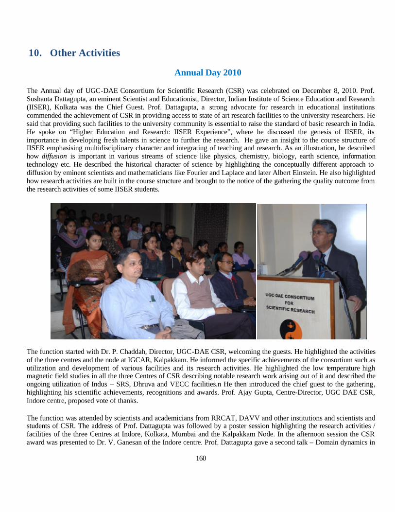
160
10. Other Activities
Annual Day 2010
The Annual day of UGC-DAE Consortium for Scientific Research (CSR) was celebrated on December 8, 2010. Prof. Sushanta Dattagupta, an eminent Scientist and Educationist, Director, Indian Institute of Science Education and Research (IISER), Kolkata was the Chief Guest. Prof. Dattagupta, a strong advocate for research in educational institutions commended the achievement of CSR in providing access to state of art research facilities to the university researchers. He said that providing such facilities to the university community is essential to raise the standard of basic research in India. He spoke on “Higher Education and Research: IISER Experience”, where he discussed the genesis of IISER, its importance in developing fresh talents in science to further the research. He gave an insight to the course structure of IISER emphasising multidisciplinary character and integrating of teaching and research. As an illustration, he described how diffusion is important in various streams of science like physics, chemistry, biology, earth science, information technology etc. He described the historical character of science by highlighting the conceptually different approach to diffusion by eminent scientists and mathematicians like Fourier and Laplace and later Albert Einstein. He also highlighted how research activities are built in the course structure and brought to the notice of the gathering the quality outcome from the research activities of some IISER students.
The function started with Dr. P. Chaddah, Director, UGC-DAE CSR, welcoming the guests. He highlighted the activities of the three centres and the node at IGCAR, Kalpakkam. He informed the specific achievements of the consortium such as utilization and development of various facilities and its research activities. He highlighted the low temperature high magnetic field studies in all the three Centres of CSR describing notable research work arising out of it and described the ongoing utilization of Indus – SRS, Dhruva and VECC facilities.n He then introduced the chief guest to the gathering, highlighting his scientific achievements, recognitions and awards. Prof. Ajay Gupta, Centre-Director, UGC DAE CSR, Indore centre, proposed vote of thanks.
The function was attended by scientists and academicians from RRCAT, DAVV and other institutions and scientists and students of CSR. The address of Prof. Dattagupta was followed by a poster session highlighting the research activities / facilities of the three Centres at Indore, Kolkata, Mumbai and the Kalpakkam Node. In the afternoon session the CSR award was presented to Dr. V. Ganesan of the Indore centre. Prof. Dattagupta gave a second talk – Domain dynamics in

161
ferroelectric films – in which he highlighted his recent research activities in this area. Following this talk, five senior research students – Bhavya Bhushan, Deepti Kothari, Suryanarayana Dash, Swati Panya and Vaishali Phatak - presented their Ph.D. thesis work and Ms. Vaishali Phatak received the best presentation award. The day ended with a classical hindusthani vocal recital by Ms. Vaishali Bakore.
ffggaannhh ii[[kk ookkMM++ ++ kk 22001100
fganh i[kokM++k ds volj ij fnukad 29 flrEcj *10 dks fuEu&lkjkaf’kr O;k[;kuksa dk vk;kstu fd;k x;kA
cgqykSgh; ¼eYVhQsjksbd½ inkFkksZa ds dqN izeq[k igyw
lq/khj dqekj ik.Ms; fo’ofo|ky; vuqnku vk;ksx & ijek.kq ÅtkZ foHkkx oSKkfud vuqla/kku ladqy
cgqykSgh; inkFkksZa us fiNys ,d n’kd esa oSKkfudx.kksa dk /;ku vkdf"kZr fd;k gSA bldk dkj.k fofHkUu ks=ksa esa bldh mi;ksfxrk ,oa vk/kkjHkwr HkSkfrd flkUrksa dks le>us ds fy, lcy volj iznku djuk gSA bu inkFkksZa dh mi;ksfxrk bl ckr ij fuHkZj djrh gS fd ;s inkFkZ izcy oS|qr&pqEcdh; ;qXeu dejs ds rkieku ij fn[kkrs gSa ;kfd ughaA bl fn’kk esa lcls cM+h pqukSrh ,sls inkFkksZa dk la’kys"k.k djuk gS tks izcy oS|qr&pqEcdh; ;qXeu dejs ds rkieku ij fn[kk,ssaA bl O;k[;ku esa ykSg&fo|qfrdh ,oa ykSg&pqEcdRo ls lacaf/kr ewyHkwr HkkSfrd flkUrksa ij izdk’k Mkys x,s rFkk buds kjk fn[kk;s tkus okys izeq[k HkkSfrd xq.kksa dh Hkh ppkZ dh xbZA mu lHkh dkjdksa ij Hkh fopkj fd;k x;k ftuls izcy oS|qr&pqEcdh; ;qXeu dks dejs ds rkieku rd yk;k tk ldsA
ngu la'ys"k.k ,oa uSuks inkFkZ vkj- izlkn
jlk;u 'kkL= v/;;u'kkyk] nsoh vfgY;k fo'ofo|ky; bUnkSj
uSuks inkFkZ ds la'ys"k.k ds fy;s ngu foKku izfØ;k eas gq, vR;k/kqfud 'kks/k ij izdk'k Mkyk x;k gSA uSuks inkFkZ ds la'ys"k.k esa iz;ksx vkus okys ijEijkxr ngu izfØ;k ij gq, cnyko dh foospuk dh xbZ gSA ngu izfØ;k ls cuk;s uSuks inkFkksZa ds mi;ksx ij fo'ks"k /;ku fn;k x;k gSA ;g fof/k cgqr gh lLrh] rst rFkk dkjxj gSA ngu fof/k kjk mPp lrg ks=Qy okys mRizsdj okgd ¼Catalyst Support½ cuk;s x;s gSA bl fof/k kjk In situ Supported Catalyst Hkh cuk;s tk ldrs gSA ?kksy ngu izfØ;k kjk T;ksfre; inkFkZ ¼Luminscent Material½] iznhIr inkFkZ ¼Phosphur Compound½ rFkk mPp /kkfjrk okys fo|qr /kkjd cuk;s x;s gSA bl fof/k kjk la'ysf"kr inkFkksZa dk mi;ksx ¼Dye Solar Cells½] Fuel Cells rFkk fjpkZtscy csSVjh esa djus ls budh kerk c<+ xbZA
lanHkZ& • Singanahally T. Aruna, Alexander S. Mukasyan, Current Opinion in Solid State and Materials Science 12 (2008)
44. • Patil KC, Hegde MS. Tanu Rattan, ST. Chemistry of namocrystalline oxide materials: combustion synthesis,
Properties and applications, Singapore: World Scientific; 2008.

162
Science Day 2011
Science Day was celebrated on 28th February at CSR, Indore centre. Two lectures were arranged on this occasion.
The first lecture was delivered by Prof. Lalit M. Kukreja of RRCAT, Indore. He started with a popular introduction to ZnO and its technological usages. This is followed by presentation of research work presently carried out at PLD laboratory of RRCAT on optical properties of ZnO quantum dots. Studies on ZnO quantum dots (ZQDs) are important to evaluate their potential for developing novel photonic and spintronic devices. Earlier their group found that the fundamental optical processes in ZQDs of sizes comparable to and smaller than the excitonic Bohr radius (~ 2.34 nm), called here as ultra-small quantum dots are radically different from those operating in their larger counterparts. In this talk Prof. Kukreja showed recent results on unusual behavior of ultra-small ZQDs in their photoluminescence (PL) related optical processes. Ten layered ensembles of alumina capped ZQDs of mean radius in the range of ~ 1 to 4 nm were grown on (0001) sapphire substrates at 250 0C in oxygen ambient at 1 x 10⁻⁴ Torr by pulsed laser deposition using a KrF Excimer laser operating at 10 Hz and 0.6 J/cm2.
Optical absorption and PL spectra followed the size dependent blue shift of the band-edges in conformity with the strong quantum confinement of electrons and holes localized in the ZQDs. Temperature dependent PL spectra of the ZQDs of mean radius of ~ 2.5 nm in the range of 10 – 70 K consisted of two peaks of different donor bound electron hole recombinations at ~ 3.368 and 3.360 eV and one acceptor bound recombination at ~ 3.333 eV. At temperatures higher than 70 K while these peaks broadened, a new feature appeared at ~ 3.376 eV which we attribute to the band-edge transitions of the strongly correlated electron hole pairs. The peak intensity of these transitions followed the normal law of thermal quenching. Band-edge of the ZQDs of mean radius 2.5 nm was calculated to be at ~ 3.558 eV, 2 but observation of the PL transitions at ~ 3.376 eV suggests the Stokes shift, commonly observed in polar materials such as ZnO. Temperature dependent PL spectra also showed significantly intense LO phonon replicas of the above stated primary transitions, which confirmed strong coupling between the carriers and the LO phonon. Temperature dependence of FWHM of the PL peaks followed the Hellmann and O’Neill models1 with carrier-LO phonon coupling coefficient (~ 980 meV) close to its value for the bulk ZnO (~ 963 meV) and higher than that reported for the larger ZQDs. This anomaly is explained through the dominance of enhanced Coulombic interaction between the electrons and holes and the ensuing Fröhlich interactions over the other size related negating effects. The temperature dependent PL peak positions followed the well known Varshni’s relation with fitting parameters close to that of the bulk ZnO. These anomalous observations have important implications on our understanding about the basic optical processes in ultra-small ZQDs.
The second lecture was given by Dr. Ram Janay Choudhary, UGC-DAE CSR, Indore on “Silence of 1/f noise”. In this talk he started giving importance of noise measurements. For any device to be applicable, it is desirable to estimate the signal to noise ratio, which finally settles on the eventual resolution available with the device made from these materials. Hence, noise measurement provides useful information concerning the limitations of the device. There are basically three kinds of noise sources in any device: White noise, Shot noise and 1/f noise. 1/f noise is known to be susceptible to any fluctuation or transition whether structural, electrical or magnetic. In this way the measurement of 1/f noise gives input regarding the conduction mechanism in the system. Also 1/f noise measurement may be a tool to probe the consequences of formation of defects on the charge transport process in thin films. Results of 1/f measurements performed on a wide range of materials in thin film form such as Fe3O4, La1-xCaxMnO3, La1-
xCexMnO3, LaFe1-xNixO3 are presented and its implications towards electrical and magneto-transport properties were highlighted. Its universal presence and occurrence in various types of natural phenomena were also discussed.

163
Awards and Recognitions
1. Staff
1. Dr. S. R. Barman was elected as Fellow of the Indian Academy of Sciences, Bangalore (2010). 2. The CSR Award for Scientific Excellence was presented to Dr. V. Ganesan, Scientist H, UGC-DAE CSR, Indore
2. Students 1. Ms. Srabanti Ghosh was awarded with cash and merit certificate for best paper presentation entitled ‘Radiation induced self-
organization of functionalized inorganic-organic hybrid nanocomposites’, S. Ghosh, A. Datta, N. Biswas, A. Datta and A. Saha in Asia-Pacific Conference on Radiation Chemistry, organized by Bhabha Atomic Research Centre, held at Lonavala on September 14-17, 2010.
2. Mr. Savan R. Mankadia, Department of Physics, Saurashtra University, Rajkot received the best poster award presentation at the International Symposium for Research Scholars on Metallurgy, Material Science and Engineering (ISRS 2010) held at IIT Madras, Chennai during Dec. 20-22, 2010.
3. Users
1. Dr. Piyush S. Solanki, Department of Physics, Saurashtra University, 1st Prize Poster Award, International Conference on Nanoscience & Nanotechnology, SRTM University, Nanded, January 11-13, 2011.
2. Mr. Ashish B. Ravalia, Department of Physics,Saurashtra University, 1st Prize Oral Presentation, Recent Advances in Materials Synthesis and Characterizations, Bhusawal, Maharashtra, January 22-23, 2011.
Foreign Visits by Faculty and Students of CSR
1. Prof. Ajay Gupta visited Photon Factory, JAPAN during April 17-28, 2010. 2. Dr. Archana Lakhani Visited KITP, University of California, Santa Barbara, USA for attending the School on “The Physics
of Glasses : Relating Mettalic Glasses to Molecular , Plymeric and Oxide glasses” during April 12 – June 12, 2010. 3. Dr. Mukul Gupta visited ILL, Grenoble, France and PSI Villigen Switzerland during July 12-31, 2010 to carry out neutron
scattering experiments. 4. Mr. Syed Mohd. Amir visited ILL, Grenoble, France and PSI Villigen Switzerland during July 13-30, 2010 to carry out
neutron scattering experiments. 5. Ms. Ranjeeta Gupta visited ILL Grenoble, France during July 13–21, 2010 to carry out neutron reflectivity experiments. 6. Dr. T. Shripathi visited PETRA III, DESY, Germany during May 16-25, 2010 for carrying out experiments. 7. Ms. Shreeja Pillai (JRF) visited PETRA III, DESY, Germany during May 16-25, 2010 for carrying out experiments. 8. Mr. Sadhan Ch. Das visited University of Griefswald, Germany during July 1-31, 2010 for academic and engineering
programme. 9. Dr. Mukul Gupta visited Horiba Jobin Yvon, France during September 27-October 3, 2010 for training and inspection of the
polarized light beam line. 10. Dr. D.M. Phase visited Horiba Jobin Yvon, France during September 27-October 3, 2010 for Technical inspection of
Polarised light beamline components. 11. Dr. Mukul Gupta visited PSI, Switzerland during October 12-19, 2010 to carry out neutron reflectivity experiments.

164
12. Mr. Sanjay Singh visited Russia to give a talk in workshop entitled “Research on ferromagnetic shape memory alloys and related materials” in National University Of Science and Technology, Moscow, Russia during the period of October 15-18, 2010.
13. Prof.Ajay Gupta visited Elettra SRS, Trieste during Oct. 17-25 20101 for carrying out experiments at SAX beamline. 14. Dr. Archana Lakhani visited Paul Scherrer Institute (PSI), Villigen Switzerland, for the Nuetron In-elastic experiments on
the project titled “Study of spin dynamics in Magnetic Glass phase of Pr0.5Ca0.5Mn0.975Al0.025 O3” using FOCUS spectrometer on Spalation Neutron Source SINQ during October 17-26, 2010.
15. Ms. Ritwika Chakrabarti visited Americal Physical Society division of Nuclear Physics Santa FE, New Maxico, USA during Nov. 2-6, 2010 for presentation at 2010 Fall Meeting of APS.
16. Dr. Anandita Chakraborty visited Chongqing, China during Nov. 13-14, 2010 to attend EPS Global International Cancer Forum Conference.
17. Dr. Kaushik visited Atlanta, Georgia, USA during Nov. 14-18, 2010 to attend 55th conference on Magnetism and Magnetic Materials.
18. Dr. S. R. Barman visted ILL, Grenoble, France for performing neutron diffraction experiments on ferromagnetic shape memory alloys and Fritz Haber Institut-Max Planck Gesellschaft, Berlin, Germany in November 18 – December 12, 2010.
19. Mr. Sanjay Singh visited ILL, Grenoble, France for performing neutron diffraction experiments on ferromagnetic shape memory alloys during the period November 22-30, 2010.
20. Ms. Pallavi Kushwaha, Senior Research Fellow has attended the “Condensed Matter and Materials Physics 2010” (CMMP10) held at Warwick University, Coventry, UK from December 14-17, 2010. She has presented a poster on “Study of magnetic anisotropy in Co-doped Mn2Sb” at the conference and delivered a talk at CMP division on “Study of magnetic phase separation and metastability across first order magnetic transition” on Dec. 17.
21. Mr. Sanjay Singh visited to Grenoble, France to attend the “FP SCHOOL-2011” held in Institut Laue Langevin from January 24 – 28, 2011.
Appointments
Dr. Gopalkrishna M. Bhalerao joined CSR in Sept 2010 as Scientist-D.
After completing MSc from Maharshi Dayanand Saraswati University Ajmer in 2000, he started his doctoral studies on growth of carbon nanostructures, their characterization and annealing studies at Raja Ramanna Centre for Advanced Technology Indore in 2003 under the supervision of Dr. R. V. Nandedkar. His post-doctoral work includes : (synchrotron radiation based at ELETTRA and SOLEIL) high-pressure XRD and EXAFS studies on ternary Zn1-xBexSe alloys at IMPMC, Université Pierre et Marie Curie, Paris (2008-2009); and growth of high performance piezoelectric single crystals with hydrothermal technique and their structural, elastic and dynamic disorder studies as a function of temperature at Université Montpellier-2 (2009-2010). Presently he has been posted at the Kalpakkam Node of the Consortium.
Dr. Sujoy Chakravarty joined CSR in Oct. 2010 as Scientist-D.
After completing M.Sc. (Physics) from St. Andrews Degree College (Affiliated to D. D. U. Gorakhpur University) in Year 2001 he joined UGC-DAE CSR as a research student. He obtained his Ph.D. from DAVV in the year 2008. From Year 2007 to 2010 he worked as a scientific coworker at Institute for Metallurgy, Technical University, Clausthal (Germany) in a project entitled “Selbstdiffusion in nanokristallinen Metallen bei niedrigen Temperaturen” (Projektnummer: SCHM1569/10-1,2). The project was sponsored by German research foundation (DFG). Presently he has been posted at the Kalpakkam Node of the Consortium.
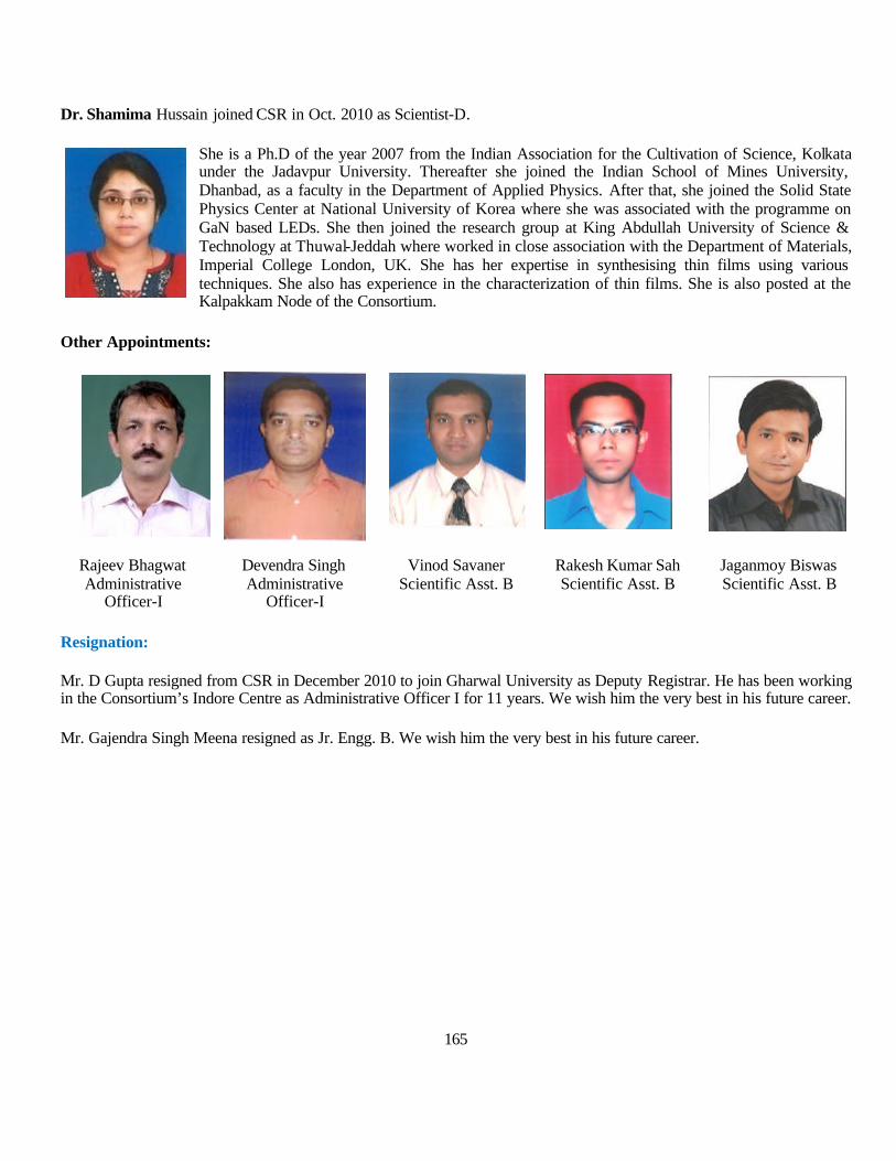
165
Dr. Shamima Hussain joined CSR in Oct. 2010 as Scientist-D.
She is a Ph.D of the year 2007 from the Indian Association for the Cultivation of Science, Kolkata under the Jadavpur University. Thereafter she joined the Indian School of Mines University, Dhanbad, as a faculty in the Department of Applied Physics. After that, she joined the Solid State Physics Center at National University of Korea where she was associated with the programme on GaN based LEDs. She then joined the research group at King Abdullah University of Science & Technology at Thuwal-Jeddah where worked in close association with the Department of Materials, Imperial College London, UK. She has her expertise in synthesising thin films using various techniques. She also has experience in the characterization of thin films. She is also posted at the Kalpakkam Node of the Consortium.
Other Appointments:
Rajeev Bhagwat Administrative
Officer-I
Devendra Singh Administrative
Officer-I
Vinod Savaner Scientific Asst. B
Rakesh Kumar Sah Scientific Asst. B
Jaganmoy Biswas Scientific Asst. B
Resignation:
Mr. D Gupta resigned from CSR in December 2010 to join Gharwal University as Deputy Registrar. He has been working in the Consortium’s Indore Centre as Administrative Officer I for 11 years. We wish him the very best in his future career.
Mr. Gajendra Singh Meena resigned as Jr. Engg. B. We wish him the very best in his future career.

166
11. List of Collaborative Research Schemes
11.1 At Kolkata Centre
Utilization of VECC, Kolkata
1. Dr.B.P.Singh - Study of fission fragments produced in heavy ion (HI) reactions. 2. Prof. L Chaturvedi - Nuclear structure study of A~130 Nuclei. 3. Dr. Madhumita Manna - Radiation and molecular immunology: Understanding the mechanism of protection against leishmania
infection using radio attenuated leishmania parasites. 4. Prof. S.C. Bhattacharya, Department of Chemistry, Jadavpur University - Effect on biomolecules by synthesized nanoparticles
using ionizing radiations 5. Dr. C.T. Aravinda Kumar, Department of Chemical Sciences, Mahatma Gandhi University Radiation Induced Modification of
DNA and Some Pyrimidine Nucleosides and Nucleotides 6. Prof. Kalyan Kumar Mukherjee, Department of Chemistry, Jadavpur University.Protection of radiation and chemical induced
DNA damage by chemicals and nano materials 7. Dr.Geeta K Sharma, Department of Chemistry, University of Pune.Radiation Induced synthesis of Metal Clusters: Effect of dose
rate and LET 8. Dr. Prabitra Chattopadhyay, Department of Chemistry, Burdwan University Study of the effects of radiation dose and ion beam
on the structure and ion-exchange property of polyoxometallates” . 9. Dr. Dilip Kumar Kakati, Department of Chemistry, Gauhati University.Studies on Ion-Beam Induced Modification and Gamma -
Radiation Induced Graft Copolymerisation of N-vinyl pyrollidone and Glycidyl methacrylate on Polyurethane ” 10. Prof.. Jag Mohan Keller, Macromolecular Research Centre, Rani Durgawati University “Study of Effect Radiation on the
Metalloid/ Oxide Incorporated Polymeric high Temperature Resistive Flexible Nano-Composite Thin Films for Microelectronics Application”
11. Dr. R. Dhanasekaran, Crystal Growth Centre, Anna University, Effect of ion implantation in GaN and ZnSe semiconductors for the fabrication of electornic and photonic devices
12. Dr. Sunita Keshri (Shaw), Birla Institute of Technology, Effect of ion implantation and gamma irradiation on wide band gap semiconductors ”
13. Dr. Abdul Khadar, Centre for Nanostructured & Nanotechnology, University of Kerla Modification of structural and optical properties of nanostructured semiconductors by ion implantation
14. Dr. Nandakumar Kalarikkal, School of Pure & Applied Physics, Mahatma Gandhi University, Ion beam Irradiation effects on the structural and ferroic properties of selected sol-gel derived films of nanomultiferroics”
15. Prof. S.C. Santra, Dept. of Environmental Science, University of Kalyani Comprehensive study on radiation induced metal tolerance in fungal strain”
16. Prof. A.R. Thakur, West Bengal State University, Evaluation of Attenuation of Infection of Human Rotavirus after Irradiation by Gamma Ray”
17. Dr. Sanjit Dey, Dept. of Physiology, Calcutta University, Radiation Protection using Moringa oleifera principles 18. Dr. Sarmistha Raychaudhuri, Department of Biophysics Molecular Biology & Bioinformatics, University Of Calcutta, Radiation
induced alterations in DNA, RNA and Polyamine levels in plants 19. Prof. Tapas Dasgupta, Department of Genetics & Plant Breeding, University Of Calcutta Radiation mediated Restructureing of
new plant type in sesame adaptable to agro-climatic condition of West Bengal. 20. Dr.Sukhendusekhar Sarkar, Deparment of Physics, Study of shape co-existence in 153Ho and few-valence particle nuclei around
146Gd Core” 21. Dr. Suresh Kumar, Deparment of Physics, Magnetic Rotational bands crossing and role of the proton and neutron orbitals in
magnetic rotation (MR) phenomena near A = 135” 22. Dr. P.V.Madhusudhana Rao, Deparment of Nuclear Physics, Search for anti-magnetic rotation in A ~ 100 nuclei 23. Prof. L. Chaturvedi, Guru Ghasidas University, Search for highly deformed structures in A ~ 130 region”. 24. Dr. Sourav Ganguly, Department of Physics, Chandernagore College, Study of Magnetic Rotation in odd-A Rb isotopes from
lifetime measurement. 25. Dr. T.R. Routray, Department of Physics , Sambalpur University, The finite range effective nuclear interaction: A complete
nuclear model .
167
26. Dr. Jag Mohan Keller, Macromolecular Research Centre, Rani Durgawati University, Study of Radiation effect on the biocompatible polymer blends of poly (vinylalcohol)/ Poly (L-lactic acid) (PLLA)/ polyglycolic acid/ hydroxyapatite”
27. Dr. Sukhendusekhar Sarkar, Department of Physics, Bengal Enginnering & Science University, Preparation of implanted targets and their application in the investigations of neclear structure and nuclear astrophysics”
Utilization of 3 MV Pelletron at IOP, Bhubaneswar
28. Budhadeb Chattopadhyay, College of Leather technology ,West Bengal Technical University, Rational and productive use of protein wastes from the tannery industries
29. Dr. Nabakanta Jana , Dept. of Zoology, Charuchandra College, Kolkata, Air pollution Bio-monitoring by lichens with respect to atmospheric pollutants with special reference to their trace element profile.
30. Dr. (Mrs.) Shanti Lata Sahoo, P. G. Dept. of Botany, Utkal University, Bhubaneswar - Regulation and Expression of Antioxidant Enzymes and Comparative Study on Metal Detection of Withania Somnifera L. and Abutilon indicum L. in Response to Various Metal Stresses.
31. Dr. Madhumita Manna, Dept. of Zoology, Bethune College, Kolkata, Influences of Trace Elements in Immune Funtion in Visceral Lesishmaiasis and post kala-azar Bermal Leishamaniasis of India.
32. Prof. S. C. Santra, Dept. of Environmental Science, University of Kalyani, Studies on trace element distribution and their role in salt stress adaptation in halophytic plants of mangrove vegetation of West Bengal.
33. Dr. R K Dutta, Dept. of Chemistry, IIT Roorkee, Roorkee, Ion Beam Assisted Synthesis and Chracterization. 34. Dr. Anup Kumar Talukdar, Dept. of Chemistry, Gauhati University, Guwahati - Studies on Effect of Ion Beam Irradiation on the
Nanostructured TiO2 prepared by Nanocasting Replication Method. 35. Dr. Shaon Ray Chaudhuri, West Bengal Univesity of Technology, Developing a laboratory scale biofilm based metal removal
system using heavy metal tolerant radioresistant marine coastal microbes. 36. Dr. A K Sen, Dept. of Earth Sciences, IIT Roorkee, Roorkee, Investigation of Potential Metalliferous Nature of Coal from India. 37. Dr. Ansuman Chattopadhyay, Dept. of Zoology, Siksha Bhavana, Visva-Bharti, Santiniketan - Arsenic induced genotoxicity and
modulation of trace elements in mammalian cells. 38. Dr. S Narayana Kalkura, Crystal Growth Centre, Anna University, Chennai, Ion Implantation and trace element analysis of
materials of importance in Biology and Medicine. 39. Dr. M L Garg, Dept of Biophysics, Panjab University, Chandigarh, Characterization of MT isolated from As treated rats for
toxicity and sensor application. 40. Dr. H.M.Agarwal, Department of Physics G.B.Pant University of Ag. & Tech., Pantnagar, Multielemental analysis of soil &
plant samples of GBPUAT Research Centres in Uttarakhand. 41. Dr. Debasis Das, Department of Chemistry, University of Burdwan, Studies on chemical speciation and metal ion separation by
some newly synthesized chelating ligands with N, S and / O donor sites: Applications to environmental samples. 42. Dr. H. N. Thatoi, Department of Biotechnology, CET, Bhubaneswar, Bioremediation of hexavalent chromium using stress
tolerant microbes isolated from chromite mine environments of Orissa. 43. Prof. Ashoke Ranjan Thakur, West Bengal University of Technology, Trace Element profiling of east Calcutta wetlands for
Phylogeny based growth condition prediction. 44. Dr. Ashutosh Naik , Department of Earth Sciences, Sambalpur University, Trace element behaviour in the granitic rocks of West
Orissa. 45. Dr. Dhrubananda Behera, Dept. of Pysics, NIT, Rourkela, Ion Beam based studies of Inter – and Intra – Granular modifications
in Yba2Du3O7 / Ag composite films. 46. Dr. P K Giri, Dept. of Physics, IIT Guwahati, Ion Beam Engineering of Carbon Nanotubes for Improved Structural and Optical
Functionalities
Utilization of 14 UD BARC-TIFR Pelletron facility at TIFR Mumbai.
47. Dr Bivash Behra, Spin distribution measurement a probe to understand the reaction mechanizm of medium mass systems. 48. Dr. Aloke De, Study of orbiting anomalies in 12C + 89Y and 16O + 89Y. 49. Dr. Surjit Mukherjee, Nuclear structure effects on breakup and multi-nucleon transfer reaction. 50. Prof N L Singh, Effect of projectile spin on fission fragement angular distribution for the system 13C , 14N + 238U.
168
51. Dr. K M Varrier, Department of Physics, University of Calicut, “Study of fission dynamics in 210Rn via the reactions 16,1 O + 194Pt and 24Mg +186Ta”
Uutilization of in-house facilities
52. Dr S. Chanda Centre For Studies in Man & Environment, Air-Borne Inorganic particulate dispersion and their accumulation on tree canopies of urban environment by using PIXE and EDXRF.
53. Dr. Avinash C Pandey, Department of Pysics,University of Allahabad, Elemental Analysis of Archaeological Findings from Kaushambi for The study of past.
54. Dr. S. Santhosh Kumar ,Mahatma Gandhi Govt. Arts College, Elemental Analysis of Traditional Medicinal Plants used in Keraleeya-ayurveda-chikitsa.
55. Prof. N. Rajmuhon Singh, Dept. of Chemistry, Manipur University, Trace Element Analysis and Biological Activities of Some Selected Medicinal Plants of Manipur.
56. Dr. B.K. Mishra, Centre of Studies in Surface Science and Technology, School of Chemistry, Sambalpur University, Selective extraction of metal ions from water sources by using surface modified silica matrix.
11.2 At Indore Centre
57. Dr. K.T. Ramakrishna Reddy, Department of Physics, Sri Venkateswara University Development of highly transparent and conducting In2O3: Mo Layers for Photovoltaic Application
58. Dr. Modi Kunal B. Department of Physics, Saurashtra University, A search for Mn-Zn based nano ferrite materials for high frequency power supplies
59. Dr. Mitesh Sarkar, Physics Department, M.S. University of Baroda, Study of dilute magnetic semiconducting alloys (DMSA)
60. Dr. S.B. Shrivastava Professor and Head, School of Studies in Physics, Vikram University,UJJAIN, Preparation and characterization of nanocrystalline materials
61. Dr. Sankar P. Sanyal, Professor in Physics, Department of Physics, Barkatullah University, Investigation of transport and magnetic properties of novel manganites
62. Dr. S. Ram, Professor, Materials Science Centre IIT, Kharagpur, Phase stability and intergranular giant-magnetoresistance properties in (La1-xEux)0.67Ca0.33MnO3 in a hybrid nanocomposite structure
63. Dr. P.C. Srivastava, Professor of Physics, Dept. of Physics, Banaras Hindu University, XPS and Valance Band Study using SR radiation along with Depth profiling studies for Magnetic metal / Semiconductor Interfaces
64. Dr. (Mrs.) Chandana Rath, Banaras Hindu University, Varanasi: Evolution of Magnetism with Oxygen Deficiency in Cobalt Doped TiO2 diluted magnetic superconductor
65. Dr. Sangeeta N. Kale, Fergusson College, Pune: To synthesize self-assembled oriented nanomagnetic particles in thin film form and study its property regime
66. Dr. Satishchandra B. Ogale, Ramanujan Fellow, NCL, Pune: Magnetic Nanostructures for Spintronics and Magneto-Optics
67. Dr. Ramphal Sharma, Dr. Babasaheb Ambedkar Marthwada University, Aurangabad: Growth and effect of SHI irradiations on structural and opto-electrical properties of nano crystalline pure and doped Zinc oxide semiconductor thin film for gas sensor application
68. Dr. P.P. Sahay, National Institute of Technology, Silchar: Studies on structural, optical and electrical properties of transparent conducting oxide (TCO) films for device applications
69. Dr. C. Venkateswaran, University of Madras: Exploration of CuInSe2 type diluted magnetic semiconductors for SPINTRONICS applications
169
70. Dr. P. Pradeep, National Institute of Technology – Calicut: Highly conducting transparent polymer films for optoelectronic & photonic application by laser ablation technique
71. Dr. Syed Rahman, Osmania University, Hyderabad: Thermal, transport and resonance studies in bismuth and germanium based glasses
72. Prof. P.B. Joshi, Solapur University: Synthesis and characterization of ferroelectric hetrostructures
73. Dr. Vilas Shelke, Barkatullah Univesrity, Bhopal: Inverstigation of wide range magnetoresistance in rare earth manganites through AgO/HgO additions
74. Dr. Rajendra Ku mar Pandey, Barkatullah University, Bhopal: Characterization of semiconductor quantum dots grown by electrochemical nanoprocesses
75. Prof. R. Bhar, Jadavpur University, Kolkata: Atomic force microscopy and confocar microscopic investigation of biomaterials under physiological and diseased conditions
76. Dr. (Smt.) L.D. Jadhav, Shivaji University, Kolhapur: Investigations on structural and electrical properties of Ni-GDC cermet as anode for solid oxide fuel cell.
77. Dr. M. Mahendran, Thiagarajar College of Engineering, Madurai: Magneto-Thermo -Mechanical characterization and phase transformation in ferromagnetic Ni-Mn-Ga shape memory alloys.
78. Dr. Jacob Mathew, S.B. College, Changanacherry: Structural and magnetic studies of some nanostructured mixed metal oxides synthesized via spray pyrolysis of polymer precursor.
79. Dr. Rachana Gupta, Acropolis Institute of Technology & Research, Indore: Microstructure of thin films prepared by reactive sputtering.
80. Dr. P.S. Goyal, Pillai’s Institute of Information Technology Engineering, Media Studies & Research, Mumbai: Non-Debye specific heat and tunneling states in mixed salts of ammonium and alkali halides.
81. Prof. Dinesh Varshney, Devi Ahilya University, Indore: Transport properties of doped ferrite thin films.
82. Prof. D.G. Kuberkar, Saurashtra University, Rajkot: Fabrication & studies on manganite based thin film devices for suitable applications.
83. Prof.. A. Mishra, Devi Ahilya University, Indore: Some studies on magnetic, optical and transport properties of BaTiO3 thin films doped with Cu/Co/Fe.
84. Dr. Atul Khanna, Guru Nanak Dev University, Amritsar: Low temperature specific heat capacity measurements and Raman spectroscopic studies in oxide glasses.
85. Dr. Prakash Nath Vishwakarma, NIT, Rourkela - Magneto-electric study of doped rare earth cobaltites
86. Prof. Ashok Rao, Manipal Institute of Technology, Manipal - Electrical and thermal properties of some doped manganites
87. Dr. R.B. Tangsali, Goa University - Study of Magnetic and Electrical Properties of Nanoparticle and Bulk Mn(x)Zn(1-x)Fe(2)O(4) prepared by Microwave Combustion.
88. Dr. V. Rama Rao Medicherla, ITER, Siksha ‘O’ Anusandhan University, Bhubaneshwar - Electronic properties of disordered transition metal alloys.
89. Dr. N. Lakshmi, M.L. Sukhadia University - Magnetic properties in Co-based multilayer systems.
90. Prof. S.I. Patil, University of Pune - Investigation of physical parameters in the context of VBS for 3d elements doping at Mn site in hole doped pervskite.
91. Dr. Ratnesh Gupta, Devi Ahilya University, Indore - To investigate the electronic structure of nitride and carburized of thick films using AIPES beamline of Indus-I.
92. Dr. Pratima Sen, School of Physics, Devi Ahilya University, Indore – Electronic and Optical properties of ZnO based multilayered thin films.

170
11.3 At Mumbai Centre
93. Dr. A. Srivastava, Punjab Univeristy, Chandigarh, “Study of arsenic and selenium toxifying using neutron activation analysis”.
94. Dr. N. Lakshmana Das, College of Science, Gitam, Rushikonda, Visakhapatanam, “Archaeometry studies using INAA”.
95. Dr. R. V. Upadhyay, Charotar Institute, Changa, Gujarat, Size dependent magnetic properties of Co-Zn spinel ferrite nanoparticles using diffraction technique
96. Dr. K. R. Priolkar, Goa University, Goa, Neutron Diffraction studies on Ni-Mn based ferromagnetic shape memory alloys
97. Dr. S. K. Kulkarni, IISER, Pune, Investigation of magnetic nano and core-shell particles
98. Dr. Mrinal Pal, Burdwan Univeristy, Burdwan, Mesoscopic structural investigation using SANS on TMI–doped nanocrystalline ZnO : Promising FMS for spintronic devices.
99. Dr. A. Srinivasan, IIT, Guwahati, Neutron Diffraction studies of structural and magnetic phases in Co-Ni-Ga alloys
100. Dr. N. Harish Kumar, IIT Madras, Chennai, Investigation of structural and magnetic properties of half metallic Heusler alloys
101. Prof. Kabir-ud-Din, AMU, Aligarh, Interaction between serum albumins and drugs
102. Prof. J.A.E. Desa, Univ. of Goa, Studies of porous materials by SANS
103. Prof. J.A.E. Desa, Univ. of Goa, Structural studies of gels by light scattering and SANS
104. Dr. S. Ravi, IIT Guwahati, Neutron powder diffraction studies in charge ordered and double exchange ferromagnetic samples of Nd-Mn-O series.
105. Dr. Sanjeev Kumar, VNSG University, Surat, Stimuli responsive solution behavior of surfactants: A SANS study.
106. Dr. Sugata Ray, IACS, Kolkata, Probing the 4d and 5d magnetism in prototypical ……. Using neutron diffraction
107. Dr. R.K. Mahajan, Gurunanak Dev University, Structural and interactional behavior of mixed micelles of twin tail surfactants and with triblock copolymers
108. Prof. A.K.Raychaudhuri, SNBCBS, Kolkata, Neutron diffraction studies of collapse of charge ordering in narrow band half doped manganite Y0.5Ca0.5MnO3.
109. Dr. Atul Khanna, Guru Nanak Dev University, Amritsar, Structural investigations of binary borate and tellurite glasses by high-Q neutron diffraction and small angle neutron scattering techniques
110. Dr. Puyam Singh Central Salt & Marine Chemicals Research Institute, Bhavnagar, “SANS investigation of the silicon rubber membranes”
111. Dr. Bragadish Iyer, Charotar Institute of Applied Sciences, Changa, “Studies on structure of microbial exopolysaccharide (EPS) solutions and hydrogels using SANS”
112. Dr. A. V. Mahajan, IIT, Mumbai, Magnetism in low-dimension and/or frustrated magnetic systems
113. Dr. Basavaraj Angadi, Bangalore University, Bangalore, “Synthesis and studies on Magneto-Electric and Spin-Lattice Coupling in Pb(Fe1/2Nb1/2)O3 based multiferroic systems”
114. Dr. N. V. Sastry, Sardar Patel University, Vallabh Vidyanagar, “Studies on Aggregation behaviour of Pyridinium based amphiphilic ionic liquids in water and in presence of aggregate growth parameters”
115. Dr. M. S. Jogad, Sharnbasveshwar College of Science, Gulbarga, “Structural study of Pb-glass by Neutron Diffraction”
116. Dr. Babita Sehgal, M. S. University of Baroda, Baroda, “Use of SANS in preparation of doped transition metal hollow spheres using carbon as template and its photo catalytic activity under visible light”
171
11. 4 At Kalpakkam Node
117. Prof. Sabu Thomas, MG Univ., Kottayam, Kerala, Carbon Nanotube Filled natural Rubber Latex Nanocomposites (Category: Physical Sciences).
118. Prof. Anand P Pathak, Univ. of Hyderabad, Hyderabad, A.P, Ion Beams synthesis of semiconductor nanocyrstals and modification of III-V semiconductor Multi Quantum Wells under SHI Irradiation (Category: Physical Sciences).
119. Prof. Prasanna S. Ghalasi, MS Univ. of Baroda, Vadodra GujaratDesigning Chiral Molecular Magnets (Category: Physical Sciences).
120. Dr. Lisa Sreejit, NIT, Calicut, Kerala, Temperature Dependent Optical Switch from Organized Assemblies (Category: Physical Sciences).
121. Prof. V. Rajendran, KSR college of Tech Tiruchengodu, T.N, Development and characterization of nano metal oxides coatings for biomedical applications (Category: Engineering Sciences).
122. Prof. N. Rajendran MIT, Anna Univ Chennai, T.N, Development of TiO2 nanotubes for biomedical application (Category: Engineering Sciences).
123. Prof. G.D. Janaki, IIT, Chennai, T.N, A Rapid Manufacturing Approach to Facilitate Joining of Stainless Steel Pipes to Titanium Pipes (Category: Engineering Sciences).
124. Prof. S. Vasantha, Associate Professor, Dept. of Chemistry, Lady Doak College, Madurai – Super hydrophobic coating for enhancing corrosion resistance.
172
12. Utilisation of in-house facilities of UGC-DAE CSR: User List
12.1 Indore Centre
Indore Centre has been attracting large number of users, and its state of art instruments have been widely used by the university researchers. The number of users is less for the more advanced facilities because of the time required and also because the usage is more specialised.
TEM 1 Dr.Bindu TIFR, Mumbai
2 Dr.Obey Kosey Kerala Univ.
3 Mr.Dharmendra Tawde Barodra Univ.
4 Prof.S.B.Rai Dept.of Physics, BHU
5 Prof.R.K.Pandey Barkatullah Univ.Bhopal
6 Dr.I.P.Jain Rajasthan Univ. Jaipur
7 Dr.Prathima Sen DAVV, Indore
8 Dr.D.P.Gogoi Tezpur Univ. Assam
9 Dr.S.D.Dhole Pune Univ.
10 Ms.Chiti, C\o.Prof.S.V.Bhoraskar Pune Univ.
11 Ms.Sunita, C\o.Dr.Anjali Rajbhoj Dr.B.A.M.Univ. Aurangabad
12 Ms.Varsha Sharma Vikram Univ., Ujjain
13 Dr. Pankaj Sagdeo BARC, Visakhapatnan
14 Dr.G.D.Verma IIT, Rurkee
15 Mr.Jitendra K. Pandey C\o. Prof.Ram Gopal Allahabad Univ.
16 Dr.Daval Modi Baroda Univ.
17 Mr.Hari Babu, C\o.Dr.Amrendra IGCAR
18 Dr.P.Shrivastav Bio-Chem.Eng, BHU
19 Mr.Nayan, C\o.Prof.R.K.Pandey Barkutullah Univ.Bhopal
20 Mr.Kamal C\o.Dr.P.K.Mishra Chem.Eng.& Tech. BHU
21 Dr.Dinesh Varshney DAVV, Indore
22 Dr.Obey Koshy Kerala Univ.
23 Mr.X.R.Babu C\o.Dr.A.Leo Rajesh Trichy, T.N
24 Mr.Ramesh C.Nagarwal Dept. Of Pharmacy, BHU
25 Mr.Dnyanesh Rasale, C\o.Dr.Apurba K. Das IIT, Indore
26 Dr.M.Baneerji DAVV,Indore
27 Prof.S.B.Rai Dept.of Physics, BHU
28 Prof.Prathima Sen DAVV, Indore
29 Prof. Samdarshi Tezpur Univ. Assam
30 Mr.Vinod, C\o.Prof.S.K.Singh Physics, Chotoo Ram Univ.
31 Mr.Tanuj, C\o.Prof.D.Kanjilal IUAC
32 Prof.S.V.Bhoraskar Pune Univ.
173
D8 Advance XRD
33 Ratnesh Gupta/Sheetal Soni DAVV, Indore
34 Dinesh Varshney/Kavita DAVV, Indore
35 Rajesh Kumar/Prashant Sahu Bhilai Inst. Techn., Durg, Chattisgarh
36 Deepshikha Barkatulla University, Bhopal
37 Amandeep Gupta/Shweta Vikram Univ, Ujjain
38 Nikhil Ranjam Jha DIMAT, Rajasthan
39 Amit Khare Barkatulla Univ., Bhopal
40 Devang D Shah MSU, Vadodra, Gujrat
41 Jashashree Roy NIT Rourkela, Orissa
42 S. Kane DAVV, Indore
43 Roja Shree D Osmania University, Hyderabad
44 Navas University of Kerala, Kerala
45 Detty Kerala University
46 Kamal Kumar Gupta IT BHU, Varanasi
47 Tarale Arjun Solapur University, Solapur, MH
48 Shushmita Chaudhri/Ritu Raj Sharma RGPV, Bhopal
49 Raju Osmania University, Hyderabad, AP
50 Sister Jessy Avinashilingam Univ. Coimbtore,TN
51 Jolly Pal Pt. RSU Univ. of Raipur, Chattisgarh
52 Rachana Gupta DAVV, Indore
53 A. Mishra/Niyati DAVV, Indore
54 Fozia Aziz IIT, Indore
55 Nanda Shakti ISM, Dhanbad, Jharkhand
56 Achnit Jain BHU, Varanasi
57 Obey Koshy University of Kerala, Kerala
58 Versha H. G. U., Sagar
59 Gautam Pt. RSU Univ. of Raipur, Chattisgarh
60 Soumya Pt. RSU Univ. of Raipur, Chattisgarh
61 Priyanka Prashar Jiwaji University, Gwalier
62 Pooja Agrawal IT BHU, Varanasi
63 Mayora Varshney S V College, Aligarh, UP
64 Sulabh Dubey Sri Ram College, Jabalpur
65 Hari Mohan Roy RGPV, Bhopal
66 Hilal Ahmed AMU, Aligarh, UP
67 D M Pimpalshinde Dr. Ambedkar College, Nagpur, MH
68 Pankaj Sagdeo BARC, Vizag, AP
69 Rajesh IT BHU, Varanasi, UP
70 Rajeshman V. G. CUSAT, Cochin, Kerala
71 Snehal Jani MSLU, Udaipur, RJ
72 Hussain Jeeva Khan RGPV, Bhopal
174
73 Suman SOP, DAVV, Indore
74 Shailendra Singh/Ashutosh BIT, Mesra, Ranchi,
75 Ramesh Nagarwal BHU, Varanasi, UP
76 Neeti Tripathi Delhi University, Delhi
77 Shribhan Singh B. I. Pharmacy, Jaipur, RJ
78 Gaurav NRI College of Phramacy, Bhopal
79 Pooja Pandey/Sachin Savant RGPV, Bhopal
80 Pooja Gupta RRCAT, Indore
81 Parul Khurand/Sheenam Thatai Banasthali Univ, Rajasthan
82 Jitendra Tripathi RDVV, Jabalpur
83 A. K. Bajpai Science college, Jabalpur
84 Stephen St. Joseph Coll, Trichi, Tamilnadu
85 B. L. Ahuja Udaipur University, Udaipur, RJ
86 Akriti Verma SGSITS, Indore
87 Pradeep Verma R. G. P. V. Bhopal
88 Dhanveer singh Rana IISER, Bhopal
89 Rizk AMU, Aligarh, UP
90 Parul/ D S Rana IISER, Bhopal
91 R. K. Shukla Lucknow University, Lucknow, UP
92 Nitesh Mangrole NRI College of Phramacy, Bhopal
93 Sanjay Pachori RGPV, Bhopal
94 Namrata Garwal University, Uttarakhand
95 Umesh GSITS, Indore
96 S Manu CNNT, Univ of Kerala
97 Vinod Kumar Sonipath, Hariyana
98 Richa Panda Barkatulla Univ., Bhopal
99 Ashish Gupta Jaypee, Guna (M. P.)
100 Neha Gupta BITS, Pilani, RJ
101 Sulekha Dable H. G. U. Sagar
102 Anjali Maurya H. G. U. Sagar
103 Kiran, Jyoti, Nupoor MLSU Udaipur
104 Adesh Sahu/Barkha MPCST, Bhopal
105 Archana Srivastava Rungta College of Eng & Tech., Bhilai
106 B. P. Joshi Solapur University, Solapur, MH
107 Chandrani Tezpur Univ, Assam
108 Vinod Kumar R MG Univ. Kotyam, Kerala
109 Mohammad Hashim AMU, Aligarh, UP
110 Pinaki Das Gupta SOP, DAVV, Indore
111 Geeta Rana G B P U A , Pantnagar, UK
112 Varsha Sharma Madhav science College, Ujjain
113 Sudheesh VD MLSU, Udaipur, RJ
114 Bindu Kumari Rani Durgawati Univ, Jabalpur

175
115 Vinay Dashore SDITS, Khandwa
116 Ravish Jain SGSITS, Indore
117 Nitya Garg Anand SGSITS, Indore
118 X Ravington St. Joseph coll Trichi, TN
119 Naveen (Mrs. P. Sen) DAVV, Indore
120 Rajeev Prakash BHU, Varanasi, UP
121 Sangeeta Malviya DAVVV, Indore
122 Paramjeet Kaur BNPG College, Udaipur, RJ
123 Shailja Tiwari MLSU, Udaipur, RJ
124 Ravalia Ashish Saurashtra University, Rajkot, Gujrat
125 Shailendra Smiriti College, Indore
126 Abhilasha Bakshi DAVV, Indore
127 Jagmohan singh Butola IT BHU, Varanasi
128 Bharat Singh Barkatulla Univ, Bhopal
129 Versha Bundela DAVV, Indore
130 Sheetal shastri DAVV, Indore
131 T. Bhattacharya IIT Indore
132 H C Nayak Govt Maharaja College, Chhatarpur, UP
133 Pateek Chawrekar Manipal Univ., Manipal, Karnataka
134 J. P. Mathews MG Univ. Kotyam, Kerala
135 Pankaj Dubey R. G. P. V. Bhopal
136 Uma Kachar Rajkot
137 Dr. Karmakar BARC, Mumabai
138 Indrajit IIT, Indore
139 Deepika Barkatulla Univ, Bhopal
140 Gagan Dixit G. B. P. U. A.. Pantnagar, Uttrakhand
141 Madhuri Gupta DAVV, Indore
142 Papanna B Belavi Karnataka Univ.
143 Tehmina Vikram Univ., Ujjain
144 Sachin Savant R. G. P. V. Bhopal
145 Versha Sharma Madav Science College, Ujjain
146 Harsha Patil DAVV, Indore
147 Rajesh K Thakur Barkatulla Univ, Bhopal
Magnetism Lab.
148 Prof. B. L. Ahuja M. L. Sukhadia University
149 Prof.N.S.Gajbhiye IIT, Kanpur
150 Prof. Balak Das Lucknow University
151 Prof. D. Behera NIT, Raourkela
152 Prof. B. L. Ahuja M. L. S. University, Udaipur
176
153 Dr. R.N. Bhowmik Pondicherry University
154 Rajendra Gulbarg University, Karnataka
155 Prof. B.L. Ahuja M. L. S. University, Udaipur
156 Prof. P. R. Sarode Goa University
157 Prof. A. Mishra DAVV, Indore
158 Dr. P. N. Vishwakarma NIT Raurkela
159 Dr. S. Srinath HCU, Hyderabad
160 Dr. K.R. Priolkar Goa University
161 Dr. R.C. Shrivastava G.B.P.U.A.&T. Pantnagar Uinv
162 Dr. Vilas Shelke Barktullah University, Bhopal University 163 Dr. H.S. Jayanna Kuvempu University Karnataka
164 Dr. Rajnish Kurchania (Sachin Sawant) MANIT, Bhopal
165 Dr. Rajnish Kurchania (Pooja Pandey) MANIT, Bhopal 166 Dr. Rajnish Kurchania (Sanjay Pachori) MANIT, Bhopal 167 Dr. Chandna Rath IT-BHU Banaras
168 Prof. O. N. Shrivastava Banaras Hindu University
169 Dr. P. N. Vishwakarma NIT Raurkela
170 Prof. S. H. Pawar D. Y. Patil University, Kolhapur
171 Dr. Abdul Khadar University of Kerla
172 R.V. Upadhyay PD Patel Institute of Applied Sciences, University Charusat Gujrat
173 Dr. Abdul Khadar University of Kerla
174 Prof. B. L. Ahuja M. L. Sukhadia University
175 Dr. Chandna Rath IT-BHU Banaras
176 Dr. R.N. Bhowmik Pondicherry University
177 Dr. B. Chandrashekar University of Hyderabad
178 Prof. R.K. Pandey Barktullah University
179 Prof.A.Choudhury Tezpur University Assam
180 Prof. D. G. Kuberkar Saurashtra University Gujrat
181 Prof. S. P. Rath IIT Kanpur
182 Prof. P. R. Sarode Goa University
183 Dr. Rachna Gopta IET, DAVV, Indore
184 Prof. Ashok Kumar Tezpur University Assam
185 Prof. A. Mishra (Niyati ) DAVV, Indore
186 Rajendra Gulbarg University, Karnataka
187 Dr. R.N. Bhowmik Pondicherry University
188 Prof. B. Appa Rao Hyderabad University
Dielectric Permittivity
189 Devang Shah C/o Dr. P.K. Mehta MSU, Baroda
190 Sushil Phadke C/o Dr. B.D. Srivastava Govt. Girls College, Dhar
177
191 Rajesh Thakur C/o Dr. N.K. Gaur Barkatullah Univ., Bhopal 192 Prof. Mahavir Singh H.P. Univ., Shimla
193 R.S. Solanki C/o Prof. D. Pandey BHU, Varanasi
Thermal Conductivity
194 Dr. V.S. Tiwari RRCAT, Indore 195 Dr. Shailendra Kumar RRCAT, Indore
DSC
196 Mayank Sharma C/o Dr. D.K. Jain I.P.S., Indore 197 Arit Verma C/o Dr. M.L. Sharma C.S.V.T.U., Bhilai, Chhattisgarh
198 Seeta Shukla C/o Dr. Anil Kumar Bajpai B.M.R.L., Jabalpur 199 Deepti Deshpande C/o Dr. Rakesh Bajpai R.D.V.V., Jabalpur 200 B.S. Rathore C/o Dr. M.S. Gaur H.C.S.T., Mathura
201 Dr. M. Roy MLS University, Udaipur 202 N. Gupta C/o Dr. A. Dalvi B.I.T.S., Pilani 203 Anji Reddy C/o Dr. Ranveer Kumar H.S.G.U., Sagar
204 Poonam Sharma C/o Dr. D. Kanchan M.S.U., Baroda 205 Dr. R. Prasad DAVV, Indore 206 V. Bundela C/o Dr. D. Varshney DAVV, Indore
207 K. Veerbhadra Rao C/o Prof. B. Appa Rao DoP, Osmania University, Hyderbad 208 Ashwini Kumar C/o Dr. D. Varshney SoP, DAVV, Indore 209 A.K. Patel C/o Prof. J.M. Keller R.D.V.V., Jabalpur
210 Sakshi Kabra C/o Prof. Anshu Rani University of Kota, Kota 211 Stuti Katara C/o Prof. Anshu Rani University of Kota, Kota 212 M. Sinha C/o Prof. R.M. Banik BHU, Varanasi
213 Aman Agrawal C/o Prof. Meena Tiwari SGSITS, Indore 214 R.C. Nagarwal C/o Prof. J.K. Pandit BHU, Varanasi 215 V. Gajbhiye C/o Prof. N.K.Jain H.S.G.U., Sagar
216 V.R. Reddy C/o Prof. P. Kistaid Osmania University, Hyderbad 217 M.A. Samee C/o Dr. Syed Raman Osmania University, Hyderbad 218 S.S. Mudgal C/o Dr. S.S. Pancholi A.C.P., KuKas, Jaipur
219 Pooja Agarwal C/o Dr. Pradeep Srivastav BHU, Varanasi 220 M. Sharda C/o Dr. Suresh Babu Osmania University, Hyderbad 221 Dr. Pooja Gupta RRCAT, Indore
222 A. Gupta C/o Dr. V.V.S. Murty Holkar Science College, Indore 223 R.Vijya Kumar C/o Dr. K. Shiva Kumar Osmania University, Hyderbad 224 Madhusmita Baral C/o Dr. Soma Banik RRCAT, Indore
225 Sangeeta Jain C/o Dr. Anil Bajpai Govt. Science College, Jabalpur

178
226 Dr. S.N. Kane DAVV, Indore 227 A.Jain C/o Prof. Sanjay Singh BHU, Varanasi
228 P. Chaubey C/o Dr. B.Mishra BHU, Varanasi
Surface Physics Laboratory
229 Umesh B. Gawas Goa Univesity
230 Ms. Rupali Kundu IOP, Bhubaneshwar 232 Mr. Prasada Rao NIT, Trichy 233 S.S. Sinde Shivaji University
233 Umesh B. Gawas Goa Univesity 234 Anjali S. Rajbhaj BAM University 235 Mr. Sunil Singh BHU, Varanasi
236 Mr. M. Earnest Stephen St. Joseph’s College, Trichi 237 Dr. Subhasish Ghosh JNU, New Delhi 238 Mr. Sandeep C/o Dr. C. Biswas S.N. Bose Centre, Kolkata
239 Mr. S. Sankar University of Kerala 240 Mr. Obey Koshy University of Kerala 241 Mr. Prashant S. Shewale Shivaji Univesity, Kolhapur
242 Mr. L. Blakrishnan NIT, Tiruchirapalli 243 Dr. Uma Subramanian Goa University 244 Mr. Sandeep Patel IIT Kanpur
245 Dr. Sudipta Bandhopadhyay Univesrity of Calcutta 246 Sr. Jessy Mathew Avinashilingam Univ. for Women,
Coimbatore
247 Ms. Usha Rajalakshmi Avinashilingam Univ. for Women, Coimbatore
Scanning Electron Microscopy
248 K.V.Bhadra Rao Osmania Univ., Hyderabad 249 Sunita Malguti DAVV, Indore
250 Prashant Shewale Shivaji Univ.Kolhapur 251 Deepti Deshpande RDVV, Jabalpur 252 Sangeeta Jain Vikram University, Ujjain
253 Preeti Shrivastav DAVV, Indore 254 Pinaki Das Gupta DAVV, Indore 255 Prof.Sadekar B.A. Marath. Univ. A’Bad
256 Renuka Tayde DAVV, Indore 257 AjitKumar Lucknow University 258 Adu Verma Lucknow University
259 Rina Singh Barkatullah Univ. Bhopal
179
260 Arti Verma CVST, Bhilai 261 Prerna Pande DAVV, Indore
262 Tahir Munaja DAVV, Indore 263 Sandeep Dhoble Ferguson College, Pune 264 Sapana Jain H.S.gaur Univ., Sagar
265 Rajesh V.G. CUSAT, Cochin 366 Kirti Samuwar Peoples College, Bhopal 267 Prof.Prasad DAVV, Indore
268 Sambhaji Shinde Shivaji Univ.Kolhapur 269 Mubarak Ali Periyar Univ. Salem 270 P.Balsubramaniyam Madurai Kamraj Univ.
271 M.Ernest Stephen Madurai Kamraj Univ. 272 Upendra Tripathi IPR, Gandhinagar 273 Smriti Parihar DAVV, Indore
274 Ashish Ravalia Saurashtra Univ.Rajkot 275 R.M.Sangashetty Gulberga University 276 Neelima Mahato BHU, Varanashi
277 Rinku Sharma DAVV, Indore 278 Pukhraj Chappnel Nahata College, Mandsaur 279 Swati Jain R.G.V.P. Bhopal
280 Bharat Katoria Saurashtra Univ.Rajkot 281 Sanjay Pachori R.G.V.P.Bhopal 282 Rajendra Mahata Sagar University
283 Aman Agarwal SGSITS, Indore 284 Sakshi Kabra Kota University, Rajasthan 285 Amita Jaiswal Smruti College, Indore
286 Varsha Sharma Madhav College, Ujain 287 Bhavna Sarvan Madhav College, Ujjain 288 Mukti Sinha BHU, Varanashi
289 Hemlata Chbra BHU, Varanashi 290 Gaurav Maria RGTU, Bhopal 291 S.N.Kane DAVV, Indore
292 Vikram Karavande SGSITS, Indore 293 Ravish Jain SGSITS, Indore 294 Jagruti Sahariya M.L.S.Univ.Udaipur
295 Anjali Soni Govt.College, Raipur 296 Chanchal Dheevar Govt.College, Raipur 297 Jassi Mathew A. Lingam Uni.Coimbtore
298 Nitish Upadhya Smruti College, Indore
180
PES Beamline
299 Mr. Amit Khare / Prof.Sanyal Barkatullah University, Bhopal 300 Dr.Pankaj Sagdeo BARC, Vizag
301 Mr.Naidu/ Prof.A.Subrahmanyam I.I.T.Chennai 302 Sheetal Soni/Dr.Ratnesh Gupta DAVV, Indore 303 Mr.Amol Vankundre/ Prof.S.I.Patil Pune University
304 Mr.S.Shinde/Dr. K.P.Adhi, Pune University, 305 Dr. Soma Banik/Dr.S.K.Deb RRCAT, Indore 306 Dr. K.J.S. Shokhey/ Mr. Sudip Nath RRCAT, Indore
307 Dr.Shailja Tiwari/ Prof.B.L.Ahuja M.L.S.University, Udaipur 308 Dr. Maheshwar Nayak/Dr.G.S.Lodha. RRCAT, Indore 309 Dr. P.Mishra/ Mr. Amit Das/Dr.Kukreja RRCAT, Indore
310 Mr.Ghosh/Dr.Ramphal Sharma B.A.M.Univ.Aurangabad 311 Kerur Patel/Prof.P.K. Mehta M.S.University, Baroda 312 Dr.S.N.Jha/ Arup Biswas BARC, Mumbai
313 Mr.Sandeep Dhoble/ Dr.S.Kale Fergusson College, Pune 314 Mr.Sanjay Brahma I.I.Sc. Banglore 315 Dr.M.V.Ramarao SOA University, Bhubaneshwar
316 Dr.Shubha Gokhale IGNOU, Delhi 317 Dr.P.Anand Kumar I.I.T.Gauhati 318 Parul Khanna/Prof.S.K.Kulkarni Banasthali University, Rajasthan
319 Dr.Shailendra Kumar RRCAT, Indore
Pulsed Laser Deposition
320 Dr. Shailja Tiwari C/o Prof. B. L. Ahuja MLS University, Udaipur
321 Mr.Jagananatha panda C/o Dr.T.K.Nath IIT Kharagpur 322 Mr.Yogesh Kumar C/o Prof. Ravi Kumar IUAC,New Delhi 323 Mr.Arvind Ghosh Dr.B.A.M.University,Aurangabad
324 Ms.Bhagwati Bisnoi M.S.University Of Baroda 325 Mr.Devang Shah M.S.University Of Baroda 326 Mr.Pankaj Mohanty IT-BHU Varansi
327 Dr. A. Dalvi BITS,Pilani 328 Mr.Sudheesh C/o Dr. N. Lakshami M.S.U. Udaipur 329 MrTarale Naganath C/o Dr.P.B.Joshi Solapur University Solapur
330 Ms.N Tripathi C/o Dr.Shyama Rath University Of Delhi 331 Mr.Ravalia Ashish C/o Prof. D.Kuberkar Sourashtra University Rajkot 332 Mr.Amit Khare C/o Prof. S. Saniyal B.U.Bhopal
333 Ms.Uma Khachar C/o Prof. D.K.Kuberkar Sourashtra University Rajkot 334 Ms.Gagan Dixit G.B.P.U.A.T. Pantnagar
181
335 Mr.Vinay Dashore Holkar Science College,Indore 336 Ms.Minaxi Sharma C/o Prof.Ravi kumar NIT Hamirpur
Low Temperature Measurements (14T/0.3K PPMS)
337 Mr. Ashish Ravalia C/o Dr. Kuberkar Saurashtra University, Rajkot
338 Mr. Koyada Pratap Kakatiya University, Warangal, AP 339 Prof. Mandal SINP, Kolkata 340 Prof. S. Goyal Pillai’s Institute, Mumbai
341 Prof. S. Ram IIT, Kharagpur 342 Prof. Kuberkar Saurashtra University, Rajkot 343 Ms. Richa Acropolis Inst. Tech and REs, Bhopal
344 Dr. Subham Majumdar IACS, Kolkatta 345 Mr. Rajesh Thakur, C/o Dr. Gour Bhopal University 346 Dr. A. Bharati IGCAR, Kalpakkam
347 Mr. Kati Raju Osmania University, Hyderabad 348 Dr. Arumugam Bharatidasan University 349 Ms. Deepshika Bhopal University
350 Prasanna C/o Prof. S. Ram IIT, Kharagpur
Heat Capacity Measurements (MTC Lab)
351 Ms. Arshpreet Kaur/Dr. Atul Khanna Guru Nanak Dev Unniversity, Amritsar 352 Mr. R. Pradeesh/ Dr. K. Sethupathi IIT, Chennai
Resistivity (77K-400K)
353 Richa Panda AITR, Bhopal 354 Amandeep Acharya Vikram Univesity, Ujjain 355 M. Shweta Vikram Univesrity, Ujjain 356 Deepika Bhavsar DAVV, Indore 357 Soniya Sharma SGSITS, Indore
Atomic Force Microscopy
358 Ms. Deepika Bhavsar SGSITS, Indore
359 Ms. Richa Panda C/o Dr.Vandana Rathore AITR, Bhopal 360 Mr. Amandeep Acharya C/o Dr. S.B.
Shrivastava Vikram University, Ujjain

182
361 Mrs. Sweta Moghe C/o Dr.SBS Vikram University, Ujjain 362 Mr. Indu Verma C/o Prof. Balak Das Gupta Lucknow University
363 Mr. Mahesh Babu Aligarh Muslim University 364 Ms. Bhagawati Bishnoi, C/o Dr. P.K. Mehta M.S. University of Baroda 365 Ms. Vidya Tour C/o Dr. Ramphal Sharma Dr. BAM University, Aurangabad
366 Mr. Tejas Tank, C/o Prof. S.P. Sanyal Barkatullah University, Bhopal 367 Dr. S. Jayakumar PSG College of Tech., Coimbatore 368 Mr. I. Navas, C/o Dr. V.P.M. Pillai University of Kerala
369 Dr. Devang Shah, C/o Dr. P.K. Mehta M.S. University, baroda 370 Mr. Arindam Ghosh, C/o Dr. Ramphal
Sharma Dr. BAM University, Aurangabad
371 Mr. B. Prkash C/o Dr. S. Umapathy St. Joseph College, Trichy 372 Mr. Mubarak Ali C/o Dr. V. Raj Priyar University, Salem 373 Mr. E. Stephen C/o Dr. S. Umpathy Madurai Kamaraj University
374 Mrs. Smriti Parihar C/o Dr. P.K. Dubey S.V. College of Pharmacy, Indore 375 Mr. Jofy P.J. Barkatullah University 376 Ms. Neelima Mahato C/o Prof. M.M. Singh BHU, Varanasi
377 Mr. U.N. Tripathi C/o Chetal Jariwala IPR, Gandhinagar 378 Ms. Maneesha Mishra C/o Dr. P. Kuppusami IGCAR, Kalpakkam 379 Mr. G.J. Shyju C/o Prof. c. Sanjeeviraja N.M. Christian College, Mmarthandam,
Kanyakumari 380 Mr. Sambhaji Shinde C/o Dr. K.Y. Rajpure Shivaji University Kolhapur 381 Mr. Sadekar H.K., C/o Dr. R.P. Sharma Dr. BAM University, Aurangabad
382 Mr. Sandeep Dhobale C/o Dr. Sangeeta Kale Fergusson College, Pune 383 Mr. R. Rajeev C/o Dr. M.A.Khadar University of Kerala 384 Mr. Bindu Kumari C/o Dr. R.K.Bajpai RDVV University, Jabalpur
385 Mrs. Deepti Deshpandey C/o Dr. Rakesh Bajpai
RDVV University, Jabalpur
386 Mr. Naveen Agrawal, C/o Dr. Mitesh Sarkar M.S. University, Baroda
387 Mr. Rounak Badera C/o Dr. N.S. Ranpise Pune University 388 Mr. Pramod M.R. C/o Prof. Sandesh R.
Jadkar Pune University
389 Dr. K.M. Jadhav Dr. BAM University, Aurangabad 390 Mr. Arjun Tarale Solapur Unversity, Solapur 391 Ms. Detty A.P. C/o Dr. V.P.M. Pillai University of Kerala
392 Sr. Jessy Y Mathew N C/o Dr. (Mrs.) Rachel Oommen
Avinashilingam Univ. for Women, Coimbatore
393 Dr. Abhijit S. Shetty SDM College of Dental Science Sattur Dharwad
394 Ms. Heena Kohad C/o Dr. T. Guha Jadavpur university 395 Ms. Usha Rajalakshmi C/o Dr. Rachel
Oommon Avinashilingam Univ. for Women, Coimbatore
396 Mr. B.T. Rao C/o Dr. L.M. Kukreja RRCAT, Indore 397 Mr. Ramesh Chand nagarwal C/o J.K. Pandit BHU, Varanasi

183
398 Ms. Anjali Soni C/o Dr. Alka Tiwari Pt. Ravishankar Shukla Univ. Raipur 399 Mr. Vijay Kumar C/o Prof. Shyam Kumar Kurukshetra University
400 Mr. Sapkal R.T. Shivaji University, Kolhapur 401 Dr. Nikita Agrawal C/o Dr. N.D. Shashikiran People’s College of Dental Science, Bhopal 402 Mr. Santosh M.V. C/o Dr. K.P. Vijayakumar Cochin University
403 V.G. Rajeshman C/o Dr. K.P. Vijayakumar Cochin University 404 Mr. Keyur Patel C/o Dr. P.K. Mehta M.S. University of Baroda 405 Mr. Ravington babu C/o Dr. A. Ieo Rajesh St. Joseph College, Trichy
406 Mr. M. Earnest Stephen C/o Dr. S. Umapathy St. Joseph College Trichy 407 Mr. Gautam Sheel Thool C/o Dr. A.K. Singh Govt. V.Y.T.P.G. College, Durg 408 Ms. Jayashree Ray C/o Dr. P.N.
Vishwakarma National Institute of Technology, Rourkela
409 Mr. Ashish Ravalia C/o Dr. D.G. Kuberkar Saurashtra University, Rajkot 410 Mr. Jolly Bose r. C/o Dr. M. Pillai University of Kerala
411 Ms. Anjali U.K. & Mr. Sabin A. C/o Dr. Abdul Khadir
University of Kerala
412 Ms. Mayora Varshney, C/o K.D. Verma S.V. College, Aligarh
413 Mr. Rajesh Kumar C/o Dr. Ruby Das Bhilai Institute of Technology, Durg 414 Mr. Adesh Kumar Shahu C/op Dr. M.K.
Rathore MPCST, Bhopal
415 Mrs. Barkha Jaiswal C/o Dr. M.K. Rathore MPCST, Bhopal 416 Mr. Praveen Kumar C/o Prof. Shyam Kumar Kurukshetra University 417 Mr. Soumya R. Deo C/o Dr. A.K. Singh Govt. V.Y.T.P.G. College, Durg
418 Mr. narinder Kumar University of Jammu 419 Mr. Vinay Dashore C/o Dr. V.V.S.Murthy Holkar Science College, Indore 420 Mr. Sheetal Soni C/o Dr. Ratnesh Gupta DAVV, Indore
421 Ms. Uma D. Khachar C/o Dr. D.G. Kuberkar Saurashtra University 422 Dr. Piyush Solanki C/o Dr. D.G. Kuberkar Saurashtra University 423 Mr. I. Navas C/o Dr.V.P.M.Pillai University of Kerala
424 Mr. Vinod Kumar C/o Dr.V.P.M.Pillai University of Kerala 425 Mr. K.B.Raulkar Vidya Bharati Mahavidyalaya, Amravati 426 Ms. Madhuri Gupta DAVV, Indore
427 Ms. Pramila Chaubey C/o Prof. B. Mishra BHU, Varanasi 428 Mr Prashant Kumar Sahu C/o Dr. Ruby Das Bhilai Institute of Technology, Durg 429 Mr. Mohammad Hasin, C/o Prof. Alimuddin Aligarh Muslim University
430 Mr. Obey Koshy C/o Dr. Abdul Khadir University of Kerala 431 Ms. Gagan Dixit, C/o Dr.H.M. Agrawal G.P.Pant University 432 Mr. Satish GSITS, Indore
433 Nataj Keru Desai C/o S.R.Patil Shivaji University, Kolhapur 434 Dr. Nikita Agrawal C/o Dr. N.D. Shashikiran People’s college of Dental Science, Bhopal 435 Ms. Jayashree Ray C/o Dr. P.N.
Vishwakarma National Institute of Technology, Rourkela
436 Mr. Vimal Kuma Tiwari C/o Prof. Pralay Mouti
BHU, Varanasi
184
Scanning Probe Microscopy
437 Mrs Preeti Schdev C/o Dr M. Banerjee DAVV, Indore 438 Prof. P.C. Srivastav BHU, Varanasi
439 Ms. Bhagawati Bishnoi C/o Dr. P.K. Mehta M.S. Univerity of Baroda 440 Ms. Dolly Singh 441 Dr. N.L. Singh M.S. University of Baroda
442 Mr. Devang Shah C/o Dr. P.K. Mehta M.S.University of Baroda 443 Mr. Naveen Agrawal C/o Dr. Mitesh Sarkar M.S. University of Baroda 444 Mr. Sanjay S. Ghosh C/o Dr. S.R. Jadkar University of Pune
445 Mr. Ashish Choudhary C/o Dr. D.K. Jain IPS Academy, Indore 446 Ms. Mukty Sinha C/o Prof. R.M. Banik BHU, Varanasi 447 Ms. Maneesha Mishra C/o Dr. P. Kuppusami IGCAR, Kalpakkam
448 Dr. R.K. Pandey Barkatullah University 449 Mr. Achint Jain C/o Prof. Sanjay Singh BHU, Varanasi 450 Ms. Pooja Agrawal C/o Dr. Pradeep
Shrivastava BHU, Varanasi
Laser Scanning Confocal Optical Microscope (LSCOM)
451 Dr. Tisson Job Coorg Inst. Of Dental Sciences, Virajpet, Karnataka
452 Mr. Aashish Choudhary IPS Academy, Indore 453 Mr. Nitish Upadhyaya Smriti College of Pharmaceutical
Education, Indore
454 Ms. Ankita Khandelswal Jaipur National University, Jaipur
Low temperature and high/ low Resistivity
455 Chandana Rath, Prof. A. Kumar Tezpur Univ., 456 K. Pratap Kakatiya University
457 Mahesh Babu Aligarh Muslim University 458 P.J. Joffy Barkatullah University 459 U.N. Tripathi IPR, Gandhinagar
Thermopower
460 Chandana Rath, Prof. A. Kumar Tezpur University
461 Mahesh Babu Aligarh Muslim University 462 P.J. Joffy Barkatullah University
185
Resistivity/Magnetoresistance measurements (MTC Lab)
463 Ms. Leena Joshi/Dr.S. Keshri Birla Institute of Technology, Mesra 464 Dr. D. Behera NIT, Rourkela 465 Dr. Sugata Ray IACS, Jadhavpur
466 Mr. Kishore Kohar K IPR, Gandhi Nagar 467 Ms. Trapti Atre/Ms. Renuka Tayade DAVV, Indore 468 Mr. Tejas Tank/Prof. S.P. Sanyal Barkatullah University
469 Ms. Reena Singh/Dr. Vilas Shelke Barkatullah University 470 Dr. B.L. Ahuja Udaipur University 471 Ashish B. Ravalia/Dr.D.G.Kuberkar Saurashtra University
472 Mr. I. Panner Muthuselvam/Dr. R. N. Bhowmik
Pondicherry University
473 Mr. Amit Vishwakarma/ Dr. D Varshney DAVV, Indore
474 Mr. Gourav Pathak/Dr. D Varshney DAVV, Indore 475 Mr. Mustaq Ahmad/ Dr. D Varshney DAVV, Indore 476 Dr. D. Bahera NIT, Rourkela
477 Mr. Bharat R. Kataria/Dr. D.G. Kuberkar Saurashtra University, Rajkot 478 Dr. M. V. Rama Rao Siksha “o” Anusandhan University,
Bhubaneswar
479 Mr. Yogesh Kumar/Dr. Ravi Kumar IUAC, New Delhi 480 Prof .O. N. Srivastava Banaras Hindu University, Varanasi 481 Mr. Irfan I. Sumara/Dr. J. A. Bhalodia Saurashtra University Rajkot
482 Mr. Ashish B Ravalia/ Dr.Nkesh Shah Saurashtra University Rajkot 483 Mr. A Sendil Kumar/Dr. S. Srinath University of Hyderabad 484 Ms.Chandrani Nath/Prof. Ashok Kumar Tezpur University
485 Dr. K.R. Priyolkar Goa University 486 Mr.Amit Khare/ Prof. S.P. Sanyal Barkatullah University 487 Mr.Kati Raju/Prof. P. Venugopal Reddy Osmania University
488 Dr. Sanjay Rai/ Kanwaljeet Singh RRCAT Indore 489 Ms. Uma D Khachar/Dr. D. G. Kuberkar Saurashtra University 490 Ms. Deepsikha Bhargava/Prof. S.P. Sanyal Barkatullah University
491 Mr.Valkunde Amol Tanaji/ Dr. S.I. Patil University of Pune 492 Mr. Hilaal Ahmed/Dr. Shakeel Khan Aligarh Muslim University 493 Ms. Mamata D Davivapra/ Prof. Ashok Rao MIT, Manipal
494 Ms. Geetha Mohlla/ Prof. Ashok Rao MIT, Manipal 495 Mr.Kati Raju/Prof. P. Venugopal Reddy Osmania University 496 Dr. Vilash Shelke Barkatullah University
186
Raman Spectroscopy
497 Ashutosh Kumar BIT Mesra, Ranchi 498 Arvind Kumar C/o. Dr. P.S. Alegaenkar DIAT, Pune
499 Mr. Shiv Kumar Barbar C/o Dr.M.Roy M.L.Sukhadia University 500 Mr. H.Srivastava RRCAT,Indore 501 Mr. Anji Reddy Poly Dr.H.S.G.University, Sagar
502 Ms. Kavita Verma DAVV, Indore 503 Ms. Ritika Swana DAVV Indore 504 Mr. Pawan K.Sharma C/o Prof.Mahavir Singh Himachal Pradesh University, Shimla
505 Mr. Avindam Ghosh Dr.B.A.M.University, Aurangabad 506 Mr. Shashikant D.Shinde C/o Dr.K.P.Adhi University Of Pune 507 Ms. Bhagwati Bisnoi C/o Dr.P.K.Mehata M.S.University Baroda
508 Mr. Nageswar Rao P RRCAT,Indore 509 Mr. Saraspreet Singh C/o Dr.Pankaj
Srivastava Delhi
510 Mr. Adinath M.Funde C/o Prof. S.R.Jadkar University Of Pune 511 Mr. Shrikant Gore C/o Prof. S.R.Jadkar University Of Pune 512 Ms. Vaishali Waman C/o Prof. S.R.Jadkar University Of Pune
513 Dr. Prasanna Ghalsasi M.S.University,Baroda 514 Mr. P.S.Shwale C/o Prof..M.D.Uplane Shivaji University, Kolhapur 515 Mr. K.Veerbhadra Rao C/o Dr.B.Appa Rao OsmaniaUniversity, Hyderabad
516 Mr. S.S.Shinde C/o Prof..C.H.Bhosale & Dr.K.Y.Rajpuri
Shivaji University, Kolhapur
517 Mr. Ashwani Kumar C/o Dr. D. Varshnay DAVV Indore
518 Mr. M.Earnest Stephen P. C/o Dr.S.Umapathey
MKU Madurai
519 Mr. B.Prakash C/o Dr.S.Umapathey MKU Madurai
520 Mr. Devang D.Shah M.S.University Baroda 521 Dr. G.S.S.Saini Chandigarh 522 Ms. Neelima Mahato C/o Prof. M M.Singh B.H.U. Varanasi
523 Mr. Vijay Kumar C/o Prof. Shyam Kumar K.U., Kurukshetra 524 Mr. Praveen Kumar C/o Prof. Shyam Kumar K.U., Kurukshetra 525 Mr. Pankag Mohanty C/o Dr. CMrs
Chandana, Rath BHU, Varanasi
526 Mr. Nagrswana Rao P. RRCAT, Indore 527 Mr. Rageev C/o- Prof. M. Abdul Kadar University of Kerala
528 Mr. Deepali Gangrade C/o Dr. Sinita Mohanty
CIPET. Bhubaneswar
529 Mr. Baban P. Dhonde C/o Dr. A. K. Tyagi IGCAR. Kalpakkam
530 Mr. Kailash Sapnar C/o Dr. Sanjay Dhol Univ. of Pune 531 Mr. Pramod MR C/o Prof. Sandesh R. Jadkar University of Pune 532 Mr. Kailash A Sapnar C/o. Dr. Sanjay Dhole University of Pune
533 Prof. Milind Deshpande Sardar Patal Uni. Gujrat
187
534 Mr. Versha Rajpoot C/o Dr. Vijay Verma Dr. H.S. Gour Central Uni. Sagar 535 Dr. P.R. Sagdeo BARC, Vishkhapattanam
536 Mr. Chiti Tank Uni. Of Pune 537 Mr. Detty Alappatt C/o Dr. V. P. Mahadevan Uni. Of Kerala 538 Dr. R. Prasad DAVV, Indore
539 Mr. Jyoy. P. Mathew C/o Dr. Jacob Mathew M.G. Uni. Kottayam, Kerala 540 Mr. Kavita Verma C/o Dr. Dinesh Varshnay DAVV Indore 541 Soni Bindiya - H S.P.U. V.V. Nagar , Gujrat
542 Mr. Nitya Garg S.P.U. V.V. Nagar 543 Pallavi Sakariya S.P.U. V.V. Nagar, Gujrat 544 Mr. Parveen Kumar & Mr.Vijay Kumar C/o
Prof. Shyam Kumar K.U.K
545 Ms. Detty A.P. C/o Dr. V.P. Mahaduan Pillai Uni. Of Kerala. Trivandram 546 Dr. Pankaj Misra Dr. M.A. Kuttappa RRCAT, Indore
547 Dr Tisson JoB Coorg Institute of Dental Sci 548 Mr. Rajeshmon V.G. Cochin University 549 Mr. Arvind Kumar Cochin University
550 Dr. P.S. Alegaonkar DIAT, Pune 551 Mr. Amit Khare BAU, Bhopal 552 Mr. V. Rajshekar Reddy C/o Dr. P. Kistaiah O.U. Hyderabad
553 Mr. Kamal Kumar Gupta BHU. Varanasi 554 Ms. Akrati Varma C/o Dr. R Prasad D.A.V.V. Indore 555 Grish Chandra C/o Dr. R.C. Srivastava G.B. Pant Uni. Pantnagar
556 Jolly Bose R C/o Prof. V.P. Mahadavan Pillai Kerala Uni., 557 Prof. K.P. Vijay Kumar Cochin University 558 Mrs. Vinay Kumari C/o Dr. D. M. Nasa Hisar
559 Neelam Godara C/o Dr. D. M. Nasa Hisar 560 Mr. Jayaprakash Karnataka Universtiy, Dharwad 561 Dr. G.S.S. Saini Punjab University, Chandigarh
562 Mr. Ajay Jha TIFR, DCS ,MUMBAI 563 Ms. Achamma George Manit, Bhopal 564 Ms. M. Sharada C/o Dr. D. Suresh Babu Osmania University, Hyderabad
565 Kavita Verma C/o. Dr. Dines Varshngy DAVV Indore 566 Priyanka. U. Londhe Dr. N.B. Chaure Uni. Of Pune 568 Mr. R.Vijaya Kumar C/o K. Siva Kumar Osmania University, Hyderabad
569 Roja Sree.D C/o- Pro. P.Yadagiri Reddi Osmania University, Hyderabad 570 Ajay Jha C/o. Dr. Joyitishman Dasgupta TIFR, Mumbai 571 T.S. Shyjv C/o Dr. R.Gopalakrishna Anna Uni.
572 Dr. S. Anandhi C/o Dr. R.Gopalakrishna Anna Uni. 573 Rajesh P.V. UGC-DAE CSR, Kolkata Center 574 Anuradha Gupta C/o. Dr. V.V.S. Murthy Holkar Sc College, Indore
575 Yashaswita B. Chavan C/o. Dr. J. Tonannavar Karnatak Uni, Dharwad 576 Shweta V. Badavi C/o. Dr. J.V. Yeragi Karnatak Uni, Dharwad
188
577 Shashikant Shinde Uni. Of Pune 578 Pramod MR Uni. Of Pune.
579 Madhushree Uni. Of Pune 580 Jagmohan Singh Butola C/o – Dr. Mrs.
Chandana Rath BHU, Varanasi
581 Pankaj Mohanty Dr. Mrs. Chandana Rath BHU, Varanasi 582 Mr. S.V. Bhoraskar Univ. of Pune 583 Kavita Verma C/o Dr. Dinesh Vaishney DAVV, Indore
584 Parul Khurana C/o. Prof. Sulabha Kulkarni Banasthali Uni 585 Gagan Dixit C/o Dr. H.M. Agrawal G.B.P.U.A.R.T., Uttrakhand 586 Geeta Rana C/o. Dr. Umesh C GBPU A&T. Panthnagar
587 Obey Koshy C/o Dr. M. Abdul Khadar Uni of Kerala 588 M.S. Yaragop Kamataka Uni, Dharwad
ESCA
589 Ms. Sonam Dwivedi DAVV, Indore
590 Mr. D. M. Pimpalshende Ambedkar College,Nagpur
591 Mr. Shivakumar Pandey BHU, Varanasi
592 Ms. Manisha Izardar DAVV Indore
593 Mr. Tulsi Ram K. V. S.V. Univ. Tirupati
594 Mr. Nilesh Shrivastava BHU Varanasi
595 Mr. Ajimsha R. S. RRCAT,Indore
596 Ms. Manju Yadav DAVV,Indore
597 Mr. Sandeep Kumar GNDU, Amritsar
598 Dr. L. M. Kukreja RRCAT,Indore
599 Ms. Darshana Inamdar Pune Univ.Pune
600 Mr. Shivacharan Sharma Univ. of Rajashtan, Jaipur
601 Mr. Razak Ali Fi AMU,Aligarh
602 Mr. Rohitranjan Shahi BHU, Varanasi
603 Mr. Dr. Rohidas Kshirsagar BARC, Mumbai
604 Mr. Rajendra Chokhare DAVV,Indore
605 Mr. Shrikant R. Naik Goa Univ.
606 Dr. Vijaykumar K. P. Cochin Univ.
607 Mr. Sovik Kondu IIT, Kharagpur
608 Mr. Anirudha Bose RRCAT,Indore
609 Ms. Sharada M. Osmania Univ.Hyderabad
610 Mr. Sachidanand Shrivastava IIT,Kanpur
611 Dr. S. K.Deb RRCAT, Indore
612 Ms. Achamma George MANIT,Bhopal

189
613 Ms. Mr. Vinay Pratap Singh IT, BHU,Varanasi
614 Ms. Manu S Univ. of Kerla
UV-Vis Spectroscopy
615 Mr. V. Rajshekhar Reddy Osmania Univ.,Hyderabad
616 Mr. Veerabhadrarao Osmania Univ.,Hyderabad
617 Mr. Vijaya Kumar Osmania Univ.,Hyderabad
618 Mr. Aman Deep Acharya Vikram Univ. Ujjain
619 Ms. Anjani Choubey ISM Dhanbad
620 Mr. Baban Dhonge IGCAR Kalpakkam
621 Ms. Bhagwati Bishnoi MS univ. Baroda
622 Ms. Bhavana Sarwan Vikram univ. Ujjain
623 Mr. Devang Shah M.S. Univ. Baroda
624 Ms. Geeta Rana GBPant University
625 Ms. Madhuri Gupta DAVV Indore
626 Mr. Naveen Kulkarni DAVV Indore
627 Ms. Nilima Mahato BHU,Varanasi
628 Ms. Pinaki Dasgupta DAVV Indore
629 Mr. Prakash bala Madurai kamraj Univ.
630 Ms. Priti Khandelwal DAVV,Indore
631 Prof. Prasad DAVV,Indore
632 Ms. Rucha Panda Acropolis institute,Bhopal
633 Mr. Sandeep Kumar GND Univ Amritsar
634 Ms. Sharda Osmania Univ.,Hyderabad
635 Ms. Shweta Moghe Vikram Univ. Ujjain
636 Ms. Sonam Dwivedi DAVV,Indore
637 Ms. Stephen Gnanadoss Madurai Kamraj Univ.
638 Ms. Stephen M. E. Madurai Kamraj univ.
639 Mr. U.N Tripathi IPR Ahmadabad
FTIR Spectroscopy
640 Ms. Stuti Katara Kota Univ.
641 Mr. U.N. Tripathi IPR Ahmedabad
642 Mr. Vijay Kaushik Kurukshetra Univ.
643 Mr. Aman Agrawal SGSITS,Indore
644 Mr. Amit Jain DAVV,Indore
645 Ms. Anjali Soni Pt. Ravishankar Univ., Raipur
190
646 Mr. Baban Dhonge IGCAR, Kalpakkam
647 Mr. Chanchal Dhiwar Pt. Ravishankar Univ., Raipur
648 Ms. Gagan Dixit Pantnagar Univ.
649 Mr. Joffy Barkatullah Univ.
650 Ms. Kawita Verma DAVV,Indore
651 Mr. Mohommad Hashim AMU, Aligarh
652 Ms. Namrata Ghildiyal HNB Gadhwal Univ., Gadhwal
653 Mr. Nitin Jaiswal SGSITS, Indore
654 Ms. Niyati Mishra DAVV,Indore
655 Mr. Pankaj Mohanty BHU,Varanasi
656 Mr. Praveen Goyal Kurukshetra Univ.
657 Prof. Prasad DAVV,Indore
658 Mr. Rajendra Sanghshetty Gulbarga Univ.
659 Mr. Rakesh Trivedi IPS academy, Indore
660 Ms. Renu Gupta Kurukshetra Univ.
661 Ms. Rinku Sharma GDC Indore
662 Ms. Ruchi Verma RRL, Bhopal
663 Ms. Shweta Jain RGPB Univ. Pantanagar
664 Ms. Sonam Dwivedi DAVV,Indore
665 Ms. Stephen M. E. Madurai Kamraj Univ.
Room temperature Mossbauer spectroscopy
666 Mr.Manu.S Kerala University
667 Mr.B.Vittal Parsad Osmania University
668 Mr.Sudeep Cochin University
669 Mr.Chandra Shekar University of Hydrabad
670 Ms.Kavita DAVV Indore
671 Mr.Sudeep Kumar IIT Kanpur
672 Ms.Snehal Udaipur University
673 Dr.N.Laxmi Udaipur University
674 Mr.Girish Pantnagar Univ.Uttrakhand
675 Mr.Pardhesh IIT Madras
676 Mr.Nawale Ashok Pune University, Pune
677 Ms.Roja Reddy Osmania University
678 Mr.Shrikant Naik Goa University
679 Dr.Chandana Rath BHU, Varanasi
680 Mr.Satyendra JNU, Delhi
681 Mr.Mohd.Hashim AMU, Aligrah

191
682 Mr.Rohit Delhi Univeristy
683 Ms.Geeta Rana Pantnagar University
684 Dr.Prasad DAVV Indore
Low temperature and high field Mossbauer spectroscopy
685 Ms.Sudesh IIT Roorkee
686 Mr.Rohit Medwal Delhi University
687 Ms.Prameela AndraUniversity
688 Dr.R.K.Dutta IIT Roorkee, Uttrakhand
689 Dr.Sanjay Kumar Jadhavpur University
690 Dr.Sreenath University of Hydrabad
691 Dr.Bhowmik Pondicherry University
692 Dr.S.N.Kane DAVV Indore
693 Dr.Jacob Mathew M.G.University, Kottayam
694 Ms.Roja Reddy Osmania University,Hydrabad
695 Mr.Satendra JNU, Delhi
696 Mr.G.S.V.R.K Choudhury Andhra Univ
697 Mr.Naresh Pondicherry University
Magneto optical Kerr effect (MOKE)
698 Mr.Arvindham Ghosh BAMU,Aurangabad
699 Ms.Razia AMU Aligrah
700 Ms.Farha Aurangabad University
701 Mr.Rohit Medwal Delhi University ,Delhi
702 Mr.Punnet Negi Pantnagar Univ,Uttrakhand
703 Prof.Ogale Pune University,Pune
704 Mr.Arjun Solapur University
705 Ms.Gagan Dixit Pant NagarUniv.Uttrakhand
706 Mr.Rajiv Kerala University ,Kerala
707 Mr.Baban IGCAR, Kalkappam
708 Mr.Naveen M.S University, Baroda
709 Mr.Yogesh IUAC , Delhi
710 Ms.Reena Verma University of Rajasthan
711 Mr.Sudheesh MLS Univ., Udaipur
712 Ms.Detty Kerala University, Kerala
713 Mr.Amit Khare Barkatulla University, Bhopal

192
714 Ms.Sheetal Soni DAVV Indore
715 Mr .Devang Shah M.S. University ,Baroda
716 Dr.Bharti IGCAR , Kalpakkam
717 Mr.Snehal M.S University, Udaipur
718 Mr.Jolly Bose Kerala University , Kerala
719 Mr.Jitendra IUAC Delhi
720 Ms.Manisha IGCAR, Kalpakkam
721 Ms.Uma Khochar Saurashtra University
722 Mr.Navas Kerala University
723 Mr.Vinod Kumar Kerala university, Kerala
724 Ms.Varsha Mehto Barkatulla University
725 Mr.Tanuj Kumar IUAC, Delhi
726 Mr.Jagannath Panda IIT Kharagpur
727 Dr.M.Sarkar M.S.Univerity,Baroda
Ferroelectric (P-E) loop tracer
728 Mr.Ramakrishna Andhra University
729 Ms. Niyati Mishra DAVV, Indore
730 Dr.Jitendra Paul IUCA, Delhi
731 Ms.J.Roy NIT, Roorkela
732 Prof. Kuberkar Saurashtra University,Rajkot
733 Mr.Papanna Dharwad Univ., Karnataka
734 Dr.Bhowmik Pondichery University
735 Ms.Shreeja Pillai BU, Bhopal
736 Prof. Ashok Kumar Tezpur University
737 Mr.Arjun Solapur University, Solapur
738 Mr.Devang Saha M.S.University, Baorda
193
12.2 Mumbai Centre
Light Scattering
739 Prof . Kabir-ud-Din AM University, Aligarh
740 Prof. S. Umapathy Madhurai Kamaraj Univ.,
Dielectric Relaxation
741 Dr.S.Patwe BARC, Mumbai
742 Dr.A.K.Tyagi BARC, Mumbai
745 Dr. Amitabh Das BARC, Mumbai
746 Dr.S.N. Acharya BARC, Mumbai
Material Preparation Lab
747 Dr. Sugata Ray, IACS, Kolkata
748 Dr. S. Srinath & A. Sendilkumar* University of Hyderabad
750 Prof. R.V. Upadhyay & Ms Harishda CHAROSAT, Changa
751 Dr.(Mrs) Srirupa Mukheri* ICL College,. Navi Mumbai
752 K.P. Muthe* BARC, Mumbai
753 R.S. Ningthoujam* BARC, Mumbai
754 Dr. Souvik Bhattacharya* BARC, Mumbai
755 Dr. A.K. Tyagi* BARC, Mumbai
756 Ms. G. Vinita* BARC, Mumbai
757 Ms. Vinila Bedekar* BARC, Mumbai
758 Mr. S. J. Patwe* BARC, Mumbai
759 Mr. B.P. Mandal* BARC, Mumbai
760 Ms. N. Farheen* BARC, Mumbai
761 Mr. R. Shukla* BARC, Mumbai
*used multiple times
CSR Neutron Diffractometer
762 Dr. Anjana Dogra National Physical Laboratory , New Delhi
763 Prof. R. Upadhyay CHARUSAT, Changa
764 Dr. Sugata Ray Indian Association for Cultivation of Sciences (IACS)
Kolkata
194
765 Dr. J. B. Singh Materials Science Division, B.A.R.C , Mumbai
766 Prof. A. K. Rastogi Jawaharlal Nehru University, New Delhi
767 Prof. A. V. Mahajan Indian Institute of Technology Bombay (IIT-B)
Mumbai
768 Dr. G. K. Padam National Physical Laboratory , New Delhi
769 Dr. K.R. Priolkar Goa University , Goa
770 Prof. A.K. Raychaudhuri S. N. Bose Centre for Basic Sciences (SNBCBS)
Kolkata
771 Dr. N. Harish Kumar Indian Institute of Technology Madras (IIT-M)
Chennai
772 Prof. Y. K. Sharma University of Rajasthan, Jaipur
773 Prof. P. L. Paulose Tata Institute of Fundamental Research (TIFR)
Mumbai
774 Prof. A. Srinivasan Indian Institute of Technology – Guwahati (IIT-G)
Guwahati
775 Dr. S. M. Yusuf Solid State Physics Division, BARC , Mumbai
776 Dr. K. Madangopal Material Science Division, BARC, MumbaI
777 Dr. R. Nirmala Indian Institute of Technology – Madras (IIT-M)
Chennai
778 Dr. Keka R. Chakraborty Solid State Physics Division, BARC , Mumbai
779 Dr. D. Bhattacharya Central Glass and Ceramics Research Institute (CGCRI)
Kolkata
780 Dr. S. Bhattacharya Technical Physics Division, BARC , Mumbai
781 Prof. R. Mahendiran National University of Singapore (NUS) , Singapore
782 Prof. E. V. Sampathkumaran Tata Institute of Fundamental Research (TIFR)
Mumbai
195
12.2 Kolkata Centre
Flow Cytometer
783 Dr.N.K.Jana Charuchandra College, Kolkata 784 Prof.S.C.Santra Kalyani University 785 Dr.Ansuman Chattopadhyay Viswabharati, Santiniketan
Cell / Tissue Culture Facilities
787 Dr.RajMohan Singh Manipur University
788 Prof.S.C.Santra Kalyani University
789 Dr.Sanjit Dey Calcutta University
790 Prof.H.N.Thattoi CET, Bhubaneswar
791 Dr.A.Chattopadyay VB, Santiniketan
792 Dr.S.Sahoo Utkal University
793 Ms. Rituparna Ghosh Calcutta University
794 Dr.RajMohan Singh Manipur University
Electrophoretic and Blotting Unit
795 Dr.Sanjit Dey Calcutta University
796 Dr.M.Manna Bethune College, Kolkata
797 Ms.Gargi Banerjee Jadavpur University
Centrifuges
798 Dr.RajMohan Singh Manipur University
799 Prof.S.C.Santra Kalyani University
800 Dr.Sanjit Dey Calcutta University
801 Prof.H.N.Thattoi CET, Bhubaneswar
802 Subhajit Bhar Kalyani University
803 Dr.A.Chattopadyay VB, Santiniketan
804 Ajit Kalekar University of Pune
805 Ms. Rituparna Ghosh Calcutta University
196
Mossbauer Spectrometer
806 Dr. Subrata Mukherjee Jadavpur University 807 Dr. Babita D. Ingale Pune University
808 Dr. G.C. Das Jadavpur University 809 Dr. Pradip Brahma Gurudas College, Calcutta 810 Dr. GVS Murthy NML, Jamshedpur 811 Dr. Zafar A. Siddiqi Aligarh Muslim University 812 Dr. Amal Kumar Das IIT, Kharagpur 813 Dr. Jay Chakraborty NML, Jamshedpur
814 Dr. Pabitra chakraborty Burdwan University 815 Dr. Sukhen Das Jadavpur University 816 Dr. Kalyan Mondal SNBNCBS, Kolkata
Positron Annihilation Spectroscopy
817 Dr. Rajesh Kumar Indraprastha University
818 Dr Asmita Sengupta Vishwabharati University,
819 Dr. S. Bandyopadhyay Calcutta University, Kolkata
820 Dr. K. S. Usha Devi N.S.S College Pandalam, Kerala
821 Dr. Chandana Rath B. H. U. (IT), Varanasi
X-Ray Fluorescence
822 Dr. Shaon Raychaudhuri West Bengal University and Tech.
823 Prof. N. Rajmuhon Singh Manipur University
824 Dr. S. Anirudhan University of Kerala
825 Dr. P. K. Chakraborty Bardwan University
826 Dr. Ansuman Chattopadhyay Visva Bharti University
827 Prof. H. N. Thatoi CET, Bhubaneswar
828 Dr. S. C. Santra Kalyani University
829 Dr. N.K. Jana Charuchandra College
830 Dr. Madhumita Manna Bethune College
831 Dr. Vipasha Chakraborty Jadavpur University
832 Dr. Santi Lata Sahoo Utkal University
UV-Vis Spectrophotometer
833 Dr. P.K. Bag Calcutta University

197
834 Dr. S.K. Das V.E.C.C. Kolkata
835 Prof. S.C. Bhattacharyya Jadavpur University, Kolkata
836 Dr. P.K. Barhai Birla Institute of Tech. Meshra.
837 Prof. Amitava Datta Power Engg. Jadavpur University, Kolkata
838 Prof. B. Ghosh School of Energy Studies, Jadavpur University
839 Dr. R. Sen C. G. C.R.I. Kolkata
840 Prof. J.M. Keller Rani Durgavati University, Jabalpur
841 Dr. Anshuman Chattopadhaya Vishwa Bharati Univ. 842 Dr. C.T. Arvindkumar Mahatma Gandhi University 843 Prof. R. Dhanasekaran Anna Univ. Chennai 844 Dr. S. De Dept. of Physiology, C.U 845 Dr. Somobrata Acharya I.A.C.S. Kolkata 846 Dr. S. Saha Benaras Hindu University 847 Prof. D. K. Kakati Gauhati University
Luminiscence Spectrometer
848 Prof. B. Ghosh School of Energy Studies, Jadavpur University 849 Prof. Amitava Datta Power Engg. Jadavpur University, Kolkata 850 Dr. P.K. Bag Calcutta University 851 Dr. S.K. Das V.E.C.C. Kolkata 852 Prof. S.C. Bhattacharyya Dept. of Chemistry, Jadavpur University, Kolkata 853 Dr. Somobrata Acharya I.A.C.S. Kolkata 854 Prof. Alokemoy Dutta S.I.N.P. Kolkata 855 Dr. S.K. Bandopadhaya V.E.C. C., Kolkata 856 Shri. Suraj Kumar Manipur University 857 Dr. P.K. Barhai Birla Institute of Tech. Meshra. 858 Dr. R. Dhanasekaran Anna Univ. Chennai 859 Dr. Sunita Keasri B.I.T. Meshra 860 Dr. C. Saha West Bengal University of Technology 861 Prof. S. K. De West Bengal University of Technology 862 Shri. Vinay Pratap Singh Benaras Hindu University.
Gamma Chamber
863 Dr. S. Raychoudhuri Calcutta University
864 Prof. S.C. Bhattacharyya Jadavpur University
865 Dr. R. Sen Central Glass & Ceramic Research Institute 866 Ms. Rituparna Ghosh Calcutta University
867 Prof. S.C. Santra Kalyani University
198
868 Dr. Tin Dalal Burdwan University
869 Dr. S. K. Das Variable Energy Cyclotron Centre
870 Dr. Satyen Saha Benaras Hindu University
871 Prof. T.K. Chaki I.I.T., Kharagpur
872 Dr. K. Nandakumar M.G. University, Kottayam, Kerala
873 Dr. Geeta Sharma, Pune University
874 Dr. C. Saha, West Bengal University of Technology
875 Dr. Somobrata Acharya Indian Association for the Cultivation of Science
876 Dr. Shaon Ray Choudhuri West Bengal University of Technology 877 Dr. C.T. Arvindakumar Mahatma Gandhi Univ. 878 Dr. Sanjit Dey, Raja Bazaar Science College, C.U.
879 Dr. B. Paul Haldia Petrochemicals
880 Dr. Amit Sadhukhan Dr. Mukherjee Homeo agency
881 Prof. J. M. Keller Rani Durgavati Univ., Jabalpur
882 Prof. Sujata Tarafdar Jadavpur University
883 Dr. D.K. kakati Guwahati University
884 Prof. N. Rajmohan Manipur University
885 Dr. Pobitra Chattopadhyay, Burdwan University
886 Dr. Sabyasachi Kundagrani, Institute of Agricultural Science,
887 Prof. S. De West Bengal University of Technology 888 Dr. D. Mohanta, Tezpur University
889 Dr. M. Manna Bethune College, Calcutta University
Dynamic Light Scattering
890 Dr. D. Mohanta Tezpur University
891 Prof. S.C. Bhattacharyya Jadavpur University 892 Dr. Nandakumar Kalarikkal Mahatma Gandhi University 893 Dr. Geetha Sharma University of Pune 894 Dr. S.K. Bandopadhaya Variable Energy Cycltron Centre 895 Prof. Amitava Datta Power Engg. Jadavpur University 896 Prof. D.K. Kakati Gauhati University 897 Prof. A. N. Talukder Gauhati University 898 Dr. Somobrata Acharya I.A.C.S. Kolkata 899 Dr. A.K. Santra Jadavpur University 900 Prof. A.K. Roychoudhuri S.N. Bose. National Centre for Basic Sciences
901 Dr. S.K. Das V.E.C.C
902 Prof. S. K. Dolui Tezpur University

199
Fourier Transform Infrared Spectrometer
903 Dr. S.K. Das V.E.C.C
904 Prof. B. Ghosh School of Energy Studies, Jadavpur University
905 Dr. D. Kakati Guwahati University
906 Dr. R. Sen Central Glass & Ceramic Research Institute 907 Prof. Amitava Datta Power Engg. Jadavpur University, Kolkata
908 Dr. Somobrata Acharya I.A.C.S. Kolkata 909 Dr. S. Paul Haldia Petrochemicals
910 Dr. Sunita Keasri B.I.T. Meshra
911 Dr. P.K. Bag Calcutta University
Sample Preparation Facilities, (Freeze drier, Pelletiser, Vacuum Filtration)
912 Prof B. G. Ghosh Jadavpur University
913 Dr. S. C. Santra Kalyani University
914 Susmita Dey BIT Mesra, Ranchi
915 Prof. A. Datta Jadavpur University
916 Dr. Ansuman Chattopadhyay Visva Bharti University
917 Prof. H. N. Thatoi CET, Bhubaneswar
918 Dr. Santi Lata Sahoo Utkal University
919 Dr. Madhumita Manna Bethuene College, Kolkata
920 Dr. Vipasha Chakraborty Jadavpur University
921 Prof. S. K. Dolni Tezpur University
922 Dr. N. Kalarikkal M. G. University
923 Sushmita Dey BIT Mesra, Ranchi
924 Dr.N.Rajmohan Singh Manipur University
925 Dr. A Chattopadhyay Visva Bharati University
926 Dr.(Ms) S.Roy Choudhury West Bengal University of technology
Low Temperature High Magnetic Field (LTHM)
927 Dr. Sudipta Bandopadhyay Physics Dept. Calcutta University
928 Dr. Pabita Chakrabarti Physica Dept , Burdwan University
929 Dr. Chandana Rath Physics Dept. Benaras Hindu University
930 Dr. T.P.Sinha Bose Instiyute ,Calcutta
931 Dr. Kalyan Mondal S.N.Bose National Centre
932 Prof. G.C.Das Dept. of Metallurgy,Jadavpur University
933 Dr. Sunita Keshri Dept of Applied Physics, BIT Mesra
200
934 Dr. Debnarayan Jana Physics Dept. Calcutta University
935 Dr. Sugata Roy IACS,Kolkata
936 Prof. S.K.Pradhan Physics Dept. Burdwan University
937 Prof. K. Ghosh Roy Saha Institute of Nuclear Physics
938 Dr. Prasanta Karmakar Variable energy Cyclotron Centre, Kolkata
939 Dr. Sukhen Das Jadavpur University . Kolkata
940 Prof. A.K. Raychaudhuri S N BOSE National Centre, Kolkata
941 Dr. Subrata Koner Dept. of Chemistry, Jadavpur University
Indian National Gamma Array
942 Dr R Palit Tata Institute of Fundamental Reserach.
943 Ms Ritwika Garg Department of Physics, University of Delhi
944 Prof Satyajit Saha Saha Institute of Nuclear Physics,
945 Dr Gopal Mukherjee Variable Energy Cycloytron Centre
946 Dr Devendra Mehta Department of Physics, Panjab University
947 Dr Tarkeshwar Trivedi Tata Institute of Fundamental Reserach
948 Dr Ajay Singh Indian Institute of Technology, Kharagpur.
949 Mr S Muralithar Inter University Accelerator Centre,
Detector Laboratory
950 Dr T R Routray P G Department of Physics, Sambalpur University
951 Prof Amitav Gupta School of Nuclear Studies & Applications, Jadavpur University.

201
13. General information on staff position
Sl. No. Designation A B
Indore Centre
01 Director of the Consortium 1 1 02 Centre-Director 1 1 03 Scientist 24 18 04 Engineer 4 3 05 Junior Engineer / Scientific Assistant 25 23 06 AO – II 1 1 07 AO – I 3 3 08 PS (to Director) 1 1 09 PA (to Centre -Director) 1 1 10 Assistant II 1 1 11 Assistant 3 3 12 Typist/Clerk/StenoTypist/ Steno-Clerk/Stenographer 4 3 13 Scientific Assistant (Library) 1 1 14 Caretaker 1 1 15 Driver/Driver-cum-Aux.Staff 3 3 16 Helper / Lab. Attendant / Aux.Staff 9 9
Kolkata Centre
01 Centre-Director 1 1 02 Scientist/Engineer 7 6 03 Junior Engineer / Scientific Assistant 14 14 04 Scientific Assistant (Library) 1 1 05 AO - I (Personnel) 1 1 06 AO – I (Accounts) 1 1 07 AO - I (Pur. & Stores) 1 1 08 PA (to Centre -Director) 1 1 09 Stenographer, Clerk/Typist 2 2 10 Guest House Attendant 1 1 11 Driver 2 2 12 Lab Attendent/Auxiliary Staff 4 4
Mumbai Centre
01 Centre-Director 1 1 02 Scientist 5 5 03 Engineer 1 1 04 Junior Engineer / Scientific Assistant 5 5 05 AO - I 2 1 06 P.A. (to Centre-Director) 1 1 07 Steno-Typist 1 1 08 Driver 1 1 09 Attendant-cum-Auxiliary Staff 1 1
A: The number of posts till 2010-2011 approved by UGC B: Existing manpower as on 31.03.2011.
202
14. Specializations and research facilities of our Scientists/Engineers
Name, Designation Specialization Facility
P. Chaddah Director
Condensed Matter Physics, Low Temperature Physics, Superconductivity
Director and overall in -charge of Head Office and facilities at the Centres
A. Gupta, Centre-Director
Nanostructured materials including nanocrystalline soft magnetic alloys, thin films and multilayers.
In-charge of facilities at Indore Centre
A. K. Sinha, Centre-Director
Experimental nuclear and accelerator physics
In-charge of facilities at Kolkata Centre
A.V. Pimpale, Centre-Director
X-ray spectroscopy, numerical simulation of X-ray and neutron instrumentation, theoretical physics
In-charge of facilities at Mumbai Centre
A. M. Awasthi, Scientist-F
Multiferroics. Solid State Battery Materials. Random covalent network systems
Modulated differential scanning calorimeter (-150 C to 550 C), quasi adiabatic calorimeter (5K to 300K), steady-state thermal conductivity set up (5K to 300K), melt -quench furnace (up to 1200 C)
P.D. Babu, Scientist-F
Magnetism of rare-earth inter-metallics, dynamical properties (phonons and magnons), Materials synthesis, Neutron Inelastic Scattering
Neutron diffraction, triple axis spectrometer, materials lab, impedance analyzer
Alok Banerjee, Scientist-G
Electron transport and magnetic properties of materials, synchrotron radiation
Ac-susceptibility setup, vibrating sample magnetometer, dielectric measurement setup, low field magnetoresistance setup
S. R. Barman, Scientist-F
Surface electronic structure Photoemission and low energy electron diffraction (LEED) experimental station
G.M. Bhalerao Scientist-D
Nanomaterials, electron and x-ray crystallography techniques
TEM
A.Chakraborty, Scientist-E
Radiation biology Fluorescent microscope, ultra centrifuge, electrophoresis set up ultra freezer, CO2-incubator
Sujoy Chakravorty Scientist-D
Thin film Deposition and Characterization using X-Ray and Neutrons, Atomic Diffusion in Thin Films.
XRD
R.J. Choudhary Scientist – E
Thin Films, Pulsed Laser Deposition, Functional Magnetic Materials
XRD
D. Das, Scientist-F
Condensed matter physics, magnetic materials,
Mössbauer spectrometer, positron annihilation spectrometer
S.K. Deshpande, Scientist-F
X-ray absorption spectroscopy, material characterization
X-ray diffractometer, impedance analyzer

203
V. Ganesan, Scientist-H
Low temperature physics, cryogenics and microscopy, ultra low temperature measurement system for T< 1K and also at high magnetic fields
Transport measurements using cryogen free environment ~10-300K, Scanning Probe Microscopy especially contact mode AFM
G. Ghosh, Scientist-E
Soft matter, fullerenes Static and dynamic light scattering
S. S. Ghugre, Scientist-F
In-beam gamma ray spectroscopy, nuclear physics
Clover array, radiation detectors and associated nuclear instrumentation, computational laboratory
Dileep Kumar Gupta Scientist – D
Condensed Matter Physics, magnetic ultra thin films and multilayers
In-situ MOKE set up
M. Gupta Scientist –E
Material Science, Neutron Diffraction
D8 Advance XRD
Shamima Hussain Scientist-D
Thin Film Synthesis and characterisations,Nanomaterials, Photoluminescence
FESEM and the material synthesis facilities
Som Datta Kaushik Engineer – D
Strongly Correlated Electron Systems & Multiferrioc materials
Neutron Diffraction Beamline / Cryocoolers
Archana Lakhani Scientist – D
Condensed Matter Physics, Low Temperature Physics, Imaging techniques
Low temperature and High Field Magnetisation.
N.P.Lalla, Scientist-F
Stuctural characterisation of materials using XRD and TEM, sysnthesis, phase transformation, quasicrystals and perovskites
.Powder XRD facility (with low-temperature-90K and high-temperature-1400K attachments), RF-induction furnace for synthesis of alloys
T. K. Mishra, Engineer E
Mechanical engineer, heat and mass transfer simulations
Mechanical workshop, vacuum systems
G. S. Okram, Scientist/Engineer – E
Experimental mesoscopic physics - development, characterization and electronic transport of low-dimensional composites
Dc electrical resistivity and thermoelectric power set up (5-300K).
Sudhir Kumar Pandey Scientist – D
Phase transition in correlated electron system
XRD
D. M. Phase, Scientist –G
Synchrotron beamline design, thin films, bilayers and multilayers, low and high energy implantation, surface and interface physics, vacuum technology
SEM-EDX Beamline for photoelectron spectroscopy at INDUS-1
V. Raghavendra Reddy, Scientist-E
Experimental condensed matter physics
Mössbauer Spectroscopy: transmission geometry( 12-1000 K ) and conversion electron, X-ray reflectivity, grazing incidence X-ray diffraction and magneto optical Kerr effect
204
R. Rawat, Scientist-E
Magneto transport and calorimetry especially in rare earth intermetallics
Resistivity / magnetoresistance set up (1.5-325 K, 0-10 Tesla), heat capacity set up (3-300 K, 0-10 Tesla)
S. Rayaprol Scientist –D
Magnetic materials & material synthesis .
Materials Lab.
A. Saha, Scientist-F
Radiation Chemistry, Macromolecular chemistry
Fluorimeter, gas chromatography, IR spectrometer, Radio-chemistry
P. Saravanan, Engineer-F
Electronics and communications Cryogenics
Liquid helium and liquid nitrogen
V. G. Sathe, Scientist-F
X-ray and neutron diffraction, EXAFS spectroscopy, synchrotron radiation and data analysis, superconductors and thin films and nano-materials
Raman spectrometer
T. Shripathi, Scientist-G
Crystal growth, electronic structure of surfaces, thin fillms, oxides, semiconductors, nanostructured materials
Electron spectroscopy for chemical analysis (ESCA and XPS), UV-Vis-NIR, FTIR and Computer Centre.
V. Siriguri, Scientist-F
Structure and magnetism of strongly correlated electron systems, disordered systems; neutron and high pressure X-ray diffraction
Neutron powder diffractometer, ac susceptometer
M. Sudarshan, Scientist-E
Trace elemental studies, proton induced X-ray emission
Energy dispersive X-ray fluorescence spectrometer, atomic absorption spectrometer, target preparation facilities -freeze drier, vacuum coating system, pelletiser
S. S. Thakur, Engineer-E
Civil engineer

205
15. List of staff at UGC-DAE CSR
Sl.No. Name Designation
1 Dr. P. Chaddah Director 2 Prof. Ajay Gupta Centre-Director 3 Dr. A.V. Pimpale Centre-Director 4 Dr. A.K. Sinha Centre-Director 5 Dr. V. Ganesan Scientist – H 6 Dr. T. Shripathi Scientist – G 7 Dr. Alok Banerjee Scientist – G 8 Dr. D.M. Phase Scientist – G 9 Dr. A.M. Awasthi Scientist – F 10 Dr. N.P. Lalla Scientist – F 11 Dr. S.R. Barman Scientist – F 12 Dr. Dipankar Das Scientist – F 13 Dr. S. Sitaram Ghughre Scientist – F 14 Dr. Vasudeva Siruguri Scientist – F 15 Dr. P.D. Babu Scientist – F 16 Dr. Abhijit Saha Scientist –F 17 Dr. S.K. Deshpande Scientist – F 18 Dr. Vasant Sathe Scientist – F 19 Dr. G.S. Okram Engineer /Scientist – E 20 Dr. Rajeev Rawat Scientist – E 21 Dr. V.R. Reddy Scientist – E 22 Dr. M. Sudarshan Scientist – E 23 Dr. (Mrs.) A. Chakraborty Scientist – E 24 Dr. Gautam Ghosh Scientist – E 25 P. Saravanan Engineer – F 26 Tapas Kumar Mishra Engineer – E 27 Sanjay Singh Thakur Engineer – E 28 Dr. Mukul Gupta Scientist – E 29 Dr. R.J. Chowdhary Scientist – E 30 Dr. S. Rayaprol Scientist – D 31 Dr. Archana Lakhani Scientist – D 32 Dr. Dileep Kumar Gupta Scientist – D 33 Dr. Sudhir Kumar Pandey Scientist – D 34 Dr.S. Datta Kaushik Engineer – D 35 Dr. G.M. Bhalerao Scientist-D
36 Dr. Sujoy Chakravorty Scientist-D
37 Dr. Shamima Hussain Scientist-D
38 Dr. J.B.M. Krishna SA-F/JE-F 39 Suresh Bhardwaj SA-F/JE – F
206
40 J.V. Joshi JE-E 41 Uday P. Deshpande JE-F 42 S.C.Das SA-F/JE-F 43 Nandkishore L. Ghodke SA-F/JE-F 44 Avinash Wadikar SA-F/JE-F 45 Satish R. Potdar SA-F/JE-F 46 Bhushan Jain SA-F/JE-F 47 B.R. Mendole SA-E 48 P.V. Rajesh SA-F/JE-F 49 Pinaki Das SA-E/JE-E 50 M.K. Verma SA-E/JE-E 51 N. Vijayakumar JE-E 52 Aparna Datta SA-E 53 S. Selvaraj SA-E 54 Kranti Kumar Sharma JE-D 55 Vinay K. Ahire JE-D 56 Mohd. Imran JE-D 57 Munshi Venugopal JE-D 58 Kaushik Basu SA-D 59 Mohan Kumar Gangrade SA-D 60 Mukesh Kumar SA-C 61 A.K. Rathore SA-C 62 Vinod Savaner SA-C 63 Jagnmoy Biswas SA-C 64 Rakesh Kumar Sah SA-C 65 C.K Modak JE-C 66 Preeti Mahajan JE-B 67 Anil Gome JE-B 68 Sachin Kumar JE-B 69 Manoj Kumar JE-B 70 B.K. Behra JE-B 71 Ms. M. Pal Technician – H 72 Ms. Mahua Kar (Ghosh) Scientific Assistant-B (Library) 73 Nitin Patil Technician – H 74 Sanjay Srivastava Technician – F 75 K. Dey Technician – G 76 D.H. Raju Technician – F 77 Prabir Kumar Das Technician – F 78 M.C. Gupta Administrative Officer –II 79 M. K. Chakraborty Administrative Officer –I (Personnel) 80 R.P. Chattopadhyay Administrative Officer-I (Accounts) 81 Sanjay Kumar Sinha Administrative Officer-I (Pur. & Stores) 82 S.S. Narayanan Administrative Officer –I 83 Ashish Upadhyay Administrative Officer – I 84 Rajeev Bhagwat Administrative Officer – I

207
85 Devendra Singh Administrative Officer - I 86 Arjun R. Sanap PS (to Director) 87 J. Viswanadha Sarma PA (to Centre -Director) 88 Phanindra Kumar PA (to Centre -Director) 89 A.K. Sen PA (to Centre -Director) 90 Mrs. Rugma Menon Steno-Typist 91 Utpal Sarkar Assistant-II 92 Ramesh Babu Assistant-I 93 Mrs. Madhulika Scientific Assistant-B (Library) 94 Mrs. Radhika Tare Assistant –I 95 C.L. Dwivedi Assistant - I 96 S.C. Chaudhury Clerk-cum-Typist 97 T.K. Gangopadhyay Clerk-cum-Typist 98 Smt. Sunita Chaudhari Clerk-Typist 99 Bireswar Pradhan Guest House Attendent 100 Ambrose Joseph Caretaker 101 Arun Yadav Driver 102 Joyanta Dhar Driver 103 Khokan Mitra Driver 104 Dilip Kaushal Driver-cum-Aux. Staff 105 Anil Rao Jadhav Driver-cum-Aux. Staff 106 Namdev Suryavanshi Driver 107 T.B. Thapa Helper 108 Iqbal Hussain Lab Attendent 109 Ram Chandra Baniya Lab Attendent 110 Kapil Nayak Lab Attendent 111 N.K. Reddy Lab Attendent 112 Ram Chandra Maity Lab Attendent 113 Deepak Yadav Aux-Staff 114 Rakesh Kumar Technician D 115 Ravindra Bhimgade Attendent/Aux-Staff I 116 Prafulla Chandra Das Aux. Staff 117 Nitin J. Patil Attendent/Aux.Staff 118 B.R.Behra Attendant 119 Manoj Kumar Sarwan Attendant

208
16. Committees
Governing Council (As on 31st March, 2011)
1 Prof. Ved Prakash, Chairperson, University Grants Commission, New Delhi 110 002
President
Ex-Officio Members:
2 Prof. S.K. Joshi Chairman, UGC-DAE CSR Governing Board
Member
3 Vice-Chairman, University Grants Commission, New Delhi – 110 002.
Member
4 Dr. S. Banerjee, Secretary, Department of Atomic Energy, and Chairman, Atomic Energy Commission Government of India, Mumbai – 400 039. or his/her nominee
Member
5 Dr. (Ms.) Niloufer A Kazmi Secretary, University Grants Commission, New Delhi – 110 002.
Member
6 Secretary, Department of Science & Technology, or his/her nominee Government of India, New Delhi 110 016
Member
7 Director General, Council for Scientific & Industrial Research, or his/her nominee Government of India, New Delhi 110 001.
Member
8. Director General, Indian Council for Medical Research, or his/her nominee New Delhi 110 029
Member
209
9. Director General, Indian Council for Agricultural Research, or his/her nominee New Delhi 110 001.
Member
10. Dr. P.K. Mishra Vice-Chancellor, Devi Ahilya University, Indore – 452 001.
Member
11 Dr. R.K. Sinha Director, Bhabha Atomic Research Centre Or his/her nominee Mumbai – 400 085.
Member
12. Dr. P.D. Gupta, Director, R.R. Centre for Advanced Technology, Indore – 452 013.
Member
13. Dr. R.K. Bhandari, Director, Variable Energy Cyclotron Centre, Kolkata – 700 064.
Member
14. Shri M.K. Sinha Joint Secretary (DL) Ministry of Human Resource Development, New Delhi – 110 001.
Member
15. Prof. A.K. Sood, Chairman, Scientific Advisory Committee, UGC-DAE CSR, Indian Institute of Science, Bangalore 560012.
Member
16. Dr. P. Chaddah, Director, UGC-DAE Consortium for Scientific Research, INDORE – 452 001.
Member-Secretary
17 Dr. P. Prakash Bureau Head (IUC), UGC
Member
Nominated Members: Two eminent scientists (nominated by UGC)
18 Prof. N.S. Gajbhiya, Vice-Chencellor, Dr.H.S. Gaur University, Sagar
Member

210
19. Prof. S.A. Ahmed,
Former Vice-Chancellor, Yenepoya University, Mangalore
Member
Member of UGC (nominated by Chairperson, UGC)
20 Prof. Shivajirao Kadam, Vice-Chancellor, Bharti Vidyapeeth Deemed University, Pune.
Member
Two Vice-Chancellors of Universities/Directors of Institute of higher learning and research (nominated by President of the Council)
21.
Prof. P.B. Sharma, Vice-Chencellor, DTU, New Delhi.
Member
22.
Prof. N.S. Gajbhiye Vice-Chancellor, Dr. Hari Singh Gaur University, Sagar.
Member
One Director of an I.I.T. (nominated by President of the Council)
23
Prof. Devang V. Khakhar, Director, Indian Institute of Technology Mumbai, Mumbai
Member
Two eminent Scientists from Physical Sciences (nominated by President of the Council)
24 Prof. O.N. Srivastava,
Dept. of Physics, Banaras Hindu University, Varanasi
Member
25.
Dr. Akhilesh Gupta Advisor – Scienist-G Dept. of Science & Technology, New Delhi.
Member

211
One Agricultural Scientist (nominated by President of the Council)
26. Prof. P.K. Joshi, Director, NAARM, Rajendra Nagar. Hyderabad.
Member
One Medical Scientist (nominated by President of the Council)
27. Dr. Jagdish Prasad, Addl. Dir. GHS, Principal & Med. Sup; VMMC Safdarjang Hospital, New Delhi
Member
Scientist (nominated by MHRD)
28.
Prof. G. Baskaran, Sr. Professor, Institute of Mathematical Sciences, Chennai
Member
212
Governing Board
(As on 31st March, 2011)
1 Prof. S.K. Joshi JNCAR Vikram Sarabhai Professor NPL, New Delhi
Chairman
Ex-Officio Members:
2 Nominee of Secretary, Department of Atomic Energy, Government of India, Mumbai – 400 039.
Member
3 Dr. (Ms.) Niloufer A. Kazmi, Secretary, University Grants Commission, New Delhi – 110 002.
Member
4. Dr. P.K. Mishra, Vice-Chancellor, Devi Ahilya University, Indore – 452 001.
Member
5. Dr. R.K. Sinha, Director, Bhabha Atomic Research Centre, Or his/her nominee Mumbai – 400 085.
Member
6. Dr. P.D.Gupta, Director, R.R. Centre for Advanced Technology, Indore – 452 013.
Member
7. Dr. R.K. Bhandari, Director, Variable Energy Cyclotron Centre, Kolkata – 700 064.
Member
8. Shri M.K. Sinha Joint Secretary (DL) Ministry of Human Resource Development, Government of India, New Delhi – 110 001.
Member
213
9. Prof. A.K. Sood, Chairman, Scientific Advisory Committee, UGC-DAE CSR, Indian Institute of Science, Bangalore 560012.
Member
10 Dr. P. Chaddah Director, UGC-DAE Consortium for Scientific Research, INDORE – 452 001 .
Member-Secretary
11 Dr. P. Prakash Bureau Head (IUC), UGC
Member
Nominated Members: Member of UGC (nominated by Chairperson, UGC)
12.
Prof.Shivajrao Kadam Vice-Chancellor Bharti Vidyapeeth Deemed University Pune
Member
Two Vice-Chancellors of Universities/Directors of Institute of higher learning and research (nominated by President of the Council)
13.
Prof. P.B. Sharma, Vice-Chancellor, DTU, New Delhi.
Member
14
Prof. N.S. Gajbhiye Vice-Chancellor, Dr. Hari Singh Gaur University, Sagar
Member
One Director of an I.I.T. (nominated by President of the Council)
15
Prof.Devang V. Khakhar, Director, Indian Institute of Technology Mumbai Mumbai.
Member
Two eminent Scientists from Physical Science (nominated by President of the Council)
16.
Prof. O.N. Srivastava, Dept. of Physics, Banaras Hindu University, Varanasi
Member
214
17. Dr. Akhilesh Gupta Advisor – Scientist – G Dept. of Science & Technology, New Delhi.
Member
One Agricultural Scientist (nominated by President of the Council)
18.
Prof. P.K. Joshi, Director, NAARM, Rajendra Nagar, Hyderabad.
Member
One Medical Scientist (nominated by President of the Council)
19.
Dr. Jagdish Prasad, Addl. Dir. GHS, Principal & Med. Sup; VMMC Safdarjang Hospital, New Delhi
Member
Scientist (nominated by MHRD)
20.
Prof. G. Baskaran, Sr. Professor, Institute of Mathematical Sciences, Chennai.
Member
215
Finance Committee
(As on 31st March, 2011)
1 Prof. S.K. Joshi Chairman, Governing Board, UGC-DAE CSR
Chairman
EX- OFFICIO
2.
Dr. (Ms.) Niloufer A Kazmi Secretary, University Grants Commission, New Delhi – 110 002.
Member
3.
Shri A.K. Dogra, Financial Advisor, University Grants Commision, New Delhi – 110 002.
Member
4.
Bureau Head, Plan Budget Section, University Grants Commission, New Delhi – 110 002.
Member
5.
Dr. P. Chaddah, Director, UGC-DAE Consortium for Scientific Research, Indore – 452 001
Member
6.
Prof. Ajay Gupta Centre-Director, UGC-DAE CSR, Indore Centre, Indore – 452 001.
Member
7. Dr. A.V. Pimpale, Centre-Director, UGC-DAE CSR, Mumbai Centre, Mumbai – 400 085.
Member
8.
Dr. A.K. Sinha Centre-Director, UGC-DAE CSR, Calcutta Centre, Kolkata
Member
9.
Dr. P. Prakash Bureau Head (IUC), UGC
Member
216
NOMINATED: Member of GB, UGC-DAE CSR
10. Dr. P.D. Gupta Director, Raja Ramanna Centre for Advanced Technology, Indore
Member
External Member (nominated by UGC)
11. Prof. V.K. Jain Jawaharlal Nehru University, New Delhi
Member
12.
Shri M.C. Gupta, Administrative Officer-II, UGC-DAE CSR, Indore.
Non-Member Secretary
217
Scientific Advisory Committee
(As on 31st March, 2011)
1. Prof. Ajay K. Sood,
Indian Institute of Science, Bangalore.
Chairman
EX-OFFICIO
2 Director (or his nominee), Bhabha Atomic Research Centre, MUMBAI – 400 085
Member
3 Director (or his nominee), Variable Energy Cyclotron Centre, Kolkata – 760 064
Member
4 Director (or his nominee) R.R. Centre for Advanced Technology, INDORE – 452 013
Member
5 Dr. A. K. Sinha, Centre-Director, UGC-DAE CSR-Kolkata Centre, Kolkata.
Member
6. Prof. Ajay Gupta, Centre-Director, UGC-DAE CSR-Indore Centre, INDORE
Member
7. Dr. A.V. Pimpale, Centre-Director, UGC-DAE CSR-Mumbai Centre, MUMBAI.
Member
8. Dr. P. Chaddah, Director, UGC-DAE CSR, Univ. Campus Khandwa Road, INDORE – 452 001.
Member - Secretary
NOMINATED
9
Dr. C.S. Sundar, Head, Materials Science Division, Indira Gandhi Centre for Atomic Research, Kalpakkam – 603 102.
Member
218
10 Prof. E.V. Sampathkumaran, Tata Institute of Fundamental Research, Mumbai – 400 005.
Member
11 Prof. Dhananjai Pandey, School of Materials Science and Technology, Institute of Technology, Banaras Hindu University Varanasi – 221 005.
Member
12 Prof. S.N. Kaul, Centra University, Hyderabad.
Member
13 Dr. V.M. Datar, BARC, Mumbai.
Member
14 Dr. S.K. Deb, RRCAT, Indore
Member
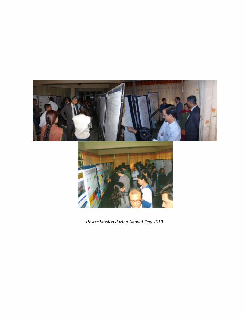
Poster Session during Annual Day 2010
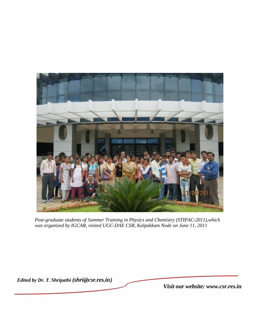
Post-graduate students of Summer Training in Physics and Chemistry (STIPAC-2011),which was organized by IGCAR, visited UGC-DAE CSR, Kalpakkam Node on June 11, 2011
Edited by Dr. T. Shripathi ([email protected])
Visit our website: www.csr.res.in