Animal models reveal role for tau phosphorylation in human disease
41
Animal models reveal role for tau phosphorylation in human disease J¨ urgen G¨ otz, Amadeus Gladbach, Luis Pennanen, Janet van Eersel, An- dreas Schild, Della David, Lars M. Ittner PII: S0925-4439(09)00219-1 DOI: doi:10.1016/j.bbadis.2009.09.008 Reference: BBADIS 63011 To appear in: BBA - Molecular Basis of Disease Received date: 16 June 2009 Revised date: 3 September 2009 Accepted date: 7 September 2009 Please cite this article as: J¨ urgen G¨ otz, Amadeus Gladbach, Luis Pennanen, Janet van Eersel, Andreas Schild, Della David, Lars M. Ittner, Animal models reveal role for tau phosphorylation in human disease, BBA - Molecular Basis of Disease (2009), doi:10.1016/j.bbadis.2009.09.008 This is a PDF file of an unedited manuscript that has been accepted for publication. As a service to our customers we are providing this early version of the manuscript. The manuscript will undergo copyediting, typesetting, and review of the resulting proof before it is published in its final form. Please note that during the production process errors may be discovered which could affect the content, and all legal disclaimers that apply to the journal pertain. peer-00616904, version 1 - 25 Aug 2011 Author manuscript, published in "Biochimica et Biophysica Acta (BBA) - Molecular Basis of Disease 1802, 10 (2010) 860" DOI : 10.1016/j.bbadis.2009.09.008
-
Upload
independent -
Category
Documents
-
view
0 -
download
0
Transcript of Animal models reveal role for tau phosphorylation in human disease
�������� ����� ��
Animal models reveal role for tau phosphorylation in human disease
Jurgen Gotz, Amadeus Gladbach, Luis Pennanen, Janet van Eersel, An-dreas Schild, Della David, Lars M. Ittner
PII: S0925-4439(09)00219-1DOI: doi:10.1016/j.bbadis.2009.09.008Reference: BBADIS 63011
To appear in: BBA - Molecular Basis of Disease
Received date: 16 June 2009Revised date: 3 September 2009Accepted date: 7 September 2009
Please cite this article as: Jurgen Gotz, Amadeus Gladbach, Luis Pennanen, Janetvan Eersel, Andreas Schild, Della David, Lars M. Ittner, Animal models reveal rolefor tau phosphorylation in human disease, BBA - Molecular Basis of Disease (2009),doi:10.1016/j.bbadis.2009.09.008
This is a PDF file of an unedited manuscript that has been accepted for publication.As a service to our customers we are providing this early version of the manuscript.The manuscript will undergo copyediting, typesetting, and review of the resulting proofbefore it is published in its final form. Please note that during the production processerrors may be discovered which could affect the content, and all legal disclaimers thatapply to the journal pertain.
peer
-006
1690
4, v
ersi
on 1
- 25
Aug
201
1Author manuscript, published in "Biochimica et Biophysica Acta (BBA) - Molecular Basis of Disease 1802, 10 (2010) 860"
DOI : 10.1016/j.bbadis.2009.09.008
ACC
EPTE
D M
ANU
SCR
IPT
ACCEPTED MANUSCRIPT
Special issue of
Biochimica et Biophysica Acta
on
‘Mouse Models of Human Neurological Disorders’ (BBADIS-09-30)
Animal models reveal role for tau phosphorylation in human
disease
Jürgen Götz1,*, Amadeus Gladbach1, Luis Pennanen2, Janet van Eersel1, Andreas Schild3,
Della David2, Lars M. Ittner1
1Alzheimer’s and Parkinson’s Disease Laboratory, Brain and Mind Research
Institute, University of Sydney, NSW, Australia, 2University of California, San
Francisco, Genentech Hall, 600 16th Street, San Francisco CA 94158-2517, 3Center for Human Genetic Research, Massachusetts General Hospital, Harvard
Medical School, Boston, MA, USA
*Address for correspondence: Alzheimer’s and Parkinson’s Disease Laboratory, Brain
and Mind Research Institute, University of Sydney, 100 Mallett St, Camperdown, NSW
2050, Australia; Tel. +61-2-9351 0789, Fax. +61-2-9351 0731; E-mail:
Keywords: Alzheimer’s disease, amyloid, Drosophila, frontotemporal dementia, kinase,
mice, nematode, phosphatase, phosphorylation, tau, transgenic
1
peer
-006
1690
4, v
ersi
on 1
- 25
Aug
201
1
ACC
EPTE
D M
ANU
SCR
IPT
ACCEPTED MANUSCRIPT
Abstract
Many proteins that are implicated in human disease are posttranslationally
modified. This includes the microtubule-associated protein tau that is deposited in a
hyperphosphorylated form in brains of Alzheimer’s disease patients. The focus of
this review article is on the physiological and pathological phosphorylation of tau,
the relevance of aberrant phosphorylation for disease, the role of kinases and
phosphatases in this process, its modeling in transgenic mice, flies and worms, and
implications of phosphorylation for therapeutic intervention.
2
peer
-006
1690
4, v
ersi
on 1
- 25
Aug
201
1
ACC
EPTE
D M
ANU
SCR
IPT
ACCEPTED MANUSCRIPT
Introduction
The Alzheimer’s disease (AD) brain is characterized by massive neuronal and synaptic
loss at specific predilection sites [1]. Together with tau-containing neurofibrillary tangles
(NFTs) and β-amyloid (Aβ) plaques, they are the defining histopathological hallmarks of
AD. Neurofibrillary lesions are found in cell bodies and apical dendrites as NFTs, in
distal dendrites as neuropil threads, and in the abnormal neurites that are associated with
some β-amyloid plaques (neuritic plaques) [2]. NFTs are also abundant, in the absence of
overt plaques, in certain subsets of frontotemporal dementia (FTD) and other tauopathies
[3]. The neurofibrillary lesions contain aggregates of the microtubule-associated protein
tau that in the course of disease is redistributed from the mainly axonal to the
somatodendritic compartment of neurons [4]. In tauopathies such as Progressive
Supranuclear Palsy (PSP) or Corticobasal Degeneration (CBD), tau forms aggregates also
in non-neuronal cells [5].
Tau is a phosphoprotein owing to its unusually high serine and threonine content. Under
pathological conditions, tau is hyperphosphorylated, meaning that it is phosphorylated to a
higher degree at normal, physiological sites, and at additional “pathological” sites [6] (Fig. 1).
Phosphorylation of tau tends to dissociate it from microtubules. Tau also undergoes a
conformational change which may assist in differential phosphorylation, or vice versa [7].
The MAPT (microtubule-associated protein tau) gene that encodes tau is located on
human chromosome 17q21; in the central nervous system it contains 15 exons, with the major
tau protein isoform being encoded by 11 exons [8]. By alternative mRNA splicing of exons 2,
3, and 10, six major tau isoforms are produced in the adult human brain. They differ by the
presence or absence of one or two short inserts in the amino-terminal half (0N, 1N, or 2N,
respectively), and have either three or four microtubule-binding repeat motifs in the carboxy-
terminal half (3R or 4R) (Fig. 1). In contrast to humans, mice express only four-repeat tau
isoforms (4R0N, 4R1N, or 4R2N) [9].
In AD, no mutations were found in the MAPT gene; these were identified in FTD
with Parkinsonism linked to chromosome 17 (FTDP-17) [10-12] (Fig. 1). This
established that dysfunction of tau in itself can cause neurodegeneration and lead to
dementia. In a subgroup of FTD, termed FTLD-U (frontotemporal lobar degeneration
3
peer
-006
1690
4, v
ersi
on 1
- 25
Aug
201
1
ACC
EPTE
D M
ANU
SCR
IPT
ACCEPTED MANUSCRIPT
with ubiquitin-immunoreactive lesions) or FTDU-17, tau-negative, ubiquitin-positive
lesions are prominent. In these patients, mutations were found in the PGRN gene
encoding progranulin, a growth factor involved in multiple physiological and
pathological processes including tumorigenesis [13, 14]. It was found that the TAR
DNA-binding protein of 43 kDa (TDP-43) is a primary constituent of the ubiquitin-
positive inclusions in FTLD-U and amyotrophic lateral sclerosis [15]. Under pathological
conditions, TDP-43 is, similar to tau, hyperphosphorylated, ubiquitinated, and carboxy-
terminally truncated [16].
Serine- and threonine-directed phosphorylation of tau
Of all post-translational modifications, protein phosphorylation in general receives particular
attention as it is an important cellular regulatory mechanism [6]. It determines enzymatic
activity, protein stability, folding properties including protein aggregation, binding of other
bio-molecules and subcellular localization. Phosphorylation occurs mainly on serine, threonine
and tyrosine residues, although proteins can be phosphorylated also on histidine, lysine and
arginine residues. Phosphorylation depends on a balanced interplay between kinases and
phosphatases that is disturbed under pathological conditions. Phosphorylation can also be
affected by O-glycosylation since some serine and threonine residues either being O-
glycosylated or phosphorylated [17-20].
Phosphorylation of normal, fetal and ‘PHF’-tau. Tau is a phosphoprotein with a remarkably
high number of potential phosphorylation sites. The longest human tau isoform in brain,
htau40, contains 45 serines and 35 threonines, of which 17 are followed by prolines (so-called
SP/TP-sites). A KXGS motif is found in each microtubule binding domain making this site a
substrate of MARK kinase [21]. In addition, there are 4 tyrosine residues [6]. Overall, 20% of
the protein is composed of amino acids that can be potentially phosphorylated. Therefore, it is
no surprise that differential phosphorylation of tau is crucial for its physiological and
pathological activities. It is then also logical that phosphorylation of tau within the
microtubule-binding domain (e.g. at the KXGS motives recognized by an antibody such as
4
peer
-006
1690
4, v
ersi
on 1
- 25
Aug
201
1
ACC
EPTE
D M
ANU
SCR
IPT
ACCEPTED MANUSCRIPT
12E8 specific for pSer262/pSer3561, see below) has functional consequences that are different
from those of phosphorylation of tau within the flanking sequences (Fig. 1).
Fetal brain contains only 3R tau that is phosphorylated at more sites than tau from adult
brain (that contains an equal mix of 3R and 4R tau), implying selective dephosphorylation of
the short isoforms during brain maturation [22]. In the developing axon, tau forms a smooth
proximo-distal phosphorylation gradient: Tau in the soma, immature dendrites and the
proximal axon is phosphorylated up to 80% at the tau-1 antibody site (Ser198/Ser199/Ser202
based on htau40), compared to only 20% in the axonal growth cone [23, 24].
In the AD brain, tau is hyperphosphorylated meaning that it is phosphorylated to a
higher degree at physiological sites as well as, in addition, at pathological sites (Fig. 1). This
tau associates to form paired helical filaments (PHFs) within NFTs. Thus pathologically
phosphorylated tau is also called "PHF"-tau [25, 26]. Pathological sites include pSer422,
pThr212/pSer214 (AT100), and pThr231/pSer235 (recognized by the conformation- and
phosphorylation-dependent antibody PHF-27/TG3) [7, 27-29]. Luckily, the tau research
community early on benefited from an excellent collection of epitope-specific antibodies
although in recent years the costs for some commercial antibodies have become exorbitantly
high (Fig. 1).
Compared to normal tau, PHF-tau displays a reduced electrophoretic mobility reflecting
an increased phosphorylation state. As phosphorylation of ‘PHF’-tau is very heterogeneous, it
runs as a smear on SDS-PAGE gels [30-34]. Upon dephosphorylation, the smear can be
resolved into discrete bands of a reduced mobility. PHF-tau also has a lower range of pI values,
indicating that it is more negatively charged than normal tau [35]. It has a reduced capacity to
bind tubulin and to promote the polymerization and stabilization of microtubules. Upon
dephosphorylation, PHF-tau binds to tubulin in a manner that is identical to that of normal tau
[36, 37]. Using mass spectrometry and phospho-specific antibodies, an increasing number of
potential phosphorylation sites have been confirmed in the AD brain, suggesting that as many
as 45 of the 84 possible sites of tau are phosphorylated in PHF-tau, compared to 15 in control
and up to 20 in fetal brain [38-43]. Obviously, there may be sites that have been missed due to
a lack of sensitivity [43], however, the question is whether these additional phospho-sites are
relevant for disease initiation and/or progression.1 For clarity, subsequently first the phosphorylated tau epitope (pS202 or S202) is listed followed by the phospho tau-specific antibody in brackets.
5
peer
-006
1690
4, v
ersi
on 1
- 25
Aug
201
1
ACC
EPTE
D M
ANU
SCR
IPT
ACCEPTED MANUSCRIPT
Many phosphorylation sites have been confirmed with phosphorylation-dependent and
epitope-specific anti-tau antibodies (Fig. 1). This assisted in correlating differential
phosphorylation with disease progression. It was shown that in the pre-NFT state, when tau has
not yet formed filaments, neurons were stained specifically with antibody TG3 (conformational
and pThr231/pSer235-specific), and the phosphorylation-dependent antibodies pSer262, and
pThr153. Intracellular NFTs were most prominently stained with antibodies against pSer46,
pThr175/pThr181, pSer214, pSer262/pSer356 (12E8) and pSer422, whereas extracellular
NFTs, the so-called ghost tangles, were most prominently stained with the AT8
(pSer202/pThr205), AT100 (pThr212/pSer214) and PHF-1/AD2 (pSer396/pSer404)
antibodies, which also stain intraneuronal NFTs. The different states of tau phosphorylation
associated with the different stages in the progression of the disease suggest a sequential
phosphorylation process which ultimately leads to NFT formation [44]. Sequential
phosphorylation is also evident with regards to individual phospho-epitopes. E.g., the AT100
epitope is generated in vitro by sequential phosphorylation, first of Ser199, Ser202 and Thr205
(flanking the AT8 epitope), then of Thr212 (by GSK-3β)‚ and finally of Ser214 (by PKA) [45].
Tyrosine-directed phosphorylation of tau
Tau in NFTs is also phosphorylated at tyrosine residues (Fig. 1). There are only four tyrosines
in tau compared to 80 serines and threonines, which may reflect the relative importance of
tyrosine phosphorylation in physiological and pathological functions, as well as the low
number of studies. As for the Ser/Thr-epitopes, there are specific antibodies for the Tyr
epitopes (reviewed in [6]). c-Abl has been suggested as a candidate kinase for Tyr394
phosphorylation, a site phosphorylated in both fetal and PHF-tau [46].
Tyrosine phosphorylation plays a pivotal role in the association of tau with the plasma
membrane, which is mediated by the amino-terminal projection domain of tau [47].
Association of tau with the plasma membrane may play a crucial role in relaying extra-cellular
signals. With its proline rich sequences in the amino-terminus, tau interacts with the SH3
domains of the tyrosine kinase Fyn that phosphorylates tau at Tyr18 [48, 49]. In a study that
goes far beyond the mapping of phosphorylation sites, Bhaskar and colleagues addressed the
question why an altered ratio of 3R to 4R tau might cause disease in FTD [50]. They used the
6
peer
-006
1690
4, v
ersi
on 1
- 25
Aug
201
1
ACC
EPTE
D M
ANU
SCR
IPT
ACCEPTED MANUSCRIPT
surface plasmon resonance biosensor technique on a panel of tau constructs and found that the
interaction between the SH3 domain of Fyn and 3R-tau was 20-fold higher than that for 4R-
tau. In addition, the affinity between 4R-tau and Fyn SH3 was increased 25-45-fold by
phosphorylation-mimicking mutations or by FTDP-17 mutations, supporting a role for the Fyn-
tau interaction in neurodegeneration [50].
Tau phosphorylation in tau transgenic mice
Classical transgenic approaches continue to be instrumental in dissecting pathogenic
mechanisms and testing therapies for AD and FTD [51, 52]. There are reports however of
viral delivery, such as with the Sindbis virus which was employed to express tau in
selected hippocampal sites, inducing a pathological conformation of tau as shown by
Alz50/MC1-reactivity, causing an accumulation of insoluble tau and inducing a region-
specific neurodegeneration [53].
The first published tau transgenic mouse model expressed the longest human four-
repeat (2N4R) tau isoform under control of the neuron-specific human Thy1 promoter [54].
Reflecting the approximately tenfold lower levels of human relative to endogenous murine tau,
the phosphorylation-specific anti-tau antibodies AT8 and PHF-1/AD2 revealed strongly labeled
neurons in many brain areas, but their numbers were small. However, as in the AD brain, tau
was re-localized to the somatodendritic domain, in addition to tau’s physiological axonal
localization. Gallyas silver impregnation failed to reveal tau filament and NFT formation, and
tau staining appeared homogenous or at the most, granular [54]. A comparable tau phenotype
was achieved in mice which expressed the shortest human tau isoform, using the murine 3-
hydroxy-methyl-glutaryl CoA reductase promoter. Here, tau was phosphorylated at the AT180
(pThr231/pSer235), AT270 (pThr181), AD2 (pSer396/pSer404), 12E8 (pSer262/pSer356), but
not the AT8 (pSer202/pThr205) epitopes [55] (Fig. 1).
Subsequently, a more advanced phenotype was achieved in three wild-type tau
transgenic strains, by choosing better expression vectors [56-58]. Tau was found to be
phosphorylated at several phospho-epitopes, but NFTs did not develop until the mice reached a
very old age [59]. In all three mouse strains, neurofilament-containing axonal spheroids formed
in brain and spinal cord, representing focal axonal dilatations. Orthograde fast axonal transport
7
peer
-006
1690
4, v
ersi
on 1
- 25
Aug
201
1
ACC
EPTE
D M
ANU
SCR
IPT
ACCEPTED MANUSCRIPT
was reduced as the mice developed a progressive motor phenotype [56]. Phosphorylation of tau
was analyzed in more detail in one of the strains, ALZ17, where it turned out to be
compartmentalized [60]. In the axons of CA1 pyramidal neurons, tau was specifically
phosphorylated at the AD2 epitope, concomitant with increased levels of cyclin-dependent
kinase-5 (cdk5). In contrast, the 12E8 (pSer262/pSer356) and AT180 (pThr231/pSer235)
epitopes were specifically phosphorylated in dendrites, and co-localized with increased levels
of GSK-3β [60]. Kinase activities have also been monitored in htau mice, which express
genomic wild-type tau (the 8c line [61]), on a tau knockout background [62]. The authors
found a significant correlation between specific phosphorylation changes and the amount of
aggregated tau. In the htau line, but not in non-NFT-forming control mice, there were increased
levels in phosphorylated (i.e. activated) p38 and the neuronal cdk5 activators, p35 and p25,
with aging, which in turn phosphorylate tau. Changes in tau kinases in the htau mice correlated
with the amount of tau present in abnormal conformations and with insoluble tau [63].
Together this shows that tau is subject to differential phosphorylation, due to differential
activities and compartmentalization of kinases and phosphatases.
The tau field experienced a major advance with the identification of both exonic and
intronic mutations in the MAPT gene encoding tau in FTDP-17, a familial dementia related to
AD [10-12]. The mutations affect the ratio of 3R to 4R tau isoforms and the propensity of tau
to form fibrils. By expressing FTDP-17 mutant tau in transgenic mice, many models were
established that represented NFT formation in both neuronal and glial cells [64, 65]. The
transgenic models were examined using transcriptomic and proteomic technology, including
the assessment of posttranslational modification [66-70], and subjected to behavioral analyses
[71, 72]. Phosphorylation patterns, however, are difficult to compare as each laboratory uses its
own set of phospho-tau-specific antibodies [5]. In P301S mutant tau transgenic mice, neuronal
loss was more pronounced than in the P301L tau models, consistent with the early onset of
FTD in patients carrying the P301S mutation [73]. In the P301S mice, the largest number of
stained neurons was observed with AT8, followed by AP422 (pSer422), AT180
(pThr231/pSer235) and PG5 (pSer409) [73]. Fewer neurons were labeled by AT100 (pThr212/
pSer214) and CP3 (pSer214), with the smallest number of neurons being stained by 12E8
(pSer262/pSer356) and PHF1 (pSer396/pSer404). Western blotting of perchloric acid-soluble
tau demonstrated that the human tau band was strongly immunoreactive with all tested
8
peer
-006
1690
4, v
ersi
on 1
- 25
Aug
201
1
ACC
EPTE
D M
ANU
SCR
IPT
ACCEPTED MANUSCRIPT
antibodies, but AT100 (pThr212/pSer214) and CP3 (pSer214). In contrast, sarkosyl-insoluble
human tau protein reacted with all phosphorylation-dependent anti-tau antibodies, including
AT100 and CP3. This indicates that immunoreactivity for pSer214 (part of epitope AT100)
closely mirrors the presence of filaments, suggesting that phosphorylation of this site occurs in
the course of, or after, filament assembly. The first P301L transgenic strain generated, JNPL3,
is characterized by a pronounced motor phenotype that is not seen in the related P301L
transgenic pR5 mice [74, 75].
When we monitored NFT formation and tau phosphorylation in these pR5 mice for up
to 24 months of age we found that NFTs first appeared in the amygdala at 6 months, while at
24 months the only other site with overt NFTs was the CA1 region of the hippocampus [76]. A
histological analysis revealed an increase in phosphorylation at the AT180 (pThr231/pSer235),
AT270 (pThr181) and 12E8 (pSer262/pSer356) epitopes with ageing whilst the AT8 (pSer202/
pThr205) and pS422 epitopes behaved differently. Firstly, whereas AT8 reactivity was found
in both the soma and dendritic branch of CA1 pyramidal neurons, pS422 staining was more
confined to the soma. Secondly, numbers of AT8-positive neurons increased from 3 to 6
months of age, but at 20 months of age, the only neurons left with AT8-reactivity were those
which had undergone NFT formation. In contrast, pS422-reactivity came up only late and
concomitantly with NFT formation [76]. As far as the disappearance in our pR5 mice of the
AT8 epitope in all but NFT-bearing neurons is concerned, one explanation may be that with
advanced pathology, neurons learn to cope with AT8 phosphorylation and the activities of the
kinase and/or phosphatase regulating phosphorylation of this epitope become balanced
resulting in a zero net phosphorylation at this epitope [6, 77]. This balancing act seems to be
specific for the AT8 epitope and the enzymes governing its phosphorylation, as other epitopes
such as AT180 or AT270 continue to be phosphorylated. In NFT-bearing neurons, however,
the AT8 phospho-epitope becomes stabilized suggesting that either a tau phosphatase may not
be able to access the phosphorylation site and/or the kinase/phosphatase equilibrium in the
dysfunctional neuron may be out of balance. Our AT8 findings are supported by data in P301S
transgenic mice which, although analyzed only until 6 months of age, reveal an increased
pS422, AT8 and AT180 phosphorylation in RIPA-extracted brains that peaks at 4-5 months
and then declines significantly at 6 months. This decline was not seen with AT270 or
phosphorylation-independent tau antibodies [78]. The authors state that the reduction at 5
9
peer
-006
1690
4, v
ersi
on 1
- 25
Aug
201
1
ACC
EPTE
D M
ANU
SCR
IPT
ACCEPTED MANUSCRIPT
months was particularly marked for the AT8 epitope [78]. The importance of both the AT8 and
pS422 epitope in tauopathies is also demonstrated by the fact that in both the JNPL3 and the
rTg4510 strain discussed below, a 170 kDa band linked to clinical features was preferentially
detected by AT8 and pS422, and this, possibly oligomeric, species negatively correlated with
memory [79].
To determine whether NFTs are central to the neurotoxic cascade in AD or
represent a protective neuronal response, P301L transgenic rTg4510 mice were generated
[80]. Here, transgene overexpression could be reduced by adding doxycycline to the
drinking water [80]. Despite the fact that even under suppressed conditions tau levels
were higher than what is generally achieved by conventional transgenic approaches,
memory function recovered and number of neurons stabilized, while NFTs continued to
accumulate under these conditions. These data show that dysfunction of tau impairs
memory, when mutant tau is massively overexpressed. They further imply that soluble
tau rather than NFTs themselves are neurotoxic [80]. Tau phosphorylation in the
rTg4510 brain was addressed in an accompanying paper, monitoring the mice from 1.3 to
8.5 months of age [81]. Differences were found between the hippocampus and cortex as
AT8 reactivity was preceded by TG3 in the hippocampal CA1 region in an order which is
opposite to that found in the cortex. In the CA1 region the epitopes appeared in the
following order: CP13 (pSer202), MC1 (conformational) and TG3 (conformational &
pThr231/pSer235) at 1.3 months, followed by PG5 (pSer409), AT8 (pSer202/pThr205),
PHF-1 (pSer396/pSer404), followed by Bielschowsky reactivity at 4-5.5 months. For the
cortex, MC1, CP-13, AT8 and PHF-1 appeared first, followed by TG3, PG5 and
Bielschowsky reactivity [81]. This appearance of phosphorylation sites raises the
question as to whether for tau to aggregate there needs to be a specific order of
phosphorylation events for the formation of tau aggregates, an issue discussed below in
more detail.
An interesting model of the specific phosphorylation profile that characterizes
Pick’s disease is the K3 strain that expresses human tau carrying the FTD mutation
K369I [82]. K3 mice develop a progressive histopathology that is reminiscent of human
FTD with the K369I mutation [83]. Specifically, as in human Pick’s disease [84] and in
the human K369I patient where the tau-containing Pick bodies are 12E8
10
peer
-006
1690
4, v
ersi
on 1
- 25
Aug
201
1
ACC
EPTE
D M
ANU
SCR
IPT
ACCEPTED MANUSCRIPT
(pSer262/pSer356)-negative [83], K3 mice also develop ovoid tau inclusions that are
12E8-negative, while additional phospho-epitopes of tau (as in humans) are strongly
phosphorylated [82]. K3 mice show an early-onset memory impairment and amyotrophy,
in the absence of overt neurodegeneration. However, as the mice age, neurodegeneration
becomes evident. Different from our previously generated tau transgenic strains, the K3
mice express the transgene in the substantia nigra (SN) and show an early-onset motor
phenotype that reproduces Parkinsonism with tremor, bradykinesia, abnormal gait and
postural instability. Interestingly, motor performance of young, but not old K3 mice
improved upon L-dopa treatment, which bears similarities to Parkinsonism in FTD. The
early-onset symptoms in the K3 mice are mechanistically related to selectively impaired
anterograde axonal transport of distinct cargos, which precedes the loss of dopaminergic
SN neurons that occurs in aged mice [82]. The impaired axonal transport in SN neurons
affects, among others, vesicles containing the dopamine-synthesizing enzyme tyrosine
hydroxylase (TH) [82]. We found that phosphorylated tau interacts pathologically with
the kinesin-associated adapter protein JIP1 both, in the mice and in AD brain [82, 85].
We proved that phosphorylation of tau is required for this pathogenic effect [85]
suggesting a pathological interaction of JIP1 and phosphorylated tau as a general
pathomechanism in tauopathies including AD.
Using an elegant stereotaxic injection approach, tau toxicity was shown to bear
resemblance to prions and to spread through the brain [86]. The study used two mouse
strains: ALZ17 mice that express high levels of wild-type human tau but reveal only a
modest pathology: amyotrophy in the absence of obvious neuronal cell loss and, despite a
massive hyperphosphorylation of tau, no formation of NFTs [58, 60]. In contrast, P301S
mice express, at levels comparable to the ALZ17 mice, a mutant form of tau found in
familial cases of FTD; the mice present with a particularly robust phenotype,
characterized by neurodegeneration in the spinal cord and an abundance of NFTs [73].
When diluted brain extracts from 6 month-old P301S mice were intracerebrally injected
into 3 month-old ALZ17 mice and the injected mice analyzed up to 15 months post-
injection, NFT formation was found as revealed by Gallyas silver impregnation, and
reactivity with antibody AT100 (pThr212/pSer 214).
11
peer
-006
1690
4, v
ersi
on 1
- 25
Aug
201
1
ACC
EPTE
D M
ANU
SCR
IPT
ACCEPTED MANUSCRIPT
Interestingly, Clavaguera and colleagues found that Gallyas reactivity (i.e. NFT
formation) was not confined to the site of injection, but rather induced up to 2 mm distant
of the injection site [86]. A time-course analysis suggests a stereotypical mode of
spreading (a feature characteristic of AD). Another remarkable finding was that insoluble
rather than soluble tau was responsible for the induction of a tau pathology. Also,
induction of the tau pathology seemed to be mediated by oligodendroglia (a cell type not
affected by tau pathology in the parental P301S mice).
Additional tau transgenic models have been discussed by us in detail elsewhere
[5, 65, 87]. Taken together, tau transgenic models have proven in vivo that the presence
of familial FTD-associated tau mutations causes tau hyperphosphorylation, aggregation,
nerve cell dysfunction as well as neuronal and glial cell loss. They have revealed that a
glial tau pathology can affect neuronal functioning and that a tau pathology in general
causes behavioural impairment [88]. Finally, the models highlight distinct
phosphorylation sites such as pS422, pThr212/pSer214 (AT100), pThr231/pSer235
(TG3) and pSer202/pThr205 (AT8) in disease initiation and progression and hence,
provide a means to target distinct kinases for therapeutic intervention.
Tau phosphorylation in kinase transgenic mice
In principal, tau phosphorylation can be brought about by the upregulation of kinases,
the downregulation of phosphatases, or both. A major question asked in the field is which
kinase(s) and phosphatase(s) in brain bring about the pathological changes that characterize
AD-tau and how these are related to neuronal demise. The human genome encodes a total of
516 protein kinases, with numbers comparable in mice. Of these kinases, several have been
identified as potential tau kinases, mostly by in vitro assays incubating recombinant tau protein
with the respective kinases. These include GSK-3β; Cdk5; the MAP kinases JNK, ERK and
p38; MARK; CK2; DYRK1A; TTBK1 and P70S6 kinase [43, 89-99]. These experiments
assisted in determining which kinases phosphorylate which phospho-epitopes of tau and
whether there is a sequential phosphorylation of sites, such as for phosphorylation of Thr231,
Ser396 and Ser400 by GSK-3β that depends on a previous phosphorylation of Ser235, Ser400
and Ser404, respectively, to provide one example [100, 101]. A few of these candidate kinases
12
peer
-006
1690
4, v
ersi
on 1
- 25
Aug
201
1
ACC
EPTE
D M
ANU
SCR
IPT
ACCEPTED MANUSCRIPT
have been expressed in transgenic mice and tau phosphorylation has been analyzed, either in
single-transgenic mice or after crossbreeding with tau transgenic mice. The findings confirmed
that kinases play an important role in NFT formation and neurodegeneration. The bulk of
studies concentrated on two kinases, GSK-3β and Cdk5, as outlined below.
The kinase GSK-3β is inactivated by phosphorylation of residue Ser9. Mice with a
constitutive active S9A form of GSK-3β showed increased activity, in the absence of
neurofibrillary pathology. Interestingly, crossing of these mice with human wild-type tau
transgenic mice markedly improved the axonopathy and motor deficits that characterize the
latter strain [102]. These findings would therefore suggest that GSK-3β is protective. However,
additional studies using different models reached the opposite conclusion. When mice with an
inducible expression of GSK-3β were crossed with mice that express tau with three FTDP-17
mutations combined (VLW mice), the double-transgenic mice developed thioflavin S-positive
tau aggregates and tau filaments. Moreover, the atrophy of the dentate gyrus of the
hippocampus, which was present in the single-transgenic GSK-3β mice, was accelerated in the
double-transgenic mice [103]. To explore whether the phenotype resulting from increased
GSK-3β activity could be reverted following restoration of normal GSK-3β levels, transgene
expression was shut down in symptomatic mice. This led to normal GSK-3β activity, normal
phospho-tau levels, diminished neuronal cell death and suppression of the cognitive deficits
[104]. When an inducible system was used to express a dominant negative mutant form of
GSK-3β, this caused apoptosis and a reversible motor deficit [105]. In these mice, AT8
phosphorylation (pSer202/pThr205) was reduced by 72% and PHF-1 phosphorylation
(pSer396/pSer404) by 46%. So in conclusion, these data identify GSK-3β as a tau kinase in
vivo.
Another kinase that has been expressed in transgenic mice is Cdk5. To address its role
in tau pathogenesis, P301L tau transgenic JNPL3 mice were crossed with mice transgenic for
the Cdk5 activator p25, the latter being expressed under control of the NSE promoter. This
caused a fivefold increase in NFT numbers in double-transgenic mice, along with
hyperphosphorylation of tau at the putative cdk5 epitopes pThr181 (AT270), pSer202,
pThr231, and pSer396/pSer404 (AD2/PHF1) [106]. On the other hand, a p25-mediated over-
activation of Cdk5 in a CaMKII promoter-driven p25 transgenic strain did not induce tau
hyperphosphorylation at a young age per se, possibly because Cdk5 over-activation inhibited
13
peer
-006
1690
4, v
ersi
on 1
- 25
Aug
201
1
ACC
EPTE
D M
ANU
SCR
IPT
ACCEPTED MANUSCRIPT
GSK-3β, by phosphorylating its inhibitory Ser9 site [107]. However, as the mice became older,
this inhibition was lost, resulting in increased GSK-3β activity that was associated with tau
hyperphosphorylation at the AT8 (pSer202/pThr205) and PHF1 (pSer396/pSer404) sites.
Together with pharmacological and co-immunoprecipitation experiments this would suggest
that GSK-3β is a key mediator of tau hyperphosphorylation, while Cdk5 acts as a modulator of
tau hyperphosphorylation via the inhibitory regulation of GSK-3β [107]. These findings were
confirmed in a second p25 transgenic model [108]. It was found that phosphorylation of
residue Ser9 of GSK-3β was mediated by an enhanced activity of the neuregulin receptor
complex, ErbB, and by activation of the downstream phosphatidylinositol 3 kinase/Akt
pathway. While young p25 mice had elevated Aβ levels, levels of phosphorylated tau were
decreased. Thus, Cdk5 appears to play a dominant role in the regulation of amyloidogenic APP
processing, whereas GSK-3β seems to play a dominant role in overall tau phosphorylation
[108].
Inducible systems were employed that caused pronounced phenotypic changes.. For
example, transgenic mice with an inducible p25 expression in the postnatal forebrain revealed a
massive neuronal loss in the cortex and hippocampus, along with forebrain atrophy,
astrogliosis, and caspase-3 activation [109]. At only 5 weeks of age, endogenous tau was
hyperphosphorylated at many epitopes, as shown for AT8 (pSer202/pThr205) and PHF1
(pSer396/pSer404). In one year-old mice, immuno-electron microscopy of sarkosyl-insoluble
fractions revealed tau filaments that were phosphorylated at the AT8, PHF1, TG3
(pThr231/pSer235) and AT100 (pThr212/pSer214) epitopes. Also, Gallyas silver impregnation
revealed NFT formation in these mice [109]. In a second inducible mouse strain, neuronal p25
triggered a similar neurodegeneration and marked neuronal loss, causing brain atrophy with a
40% loss at 5 months of age resulting in an almost complete elimination of the hippocampus.
Interestingly, this type of neurodegeneration was not associated with hyperphosphorylation of
tau or Aβ generation [110]. One likely explanation is that p25 neuro-toxicity is related to
substrates other than tau or APP.
The tau-tubulin kinase (TTBK) family consists of TTBK1 and TTBK2 that belong to
the casein kinase 1 superfamily. Different from TTBK2, TTBK1 is specifically expressed in
neurons. It can phosphorylate tau directly at multiple Ser/Thr residues that are found in PHF-
tau in AD brain. Furthermore, TTBK1 is expressed in NFT-bearing neurons in the AD cortex
14
peer
-006
1690
4, v
ersi
on 1
- 25
Aug
201
1
ACC
EPTE
D M
ANU
SCR
IPT
ACCEPTED MANUSCRIPT
[99]. Transgenic mice expressing full-length human TTBK1 show an age-dependent memory
impairment accompanied by increased phosphorylation of tau and neurofilaments, and
increased levels of p25 and p35 [111].
Finally, another interesting kinase is DYRK1A (dual specificity tyrosine-regulated
kinase-1A) that has been implicated in Down’s syndrome (DS), due to the location of the
Dyrk1a gene in the critical region of human chromosome 21 [96]. DS is a genetic disorder in
humans caused by partial or complete trisomy of chromosome 21. People with DS show a
tendency towards premature aging and an increased risk for AD [112]. DS patients show an
early-onset tauopathy that resembles AD in many ways. In addition to mental problems, motor
dysfunction is highly prevalent in DS. Dyrk1a transgenic mice express the transgene in several
areas of the mid- and hindbrain [113]. When assessed in motor tests such as the treadmill, the
mice showed impairment in some parameters; in particular, they required longer training
periods [113]. A transcriptomic analysis revealed upregulation of the NMDA receptor subunit
2A which may explain the altered excitatory transmission reported in humans with DS and in
DS mouse models [114]. Altered synaptic plasticity along with learning and memory deficits
was reported for a second strain, that used a BAC clone of Dyrk1a [115]. A similar memory
phenotype was found in Dyrk1a haplo-insufficient mice [116]. In one of the mouse models, tau
phosphorylation was addressed: tau in Dyrk1a transgenic mice was hyperphosphorylated at
Thr212, Ser202 and Ser404; furthermore, phosphorylation by DYRK1A strongly inhibited the
ability of tau to promote microtubule assembly [117]. The finding that gene dosage of Dyrk1
affects tau splicing may explain the early onset tauopathy in individuals with DS [118]
Whilst these studies always only test a subset of tau phosphorylation sites, they still highlight a
role for cdk5 and its activator p25 in Aβ pathology, and for GSK-3β, DYRK1A and TTBK1 in
tau aggregation. Whether targeting a specific kinase in the development of an AD therapy will
be a fruitful approach is still a matter of debate. It may well be that in a human setting a general
subtle down-regulation of kinase(s) in the brain may be more beneficial.
Tau phosphorylation in phosphatase transgenic mice
Of the serine/threonine-specific phosphatases that are abundant in the brain, protein
phosphatase 2A (PP2A) is a major phosphatase implicated in tau dephosphorylation [119],
15
peer
-006
1690
4, v
ersi
on 1
- 25
Aug
201
1
ACC
EPTE
D M
ANU
SCR
IPT
ACCEPTED MANUSCRIPT
perhaps more so because PP2A can bind directly to tau [120-122]. PP2A is a trimeric
holoezyme that consists of a catalytic subunit C and a scaffolding subunit A. The A/C core
enzyme associates with variable regulatory subunits of the PR55(B), PR56/61(B’),
PR59/72/130(B’’) and PR93/110(B’’’) families to form heterotrimers [123]. Recruitment of
regulatory B subunits into the holoenzyme is dependent upon the highly conserved DYFL
motif in the carboxy-terminus of subunit C that undergoes methylation at Leu309, thereby
affecting subunit B recruitment [124, 125]. The resolution of the structure of the B subunit
provides significant insight into how PP2A dephosphorylates tau [126]. Interestingly,
methylation of the C subunit does not seem to be required for in vitro assembly of the PP2A
holoenzyme involving either B or B’. The authors argue that the regulatory subunits may be
sequestered in a specific cellular compartment and that the methylated carboxy-terminus of the
C subunit may allow subcellular targeting for holoenzyme assembly [126].
Dephosphorylation of tau can be blocked in cells by okadaic acid (OA), an inhibitor of
the two phosphatases PP1 and PP2A [127, 128]. When rat brain slices were incubated with
OA, tau became phosphorylated at multiple sites [129]. A role for PP2A in AD is implicated by
the finding that its activity is reduced in AD brain [130]; furthermore, association analyses
indicate that a CAG repeat polymorphism in one of the B subunits may confer susceptibility to
AD [131].
Transgenic strategies targeting PP2A include gene knockouts, overexpression of
regulatory subunits and dominant negative mutant approaches [132]. A knockout of the major
catalytic subunit Cα caused delayed embryonic lethality [133], as the highly homologous Cβ
subunit failed to complement the lack of Cα in mesoderm formation [134], due to a different
subcellular localization of the two catalytic subunits [135]. Under physiological conditions, the
A and C subunits are ubiquitously expressed, whereas the regulatory B subunits show a tissue-
specific expression; they also reveal a differential expression pattern in brain and during
neuronal differentiation [136-138].
To address the role of PP2A in tau phosphorylation in vivo, transgenic mice were
generated that express a dominant negative mutant form of the catalytic subunit Cα of PP2A,
L199P, in neurons. The transgenic mice have a reduced PP2A activity resulting in a pre-NFT
phenotype, with phosphorylation of endogenous murine tau at the AT8 (pSer202/pThr205) and
Ser422 epitopes [139]. This effect may be directly mediated by PP2A, but could also occur
16
peer
-006
1690
4, v
ersi
on 1
- 25
Aug
201
1
ACC
EPTE
D M
ANU
SCR
IPT
ACCEPTED MANUSCRIPT
indirectly, via deregulated kinases such as ERK and JNK, that are themselves substrates of
PP2A [140]. The carboxy-terminal DYFL motif of the catalytic subunit has a role in the
recruitment of B subunits into the PP2A complex, by methylating leucine 309 and
phosphorylating tyrosine 307 [123]. To determine the role of the DYFL motif in PP2A activity
in vivo, a second dominant negative mutant strain was established that expressed the L309A
mutant form of Cα in neurons. This caused an altered subunit composition of the PP2A
holoenzyme in vivo. In the brain, tau was hyperphosphorylated at Ser202/Thr205 (AT8/CP13);
furthermore, there was an impaired dephosphorylation of the intermediate filament protein
vimentin [141]. The additional expression of the L309A transgene expression in the Harderian
(lacrimal) gland caused a delayed postnatal development and hypoplasia of this gland, causing
enophthalmos [142]; .
When the PP2A L309A mutant mice were crossed with P301L tau mutant pR5 mice,
this caused a seven-fold increased number of hippocampal neurons that specifically s
phosphorylated the Ser422 epitope of tau. The double-transgenic mice showed eight-fold
increased numbers of NFTs compared to pR5 mice, in agreement with the previous finding that
NFT formation is correlated with and preceded by phosphorylation of tau at the Ser422 epitope
[143]. The critical role of the DYFL motif in PP2A function, with particular regards to the
cytoskeleton, is further demonstrated by cell culture experiments, which showed that
expression of mutants such as L309Δ induces a loss of microtubules [144]. As mentioned
above, in addition to methylation at the carboxy-terminal Leu309, phosphorylation at Tyr307
also regulates PP2A function [123]. PP2A phosphorylated at Tyr307 associates with pre-
tangles and NFTs in areas such as the entorhinal cortex and the hippocampus, brain areas
where the neurofibrillary changes are initiated [145]. Together, these studies demonstrate a
crucial role for PP2A in tau phosphorylation. As many kinases are themselves substrates of
PP2A, the control of PP2A goes beyond the reversible phosphorylation of its non-enzymatic
substrates. It would not be surprising to find that targeting of PP2A, in particular of its
regulatory subunits, represents a valid means of treating AD.
Tau phosphorylation in mice with an amyloid pathology
17
peer
-006
1690
4, v
ersi
on 1
- 25
Aug
201
1
ACC
EPTE
D M
ANU
SCR
IPT
ACCEPTED MANUSCRIPT
Tau phosphorylation is part of the phenotypic characterization of APP mutant mouse models.
In the following, we selected a few examples to demonstrate how Aβ affects endogenous tau
phosphorylation in vivo. For example, in one of the earliest reports of a strong Aβ-plaque
forming mouse model, the APP23 mice, increased tau phosphorylation was found to parallel
Aβ peptide deposition [146]. APP23 mice have been thoroughly investigated, recently by using
3D reconstruction techniques to determine how small Aβ aggregates associated with the
microvasculature lead to morphological and architectural alterations of the vasculature, thus
resulting in an altered local blood flow [147]. Even more studies have been conducted with the
APP mutant mouse strain Tg2576 in which an anti-Aβ treatment provided evidence for a slow
seeding mechanism preceding a rapid fibrillogenesis in determining the extent of Aβ deposition
[148]. Formation of AT8 (pSer202/pThr205)-positive dystrophic neurites was found to occur
simultaneously with Congo red-positive plaque development [149]. Oxidative stress seems to
play an important role in the Aβ pathology. Superoxide dismutase 2 (SOD2) is an enzyme
which detoxifies reactive oxygen species that are produced predominantly by mitochondria.
Mice lacking SOD2 die within the first week of life, and develop a complex heterogeneous
phenotype arising from mitochondrial dysfunction and oxidative stress [150]. By crossing the
Tg2576 mice onto an SOD2 heterozygous knockout background, it was found that this
mitochondrial SOD2 deficiency both exacerbated Aβ plaque burden and tau phosphorylation at
Ser396 [151].
Combinatorial approaches were particularly helpful in elucidating the role of Aβ in tau
phosphorylation and aggregation. When P301L tau transgenic JNPL3 mice were crossed with
Aβ plaque-forming Tg2576 mice, a sevenfold increase in NFT induction was seen [152]. In a
parallel study with P301L tau transgenic pR5 mice, we found that stereotaxic injections with
Aβ42 fibrils caused a fivefold increase in NFT formation compared to uninjected mice [153].
NFT formation was tightly correlated with phosphorylation of tau at Ser422 and AT100
(pThr212/pSer214), but not the AT8 (pSer202/pThr205) epitope. Mutagenesis of phospho-sites
in a human SH-SY5Y tissue culture system revealed that the Ser422 epitope is required for the
Aβ-mediated formation of tau filaments [154-156]. Aβ further exacerbates a mitochondrial
dysfunction that characterizes the pR5 mice [157, 158]. In a third study, Aβ plaque and NFT
pathology was combined in a single animal by a triple transgenic approach [159]. In these
3xtg-AD mice (that express P301L mutant tau as well as mutant APP and PS1), synaptic
18
peer
-006
1690
4, v
ersi
on 1
- 25
Aug
201
1
ACC
EPTE
D M
ANU
SCR
IPT
ACCEPTED MANUSCRIPT
dysfunction, including deficits in long-term potentiation, was found to precede Aβ plaque and
NFT formation. The mice showed a remarkable phosphorylation pattern in that AT8 (pSer202/
pThr205) staining was only evident after 6 months and PHF1 (pSer396/pSer404) only at
around 18 months of age, as shown for the hippocampus and cortex, despite a markedly high
transgene expression and prominent HT7 (human tau) reactivity [160]. In a follow-up of the
stereotaxic injection approach, Bolmont and colleagues diluted brain extracts from aged Aβ
plaque-forming APP23 transgenic mice and intracerebrally infused these in young P301L tau
transgenic mice. They found that six months after the infusion a tau pathology was induced in
the injected hippocampus but also in brain regions well beyond the injection sites such as the
entorhinal cortex and amygdala, areas with neuronal projection to the injection site [161]. This
is similar to findings in pR5 mice where Aβ while injected into the hippocampus and
somatosensory cortex, induced NFT formation in the amygdala, aan area with neuronal
projections to the injection site [153]. Together, this demonstrates that Aβ can induce tau
hyperphosphorylation and augment a pre-existing tau pathology in mice.
Tau phosphorylation in flies and worms
More recently, invertebrate species have gained considerable attention. Despite their reduced
complexity, they offer distinct advantages compared to mice. The life span of both the
nematode Caenorhabditis (C.) elegans and the fruitfly Drosophila melanogaster is short. Costs
for maintenance are low, handling is relatively easy, mass production is possible and screening
for mutations simple. Also, working with these two species does not need approval from
animal ethics committees.
Several of the genes that are implicated in AD pathogenesis have been expressed in
flies and worms. For example, both wild-type and FTDP-17 mutant human tau has been
expressed in the fly. These reproduced key features of the human disease, including adult
onset, progressive neurodegeneration, enhanced toxicity of mutant tau, accumulation of
abnormal tau and anatomical selectivity. Interestingly, immunoreactivity for phospho-epitopes
such as 12E8 (pSer262/pSer356), AT100 (pThr212/pSer214), and AT180 (pThr231/pSer235)
increased as the flies aged, whereas immunoreactivity using a phosphorylation-independent
antibody was unaltered. Neurodegeneration, however, can occur without NFT formation [162],
19
peer
-006
1690
4, v
ersi
on 1
- 25
Aug
201
1
ACC
EPTE
D M
ANU
SCR
IPT
ACCEPTED MANUSCRIPT
which is consistent with studies in mice [80]. When wild-type human tau was expressed in
combination with Shaggy, the Drosophila GSK-3β homolog and wnt signaling pathway
component, this lead to a neurofibrillary pathology with tau filaments [163].
When going through the vast literature on animal models of AD, it becomes obvious
that most of the studies addressing the role of tau phosphorylation in tau aggregation and
neurodegeneration are descriptive, although a few mutagenesis studies have been performed.
To address the role of distinct tau phosphorylation sites in controlling tau neurotoxicity, a
mutagenesis study was performed in Drosophila which indicates that here, no single
phosphorylation residue plays a dominant role in controlling tau toxicity, but rather that tau
phosphorylation sites work in concert to promote neurotoxicity in vivo [164].
The 12E8 epitope, located in the microtubule-binding domain of tau, is a substrate of
MARK [44, 165]. Work in Drosophila showed that the MARK homologue PAR-1 is a
physiological tau kinase that plays a central role in regulating tau phosphorylation and toxicity,
without promoting NFT formation [165]. Mutating the PAR-1 phosphorylation site
Ser262/Ser356 (12E8) by alanine substitution abolished tau toxicity. When human tau was
expressed in the Drosophila eye along with either GSK-3β/Shaggy or Cdk5, this enhanced
toxicity of tau [166]. Lithium administration inhibited GSK-3β activity and thus reduced tau
phosphorylation but did not ameliorate tau-induced toxicity, possibly reflecting high levels of
tau expression [166].
Compared to Drosophila, the nematode is even easier to work with [167]. What is
unique to C. elegans is that wild-type adult worms contain a constant 959 somatic cells. Not
only is the cell number constant but also the position of each cell. Moreover, the worm is
transparent and hence, it is easy to track cells and follow cell lineages. The nervous system of
an adult hermaphrodite C. elegans consists of only 302 neurons that form approximately 7000
synapses. Transgenic animals can be generated either using microinjection or ballistic
approaches [168]. Increasingly, C. elegans is employed for studies into diabetes and AD.
Regarding the latter, research currently progresses along two lines, firstly the characterization
of C. elegans homologues of AD-related genes, and secondly the expression of human AD
genes in C. elegans [167].
Expression of P301L and V337M mutant tau in C. elegans lead to behavioral,
synaptic and pathological abnormalities, and caused an earlier and more severe
20
peer
-006
1690
4, v
ersi
on 1
- 25
Aug
201
1
ACC
EPTE
D M
ANU
SCR
IPT
ACCEPTED MANUSCRIPT
phenotype than over-expression of wild-type human tau. Substantial neurodegeneration
followed by loss of neurons occurred after insoluble tau began to accumulate. Tau was
phosphorylated at the PHF1 (pSer396/pSer404), AT270 (pThr181), and 12E8
(pSer262/pSer356) epitopes in both the soluble and insoluble fraction, and at Ser422 and
AT8 (pSer202/pThr205) only in the soluble fraction. However, tau phosphorylation did
not appear to correlate with the severity of the phenotype [169]. The fact that neurological
symptoms were apparent before insoluble tau accumulated, indicates that toxicity does
not depend on the formation of large tau aggregates. To determine the role of tau
phosphorylation in disease more directly, a short, fetal isoform of human tau was
expressed, with a total of ten serines and threonines replaced by either glutamate (a so-
called pseudophosphorylation or PHP construct) or alanine [170]. Both, wild-type and
PHP tau induced a progressive age-dependent phenotype of uncoordinated locomotion
(unc) in the absence of neuronal degeneration. In comparison, the alanine mutant
transgenic worms displayed a reduced survival and developed an earlier unc phenotype,
indicating that phosphorylation at these sites alone is not the cause of the observed
defects [170]. However, it is difficult to draw any conclusions with regards to the role of
single phospho-epitopes in pathogenesis, as in the PHP construct, ten sites were
phosphorylated simultaneously.
Implications of animal work for pathogenesis and therapy
Which phosphorylation sites are critical in triggering the cascade of neuronal dysfunction and
eventually, neuronal loss, in tauopathies? What are the implications of data obtained in animal
models for the development of therapies? Obviously, the promiscuity of kinases and
phosphatases and the interdependence of phosphorylation sites, along with a profile that can
differ from one neuron to the next, renders it difficult at present to pinpoint specific
phosphorylation sites and their regulation [171-173].
We have found in vivo, that NFT formation is associated with the phosphorylation
of the pSer422 and pThr212/pSer214 (AT100) epitope [153] (Fig. 1). This and additional
studies including site-directed mutagenesis of tau phosphorylation sites suggest a role for
specific phospho-epitopes and hence kinases and phosphatases, in tau aggregation and
21
peer
-006
1690
4, v
ersi
on 1
- 25
Aug
201
1
ACC
EPTE
D M
ANU
SCR
IPT
ACCEPTED MANUSCRIPT
neuronal dysfunction [63, 154, 174, 175]. Other studies support the notion that a
generally increased phosphorylation rather than phosphorylation of specific sites is
needed [156, 164]. This concept is also supported by a recent analysis of tau aggregation
and the role of phosphorylation in this process in an inducible N2a cell line that expresses
ΔK280 full-length tau with and without truncated forms of tau [176]. While this study is
restricted to 4R tau and needs to be validated in vivo, interestingly, aggregation of full-
length tau was found to be triggered by truncated fragments and was associated with the
phosphorylation of most of the few sites analyzed. These were distributed between both
soluble and aggregated tau, suggesting that none of the sites determines aggregation in an
all-or-none manner [176]. Overall, whereas phosphorylation in the repeat domain tends to
inhibit aggregation, other sites show a tendency to support aggregation. Together, this has
obvious implications for treatment strategies as it would imply that phosphorylation in
general, rather than specific tau kinases, needs to be blocked [87].
Other take home lessons are that transgenic animal models prove that expression
of FTDP-17 mutant tau accelerates tau pathology, causing NFT formation within the
lifetime of mice. They underscore the role of serine/threonine-specific phosphorylation in
tau aggregation. Strong evidence has been provided for a role of the tau phospho-epitopes
AT100 (pThr212/pSer214) and S422 in tau aggregation as indicated above, although a
map of “necessary” and “sufficient” epitopes of tau is still lacking. Based on the currently
available data it appears that phosphorylation of tau is closely linked to tau aggregation
and fibril formation.
A tau-directed treatment strategy may target any step involved in converting tau
to a toxic species [177]. Alternatively, tau levels (and hence levels of phosphorylated tau)
may be altogether reduced as it has been shown that Aβ toxicity is exerted via tau and
that excitotoxicity can be rescued when tau levels are reduced [178].
Several therapeutic strategies for treating AD on the basis of tau hyperphosphorylation
are available as discussed recently [179]: these include (1) inhibition of GSK-3β, Cdk5 and
other tau kinases; (2) restoration of PP2A activity; and (3) targeting O-glycosylation of tau
[179].
A more recent study evaluated the concept of the promiscuity of kinases by using an
orally bioavailable and blood-brain barrier (BBB)-penetrating analog of the relatively non-
22
peer
-006
1690
4, v
ersi
on 1
- 25
Aug
201
1
ACC
EPTE
D M
ANU
SCR
IPT
ACCEPTED MANUSCRIPT
specific protein kinase inhibitor K252a [180]. This compound prevented motor deficits in the
P301L tau transgenic mouse line JNPL3 and reduced levels of soluble aggregated
hyperphosphorylated tau. Interestingly, NFT numbers were not reduced, suggesting that the
main cytotoxic effects of tau are not exerted by NFTs, but by lower molecular mass aggregates.
This finding is in line with studies where a reduction in the expression of transgenic human
P301L tau led to a recovery of memory function and stabilization of neuron numbers, despite
the continued accumulation of NFTs [80].
In many studies the GSK-3β inhibitor lithium has been evaluated. For example, in
3xTg-AD mice that develop both plaques and tangles, lithium chloride reduced tau
phosphorylation but it did not significantly alter the Aβ load [181]. In a second mutant human
tau transgenic mouse model, treatment with lithium chloride resulted in a significant inhibition
of GSK-3 activity. After normalization to total levels of tau, lithium chloride treatment was
found to result in a significantly decreased phosphorylation at putative GSK-3-directed sites,
including pSer202 (CP13) and pSer396/pSer404 (PHF-1). Phosphorylation at sites not
recognized by GSK-3 such as pSer422 or pSer262 appeared not to be affected. There were
significantly reduced levels of aggregated, insoluble tau. Administration of another GSK-3
inhibitor also correlated with reduced insoluble tau levels, supporting the idea that lithium
exerts its effect through GSK-3 inhibition. Levels of aggregated tau correlated strongly with
the degree of axonal degeneration, and lithium-chloride-treated mice showed less degeneration
when administration was started during early stages of NFT development [182]. Very recently,
a zebrafish model has been established which reproduced tau hyperphosphorylation, NFT
formation, neuronal and behavioral disturbances as well as cell death. Of the many inhibitors of
GSK-3β tested in the fish model, a compound called AR-534 turned out to reduce tau
hyperphosphorylation in vivo, without causing toxic side-effects [183].
Furthermore, the protein kinase inhibitor rapamycin has been reported to reduce
toxicity in Drosophila expressing either wild-type or mutant forms of tau, probably by reducing
the amount of insoluble tau [184]. Rapamycin induces autophagy through inhibition of the
protein kinase mammalian target of rapamycin (mTOR) [185]. It remains to be seen whether
the beneficial effects of rapamycin are related to TOR-dependent abnormal cell cycle
activation that has been described in Drosophila tauopathy models.
23
peer
-006
1690
4, v
ersi
on 1
- 25
Aug
201
1
ACC
EPTE
D M
ANU
SCR
IPT
ACCEPTED MANUSCRIPT
While these data encourage the use of kinase inhibitors, at the same time,
microtubules, known to be stabilized by tau, have also emerged as a drug target, as they
can be strengthened by drugs such as paclitaxel [186]. Which of the approaches will be
translated into human practice remains to be seen. As selective vulnerability characterizes
the AD brain, different brain areas may be differently susceptible to any of these
treatments [187].
Acknowledgements
JG is a Medical Foundation Fellow. This work has been supported by the University of
Sydney, the National Health & Medical Research Council (NHMRC), the Australian
Research Council (ARC), the New South Wales Government through the Ministry for
Science and Medical Research (BioFirst Program), the Nerve Research Foundation, the
Medical Foundation (University of Sydney) and the Judith Jane Mason & Harold Stannett
Williams Memorial Foundation. LI has been supported by the NHMRC and ARC.
References
[1] D.J. Selkoe, Alzheimer's disease is a synaptic failure, Science 298 (2002) 789-791.
[2] M. Goedert, R. Jakes, M.G. Spillantini, R.A. Crowther, P. Cohen, E. Vanmechelen, A. Probst, J. Gotz, K. Burki, Tau protein in Alzheimer's disease, Biochem Soc Trans 23 (1995) 80-85.
[3] V.M. Lee, M. Goedert, J.Q. Trojanowski, Neurodegenerative tauopathies, Annu Rev Neurosci 24 (2001) 1121-1159.
[4] M. Goedert, C.M. Wischik, R.A. Crowther, J.E. Walker, A. Klug, Cloning and sequencing of the cDNA encoding a core protein of the paired helical filament of Alzheimer disease: identification as the microtubule-associated protein tau, Proc Natl Acad Sci U S A 85 (1988) 4051-4055.
[5] J. Gotz, Tau and transgenic animal models, Brain Res Brain Res Rev 35 (2001) 266-286.
[6] F. Chen, D. David, A. Ferrari, J. Gotz, Posttranslational modifications of tau - Role in human tauopathies and modeling in transgenic animals, Curr Drug Targets 5 (2004) 503-515.
[7] G.A. Jicha, E. Lane, I. Vincent, L. Otvos, Jr., R. Hoffmann, P. Davies, A conformation- and phosphorylation-dependent antibody recognizing the paired helical filaments of Alzheimer's disease, J Neurochem 69 (1997) 2087-2095.
24
peer
-006
1690
4, v
ersi
on 1
- 25
Aug
201
1
ACC
EPTE
D M
ANU
SCR
IPT
ACCEPTED MANUSCRIPT
[8] A. Andreadis, W.M. Brown, K.S. Kosik, Structure and novel exons of the human tau gene, Biochemistry 31 (1992) 10626-10633.
[9] M. Goedert, R. Jakes, Expression of separate isoforms of human tau protein: correlation with the tau pattern in brain and effects on tubulin polymerization, Embo J 9 (1990) 4225-4230.
[10] M. Hutton, C.L. Lendon, P. Rizzu, M. Baker, S. Froelich, H. Houlden, S. Pickering-Brown, S. Chakraverty, A. Isaacs, A. Grover, J. Hackett, J. Adamson, S. Lincoln, D. Dickson, P. Davies, R.C. Petersen, M. Stevens, E. de Graaff, E. Wauters, J. van Baren, M. Hillebrand, M. Joosse, J.M. Kwon, P. Nowotny, P. Heutink, et al., Association of missense and 5'-splice-site mutations in tau with the inherited dementia FTDP-17, Nature 393 (1998) 702-705.
[11] P. Poorkaj, T.D. Bird, E. Wijsman, E. Nemens, R.M. Garruto, L. Anderson, A. Andreadis, W.C. Wiederholt, M. Raskind, G.D. Schellenberg, Tau is a candidate gene for chromosome 17 frontotemporal dementia, Ann Neurol 43 (1998) 815-825.
[12] M.G. Spillantini, J.R. Murrell, M. Goedert, M.R. Farlow, A. Klug, B. Ghetti, Mutation in the tau gene in familial multiple system tauopathy with presenile dementia, Proc Natl Acad Sci U S A 95 (1998) 7737-7741.
[13] M. Baker, I.R. Mackenzie, S.M. Pickering-Brown, J. Gass, R. Rademakers, C. Lindholm, J. Snowden, J. Adamson, A.D. Sadovnick, S. Rollinson, A. Cannon, E. Dwosh, D. Neary, S. Melquist, A. Richardson, D. Dickson, Z. Berger, J. Eriksen, T. Robinson, C. Zehr, C.A. Dickey, R. Crook, E. McGowan, D. Mann, B. Boeve, H. Feldman, M. Hutton, Mutations in progranulin cause tau-negative frontotemporal dementia linked to chromosome 17, Nature (2006).
[14] M. Cruts, I. Gijselinck, J. van der Zee, S. Engelborghs, H. Wils, D. Pirici, R. Rademakers, R. Vandenberghe, B. Dermaut, J.J. Martin, C. van Duijn, K. Peeters, R. Sciot, P. Santens, T. De Pooter, M. Mattheijssens, M. Van den Broeck, I. Cuijt, K. Vennekens, P.P. De Deyn, S. Kumar-Singh, C. Van Broeckhoven, Null mutations in progranulin cause ubiquitin-positive frontotemporal dementia linked to chromosome 17q21, Nature (2006).
[15] M. Neumann, D.M. Sampathu, L.K. Kwong, A.C. Truax, M.C. Micsenyi, T.T. Chou, J. Bruce, T. Schuck, M. Grossman, C.M. Clark, L.F. McCluskey, B.L. Miller, E. Masliah, I.R. Mackenzie, H. Feldman, W. Feiden, H.A. Kretzschmar, J.Q. Trojanowski, V.M. Lee, Ubiquitinated TDP-43 in frontotemporal lobar degeneration and amyotrophic lateral sclerosis, Science 314 (2006) 130-133.
[16] C. Cook, Y.J. Zhang, Y.F. Xu, D.W. Dickson, L. Petrucelli, TDP-43 in neurodegenerative disorders, Expert Opin Biol Ther 8 (2008) 969-978.
[17] J. Guevara, B. Espinosa, E. Zenteno, L. Vazguez, J. Luna, G. Perry, R. Mena, Altered glycosylation pattern of proteins in Alzheimer disease, J Neuropathol Exp Neurol 57 (1998) 905-914.
[18] P.J. Yao, P.D. Coleman, Reduction of O-linked N-acetylglucosamine-modified assembly protein-3 in Alzheimer's disease, J Neurosci 18 (1998) 2399-2411.
[19] L.A. Robertson, K.L. Moya, K.C. Breen, The potential role of tau protein O-glycosylation in Alzheimer's disease, J Alzheimers Dis 6 (2004) 489-495.
25
peer
-006
1690
4, v
ersi
on 1
- 25
Aug
201
1
ACC
EPTE
D M
ANU
SCR
IPT
ACCEPTED MANUSCRIPT
[20] F. Liu, K. Iqbal, I. Grundke-Iqbal, G.W. Hart, C.X. Gong, O-GlcNAcylation regulates phosphorylation of tau: a mechanism involved in Alzheimer's disease, Proc Natl Acad Sci U S A 101 (2004) 10804-10809.
[21] E.M. Mandelkow, E. Thies, B. Trinczek, J. Biernat, E. Mandelkow, MARK/PAR1 kinase is a regulator of microtubule-dependent transport in axons, J Cell Biol 167 (2004) 99-110.
[22] M. Goedert, M.G. Spillantini, R. Jakes, R.A. Crowther, E. Vanmechelen, A. Probst, J. Gotz, K. Burki, P. Cohen, Molecular dissection of the paired helical filament, Neurobiol Aging 16 (1995) 325-334.
[23] T. Tanaka, K. Iqbal, E. Trenkner, D.J. Liu, I. Grundke-Iqbal, Abnormally phosphorylated tau in SY5Y human neuroblastoma cells, FEBS Lett 360 (1995) 5-9.
[24] J.W. Mandell, G.A. Banker, A spatial gradient of tau protein phosphorylation in nascent axons, J Neurosci 16 (1996) 5727-5740.
[25] I. Grundke-Iqbal, K. Iqbal, Y.C. Tung, M. Quinlan, H.M. Wisniewski, L.I. Binder, Abnormal phosphorylation of the microtubule-associated protein tau (tau) in Alzheimer cytoskeletal pathology, Proc Natl Acad Sci U S A 83 (1986) 4913-4917.
[26] Y. Ihara, N. Nukina, R. Miura, M. Ogawara, Phosphorylated tau protein is integrated into paired helical filaments in Alzheimer's disease, J Biochem (Tokyo) 99 (1986) 1807-1810.
[27] M. Hasegawa, R. Jakes, R.A. Crowther, V.M. Lee, Y. Ihara, M. Goedert, Characterization of mAb AP422, a novel phosphorylation-dependent monoclonal antibody against tau protein, FEBS Lett 384 (1996) 25-30.
[28] R. Hoffmann, V.M. Lee, S. Leight, I. Varga, L. Otvos, Jr., Unique Alzheimer's disease paired helical filament specific epitopes involve double phosphorylation at specific sites, Biochemistry 36 (1997) 8114-8124.
[29] T. Bussiere, P.R. Hof, C. Mailliot, C.D. Brown, M.L. Caillet-Boudin, D.P. Perl, L. Buee, A. Delacourte, Phosphorylated serine422 on tau proteins is a pathological epitope found in several diseases with neurofibrillary degeneration, Acta Neuropathol (Berl) 97 (1999) 221-230.
[30] S. Flament, A. Delacourte, B. Hemon, A. Defossez, [Direct demonstration of abnormal phosphorylation of Tau microtubular proteins in Alzheimer's disease], C R Acad Sci III 308 (1989) 77-82.
[31] D.P. Hanger, J.P. Brion, J.M. Gallo, N.J. Cairns, P.J. Luthert, B.H. Anderton, Tau in Alzheimer's disease and Down's syndrome is insoluble and abnormally phosphorylated, Biochem J 275 ( Pt 1) (1991) 99-104.
[32] M. Goedert, M.G. Spillantini, N.J. Cairns, R.A. Crowther, Tau proteins of Alzheimer paired helical filaments: abnormal phosphorylation of all six brain isoforms, Neuron 8 (1992) 159-168.
[33] S.G. Greenberg, P. Davies, J.D. Schein, L.I. Binder, Hydrofluoric acid-treated tau PHF proteins display the same biochemical properties as normal tau, J Biol Chem 267 (1992) 564-569.
[34] T.D. Garver, K.A. Harris, R.A. Lehman, V.M. Lee, J.Q. Trojanowski, M.L. Billingsley, Tau phosphorylation in human, primate, and rat brain: evidence that a
26
peer
-006
1690
4, v
ersi
on 1
- 25
Aug
201
1
ACC
EPTE
D M
ANU
SCR
IPT
ACCEPTED MANUSCRIPT
pool of tau is highly phosphorylated in vivo and is rapidly dephosphorylated in vitro, J Neurochem 63 (1994) 2279-2287.
[35] H. Ksiezak-Reding, L.I. Binder, S.H. Yen, Alzheimer disease proteins (A68) share epitopes with tau but show distinct biochemical properties, J Neurosci Res 25 (1990) 420-430.
[36] G. Drewes, E.M. Mandelkow, K. Baumann, J. Goris, W. Merlevede, E. Mandelkow, Dephosphorylation of tau protein and Alzheimer paired helical filaments by calcineurin and phosphatase-2A, FEBS Lett 336 (1993) 425-432.
[37] K. Iqbal, T. Zaidi, C. Bancher, I. Grundke-Iqbal, Alzheimer paired helical filaments. Restoration of the biological activity by dephosphorylation, FEBS Lett 349 (1994) 104-108.
[38] M. Hasegawa, M. Morishima-Kawashima, K. Takio, M. Suzuki, K. Titani, Y. Ihara, Protein sequence and mass spectrometric analyses of tau in the Alzheimer's disease brain, J Biol Chem 267 (1992) 17047-17054.
[39] M. Morishima-Kawashima, M. Hasegawa, K. Takio, M. Suzuki, H. Yoshida, K. Titani, Y. Ihara, Proline-directed and non-proline-directed phosphorylation of PHF-tau, J Biol Chem 270 (1995) 823-829.
[40] M. Morishima-Kawashima, M. Hasegawa, K. Takio, M. Suzuki, H. Yoshida, A. Watanabe, K. Titani, Y. Ihara, Hyperphosphorylation of tau in PHF, Neurobiol Aging 16 (1995) 365-371; discussion 371-380.
[41] D.P. Hanger, J.C. Betts, T.L. Loviny, W.P. Blackstock, B.H. Anderton, New phosphorylation sites identified in hyperphosphorylated tau (paired helical filament-tau) from Alzheimer's disease brain using nanoelectrospray mass spectrometry, J Neurochem 71 (1998) 2465-2476.
[42] P. Friedhoff, E.M. Mandelkow, Tau protein, In; Guidebook to the cytoskeletal and motor proteins.(Editors: Kreis T and Vale R),Oxford University Press, Oxford (1999) 230-236.
[43] D.P. Hanger, H.L. Byers, S. Wray, K.Y. Leung, M.J. Saxton, A. Seereeram, C.H. Reynolds, M.A. Ward, B.H. Anderton, Novel phosphorylation sites in tau from Alzheimer brain support a role for casein kinase 1 in disease pathogenesis, J Biol Chem 282 (2007) 23645-23654.
[44] J.C. Augustinack, A. Schneider, E.M. Mandelkow, B.T. Hyman, Specific tau phosphorylation sites correlate with severity of neuronal cytopathology in Alzheimer's disease, Acta Neuropathol (Berl) 103 (2002) 26-35.
[45] Q. Zheng-Fischhofer, J. Biernat, E.M. Mandelkow, S. Illenberger, R. Godemann, E. Mandelkow, Sequential phosphorylation of Tau by glycogen synthase kinase-3beta and protein kinase A at Thr212 and Ser214 generates the Alzheimer-specific epitope of antibody AT100 and requires a paired-helical-filament-like conformation, Eur J Biochem 252 (1998) 542-552.
[46] P. Derkinderen, T.M. Scales, D.P. Hanger, K.Y. Leung, H.L. Byers, M.A. Ward, C. Lenz, C. Price, I.N. Bird, T. Perera, S. Kellie, R. Williamson, W. Noble, R.A. Van Etten, K. Leroy, J.P. Brion, C.H. Reynolds, B.H. Anderton, Tyrosine 394 is phosphorylated in Alzheimer's paired helical filament tau and in fetal tau with c-Abl as the candidate tyrosine kinase, J Neurosci 25 (2005) 6584-6593.
27
peer
-006
1690
4, v
ersi
on 1
- 25
Aug
201
1
ACC
EPTE
D M
ANU
SCR
IPT
ACCEPTED MANUSCRIPT
[47] R. Brandt, J. Leger, G. Lee, Interaction of tau with the neural plasma membrane mediated by tau's amino-terminal projection domain, J Cell Biol 131 (1995) 1327-1340.
[48] G. Lee, S.T. Newman, D.L. Gard, H. Band, G. Panchamoorthy, Tau interacts with src-family non-receptor tyrosine kinases, J Cell Sci 111 (1998) 3167-3177.
[49] G. Lee, R. Thangavel, V.M. Sharma, J.M. Litersky, K. Bhaskar, S.M. Fang, L.H. Do, A. Andreadis, G. Van Hoesen, H. Ksiezak-Reding, Phosphorylation of tau by fyn: implications for Alzheimer's disease, J Neurosci 24 (2004) 2304-2312.
[50] K. Bhaskar, S.H. Yen, G. Lee, Disease-related modifications in tau affect the interaction between Fyn and Tau, J Biol Chem 280 (2005) 35119-35125.
[51] L.M. Ittner, J. Gotz, Pronuclear injection for the generation of transgenic mice, Nat Protoc 2 (2007) 1206-1215.
[52] J. Gotz, L.M. Ittner, Animal models of Alzheimer's disease and frontotemporal dementia, Nat Rev Neurosci 9 (2008) 532-544.
[53] N. Shahani, S. Subramaniam, T. Wolf, C. Tackenberg, R. Brandt, Tau aggregation and progressive neuronal degeneration in the absence of changes in spine density and morphology after targeted expression of Alzheimer's disease-relevant tau constructs in organotypic hippocampal slices, J Neurosci 26 (2006) 6103-6114.
[54] J. Gotz, A. Probst, M.G. Spillantini, T. Schafer, R. Jakes, K. Burki, M. Goedert, Somatodendritic localization and hyperphosphorylation of tau protein in transgenic mice expressing the longest human brain tau isoform, Embo J 14 (1995) 1304-1313.
[55] J.P. Brion, G. Tremp, J.N. Octave, Transgenic expression of the shortest human tau affects its compartmentalization and its phosphorylation as in the pretangle stage of Alzheimer's disease [see comments], Am J Pathol 154 (1999) 255-270.
[56] T. Ishihara, M. Hong, B. Zhang, Y. Nakagawa, M.K. Lee, J.Q. Trojanowski, V.M. Lee, Age-dependent emergence and progression of a tauopathy in transgenic mice overexpressing the shortest human tau isoform, Neuron 24 (1999) 751-762.
[57] K. Spittaels, C. Van den Haute, J. Van Dorpe, K. Bruynseels, K. Vandezande, I. Laenen, H. Geerts, M. Mercken, R. Sciot, A. Van Lommel, R. Loos, F. Van Leuven, Prominent axonopathy in the brain and spinal cord of transgenic mice overexpressing four-repeat human tau protein, Am J Pathol 155 (1999) 2153-2165.
[58] A. Probst, J. Gotz, K.H. Wiederhold, M. Tolnay, C. Mistl, A.L. Jaton, M. Hong, T. Ishihara, V.M. Lee, J.Q. Trojanowski, R. Jakes, R.A. Crowther, M.G. Spillantini, K. Burki, M. Goedert, Axonopathy and amyotrophy in mice transgenic for human four-repeat tau protein, Acta Neuropathol (Berl) 99 (2000) 469-481.
[59] T. Ishihara, B. Zhang, M. Higuchi, Y. Yoshiyama, J.Q. Trojanowski, V.M. Lee, Age-Dependent Induction of Congophilic Neurofibrillary Tau Inclusions in Tau Transgenic Mice, Am J Pathol 158 (2001) 555-562.
[60] J. Gotz, R.M. Nitsch, Compartmentalized tau hyperphosphorylation and increased levels of kinases in transgenic mice, Neuroreport 12 (2001) 2007-2016.
[61] K. Duff, H. Knight, L.M. Refolo, S. Sanders, X. Yu, M. Picciano, B. Malester, M. Hutton, J. Adamson, M. Goedert, K. Burki, P. Davies, Characterization of
28
peer
-006
1690
4, v
ersi
on 1
- 25
Aug
201
1
ACC
EPTE
D M
ANU
SCR
IPT
ACCEPTED MANUSCRIPT
pathology in transgenic mice over-expressing human genomic and cDNA tau transgenes, Neurobiol Dis 7 (2000) 87-98.
[62] K.L. Tucker, M. Meyer, Y.A. Barde, Neurotrophins are required for nerve growth during development, Nat Neurosci 4 (2001) 29-37.
[63] I. Kelleher, C. Garwood, D.P. Hanger, B.H. Anderton, W. Noble, Kinase activities increase during the development of tauopathy in htau mice, J Neurochem 103 (2007) 2256-2267.
[64] J. Gotz, M. Tolnay, R. Barmettler, F. Chen, A. Probst, R.M. Nitsch, Oligodendroglial tau filament formation in transgenic mice expressing G272V tau, Eur J Neurosci 13 (2001) 2131-2140.
[65] J. Gotz, N. Deters, A. Doldissen, L. Bokhari, Y. Ke, A. Wiesner, N. Schonrock, L.M. Ittner, A Decade of Tau Transgenic Animal Models and Beyond Brain Pathol 17 (2007) 91-103.
[66] F.J. Hoerndli, M. Toigo, A. Schild, J. Gotz, P.J. Day, Reference genes identified in SH-SY5Y cells using custom-made gene arrays with validation by quantitative polymerase chain reaction, Anal Biochem 335 (2004) 30-41.
[67] D. David, F. Hoerndli, J. Gotz, Functional Genomics meets neurodegenerative disorders Part I: Transcriptomic and proteomic technology, Prog Neurobiol 76 (2005) 153-168.
[68] F. Hoerndli, D. David, J. Gotz, Functional genomics meets neurodegenerative disorders. Part II: Application and data integration, Prog Neurobiol 76 (2005) 169-188.
[69] D.C. David, L.M. Ittner, P. Gehrig, D. Nergenau, C. Shepherd, G. Halliday, J. Gotz, ß-Amyloid treatment of two complementary P301L tau-expressing Alzheimer's disease models reveals similar deregulated cellular processes, Proteomics 6 (2006) 6566-6577.
[70] R.A. Sowell, J.B. Owen, D.A. Butterfield, Proteomics in animal models of Alzheimer's and Parkinson's diseases, Ageing Res Rev 8 (2009) 1-17.
[71] L. Pennanen, H. Welzl, P. D'Adamo, R.M. Nitsch, J. Gotz, Accelerated extinction of conditioned taste aversion in P301L tau transgenic mice, Neurobiol Dis 15 (2004) 500-509.
[72] L. Pennanen, D.P. Wolfer, R.M. Nitsch, J. Gotz, Impaired spatial reference memory and increased exploratory behavior in P301L tau transgenic mice, Genes Brain Behav 5 (2006) 369-379.
[73] B. Allen, E. Ingram, M. Takao, M.J. Smith, R. Jakes, K. Virdee, H. Yoshida, M. Holzer, M. Craxton, P.C. Emson, C. Atzori, A. Migheli, R.A. Crowther, B. Ghetti, M.G. Spillantini, M. Goedert, Abundant tau filaments and nonapoptotic neurodegeneration in transgenic mice expressing human P301S tau protein, J Neurosci 22 (2002) 9340-9351.
[74] J. Lewis, E. McGowan, J. Rockwood, H. Melrose, P. Nacharaju, M. Van Slegtenhorst, K. Gwinn-Hardy, P.M. Murphy, M. Baker, X. Yu, K. Duff, J. Hardy, A. Corral, W.L. Lin, S.H. Yen, D.W. Dickson, P. Davies, M. Hutton, Neurofibrillary tangles, amyotrophy and progressive motor disturbance in mice expressing mutant (P301L) tau protein, Nat Genet 25 (2000) 402-405.
[75] J. Gotz, F. Chen, R. Barmettler, R.M. Nitsch, Tau Filament Formation in Transgenic Mice Expressing P301L Tau, J Biol Chem 276 (2001) 529-534.
29
peer
-006
1690
4, v
ersi
on 1
- 25
Aug
201
1
ACC
EPTE
D M
ANU
SCR
IPT
ACCEPTED MANUSCRIPT
[76] N. Deters, L.M. Ittner, J. Gotz, Divergent phosphorylation pattern of tau in P301L tau transgenic mice, Eur J Neurosci. 28 (2008) 137-147.
[77] K. Iqbal, A.C. Alonso, C.X. Gong, S. Khatoon, J.J. Pei, J.Z. Wang, I. Grundke-Iqbal, Mechanisms of neurofibrillary degeneration and the formation of neurofibrillary tangles, J Neural Transm Suppl 53 (1998) 169-180.
[78] P. Delobel, I. Lavenir, G. Fraser, E. Ingram, M. Holzer, B. Ghetti, M.G. Spillantini, R.A. Crowther, M. Goedert, Analysis of tau phosphorylation and truncation in a mouse model of human tauopathy, Am J Pathol 172 (2008) 123-131.
[79] Z. Berger, H. Roder, A. Hanna, A. Carlson, V. Rangachari, M. Yue, Z. Wszolek, K. Ashe, J. Knight, D. Dickson, C. Andorfer, T.L. Rosenberry, J. Lewis, M. Hutton, C. Janus, Accumulation of pathological tau species and memory loss in a conditional model of tauopathy, J Neurosci 27 (2007) 3650-3662.
[80] K. Santacruz, J. Lewis, T. Spires, J. Paulson, L. Kotilinek, M. Ingelsson, A. Guimaraes, M. DeTure, M. Ramsden, E. McGowan, C. Forster, M. Yue, J. Orne, C. Janus, A. Mariash, M. Kuskowski, B. Hyman, M. Hutton, K.H. Ashe, Tau suppression in a neurodegenerative mouse model improves memory function, Science 309 (2005) 476-481.
[81] M. Ramsden, L. Kotilinek, C. Forster, J. Paulson, E. McGowan, K. SantaCruz, A. Guimaraes, M. Yue, J. Lewis, G. Carlson, M. Hutton, K.H. Ashe, Age-dependent neurofibrillary tangle formation, neuron loss, and memory impairment in a mouse model of human tauopathy (P301L), J Neurosci 25 (2005) 10637-10647.
[82] L.M. Ittner, T. Fath, Y.D. Ke, M. Bi, J. van Eersel, K.M. Li, P. Gunning, J. Gotz, Parkinsonism and impaired axonal transport in a mouse model of frontotemporal dementia, Proc Natl Acad Sci U S A 105 (2008) 15997-16002.
[83] M. Neumann, W. Schulz-Schaeffer, R.A. Crowther, M.J. Smith, M.G. Spillantini, M. Goedert, H.A. Kretzschmar, Pick's disease associated with the novel Tau gene mutation K369I, Ann Neurol 50 (2001) 503-513.
[84] J. van Eersel, M. Bi, Y.D. Ke, J.R. Hodges, J.H. Xuereb, G.C. Gregory, G.M. Halliday, J. Gotz, J.J. Kril, L.M. Ittner, Phosphorylation of soluble tau differs in Pick's disease and Alzheimer's disease brains, J Neural Transm (2009), in press.
[85] L.M. Ittner, Y.D. Ke, J. Gotz, Phosphorylated Tau Interacts with c-Jun N-terminal Kinase-interacting Protein 1 (JIP1) in Alzheimer Disease, J Biol Chem 284 (2009) 20909-20916.
[86] F. Clavaguera, T. Bolmont, R.A. Crowther, D. Abramowski, S. Frank, A. Probst, G. Fraser, A.K. Stalder, M. Beibel, M. Staufenbiel, M. Jucker, M. Goedert, M. Tolnay, Transmission and spreading of tauopathy in transgenic mouse brain, Nat Cell Biol 11 (2009) 909-913.
[87] J. Gotz, J.R. Streffer, D. David, A. Schild, F. Hoerndli, L. Pennanen, P. Kurosinski, F. Chen, Transgenic animal models of Alzheimer's disease and related disorders: Histopathology, behavior and therapy, Mol Psychiatry 9 (2004) 664-683.
[88] P. Kurosinski, J. Gotz, Glial cells under physiologic and pathological conditions, Arch Neurol 59 (2002) 1524-1528.
[89] D.P. Hanger, K. Hughes, J.R. Woodgett, J.P. Brion, B.H. Anderton, Glycogen synthase kinase-3 induces Alzheimer's disease-like phosphorylation of tau:
30
peer
-006
1690
4, v
ersi
on 1
- 25
Aug
201
1
ACC
EPTE
D M
ANU
SCR
IPT
ACCEPTED MANUSCRIPT
generation of paired helical filament epitopes and neuronal localisation of the kinase, Neurosci Lett 147 (1992) 58-62.
[90] E.M. Mandelkow, G. Drewes, J. Biernat, N. Gustke, J. Van Lint, J.R. Vandenheede, E. Mandelkow, Glycogen synthase kinase-3 and the Alzheimer-like state of microtubule-associated protein tau, FEBS Lett 314 (1992) 315-321.
[91] K. Baumann, E.M. Mandelkow, J. Biernat, H. Piwnica-Worms, E. Mandelkow, Abnormal Alzheimer-like phosphorylation of tau-protein by cyclin-dependent kinases cdk2 and cdk5, FEBS Lett 336 (1993) 417-424.
[92] B.T. Hyman, T.E. Elvhage, J. Reiter, Extracellular signal regulated kinases. Localization of protein and mRNA in the human hippocampal formation in Alzheimer's disease, Am J Pathol 144 (1994) 565-572.
[93] B. Trinczek, J. Biernat, K. Baumann, E.M. Mandelkow, E. Mandelkow, Domains of tau protein, differential phosphorylation, and dynamic instability of microtubules, Mol Biol Cell 6 (1995) 1887-1902.
[94] C.H. Reynolds, M.A. Utton, G.M. Gibb, A. Yates, B.H. Anderton, Stress-activated protein kinase/c-jun N-terminal kinase phosphorylates tau protein, J Neurochem 68 (1997) 1736-1744.
[95] C.H. Reynolds, A.R. Nebreda, G.M. Gibb, M.A. Utton, B.H. Anderton, Reactivating kinase/p38 phosphorylates tau protein in vitro, J Neurochem 69 (1997) 191-198.
[96] J. Guimera, C. Casas, X. Estivill, M. Pritchard, Human minibrain homologue (MNBH/DYRK1): characterization, alternative splicing, differential tissue expression, and overexpression in Down syndrome, Genomics 57 (1999) 407-418.
[97] I. Ferrer, T. Gomez-Isla, B. Puig, M. Freixes, E. Ribe, E. Dalfo, J. Avila, Current advances on different kinases involved in tau phosphorylation, and implications in Alzheimer's disease and tauopathies, Curr Alzheimer Res 2 (2005) 3-18.
[98] J.J. Pei, W.L. An, X.W. Zhou, T. Nishimura, J. Norberg, E. Benedikz, J. Gotz, B. Winblad, P70 S6 kinase mediates tau phosphorylation and synthesis, FEBS Lett 580 (2006) 107-114.
[99] S. Sato, R.L. Cerny, J.L. Buescher, T. Ikezu, Tau-tubulin kinase 1 (TTBK1), a neuron-specific tau kinase candidate, is involved in tau phosphorylation and aggregation, J Neurochem 98 (2006) 1573-1584.
[100] T. Li, H.K. Paudel, Glycogen synthase kinase 3beta phosphorylates Alzheimer's disease-specific Ser396 of microtubule-associated protein tau by a sequential mechanism, Biochemistry 45 (2006) 3125-3133.
[101] T. Li, C. Hawkes, H.Y. Qureshi, S. Kar, H.K. Paudel, Cyclin-dependent protein kinase 5 primes microtubule-associated protein tau site-specifically for glycogen synthase kinase 3beta, Biochemistry 45 (2006) 3134-3145.
[102] K. Spittaels, C. Van Den Haute, J. Van Dorpe, H. Geerts, M. Mercken, K. Bruynseels, R. Lasrado, K. Vandezande, I. Laenen, T. Boon, J. Van Lint, J. Vandenheede, D. Moechars, R. Loos, F. Van Leuven, Glycogen synthase kinase-3beta phosphorylates protein tau and rescues the axonopathy in the central nervous system of human four-repeat tau transgenic mice, J Biol Chem 275 (2000) 41340-41349.
31
peer
-006
1690
4, v
ersi
on 1
- 25
Aug
201
1
ACC
EPTE
D M
ANU
SCR
IPT
ACCEPTED MANUSCRIPT
[103] T. Engel, J.J. Lucas, P. Gomez-Ramos, M.A. Moran, J. Avila, F. Hernandez, Cooexpression of FTDP-17 tau and GSK-3beta in transgenic mice induce tau polymerization and neurodegeneration, Neurobiol Aging (2005).
[104] T. Engel, F. Hernandez, J. Avila, J.J. Lucas, Full reversal of Alzheimer's disease-like phenotype in a mouse model with conditional overexpression of glycogen synthase kinase-3, J Neurosci 26 (2006) 5083-5090.
[105] R. Gomez-Sintes, F. Hernandez, A. Bortolozzi, F. Artigas, J. Avila, P. Zaratin, J.P. Gotteland, J.J. Lucas, Neuronal apoptosis and reversible motor deficit in dominant-negative GSK-3 conditional transgenic mice, Embo J 26 (2007) 2743-2754.
[106] W. Noble, V. Olm, K. Takata, E. Casey, O. Mary, J. Meyerson, K. Gaynor, J. LaFrancois, L. Wang, T. Kondo, P. Davies, M. Burns, Veeranna, R. Nixon, D. Dickson, Y. Matsuoka, M. Ahlijanian, L.F. Lau, K. Duff, Cdk5 is a key factor in tau aggregation and tangle formation in vivo, Neuron 38 (2003) 555-565.
[107] F. Plattner, M. Angelo, K.P. Giese, The roles of cyclin-dependent kinase 5 and glycogen synthase kinase 3 in tau hyperphosphorylation, J Biol Chem 281 (2006) 25457-25465.
[108] Y. Wen, E. Planel, M. Herman, H.Y. Figueroa, L. Wang, L. Liu, L.F. Lau, W.H. Yu, K.E. Duff, Interplay between cyclin-dependent kinase 5 and glycogen synthase kinase 3 beta mediated by neuregulin signaling leads to differential effects on tau phosphorylation and amyloid precursor protein processing, J Neurosci 28 (2008) 2624-2632.
[109] J.C. Cruz, H.C. Tseng, J.A. Goldman, H. Shih, L.H. Tsai, Aberrant Cdk5 activation by p25 triggers pathological events leading to neurodegeneration and neurofibrillary tangles, Neuron 40 (2003) 471-483.
[110] D. Muyllaert, D. Terwel, A. Kremer, K. Sennvik, P. Borghgraef, H. Devijver, I. Dewachter, F. Van Leuven, Neurodegeneration and neuroinflammation in cdk5/p25-inducible mice: a model for hippocampal sclerosis and neocortical degeneration, Am J Pathol 172 (2008) 470-485.
[111] S. Sato, J. Xu, S. Okuyama, L.B. Martinez, S.M. Walsh, M.T. Jacobsen, R.J. Swan, J.D. Schlautman, P. Ciborowski, T. Ikezu, Spatial learning impairment, enhanced CDK5/p35 activity, and downregulation of NMDA receptor expression in transgenic mice expressing tau-tubulin kinase 1, J Neurosci 28 (2008) 14511-14521.
[112] W.B. Zigman, I.T. Lott, Alzheimer's disease in Down syndrome: neurobiology and risk, Ment Retard Dev Disabil Res Rev 13 (2007) 237-246.
[113] M. Martinez de Lagran, X. Altafaj, X. Gallego, E. Marti, X. Estivill, I. Sahun, C. Fillat, M. Dierssen, Motor phenotypic alterations in TgDyrk1a transgenic mice implicate DYRK1A in Down syndrome motor dysfunction, Neurobiol Dis 15 (2004) 132-142.
[114] X. Altafaj, J. Ortiz-Abalia, M. Fernandez, M.C. Potier, J. Laffaire, N. Andreu, M. Dierssen, C. Gonzalez-Garcia, V. Cena, E. Marti, C. Fillat, Increased NR2A expression and prolonged decay of NMDA-induced calcium transient in cerebellum of TgDyrk1A mice, a mouse model of Down syndrome, Neurobiol Dis 32 (2008) 377-384.
32
peer
-006
1690
4, v
ersi
on 1
- 25
Aug
201
1
ACC
EPTE
D M
ANU
SCR
IPT
ACCEPTED MANUSCRIPT
[115] K.J. Ahn, H.K. Jeong, H.S. Choi, S.R. Ryoo, Y.J. Kim, J.S. Goo, S.Y. Choi, J.S. Han, I. Ha, W.J. Song, DYRK1A BAC transgenic mice show altered synaptic plasticity with learning and memory defects, Neurobiol Dis 22 (2006) 463-472.
[116] G. Arque, V. Fotaki, D. Fernandez, M. Martinez de Lagran, M.L. Arbones, M. Dierssen, Impaired spatial learning strategies and novel object recognition in mice haploinsufficient for the dual specificity tyrosine-regulated kinase-1A (Dyrk1A), PLoS ONE 3 (2008) e2575.
[117] S.R. Ryoo, H.K. Jeong, C. Radnaabazar, J.J. Yoo, H.J. Cho, H.W. Lee, I.S. Kim, Y.H. Cheon, Y.S. Ahn, S.H. Chung, W.J. Song, DYRK1A-mediated hyperphosphorylation of Tau. A functional link between Down syndrome and Alzheimer disease, J Biol Chem 282 (2007) 34850-34857.
[118] J. Shi, T. Zhang, C. Zhou, M.O. Chohan, X. Gu, J. Wegiel, J. Zhou, Y.W. Hwang, K. Iqbal, I. Grundke-Iqbal, C.X. Gong, F. Liu, Increased dosage of Dyrk1A alters alternative splicing factor (ASF)-regulated alternative splicing of tau in Down syndrome, J Biol Chem 283 (2008) 28660-28669.
[119] M. Goedert, R. Jakes, Z. Qi, J.H. Wang, P. Cohen, Protein phosphatase 2A is the major enzyme in brain that dephosphorylates tau protein phosphorylated by proline-directed protein kinases or cyclic AMP-dependent protein kinase, J Neurochem 65 (1995) 2804-2807.
[120] E. Sontag, V. Nunbhakdi-Craig, G. Lee, G.S. Bloom, M.C. Mumby, Regulation of the phosphorylation state and microtubule-binding activity of Tau by protein phosphatase 2A, Neuron 17 (1996) 1201-1207.
[121] E. Sontag, V. Nunbhakdi-Craig, G. Lee, R. Brandt, C. Kamibayashi, J. Kuret, C.L. White, 3rd, M.C. Mumby, G.S. Bloom, Molecular interactions among protein phosphatase 2A, tau, and microtubules. Implications for the regulation of tau phosphorylation and the development of tauopathies, J Biol Chem 274 (1999) 25490-25498.
[122] F. Liu, I. Grundke-Iqbal, K. Iqbal, C.X. Gong, Contributions of protein phosphatases PP1, PP2A, PP2B and PP5 to the regulation of tau phosphorylation, Eur J Neurosci 22 (2005) 1942-1950.
[123] V. Janssens, J. Goris, Protein phosphatase 2A: a highly regulated family of serine/threonine phosphatases implicated in cell growth and signalling, Biochem J 353 (2001) 417-439.
[124] H. Xie, S. Clarke, Protein phosphatase 2A is reversibly modified by methyl esterification at its C-terminal leucine residue in bovine brain, J Biol Chem 269 (1994) 1981-1984.
[125] J.C. Bryant, R.S. Westphal, B.E. Wadzinski, Methylated C-terminal leucine residue of PP2A catalytic subunit is important for binding of regulatory Balpha subunit, Biochem J 339 ( Pt 2) (1999) 241-246.
[126] Y. Xu, Y. Chen, P. Zhang, P.D. Jeffrey, Y. Shi, Structure of a protein phosphatase 2A holoenzyme: insights into B55-mediated Tau dephosphorylation, Mol Cell 31 (2008) 873-885.
[127] T. Arendt, M. Holzer, R. Fruth, M.K. Bruckner, U. Gartner, Paired helical filament-like phosphorylation of tau, deposition of beta/A4-amyloid and memory impairment in rat induced by chronic inhibition of phosphatase 1 and 2A, Neuroscience 69 (1995) 691-698.
33
peer
-006
1690
4, v
ersi
on 1
- 25
Aug
201
1
ACC
EPTE
D M
ANU
SCR
IPT
ACCEPTED MANUSCRIPT
[128] E. Planel, K. Yasutake, S.C. Fujita, K. Ishiguro, Inhibition of protein phosphatase 2A overrides tau protein kinase I/glycogen synthase kinase 3 beta and cyclin-dependent kinase 5 inhibition and results in tau hyperphosphorylation in the hippocampus of starved mouse, J Biol Chem 276 (2001) 34298-34306.
[129] J.J. Pei, C.X. Gong, W.L. An, B. Winblad, R.F. Cowburn, I. Grundke-Iqbal, K. Iqbal, Okadaic-acid-induced inhibition of protein phosphatase 2A produces activation of mitogen-activated protein kinases ERK1/2, MEK1/2, and p70 S6, similar to that in Alzheimer's disease, Am J Pathol 163 (2003) 845-858.
[130] C.X. Gong, T.J. Singh, I. Grundke-Iqbal, K. Iqbal, Phosphoprotein phosphatase activities in Alzheimer disease brain, J Neurochem 61 (1993) 921-927.
[131] C.M. Chen, Y.T. Hou, J.Y. Liu, Y.R. Wu, C.H. Lin, H.C. Fung, W.C. Hsu, Y. Hsu, S.H. Lee, H.M. Hsieh-Li, M.T. Su, S.T. Chen, H.Y. Lane, G.J. Lee-Chen, PPP2R2B CAG repeat length in the Han Chinese in Taiwan: Association analyses in neurological and psychiatric disorders and potential functional implications, Am J Med Genet B Neuropsychiatr Genet 150B (2009) 124-129.
[132] J. Gotz, A. Schild, Transgenic and knockout models of PP2A, Methods Enzymol 366 (2003) 390-403.
[133] J. Gotz, A. Probst, E. Ehler, B. Hemmings, W. Kues, Delayed embryonic lethality in mice lacking protein phosphatase 2A catalytic subunit Calpha, Proc Natl Acad Sci U S A 95 (1998) 12370-12375.
[134] J. Gotz, W. Kues, The role of protein phosphatase 2A catalytic subunit Calpha in embryogenesis: evidence from sequence analysis and localization studies, Biol Chem 380 (1999) 1117-1120.
[135] J. Gotz, A. Probst, C. Mistl, R.M. Nitsch, E. Ehler, Distinct role of protein phosphatase 2A subunit calpha in the regulation of E-cadherin and beta-catenin during development, Mech Dev 93 (2000) 83-93.
[136] K. Schmidt, S. Kins, A. Schild, R.M. Nitsch, B.A. Hemmings, J. Gotz, Diversity, developmental regulation and distribution of murine PR55/B subunits of protein phosphatase 2A, Eur J Neurosci 16 (2002) 2039-2048.
[137] E. Martens, S. Stevens, J. Janssens, J. Gotz, J. Goris, C. van Hoof, Genomic organisation, chromosomal localisation, tissue distribution and developmental regulation of the PR61/B' regulatory subunits of protein phosphatase 2A in mice, J Mol Biol 336 (2004) 971-986.
[138] A. Schild, K. Schmidt, Y.A. Lim, Y. Ke, L.M. Ittner, B.A. Hemmings, J. Gotz, Altered levels of PP2A regulatory B/PR55 isoforms indicate role in neuronal differentiation, Int J Dev Neurosci 24 (2006) 437-443.
[139] S. Kins, A. Crameri, D.R. Evans, B.A. Hemmings, R.M. Nitsch, J. Gotz, Reduced PP2A activity induces hyperphosphorylation and altered compartmentalization of tau in transgenic mice, J Biol Chem 276 (2001) 38193-38200.
[140] S. Kins, P. Kurosinski, R.M. Nitsch, J. Gotz, Activation of the ERK and JNK signaling pathways caused by neuron specific inhibition of PP2A in transgenic mice, Am J Pathol 163 (2003) 833-843.
[141] A. Schild, L.M. Ittner, J. Gotz, Altered phosphorylation of cytoskeletal proteins in mutant protein phosphatase 2A transgenic mice, Biochem Biophys Res Commun 343 (2006) 1171-1178.
34
peer
-006
1690
4, v
ersi
on 1
- 25
Aug
201
1
ACC
EPTE
D M
ANU
SCR
IPT
ACCEPTED MANUSCRIPT
[142] A. Schild, S. Isenmann, N. Tanimoto, F. Tonagel, M.W. Seeliger, L.M. Ittner, A. Kretz, E. Ogris, J. Gotz, Impaired development of the Harderian gland in mutant protein phosphatase 2A transgenic mice, Mech Dev 123 (2006) 362-371.
[143] N. Deters, L.M. Ittner, J. Gotz, Substrate-specific reduction of PP2A activity exaggerates tau pathology, Biochem Biophys Res Commun (2009).
[144] V. Nunbhakdi-Craig, S. Schuechner, J.M. Sontag, L. Montgomery, D.C. Pallas, C. Juno, I. Mudrak, E. Ogris, E. Sontag, Expression of protein phosphatase 2A mutants and silencing of the regulatory B alpha subunit induce a selective loss of acetylated and detyrosinated microtubules, J Neurochem 101 (2007) 959-971.
[145] R. Liu, X.W. Zhou, H. Tanila, C. Bjorkdahl, J.Z. Wang, Z.Z. Guan, Y. Cao, J.A. Gustafsson, B. Winblad, J.J. Pei, Phosphorylated PP2A (tyrosine 307) is associated with Alzheimer neurofibrillary pathology, J Cell Mol Med 12 (2008) 241-257.
[146] C. Sturchler-Pierrat, D. Abramowski, M. Duke, K.H. Wiederhold, C. Mistl, S. Rothacher, B. Ledermann, K. Burki, P. Frey, P.A. Paganetti, C. Waridel, M.E. Calhoun, M. Jucker, A. Probst, M. Staufenbiel, B. Sommer, Two amyloid precursor protein transgenic mouse models with Alzheimer disease-like pathology, Proc Natl Acad Sci U S A 94 (1997) 13287-13292.
[147] E.P. Meyer, A. Ulmann-Schuler, M. Staufenbiel, T. Krucker, Altered morphology and 3D architecture of brain vasculature in a mouse model for Alzheimer's disease, Proc Natl Acad Sci U S A 105 (2008) 3587-3592.
[148] R.A. Karlnoski, A. Rosenthal, D. Kobayashi, J. Pons, J. Alamed, M. Mercer, Q. Li, M.N. Gordon, P.E. Gottschall, D. Morgan, Suppression of amyloid deposition leads to long-term reductions in Alzheimer's pathologies in Tg2576 mice, J Neurosci 29 (2009) 4964-4971.
[149] K. Noda-Saita, K. Terai, A. Iwai, M. Tsukamoto, Y. Shitaka, S. Kawabata, M. Okada, T. Yamaguchi, Exclusive association and simultaneous appearance of congophilic plaques and AT8-positive dystrophic neurites in Tg2576 mice suggest a mechanism of senile plaque formation and progression of neuritic dystrophy in Alzheimer's disease, Acta Neuropathol 108 (2004) 435-442.
[150] S. Melov, P. Coskun, M. Patel, R. Tuinstra, B. Cottrell, A.S. Jun, T.H. Zastawny, M. Dizdaroglu, S.I. Goodman, T.T. Huang, H. Miziorko, C.J. Epstein, D.C. Wallace, Mitochondrial disease in superoxide dismutase 2 mutant mice, Proc Natl Acad Sci U S A 96 (1999) 846-851.
[151] S. Melov, P.A. Adlard, K. Morten, F. Johnson, T.R. Golden, D. Hinerfeld, B. Schilling, C. Mavros, C.L. Masters, I. Volitakis, Q.X. Li, K. Laughton, A. Hubbard, R.A. Cherny, B. Gibson, A.I. Bush, Mitochondrial oxidative stress causes hyperphosphorylation of tau, PLoS ONE 2 (2007) e536.
[152] J. Lewis, D.W. Dickson, W.-L. Lin, L. Chisholm, A. Corral, G. Jones, S.-H. Yen, N. Sahara, L. Skipper, D. Yager, C. Eckman, J. Hardy, M. Hutton, E. McGowan, Enhanced neurofibrillary degeneration in transgenic mice expressing mutant Tau and APP, Science 293 (2001) 1487-1491.
[153] J. Gotz, F. Chen, J. van Dorpe, R.M. Nitsch, Formation of neurofibrillary tangles in P301L tau transgenic mice induced by Abeta 42 fibrils, Science 293 (2001) 1491-1495.
35
peer
-006
1690
4, v
ersi
on 1
- 25
Aug
201
1
ACC
EPTE
D M
ANU
SCR
IPT
ACCEPTED MANUSCRIPT
[154] A. Ferrari, F. Hoerndli, T. Baechi, R.M. Nitsch, J. Gotz, Beta-amyloid induces PHF-like tau filaments in tissue culture, J Biol Chem 278 (2003) 40162-40168.
[155] J. Gotz, A. Schild, F. Hoerndli, L. Pennanen, Amyloid-induced neurofibrillary tangle formation in Alzheimer's disease: insight from transgenic mouse and tissue-culture models, Int J Dev Neurosci 22 (2004) 453-465.
[156] L. Pennanen, J. Gotz, Different tau epitopes define Abeta(42)-mediated tau insolubility, Biochem Biophys Res Commun 337 (2005) 1097-1101.
[157] D.C. David, S. Hauptmann, I. Scherping, K. Schuessel, U. Keil, P. Rizzu, R. Ravid, S. Dröse, U. Brandt, W.E. Müller, E. Eckert, J. Gotz, Proteomic and functional analysis reveal a mitochondrial dysfunction in P301L tau transgenic mice, J Biol Chem 280 (2005) 23802-23814.
[158] A. Eckert, S. Hauptmann, I. Scherping, J. Meinhardt, V. Rhein, S. Drose, U. Brandt, M. Fandrich, W.E. Muller, J. Gotz, Oligomeric and fibrillar species of β-amyloid (Aβ42) both impair mitochondrial function in P301L tau transgenic mice, J Mol Med 86 (2008) 1255-1267.
[159] S. Oddo, A. Caccamo, J.D. Shepherd, M.P. Murphy, T.E. Golde, R. Kayed, R. Metherate, M.P. Mattson, Y. Akbari, F.M. LaFerla, Triple-transgenic model of Alzheimer's disease with plaques and tangles. Intracellular abeta and synaptic dysfunction, Neuron 39 (2003) 409-421.
[160] S. Oddo, A. Caccamo, M. Kitazawa, B.P. Tseng, F.M. LaFerla, Amyloid deposition precedes tangle formation in a triple transgenic model of Alzheimer's disease, Neurobiol Aging 24 (2003) 1063-1070.
[161] T. Bolmont, F. Clavaguera, M. Meyer-Luehmann, M.C. Herzig, R. Radde, M. Staufenbiel, J. Lewis, M. Hutton, M. Tolnay, M. Jucker, Induction of tau pathology by intracerebral infusion of amyloid-beta -containing brain extract and by amyloid-beta deposition in APP x Tau transgenic mice, Am J Pathol 171 (2007) 2012-2020.
[162] C.W. Wittmann, M.F. Wszolek, J.M. Shulman, P.M. Salvaterra, J. Lewis, M. Hutton, M.B. Feany, Tauopathy in Drosophila: neurodegeneration without neurofibrillary tangles, Science 293 (2001) 711-714.
[163] G.R. Jackson, M. Wiedau-Pazos, T.-K. Sang, N. Wagle, C.A. Brown, S. Massachi, D.H. Geschwind, Human wild-type tau interacts with wingless pathway components and produces neurofibrillary pathology in Drosophila, Neuron 34 (2002) 509-519.
[164] M.L. Steinhilb, D. Dias-Santagata, T.A. Fulga, D.L. Felch, M.B. Feany, Tau phosphorylation sites work in concert to promote neurotoxicity in vivo, Mol Biol Cell 18 (2007) 5060-5068.
[165] I. Nishimura, Y. Yang, B. Lu, PAR-1 kinase plays an initiator role in a temporally ordered phosphorylation process that confers tau toxicity in Drosophila, Cell 116 (2004) 671-682.
[166] K.W. Chau, W.Y. Chan, P.C. Shaw, H.Y. Chan, Biochemical investigation of Tau protein phosphorylation status and its solubility properties in Drosophila, Biochem Biophys Res Commun 346 (2006) 150-159.
[167] M. Morcos, H. Hutter, The Model Caenorhabditis elegans in Diabetes Mellitus and Alzheimer's Disease, J Alzheimers Dis 16 (2009) 897-908.
36
peer
-006
1690
4, v
ersi
on 1
- 25
Aug
201
1
ACC
EPTE
D M
ANU
SCR
IPT
ACCEPTED MANUSCRIPT
[168] R.A. Green, A. Audhya, A. Pozniakovsky, A. Dammermann, H. Pemble, J. Monen, N. Portier, A. Hyman, A. Desai, K. Oegema, Expression and imaging of fluorescent proteins in the C. elegans gonad and early embryo, Methods Cell Biol 85 (2008) 179-218.
[169] B.C. Kraemer, B. Zhang, J.B. Leverenz, J.H. Thomas, J.Q. Trojanowski, G.D. Schellenberg, From the Cover: Neurodegeneration and defective neurotransmission in a Caenorhabditis elegans model of tauopathy, Proc Natl Acad Sci U S A 100 (2003) 9980-9985.
[170] R. Brandt, A. Gergou, I. Wacker, T. Fath, H. Hutter, A Caenorhabditis elegans model of tau hyperphosphorylation: induction of developmental defects by transgenic overexpression of Alzheimer's disease-like modified tau, Neurobiol Aging 30 (2009) 22-33.
[171] F. Hernandez, F. Lim, J.J. Lucas, C. Perez-Martin, F. Moreno, J. Avila, Transgenic mouse models with tau pathology to test therapeutic agents for Alzheimer's disease, Mini Rev Med Chem 2 (2002) 51-58.
[172] G. Li, A. Faibushevich, B.J. Turunen, S.O. Yoon, G. Georg, M.L. Michaelis, R.T. Dobrowsky, Stabilization of the cyclin-dependent kinase 5 activator, p35, by paclitaxel decreases beta-amyloid toxicity in cortical neurons, J Neurochem 84 (2003) 347-362.
[173] C.J. Phiel, C.A. Wilson, V.M. Lee, P.S. Klein, GSK-3alpha regulates production of Alzheimer's disease amyloid-beta peptides, Nature 423 (2003) 435-439.
[174] T. Bussiere, P.R. Hof, C. Mailliot, C.D. Brown, M.L. Caillet-Boudin, D.P. Perl, L. Buee, A. Delacourte, Phosphorylated serine422 on tau proteins is a pathological epitope found in several diseases with neurofibrillary degeneration, Acta Neuropathol (Berl) 97 (1999) 221-230.
[175] A. Schneider, J. Biernat, M. von Bergen, E. Mandelkow, E.M. Mandelkow, Phosphorylation that detaches tau protein from microtubules (Ser262, Ser214) also protects it against aggregation into Alzheimer paired helical filaments, Biochemistry 38 (1999) 3549-3558.
[176] Y.P. Wang, J. Biernat, M. Pickhardt, E. Mandelkow, E.M. Mandelkow, Stepwise proteolysis liberates tau fragments that nucleate the Alzheimer-like aggregation of full-length tau in a neuronal cell model, Proc Natl Acad Sci U S A 104 (2007) 10252-10257.
[177] C. Ballatore, V.M. Lee, J.Q. Trojanowski, Tau-mediated neurodegeneration in Alzheimer's disease and related disorders, Nat Rev Neurosci 8 (2007) 663-672.
[178] E.D. Roberson, K. Scearce-Levie, J.J. Palop, F. Yan, I.H. Cheng, T. Wu, H. Gerstein, G.Q. Yu, L. Mucke, Reducing endogenous tau ameliorates amyloid beta-induced deficits in an Alzheimer's disease mouse model, Science 316 (2007) 750-754.
[179] C.X. Gong, K. Iqbal, Hyperphosphorylation of microtubule-associated protein tau: a promising therapeutic target for Alzheimer disease, Curr Med Chem 15 (2008) 2321-2328.
[180] S. Le Corre, H.W. Klafki, N. Plesnila, G. Hubinger, A. Obermeier, H. Sahagun, B. Monse, P. Seneci, J. Lewis, J. Eriksen, C. Zehr, M. Yue, E. McGowan, D.W. Dickson, M. Hutton, H.M. Roder, An inhibitor of tau hyperphosphorylation
37
peer
-006
1690
4, v
ersi
on 1
- 25
Aug
201
1

ACC
EPTE
D M
ANU
SCR
IPT
ACCEPTED MANUSCRIPT
prevents severe motor impairments in tau transgenic mice, Proc Natl Acad Sci U S A 103 (2006) 9673-9678.
[181] A. Caccamo, S. Oddo, L.X. Tran, F.M. LaFerla, Lithium reduces tau phosphorylation but not A beta or working memory deficits in a transgenic model with both plaques and tangles, Am J Pathol 170 (2007) 1669-1675.
[182] W. Noble, E. Planel, C. Zehr, V. Olm, J. Meyerson, F. Suleman, K. Gaynor, L. Wang, J. LaFrancois, B. Feinstein, M. Burns, P. Krishnamurthy, Y. Wen, R. Bhat, J. Lewis, D. Dickson, K. Duff, Inhibition of glycogen synthase kinase-3 by lithium correlates with reduced tauopathy and degeneration in vivo, Proc Natl Acad Sci U S A 102 (2005) 6990-6995.
[183] D. Paquet, R. Bhat, A. Sydow, E.M. Mandelkow, S. Berg, S. Hellberg, J. Falting, M. Distel, R.W. Koster, B. Schmid, C. Haass, A zebrafish model of tauopathy allows in vivo imaging of neuronal cell death and drug evaluation, J Clin Invest 119 (2009) 1382-1395.
[184] Z. Berger, B. Ravikumar, F.M. Menzies, L.G. Oroz, B.R. Underwood, M.N. Pangalos, I. Schmitt, U. Wullner, B.O. Evert, C.J. O'Kane, D.C. Rubinsztein, Rapamycin alleviates toxicity of different aggregate-prone proteins, Hum Mol Genet 15 (2006) 433-442.
[185] T. Noda, Y. Ohsumi, Tor, a phosphatidylinositol kinase homologue, controls autophagy in yeast, J Biol Chem 273 (1998) 3963-3966.
[186] B. Zhang, A. Maiti, S. Shively, F. Lakhani, G. McDonald-Jones, J. Bruce, E.B. Lee, S.X. Xie, S. Joyce, C. Li, P.M. Toleikis, V.M. Lee, J.Q. Trojanowski, Microtubule-binding drugs offset tau sequestration by stabilizing microtubules and reversing fast axonal transport deficits in a tauopathy model, Proc Natl Acad Sci U S A 102 (2005) 227-231.
[187] J. Gotz, N. Schonrock, B. Vissel, L.M. Ittner, Alzheimer's Disease Selective Vulnerability and Modelling in Transgenic Mice, J Alzheimers Dis (2009), in press.
38
peer
-006
1690
4, v
ersi
on 1
- 25
Aug
201
1
ACC
EPTE
D M
ANU
SCR
IPT
ACCEPTED MANUSCRIPT
Figure 1
The microtubule-associated protein tau. (A) Tau is composed of an amino-terminal
projection domain, a microtubule-binding domain (MBD) and a carboxy-terminus.
Human brain expresses six major tau isoforms. They differ by the presence or absence of
one or two 29 amino acid inserts (E2, E3) in the amino-terminal half (0N, 1N, or 2N,
respectively), and have either three or four microtubule-binding repeat motifs in the
carboxy-terminal half (3R or 4R). (B) Until today, 42 pathogenic exonic and intronic
mutations have been identified in the MAPT gene encoding tau. Most of the exonic
mutations are clustered around the MBD. Of these mutations, several have been
expressed in transgenic mice including N279K, ΔK280, G272V, P301L, P301S, V337
and R406W. (C) Selected phospho-epitopes mentioned in this review are shown for
clarity along with the name of the phospho-tau-specific antibody. Note: These phospho-
epitopes are conserved between humans and mice.
39
peer
-006
1690
4, v
ersi
on 1
- 25
Aug
201
1
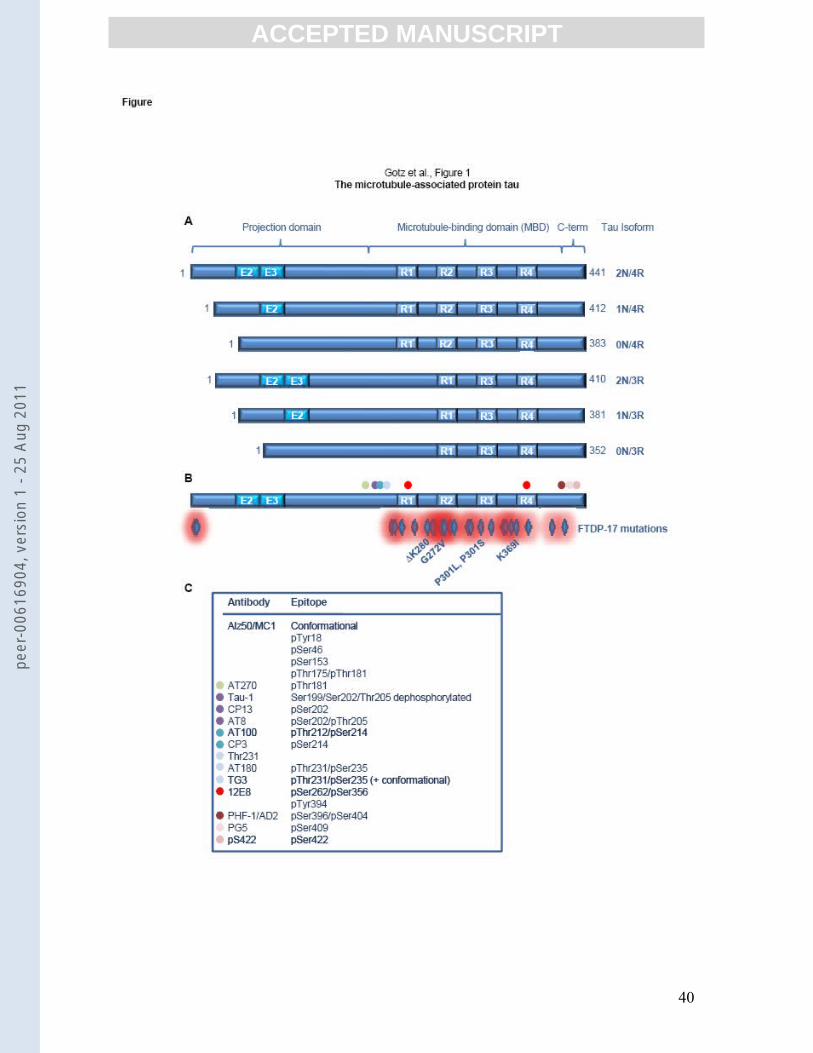
ACC
EPTE
D M
ANU
SCR
IPT
ACCEPTED MANUSCRIPT
40
peer
-006
1690
4, v
ersi
on 1
- 25
Aug
201
1