Anatomical differences and network characteristics underlying smoking cue reactivity
27
Anatomical differences and network characteristics underlying smoking cue reactivity Xiaochu Zhang, Betty Jo Salmeron, Thomas J Ross, Hong Gu, Xiujuan Geng, Yihong Yang, and Elliot A Stein * Neuroimaging Research Branch, Intramural Research Program, National Institute on Drug Abuse, NIH, Baltimore, MD Abstract A distributed network of brain regions is linked to drug-related cue responding. However, the relationships between smoking cue-induced phasic activity and possible underlying differences in brain structure, tonic neuronal activity and connectivity between these brain areas are as yet unclear. Twenty-two smokers and 22 controls viewed smoking-related and neutral pictures during a functional arterial spin labeling scanning session. T1, resting functional, and diffusion tensor imaging data were also collected. Six brain areas, dorsal lateral prefrontal cortex (dlPFC), dorsal medial prefrontal cortex (dmPFC), dorsal anterior cingulate cortex/cingulate cortex, rostral anterior cingulate cortex (rACC), occipital cortex, and insula/operculum, showed significant smoking cue-elicited activity in smokers when compared with controls and were subjected to secondary analysis for resting state functional connectivity (rsFC), structural, and tonic neuronal activity. rsFC strength between rACC and dlPFC was positively correlated with the cue-elicited activity in dlPFC. Similarly, rsFC strength between dlPFC and dmPFC was positively correlated with the cue-elicited activity in dmPFC while rsFC strength between dmPFC and insula/operculum was negatively correlated with the cue-elicited activity in both dmPFC and insula/operculum, suggesting these brain circuits may facilitate the response to the salient smoking cues. Further, the gray matter density in dlPFC was decreased in smokers and correlated with cue-elicited activity in the same brain area, suggesting a neurobiological mechanism for the impaired cognitive control associated with drug use. Taken together, these results begin to address the underlying neurobiology of smoking cue salience, and may speak to novel treatment strategies and targets for therapeutic interventions. Keywords Smoking cue; anatomical; ASL; DTI; VBM; resting state functional connectivity * Corresponding to Dr. Stein, Neuroimaging Research Branch, Intramural Research Program, National Institute on Drug Abuse, Baltimore, MD 21224, USA., Tel: 443-740-2650 Fax: 443-740-2816, [email protected]. Disclosure/Conflict of Interest None The authors reported no financial interests or potential conflicts of interest. Publisher's Disclaimer: This is a PDF file of an unedited manuscript that has been accepted for publication. As a service to our customers we are providing this early version of the manuscript. The manuscript will undergo copyediting, typesetting, and review of the resulting proof before it is published in its final citable form. Please note that during the production process errors may be discovered which could affect the content, and all legal disclaimers that apply to the journal pertain. NIH Public Access Author Manuscript Neuroimage. Author manuscript; available in PMC 2012 January 1. Published in final edited form as: Neuroimage. 2011 January 1; 54(1): 131–141. doi:10.1016/j.neuroimage.2010.07.063. NIH-PA Author Manuscript NIH-PA Author Manuscript NIH-PA Author Manuscript
Transcript of Anatomical differences and network characteristics underlying smoking cue reactivity

Anatomical differences and network characteristics underlyingsmoking cue reactivity
Xiaochu Zhang, Betty Jo Salmeron, Thomas J Ross, Hong Gu, Xiujuan Geng, Yihong Yang,and Elliot A Stein*Neuroimaging Research Branch, Intramural Research Program, National Institute on Drug Abuse,NIH, Baltimore, MD
AbstractA distributed network of brain regions is linked to drug-related cue responding. However, therelationships between smoking cue-induced phasic activity and possible underlying differences inbrain structure, tonic neuronal activity and connectivity between these brain areas are as yet unclear.Twenty-two smokers and 22 controls viewed smoking-related and neutral pictures during a functionalarterial spin labeling scanning session. T1, resting functional, and diffusion tensor imaging data werealso collected. Six brain areas, dorsal lateral prefrontal cortex (dlPFC), dorsal medial prefrontal cortex(dmPFC), dorsal anterior cingulate cortex/cingulate cortex, rostral anterior cingulate cortex (rACC),occipital cortex, and insula/operculum, showed significant smoking cue-elicited activity in smokerswhen compared with controls and were subjected to secondary analysis for resting state functionalconnectivity (rsFC), structural, and tonic neuronal activity. rsFC strength between rACC and dlPFCwas positively correlated with the cue-elicited activity in dlPFC. Similarly, rsFC strength betweendlPFC and dmPFC was positively correlated with the cue-elicited activity in dmPFC while rsFCstrength between dmPFC and insula/operculum was negatively correlated with the cue-elicitedactivity in both dmPFC and insula/operculum, suggesting these brain circuits may facilitate theresponse to the salient smoking cues. Further, the gray matter density in dlPFC was decreased insmokers and correlated with cue-elicited activity in the same brain area, suggesting a neurobiologicalmechanism for the impaired cognitive control associated with drug use. Taken together, these resultsbegin to address the underlying neurobiology of smoking cue salience, and may speak to noveltreatment strategies and targets for therapeutic interventions.
KeywordsSmoking cue; anatomical; ASL; DTI; VBM; resting state functional connectivity
*Corresponding to Dr. Stein, Neuroimaging Research Branch, Intramural Research Program, National Institute on Drug Abuse, Baltimore,MD 21224, USA., Tel: 443-740-2650 Fax: 443-740-2816, [email protected]/Conflict of InterestNoneThe authors reported no financial interests or potential conflicts of interest.Publisher's Disclaimer: This is a PDF file of an unedited manuscript that has been accepted for publication. As a service to our customerswe are providing this early version of the manuscript. The manuscript will undergo copyediting, typesetting, and review of the resultingproof before it is published in its final citable form. Please note that during the production process errors may be discovered which couldaffect the content, and all legal disclaimers that apply to the journal pertain.
NIH Public AccessAuthor ManuscriptNeuroimage. Author manuscript; available in PMC 2012 January 1.
Published in final edited form as:Neuroimage. 2011 January 1; 54(1): 131–141. doi:10.1016/j.neuroimage.2010.07.063.
NIH
-PA Author Manuscript
NIH
-PA Author Manuscript
NIH
-PA Author Manuscript
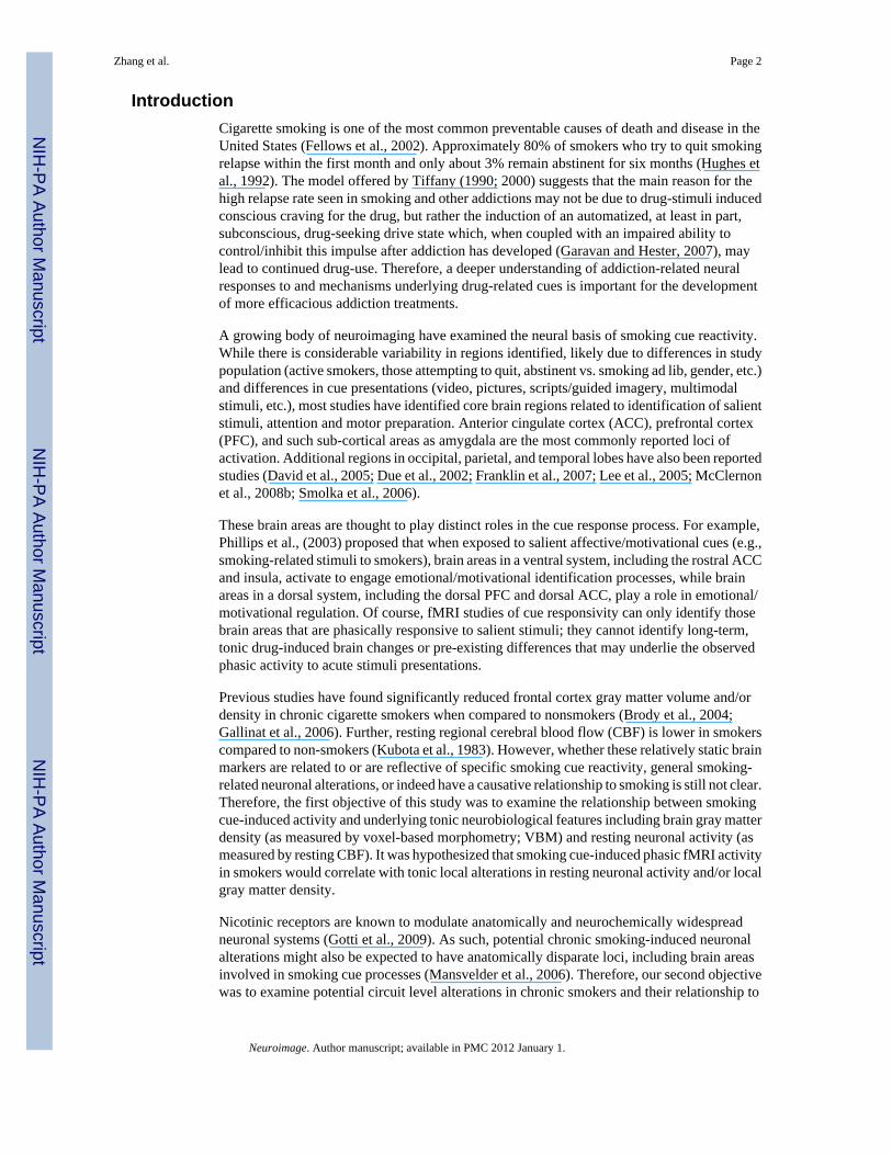
IntroductionCigarette smoking is one of the most common preventable causes of death and disease in theUnited States (Fellows et al., 2002). Approximately 80% of smokers who try to quit smokingrelapse within the first month and only about 3% remain abstinent for six months (Hughes etal., 1992). The model offered by Tiffany (1990; 2000) suggests that the main reason for thehigh relapse rate seen in smoking and other addictions may not be due to drug-stimuli inducedconscious craving for the drug, but rather the induction of an automatized, at least in part,subconscious, drug-seeking drive state which, when coupled with an impaired ability tocontrol/inhibit this impulse after addiction has developed (Garavan and Hester, 2007), maylead to continued drug-use. Therefore, a deeper understanding of addiction-related neuralresponses to and mechanisms underlying drug-related cues is important for the developmentof more efficacious addiction treatments.
A growing body of neuroimaging have examined the neural basis of smoking cue reactivity.While there is considerable variability in regions identified, likely due to differences in studypopulation (active smokers, those attempting to quit, abstinent vs. smoking ad lib, gender, etc.)and differences in cue presentations (video, pictures, scripts/guided imagery, multimodalstimuli, etc.), most studies have identified core brain regions related to identification of salientstimuli, attention and motor preparation. Anterior cingulate cortex (ACC), prefrontal cortex(PFC), and such sub-cortical areas as amygdala are the most commonly reported loci ofactivation. Additional regions in occipital, parietal, and temporal lobes have also been reportedstudies (David et al., 2005; Due et al., 2002; Franklin et al., 2007; Lee et al., 2005; McClernonet al., 2008b; Smolka et al., 2006).
These brain areas are thought to play distinct roles in the cue response process. For example,Phillips et al., (2003) proposed that when exposed to salient affective/motivational cues (e.g.,smoking-related stimuli to smokers), brain areas in a ventral system, including the rostral ACCand insula, activate to engage emotional/motivational identification processes, while brainareas in a dorsal system, including the dorsal PFC and dorsal ACC, play a role in emotional/motivational regulation. Of course, fMRI studies of cue responsivity can only identify thosebrain areas that are phasically responsive to salient stimuli; they cannot identify long-term,tonic drug-induced brain changes or pre-existing differences that may underlie the observedphasic activity to acute stimuli presentations.
Previous studies have found significantly reduced frontal cortex gray matter volume and/ordensity in chronic cigarette smokers when compared to nonsmokers (Brody et al., 2004;Gallinat et al., 2006). Further, resting regional cerebral blood flow (CBF) is lower in smokerscompared to non-smokers (Kubota et al., 1983). However, whether these relatively static brainmarkers are related to or are reflective of specific smoking cue reactivity, general smoking-related neuronal alterations, or indeed have a causative relationship to smoking is still not clear.Therefore, the first objective of this study was to examine the relationship between smokingcue-induced activity and underlying tonic neurobiological features including brain gray matterdensity (as measured by voxel-based morphometry; VBM) and resting neuronal activity (asmeasured by resting CBF). It was hypothesized that smoking cue-induced phasic fMRI activityin smokers would correlate with tonic local alterations in resting neuronal activity and/or localgray matter density.
Nicotinic receptors are known to modulate anatomically and neurochemically widespreadneuronal systems (Gotti et al., 2009). As such, potential chronic smoking-induced neuronalalterations might also be expected to have anatomically disparate loci, including brain areasinvolved in smoking cue processes (Mansvelder et al., 2006). Therefore, our second objectivewas to examine potential circuit level alterations in chronic smokers and their relationship to
Zhang et al. Page 2
Neuroimage. Author manuscript; available in PMC 2012 January 1.
NIH
-PA Author Manuscript
NIH
-PA Author Manuscript
NIH
-PA Author Manuscript
regional, phasic smoking cue activation. Synchronized spontaneous fluctuations in the resting-state fMRI signal are thought to reflect anatomically constrained, intrinsic, dynamicinteractions between brain regions (Vincent et al., 2007). Resting state functional connectivity(rsFC) maps have been observed in specific brain circuits (Albert et al., 2009; Greicius,2008; Waites et al., 2005), alterations of which have been demonstrated in such disorders asAlzheimer’s disease, schizophrenia, major depression and epilepsy (reviewed by Greicius,2008). Although rsFC does not, by definition, measure brain activation during taskperformance, the strength of such resting connectivity has been demonstrated to predictperformance on behavioral tasks requiring that circuit (Anticevic et al., 2010; Hampson et al.,2006; Kelly et al., 2008; Tambini et al., 2010). Therefore, rsFC was employed to determinepossible circuit level characteristics of chronic smokers. It was hypothesized that brain areasactivated by phasic smoking cues will exhibit altered rsFC in chronic smokers and that rsFCwill be related to cue-induced activity.
While rsFC has been shown to be anatomically constrained (Greicius et al., 2009), itnevertheless does not provide direct anatomical information. We therefore employed diffusiontensor imaging (DTI), a neuroimaging technique that assesses white matter integrity bymeasuring the freedom of water movement. We previously found prefrontal white matterintegrity alterations related to cigarette smoking (unpublished). Therefore, our third objectivewas to examine whether smoking-related impaired prefrontal white matter integrity isassociated with prefrontal rsFC strength alterations and/or phasic regional smoking cue-elicitedactivity. It was hypothesized that there is a direct link between these anatomical and functionalmeasures.
Materials and MethodsParticipants
Data were collected from 22 smokers (11 females; mean (±SD) age: 31.0±9.4; range: 21 to 50)and 22 age and gender matched controls (10 females; mean age: 29.6±7.0; range: 21 to 42).Smokers used cigarettes for an average of 12.0±7.3 years (range: 2 to 28 years) and smoked amean of 21.1±4.3 cigarettes/day (range: 15 to 30), with an average Fagerström Test for NicotineDependence (FTND) score of 5.4±1.9 (range: 3 to 10) and an average lifetime cigarette usageof 12.7±7.5 pack-years (range: 1.5 to 27). Smokers were required to not smoke for at least twohours before their MRI session and compliance was monitored by a member of the study team.A urine sample was collected and assessed for common drugs of abuse (TRIAGE®) before thescanning session. Participants were excluded if they had any major medical illnesses, currentmajor psychiatric disorders, neurological illnesses, or if their T1 weighted images revealedgross structural abnormalities. No participants had a history of dependence (current or past)on any drug other than nicotine based on the computerized Structured Clinical Interview forDSM-IV, drug use survey and clinical interview.
All participants gave written informed consent to protocols approved by the NIDA-IRPInstitutional Review Board and were compensated for their participation. Cue-elicited andresting Arterial Spin Label (ASL) cerebral blood flow (CBF) scans were obtained from allsubjects along with DTI and anatomical T1 data. Resting state BOLD fMRI data were obtainedon all but four smokers and four controls.
Experimental ParadigmIn a block design paradigm, participants were presented with two kinds of visual cues, i.e.,smoking-related and neutral pictures. The neutral pictures were similar in content to thesmoking-related ones, but absent any smoking-related properties (Figure 1). All pictures wereselected from those of Mucha et al., (1999) and the International Smoking Image Series (Gilbert
Zhang et al. Page 3
Neuroimage. Author manuscript; available in PMC 2012 January 1.
NIH
-PA Author Manuscript
NIH
-PA Author Manuscript
NIH
-PA Author Manuscript

and Rabinovich, 1999). In each of two scanning runs within a single scanning session, thirteen30s blocks, consisting of six cue and seven interspersed fixation/resting blocks, were presented(Figure 1). Each cue block consisted of 10 corresponding pictures, each presented for 1s witha 2s fixation gap, about 5°X5° visual angle. No subjective measurements (e.g., craving self-report) were collected during scanning because it has been proposed that to do so would engagetop-down appraisal processes that would likely alter neural activity reflecting the automaticconsequence of the smoking-related emotional stimuli (Ochsner and Gross, 2005;Taylor et al.,2003), which was the main focus of this study. The order of the neutral and the smoking-relatedblocks was counter-balanced across the participants.
Data acquisitionExperiments were performed on a Siemens 3T Allegra MRI scanner equipped with a standardRF birdcage head coil to acquire the neuroimaging data. A pulsed ASL sequence (Luh et al.,1999; Wong et al., 1997) was used to quantify perfusion changes associated with brainactivation in response to the visual stimuli. Eleven oblique 8mm thick axial slices encompassedmost of the brain. Some parietal areas were not covered in some participants. The PASLparameters are as follows: the gap between the labeling slab and the proximal slice=10mm,TI1=700ms, TI1 stop-time=1300ms, TI2=1400ms, with crusher bipolar gradients switchedbetween slice excitation and readout to reduce signal from large vessels. Further parameterswere: TE/TR=15ms/3000ms, number of measurements=130/run, imaging matrix=64×64,FOV=220mm×220mm, partial (6/8) Fourier acquisition, BW=3004Hz/Px, echospacing=0.4ms. Whole-brain T1-weighted structural images (MPRAGE) (1-mm3 isotropicvoxels, TE/TR=4.38ms/2500ms, FA=8°) were acquired for anatomical reference and VBManalyses. A resting-state BOLD fMRI scanning run was acquired using a gradient-echo EPIsequence (150 volumes, TE/TR=27ms/2000ms, FA=80°, FOV=220mm×220mm, imagingmatrix=64×64; 33 5mm-sagital slices in one smoker and two controls and 39 4mm-axial slicesin the others). DTI data were acquired using a single-shot, spin-echo echo-planar imagingtechnique (TE/TR=87ms/5000ms, BW=1700Hz/Pixel, FOV=220mm×220mm, imagingmatrix=128x128, 35 slices, thickness=4mm, b value= 0 and 1000s/mm2). Thirteen uniquevolumes were collected to compute the tensor: a b=0s/mm2 image and 12 images with diffusiongradients applied in 12 noncollinear directions (Gx, Gy, Gz: [1.0, 0.0, 0.5], [0.0, 0.5, 1.0], [0.5,1.0, 0.0], [1.0, 0.5, 0.0], [0.0, 1.0, 0.5], [0.5, 0.0, 1.0], [1.0, 0.0, −0.5], [0.0, −0.5, 1.0], [−0.5,1.0, 0.0], [1.0, −0.5, 0.0], [0.0, 1.0, −0.5], [−0.5, 0.0, 1.0]). Participant head movement wasminimized by the use of soft foam padding, a vacuum bag, and/or hardened polyurethane foam.
Whole brain analysis for ASL fMRI dataFunctional imaging data were analyzed with the Analysis of Functional Neuro-Images (AFNI)software package (Cox, 1996). After motion and acquisition time corrections, perfusion-weighted image series were generated by pair-wise subtraction (i.e., the effective TR is 6s.) ofthe label and control images (Liu and Wong, 2005), followed by conversion to absolute CBFbased on Equation 1. The CBF data were subjected to a whole brain 2×2 repeated-measuresanalysis of variance (ANOVA) with group (smoker and control) as a between-subjects factorand cue (smoking and neutral) as a within-subject factor.
(1)*
*:ΔM is the difference signal (control–labeled); M0b is the fully relaxed magnetization ofarterial blood†; TI1=0.7s, TI2=1.4s, and T1b=1.613s (the T1 decay time for labeled blood at 3T (Lu et al., 2004)). †: M0b is calculated from the steady-state signal of the control images.
Zhang et al. Page 4
Neuroimage. Author manuscript; available in PMC 2012 January 1.
NIH
-PA Author Manuscript
NIH
-PA Author Manuscript
NIH
-PA Author Manuscript

Secondary ROI analysisClusters showing significant interaction (FWE corrected p<0.05) between cue (smoking vs.neutral) and group (smokers vs. controls) in the whole-brain ASL results were considered asregions of interest (ROIs) and subjected to secondary analyses with an uncorrected thresholdof p=0.05, which was chosen to reflect the exploratory nature of the post-hoc analyses. Ageand gender were used as covariates for all correlation analyses in this secondary analysis.
Smoking cue-elicited changes in CBF [smoking-neutral] derived from the smoking and neutralcue conditions were averaged across voxels in each ROI from the corresponding blocks. Signalfrom the first effective TR in each block was removed to minimize the effects of hemodynamicdelay. A paired t-test was applied to the CBF data between the smoking and the neutral cueconditions in smokers to determine if the whole brain analysis [cue X group] result reflected,at least in part, differential activation in smokers to smoking cues. Additional exploratorycorrelations were performed between cue-elicited CBF changes and lifetime cigarette usageand FTND.
Based on our first hypothesis, resting neuronal activity (in the form of resting CBF) and brainstructure (gray matter density) were calculated for each ROI in each participant and comparedbetween controls and smokers via group t-tests. The resting CBF was calculated from fixationblocks in the ASL scans. Similar to cue-elicited CBF data, the first effective TR in each blockwas also removed. The gray matter density was calculated as described below from T1-weighted structural data via FSL-VBM, a voxel-based morphometry analysis package(Ashburner and Friston, 2000). First, structural images were skull stripped and then tissuesegmented into gray matter, white matter and CSF. The resulting gray matter partial volumeimages were aligned to standard space using an affine registration with the Image RegistrationToolkit (Rueckert et al., 1999). The resulting images were averaged to create a study-specifictemplate to which the native gray matter images were non-linearly re-registered. The registeredpartial volume images were then modulated (to correct for local expansion or contraction) bythe Jacobian of the warp field and then smoothed with an isotropic Gaussian kernel withFWHM=8mm.
To test whether resting CBF and gray matter density were related to cue-elicited phasic CBFchanges, Pearson’s correlation analyses were performed in each ROI between the cue-elicitedCBF values (smoking-neutral condition) and both gray matter density and resting CBF.Additional exploratory correlations were performed between these measures and lifetimecigarette usage and FTND.
Based on our second hypothesis, rsFC strength was calculated between different cue-activatedROIs using resting-state BOLD fMRI data via AFNI and MATLAB (The MathWorks, Inc.).First, volumes were slice-timing aligned and motion corrected. After linear detrending of thetime-course of each voxel, volumes were spatially normalized and resampled to Talairachspace at 2×2×2mm3 and temporally low-pass filtered (f cutoff =0.1Hz) (Cordes et al., 2001).The time courses from all voxels in each ROI were averaged in each participant, followed byPearson’s correlation analyses between the different ROIs. Fluctuations unlikely to be relevantto neuronal activity were regressed out as covariates including the six rigid head-motionparameter time-courses and the average time-courses in white matter and CSF (Fox et al.,2005; Lund et al., 2006). Correlation coefficients were transferred to z scores and thencompared between groups and correlated to smoking cue-elicited CBF in corresponding ROIsand correlated with lifetime cigarette usage and FTND in exploratory within-smoker groupanalyses.
DTI analysis was based on tract-based spatial statistics (TBSS) (Smith et al., 2006), anautomated, observer-independent method to allow group-wise comparisons, employed with an
Zhang et al. Page 5
Neuroimage. Author manuscript; available in PMC 2012 January 1.
NIH
-PA Author Manuscript
NIH
-PA Author Manuscript
NIH
-PA Author Manuscript

improved fractional anisotropy (FA) alignment method (Geng et al., 2009). According to themethod of Pierpaoli et al., (1996), FA images were created by fitting the raw diffusion data toa tensor model. All FA datasets were simultaneously registered onto an implicit referencecorresponding to the group average using implicit reference-based group (IRG) registration(Geng et al., 2009). Compared with conventional methods, which register each group imageto a selected reference, the IRG registration eliminates the bias associated with referenceselection and produces smaller registration error. All FA datasets were then transformed intostandard space using an affine transformation. The mean FA image was built to create a meanFA skeleton via FSL (Smith et al., 2004). This skeleton represents the centers of all tractscommon to the group. Each participant’s aligned FA data were then projected onto thisskeleton.
Because our previous smoking-related FA results (unpublished) and the observed alterationsin smoking cue-related rsFC strength (see Results) suggest only frontal cortex involvement, afrontal white matter skeleton mask was created and group comparisons with permutation-basedtesting (Nichols and Holmes, 2002) were performed within this mask to address our thirdhypothesis. The FA values from clusters showing significant group difference were averagedand correlated to the smoking cue-elicited CBF changes in frontal ROIs and the rsFC strengthsbetween the corresponding ROIs.
ResultsWhole brain CBF analysis of smoking cue provocation
Six clusters showed a significant (p<0.005 (F value>8.76) combined with a minimum clustersize of 1226 mm3 yields an FWE corrected p<0.05) interaction between group (smoker vs.control) and cue (smoking vs. neutral) from the whole brain ANOVA analysis. These clustersinclude bilateral dorsal medial prefrontal cortex (dmPFC), right dorsal lateral prefrontal cortex(dlPFC), bilateral dorsal anterior cingulate cortex/cingulate cortex (dACC/CC), right middleoccipital gyrus (MOG), left insula/operculum (including operculum temporale into superiortemporal gyrus and operculum frontoparietale into postcentral gyrus and inferior parietal)) (I/O) and bilateral rostral anterior cingulate cortex (rACC) (see Figure 2, Table 1). Data fromeach of these ROIs were used in subsequent secondary analyses.
Secondary ROI analysesSmoking cue-elicited CBF phasic activity—The averaged CBF for each cue identifiedROI during each of the cue conditions in both groups are listed in Table 2. There was asignificant CBF increase in smokers to the smoking cue compared to the neutral cue conditionin all six brain regions (see Table 2). Lifetime cigarette usage was positively correlated withthe cue-elicited CBF change (smoking–neutral) in the rACC (r[18]=0.53, p=0.017; Figure 3A)and negatively related to that in the dlPFC (r[18]= −0.46, p=0.042; Figure 3B). There was nosignificant correlation between smoking cue-elicited CBF and FTND in any ROI.
Resting CBF and gray matter density—To address our first hypothesis, gray matterdensity and baseline neuronal activity (i.e. resting CBF) and their relationship to smoking cue-elicited phasic CBF in each ROI were calculated (Table 3 and 4). Significantly lower restingCBF was found in the I/O (t[42]= −2.83, p=0.007), the rACC (t[42]= −2.79, p=0.008), and thedACC (t[42]= −2.15, p=0.037) in smokers compared to controls. There were no significantassociations between resting CBF and the phasic smoking cue-elicited CBF changes, lifetimecigarette usage or FTND (see Table 3). Further, the gray matter density in dlPFC in smokerswas significantly lower (t[42]= −2.62, p=0.012) than in controls and was positively correlatedwith smoking-related CBF cue changes (r[18]=0.54, p=0.013; Figure 3C), with a trend towardsa negative correlation with lifetime cigarette usage (r[18]= −0.43, p=0.060). The gray matter
Zhang et al. Page 6
Neuroimage. Author manuscript; available in PMC 2012 January 1.
NIH
-PA Author Manuscript
NIH
-PA Author Manuscript
NIH
-PA Author Manuscript

in the I/O in smokers was also lower in controls (t[42]= −2.07, p=0.045). No other groupdifferences or correlations were seen.
RsFC—In reference to our second hypothesis, rsFC strength between the dlPFC and rACCwas positively correlated to the cue-elicited CBF change in the dlPFC (r[14]=0.64, p=0.007;Figure 4A), while the rsFC strength between the dmPFC and dlPFC was positively related tothe smoking cue-elicited CBF change in dmPFC (r[14]=0.62, p=0.010; Figure 4B). Further,rsFC strength between the dmPFC and the I/O was negatively correlated with the smoking cue-elicited CBF change in both dmPFC (r[14]= −0.55, p=0.027; Figure 4C) and I/O (r[14]= −0.55,p=0.026; Figure 4D). No group differences or additional significant correlations were found(Table 5).
White matter integrity (DTI)—Consistent with our previous observations (unpublished),one cluster located in the left prefrontal area showed significantly lower FA in smokers thanin controls (uncorrected p=0.005 and volume=45mm3 (yields an FWE corrected p=0.034),Talairach coordinate: x=19.5mm, y=−38.1mm, z=10.2mm; see Figure 5A). To address ourthird hypothesis, correlations between the FA data averaged from this cluster and the rsFCstrength between frontal ROIs and phasic cue-elicited CBF activity in frontal ROIs werecalculated. This regional FA was significantly correlated with the rsFC strength in controlsbetween dACC and dmPFC (r[14]=0.59, p=0.015; Figure 5B) and between the dmPFC andthe dlPFC (r[14]=0.53, p=0.036; Figure 5C). Finally, this FA reduction was not correlated withany other rsFC circuit between any of the frontal ROIs in either smokers or controls, nor didit correlate with phasic smoking cue-elicited activity in any frontal ROI in smokers (Table 6).
DiscussionIn this study, we measured changes in regional cerebral blood flow (CBF) while both smokersand non-smokers were exposed to smoking-related and matched neutral pictures. We foundsix brain areas that showed greater smoking cue-elicited activity in smokers, including dorsalmedial prefrontal cortex (dmPFC), right dorsal lateral prefrontal cortex (dlPFC), dorsal anteriorcingulate cortex/cingulate cortex (dACC/CC), right middle occipital gyrus (MOG), left insula/operculum and rostral anterior cingulate cortex (rACC). Using these 6 functionally identifiedROIs as nodes in a rsFC analysis, we found that the connectivity strength between rACC anddlPFC was positively correlated with subsequent cue-elicited activity in dlPFC, while the rsFCstrength between dlPFC and dmPFC was positively correlated with cue-elicited activity indmPFC. Further, the rsFC strength between dmPFC and insula/operculum was negativelyrelated to cue-elicited activity in both dmPFC and insula/operculum. In addition, gray matterdensity in the dlPFC was lower in smokers compared to controls and positively related tosmoking cue-elicited activity in the same brain area. Further, lifetime exposure to cigaretteswas correlated to smoking cue-elicited activity in dlPFC and rACC. Finally, prefrontal FA wasreduced in smokers while resting CBF was reduced in three ROIs. Insofar as these secondaryROI analyses were not corrected for multiple comparisons, they should be consideredexploratory. However the network and relationships they reveal are intriguing (see Figure 6).The implications of these results are discussed below.
Smoking cue-elicited CBF phasic changesSix brain regions (dlPFC, dmPFC, dACC/CC, rACC, MOG, and insula/operculum, see Figure2) demonstrated a significant increase in activation to cue presentation that was both group(smoker vs. control) specific and cue (smoking vs. neutral) selective. These functionallyidentified clusters were subjected to secondary ROI analysis (discussed below) in the presentstudy partially because previous studies (David et al., 2005;Due et al., 2002;Franklin et al.,2007;Lee et al., 2005;McClernon et al., 2008b;Smolka et al., 2006) have not reported consistent
Zhang et al. Page 7
Neuroimage. Author manuscript; available in PMC 2012 January 1.
NIH
-PA Author Manuscript
NIH
-PA Author Manuscript
NIH
-PA Author Manuscript

results. For example, Franklin et al., (2007) and Lee et al., (2005) reported brain networks thathad almost no overlapping areas. Lee et al., (2005) found activation almost entirely corticallylocated, including the prefrontal cortex, anterior cingulate gyrus, and supplementary motorarea, while Franklin et al., (2007) reported predominantly sub-cortical areas, including ventralstriatum, amygdala, hippocampus and medial thalamus. Further, McClernon et al. (2008b) andSmolka et al. (2006) both looked at correlations between smoking cue-evoked fMRI andFTND, and reported opposite results.
While there are many possible explanations for these discrepancies, different experimentalparadigms (event-related design vs block design), neuroimaging tools (BOLD vs ASL), subjectsmoking status (ad lib smoking, time of abstinence), and/or data analysis method, are likelycontributory. Indeed, Franklin et al., (2007) and Due et al., (2002) only analyzed data from apriori-selected brain areas, not from whole brain. In particular, Franklin et al., (2007) employed10-minutes video/audio clip, which likely activated circuits distinct from those processingdiscrete pictures presented for only 5 sec by Lee et al., (2005). We also used a brief, discretestimulus (1 sec in the present study), and interestingly, we too see predominantly corticalactivation, with very similar regional distribution. Cue duration and the embedded context(complex in a video, discrete and isolated for a picture) likely activate and maintain (or not)different attentional networks that can have dramatic profound effects on cue processing.
The difference roles of these brain areas during smoking-related cue process may also helpexplain the disparate literature. Based on a prominent model of emotional processing (Phillipset al., 2003), these six brain areas may be divided into two groups, a ventral and a dorsal system.The ventral system, including rostral ACC and insula (and possibly opercular and occipitalgyrus), has been implicated in emotional/motivational identification processes, and theiractivation in this study may thus reflect the motivational salience engaged in smokers by thesmoking cues. The dorsal system, which includes the dorsal medial/lateral PFC and dorsalACC, is thought to play a role in motivational regulation, suggesting that response regulatoryresources, perhaps including response inhibition, are being engaged in response to smokingcues, especially inasmuch as subjects seeing these cues were required to remain in the scannerand in the lab for some time and could not freely act upon any cue-induced drive state.
Further, since McClernon et al., (2008a) and David et al., (2007) found that acute nicotineabstinence significantly alters BOLD activation to smoking cue in cigarette smokers, and sincesmokers in the current study were not permitted to smoke for at least 2 hours before scanning,our observed [smoker-nonsmoker] differences may reflect not only the learning-inducedenhanced salience of the cues to smokers, but also the acute effects of very early nicotinewithdrawal. Notably, the CBF difference between the smoking and neutral cue conditions inthe rACC in smokers was positively correlated to total lifetime nicotine exposure, but not toFTND, a measure of addiction severity (Heatherton et al., 1991), suggesting a potentialsmoking-related compensation and/or learning effect.
Relationship between phasic cue activity and functional network/anatomical alterationsIn an attempt to identify underlying structural and tonic functional brain alterations that mighthelp explain the acute cue responses reported above, we performed exploratory correlationanalyses between the phasic cue responses, rsFC circuits and gray matter density alterations.Each is discussed below.
Relationship between rsFC and smoking cue-induced phasic activation—It hasrecently been suggested that the strength of rsFC circuits reflect the ability to engage thosecircuits during active task processing and may thus serve as surrogates for behavioralperformance and/or learning-induced plasticity. For example, motor learning modulatesfronto-parietal resting state networks (Albert et al., 2009) and the performance on a language
Zhang et al. Page 8
Neuroimage. Author manuscript; available in PMC 2012 January 1.
NIH
-PA Author Manuscript
NIH
-PA Author Manuscript
NIH
-PA Author Manuscript
task affects resting state networks in regions associated with language (Waites et al., 2005).Further, Anticevic et al., (2010), Tambini et al., (2010), and Hampson et al., (2006) alsoreported that rsFC between posterior cingulate cortex, hippocampus, and frontal corticalregions can predict performance on working memory tasks.
In the present study, the rsFC strength between dlPFC and rACC was positively correlatedwith the increased phasic smoking cue activity magnitude in the dlPFC itself, suggesting thatthis circuit is engaged when a smoker is processing smoking cues. The rostral ACC belongsto the affective division of the cingulate cortex and is engaged by emotionally laden tasks inboth normal healthy volunteers and in those with psychiatric disorders such as depression anddrug dependence (Bush et al., 2000). Previous studies (Bush et al., 2000; Posner et al., 2007)have shown that the ventral/rostral ACC receives motivational/emotional information fromsub-cortical regions (e.g., amygdala) and then transfers this information to and receivesmodulations from such higher-level cortical regions as the dlPFC. Further, convergent evidencesupports a role for the dlPFC in cognitive control processes, including regulation of affectiveinformation induced by affective/emotional cues (reviewed by Badre, 2008; Phillips et al.,2003). Therefore, the positive correlation between rsFC strength of a dlPFC-rACC circuit andthe increased dlPFC response to smoking cues suggests that this brain circuit is involved inprocessing the enhanced salience of smoking cues in smokers, most likely by regulating thesalience-identifying response of the rACC. This raises the interesting possibility thatconnectivity in this circuit might be a potential target for therapeutic interventions.
Similarly, smoking cue-elicited activity in dmPFC was correlated to rsFC strength betweendmPFC and dlPFC in smokers, further supporting a role for a dorsal PFC network in processingsalient cues. It has been suggested that the dlPFC and dmPFC/ACC play different roles incognitive control processes (Ridderinkhof et al., 2004), with the dmPFC/ACC involved inperformance monitoring (e.g., cue-elicited arousal or motivation monitoring during cueprovocation), while the dlPFC plays a role in subsequent performance control adjustments(e.g., arousal control/inhibition or action/motivation induced by cues). The positiveconnectivity strength between monitoring and control adjustment brain systems and theircorrelation with activity following cue presentation in the monitoring system componentsuggests a role for this circuit in salient cue processing.
Further, the rsFC strength between dmPFC and insula/operculum was negatively correlatedwith the phasic smoking cue activity magnitude in both dmPFC and insula/operculum. Asdiscussed above, dmPFC plays a role in cue-elicited arousal or motivation monitoring. Theinsula is a key neural structure for representing the interoceptive effects of drug use, which areassociated with conscious cue-induced urges (reviewed by Naqvi and Bechara, 2009). Naqviet al., (2007) observed that cerebrovascular strokes in smokers limited to the insula lead to aprofound reduction in post recovery smoking behavior and craving for cigarette. Further,previous studies also reported that the operculum (in particular opercular/SII) plays animportant role in somatosensory and pain responding (Craig et al., 1996; Mazzola et al.,2006) and is related to anticipation of palatable food intake and consumption (Stice et al.,2009). The latter result appears consistent with our findings in that craving induced by cues isan anticipation of future consumption of a desirable commodity (cigarettes). Although therelationship of the dmPFC to the insula/operculum is not yet well understood, greaterconnectivity in the current study was associated with less cue reactivity in both regions,implying a sort of mutual inhibition of cue reactivity. Intriguingly, there was a trend towardsa positive correlation between rsFC in the insula/operculum-rACC circuit and cue reactivityin the rACC (p=0.056), pointing to a possible mechanism for this region to influence (or beinfluenced by) cue reactivity in those prefrontal regions related to salience detection that isregulated by connectivity between insula and dmPFC, thus completing a brain circuit (i.e.,rACC to dlPFC to dmPFC to insula to rACC) involved in smoking-related cue process.
Zhang et al. Page 9
Neuroimage. Author manuscript; available in PMC 2012 January 1.
NIH
-PA Author Manuscript
NIH
-PA Author Manuscript
NIH
-PA Author Manuscript

Intriguingly and contrary to our hypothesis, while the cue provocation led to increased phasicactivation in smokers, the rsFC strength in the above circuits did not differ between smokersand controls. It is thus possible that the rsFC relationship with regional cue-related activationmay not be unique to smoking-related information per se, but rather a response characteristicof appetitive/motivational stimuli in general, of which smoking-related information in smokersis only one of many such categorical stimuli. As there were no appetitive/motivational stimulipresented to our subjects (e.g. appetitive food cues), this hypothesis cannot be tested using thecurrent dataset, but would be of interest for future studies.
dlPFC in nicotine dependence—The dlPFC gray matter density was significantly lowerin smokers compared to controls, which is consistent with previous reports (Brody et al.,2004; Gallinat et al., 2006) that also found a negative correlation with lifetime cigaretteconsumption. We also observed a similar negative correlation, but at a trend level (p = 0.060).Brody et al (2004) suggest that such group differences in dlPFC gray matter might reflect thecumulative effects of thousands of cigarette cue presentations or the pharmacological orneurotoxic properties of cigarettes. Given convergent data suggesting that smoking damagesPFC functions (Ernst et al., 2001; Jacobsen et al., 2005) and the positive correlation of cue-induced activation with gray matter density (i.e., more ‘normalized’ gray matter density isassociated with more activation to cues), it is possible that dlPFC activation to cues may reflectengagement of control systems to deal with the appetitive cue response, which weaken withextended exposure to cigarettes.
In further support of this interpretation, the dlPFC has been implicated in, among otherfunctions, cognitive control and/or inhibitory processes, including regulation of informationinduced by affective/emotional cues (Badre, 2008; Phillips et al., 2003). Recently, Artiges etal., (2009) reported that smoking cues trigger an adaptive top-down process, e.g., cue-inducedcraving may induce inhibitory processing in smokers involving the right dlPFC. Further, dlPFCgray matter has been found to correlate positively to the inhibitory control necessary to performthe Stroop task (Haldane et al., 2008). We observed cue-induced activity in rACC that increaseswith lifetime exposure to cigarettes, while dlPFC cue-induced activity decreased with lifetimeexposure, and is related to the strength of rsFC between rACC and dlPFC. In addition, dlPFCgray matter density trended towards a negative correlation with lifetime exposure, a resultalready demonstrated by several other groups. Taken together, a picture starts to emerge ofpossible loss of dlPFC regulation of cue responsivity in rACC related to continuous smokingbehavior. The rACC cue response thus grows due to loss of descending regulatory input which,based on the relationship between rsFC and cue-induced activation, likely involves a circuitfrom rACC to dlPFC to dmPFC to insula/ST to rACC. Taking into account the exploratorycharacter of our statistical analysis, this conclusion needs further empirical testing, perhapsusing effective connectivity analysis.
Tonic brain differences unrelated to smoking cuesFinally, we identified a white matter cluster in the prefrontal lobe that was significantly reducedin smokers (Figure 5A), using a recently developed voxel-wise DTI analysis method (Geng etal., 2009; Smith et al., 2006). Further, the impaired prefrontal white matter integrity in thisregion was significantly correlated with rsFC strength between the dACC/CC and the dmPFCand between the dmPFC and the dlPFC in nonsmokers, suggesting that the absence of such arelationship in smokers may reflect the absence of coherence between white matter and rsFCthat is normally present. Since FA in this cluster did not correlate with phasic cue-elicitedactivity in dACC/CC, dmPFC or dlPFC, the reduced FA is not likely to be specific to smokingcue reactivity. However, because the rsFC between dmPFC and dlPFC was significantlycorrelated to smoking-related cue activity (in smokers) and this FA cluster (in nonsmokers),this prefrontal white matter impairment may indirectly influence smoking-related cue
Zhang et al. Page 10
Neuroimage. Author manuscript; available in PMC 2012 January 1.
NIH
-PA Author Manuscript
NIH
-PA Author Manuscript
NIH
-PA Author Manuscript

processes via general smoking-related alterations in prefrontal areas, e.g., inhibition or controlfunctions.
Further, rACC, insula/operculum and dACC/CC showed significantly lower resting CBF insmokers. This result is consistent with previous reports (Kubota et al., 1983; Siennicki-Lantzet al., 2002) of lower regional CBF in frontal and temporal regions in smokers. Interestingly,Kosten et al., (1998) and Pezawas et al., (2002) found a similar CBF decrease in mixed cocaine-alcohol and opioid users, respectively. The coupling between neuronal activity, metabolismand CBF is the basis of most functional neuroimaging methods (Villringer and Dirnagl,1995; Wong et al., 1997). Therefore, lower regional CBF likely reflects lower tonic neuralactivity in smokers and is consistent with a previous study demonstrating reduced frontal brainmetabolism in cocaine addicts (Volkow et al., 1992). Further, Peoples et al., (2007) reportedthat not only did prolonged cocaine self-administration reduce tonic accumbal neural activityin rats, short-term drug exposure was still able to increase activity in the same area, providinga potential mechanism underlying a similar CBF pattern in the present study, wherein weobserved increased phasic cue-induced activity in the face of tonic decreased resting neuralactivity in the same areas.
While it is most likely that the resting CBF findings reflect lower tonic neural activity insmokers, one cannot eliminate potential alternative effects of chronic cigarette smoke and/ornicotine on rCBF regulatory mechanisms, vessel alterations due to embolism and/or vascularinjury, alterations in blood viscosity or oxygen levels, and/or neuronal morphologicalalterations (Mathew and Wilson, 1991). A number of factors would argue against such ‘non-specific contributors to the observed signal changes. For example, reduced gray matter densitywas seen only in insula/operculum and dlPFC, limiting any morphological contributions to theregional effects reported. Further, although all our participants presented to scanning sessionswith normal hematocrit and oxygen saturation levels, chronic smoking, perhaps due to elevatedCO levels or other constituents in tobacco smoke, may nevertheless have damaged localcerebral vasculature (Leone, 2007). However, Murphy et al (2006) report that neuronalactivity-BOLD coupling remains unchanged in chronic smokers and thus does not support aninterpretation of ‘non-specific’ effect of nicotine and/or cigarette smoke on blood flow oralterations in coupling. As such, we suggest that alterations in resting CBF, rsFC and phasicCBF alterations to salient smoking pictures reported herein, accurately reflect alterations inneuronal activity.
Limitations and ConclusionSeveral potential limitations of the present study merit consideration. As pointed out earlier,no direct behavioral measure of cue induced craving or drug seeking or control over this drivestate were collected. According to Tiffany’s (2000) ‘cognitive process’ model, the drug-seeking drive elicited by cues becomes automatized, suggesting that drug-related stimulipresented in the current study should have resulted in an enhanced drive state. We elected notto gather craving ratings, as our primary focus was on neural responses to cues, and the veryact of rating can alter such responses (Ochsner and Gross, 2005; Taylor et al., 2003). Whilethis approach allowed us to assess neural responding to the cues unencumbered by otherpsychological processes, we have no measure that participant’s paid attention to the cues, oreven looked at them. However, based on the commonality of regions we obtained and thoseseen in other cue reactivity studies, it would appear that subjects were compliant with taskinstructions, but there is no definitive verification of this in our design. Another limitation isthe absence of smoking cue-elicited activity in sub-cortical areas, including the amygdala,hippocampus and nucleus accumbens is curious, as these regions have been reported in one ormore drug-related cue studies (David et al., 2005; Franklin et al., 2007; Grant et al., 1996; Kiltset al., 2001; McClernon et al., 2007). Partial volume-induced signal reductions due to the
Zhang et al. Page 11
Neuroimage. Author manuscript; available in PMC 2012 January 1.
NIH
-PA Author Manuscript
NIH
-PA Author Manuscript
NIH
-PA Author Manuscript

relatively thick 8mm slices used in the ASL acquisition may help explain this null result. It isalso possible, however, that, due to the complex interactions cited above for controllingresponding to salient cues, some cues responses were obscured. Future connectivity studies,both at rest and during cue exposure, might help clarify this issue. Finally, multiple comparisoncorrection was not employed in the secondary ROI analyses, reflecting the exploratory natureof this approach. As such, our findings should be seen as preliminary, with a large samplereplication with more focused hypotheses warranted.
In conclusion, the present study attempted to expand our understanding of the neural basis forthe enhanced salience of cues in chronic smokers by applying multi-modal imaging techniquesto extend traditional differential activation to smoking cues. We found numerous changes inPFC, especially dlPFC, in smokers along with decreases in resting CBF, gray matter density,and the relationship between rsFC strength and phasic cue response magnitude. Whileconsidered preliminary, many of our findings are consistent with the extant literature,suggesting candidate neural circuitry underlying smoking cue responses along with changesin the circuitry with repeated smoking and daily environmental cue exposure. Understandingthe changing role of the dlPFC and overall alterations in PFC function may be fruitfully appliedin developing clinical strategies to assist with smoking cessation.
Supplementary MaterialRefer to Web version on PubMed Central for supplementary material.
AcknowledgmentsThis research was supported by the Intramural Research Program of the National Institute on Drug Abuse, NIH. Wethank Kimberly Modo and Loretta Spurgeon for their assistance in the conduct of some of the studies reported hereinand Mary Pfeiffer for editing assistance.
Reference ListAlbert NB, Robertson EM, Miall RC. The resting human brain and motor learning. Curr Biol
2009;19:1023–1027. [PubMed: 19427210]Anticevic A, Repovs G, Shulman GL, Barch DM. When less is more: TPJ and default network
deactivation during encoding predicts working memory performance. Neuroimage 2010;49:2638–2648. [PubMed: 19913622]
Artiges E, Ricalens E, Berthoz S, Krebs MO, Penttila J, Trichard C, Martinot JL. Exposure to smokingcues during an emotion recognition task can modulate limbic fMRI activation in cigarette smokers.Addict Biol 2009;14:469–477. [PubMed: 19650816]
Ashburner J, Friston KJ. Voxel-based morphometry--the methods. Neuroimage 2000;11:805–821.[PubMed: 10860804]
Badre D. Cognitive control, hierarchy, and the rostro-caudal organization of the frontal lobes. TrendsCogn Sci 2008;12:193–200. [PubMed: 18403252]
Brody AL, Mandelkern MA, Jarvik ME, Lee GS, Smith EC, Huang JC, Bota RG, Bartzokis G, LondonED. Differences between smokers and nonsmokers in regional gray matter volumes and densities. BiolPsychiatry 2004;55:77–84. [PubMed: 14706428]
Bush G, Luu P, Posner MI. Cognitive and emotional influences in anterior cingulate cortex. Trends CognSci 2000;4:215–222. [PubMed: 10827444]
Cordes D, Haughton VM, Arfanakis K, Carew JD, Turski PA, Moritz CH, Quigley MA, Meyerand ME.Frequencies contributing to functional connectivity in the cerebral cortex in “resting-state” data. AJNRAm J Neuroradiol 2001;22:1326–1333. [PubMed: 11498421]
Cox RW. AFNI: software for analysis and visualization of functional magnetic resonance neuroimages.Comput Biomed Res 1996;29:162–173. [PubMed: 8812068]
Zhang et al. Page 12
Neuroimage. Author manuscript; available in PMC 2012 January 1.
NIH
-PA Author Manuscript
NIH
-PA Author Manuscript
NIH
-PA Author Manuscript

Craig AD, Reiman EM, Evans A, Bushnell MC. Functional imaging of an illusion of pain. Nature1996;384:258–260. [PubMed: 8918874]
David SP, Munafo MR, Johansen-Berg H, Mackillop J, Sweet LH, Cohen RA, Niaura R, Rogers RD,Matthews PM, Walton RT. Effects of Acute Nicotine Abstinence on Cue-elicited Ventral Striatum/Nucleus Accumbens Activation in Female Cigarette Smokers: A Functional Magnetic ResonanceImaging Study. Brain Imaging Behav 2007;1:43–57. [PubMed: 18458752]
David SP, Munafo MR, Johansen-Berg H, Smith SM, Rogers RD, Matthews PM, Walton RT. Ventralstriatum/nucleus accumbens activation to smoking-related pictorial cues in smokers and nonsmokers:a functional magnetic resonance imaging study. Biol Psychiatry 2005;58:488–494. [PubMed:16023086]
Due DL, Huettel SA, Hall WG, Rubin DC. Activation in mesolimbic and visuospatial neural circuitselicited by smoking cues: evidence from functional magnetic resonance imaging. Am J Psychiatry2002;159:954–960. [PubMed: 12042183]
Ernst M, Heishman SJ, Spurgeon L, London ED. Smoking history and nicotine effects on cognitiveperformance. Neuropsychopharmacology 2001;25:313–319. [PubMed: 11522460]
Fellows J, Trosclair A, Adams E, Rivera C. Annual smoking-attributable mortality, years of potentiallife lost, and economic costs: United States, 1995–1999. MMWR Morb Mortal Wkly Rep2002;51:300–303. [PubMed: 12002168]
Fox MD, Snyder AZ, Vincent JL, Corbetta M, Van E, Raichle ME. The human brain is intrinsicallyorganized into dynamic, anticorrelated functional networks. Proc Natl Acad Sci USA2005;102:9673–9678. [PubMed: 15976020]
Franklin TR, Wang Z, Wang J, Sciortino N, Harper D, Li Y, Ehrman R, Kampman K, O’Brien CP, DetreJA, Childress AR. Limbic activation to cigarette smoking cues independent of nicotine withdrawal:a perfusion fMRI study. Neuropsychopharmacology 2007;32:2301–2309. [PubMed: 17375140]
Gallinat J, Meisenzahl E, Jacobsen LK, Kalus P, Bierbrauer J, Kienast T, Witthaus H, Leopold K, SeifertF, Schubert F, Staedtgen M. Smoking and structural brain deficits: a volumetric MR investigation.Eur J Neurosci 2006;24:1744–1750. [PubMed: 17004938]
Garavan H, Hester R. The role of cognitive control in cocaine dependence. Neuropsychol Rev2007;17:337–345. [PubMed: 17680368]
Geng X, Christensen GE, Gu H, Ross TJ, Yang Y. Implicit reference-based group-wise image registrationand its application to structural and functional MRI. Neuroimage 2009;47:1341–1351. [PubMed:19371788]
Gilbert, DG.; Rabinovich, NE. International Smoking Image Series (With Neutral Counterparts), version1.2 ed. Carbondale: Integrative Neuroscience Laboratory; 1999.
Gotti C, Clementi F, Fornari A, Gaimarri A, Guiducci S, Manfredi I, Moretti M, Pedrazzi P, Pucci L,Zoli M. Structural and functional diversity of native brain neuronal nicotinic receptors. BiochemPharmacol 2009;78:703–711. [PubMed: 19481063]
Grant S, London ED, Newlin DB, Villemagne VL, Liu X, Contoreggi C, Phillips RL, Kimes AS, MargolinA. Activation of memory circuits during cue-elicited cocaine craving. Proc Natl Acad Sci USA1996;93:12040–12045. [PubMed: 8876259]
Greicius M. Resting-state functional connectivity in neuropsychiatric disorders. Curr Opin Neurol2008;21:424–430. [PubMed: 18607202]
Greicius MD, Supekar K, Menon V, Dougherty RF. Resting-state functional connectivity reflectsstructural connectivity in the default mode network. Cereb Cortex 2009;19:72–78. [PubMed:18403396]
Haldane M, Cunningham G, Androutsos C, Frangou S. Structural brain correlates of response inhibitionin Bipolar Disorder I. J Psychopharmacol 2008;22:138–143. [PubMed: 18308812]
Hampson M, Driesen NR, Skudlarski P, Gore JC, Constable RT. Brain connectivity related to workingmemory performance. J Neurosci 2006;26:13338–13343. [PubMed: 17182784]
Heatherton TF, Kozlowski LT, Frecker RC, Fagerstrom KO. The Fagerstrom Test for NicotineDependence: a revision of the Fagerstrom Tolerance Questionnaire. Br J Addict 1991;86:1119–1127.[PubMed: 1932883]
Hughes JR, Gulliver SB, Fenwick JW, Valliere WA, Cruser K, Pepper S, Shea P, Solomon LJ, FlynnBS. Smoking cessation among self-quitters. Health Psychol 1992;11:331–334. [PubMed: 1425551]
Zhang et al. Page 13
Neuroimage. Author manuscript; available in PMC 2012 January 1.
NIH
-PA Author Manuscript
NIH
-PA Author Manuscript
NIH
-PA Author Manuscript

Jacobsen LK, Krystal JH, Mencl WE, Westerveld M, Frost SJ, Pugh KR. Effects of smoking and smokingabstinence on cognition in adolescent tobacco smokers. Biol Psychiatry 2005;57:56–66. [PubMed:15607301]
Kelly AM, Uddin LQ, Biswal BB, Castellanos FX, Milham MP. Competition between functional brainnetworks mediates behavioral variability. Neuroimage 2008;39:527–537. [PubMed: 17919929]
Kilts CD, Schweitzer JB, Quinn CK, Gross RE, Faber TL, Muhammad F, Ely TD, Hoffman JM, DrexlerKP. Neural activity related to drug craving in cocaine addiction. Arch Gen Psychiatry 2001;58:334–341. [PubMed: 11296093]
Kosten TR, Cheeves C, Palumbo J, Seibyl JP, Price LH, Woods SW. Regional cerebral blood flow duringacute and chronic abstinence from combined cocaine-alcohol abuse. Drug Alcohol Depend1998;50:187–195. [PubMed: 9649971]
Kubota K, Yamaguchi T, Abe Y, Fujiwara T, Hatazawa J, Matsuzawa T. Effects of smoking on regionalcerebral blood flow in neurologically normal subjects. Stroke 1983;14:720–724. [PubMed: 6658956]
Lee JH, Lim Y, Wiederhold BK, Graham SJ. A functional magnetic resonance imaging (FMRI) study ofcue-induced smoking craving in virtual environments. Appl Psychophysiol Biofeedback2005;30:195–204. [PubMed: 16167185]
Leone A. Smoking, haemostatic factors, and cardiovascular risk. Curr Pharm Des 2007;13:1661–1667.[PubMed: 17584096]
Liu TT, Wong EC. A signal processing model for arterial spin labeling functional MRI. Neuroimage2005;24:207–215. [PubMed: 15588612]
Lu H, Clingman C, Golay X, van Zijl PC. Determining the longitudinal relaxation time (T1) of blood at3.0 Tesla. Magn Reson Med 2004;52:679–682. [PubMed: 15334591]
Luh WM, Wong EC, Bandettini PA, Hyde JS. QUIPSS II with thin-slice TI1 periodic saturation: a methodfor improving accuracy of quantitative perfusion imaging using pulsed arterial spin labeling. MagnReson Med 1999;41:1246–1254. [PubMed: 10371458]
Lund TE, Madsen KH, Sidaros K, Luo WL, Nichols TE. Non-white noise in fMRI: does modelling havean impact? Neuroimage 2006;29:54–66. [PubMed: 16099175]
Mansvelder HD, van Aerde KI, Couey JJ, Brussaard AB. Nicotinic modulation of neuronal networks:from receptors to cognition. Psychopharmacology (Berl) 2006;184:292–305. [PubMed: 16001117]
Mathew RJ, Wilson WH. Substance abuse and cerebral blood flow. Am J Psychiatry 1991;148:292–305.[PubMed: 1992832]
Mazzola L, Isnard J, Mauguiere F. Somatosensory and pain responses to stimulation of the secondsomatosensory area (SII) in humans. A comparison with SI and insular responses. Cereb Cortex2006;16:960–968. [PubMed: 16177270]
McClernon FJ, Hiott FB, Liu J, Salley AN, Behm FM, Rose JE. Selectively reduced responses to smokingcues in amygdala following extinction-based smoking cessation: results of a preliminary functionalmagnetic resonance imaging study. Addict Biol 2007;12:503–512. [PubMed: 17573781]
McClernon FJ, Kozink RV, Lutz AM, Rose JE. 24-h smoking abstinence potentiates fMRI-BOLDactivation to smoking cues in cerebral cortex and dorsal striatum. Psychopharmacology (Berl). 2008a
McClernon FJ, Kozink RV, Rose JE. Individual differences in nicotine dependence, withdrawalsymptoms, and sex predict transient fMRI-BOLD responses to smoking cues.Neuropsychopharmacology 2008b;33:2148–2157. [PubMed: 17987060]
Mucha RF, Geier A, Pauli P. Modulation of craving by cues having differential overlap withpharmacological effect: evidence for cue approach in smokers and social drinkers.Psychopharmacology (Berl) 1999;147:306–313. [PubMed: 10639690]
Murphy K, Dixon V, LaGrave K, Kaufman J, Risinger R, Bloom A, Garavan H. A validation of event-related FMRI comparisons between users of cocaine, nicotine, or cannabis and control subjects. AmJ Psychiatry 2006;163:1245–1251. [PubMed: 16816231]
Naqvi NH, Bechara A. The hidden island of addiction: the insula. Trends Neurosci 2009;32:56–67.[PubMed: 18986715]
Naqvi NH, Rudrauf D, Damasio H, Bechara A. Damage to the insula disrupts addiction to cigarettesmoking. Science 2007;315:531–534. [PubMed: 17255515]
Nichols TE, Holmes AP. Nonparametric permutation tests for functional neuroimaging: a primer withexamples. Hum Brain Mapp 2002;15:1–25. [PubMed: 11747097]
Zhang et al. Page 14
Neuroimage. Author manuscript; available in PMC 2012 January 1.
NIH
-PA Author Manuscript
NIH
-PA Author Manuscript
NIH
-PA Author Manuscript

Ochsner KN, Gross JJ. The cognitive control of emotion. Trends Cogn Sci 2005;9:242–249. [PubMed:15866151]
Peoples LL, Kravitz AV, Lynch KG, Cavanaugh DJ. Accumbal neurons that are activated during cocaineself-administration are spared from inhibitory effects of repeated cocaine self-administration.Neuropsychopharmacology 2007;32:1141–1158. [PubMed: 17019407]
Pezawas L, Fischer G, Podreka I, Schindler S, Brucke T, Jagsch R, Thurnher M, Kasper S. Opioidaddiction changes cerebral blood flow symmetry. Neuropsychobiology 2002;45:67–73. [PubMed:11893862]
Phillips ML, Drevets WC, Rauch SL, Lane R. Neurobiology of emotion perception I: The neural basisof normal emotion perception. Biol Psychiatry 2003;54:504–514. [PubMed: 12946879]
Pierpaoli C, Jezzard P, Basser PJ, Barnett A, Di CG. Diffusion tensor MR imaging of the human brain.Radiology 1996;201:637–648. [PubMed: 8939209]
Posner MI, Rothbart MK, Sheese BE, Tang Y. The anterior cingulate gyrus and the mechanism of self-regulation. Cogn Affect Behav Neurosci 2007;7:391–395. [PubMed: 18189012]
Ridderinkhof KR, Ullsperger M, Crone EA, Nieuwenhuis S. The role of the medial frontal cortex incognitive control. Science 2004;306:443–447. [PubMed: 15486290]
Rueckert D, Sonoda LI, Hayes C, Hill DL, Leach MO, Hawkes DJ. Nonrigid registration using free-formdeformations: application to breast MR images. IEEE Trans Med Imaging 1999;18:712–721.[PubMed: 10534053]
Siennicki-Lantz A, Lilja B, Elmstahl S. Cerebral perfusion deficits in age-associated memory impairment.The role of tobacco smoking. Aging Clin Exp Res 2002;14:108–116. [PubMed: 12092784]
Smith SM, Jenkinson M, Johansen-Berg H, Rueckert D, Nichols TE, Mackay CE, Watkins KE, CiccarelliO, Cader MZ, Matthews PM, Behrens TE. Tract-based spatial statistics: voxelwise analysis of multi-subject diffusion data. Neuroimage 2006;31:1487–1505. [PubMed: 16624579]
Smith SM, Jenkinson M, Woolrich MW, Beckmann CF, Behrens TE, Johansen-Berg H, Bannister PR,De LM, Drobnjak I, Flitney DE, Niazy RK, Saunders J, Vickers J, Zhang Y, De SN, Brady JM,Matthews PM. Advances in functional and structural MR image analysis and implementation as FSL.Neuroimage 2004;23(Suppl 1):S208–S219. [PubMed: 15501092]
Smolka MN, Buhler M, Klein S, Zimmermann U, Mann K, Heinz A, Braus DF. Severity of nicotinedependence modulates cue-induced brain activity in regions involved in motor preparation andimagery. Psychopharmacology (Berl) 2006;184:577–588. [PubMed: 16133128]
Stice E, Spoor S, Ng J, Zald DH. Relation of obesity to consummatory and anticipatory food reward.Physiol Behav 2009;97:551–560. [PubMed: 19328819]
Tambini A, Ketz N, Davachi L. Enhanced brain correlations during rest are related to memory for recentexperiences. Neuron 2010;65:280–290. [PubMed: 20152133]
Taylor SF, Phan KL, Decker LR, Liberzon I. Subjective rating of emotionally salient stimuli modulatesneural activity. Neuroimage 2003;18:650–659. [PubMed: 12667842]
Tiffany ST. A cognitive model of drug urges and drug-use behavior: role of automatic and nonautomaticprocesses. Psychol Rev 1990;97:147–168. [PubMed: 2186423]
Tiffany ST, Conklin CA. A cognitive processing model of alcohol craving and compulsive alcohol use.Addiction 2000;95(Suppl 2):S145–S153. [PubMed: 11002909]
Villringer A, Dirnagl U. Coupling of brain activity and cerebral blood flow: basis of functionalneuroimaging. Cerebrovasc Brain Metab Rev 1995;7:240–276. [PubMed: 8519605]
Vincent JL, Patel GH, Fox MD, Snyder AZ, Baker JT, Van E, Zempel JM, Snyder LH, Corbetta M,Raichle ME. Intrinsic functional architecture in the anaesthetized monkey brain. Nature2007;447:83–86. [PubMed: 17476267]
Volkow ND, Hitzemann R, Wang GJ, Fowler JS, Wolf AP, Dewey SL, Handlesman L. Long-term frontalbrain metabolic changes in cocaine abusers. Synapse 1992;11:184–190. [PubMed: 1636149]
Waites AB, Stanislavsky A, Abbott DF, Jackson GD. Effect of prior cognitive state on resting statenetworks measured with functional connectivity. Hum Brain Mapp 2005;24:59–68. [PubMed:15382248]
Wong EC, Buxton RB, Frank LR. Implementation of quantitative perfusion imaging techniques forfunctional brain mapping using pulsed arterial spin labeling. NMR Biomed 1997;10:237–249.[PubMed: 9430354]
Zhang et al. Page 15
Neuroimage. Author manuscript; available in PMC 2012 January 1.
NIH
-PA Author Manuscript
NIH
-PA Author Manuscript
NIH
-PA Author Manuscript

Figure 1.Experimental paradigm of an ASL scanning run
Zhang et al. Page 16
Neuroimage. Author manuscript; available in PMC 2012 January 1.
NIH
-PA Author Manuscript
NIH
-PA Author Manuscript
NIH
-PA Author Manuscript

Figure 2.Significant interactions (FWE corrected p<0.05, i.e., uncorrected p<0.005 and minimalvolume=1226mm3) between group (smokers vs. controls) and stimulus cue type (smoking vs.neutral). 1: bilateral dorsal medial prefrontal cortex (dmPFC), 2: right dorsal lateral prefrontalcortex (dlPFC); 3: bilateral dorsal anterior cingulate cortex/cingulate cortex (dACC/CC)), 4:right middle occipital gyrus (MOG), 5: left Insula/operculum, and 6: bilateral rostral anteriorcingulate cortex (rACC).
Zhang et al. Page 17
Neuroimage. Author manuscript; available in PMC 2012 January 1.
NIH
-PA Author Manuscript
NIH
-PA Author Manuscript
NIH
-PA Author Manuscript

Figure 3.A. The smoking cue-elicited CBF change in rACC was positively correlated with lifetimecigarette usage (pack-years). B. The smoking cue-elicited CBF change in dlPFC was negativelycorrelated with lifetime cigarette usage (pack-years). C. The smoking cue-elicited CBF changein dlPFC was positively correlated with gray matter density in the same area. All data werecorrected for age and gender.
Zhang et al. Page 18
Neuroimage. Author manuscript; available in PMC 2012 January 1.
NIH
-PA Author Manuscript
NIH
-PA Author Manuscript
NIH
-PA Author Manuscript

Figure 4.A. The rsFC strength between dlPFC and rACC was positively correlated with the smokingcue-elicited CBF change in dlPFC. B. The rsFC strength between dmPFC and dlPFC waspositively correlated with smoking cue-elicited CBF change in dmPFC. C. The rsFC strengthbetween dmPFC and Insula/operculum was negatively correlated with smoking cue-elicitedCBF change in dmPFC. D. The rsFC strength between dmPFC and Insula/operculum wasnegatively correlated with smoking cue-elicited CBF change in Insula/operculum. All datawere corrected for age and gender.
Zhang et al. Page 19
Neuroimage. Author manuscript; available in PMC 2012 January 1.
NIH
-PA Author Manuscript
NIH
-PA Author Manuscript
NIH
-PA Author Manuscript
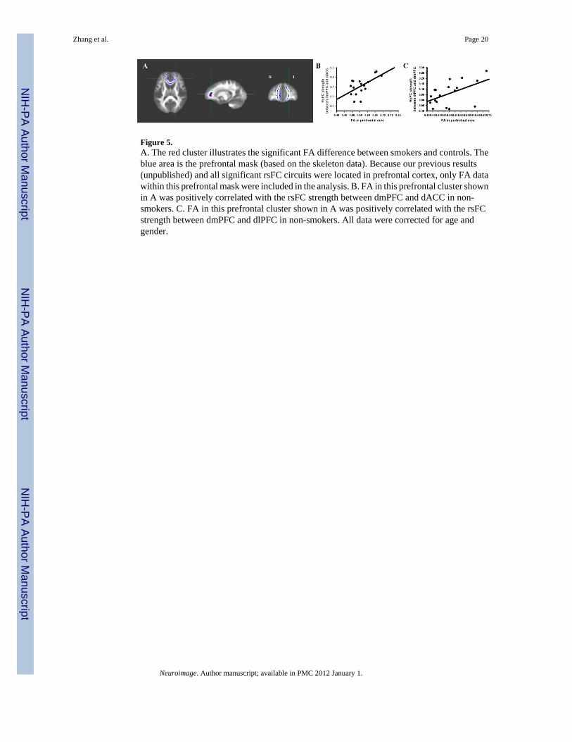
Figure 5.A. The red cluster illustrates the significant FA difference between smokers and controls. Theblue area is the prefrontal mask (based on the skeleton data). Because our previous results(unpublished) and all significant rsFC circuits were located in prefrontal cortex, only FA datawithin this prefrontal mask were included in the analysis. B. FA in this prefrontal cluster shownin A was positively correlated with the rsFC strength between dmPFC and dACC in non-smokers. C. FA in this prefrontal cluster shown in A was positively correlated with the rsFCstrength between dmPFC and dlPFC in non-smokers. All data were corrected for age andgender.
Zhang et al. Page 20
Neuroimage. Author manuscript; available in PMC 2012 January 1.
NIH
-PA Author Manuscript
NIH
-PA Author Manuscript
NIH
-PA Author Manuscript
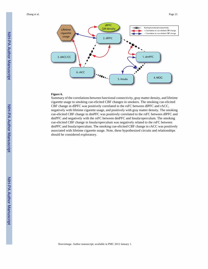
Figure 6.Summary of the correlations between functional connectivity, gray matter density, and lifetimecigarette usage to smoking cue-elicited CBF changes in smokers. The smoking cue-elicitedCBF change in dlPFC was positively correlated to the rsFC between dlPFC and rACC,negatively with lifetime cigarette usage, and positively with gray matter density. The smokingcue-elicited CBF change in dmPFC was positively correlated to the rsFC between dlPFC anddmPFC and negatively with the rsFC between dmPFC and Insula/operculum. The smokingcue-elicited CBF change in Insula/operculum was negatively related to the rsFC betweendmPFC and Insula/operculum. The smoking cue-elicited CBF change in rACC was positivelyassociated with lifetime cigarette usage. Note, these hypothesized circuits and relationshipsshould be considered exploratory.
Zhang et al. Page 21
Neuroimage. Author manuscript; available in PMC 2012 January 1.
NIH
-PA Author Manuscript
NIH
-PA Author Manuscript
NIH
-PA Author Manuscript

NIH
-PA Author Manuscript
NIH
-PA Author Manuscript
NIH
-PA Author Manuscript
Zhang et al. Page 22
Tabl
e 1
Bra
in re
gion
s sho
win
g si
gnifi
cant
inte
ract
ion
betw
een
grou
p (s
mok
ers v
s. co
ntro
ls) a
nd c
ue ty
pe (s
mok
ing
vs. n
eutra
l)
RO
IB
rain
reg
ion
Vol
ume
(mm
3 )T
alai
rach
coo
rdin
ate
(mm
)
corr
ecte
d P
xy
z
dmPF
C1
Bila
tera
l dor
sal m
edia
l pre
fron
tal c
orte
x41
663.
941
.239
.7<0
.001
dlPF
C1
Rig
ht m
iddl
e/su
perio
r pre
fron
tal g
yrus
(dor
sal l
ater
al p
refr
onta
l cor
tex)
3954
24.0
29.4
34.1
<0.0
01
dAC
C/C
CB
ilate
ral d
orsa
l ant
erio
r cin
gula
te c
orte
x47
620.
3−9
.141
.7<0
.001
MO
GR
ight
mid
dle
occi
pita
l gyr
us19
5747
.5−7
6.7
−0.3
0.00
3
I/OIn
sula
;/lef
t sup
erio
r tem
pora
l gyr
us/
1644
−55.
8−2
6.3
18.5
0.01
0
rAC
CB
ilate
ral r
ostra
l ant
erio
r cin
gula
te c
orte
x12
394.
141
.1−5
.70.
046
1 Thes
e cl
uste
rs a
ppea
red
conn
ecte
d at
unc
orre
cted
p=0
.005
and
wer
e se
para
ted
at u
ncor
rect
ed p
=.00
1 on
the
activ
atio
n m
ap b
ased
on
stru
ctur
al in
depe
nden
ce (s
ee su
pple
men
tary
mat
eria
ls).
Neuroimage. Author manuscript; available in PMC 2012 January 1.

NIH
-PA Author Manuscript
NIH
-PA Author Manuscript
NIH
-PA Author Manuscript
Zhang et al. Page 23
Tabl
e 2
Cue
-elic
ited
CB
F (m
l/100
g/m
in) a
cros
s gro
ups a
nd c
ondi
tions
RO
I
Smok
erC
ontr
olSm
okin
g vs
neu
tral
cue
in sm
oker
s
Cor
rela
tion
with
life
time c
igar
ette
usa
gein
smok
ers
Cor
rela
tion
with
FT
ND
in sm
oker
s
Smok
ing
cue
Neu
tral
cue
Smok
ing
cue
Neu
tral
cue
mea
n±SD
mea
n±SD
mea
n±SD
mea
n±SD
tp
Rp
rp
dmPF
C44
.7±1
4.9
38.7
±13.
639
.9±1
7.3
45.2
±15.
56.
15<0
.001
−0.0
80.
725
0.16
0.49
4
dlPF
C29
.1±9
.025
.1±7
.926
.1±1
0.8
33.6
±10.
66.
29<0
.001
−0.4
60.
042
−0.1
70.
477
dAC
C/C
C49
.5±1
5.9
42.8
±17.
252
.1±2
0.2
58.7
±17.
06.
27<0
.001
0.18
0.44
9−0
.09
0.71
3
MO
G45
.1±1
7.8
40.9
±18.
949
.2±3
0.2
55.4
±30.
25.
11<0
.001
−0.0
30.
991
−0.2
70.
255
I/O42
.0±1
8.8
36.4
±18.
851
.2±2
8.5
58.3
±28.
15.
35<0
.001
0.26
0.26
9−0
.02
0.93
9
rAC
C56
.2±1
6.8
46.0
±18.
159
.6±2
6.7
64.0
±25.
55.
14<0
.001
0.53
0.01
70.
080.
754
Neuroimage. Author manuscript; available in PMC 2012 January 1.

NIH
-PA Author Manuscript
NIH
-PA Author Manuscript
NIH
-PA Author Manuscript
Zhang et al. Page 24
Tabl
e 3
Res
ting
CB
F (m
l/100
g/m
in) a
nd c
orre
latio
ns w
ith sm
okin
g cu
e-el
icite
d C
BF,
life
time
ciga
rette
usa
ge a
nd F
TND
RO
IC
ontr
olSm
oker
Gro
up t-
test
Cor
rela
tion
to sm
okin
g cu
e-el
icite
d C
BF
insm
oker
sC
orre
latio
n w
ith li
fetim
e ci
gare
tte u
sage
insm
oker
sC
orre
latio
n to
FT
ND
in sm
oker
s
mea
n±SD
mea
n±SD
tp
rp
rp
rP
dmPF
C44
.1±1
5.9
41.2
±13.
6−0
.63
0.53
00.
270.
245
0.02
0.94
40.
240.
314
dlPF
C33
.0±1
2.2
27.0
±8.3
−1.8
80.
067
0.35
0.13
60.
240.
311
0.17
0.47
3
dAC
C/C
C57
.8±1
8.1
46.7
±16.
2−2
.15
0.03
7−0
.25
0.29
7−0
.09
0.71
00.
190.
421
MO
G50
.1±2
8.7
40.6
±18.
6−1
.31
0.20
0−0
.19
0.41
50.
300.
197
0.39
0.09
1
I/O57
.3±2
6.8
38.4
±16.
2−2
.83
0.00
7−0
.24
0.32
0−0
.16
0.50
50.
140.
554
rAC
C68
.7±2
2.5
52.7
±14.
8−2
.79
0.00
8−0
.25
0.27
9−0
.26
0.27
00.
040.
884
Neuroimage. Author manuscript; available in PMC 2012 January 1.

NIH
-PA Author Manuscript
NIH
-PA Author Manuscript
NIH
-PA Author Manuscript
Zhang et al. Page 25
Tabl
e 4
Gra
y m
atte
r den
sity
and
cor
rela
tions
with
smok
ing
cue-
elic
ited
CB
F, li
fetim
e ci
gare
tte u
sage
and
FTN
D
RO
IC
ontr
olSm
oker
Gro
up t-
test
Cor
rela
tion
to sm
okin
g cu
e-el
icite
d C
BF
insm
oker
sC
orre
latio
n w
ith li
fetim
e ci
gare
tte u
sage
insm
oker
sC
orre
latio
n to
FT
ND
in sm
oker
s
mea
n±SD
mea
n±SD
tp
rP
rp
rp
dmPF
C0.
598±
0.09
60.
571±
0.06
1−1
.09
0.28
20.
030.
916
−0.1
40.
550
0.08
0.75
4
dlPF
C0.
430±
0.08
50.
364±
0.08
3−2
.62
0.01
20.
540.
013
−0.4
30.
060
−0.0
60.
797
dAC
C/C
C0.
631±
0.07
20.
673±
0.10
41.
570.
125
−0.0
60.
796
0.35
0.12
70.
440.
050
MO
G0.
579±
0.13
00.
575±
0.09
6−0
.11
0.91
20.
120.
626
0.11
0.65
50.
120.
604
I/O0.
597±
0.11
30.
533±
0.09
0−2
.07
0.04
5−0
.39
0.09
0−0
.25
0.28
90.
220.
346
rAC
C0.
652±
0.13
20.
585±
0.10
3−1
.87
0.06
90.
230.
323
−0.0
30.
903
0.23
0.32
0
Neuroimage. Author manuscript; available in PMC 2012 January 1.

NIH
-PA Author Manuscript
NIH
-PA Author Manuscript
NIH
-PA Author Manuscript
Zhang et al. Page 26
Tabl
e 5
Ave
rage
d rs
FC z
scor
es b
etw
een
RO
I pai
rs a
nd c
orre
latio
ns w
ith sm
okin
g cu
e-el
icite
d C
BF,
life
time
ciga
rette
usa
ge a
nd F
TND
RO
I to
RO
ISm
oker
Con
trol
Gro
up t-
test
Cor
rela
tion
with
smok
ing
cue-
elic
ited
CB
F ch
ange
s in
smok
ers
Cor
rela
tion
with
life
time
ciga
rette
usa
ge in
smok
ers
Cor
rela
tion
to F
TN
D in
smok
ers
Firs
t RO
ISe
cond
RO
I
mea
n±SD
mea
n±SD
tp
rp
rp
rP
rp
dmPF
C to
dlP
FC0.
158±
0.18
30.
060±
0.11
31.
920.
063
0.62
0.01
00.
080.
780
0.09
0.73
40.
220.
406
dmPF
C to
dA
CC
/CC
0.09
8±0.
161
0.11
0±0.
096
−0.2
80.
784
−0.1
50.
574
0.41
0.11
70.
020.
947
0.07
0.81
0
dmPF
C to
MO
G−0
.005
±0.0
85−0
.007
±0.0
90−0
.40
0.69
4−0
.36
0.16
90.
050.
860
0.13
0.64
50.
220.
412
dmPF
C to
I/O
0.04
8±0.
123
0.04
7±0.
094
0.03
0.97
7−0
.55
0.02
7−0
.55
0.02
60.
110.
696
0.11
0.69
0
dmPF
C to
rAC
C/C
C0.
102±
0.13
10.
099±
0.07
30.
070.
943
0.03
0.92
50.
070.
809
−0.1
00.
727
−0.4
00.
124
dlPF
C to
dA
CC
/CC
0.03
2±0.
162
0.03
0±0.
095
0.05
0.96
00.
100.
726
−0.1
00.
714
−0.2
50.
353
0.26
0.32
7
dlPF
C to
MO
G−0
.021
±0.1
00−0
.002
±0.0
66−0
.67
0.51
2−0
.24
0.37
10.
360.
170
−0.3
00.
268
−0.1
30.
645
dlPF
C to
I/O
−0.0
10±0
.105
−0.0
01±0
.074
−0.2
90.
774
0.11
0.69
6−0
.13
0.64
3−0
.20
0.46
10.
230.
387
dlPF
C to
rAC
C0.
025±
0.12
60.
008±
0.11
70.
420.
680
0.64
0.00
70.
040.
888
−0.2
70.
319
0.23
0.38
4
dAC
C/C
C to
MO
G0.
018±
0.10
50.
029±
0.05
3−0
.41
0.68
50.
250.
344
−0.1
70.
522
0.30
0.25
80.
060.
829
dAC
C/C
C to
I/O
0.16
5±0.
115
0.21
8±0.
114
−1.4
00.
171
0.41
0.11
5−0
.10
0.72
10.
290.
283
0.04
0.87
2
dAC
C to
rAC
C0.
075±
0.11
00.
067±
0.09
10.
260.
800
0.39
0.98
90.
060.
834
−0.2
80.
292
−0.1
10.
696
MO
G to
I/O
0.02
2±0.
090
0.03
9±0.
102
−0.5
30.
599
−0.1
40.
602
−0.0
10.
984
0.20
0.46
1−0
.13
0.64
4
MO
Gto
rAC
C−0
.041
±0.0
78−0
.003
±0.0
49−1
.75
0.09
00.
330.
209
−0.3
10.
247
−0.2
60.
323
−0.0
50.
852
I/O to
rAC
C−0
.005
±0.1
030.
030±
0.08
6−1
.11
0.27
40.
110.
679
0.49
0.05
60.
290.
270
0.29
0.26
9
Neuroimage. Author manuscript; available in PMC 2012 January 1.

NIH
-PA Author Manuscript
NIH
-PA Author Manuscript
NIH
-PA Author Manuscript
Zhang et al. Page 27
Table 6
Correlation between FA in the frontal cluster and rsFC strengths between frontal ROIs
Control Smoker
r p r p
dmPFC to rACC 0.16 0.561 0.42 0.102
dlPFC to rACC −0.25 0.349 −0.20 0.453
dACC/CC to rACC 0.45 0.078 0.13 0.643
dmPFC to dlPFC 0.53 0.036 0.39 0.134
dmPFC to dACC/CC 0.59 0.015 −0.11 0.686
dlPFC to dACC/CC 0.01 0.962 −0.31 0.237
Neuroimage. Author manuscript; available in PMC 2012 January 1.