Analysis of xer site-specific recombination in vivo A thesis ...
239
Analysis of xer site-specific recombination in vivo A thesis submitted for the degree of Doctor of Philosophy at the University of Glasgow by Richard McCulloch Institute of Genetics University of Glasgow Church Street Glasgow December 1992
-
Upload
khangminh22 -
Category
Documents
-
view
1 -
download
0
Transcript of Analysis of xer site-specific recombination in vivo A thesis ...

Analysis of xer site-specific recombination in vivo
A thesis submitted for
the degree of
Doctor of Philosophy
at the University of Glasgow
by
Richard McCulloch
Institute of Genetics
University of Glasgow
Church Street
Glasgow
December 1992

ProQuest Number: 11007935
All rights reserved
INFORMATION TO ALL USERS The quality of this reproduction is dependent upon the quality of the copy submitted.
In the unlikely event that the author did not send a com p le te manuscript and there are missing pages, these will be noted. Also, if material had to be removed,
a note will indicate the deletion.
uestProQuest 11007935
Published by ProQuest LLC(2018). Copyright of the Dissertation is held by the Author.
All rights reserved.This work is protected against unauthorized copying under Title 17, United States C ode
Microform Edition © ProQuest LLC.
ProQuest LLC.789 East Eisenhower Parkway
P.O. Box 1346 Ann Arbor, Ml 48106- 1346

QLf OOU l s n l S ^ )
GLASGOWUNIVERSITYLIBRARY

Dedicated to my mum and dad.

The research reported in this thesis is my own
and original work except where otherwise stated
and has not been submitted for any other degree.

CONTENTS
Abbreviations i
Acknowledgements ii
Summary iii
Chapter 1: Introduction 1
Chapter 2:- Materials and methods 23
Chapter 3 Analysis of the role of aminopeptidase A in cer site-specific
recombination by site-directed mutagenesis 33
3.1 Introduction 34
3.2 Site-directed mutagenesis of pepA 37
3.2 Determination of the peptidase activity of PepA E354A in vitro 41
3.3 Determination of the Xer phenotype of PepA E354A in vivo 44
3.4 Determination of the peptidase activity of PepA E354A in vivo 45
3.5 Complementation in CSX 17 using pRM21 48
3.6 Discussion 50
Chapter 4 Analysis of cer site-specific recombination in vivo, using
the controllable strain RM40 54 -
4.1 Introduction 55
4.2 Construction of strains RM10, RM20, RM30 and RM40 57
4.3 Controllable in vivo recombination in strains RM10, 20, 30 and 40 60
4.4 Analysis of pSD 115 recombination in RM40 61
4.5 Electrophoretic analysis of putative Holliday junctions 64
4.6 The putative Holliday junctions are substrates for cleavage
by RuvC in vitro 66
4.7 Electron microscopy of isolated % structures 69
4.8 The nature of the first pair of strand exchanges 71
4.9 Construction of RM50, and in vivo recombination in this strain 75
4.10 Controlled in vivo recombination using the plasmid pRM60 77
4.11 Do pKS455 and pCS202 make stable Holliday junctions ? 80

4.12 Discussion 83
Chapter 5: Further characterisation of xer site-specific recombination in vivo 90
5.1 Introduction 91
5.2 cer recombination in argR~ and pepA" derivatives of RM40 92
5.3 Site-directed mutational analysis of the putative cer overlap sequence
(A) Determining whether the-GGG motif in the cer overlap is the
cause of Holliday junction accumulation 96
(B) Determining whether overlap sequence heterologies affect the
efficiency of cer recombination 99
5.4 Are the stable Holliday junctions protein-bound in vivo ? 104
5.5 The effect of RuvC on cer recombination 109
5.6 Comparing cer, type H hybrid and d/f recombination in RM40 113
5.7 The effect of mutations in dapFt orf235 and orf238
on ^/recombination 115
5.8 pSD126 recombination controlled by pRM60 117
5.9 Comparison of pSD126 and pSD124 recombination in RM40 118
5.10 Genetic characterisation of a fourth xer gene: xprB 119
5.11 Analysis of cer and di/recombination in RM40/pRM 135 124
5.12 Discussion 127
Chapter 6: Concluding remarks 134
Bibliography 140

ABBREVIATIONS
Chemicals
ATP adenosine triphosphate
CTP cytidine triphosphate
DNA 2' deoxyribonucleic acid
dNTP 2' deoxy (nucleotide)
EDTA ethylene diamine tetra-acetic acid (disodium salt)
IPTG isopropyl B-D-thiogalactoside
RNA ribonucleic acid
SDS sodium dodecyl sulphate
Tris tris (hydroxymethyl) amino ethane
Antibiotics
Ap ampicillin
Cm chloramphenicol
Km kanamycin
Tet tetracycline
Measurements
bp base pair
kbp kilobase pair (10^ bp)
kDa kilodalton (10^ dalton)
min minute
Miscellaneous
UV ultra violet light
DNAase deoxyribonuclease

ACKNOWLEDGMENTS
Firstly I thank Dave for all his help and encouragement during this thesis, and Mary
for her patient advice and for showing where and what everything is in the lab. Thanks also
to the "media ladies" for glassware, solutions and cakes and to Margaret, Linda and Diane.
Thanks to S.West and L.Coggins for the experiments they performed and helped me
perform. Also, thanks to S.Lovett, R.Lloyd and G.Sharpies for their kind gifts of strains
and plasmids.
Special thanks go to Marshall for both reading this thesis and being kind enough to
require only a few drinks in return for this horrendous experience. Thanks to everyone else
in my lab for making the last three years enjoyable in work and, more importantly, outside
of work - Dave, Jen, Gerhard, Stephen (thanks for 5 years in this case, and plenty of
tequila during it), Garry, Nick, Martin, Sally, Shahnaz, Angela and the old cer workers
(Sean and George). Important thanks go to the following groups of people: the football
team, the multisport team (not unsurprisingly for the drinking rather than the sport) and all
other friends I have spent productive hours in Tennants with. Thanks to C.Spence for the
kind use of his computer (games), and Gillian and Andy for supplying me with t-shirts and
loans.
The most important thank-you goes to Heather for all the times we have spent
together, for magnificent "gourmet" Mexican rice and extra-curricular mambo.

SUMMARY
The xer site-specific recombination system is employed in two related biological
processes. The first of these roles is in resolving multimers of high copy number,
randomly partitioned plasmids in order to ensure their heritable stability. Plasmid multimers
arise through homologous recombination. Their resolution to monomers by xer site-
specific recombination requires a recombination locus named cer that was originally
described in the naturally occurring plasmid ColEl, although similar sites have been
isolated in many other plasmids (Summers and Sherratt, 1984; Summers et al, 1985). The
second role of the xer system is in bacterial chromosome partition. A locus has been
described, named dif, in the terminus region of the Escherichia coli chromosome that is a
substrate for xer catalysed site-specific recombination (Blakely etal, 1991; Kuempel et al,
1991). It is believed xer recombination activity is required at d if because dimers of
replicating chromosomes are produced by homologous recombination; these dimers cannot
be partitioned into daughter cells as the host cell divides, and hence xer recombination
converts the chromosomal dimers into monomers and allows cell division.
The proteins that act in xer site-specific recombination are encoded by the E.coli
chromosome. At the start of this work three genes (xer genes) had been shown to be
absolutely required for cer recombination, and one for dif recombination. xerC has been
cloned and sequenced and is required for both cer and dif recombination (Colloms et al,
1990; Blakely et al, 1991). The protein encoded by xerC binds to both recombination sites
in vitro and has sequence homologies to the X integrase family of site-specific
recombinases. xer A and xerB encode the proteins ArgR and aminopeptidase A (PepA)
respectively; both are essential for cer recombination and are believed to be "accessory
factors" in the reaction (Stirling et al, 1988 and 1989). While this work was being
performed a fourth xer gene was identified, called xprB. Genetic evidence is presented that

the XprB is essential for both cer and d if recombination and that it shows amino acid
sequence homologies to the X integrase family of recombinases.
PepA is an amino-terminal exopeptidase whose role in the cer recombination reaction
was not understood at the start of this work. Site-directed mutagenesis of the gene
encoding PepA is described, and evidence presented that the mutant enzyme (named
E354A) encoded by the altered pepA gene lacks any aminopeptidase activity both in vitro
and in vivo but is still able to support cer recombination. The relevance of this observation
to how PepA is employed by the xer site-specific recombination reaction is discussed.
The construction of an controllable in vivo recombination system and its use in
analysing the mechanism of the xer recombination reaction is described. The system
comprises a derivative of E.coli K12, named RM40, in which the expression of xerC is
controlled by the lac promoter and operator sequences rather than its natural promoter
sequences. Analysing the products derived from c^r-mediated recombination of reporter
plasmids demonstrated the existence of Holliday junction structures produced during the
reaction. These putative recombination intermediates were shown to have arisen by the
exchange of a specific pair of strands within the cer sites of the substrate DNA molecule
used. These results suggest that cer site-specific recombination involves the same form of
strand exchange mechanism that has been described in vitro for other members of the X
integrase family (Int, FLP and Cre; Staik et al, 1992). The Holliday junctions were isolated
in anomalously large quantities when compared to the isolation of such reaction
intermediates in other, related systems. The possible relevance of this to the xer
recombination mechanism was analysed by comparing cer and recombination in RM40,
and by altering the cer substrate molecules and reaction conditions employed.

Chapter 1
In troduction
1
1.1 General introduction
Site-specific recombination is a controlled process found in both eukaryotes and
prokaryotes which generates precise rearrangements of DNA molecules at defined
positions. The rearrangements are catalysed by a variety of proteins which are
collectively termed "recombinases", and are achieved by cutting the DNA molecules at
specific points and joining the ends to new DNA partners. Site-specific recombination
is distinguished from homologous recombination in a number of ways. In homologous
recombination the recombining DNA segments recognise their partners by comparing
the DNA sequences through Watson-Crick base-pairing between the partner duplexes
(a process which is mediated by RecA-like proteins), and therefore extensive homology
is required between the two DNA molecules. In contrast, there is no requirement for
extensive homology between DNA molecules during site-specific recombination
because the recombination sites are brought together primarily through interactions
between DNA binding proteins. Moreover, no base-pairing between the two
recombining DNA duplexes is required before strand cleavage in site-specific
recombination. The two processes are also distinguished by the fact that the proteins
involved in the reactions are unrelated, and site-specific recombination is probably a
more regulated process.
Site-specific recombination is related to another type of non-homologous
recombination process called transposition. These processes differ in their biological
and genetic consequences and in their reaction mechanisms. In site-specific
recombination both recombining molecules are cleaved at two precise positions within a
very short region of homology, and there is no synthesis or degradation of DNA during
the strand exchange reaction (which in both processes involves a series of precise
catalytic steps). In transposition there is no requirement for homology in the
recombination sites and DNA synthesis is utilised during the strand breakage and
reunion steps. During replicative transposition the entire transposing DNA segment is
replicated, whereas in conservative (or "cut and paste") transposition the DNA
2

synthesis involves only a few bases of sequence. Reviews of transposition have been
published by Craig and Kleckner (1987) and Derbyshire and Grindley (1987) and it
will not be considered further here.
1.2 Consequences of site-specific recombination
Site-specific recombination alters the structure of DNA molecules in a number of
ways which depend on the organisation of the recombination sites (see fig. 1.1).
Recombination between sites on different molecules is possible, and this causes the two
substrate molecules (which could, for instance, be plasmids) to become fused; site-
specific recombination of this sort is termed "intermolecular". Intramolecular
recombination (between sites on the same molecule) is also possible, and has two
possible outcomes depending on the relative orientations of the recombination sites.
When the sites are in direct repeat in a circular substrate molecule, site-specific
recombination results in deletion of the DNA intervening between the sites and
generates two smaller product molecules; this reaction is termed "resolution" in this
thesis, but has also been called "excision" in other systems (see below). In contrast,
when recombination sites are in inverted repeat relative to each other, recombination
causes the DNA sequences between the sites to invert in relation to the rest of the
molecule. These reaction outcomes are a consequence of the fact that site-specific
recombination sites are asymmetric, and the asymmetry is retained during
recombination such that the left hand side of one site is always fused to right hand side
of the other site, and vice versa. It should be noted, however, that in some
circumstances this asymmetry can be overcome (see, e.g., Hoess et al, 1986).
Although the intramolecular reaction products were described above as being
simple circles, it has been shown that topologically more complex products can be
created during site-specific recombination reactions in vitro. This is illustrated, as an
example, in Figure 1.1 by showing that catenated products can be generated during
resolution reactions. Analysis of the topological nature of these, and other, in vitro
3

o
Figure 1.1. Physical consequences of site-specific recom bination.Recombination sites are shown as arrows whose direction indicates the site's orientation. A Intermolecular recombination results in the fusion of two substrate DNA circles. B Intramolecular recombination in a circular substrate DNA molecule
containing directly repeated sites results in two product circles; this is termed resolution in this thesis. C Intramolecular recombination of a substrate containing sites in inverted orientation results in inversion of the DNA between the sites. D Site-specific recombination in vitro in a supercoiled substrate molecule can generate topologically complex products; in this case resolution generates a multiply linked catenane because substrate supercoils were trapped during the reaction. (Adapted from Craig, 1988).
products has allowed information to be derived regarding the detailed mechanisms of
various site-specific recombination reactions (this is discussed below).
The biological consequences of site-specific recombination are very diverse and
incorporate the structural considerations described above. This is illustrated in the
following examples:
(i) Integration and excision of temperate bacteriophages into and out of the host
organism's chromosome. Integration of the phage generates a prophage, in which the
phage genome becomes part of the host genome and is replicated by the host
machinery. Excision is required to release the phage and allow it to spread horizontally,
by infection, into other cells. Bacteriophage X is notable because it was here that site-
specific recombination was first described (reviewed by Landy, 1989), but other
phages also employ this strategy (e.g. 080 and P22; Leong et al, 1985).
(ii) Resolution of cointegrate intermediates during replicative, inter-replicon
transposition of type II transposons. These Tn3-like transposons move by a replicative
mechanism during which the donor and recipient replicons become fused. Resolution
of these cointegrate structures is achieved by site-specific recombination of directly
repeated transposon-borne res sites, and is catalysed by transposon-encoded
recombinases called resolvases (Sherratt, 1989; Stark et al, 1989).
(iii) Control of gene expression by "switching" of DNA segments. The Hin, Gin,
Cin and Pin systems encode a family of DNA invertase recombinases that execute
inversions of DNA segments (either containing genes or the promoter sequences of
genes), and thereby mediate the variation of flagellar antigens in Salm onella
typhimurium and tail fibre proteins in bacteriophages Mu, PI and relatives (reviewed
by Glasgow et al, 1989). fimB and fimE encode Escherichia coli recombinases that are
unrelated to the DNA invertases which catalyse the inversion of a small chromosomal
region containing the promoter for the gene fimA, and therefore alter the fimbriation
state of the cell (Klemm, 1986).
(iv) Stabilisation of low copy number plasmids by multimer resolution.
Bacteriophage PI does not integrate into the host's chromosome during lysogeny, but
4

instead exists as a low copy number circular plasmid. The phage-encoded recombinase
Cre is required to resolve dimers of PI which arise through homologous recombination
to ensure that the lysogenic phage is efficiently segregated into daughter cells during
host division (reviewed by Sadowski, 1986). Similar systems are employed in other
low copy number plasmids (e.g. the plasmid R46 also encodes a recombinase; see
Dodd and Bennett, 1987).
(v) FLP is a recombinase encoded by the 2-micron plasmid found in
Saccharomyces cerevisiae which acts on plasmid-borne FRT sites to cause inversion of
a large DNA segment and promote amplification of the plasmid to high copy numbers
in the cell (Cox, 1989).
1.3 General reaction mechanisms in site-specific recombination
A number of site-specific recombination sytems have been reconstituted in vitro,
using either crude cell extracts or purified proteins, simple buffer solutions and defined
substrate molecules. The reactions that have been established in this way include those
catalysed by X Integrase (Int; Nash, 1975), yeast FLP (Vetter et al, 1983),
bacteriophage PI Cre (Abremski et al, 1983) and some of the resolvases and DNA
invertases (Hatfull and Grindley, 1988). Analysing the reactions in vitro has shown
that the basic reaction mechanisms of the different systems (described above) are
similar, but the detailed biochemistry of the reactions can be divided into two classes;
the integrase-like and the resolvase/invertase class of reactions (see below). The
distinction between the reaction mechanisms is reflected in the fact that all recombinases
can be assigned as being part of one of the two classes because the proteins are
homologous within the two classes (see below).
The various recombination sites in the different systems share little sequence
homoloj^ , and in fact the two recombining sites in a given system can be different in
primary sequence. However, in all site-specific recombination reactions a small region
5

of sequence identity between the recombination sites is essential. This is where the
strand exchanges that occur during the recombination reaction are found. The parental
DNA molecules are broken at specific, fixed positions within these regions and the
recombinational strand exchanges occur by joining the broken ends of one parental
duplex to the ends of the other broken parental duplex. Recombinant DNA molecules
generated in this way contain no gaps or nicks. The breakage positions in the top and
bottom strands of both parental duplexes are staggered (see figures 1.3 and 1.7), and
consequently the recombinant DNA molecules contain some heteroduplex DNA. The
size of this stagger varies from 2-8 bp, and is one of the features that distinguish the
two classes of site-specific recombination reaction (see below).
The strand exchange mechanism of all site-specific recombination reactions
involves a transient, covalent linkage between the substrate DNA and recombinase
proteins (see figs. 1.3 and 1.7). Correlating with this is the fact that in all
recombination sites the recombinases bind to DNA sequences surrounding the points of
strand exchange. The DNA sequence which encompasses the recombinase binding sites
and staggered cleavage positions is termed the "crossover" region or "core"
recombination site. The form that the covalently linked protein-DNA intermediate takes
is different in the two classes of recombination reaction and is conserved within each
class (see below). The function of this intermediate is to conserve the energy of the
substrate DNA molecule's phosphodiester backbone, and hence no site-specific
recombination reactions utilise high energy co-factors.
In some site-specific recombination systems the crossover region contains all the
sequence information that is required in the reaction (e.g. the loxP site of Cre, the FRT
site of FLP and the attB site of phage X; below). In other cases, the recombination sites
have a more more elaborate architecture, and can employ additional proteins and protein
binding sites (e.g. in X, the attP> attL and attR sites contain binding sequences for the
proteins IHF, Xis and Fis). In all cases, however, the catalytic steps of cutting,
exchanging and ligating the DNA are mediated by the recombinases, and the other so-
called "accessory factors" have various supporting roles in the reactions.
6

Current understanding of the biochemical details of resolvase/DNA invertase and
X integrase-like site-specific recombination reactions, and the functions of the site-
specific recombination proteins, are described separately below. It should be noted,
however, that these families cannot be separated in a biological context, since similar
reactions are performed by members of both classes.
1.4 The resolvase/D N A invertase class o f s ite -sp ec ific
recombination system
As suggested by the name, this family is divided into two sub-families on the
basis of the type of reaction that is executed, despite the fact that they apparently share
the same reaction mechanism. The resolvases catalyse resolution reactions and are
primarily involved in recombining cointegrate structures (described above), although
they have also been described in plasmid-stabilising monomerisation (e.g. R46; Dodd
and Bennett, 1987). The resolvases encoded by the transposons Tn3 and y5 are the best
studied in vitro (although resolvases from other transposons have also been described,
e.g. Tn21, Tn552 and Tnl721; reviewed by Hatfull and Grindley, 1988; Stark et al,
1989 and Sherratt, 1989). The functions of the invertases Gin, Hin, Cin and Pin have
been discussed (Section 1.2) and the sites of all these systems are interchangeable
(Glasgow et al, 1989). Recombination of Gin and Hin have been extensively analysed
in vitro (Kahmann et al, 1985; Bruist et al, 1987).
These recombinases are small proteins of highly conserved size (all approximately
180 amino acids) and sequence (see fig. 1.2). The proteins appear to be comprised of
two structural domains that can be separated by proteolytic digestion; a C-terminal
domain of approximately 40 amino acids that is mainly responsible for sequence
specific DNA binding (Abdel-Meguid et al, 1984), and an N-terminal domain (whose
X-ray crystallographic structure has been solved; Sanderson et al, 1990) that contains
the catalytic site and is responsible for protein-protein interactions.
7

active site serine
Th3 H R I F G Y A R V S T S O O S L D I C I R A L K D A G _ _ V K A N R I F T D K A S C ss _ - T D R E C L D L L R M K V E E C D V I L V K K L D R L G RThlOOO H R L F G Y A R V S T S O Q S L D I C V R A L K D A G - - VK A N R I F T D K A S C ss - - S D R K G L D L L R H K V E E G D V I L V K X L D R L G RRMS H R L F G Y A R V S T S Q Q S L D I c I K C L K E AG - - V K A S R I F T D K A S C ss - - T D R K G L D L L R H K V E E G D V T L V K K L D R L G RTh21 H T G Q R I - G Y I R V S T F D Q M P E R Q ------ - L E G - - V K V D R A F S D K A S C K D - - VK R P Q L E A L I S F A R T G D T V W H S H D R L A RTH501 H O G H R I - G Y V R V S S F D O N P E R C - L E O - - T Q V S K V F T D K A S C K D - - T O R P Q L E A L L S F V R E G D T V W H S H D R L A RTh2501 H S R V F A Y C R V S T L E Q T T E N 0 R R E I E A A G F A I R P Q R L I E E H I S C S V A A S E R P G F I R L L D R H E N G D V L I V T K L D R L G Rp IF O ) H L V - G Y A R V S T E E O S L N R 0 I D F L V D Y G - - V D K R N I Y 0 E K I S C H K - - P N R E Q L D K H I D E L Q E G D T V I I T D L T R I S RTH917 H - I F G Y A R V S T D D Q N L S L Q I D A L T H T G - - I - - D K L F 0 E L V T C A L - - L D R P Q L E E H I N L L R E G D S W I Y K L D R I S RHi n H A T I - G Y I R V S T I D Q N I D L 0 R N A L T S AN - - — C D R I F E D R I S C K I - - A N R P G L K R A L K Y V N K G D T L V V W K L D R L G RGi n H L I - G Y V R V S T N D Q N T D L C R N A L V C A G - - - - C E 0 I F E D K L S G T R - - T D R P G L K R A L K R L Q K G D T L V V W K L D R L G RPi n H L I - G Y V R V S T N D Q N T D L 0 R N A L N C A G - - — C E L I F E D K I S C T K - - S E R P G L K K L L R T L S A G D T L W W K L D R L G RCl n H L I - G Y V R V S T N E O N T A L 0 R N A L E S AG - - — C E L I F E D K A S C KK - - A E R P G L K K V L R H L S R G D T L W W K L D R L G R
Th3 D T A D H I C L IThlOOO D T A D H I Q L IRU6 D T A D H I 0 L TTh21 N L D D L R R I VTY601 N L D D L R R L VThSOI N A H D I R K T VpIPHCW S T K D L L N I ITh9T7 S T L H L I E L SHin S V K H L V A L IGin S H K H L I S L VPin S H R H L VV L VC ln S H R H L V V L V
K E F D A O K E F D A O K E F D A Q Q T L T Q R Q K L T O R E O L A S S D R I K A D E L F E E L S E L H E R C E L R E R E E L R E R E E L R D R
Th3 R T V — D R N V V L - T L H Q K ThlOQO R K I — D R D A V L - N H W Q C RM6 R I I - - O R N S V L - A L H O C Th21 K S L S S E R I A E L R O R V E A TJ601 K A L S D E Q A A T L R Q R A T A Th2501 S A L N E E O O L T V I A R I N A p I F O i S K R N - D K A D T V G L L Y R E Th9T7 S K G K L S I D L A L - K H Y D S t t t n R A I N K H E O E O I S R L L E K Gi n P K L T K A E W E Q A G R L L A C Pi n P K L T P E Q U A O A G R L I A A Cl n P K Y Q E E T W Q Q H R R L L E K Aero
A V R F S I R F A V R F H I E F R I E F R V H C S I K S H F I S H F H S N F R S N F R S N F R S
T G A T E L G A S H T G A T D E Q K T K E P K AC I S I S A Y K I V D E Y S I R H P R O C I P R K C T P R Q K I P R K C F G Q T K
G - - I S G - - I S G - - I S H - L S F G - L V F G G V D L T - W L D f l - . V D S - - I D S - - I D S - - I D S - - I D
I A H Q L S I S K T H K I A R R L S
A R E F C A R E F N
I A R E F F I V K Q T C 0 1 L D A S
A I I F C A L I Y D A I I Y D
VA I I Y D A K D L G V
D G D - - D G E - - D C E G E D S P G E D S P S A A G R S S D N P S T - - S S - - S A S - - S P S - - T P S - - T P
A R S A R S A R S S R E S R E T R Q S R A L K T C V S A L S C V S
T V Y T V Y T V Y T L Y T L Y T I L T V Y T F Y T L Y T L Y T L Y
G Q M V V TG K H V V TG K H V V TA N L H L SA N L M L ST H Q V IN S F L L TG R F F F RG R F F F HG R F F F HG R F F F HG R F F F H
I L S A V A I L S A V A I L S A V A V H G A F A V M G A F A — S A V A V H S G L S V H A S L A V H S A L A V H G A L A V H G A L A V H G A L A
E R T E R T E RT E R C E R C E R T Q R T E R T E R T E R T E R T E R T
R Q E R Q E R Q E I A L I T L I A R L K S L K A L A A L A A L E T L D A
K L K H AK K L K K Q R K Q R K A T K A R R V R R A O R N K R A Q R A E
I K F V V F I R F A Y R A Y R K R F R NG K K G R L G R I G R I G R I G
K I L E D E R A SK V I N E S NK I L E D E S R V N LO Y L R T D QQ Y L R T D DR V K A G Q Q S SR V L N D L K L KR Y L N K R Y AR Y F P A S S I K K R H NK K H P A K R A H I E N D D R I NK R F P A G D K
A V S l T L Y l K K F P A S S F O SY Q S A I N K A I H
h e lix -tu r n -h e lix
F igure 1.2. Amino acid sequence alignm ent of some m em bers of the resolvase/D N A invertase fam ily of site-specific recom binases. These proteins are believed to interact with their DNA binding sites via a helix-turn-helix motif, as shown. Also indicated is the proposed active site serine residue. (Taken from Sherratt, 1989).

The recombination site of Tn3 (called res) is approximately 120 bp in length and
contains 3 sub-sites (named I, II and III), each of which has dyad symmetry and is
believed to bind dimers of resolvase (Grindley et al, 1982; Kitts et al, 1983). Strand
exchange occurs exclusively at the central AT sequence of sub-site I, but II and III are
essential for recombination. All the different res sites have the same organisation,
although the size of the subsites and spacings between them vary considerably
(M.Boocock, pers.comm.). The different activities of resolvase when bound to the
different subsites is probably due to differences in the size and sequences of the sub
sites altering the nature of the protein-DNA interactions. Resolvases subunits bound at
sub-sites II and III might act as accessory factors that facilitate the formation of a
nucleoprotein synaptic complex in which the recombination reaction occurs (Stark et al,
1989; see fig. 1.4).
The recombination sites of the DNA invertases are 26 bp in length and, like res
sub-sites, have dyad symmetry and probably bind dimers of the recombinase (Mertens
et al, 1988). The strand exchange reactions occur at the central 2 bp of the sites (Klippel
et al, 1988). They are only efficiently recombined, however, if an enhancer sequence
(called sis), which binds the host protein Fis, is present in the same DNA substrate
molecule (Kahmann et al, 1985). Fis therefore represents an accessory factor that
possibly performs a similar function in the invertase reaction to resolvase at sub-sites II
and III during resolvase recombination (Bednarz et al, 1990; see fig. 1.4).
The recombination reactions catalysed by resolvases and DNA invertases are
mechanistically very similar. The top and bottom strand cuts are staggered by 2 bp and
create recessed 5' ends and overhanging 3'OHs (fig. 1.3). Putative intermediates have
been isolated using abnormal in vitro reaction conditions in which the substrate
molecules have been cut at all four cleavage positions, suggesting that the
recombination reactions proceed by double strand breaks, duplex rotation and ligation
(Klippel et al, 1988; Reed and Grindley, 1981). The isolated intermediates are
covalently linked at each broken 5' end to a resolvase/DNA invertase protein, and it is
believed that an absolutely conserved serine residue present near the N-terminus of the
8

F ig u re 1.3. Events a t the crossover site d u rin g resolvase/D N A invertase-cata lysed site-specific recom bination reactions. Recombinase subunits are depicted as shaded ovals; the ends of the crossover sites by inverted arrows; the two base pairs between the staggered cleavages (the overlap) as vertical lines; and the phosphates that are attacked by the recombinases as black diamonds. The four DNA strands (thick and thin lines) of the two crossover sites (differentiated by shading) are cleaved and covalently linked protein-DNA intermediates form. Recombination occurs by rotation of the left half-sites relative to the right half-sites and subsequent joining of the broken DNA ends to a new half site. (Adapted from Stark et al, 1992).
recomDirarion site ^ / ermancer
invertase
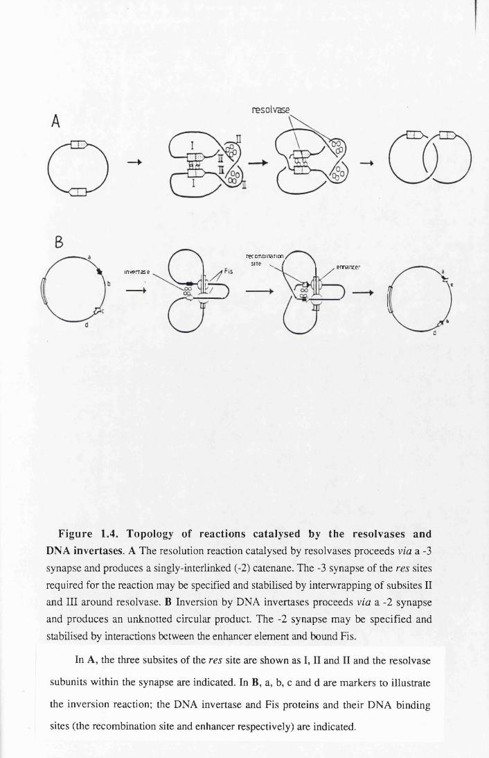
Figure 1.4. Topology of reactions catalysed by the resolvases and DNA invertases. A The resolution reaction catalysed by resolvases proceeds via a -3 synapse and produces a singly-interlinked (-2) catenane. The -3 synapse of the res sites required for the reaction may be specified and stabilised by interwrapping of subsites II and III around resolvase. B Inversion by DNA invertases proceeds via a -2 synapse and produces an unknotted circular product. The -2 synapse may be specified and stabilised by interactions between the enhancer element and bound Fis.
In A, the three subsites of the res site are shown as I, II and II and the resolvase
subunits within the synapse are indicated. In B, a, b, c and d are markers to illustrate
the inversion reaction; the DNA invertase and Fis proteins and their DNA binding
sites (the recombination site and enhancer respectively) are indicated.

proteins (fig. 1.2) is the catalytic residue involved in the phosphodiester linkage
(Klippel et al, 1988; Hatfull and Grindley, 1986).
Tn3 res sites are only efficiently recombined in vitro when they are present in
direct repeat on the same, supercoiled DNA molecule (Kitts et al, 1983). Similarly,
DNA invertase sites must also be present on the same, supercoiled substrate, but, in
contrast to res sites, they must be in inverted repeat (Kahmann et al, 1985). In addition
to this site orientation selectivity the resolvase/DNA invertase reactions in vitro have
topological selectivities (summarised in fig. 1-4 resolvase-catalysed deletion reactions
normally produce a specific (-2) catenane product and involve a linkage change of +4
(Wassermann and Cozzarelli, 1985; Boocock et al, 1987); DNA invertase-catalysed
inversion normally produces an unknotted circular product and has a +4 linkage change
(Kahmann et al, 1987; Kanaar et al, 1988). These results have suggested that the strand
exchange reactions only occur when the recombining sites form a specifically
interwrapped synaptic complex, and that the strand exchange is a simple right-handed
180° rotation of the cleaved DNA duplexes and subsequent ligation. The differences in
the two reactions' product topologies can be accounted for by the different synaptic
topologies required for strand exchange. Resolvase reactions occur via a -3 synapsis
which is thought to be stabilised by resolvase subunits bound to the interwrapped
accessory sites (Stark et al, 1989a and 1989b), whilst the inversion reactions occur via
a -2 synapse that is stabilised by Fis bound to the sis enhancer (Bruist et al, 1987;
Kanaar etal, 1989).
1.5 The X integrase class of site-specific recombination system
The second class of site-specific recombination reaction is called the X integrase
class because of the sequence homologies of the recombinase proteins to X Int. The
functions of some of these reactions have been discussed (X, Int, FimB/E, FLP and Cre;
see above), but many other X integrase-like recombinases have been described; e.g.
9

Tnpl of Tn4430 mediates cointegrate resolution (Mahillon and Lereclus, 1988) and the
D protein encoded by the E.coli F factor is involved in plasmid-stabilising
monomerisation (Lane et al, 1986 and O'Connor et al, 1986).
This family of recombinases has no sequence homologies with the
resolvase/DNA invertases and the proteins display much greater variation in size and
sequence. Two regions of amino acid similarity have been identified by comparing the
recombinase sequences, termed domains 1 and 2 (Argos et al, 1986; Abremski and
Hoess, 1992; see fig.5.17). Only four residues are completely conserved in all
published X integrases. The reactions catalysed by X Int, PI Cre and FLP are the only
ones to have been analysed in vitro, and therefore only they will be described in detail
(although it is likely that all the X integrase-like systems have a common basic reaction
mechanism).
The recombination sites of FLP and Cre (called FRT and loxP respectively) are
simpler than the att sites of X Int (see figs. 1.5 and 1.6). loxP comprises inverted 13 bp
Cre binding sites surrounding an 8 bp spacer sequence, and therefore it is simply a
crossover site (Hoess and Abremski, 1985). FRT also comprises two 13 bp inverted
recombinase binding sites surrounding an 8 bp spacer, but differs from loxP in that it
has an additional 13 bp FLP binding site that is not essential for recombination
(Gronostajski and Sadowski, 1985a; Andrews et al, 1987). Neither of these site-
specific recombination reactions require any accessory factors. In contrast, the att sites
of X are complex in structure and Int-catalysed recombination requires accessory
factors. Two pathways for X recombination exist, one for phage integration and the
other for prophage excision (fig. 1.6). The integration reaction recombines the phage-
borne attP site with the E.coli chromosomal attB site and generates attL and attR sites; it
requires the host-encoded IHF accessory protein in addition to Int. Excisive
recombination recombines attL and attR to regenerate attP and attB; it is not, however,
the exact reverse since it requires Int, IHF, phage-encoded Xis and is stimulated by
host-encoded Fis. The two reaction pathways are carefully regulated (reviewed by
Landy, 1989). The crossover sites of attP and attB are not identical, but have a similar
10

sp acer
sequ en ces
*attB AGCCTGCTTTTTTATACTAACTTGA
TCGGACGAAAAAATATGATTGAACTt
attP GTTCAGCTTTTTTATACTAAGTTGG
CAAGTCGAAAAAATATGATTCAACC
T i
loxP ATAACTTCGTATAATGTATGCTATACGAAGTTAT
TATTGAAGCATATTACATACGATATGCTTCAATAT ' --------------------------
iFRT GAAGTTCCTATACTTTCTAGAGAATAGGAACTTCGGAATAGGAACTTC
CTTCAAGGATATGAAAGATCTCTTATCCTTGAAGCCTTATCCTTGAAGr -----------------------------'
F igure 1.5. C om parison of the crossover sites fo r X In t (a ttP and
attB), FLP (FRT) and Cre (loxP). Each site consists of two binding sites for the recombinases (which are inverted repeats in loxP and FRT) surrounding the spacer
sequence. In FRT the right-most FLP binding site can be deleted. The sites are cleaved by their respective recombinases on either side of the overlap, as shown.

attP
attB
attL
attR
xis
B B'
A
INTIMF
V
fXISI
' f ! S
Figure 1.6. Phage X integrative and excisive recom bination pathways.Integration of phage X into the host chromosome involves recombination between
phage-borne attP and chromosomal attB and generates attL and attR, excision regenerates attP and attB. The proteins required for the two reactions are indicated, as are the proteins' binding sites: IHF (H), Xis (X), Fis (F), arm-type Int (P) and core- type Int (C or B).

organisation to loxP and FRT sites (fig. 1.5) since they comprise two Int binding sites
(so-called core sites; see below) around a 7 bp spacer sequence. The full attP site is 240
bp in size and is made up of, in addition to the crossover region, multiple binding sites
for Int (arm-type binding sites; see below), IHF, Xis and Fis (Landy, 1989; fig. 1.6).
Int's ability to bind core and arm sequences is a consequence of the fact that the protein
contains two DNA-binding domains which can be separated by proteolytic cleavage; a
7.5 kDa N-terminal fragment is made which binds the arm sites and a 32 kDa C-
terminal fragment that binds the core sites (Moitoso de Vargas et al, 1988).
In all these recombination reactions the strand exchanges are made within the
crossover sites, and the top and bottom strand cuts are made at fixed positions near the
edges of the spacer sequences (fig. 1.5). The size of the stagger between the cut
positions is greater than in the resolvase/DNA invertase reactions and varies from 6 to 8
bp; the sequence between the cuts is called the ’’overlap" sequence. Notice that the
spacer and overlap sequences are not necessarily equivalent, since in loxP the spacer
between the Cre binding sites is 8 bp and the stagger between cleavage positions is 6 bp
(fig. 1.5). A further difference between the integrases and resolvese/invertases in the
cleavage step of the reactions is that cleavage on both strands of integrase-like sites
would generate a protruding 5'OH and a recessed 3' phosphate. In certain in vitro
reaction conditions, the integrase recombinases become covalently linked to their
substrates via a phosphodiester link involving the 3' end of the cleaved DNA and the
absolutely conserved tyrosine found in domain 2 (for Int see Craig and Nash, 1983 and
Pargellis et al, 1988; for FLP see Andrews et al, 1987 and Gronostajski and Sadowski,
1985b; for Cre see Hoess and Abremski, 1985).
The mechanism of the strand exchange reaction employed by the X integrases is
quite different to the double strand breakages used by the resolvose/invertase class. The
reactions proceed via two independent pairs of strand exchanges, as shown in
figure 1.7. For the Int and Cre reactions it has been demonstrated that the "top" pair of
strand exchanges is always made first (Kitts and Nash, 1985; Hoess et al, 1987), but
this may not always be true in FLP recombination (Jayaram et al, 1988). In all cases,

F igure 1.7. Events at the crossover site d u rin g recom bination reactions catalysed by X integrase-like recombinases. Recombinase subunits
are depicted as shaded ovals; the ends of the crossover sites by inverted arrows; the base pairs between the staggered cleavages (the overlap region) as vertical lines (note the size of this can vary from 6-8 bp); and the phosphates that are attacked by the recombinases as black diamonds. A single DNA strand (thick or thin line) from each crossover site (the sites are differentiated by shading) is cleaved and covalently linked protein-DNA intermediates form. The strands are exchanged and a Holliday intermediate is created. Recombinant product is made by branch migration of the Holliday junction across the overlap and a second pair of strand exchanges; it is not intended that this figure imply that two 4-way junctions are present simultaneously. (Adapted from Stark et al, 1992).

exchange of the first pair of strands creates a Holliday junction intermediate that is
converted to full recombinant product molecules by the second pair of strand
exchanges. These Holliday intermediates have been isolated using specific in vitro
reaction conditions and substrate molecules (For Int see Nunes-Duby et al, 1987 and
Kitts and Nash, 1988; for FLP see Jayaram et al, 1988 and Meyer-Leon et al, 1988 and
1990; for Cre see Hoess et al, 1987). Resolution of the Holliday junctions by the
second pair of strand exchanges requires that they branch migrate from the point that
they are generated (by the first pair of strand exchanges) across the overlap sequence,
as shown. It is proposed that this is the reason that the overlap sequences of the
recombining sites must be homologous (see Stark et al, 1992 for review). Integrase-
like recombination is therefore sub-divisible into two stages, each involving cutting and
rejoining of the DNA strands.
The rigid substrate requirements described for resolvase/DNA invertase
recombination is not reflected in integrase-like reactions. Neither FLP nor Cre require
supercoiling of their substrate molecules in vitro, and both enzymes can catalyse both
intermolecular and intramolecular (deletion and inversion) recombination (Vetter et al,
1984;Gronostajski and Sadowski, 1985c; Abremski and Hoess, 1984). Integrative
recombination catalysed by Int does require a supercoiled attP substrate molecule
(though not attB), whilst excisive recombination does not need supercoiled substrates
(Landy, 1989). This lack of selectivity may suggest that the integrase family of site-
specific recombination reactions does not have the same restrictions as the
resolvase/invertase family in forming catalytically competent synapses.
1.6 Identification of the xer site-specific recombination system
The xer site-specific recombination system was identified during analysis of the
mechanisms employed by the naturally occurring, high copy number plasmid ColEl for
its stable maintenance in growing cell cultures. Various plasmid functions have been
12

described which ensure the stable maintenance of plasmids during cell growth and
division (reviewed by Nordstrom and Austin, 1989); the two basic requirements are
that the rate of plasmid replication matches the rate of replication of the host's
chromosome(s), and that after cell division each daughter cell receives at least one copy
of the plasmid. The first of these requirements is satisfied by the different mechanisms
used by plasmids to maintain their copy number at a set level in the host cell's
cytoplasm. It was during analysis of the way that ColEl fulfills the second requirement
that the xer recombination system was discovered.
Plasmids can be partitioned by either an active or a random process as the host
cell divides. There is evidence that low copy number plasmids employ the former
partitioning mechanism. The F factor of E.coli and the plasmids R1 and PI all have
systems comprising cis-acting sites and plasmid-encoded trans-acting par proteins
which interact with the host's segregation "machinery" and place a copy of the plasmids
into each daughter cell as the host cell divides (reviewed by Austin and Nordstrom,
1990). In contrast, there is no evidence for active partitioning of high copy number
plasmids such as ColEl (and its relatives), and indeed all evidence suggests that these
plasmids segregate into the daughter cells at random during host division (Summers
and Sherratt, 1984). It is calculated that a random mechanism of this sort would
generate plasmid-free segregants with a probability of 2^~n, where n represents the
number of independently segregating plasmid units. This means that an experimentally
undetectable frequency of <10"5 plasmid-free daughter cells per cell division is
produced when the plasmid copy number is >18. Correlating with this is the
observation that ColEl has an estimated copy number of 30 at division (Timmis,
1981), and ColEl-free segregants have never been found in non-selective cell cultures.
Paradoxically, it was found that many commonly used cloning vectors (such as
pACYC184) are lost from bacterial cultures at frequencies of 1 0 “ 2 - 1 0 ' 5 , despite the fact
that these vectors are derived from ColEl (or its relative pM Bl) and have copy
numbers in excess of the naturally ocurring plasmids (Summers and Sherratt, 1984).
Summers and Sherratt (1984) showed that in Rec+ strains pACYC184 generates
13

plasmid multimers while ColEl does not, and that this multimerisation is associated
with an increase in the instability of the cloning vector. They also demonstrated that
dimers of ColEl introduced into these strains are rapidly converted to monomers while
pACYC184 dimers are not, and that the multimers of pACYC184 are present at lower
copy numbers in the cell. The conclusions derived from these results are two-fold:
(i) The instability of the high copy number cloning vectors is a result of
homologous recombination causing multimerisation of the plasmids. Multimerisation
results in an increase in the frequency of plasmid-free segregants because the multimers
have numerous origins of replication, and, because copy number control mechanisms
"count" the number of plasmid replication origins, the multimers are maintained at a
lower copy number in the cell. The decrease in the number of independently
segregating units increases the probability of generating daughter cells that are free of
plasmid at host division (see above).
(ii) ColEl contains a determinant, that is absent from cloning vectors, which acts
to resolve dimers into monomers and therefore maximise the plasmid's copy number
and stability.
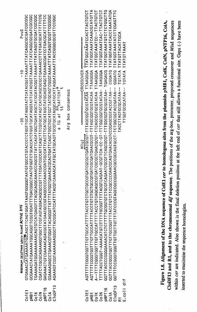
1.7 Characterisation of the ColEl cer recombination site
The determinant of ColEl that causes dimer resolution was isolated by a
combination of sub-cloning fragments of the plasmid, deletion mutagenesis and
sequencing. It was originally defined as a 284 bp Hpall-Taql fragment, and is named
cer (Summers and Sherratt, 1984; Summers et aly 1985; Summers and Sherratt, 1985;
see fig. 1.8). A plasmid with two cer sites present in direct repeat acts as a substrate for
site-specific recombination. When the cer sequence is deleted from ColEl the plasmid
becomes unstable and multimerises in Rec+ strains. If it is cloned into pUC8, cer
increases the stability of the cloning vector and stops it from multimerising in Rec+
strains. The site-specific recombination reaction at cer sites is highly directional;
14
0 O O CD O O 0CD CD CD O O CD CDCD O CD O *— CD O0 O O t— \— CD OCD CD CD b - 1— CD O0 O O ♦— 1— O t—
1—( b - K 1— t— b ~ b - H -d < < < t— < f— H -> 0 CD CD < O CD CD
CL CD O O O O O OCD CD O CD O O CD0 O I- CD < H— O0 CD CD < CD CD CD0 CD CD CD H“ CD CD< < < O < CD0 O O O O O Ob - t— b - t— !— f— O< < < H - < < <(— 1— 1— O CD f— 1 -I— 1— (— O O (— b -1— t— b - < CD t— b -< < < < < < << < < < < < <
O < < < < < < <■*— < < < CD O < <
1 I— b - t— b~ H - b— b -h - b— CD O O <
CD CD CD CD CD CD CDCD CD CD O < CD CDO O a CD CD CD << < < O f— f— T a t
t— 1— b - CD CD CD W< < < < < < < < Z3O O 0 O O O 0 O V)I— CD O CD O 0 I— cb - t— u b~ b - b - w— CD< < < < < < < < V)H - b ~ 1— O b - b - 0 4-> cCD CD 0 O b— CD CD +-> 0CD CD CD CD CD CD CDCC + J CD< < < < < CD <H— b ~ t— 1— (— b ~ f— u _ X< < < < < < < CO 0
ID O O < O O O O - QCO CD CD a O O CD CD CD
1 t— h - 1— b - 1— H - H-* b - CDb — 1— 0 CD < O CD L .CD CD CD O O CD CD CD <CD CD CD b - b ~ CD CDb - ♦— 1— H - t—O O < 1— b ~ O b~b - t— O O O CD CDO < < CD < < <O CD O < O O OO O f— O I— t— CD< < h - 1— b - b ~ b -h — O < b - < < <CD CD 1— < O CD h -H - H - O O f— OO O CD O CD O CDO f— O CD CD < ►—CD CD cd CD 1— CD <CD CD < 1— < CD b -H - b— O < CD O <CD CD O CD CD <C O1— 1— < O O O << < < »— < < << < < H* < < <b - b ~ < f— < < <CD CD O 1— O CD OCD CD CD a O b - (—CD CD CD 0 O CD OCD O < 0 O CD OCD O O CD O I— <b - < CD 0 CD < t—CD CD O CD < f— I—< b ~ < < < »— 1—< I— 1— O H— b - 1—b - t— b - < CD b - <1 - t— f— O O < OU f < < CD CD O b - CDCD CD CD < CD f— I—t o CD O < t— CD CD <
UJ H - t— O CD < CD O> CD CD 1— < < CD OP < CD < b~ CD < OO O O O CD 1— O O< b ~ 1— f— O < < <
>— H - O b— H— b - b -3 O O b — t— CD b -3 CD CD CD O O < CD—I < < < CD O CD CD
O O CD < CD CD* D CD < ►— CD 1— CD
— CD CD < CD < O <1— h - < O O O << < < < < < <Z < < < < < < <e < < < < < < <
h < < < CD < CD <u 1 < < < < O H - <LI CD O CD O O CD H*O b ~ f— CD O h™ O Os: < < b - O CD CD CD
O H* < CD H - < OT O O CD CD O CD f -X < < < > - < < CD< < < < O < < <x < < < b - < < <
O CD < < CD < <O CD CD < O CD
CD CD b - O CD CD <
COto T—T— Li.
LU T— Z (— < Or— CD r— r— a . r — OO x O O z 0
O Q . O O Q . CD O
1— < h— H - CD < OO 1— O O b ~ CD f—b ~ t ~ b— h— b — ►— 1—CD CD CD CD CD < >—H - t - f—■ CD b - CDO < O O CD < << < < b ~ < CD1— CD I— h - CD < Ob - O b - 1— b ~ H - t—CD < 1 1 b - O h - << < 1 O CD O O OO < O < H* < b - CDO O O O CD O < b -1— b - 1— f— b - b - b - b - b -< < < < < < < < <
« < < < < < < < O << < < < < < < < <b — >— H- H— b - H - h -CD CD CD CD O CD CD (—CD CD CD CD CD CD CD CD CD1— t— b - 1— b - b - b - 1— 1—< < < < < < < < <►— t— 1— b - b - ►- f— 1— b -
■ h - b - 1— b - 1— 1— b - b -
< < < < CD CD CD < <CD CD CD CD CD b— (— b - b -CD CD CD CD < < < < <CD CD CD CD O CD CD b -< < < < < CD CD CD CD< < < < O CD CD b -1— b ~ (— b ~ 1—b - b - 1— 1 1 1 1
1 < < < < < <1
<1
<1
<< < < < < < < < <O O O O H - b - I— 1— t—< < < < CD V— < < <b— b - h— O CD CD CD OCD CD CD CD CD CD O CD CDO CD O O O CD O O OCD CD CD CD O < < CD CD
I f - b ~ H* t— b -CD CD CD CD CD CD CD CD CDCD CD CD CD CD CD CD < CDO O O O CD CD O t— b~CD CD CD CD CD CD CD h - b -< < < CD CD < CD OI— t— O b - b - CD b - O1— b ~ t— t— b - CD CD <b - b ~ (— 1— b - CD b - (—
1 1 1 1 1 CD 1f— I— b - b - H— b - 1—O CD CD CD CD CD OO O O 1 O CD OCD CD CD O CD < OO CD O O O CD b -
. < < < 1 CD < CD< < < < < < <CD CD CD O O CD CDb ~ b - f - H* b - CDO < O b - b - CD CDCD CD CD CD CD < CDCD O O O CD O CDCD CD CD CD CD CD CDCD O O 1 CD O CDb - t— 1— t— b ~ < << < < < < < <CD CD CD < < O <
1— CD CD CD CDCD CD CD < < CD CDCD O O O O CD CDO O O h — CD CDCD CD CD CD CD CD CDH - h— < O O CD CDO O O < CD CD CDCD CD CD b - b ~ t— CDh - h— < b - CD b— <CD O O b - b— CD b -f - b— b - CD b - CDCD CD CD f— CD CDb ~ f— t— CD CD h - CDO O O CD CD O CDO O O O CD O I—< < < O CD < <b - h - 1— 1— b - b ~ f—b - b - t— b - b - b - >—b - b - 1— b - 1— b ~ 1—b - b - 1— b— 1— H - t—b - 1— f— H— CD CD< < < O O CD b -O O O 1— L - CDO O O CD O CD OCD CD CD h - t— CD CDb - t— f - < < t— 1—h - b ~ H— O O h - >—CD CD CD < < CD CDb - t— 1— < < b - I—b - 1— 1— < < b— h -H— h - H - < < b— CDCD CD CD 1 CD CD OCD CD CD * CD CD CDt— t - t— CD CD b—CD CD CD CD CD CD CDO CD CD CD O CD CDCD CD CD H* CD CD CDO O O O CD CD Ob - b - b ~ O CD CD Ob ~ b - t— < < < H -b - h - t— 1— b - I— b -b - b - 1— 1— b - b ~ b -b - ( - 1 1— f— b - b—CD CD 1 b - CD CD CD< < < b - CD CD CD 4 -
T JCO
CD T~ •r—T~ t— LL r —LXI T— Z b ~ < O Or— CO r — r— CL r— O OO z O O z O T—
O Q . O O Q . O O c d LU
Figu
re
1.8.
Alig
nmen
t of
the
DNA
sequ
ence
of
Col
El
cer
to ho
mol
ogou
s sit
es
from
the
plas
mid
s pM
Bl,
Col
K,
CoI
N,
pNT
P16,
Col
A,
CloD
F13
and
Rl,
and
to the
ch
rom
osom
al d
if se
quen
ce.
The
posit
ions
of
the
arg-
box,
pro
mot
er,
prop
osed
cr
osso
ver
and
Mlu
l se
quen
ces
with
in cer
are
in
dica
ted.
Also
sh
own
is the
fin
al d
eletio
n po
sitio
n at
the
left
end
of ce
r th
at sti
ll al
lows
a
func
tiona
l sit
e. Ga
ps
(-)
have
be
en
inse
rted
to m
axim
ise
the
sequ
ence
ho
mol
ogie
s.

Left arm recombinase Right arm recombinase
binding site Spacer binding site
ColEl GCGGTGCGTACAA TTAAGGGA TTATGGTAAATColK GCGGTGCGTACAA TTAAGGGA TTATGGTAAATpMBl GCGGTGCGTACAA TTAAGGGA TTATGGTAAATColN GCGGTGCGTACAA -TAAGGGA TTATGGTAAATColA GCGGTGCGTACAA --CGGATG TTATGGTAAATNPT16 GCGGTGCGCGTAA -TGAGACG TTATGGTAAATColE2 GGGGGGCGTACAA --CGGGAG TTATGGTAAATColE3 GGGGTGCGTACAA --CGGGAG TTATGGTAAATpSClOl GCGGTGCGCGCAA --GATCCA TTATGTTAAACCloDFl3 GCGGTACCGATAA --GGGATG TTATGGTAAATtype I hybrid GCGGTGCGTACAA TTGGGATG TTATGGTAAATtype II hybrid GCGGTGCGTACAA --GGGATG TTATGGTAAATdif. TTGGTGCGCATAA --TGTATA TTATGTTAAATRl TTAGTGCGCATAA --TGTATA TTATGTTACAT
Figure 1.9. Comparison of the putative crossover sites from different cer-like sites. The conserved, putative left- and right-arm recombinase binding sites are indicated, as are the spacer sequences which, as shown, vary in their putative sequences and sizes (from 6-8 bp).This diagram was kindly supplied by J.Roberts.

plasmids containing directly repeated cer sites are resolved by intramolecular
recombination and do not recombine intermolecularly (see e.g. Colloms, 1990 and this
thesis). This directionality is expected from the site's function, since intermolecular
multimerisation would lead to destabilisation and not stabilisation of ColEl.
Highly homologous sites have been identified from a large number of related,
naturally occuring plasmids: ColK (Summers et al, 1985), CloDF13 (Hakkaart et al,
1984), pMBl (Greene et al, 1981), ColA (Morion et al, 1988), pNTP16 (P.Strike,
pers.comm.) and ColN (Kolot, 1990) all contain sites that have homology with the
entire 284 bp of cer (see fig. 1.8); in addition the plasmid R1 contains a plasmid
stabilisation site with homology to the proposed crossover region of cer (see below and
fig. 1.8; Clerget, 1984). It is likely that most, if not all, plasmids contain sites that
function in resolving multimers which arise by homologous recombination, although in
many cases they are not homologous in sequence to cer. For example, PI encodes the
Cre protein that acts at loxP sites (Austin et al, 1981; see above), the F factor encodes
the D protein which acts at rfsF sites (O'Connor et al, 1986) and a resolvase
recombinase acts at the R46 per site (Dodd and Bennett, 1987).
The position where the strand exchange reactions occur in cer have been coarsely
mapped by sequencing the reaction products of recombination between ColEl cer and
the cer-\ike sites from ColK and CloDF13 (Summers et al, 1985; Summers, 1989).
This suggested that the exchanges had occurred within a 35 bp region at the right-hand
end of the cer site. Comparing the numerous cer-like sites in this region showed that it
is the most conserved sequence within the sites, and suggested that it might be the
crossover sequence of cer with a similar organisation to the crossover sequences of X
att sites, PI loxP sites and the yeast 2-micron plasmid FRT sites (see figs.1.9 and 1.5).
It is hypothesised that the cer-like crossover regions comprise imperfect and highly
conserved, inverted repeat recombinase binding sites flanking a less conserved spacer
sequence. Notice that if this analysis is correct then the recombinase(s) that act on the
cer-hke sites are able to perform strand exchange reactions on various sites that have
spacer regions which vary in their sequence and in their size (from 6-8 bp). The
15
positions of strand cleavage within this presumptive crossover sequence are not
known, but, by analogy with other X integrase-like systems, it is hypothesised that the
cleavages are within the spacer sequence (see Chapter 5).
Deletion analysis determined that 200 bp of the sequence upstream of the putative
crossover region is all that is needed for cer recombination, therefore suggesting that
approximately 250 bp of the 284 Hpall-Tapl fragment comprise the actual cer site
(Summers and Sherratt, 1988). Within this 200bp region is a conserved "arg-box"
sequence that is essential for the recombination reaction (see fig. 1.10 and below;
Stirling et al, 1988). The arg-box is only some 18 bp in size, however, and it is
therefore necessary to explain the function of the remaining 180 bp (approximately) of
sequence upstreamof the cer crossover. The sequences upstream of the arg-box are not
believed to bind any proteins, but instead to act as a "flexible” region; this is based on
the observation that they can be replaced by unrelated sequences that have alternating
AT and GC tracts which are found in bent DNA (Summers and Sherratt, 1988; Drew
and Travers, 1985). The region between the arg-box and putative crossover site is more
conserved in sequence than the "flexible region", and the distance between the two
sequence motifs is also conserved, but despite this its function is not yet understood. A
transcript is made in this region, but does not appear to be involved in cerof
recombination since a mutation that reduces the level^its expression 60-fold has no
effect on cer recombination (Summers and Sherratt, 1988).
Recombination between ColEl cer and CloDF13 parB sites is inefficient and
generates two cer-like sites (termed the typel and typell hybrids; see fig. 1.8) whose
recombination characteristics have important implications (see sections 1.8.1 and 1.8.2,
below). The typell hybrid site supports both intramolecular and intermolecular
recombination, unlike cer which recombines in an exclusively intramolecular direction
(Summers, 1989). All the sequences necessary for the typell hybrid recombination
reactions are present in the region downstream of the cer Mlul site, therefore offering
further evidence that this is the crossover region. The reasons for these altered reaction
properties are currently being investigated, and it is likely that they are due to the
16

differences between the cer and typell hybrid sites in their overlap and left arm
crossover sequences (J.Roberts, pers.comm.).
1.8 The E.coli chromosomally encoded xer genes
As stated above, the only sequences of ColEl required for xer site-specific
recombination are within the 250 bp of the cer site. Although a transcript is expressed
from cer, it has very limited coding potential and is not conserved in the other cer-like
sites, and it was therefore suggested that the protein(s) which recombine cer are
encoded by the E.coli chromosome (Summers et al, 1985). For this reason the
transposon Tn5 was used to mutagenise E.coli, and xer mutants were identified by their
inability to resolve a cer reporter plasmid, pKS455 (see fig.4.20), that carries selectable
drug resistence markers. Three genes were identified by this technique as being
essential for cer recombination; xerA,xerB (Stewart, 1986; Stirling et al, 1988 and
1989) and xerC (Colloms et al, 1990). All have been cloned and sequenced, and are
discussed below. (While work was being performed for this thesis a fourth xer gene
(xetD/xprB) was identified by different methods; it is described in Chapter 5.)
1.8.1 x e r A
The map position in the E.coli chromosome (70.5 mins) and nucleotide sequence
of the cloned xer A gene were shown to be identical to the sequence and map position
determined for argR, which encodes the arginine biosynthetic repressor (Stirling et al,
1988; Lim et al, 1987). ArgR (in conjunction with its corepressor, L-arginine) is a
negative regulator of the expression of the genes involved in arginine biosynthesis (for
review, see Glansdorff, 1987). Gel retardation and footprinting experiments using the
purified protein have shown that ArgR binds to an 18 bp sequence in cer that has
homologies with the operator sequences found in the promoter regions of the arginine
biosynthesis operons (Stirling et al, 1988; see fig. 1.10). It is intriguing that the cer site
17

appears to contain only one copy of this 18 bp arg-box sequence, whilst the arginine
biosynthesis genes, as well as argR itself, contain two repeats of the loosely conserved
arg-box. The cer footprinting data that are available do not detail whether ArgR/XerA
binds to the region directly downstream of the arg-box (which would be the location of
a second arg-box if it was present) because its sequence has innate resistance to
DNAasel cleavage, and therefore the possibility that this may reflect an alteration of
ArgR binding to cer (when compared to other operators) that has significance in terms
of cer recombination remains to be investigated.
A number of results show that ArgR is not the cer recombinase, but is an
accessory factor in the reaction; it displays no sequence homologies to either class of
recombinases, it binds approximately 100 bp from the proposed strand exchange
positions and typell hyrid sites (see above) are able to recombine in the absence of
functional ArgR (Summers, 1989). It seems unlikely that the role of ArgR during cer
recombination is to repress transcription from the promoter which overlaps with the cer
arg-boxes. This can be said because alterations in the level of expression of the
transcript have no effect on cer recombination unless the mutations also affect ArgR
binding (Summers and Sherratt, 1988; it may in fact be argued that the existence of the
transcript and promoter sequences in the cer site are simply a consequence of the
incorporation of the argR binding site during cer evolution). The role of ArgR may be
analogous to that of IHF during X recombination, and is in the assembly of higher-
order protein-DNA complexes in which cer recombination occurs. In this role ArgR
(along with the upstream sequences and PepA; see below) could contribute to the
intramolecular directionality of the cer recombination reaction.
1.8.2 x e r B
Database searching using the predicted amino acid sequence of XerB (derived
from the cloned gene) revealed 31% identity to bovine lens leucine aminopeptidase
(Stirling et al, 1989; see fig.3.2). This raised the possibility that xerB may be an E.coli
gene encoding an aminopeptidase. Analysis of the Xer phenotype of various pep
18

a r g Fa r g largECBHcarABa r g Aarg Rc e r
co n cen su s
AATGAATAATTACACATA
-G ----------------C -T C ---------
T - - C --------T -C -T G --G -
TG---------- T - A - - T G - - A -
-C A ------------- A A -T -----C -
T T - -C --------A A -T T ------ C
G T --C -------G G --T T -------
TATGAATAATnATnCAnT
A TA
t a a AGTGAATTTTAATTCAAT
t a a - T ------------------------------ T -
a t t T A AAA A - - C -
t a a G-GAAT T C -
a ta T T C A A -C --G A
t c r t T A --C -C A A GTTTG
cggT T A A ATCAGGCGC
TATGAATAATnATnCAnT
A TA
Figure 1.10. Comparison of the ArgR DNA binding sequence in cer to the arg-boxes from the arginine biosynthetic genes. The sequences are aligned against the arg-boxes found in the argF promoter region, bases unchanged are denoted by a dash and the boxes are framed. (Sequences taken from Glansdorff, 1987 and Stirling et al, 1988).

mutants of S.typhimurium suggested that xerB may be the E.coli equivalent of pepA\ it
was necessary to perform the genetic analysis in S.typhimurium because the pep genes
of this organism have been more extensively mapped and analysed (Miller, 1987). It
was further shown that a clone of the S.typhim urium pep A gene was able to
complement an xerB mutation in E.coli and allow cer recombination (Colloms, 1990),
therefore offering compelling evidence that xerB is pepA and encodes E.coli
aminopeptidase A.
Aminopeptidase A (PepA) was previously purified from E.coli and named
aminopeptidase I by Vogt (1970). Analysis of the purified enzyme in vitro has shown
that it is heat stable (70 °C for 5 mins), has an approximate molecular weight of 52
kDa, aggregates to form a larger molecular weight species in low ionic strength buffer
and that it is an exopeptidase which cleaves amino-terminal residues from various
peptide substrates. E.coli and S.typhimurium encode a number of aminopeptidases
(PepA, PepB, PepM, PepN and PepP) - as well as four dipeptidases (PepD, PepQ,
PepE and PepG), a tripeptidase (PepT) and several carboxypeptidases - which all have
non-specific substrate specificities (Miller and McKinnon, 1974; Miller and Schwartz,
1978; Miller, 1987). The exclusive requirement for PepA rather than the other
aminopeptidases in cer site-specific recombination is therefore surprising, and is not yet
understood, but it could function in either an enzymatic or structural capacity (this is
considered in more detail in Chapter 3). It is clear, however, that PepA is an accessory
factor (in conjunction with ArgR), since it has no sequence homologies to either class
of recombinases and because type II hybrid sites will recombine in pepA strains
(Summers, 1989).
1.8.3 x e r C
This was the last xer gene to be identified, and has been mapped to the 85 minute
region of the E.coli chromosome, between the genes for adenylate cyclase (cya) and
DNA helicase II (uvrD; Colloms et al, 1990). The translated amino acid sequence of
XerC has homologies to the X integrase class of recombinases (see fig.5.17), and
19

partially purified preparations bind to the crossover region of the cer site (Colloms,
1990). This suggests that XerC is the recombinase responsible for the xer strand
cleavage and exchange reactions, and it is likely that the recombination mechanisms will
correspond to the scheme described for Int, FLP and Cre-catalysed recombination (see
above).
xerC constitutes the third member of an operon which also contains the
previously cloned gene dapF , encoding the enzyme diaminopimelate epimerase
(Colloms et al, 1990; Richaud et al, 1987; Richaud and Printz, 1988). The co
transcribed genes in the operon are, in order, dapF, orf235, xerC and orf238 (see
fig.4.1). The roles of the proteins encoded by the two open reading frames are not
known and no homologies to published proteins have been found. Why xerC should be
part of an operon, and how its function may be related to, or regulated by, the other
proteins is not understood.
1.9 A cellular role for xer site-specific recombination
Insight into the cellular role of the xer site-specific recombination system was
gained through the observation that xerC mutant strains have a tendency to produce
filaments and have aberrant and amplified nucleoids (Blakely et al, 1991). This
suggested that xerC mutants, though viable, have defects in cell division and nucleoid
segregation. The same phenotype is observed in strains carrying deletions in a region of
the E.coli chromosome close to the terminus of replication called d if (Kuempel et al,
1991). Analysing the sequence of the dif region revealed a 33 bp sequence similarity to
the crossover region of the cer site (see figs. 1.8 and 1.9), and it was therefore
proposed that d if is an E.coli chromosomal substrate for xer site-specific
recombination.
The 33 bp d if sequence is sufficient to act as a substrate for site-specific
recombination when cloned into plasmids, and XerC binds specifically to it in gel
2 0

retardation assays (Blakely et al, 1991). Recombination of dif does not require either of
the cer accessory factors ArgR or PepA, which is consistent with the fact that dif and
cer are only homologous at their crossover sites and argR and pepA strains do not
have a filamentous phenotype. The dif site-specific recombination reaction, like
recombination of the typell hybrid (which can also function as a simple crossover site
without accessory sequences), shows no directionality - i.e. it proceeds both
intramolecularly and intermolecularly.
The above results have led to the hypothesis that the cellular role of the xer site-
specific recombination system is in chromosome partitioning. Odd numbers of
homologous recombinational exchanges between monomeric sister chromosomes
(either during or after their replication) will generate chromosomal dimers. Dimers
formed in this way cannot be partitioned into the daughter cells at cell division.
Therefore it is proposed that xer site-specific recombination at dif acts to convert these
chromosomal dimers into segregateable monomers, in an analogous function to plasmid
stabilisation by recombination at cer sites (Blakely et al, 1991). It is believed that the
positioning of the dif locus at the replication terminus allows all dimers to be resolved
just prior to termination of replication, and it could minimise the decatenation required
to separate the monomerised chromosomes. The apparent lack of directionality of the
dif recombination reaction would create as well as resolve chromosome dimers, but it is
possible that this reaction mechanism is needed because the sites are unable to
determine when the chromosomes are dimeric, and therefore must rapidly recombine
irrespective of the chromosomal configuration.
Support for the above hypothesis is provided by the fact that the filamentous
phenotype and aberrant nucleoids of xerC strains are overcome when they are made
recA, suggesting that without homologous recombination generating chromosome
dimers dif recombination is not necessary (Blakely et al, 1991). It is also strengthened
by the fact that xerC is widely distributed in bacteria (G.Blakely, pers.comm.). The
hypothesis, however, still requires formal demonstration of xer recombination at the

chromosomal dif locus, perhaps by analysing the dimeric state of XerC+ and XerC'
cells.
22
Chapter 2
Materials and Methods
23

Table 2.1 Bacterial Strains
Strain Genotype Source/referenceAB1157 thr-1, leuB6, hisG4, thi-1, ara-14,
A(gpt-proA)62, argE3, galK2, supE44,
xyl-5, mtl-1, tsx-33, lacYl, rpsL31 Bachmann, 1972DS941 AB1157, but recF143, /acZAM15, lacIQ D. SherrattDS942 DS941, but lacZAH220 (lad) D. Sherratt
DS956 DS941, butxerA9 (argR::fo\) D. SherrattCSX17 DS941, butxerBl (pepA::Tn5) C.Stirling, 1989HOM38b DS941, but pepA7 H.O’MaraDS980 DS941, buto/;/235::Tn5 S.CollomsDS981 DS941, but .xe/ C::Kan P.SykoraDS982 DS941, but orf238Y2 (::mini Mu) S.CollomsDS984 DS941, but xerCY17(::miniTnlO) S.CollomsDS9008 DS941, but xprB::miniTnlO M.BurkeSTL116 AB1157, butx/;/B::miniTnlO S.LovettCS85 AB1157, but ruvC53, eda-51 B.LloydJC7623 AB1157, but recBC, sbcB C.RichaudBMH 71-18 thi, supE, A (lac-proAB) mutSv.TnlO
(F lacIQ lacZAMI5) Promega
EMI DS941, but mutS E.MorrellRM10 DS941, but lacPOxerC, dapF/otf238::Km This workRM20 RM30 RM40RM41 RM40, but xerA9RM42 HO'M38b, but lacPOxerC, dapF/otf238::Km "RM43 RM40, but ruvC53RM50 DS941, but lacPOdapFRM60 DS942, but xerC: :KanRM61 DS942, but xerCY17RM62 DS942, but xprB: :miniTn 10
2 4

Table 2.2 Plasmids
Plasmid/resistance DescriptionpBR322 (Ap, Tet) vector derived from pMB 1pUCl 8 (Ap) vector derived from pBR322pUC19 (Ap)pUC9 (Ap)pKK223-3 (Ap)pIC20R (Ap)pAT153 (Ap)pSELECT (Tet) pBR322-derived mutagenesis vector pUC71K (Ap, Km) pUC19 + kanamycin resistence genepCT1050 (Ap)
pGPl-2 (Cm) pGPl-2Km (Km) pBAD (Ap) pCB 105/6 (Cm)
Source/ReferenceSutcliffe, 1978
Yanisch-Perron et al, 1985
Pharmacia R. Wilson A.Twigg Promega
Pharmacia R.Thompson
Tabor and Richardson, 1985 S.Rowlands A.C.Boyd
D.Summers C.Stirling, 1987
p ATI 53 + APl promoter T7 polymerase expression vector vector derived from pGPl-2 pKK223-3 derived Ptac expresion vector A,dv-based vector
pKS455 (Ap, Cm) pUC9-based 2-cer reporter plasmid pCS202 (Cm, Tet) Adv-based 2-cer reporter plasmid
pBAD +1.9 kbp HindlU pep A fragment "pCB 106 + 1.9 kbp HindlU pep A fragment "pCB 106 + 920 bp Sphl-Accl argR rfagment "pBAD+1.2 kbpHindlU-EcoRl xerC fragment S.Colloms, 1990 pTZ18R + 3.8 kbp HindUl-Bgtll xerC fragment "pBR322-based 2-typeII reporter plasmid pBR322 + 300 bp cer fragment in EcoRl-HindUl "pSDl 13 + 300 bp cer in PvuU "
pSD 124 (Ap, Km) pUCl 8-based 2-dif reporter plasmid pSD126 (Ap, Km) pBR322-based 2-di/reporter plasmid
pUC18 + 1.12 kbp ruvC fragment pBR322 + 3.8 kt-j5̂ r| fragment pIC20R + 450 bp Sspl-Hindlll rrnB terminator from pKK223-3 pRMIO +1.3 kbp Pstl kanamycin (ex pUC7IK) "pSELECT +1.9 kbp HindlU pep A fragment This workpRM20, but containing pepAG.354A "pBAD + 1.9 kbp HindUl pepAE354A (ex pRM21) pCT1050 + 1.2 kbp HindUl-EcoRl xerC fragment "
pCS126 (Ap) pCS118 (Cm) pCS350 (Cm) pSD105 (Ap) pSD102 (Ap)
pSDllO (Ap) pSD113 (Ap) pSD115 (Ap)
pGS762 (Ap) pJC763 (Ap) pRMIO (Ap)
Blakely ef al, 1991t t
Sharpies and Lloyd, 1991 Lovett and Kolodner, 1991
This workpRM ll (Ap, Km) pRM20 (Tet) pRM21 (Ap, Tet) pRM40 (Ap) pRM50 (Ap)
25

Table 2.2 (continued)pRM60 (Cm) pGPl-2 +1.6 kbp EcoRI-BamHI
XPl-xerC fragment from pRM50 This work
pRM65 (Km) pGPl-2Km + 1.6 kbp EcoRI-BamHIXPL-xerC fragment from pRM50 "
pRM70 (Ap) pUC18 + 2.1 kbp Pvull-Stul xerC frag, (ex pSD102) "pRM71 (Ap, Km) pRM70 + 2.4 kbp BamHl fragment from pRM 102 "pRM80 (Cm) pCB105 + 1.12 kbp HindUl-EcoRl ruvC
fragment from pGS762 "pRM90 (Ap) cer MluY derivative of pSDl 13 "pRM91 (Ap) pRM90 + wild type cer at Pvull site (see pSDl 15) "pRM92 (Ap) pSDl 13 + MluY cer at PvuU site (see pSDl 15) "pRM93 (Ap) pRM90 + MluY cer at PvuU site (see pSD 115) "pRMlOl (Ap) pSD102 + 230 bp Pvull-Smal lacPO in Nrul site "pRM102 (Ap, Km) pRMlOl + 1.8 kbp EcoRl Kanamycin-mzB
terminator cassette from pRM 11 "pRM 120 (Ap) GGG-A AT cer site derivative of pSD 113 "pRM121 (Ap) pRM120 + wild type cer in PvuU (see pSDl 15) "pRM122 (Ap) pSDl 13 + GGG-AAT cer in PvuU (see pSDl 15) "pRM 123 (Ap) pRM 120 + GGG-AAT cer in PvuU (see pSD 115) 11pRM130 (Ap) pUC19 + 1.7 kbp HindUl-EcoRl xprB
fragment from pJC763 "pRM 131 (Ap) 760 bp SacU deletion derivative of pRM 130 "pRM132 (Ap) pUC19 + 1.3 kbp HindlU-Haell xprB
fragment from pJC763 "pRM133 (Ap, Gen) pRM130 5acII+ 2.0 kbp HindlU gentamycin
resistance gene "pRM134 (Ap, Gen) pRM130 + 2.0 kbp HindlU gentamycin
resistance gene in EcoRV site "pRM135 (Cm) pCB106 + 1.7 kbp HindUl-EcoRl xprB
fragment from pJC763 "pRM140 (Cm) replacement of xerC in pRM60with 1.7 kbp
HindlU-EcoRl xprB from pRM 130 "
2 6

2.1 Bacterial strains and plasmids. The derivatives of Escherichia coli K-
12 used in this work are listed in Table 2.1; new strains were constructed either by PI
transduction (Miller, 1972) or by linear transformation of mutant, cloned genes
(Winans et al, 1985; see Chapter 4). The plasmids that were used and constructed for
this work are listed in Table 2.2.
2.2 Chemicals
CHEMICALS
General chemicals, biochemicals
and organic solvents
Media
Agarose
IPTG
Radiochemicals
Nucleotides
Antibiotics
Val-Leu-NH2
SOURCE
BDH, May and Baker, Sigma
Difco, Oxoid
BRL, Flowgen
BRL
NEN
Boehringer Mannheim, Promega
Sigma
BioMac
2.3 Bacterial growth media and conditions. Bacteria were grown in
standard L-broth (Miller, 1972) and L-agar at 37 °C, or in minimal agar (100 ml of
which contained 25 ml D&M salts, 75 ml 2% agar in distilled water, 0.2% glucose, 20
ug/ml vitamin B1 and the following amino acids at 30 ug/ml: threonone, leucine,
histidine, arginine and proline) at the same temperature. Antibiotics and other
supplements were added where appropriate (see text). The bacterial strains were stored
in 50% L-broth, 20% glycerol and 1 % peptone at -20 °C, or in thiamine-containing
slopes at room temperature.
2 7

The antibiotic concentrations used for both liquid and plate selection were as
follows:
Antibiotic Stock Solution Selective concentration
Ampicillin 5 mg/ml water
Tetracycline 1.25 mg/ml 10 mM HC1
50 ug/ml
12.5 ug/ml
25 ug/ml
50 ug/ml
100 ug/ml
10 ug/ml
10 ug/ml
Chloramphenicol 2.5 mg/ml ethanol
Kanamycin 5 mg/ml water
Streptomycin 10 mg/ml water
Trimethoprin 1 mg/ml water
Gentamycin 1 mg/ml water
All stocks were stored at 4 °C and were added to molten agar cooled to 55 °C.
2.4 Bacterial transformation. All transformations of plasmid DNA used
standard CaCl2 treatment of cells (Sambrook et al, 1989; Cohen and Hsu, 1972).
2.5 Isolation of plasmid DNA. Four methods for preparing plasmid DNA
from bacterial cultures were used:
(i) Small scale DNA preparations used a modification of the boiling (STET)
method of Holmes and Quigley (1981) that is in common use in our laboratory
(Colloms, 1990); where stated, a phenol/chloroform extraction step was included
before precipitation of the plasmid DNA with isopropanol. DNA was made from
"patched" cultures of E.coli from L-agar plates, or by harvesting 3 ml of an L-broth
culture at mid-log phase (e.g. in in vivo recombination assays) or at stationary phase.
(ii) Mid scale preparations used Qiagen columns and followed the manufacturers'
instructions.
2 8

(iii) Large scale preparations for long-term storage of the DNA used a
modification of the technique of Birnboim and Doly (1976) developed by C.Boyd
(1985).
(iv) For rapid analysis of the plasmid content of a bacterial culture, the single
colony lysis technique was used. In this, a 1 cm patch of a single transformant was
collected and resuspended in 100-200 ul of single colony gel buffer (2% ficoll, 1%
SDS, 0.01% bromophenol blue in Tris-acetate buffer). The cells were allowed to lyse
at room temperature for 15 mins, cell debris and chromosomal DNA spun down in a
microfuge for 30 mins at 4 °C and 30 ul of the supernatant loaded onto an agarose gel.
2.6 in vitro DNA manipulations. DNA manipulations were essentially as
described in Sambrook et al (1989).
Restriction digestions were performed in 10-30 ul of IX restriction buffer
(purchased from BRL) and contained 0.3-3 ug of DNA. 5-30 units of restriction
enzymes (purchased from BRL, Pharmacia, BIOLABS and Promega) were used in the
digestions.
The ends of restricted DNA fragments were filled-in using either the Klenow
fragment of E.coli DNA polymerase I or phage T4 polymerase (both purchased from
BRL; see Sambrook et al, 1989).
DNA fragments were ligated for 2-5 hours at room temperature in 20 ul of IX
ligation buffer using 1-3 units of T4 DNA ligase (both purchased from BRL) before
bacterial transformation.
End-labelling of DNA fragments was performed as described in Sambrook et al
(1989) using the Klenow fragment of E.coli DNA polymerase I (purchased from BRL)
and 10-20 uCi of a[32p]dATP/dCTP (purchased from NEN).
2 9
2.7 Site-directed mutagenesis of DNA. Two site-directed mutagenesis
techniques were employed:
(i) The pep A gene was mutagenised after being cloned into pS ELECT (purchased
from Promega) following the instructions supplied. The repair-deficient strain EMI
was used in preference to BMH71-18.
(ii) Mutagenesis of the cer site in pSD113 followed a protocol developed by
M.Stark (unpublished). Approximately 2 ug of plasmid DNA was nicked in IX
restriction buffer (containing 300 ug/ml ethidium bromide) using 2 ug/ml DNAasel
(Stark et al, 1991), purified by ethanol precipitation after phenol/chloroform extraction
(Sambrook et al, 1989) and then denatured by boiling at 100 °C for 10 mins in 25 ul of
IX annealing buffer (purchased from Promega). The denatured DNA was placed on ice
and 2 pmoles of both the mutagenic oligonucleotide and Seal deletion oligonucleotide
(kindly supplied by M.Stark) were added; annealing of the oligos was performed by
incubating the DNA at 55 °C for 2 mins before it was cooled slowly to room
temperature. The plasmid DNA was repaired from the oligonucleotides in 40 ul of IX
synthesis buffer (purchased from Promega) using T4 DNA polymerase and T4 ligase
(purchased from BRL) following the procedures described for pSELECT mutagenesis
(Promega). The repaired DNA was transformed into the either of the repair deficient
strains EMI or BMH71-18 and grown overnight. Qiagen preparations of the plasmid
DNA were performed from overnight cultures and samples digested with a large excess
of Seal (25 units for 3 hours) before being transformed into DS941. Plasmid DNA was
prepared from a number of the Scal-resistant clones and the desired site-directed
mutants selected by further restriction digestion.
The mutagenic oligonucleotides that were used are described in the text. The Seal
deletion oligonucleotide had the sequence: 5'GTGACTGGTGAGTATTCAACCAA
GTCATTC 3'. All the oligos were synthesised on an Applied Biosystems PCR-mate
oligonucleotide synthesiser and were not purified before use; they were phosphorylated
using T4 kinase as described in the pSELECT mutagenesis protocol (Promega).
3 0

2.8 Gel electrophoresis of DNA. DNA was analysed on three kinds of
horizontal agarose gels: standard 0.7-1.2% gels, 1.2% low melting point (LMP) gels
and 1.2% alkaline denaturing gels. The DNA samples were applied to the gels in Ficoll-
containing loading buffer (Sambrook et al, 1989). Standard and LMP gels were made
and run in Tris-acetate buffer (40 mM Tris.Acetate [pH 8.2], 20 mM Na. Acetate, 1 mM
EDTA). The preparation and running of the LMP gels was as described by the agarose
manufaturers' instructions (FMC Bioproducts), whilst the standard and alkaline
denaturing gels were as detailed in Sambrook et al (1989). Custom-made gel
electrophoresis kits were used which made 23 cm long gels and contained 3 litres of
buffer; the gels were routinely run for 15-18 hours at 1.5-2.5 volts/cm.
The DNA was visualised either by 254 n.m. UV illumination after staining with
ethidium bromide, or by autoradiography (with and without vacuum drying of the
gels). Ethidium bromide-stained gels were photographed on a Pentax 35 mm SRL
camera fitted with a Kodak Wratten no.23A filter using Ilford HP5 film;
autoradiographs used Fiji NIF RX Medical X-ray film.
2.9 Extraction of DNA from agarose gels. DNA was purified from
standard agarose gels by low speed centrifugation through siliconised glass wool
(Heery et al, 1990) and from LMP gels by phenol/chloroform extraction (following the
agarose manufacturers’ instructions).
2.10 Electron microscopy. Electron microscopic (EM) examination of
purified DNA was performed essentially as described by Coggins (1987). The DNA
was prepared for EM by spreading in 40% formamide, IX TE (0.1 M Tris [pH 8.3],
10 mM EDTA), 0.1 mg/ml cytochrome c on a hypophase of 10% formamide, 0.1X
TE. Contrast was enhanced by staining with uranyl.acetate, and low angle (8-10°)
shadowing was performed by vacuum evaporation of Platinum : Palladium (80 : 20)
wire.

2.11 Gel electrophoresis of protein. PepA was analysed on 12% SDS-
polyacrylamide gels as described by Laemmli (1970) using vertical kits purchased from
BioRad. The protein samples were applied to the gel in a loading buffer that contained
5% SDS, 50% glycerol, 0.01% bromophenol blue, 50 mM Tris-HCl (pH 8.0) and 5%
beta-mercaptoethanol. The proteins were visualised (after fixing in 40% methanol, 10%
acetic acid) by staining with Coomassie blue in 50% methanol, 25% TCA.
2.12 Preparation of concentrated cell extracts for aminopeptidase
assays. 100 ml cell cultures were harvested by centrifugation and resuspended in 20
mM Tris-HCl (pH 8.2), 100 mM KC1,0.1 mM EDTA, 1 mM MgOAc. They were then
lysed by the addition of T4 lysozyme at 4 ug/ml and repeated cycles of freezing in
liquid nitrogen and thawing by incubation at 37 °C. Cell debris were removed by
centrifugation.
2.13 in vitro aminopeptidase assays. The enzymatic substrate used was
leucine-p-nitroanilide (purchased from Sigma). The assays were performed at 37 °C in
20 mM Tris-HCl pH 8.2, 100 mM KC1, 1 mM MnCl2, 0.1 mM EDTA, 1 mM L-
leucine-p-nitroanilide.l ml samples were removed from a total assay volume of 14 ml at
the times stated. The reaction was measured by the absorbance change at 400 n.m and
the amount of reaction product (p-nitroaniline) in the assay was calculated using the
extinction coefficient taken from the Handbook of Chemistry and Physics (53rd edition;
logio Extinction Coefficient is 4.19). Aminopeptidase units were expressed as nmoles
of p-nitroaniline produced per milligram of protein per min.
32

Chapter 3
Analysis of the role of aminopeptidase A in cer site-specific
recombination by site-directed mutagenesis
33

3.1 Introduction
The role that Aminopeptidase A (PepA) plays in the cer site-specific
recombination reaction is not yet understood. The evidence that the protein is essential
for cer recombination, and not required for d if recombination, is genetic. Strains
containing Tn5 insertions within the pepA gene (e.g. CSX17) will not recombine
reporter plasmids containing directly repeated cer sites, but will recombine similar
plasmids if they contain dif sites (Stirling et al, 1989; Blakely et al, 1991); these X er
mutations can be complemented by a minimal pepA clone. Because it has not been
possible to recreate the cer recombination system in vitro, experiments to analyse the
role of PepA have been limited (e.g. attempting to see if it has DNA binding properties;
see below). This has meant that considerations regarding its function have been
necessarily speculative, and have centred on the question of whether the protein is
employed by the cer system for its catalytic properties (it is an amino-exopeptidase) or
in a structural capacity (see Stirling et al, 1989). Definitive experimental examination of
these alternative (though not exclusive) roles has so far proved elusive (G. Szatmari,
H.O'Mara, pers.comms.; see Discussion).
Analysis of the proposed cellular functions of PepA does not offer any answers to
these questions. Vogt (1970) isolated, characterised and crystallised a hexameric, 323
kDa aminopeptidase from E.coli whose catalytic activity was dependent on the divalent
cation Mn2+. This protein, called aminopeptidase I, is very likely to be PepA, since this
heat-resistant peptidase activity is absent from DS941pepA::Tn5 strains (CSX17;
Stirling et al, 1989) and pepA mutants selected by their resistance to the toxic dipeptide
L-Valyl-L-Leucine-amide lack a protein of the size described for Pep I (Miller and
Schwartz, 1978). The in vitro characterisation of PepA by Vogt demonstrated that it is
an aminopeptidase that will processively cleave the amino-terminal residues from many
different peptide molecules (in size and sequence) to produce free amino acids. This so-
called "broad-specificity" of PepA is similar to other aminopeptidases from E.coli and
S.typhimurium (PepN and PepB; see Chapter 1), but it should be noted that the
3 4

substrate profiles of these enzymes can be distinguished since they do not recognize
peptides with every possible amino terminal residue (Miller, 1987). The
aminopeptidases appear to be involved in the degradation of peptides derived from
cleavage (by other enzymes) of abnormal or truncated proteins in the cell, and also in
breaking down exogenous peptides which are transported into the cell so that they can
be utilised as nutrient sources. This analysis is derived from metabolic examination of
S.typhimurium strains mutant in aminopeptidases A, B, D and N; it is likely, however,
that E.coli encodes the same set of enzymes (Miller, 1987; Miller and Schwartz, 1978).
Although there is no direct evidence, either from in vitro or in vivo experiments,
that proteins are substrates for PepA, the possibility cannot be ruled out that it
processes, and therefore activates, one of the xer proteins (i.e. ArgR, XerC, XprB or
indeed a protein not yet identified). Activation would involve the cleavage of one or
more N-terminal amino acids, and must be due to a substrate specificity for the Xer
protein(s) possessed by PepA that the other cellular aminopeptidases do not have. No
experiments have been able to identify any such amino-terminal cleavage of these
proteins, but this has not been exhaustively analysed and remains possible. Since XerC
and XprB are able to recombine type II hybrid and dif sites in the absence of functional
PepA, this suggests that the putative recombinases are not the targets for PepA-
activation. If this were how PepA functioned in cer recombination then the amino
teminus of the target protein(s) must be free on the surface of the molecule to allow
access to PepA; alternatively the cleavage may occur during the translation of the xer
proteins.
An alternative target for aminopeptidase cleavage by PepA in cer recombination
could be small peptide molecules, which are the natural substrates for this enzyme.
These have not been identified, and are therefore purely hypothetical, but it is
conceivable they could function either as activators that require cleavage or as inhibiors
that are inactivated by cleavage. This possibility has the same problem as xer protein-
cleavage in explaining why PepA alone of all the broad-specificity peptidases in the cell
is active in this role.
35

Whilst there are difficulties in imagining how PepA acts in an enzymatic role
during cer recombination, it is equally problematic to explain how it might act
structurally. If PepA were involved in the nucleoprotein complex which forms during
the recombination reaction (either directly binding to the cer site, or interacting with the
other xer proteins) this would answer why PepA and no other peptidases are
specifically involved in the reaction. However, no cer-specific DNA binding activity
has been described for this protein (C.Stirling, pers.comm.), and no experiments have
been performed to attempt to address the possibility of it forming protein-protein
interactions with either ArgR, XerC or XprB (e.g. by gel binding assays). Clearly,
however, the fact that these experiments yielded negative results cannot exclude these
possible roles since they may be a consequence of having used the incorrect reaction
conditions.
This chapter describes site-directed mutagenesis of the pepA gene in an attempt to
analyse the above possibilities. Site-directed mutagenesis has been used to analyse the
catalytic mechanisms of a number of enzymes (Leatherbarrow and Ferscht, 1896). The
experiments in this study, however, were not intended as an analysis of the amino acid
residues involved in the active site of PepA. Instead the idea was to target specific
residues in PepA's active site in the hope of creating derivatives that lack any peptidase
activity which could then be tested in their ability to support cer site-specificAl
recombination. This experiment approach is therefore asking the question: is PepA's
aminopeptidase activity required for cer recombination ?. The implicit assertion of this
approach is that PepA acts as a structural component of the xer machinery, and does not
utilise its enzymatic activity during the cer reaction. The reason that this hypothesis
must be made is because a mutant PepA which is both X er and peptidase" is unable to
show that PepA acts enzymatically, because a protein with both phenotypes may have
structural perturbations as a result of the site-directed amino acid alterations.
36

3.1 Site-directed mutagenesis of pepA
The three dimensional structure of bovine lens leucine aminopeptidase (henceforth
called BLLAP) published by Burley et al (1990) was used to design the mutagenesis
strategy. The crystallographic structure of this enzyme (to 2.7A resolution) was
reported at the start of this work, and later a more refined structure (as well as the
structure of the protein complexed with the inhibitor bestatin) was reported (Burley et
al, 1991 and 1992). BLLAP is an aminopeptidase which, like PepA, displays a wide
substrate profile in vitro. The reason that the structure of this protein was believed to be
the related to that of PepA is because of the high sequence conservation between the
two enzymes (Stirling et al, 1989; see below). Described below are some of the
important structural features of BLLAP (taken from the 3-D structure) which
determined the PepA mutation that was made in this study.
BLLAP is active as a 324 kDa hexamer consisting of six identical subunits, each
487 amino acids in size. The protein contains 12 Zn2+ ions (two in each monomer)
which are essential for its enzymatic activity; they can, however, be replaced by Mg2+,
Mn2+ or Co2+, although each reduces the Km of the enzyme. The N-terminal 137
amino acids of each BLLAP monomer can be cleaved using trypsin and the remaining
C-terminal portions retain the hexamer structure and are catalytically active (van Loon-
Klassen et al, 1979)
Isolated BLLAP monomers have an approximate "comma" shape, comprising an
N-terminal and a C-terminal domain. The C-terminal domain is 327 amino acids in size,
comprises the body of the comma and contains the proposed active site and its
associated Zn2+ ions. This is consistent with photoaffinity labelling experiments that
localised the active site to within the larger of the trypsin-cleaved fragments. The
hexamer has 32 symmetry, in which the catalytic domains are clustered around the
threefold symmetry axis. The 160 residue, N-terminal domains extend outwards, and
away from, this catalytic core. It is believed that the residues responsible for
trimerisation are within the C-terminal domain, whilst those involved in trimer-trimer
37

Asp
Asp
AAsp
Figure 3.1. Stick figure representation of the active site residues of bovine lens leucine aminopeptidase. Each a-carbon atom is labelled and the two
active site zinc ions are shown as crosses, the uppermost of which is in the readily exchangeable subsite (site l).(Adapted from Burley et al, 1992).

interactions are in the N-terminal domain (this, however, disagrees somewhat with the
fact that trypsin cleavage of the majority of the N-terminus does not destabilise the
hexamer). The six active sites are located in the interior of the hexamer and line the
walls of a disc-shaped cavity. Access to this interior cavity for substrate molecules is
believed to be via three solvent channels that run along the 2-fold symmetry axis. The
maximal dimensions of molecules wishing to gain access to the active sites are
approximately 7A, meaning that it is that unlikely BLLAP could utilise proteins as
substrates unless their N-termini are in an extended form; whether this would also be
true for PepA depends on whether it is also active as a hexamer and whether it retains
these structural features.
The active site residues of BLLAP (described below; fig.3.1) were identified on
the basis of their proximity to the bound Zn2+ ions. Because two Zn2+ ions are used
by each monomer the active site is described as consisting of two subsites. The subsites
do not appear to be equivalent, however, since one (site 1, the activation site) will
readily exchange the bound Zn2+ ion with a variety of other divalent cations (Mg2+,
Mn2+ and Co2+), whereas the other (site 2) binds Zn2+ more tightly and will only
accept other ions when it is unoccupied. The crystallographic structure suggested that
the metal ion in site 1 is co-ordinated by one carboxylate oxygen atom from the residues
Asp273 and Glu334, and by the side-chain amino group of Lys250 (see fig.3.1). In
site 2 the co-ordination is achieved by one carboxylate oxygen from each of Asp255,
Asp332 and Glu334, and by the carbonyl oxygen of Asp332. This illustrates that each
Zn2+ ion is chelated by more than one amino acid residue, and that Glu334 is involved
in the co-ordination at both subsites. Two positively charged residues (Lys262 and
Arg336) are also described as being in the active site because they extend into the
region around the Zn2+ ions. They are, however, not thought to be involved in metal
ion chelation and their inclusion in the defined active site is mainly a consequence of
their polar character and proximity.
It was important to ask at this stage whether the structure of BLLAP would allow
a reasonable mutagenesis strategy to be extrapolated for PepA, for which no 3-D
38

BLLAP 1 TKGLVLGIYSKEKEEDEPQFTSAGENFNKLVSGKLREILNISGPPLKAGK, A * A . . • , • A • • A . A # .A • . A , A
50
P ep A 15 S A C IW G V F ------------ EPRRLSPIAEQLDKISDGYISALLRRGELEGKPGQ 58
5 1 TRTF YGLHEDFPSWWGLGKKTAGIDEQENWHEGKENIRAAVAAGCRQI★ • • • • • • • * • • A A • • * A . .
1 0 0
59 TLLLHHVPNVLSERILLIGCGKERELDERQYKQVIQKTINTLNDTGSMEA 1 0 8
1 0 1 QDL - - EIPSVEVDPCGDAQAAAEGAVLGLYEYDDLKQK---------------- R K W. * . . . . * * * * * . * . * ★ * * . *
1 4 0
1 0 9 VCFLTELHVKGRNNYWKVRQAVETAKETLYSFDQLKTNKSEPRRPLRKMV 1 5 8
1 4 1 --VSAKLHGSEDQEAWQRGVLFASGQNLARRLMETPANEMTPTKFAEIVEA • . . A A . A . * A A A . A . A . A . .A
1 8 8
1 5 9 FNVPTRRELTSGERAIQHGLAIAAGIKAAKDLGNMPPNICNAAYLASQAR 2 0 8
1 8 9 ENLKSASIKTDVFIRPKSW IEEQEM GSFLSVAKGSEEPPVFLEIHYKGSP• A A . . A * A A , A A * A A . # * # A A A A .
2 3 8
2 0 9 QLADSYSKNVITRVIGEQQMKELGMHSYLAVGQGSQNESLMSVIEYKGNA 2 5 0 2 5 5 2 6 2 273
2 5 8
2 3 9 NASEPPLVFVG^G ITFDSGGISIKAAANMDLMRADMGGAATICSAIVS AAA*A*AAAA. AAAAAAAAAA* A A A ♦ A A AAA • • • • A
2 8 8
2 5 9 SEDARPIVLVGKGLTFDSGGISIKPSEGMDEMKYDMCGAAAVYGVMRMVA332 334 33 6
3 0 8
2 8 9 KLDLPINIVGLAPLCENMPSGKANKPGDWRARNGKTIQVDNTDAEGELIA. AAAA. * A, • AAAAA*A*A ■ A A A A . A A • • A AAAAAAAA.
3 3 8
3 0 9 ELQLPINVIGVLAGCENMPGGRAYRPGDVLTTMSGQTVEVLNTDAEGELV 3 5 8
3 3 9 LADALCYAHTFNPKVIINAATLTGAMDIALGSGATGVFTN-SSWMNKLFE★ A A A A . A • A • ★★★★★'A' A A A A A A , . A , , A,
3 8 7
3 5 9 LCDVLTYVERFEPEAVIDVATLTGACVIALGHHITGLMANHNPLAHELIA 4 0 8
3 8 8 ASIETGDRVWRMPLFEHYTRQVIDCQLADVNNIGKYRSAGACTAAAFLKEAA * AAA AA , AA * A A . • r . A A * AAA A , AA AA , AA
4 3 7
4 0 9 ASEQSGDRAWRLPLGDEYQEQ-LESNFADMANIGG RPGGAITAGCFLSR 4 5 6
4 3 8 FVTHPKWAHLDIAGVMTNKDEVPYLRKGMAGRPTRFSQD 4 7 6A AAAAAAAA • AA AAA • •
4 5 7 FTRKYNWAHLDIAGTAWRSGKA--------KGATGRPVALLAQ 4 9 1
Figure 3.2. Com parison of the protein sequences of PepA and bovine lens leucine aminopeptidase. Gaps (-) have been introduced in the sequences to maximise homology. Identical residues are indicated by an asterisk (*) and conservative changes (within the exchange groups (V, L, I, F, Y, M, W) - (A, T, G, S, C) - (H, K, R) - (D, E, Q, N)) are shown by a colon (:). The seven active site residues are underlined and their amino acid position within the bovine enzyme's sequence detailed.

structural data we available (work is in progress to determine this, however; N.Isaacs
and A.Lipscomb, pers.comms.). PepA has a molecular weight of approximately 55kDa
(based on its constituent amino acids). In a low ionic strength buffer it aggregates and
becomes too large to migrate into non-denaturing protein gels (Miller and Schwartz,
1978). This is consistent with the protein, like BLLAP, being hexameric in nature. An
alignment of the protein sequences of PepA and BLLAP (see fig.3.2) reveals that they
have an overall sequence identity of 31%, and this increases to 52% in the C-terminal
portion of the proteins (residues 210 to 451 of BLLAP and 230 to 470 of PepA). This
is a very high level of homology between proteins from such diverged organisms. The
greater homology between the C-termini reflects the fact that N-terminal 160 residues of
BLLAP can be cleaved off without compromising the activity of the enzyme, and may
indicate that the catalytic core of PepA also resides in the C-terminus. Figure 3.2 also
shows that all the seven residues implicated in the active site of BLLAP are conserved
in PepA. It should be noted, however, that PepA is a Mn2+-dependent aminopeptidase,
and is inhibited by Zn2+ (Stirling et al, 1989); it must therefore be assumed that the
residues (described above) which chelate Zn2+ in BLLAP perform the same function
with Mn2+ in PepA. Finally, it has been shown that the compound bestatin is capable
of allowing E.coli transformed with a PepA expression vector to grow on media
containing the toxic dipeptide Val-Leu-NH2 (G.Szatmari, pers.comm.; see section
3.5). Bestatin is a slow binding inhibitor of BLLAP in vitro and its co-structure with
the enzyme has recently been solved (Burley et al> 1992); its ability to inhibit PepA
further supports the belief that the structures, and perhaps catalytic mechanisms, of the
two proteins are conserved.
It was decided that the residues in PepA that putatively chelate Mn2+ (which were
implicated by the BLLAP Zn2+-co-ordination residues) would be targetted in the site-
directed mutagenesis. Lys262 and Arg336 were decided against because the detailed
catalytic mechanism used by these enzymes had not been analysed, and consequently
the involvement of these residues was speculative. In contrast, it was known that
binding of metal cations was essential for the activity of both enzymes, and the co
39
ordination residues therefore offered the best possibility of creating a peptidase-
deficient PepA derivative.
The residue Glu354 in PepA was chosen as the single amino acid to be altered in
this experiment. It was targetted because it is PepA's equivalent of Glu334 in BLLAP
(fig.3.2). Since it is the only residue implicated in the chelation of both Mn2+ ions, its
alteration was thought to be likely to cause the largest reduction in the catalytic activity
of PepA. Only one mutant was made, for two reasons. This experimental approach
relies on creating a PepA mutant which is peptidase-deficient, because a mutant that is
Xer+ but has detectable residual catalytic activity cannot determine that PepA is used in
a purely structural role in the cer recombination reaction (as noted a peptidase-, X er
mutant is a null result in this approach). Because multiple residues are used in the co
ordination of metal ions in PepA/BLLAP it is possible that mutating only one residue
would be insufficient to make such a mutant, and therefore the E354 mutant of PepA
could be used as a source to create double mutants with even greater reductions in
catalytic activity. The other reason for this cautious approach was the fact that the PepA
mutagenesis was based on the structural data of BLLAP. Since it is possible that the
enzymes in fact do not have the the same structure, a single ("test") mutant was made
which could be subsequently compared to mutations in the other residues if necessary.
The mutagenesis strategy is diagrammed in figure 3.3 and used the pSELECT
plasmid and protocols developed by Promega (Materials and Methods). The gene
encoding PepA was cloned as 1.9 kbp Hindlll fragment from pCS126 (Stirling et al,
1989) into pSELECT, creating pRM20. This allowed the production of single-stranded
DNA which served as a pepA template for mutagenesis using the oligonucleotide
described (fig.3.3). The pepA insert in pRM20 was orientated by EcoRW restriction
(which cuts once once in pepA and once in pSELECT) and, as shown, the gene is
transcribed from the T7 Polymerase promoter. The mutagenesis reaction was
performed following the pSELECT suppliers' instructions.
Mutation by the oligonucleotide in this experiment has two consequences. Firstly,
it creates a novel Pstl site in the pepA gene. This meant that the mutation in the gene
4 0

P me ™ POP*
p B A D
p C S 126
P lac
p S E L E C T
pRM20
AACACCGACGCTGAAGGCCGCCTGGTA TTGTGGCTGCGACTTCCGGCGGACCAT
w ild ty p e P epA N T D A E G R L V
mutagenicoligonucleotide
5' CACCGACGCTGCAGGCCGCCTG 3 ' P s t l
AACACCGACGCTGCAGGCCGCCTGGTA TTGTGGCTGCGACGTCCGGCGGACCAT
P epA E 3 5 4 A N T D A A G R L V
TYrn Hin pep A E3S4A Hin PT7 . , ————---------------- , ̂ P/ac
p S E L E C T
pRM21
HHI pepA E3S4A Hinvtac ^
pRM40
Figure 3.3. Strategy used in the site-directed m utagenesis of p ep A.Cloning steps are represented by ( ), the mutagenesis step is represented by( ) 7 he details of the pSELECT and pBAD vectors are discussed in the text.
The sequence shown represents part of the pepA gene and transcribed protein; it is taken from Stirling et al (1989) and centres on the region of the gene that has been mutagenised. The sequences in bold text are the codon that has been altered and the resultant PepA amino acid change. Note that the mutation made introduces a Pstl recognition site.

could be directly selected by Pstl restriction of the ampicillin resistant clones made
during the mutagenesis, and it was not necessary to sequence the mutant pRM20
derivative that was chosen - called pRM21. The second consequence was that it
changed the glutamic acid residue at position 354 in PepA to an alanine. Alanine was
chosen as the replacement residue because the lack of a polar side chain at position 354
should mean that the active site of PepA becomes disrupted in its ability to chelate
Mn2+, and should therefore be reduced in its catalytic competence. Furthermore, the
smaller size of alanine relative to glutamic acid should not structurally perturb the
protein and therefore not compromise its putative structural role in cer recombination.
The 1.9 kbp insert of pRM21 was next cloned into ////i^dll-restricted pBAD, and
its orientation selected (by EcoRl and Pstl digestion) such that pepA is transcribed from
the vector's tac promoter. The plasmid that was created in this way is called pRM40
and is exactly equivalent to the expression vector pCS126 except that it expresses the
E354A derivative of PepA rather than wild type (the Pstl restriction confirmed that
pRM40 contained a Pstl recognition site at the correct position in pepA). This plasmid
allowed the mutant PepA to be purified (by IPTG induction of pepA expression; see
below) to determine if the Glu to Ala mutation had altered its catalytic activity, and
allowed complementation assays to be performed to determine if PepA E354A is Xer+
or X er.
3.3 Determination of the aminopeptidase activity of PepA E354A in
vitro
Wild type and mutant PepA (expressed from pCS126 and pRM40 respectively)
were purified from the strain CSX17 (DS941pepA::Tn5) by Mary Burke, using an
adaptation of the methods described by Vogt (1970) and Stirling et al (1989). A heat
step (70 °C for 5 mins) can be used in these purifications to distinguish PepA activity
from the other, non-heat stable, cellular aminopeptidases. However, because the Glu to
41

oo
(15 O
~ < "D Q.•"= CD £ CL
05Q .
~ < ■O Q .= 05S £L
<inooLU
<Q .05
CL
F igure 3.4. 12% SDS-poIyacrylamide electrophoresis of purified wild type and m utant PepA. The proteins were over-expressed (from the tac promoter) by IPTG induction of the plasmids pCS126 and pRM40 in the strain CSX 17. One sample of wild type PepA was purified following the technique described by Vogt (1970) and involved a 70 °C heat step. The other wild type PepA sample and the mutant (E354A) PepA sample used an adaptation (similar to Stirling et al, 1989) of this technique in which no heat step was used. The positions of the molecular size markers (in kDa) are indicated to the left.

2000
^ < < < Q u aQU O) O) O) pH O-l Pm j—» j—»
> OJD wd M 3 3^ in m m h-ro oo
+ I *
^ < < <m m
in ro ro ro W Ww bJD bO 0X) 3 33 o oO (N 00 O O in *"H t-h
X)<uC/23on>>cd
cd<Dtflcd."2*4—>cxDCXV
. 3H<a.a>Sm<inmW
o
L O i 00
OC\J
OOO
cnC
a)s
oo
■os«sa>a
■oa>ISu3a
<->3a>ViaJO
"■*-»a&I* M
«
^S S B /3 U I|lU B 0 j;iU -x fS310UIU
cou3Q.EoUtnroO)u3OX)
leuc
ine-
p-ni
troan
ilide
as
a su
bstra
te and
the
ac
tiviti
es
are
show
n as
the
amou
nt o
f p-
nitro
anili
ne
prod
uct
mad
e at
three
co
ncen
tratio
ns
of the
tw
o en
zym
es.
Prod
uct
conc
entra
tions
we
re sp
ectro
phot
omet
rical
ly
calc
ulat
ed
at 40
0 n.
m.
using
the
pr
evio
usly
de
term
ined
ex
tinct
ion
coef
ficie
nt f
or
p-ni
troan
iline
(1
.55
X 10
;̂ 53
rd ed
ition
of
the
Han
dboo
k of
Chem
istry
and
Ph
ysic
s).
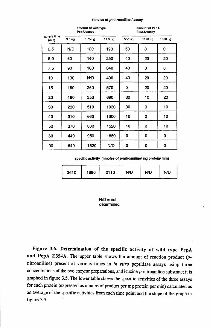
nmoles of p-nitroaniline / assay
amount of wild type amount of PepAPepA/assay E354A/assay
sample time ----------------------- --------------------- --------------------------------------------(min) 3.5 ug 8.75 ug 17.5 ug 560 ug 1120ug 1680 ug
2.5 N/D 120 190 50 0 0
5.0 60 140 250 40 20 20
7.5 90 180 340 40 0 0
10 130 N/D 400 40 20 20
15 160 260 570 0 20 20
20 190 350 690 30 10 20
30 230 510 1030 30 0 10
40 310 660 1300 10 0 10
50 370 800 1520 10 0 10
60 440 950 1650 0 0 0
90 640 1320 N/D 0 0 0
specific activity (nmoles of p-nitroaniline/ mg protein/ min)
2610 1980 2110 N/D N/D N/D
N/D = not determined
Figure 3.6. Determ ination of the specific activity of wild type PepA and PepA E354A. The upper table shows the amount of reaction product (p- nitroaniline) present at various times in in vitro peptidase assays using three concentrations of the two enzyme preparations, and leucine-p-nitroanilide substrate; it is graphed in figure 3.5. The lower table shows the specific activities of the three assays for each protein (expressed as nmoles of product per mg protein per min) calculated as an average of the specific activities from each time point and the slope of the graph in figure 3.5.

Ala change in PepA E354A may result in a temperature sensitive mutant no such step
was utilised in this purification; the maximum temperature that the proteins were
exposed to was 37 °C as the E.coli culture was grown. Figure 3.4 shows a SDS-
polyacrylamide gel of the purified proteins, and compares them to PepA previously
prepared following the Vogt protocol. This shows that both the wild type and mutant
proteins were purified to near homogeneity in this preparation, and that they both
appeared to be the same size (approximately 55kDa) as heat-purified PepA. This
suggests that the mutation made in pRM40 has not introduced a stop codon in pep A s
open reading frame or a protease cleavage site in PepA, both of which would result in
production of a truncated enzyme.
Aminopeptidase assays were performed on the purified enzymes using leucine-p-
nitroanilide (purchased from Sigma) as a substrate (see Materials and Methods). Figure
3.5 shows a graph of the spectrophotometrically determined amounts of reaction
product (p-nitroaniline) made over time in these assays. The assays were performed
using three different concentrations of both wild type and mutant PepA, and, for
comparison, the result is shown in tabular form along with the specific activities
calculated from the assays (fig.3.6).
As expected, wild type PepA produced increasing amounts of p-nitroaniline with
time, and the rate of this production increased as the concentration of the enzyme was
increased. In contrast, no reaction product was detected in the assays involving the
mutant PepA preparation. It should be noted that the concentrations of PepA E354A
used in this experiment were substantially higher than the concentrations of wild type
PepA: 0.25, 0.62 and 1.25 ug/ml of wild type; and 40, 80 and 120ug/ml of mutant (i.e.
approximately 100X more mutant than wild type in each assay). The product
concentrations from each reaction time point, and the slopes of the graph in figuip3.5,
were then used to calculate specific activites for each concentration of wild type PepA.
These were compared and the average specific activity for the wild type preparation was
calculated as 2230U/mg of protein, which is higher than the activity determined
previously for PepA (1300U/mg; Stirling et al, 1989, and Szatmari pers.comm.). The
4 2

lack of reaction products seen using PepA E354A meant it was not possible to
determine a specific activity for this protein preparation. This experiment was repeated
on two separate occasions by myself and independently by Mary Burke, and
comparable specific activities for wild type PepA were determined in each case.
Similarly, no detectable activity was seen using the mutant PepA preparation in these
assays - even when the reaction was allowed to proceed for three hours.
These experiments suggest that the mutant PepA used in these assays is highly
deficient in aminopeptidase activity. It was not possible to quantify the amount that the
catalytic activity is reduced in this protein preparation since no reaction products could
be detected in the assay used. The lack of reaction product suggests that PepA E354A ist o
unable^catalyse the hydrolysis of leucine-p-nitroanilide, or at least that any residual
activity in this enzyme preparation is beyond the limits of detection of this assay.
It seems unlikely that the inactivity of the E354A mutant PepA preparation is
simply a result of the treatment of this particular purified sample. This can be concluded
because the wild type enzyme was prepared in the same manner and at the same time
and was highly active, and also because the enzyme appeared to be the correct size on
protein gels. This therefore suggests that the lack of aminopeptidase activity in the
mutant is a result of the site-directed alteration to PepA’s amino acid sequence. The
glutamic acid to alanine change (or indeed other chance, uncharacterised changes made
during the mutagenesis; see discussion) could have this effect either by perturbing the
structure of PepA or by specifically disrupting the integrity of the active site.
Structural perturbations of the protein could either be general conformational
changes because E354 is involved in organising the tertiary structure of PepA, or could
be local changes to the active site region which make the mutant unable to recognise
leucine-p-nitroanilide. The latter possibility is a problem in this analysis because such
local changes may simply be confined to leucine-p-nitroanilide as a substrate, and other
(natural) substrates may still be readily processed by PepA E354A (this is considered
more fully in later sections). Available evidence from other experiments suggests that
conservative site-directed changes in protein sequence (where the new amino acid's
43

side chain occupies less space than the natural residue and does not form novel
interactions) rarely cause widespread conformational alterations (see e.g.
Leatherbarrow and Ferscht, 1986; Brodo and Argos 1990). However, since there is no
satisfactory method of determining whether a given site-directed change has this effect
(short of solving the 3-D structure of the mutant protein), this possibility cannot be
excluded in explaining the reduction in peptidase activity of PepA E354A.
The importance of the above result as regards analysing the role of PepA in cer
site-specific recombination is that the E354A mutant is as depleted in peptidase activity
as can be detected in in vitro assays. For this reason it was not felt necessary to create
further PepA mutants before analysing the Xer phenotype of this PepA derivative.
3.4 Determination of the xer phenotype of PepA E354A in vivo
Ideally the ability of this PepA E354A to support cer recombination would have
been tested in vitro, since the same reaction conditions employed for determining its
peptidase activity could have been used in cer recombination assays and a strong
correlation between the two properties could have been derived. Unfortunately, the cer
reaction has not yet been recreated in vitro and it was therefore necessary to determine
the xer phenotype of PepA E354A in vivo. This was achieved by doubly transforming
pRM40 and pCS202 (a cer reporter plasmid; see fig.4.21), or pCS126 and pCS202,
into DS941pepA::Tn5 (CSX17). The transformants were selected on media containing
both ampicillin and chloramphenicol, and their DNA was analysed by boiling
preparations and agarose gel electrophoresis (fig.3.7). This showed that both pCS126
and pRM40 were able to complement the chromosomal pep A:: Tn5 mutation and allow
the complete recombination of pCS202 to its resolution product (pCS203, which is
indicated). Restriction of the DNA samples with Pstl confirmed that pCS126 contained
wild type pepA and pRM40 contained p^pAE354A. This suggests that the E354A PepA
mutant, which is peptidase- in vitro, is Xer+ in vivo.
4 4

CSX17 Psndigested
o s S aCM O C M ^
CO ™ CO $CO f < CO
$■ Q. S' Q.C M C O CM C O + C O _ C O + C O _O O O C M 2 C M 2 C M 2 c m 2C M C M CM T - 2 J - ^c o c o c o c o ^ E CO 2 CO S co - Eo o o o i r o c c o a ; o ocQ . Q . Q . Q . Q . a a a a Q . Q .
pCS202
pCS203__
p C S 1 2 6 / ^pRM40
Figure 3.7. Complementation of an X er- pepA ::T n5 insertion by wild type PepA and PepA E354A. DS941pepA::Tn5 (CSX 17) was transformed with pCS202 alone, with pCS202 and pCS126 or with pCS202 and pRM40. Plasmid DNA
was isolated and run on a 1.2% agarose gel before and after Pstl restriction. The reporter plasmid pCS202 is resolved in Xer+ strains to give the plasmid pCS203. pCS126 strongly expresses wild type PepA from the tac promoter; pRM40 is the same plasmid as pCS126, but expresses PepA E354A. Pstl restriction identifies the site- directed sequence alteration to the pep A gene that distinguishes the two plasmids

Interpretation of this experiment is complicated by the numerous potential
differences between the analysis of the activities of PepA E354A in vitro and in vivo.
One possible interpretation of the fact that the mutant appears to be Xer+ is that the
peptidase deficiency described in vitro is reflected in vivo, and therefore the role that
PepA plays in the cer recombination reaction does not require that PepA be active as an
aminopeptidase, or that it has other catalytic activities. A number of objections to this
are possible, however, and all would mean that this experiment could not determine
whether the peptidase activity of PepA is required for the cer reaction:
(i) It is conceivable that the description of PepA E354A as being peptidase' is
only due to the insensitivity of the leucine-p-nitroanilide assay, and in vivo there is
sufficient enzymatic activity from the protein expressed from pRM40 to allow cer
recombination.
(ii) The lack of activity in vitro when using PepA E354A may have been
overcome in the different in vivo reaction conditions. This could, for instance, have
been due to local structural alterations resulting from the glutamic acid to alanine change
being overcome in the cell.
(iii) It is possible that the mutant PepA has been specifically altered in its ability to
recognise the substrate analogue leucine-p-nitroanilide, and this does not reflect any
change in the activity of the enzyme on other substrates. For instance, it is possible that
PepA has a capacity to specifically recognise one of the xer proteins and this enzymatic
activity is unaltered in the E354A mutant.
3.5 Determination of the peptidase activity of PepA E354A in vivo
E.coliK ll is sensitive to valine and valine-containing peptides when grown on
minimal medium. This is because valine is feedback inhibitor of both its own synthesis
and that of isoleucine, and therefore if isoleucine is not included in the medium the cells
cannot grow (Miller, 1987). This property has been exploited by using various valine-

0.05 mM 0 mM 0.5 mM
0.1 mM 0.2 mM
pCS126/DS941
pRM40/DS941
pBAD/DS941
pBAD/CSX17
pRM40/CSX17
pCS126/CSX17
F igure 3.8. D eterm ination of the peptidase activ ities of wild type PepA and PepA E354A in vivo. Both DS941 and DS941p^pA::Tn5 (CSX17) were transformed with pBAD, pCS126 or pRM40 and grown for approximately 15 hours on minimal agar containing with either 0, 0.05, 0.1, 0.2 or 0.5 mM Val-Leu- N H 2 . The organisation of each plate into six sectors is diagrammed and the
concentration of Val-Leu-NH2 in each plate is indicated. Val-Leu-NH2 is cleaved by aminopeptidase A and releases valine, which is toxic to E.coli. pCS126 expresses wild type PepA, pRM40 expresses PepA E354A and pBAD is the vector used in these plasmids.

containing peptides to select for E.coli derivatives that were mutant in aminopeptidases
and in peptide uptake (valine resistant mutants; Miller and Schwartz, 1978). The
molecule Val-Leu-NH2 was demonstrated to be a relatively specific substrate ffor
aminopeptidase A in E.coli because all strains isolated which were resistant to this
dipeptide but still sensitive to valine were mutant in pep A. Val-Leu-NH2 has also beten
used more recently in our laboratory to attempt to select for peptidase", X er+/X er
mutants (H.O'Mara, pers.comm.; see discussion). These experiments suggested that
the dipeptide would be a suitable molecule to determine whether the peptidase
deficiency of PepA E354A is seen in vivo, and therefore to address some of the
considerations discussed in section 3.4.
This analysis was performed by transforming pCS126 (pepA wild type), pRM40
(pepA E354A) and pBAD (expression vector alone) into both DS941 arnd
DS941p^pA::Tn5 (CSX17) and growing the cells overnight in L-agar supplemented
with ampicillin. Transformants from each plate were then streaked onto minimal media
(containing ampicillin and the amino acid supplements detailed in materials and
methods) containing the following concentrations of Val-Leu-NH2: 0, 0.05, 0.1, 0.2
and 0.5 mM. The cells were allowed to grow overnight before being photographed
(fig.3.8).
Without Val-Leu-NH2 all the cells grew equally well. Increasing concentrations
of Val-Leu-NH2 were generally toxic to both DS941 and CSX17 cells containing
pBAD, since they grew progressively more poorly. This suggests that the dipeptide can
infact be cleaved in the absence of functional PepA, presumably by the other
aminopeptidases. However, at 0.5 mM Val-Leu-NH2, pBAD/CSX17 grew while
pBAD/DS941 did not. This shows that this assay is able to detect the amount of PepA
made from the single chromosomal pepA gene in wild type cells and distinguish that
amount of activity from the peptidase activity in pepA mutant strains; it also illustrates
that PepA is the enzyme that predominantly cleaves this dipeptide in the cell. Both
strains grew very poorly when they contained pCS126; they did not grow detectably
even at 0.05 mM Val-Leu-NH2. This suggests that the amount of peptidase activity in
4 6

these cells is substantially higher than cells without the expression vector, and that the
level of expression of PepA from pCS126 more than compensates for the loss of the
enzyme in CSX 17. Both DS941 and CSX 17 containing pRM40 grew as well as the
same cells containing the pBAD vector alone; there was no evidence of the expression
of PepA from this plasmid causing the cells to efficiently cleave Val-Leu-NH2 as in
pCS126-containing cells. Significantly there was as much growth of pRM40/CSX17 at
0.5 mM Val-Leu-NH2 as there was of pBAD/CSX17. This suggests that there is no
more peptidase activity in cells containing pRM40 than there is in pep A:: Tn5 mutants,
despite the fact that PepA should be expressed to the same high levels as in the cells
containing pCS126. Furthermore, three separate repetitions of this experiment showed
some growth of pRM40/DS941 at 0.5 mM Val-Leu-NH2 whilst there was no growth
of pBAD/DS941. This may be a consequence of PepA E354A monomers expressed
from pRM40 forming heterohexamers with wild type PepA monomers expressed from
the chromosome, and resulting in a population of hexameric PepA in the cells which
cannot cleave the toxic dipeptide.
This experiment confirms that the lack of PepA E354A-catalysed hydrolysis of
leucine-p-nitroanilide seen in vitro reflects a more general reduction in the mutant's
enzymatic activity. This is important because it rules out the possibility that the Xer+
phenotype of this protein in vivo is a consequence of the in vitro peptidase" phenotype
being overcome by the different cellular reaction conditions. The fact that PepA E354A
is reduced in its ability to cleave two different substrates suggests that the mutation
might have a general effect. This, however, cannot exclude the possibility that the
mutant enzyme is still capable of processing other substrates (e.g. xer proteins).
A difficulty in correlating the reduction of enzymatic activity in vitro with the xer
activity in vivo was the possibility that pRM40 expressed sufficient protein for the level
of enzymatic activity to be high enough in the cells to allow cer recombination
(assuming that PepA acts enzymatically in the xer reaction). Within the levels of its
sensitivity, this experiment suggests thatpRM40 expresses no more peptidase activity
than a pepAv.Tn5 strain (CSX17). This means that, at least as regards Val-Leu-NH2, a
4 7

PepA protein (in this case PepAE354A) can be expressed in in vivo assays where it has
no more peptidase activity than an X er, pepA knockout strain and it can support cer
recombination. The problem with concluding that this means that PepA has a structural
role in cer recombination is that these assays may not be sensitive enough to detect the
residual amount of peptidase activity that is allowing the cer reaction to proceed, and the
fact that the mutant enzyme may still have full enzymatic activity when it is directed
against substrates for which it has a higher specificity.
3.6 Complementation in CSX17 using pRM21
It was intended that this analysis would be concluded by replacing the wild type
DS941 pep A gene with the pepAE354A site-directed mutant. Construction of such a
strain would have allowed the mutant pepA to be transcribed from the gene's natural
promoter, and therefore would have allowed cer recombination assays to be performed
where the mutant enzyme was expressed at wild type levels in the cell. This would havebe
meant that any residual peptidase activity from PepA E354A would^reduced below that
made from pRM40, and therefore if the protein was being used as a enzyme in the cer
reaction this may detect it. To do this the plasmids pRM41 and pRM45 were
constructed. These are derivatives of pRM40 and pCS126 respectively, in which a
kanamycin resistence gene is inserted into a unique Stul recognition sequence between
the pep A gene and downstream orfl3 (see Colloms, 1990 and Stilling et al, 1989 for
details of this open reading frame). The plasmids were linearised by restriction with
B am H I and transformed into the strain JC7623 (the use of JC7623 for strain
construction is discussed in Chapter 4; see also Winans et al, 1985). It was hoped that
this linear transformation would allow the plasmid-borne (kanamycin resistant) pepA
genes to exchange with the chromosomal copies of the genes by homologous
recombination, thereby creating a selectable strain containing the pepAE354k gene in
the natural chromosomal location of pep A, as well as a control strain. Several attempts
failed to yield any kanamycin resistant JC7623 derivatives (perhaps because of the
4 8
specific activities (nmoles p*nitroaniline/mg extract/min)
strain no heat 70 oC heatextract treatment treatment
DS941 16.6 15.4
CSX17 4.7 0
pCS126/CSX17 95.2 91.2
pRM40/CSX17 5.1 0
pRM20/CSX17 17.8 17.6
pRM21/CSX17 9.9 0
F igu re 3.9. Am inopeptidase activities of cell ex tracts of p e p A wild type and m utant strains, and strains containing plasmids expressing wild type PepA or PepA E354A. DS941pepA::Tn5 (CSX 17) was transformed with pRM20, pRM21, pCS126 or pRM40. 100 ml cultures of these transformants, and of DS941 and CSX17, were grown overnight at 37 °C and concentrated cell extracts prepared. As in figures 3.5 and 3.6 the activities were determined using leucine-p- nitroanilide as a substrate and the amount of p-nitroaniline product was calculated spectrophotometrically. The specific activities are expressed as nmoles of product per mg of extract per min, and are the average that was determined for two concentrations of the various extracts, before and after heating to 70 °C for 5 mins.

CSX17 pst\d ig e s te d
CM CM CM CMO O O OCM CM CM CMX CO CO CO CO
CO O O O Oo C L CL CL CLCM OO CMo o o A 4 : O t - O T— OC \| CM CM CM CM CM CM CM CM CM CMCO C O C O 5 2 2 2 2 2 2O O O cr cr e ra : a: a: cr crC L a . CL c l a. C L CL CL C L CL CL
pRM20/pRM21
F igure 3.10. Com plem entation of an X er- p ep A ::Tn5 insertion by pRM 20 and pRM21. DS941/?epA::Tn5 (CSX17) was transformed with either pCS202, pRM20 or pRM21. The strains containing pRM20 and pRM21 were then transformed with pCS202. The plasmid DNA was run on a 1.2% agarose gel, before and after digestion with Pstl. pCS202 is resolved by xer recombination to give pCS203. pRM20 expresses wild type PepA, pRM21 expresses PepA E354A (the plasmids are distinguished by Pstl digestion); the genes encoding the two proteins are poorly transcribed from the T7 polymerase promoter in both plasmids, and therefore the levels of enzyme in the cells are lower than in cells containing pCS 126 and pRM40
(see fig.3.9).
pCS203

disruption to the expression of orfl3, whose function is not understood) and therefore
it was not possible to perform this experimental analysis.
In the plasmids pRM20 and pRM21 the expression of the pepA genes is driven
by the promoter from bacteriophage T7 (fig.3.3). Because this promoter is not
recognised by E.coli RNA polymerase (Tabor and Richardson, 1986) the transcription
level of the pepA genes is likely to be very poor, and therefore these plasmids offer the
opportunity to express PepA E354A at low levels in the cell and perform an equivalent
experiment to the strain construction described above. To determine the level of PepA
expressed from the plasmids, crude cell extracts were prepared from 100 ml cultures of
the following strains: DS941, CSX17, pRM20/CSX17, pRM21/CSX17,
pCS126/CSX17 and pRM40/CSX17 (Materials and Methods). Aminopeptidase assays
were then performed (using leucine-p-nitroanilide as a substrate) on the extracts before
and after heating to 70 °C for five mins. The specific activities determined from these
assays are tabulated in figure 3.9.
CSX17 (DS941/?£/?A::Tn5) displayed a low amount of peptidase activity which
was reduced to 0 U/mg of extract after heating. This is because PepA is the only heat
stable aminopeptidase in E.coliK ll. The activity seen before heating represents
cleavage of leucine-p-nitroanilide by the other cellular aminopeptidases. DS941 had
approximately 15 U/mg extract after heating; this agreed with the activity determined
previously (Stirling etal, 1989) and represents the activity derived from chromosomally
encoded PepA. Both before and after heat treatment, the cells containing pCS126
showed a high level of peptidase activity (approx. 96 U/mg extract) that was absent
from pRM40-containing cells (this activity is lower than the 600U/mg determined by
Stirling et at). This is in agreement with the other experiments of this chapter which
suggested that pCS126 expresses PepA to levels considerably exceeding that made
from chromosomal pep A, and that pRM40 expresses PepA E354A which has
undetectable peptidase activity. When wild type PepA was expressed from pRM20 the
amount of activity was approximately 17 U/mg extract, which was approximately 10-
fold lower than pCS126-containing cells and was comparable to DS941. This suggests
| 4 9ii

that the amount of PepA made from pRM20 is little more than is made from the
chromosomal pepA gene, and consequently the level of any peptidase activity
expressed from pRM21 will be 5-fold lower than from pRM40.
To determine the xer activities of pRM20 and pRM21 they were transformed into
CSX 17. After selecting for the presence of these plasmids by growth on tetracycline,
the reporter plasmid pCS202 was transformed into the cells and double transformants
were selected on media containing chloramphenicol and tetracycline. It was necessary
to establish the expression vectors in CSX17 before transforming in pCS202 because
the resistance determinant of the reporter plasmid that is lost during cer resolution is
tetracycline. The DNA from the double transformants was prepared by the boiling
technique and analysed by agarose gel electrophoresis (fig.3.10). Both pRM20 and
pRM21 were able to complement the pep A:: Tn5 mutation in CSX 17 and allow the
complete recombination of pCS202. This suggests that the PepA E354A is Xer+ even
when expressed at low levels that are comparable to the expression of PepA from the
chromosome, and therefore if PepA acts enzymatically during the cer reaction the low
levels of peptidase activity expressed from pRM21 are sufficient to achieve this
function.
3.7 Discussion
The experiments in this chapter describe the catalytic properties of a single site-
directed derivative of E.coli aminopeptidase A, and utilise this mutant enzyme to
analyse the function of PepA in the cer site-specific recombination reaction. In the
mutagenesis, the amino acid residue Glu354 was replaced by alanine. This specific
alteration was based on the published structure of bovine lens leucine aminopeptidase
(Burley et al, 1990 and 1992). Glu334 was implicated as one of the seven active site
residues in BLLAP that was involved in the chelation of Zn2+ ions; Glu 354 was

chosen for mutation in PepA because it is the equivalent residue to Glu334 and is
therefore believed to co-ordinate the binding of Mn2+.
The mutant derivative of PepA (PepA E354A) is deficient in its ability to
hydrolyse substrates both in vitro and in vivo (leucine-p-nitroanilide and Val-Leu-
NH2, respectively). This suggests that Glu354 might be at the catalytic site of the
enzyme and may be important in the catalytic mechanism of PepA, since replacement of
the residue with Ala has compromised the active site of the enzyme. However,
discussion of the catalytic role of Glu334 and the implications of the mutagenesis
experiment for considerations regarding the enzymatic mechanism of BLLAP and PepA
are inappropriate. This is for a number of reasons: the lack of aminopeptidase activity
may be due to tertiary structural alterations in PepA and not purely a result of the
enzyme being unable to chelate Mn2+; the pepAE354A gene was not sequenced and
therefore the possibility of mutations in other active site residues cannot be excluded;
and there is no available model for the mechanism of peptide hydrolysis by these
enzymes.
The mutagenesis described above was not performed in order to analyse the
catalytic mechanism of PepA, but instead to examine the protein's role in the cer site-
specific recombination reaction. Explicitly, this experimental approach was attempting
to create a peptidase-deficient PepA derivative which could be tested for its ability to
support cer recombination. This was done because of our inability to examine the role
of PepA in in vitro assays (e.g. it would be relatively simple in vitro to remove Mn2+
ions from the reaction buffer and determine whether recombination could proceed). The
fact that PepA E354A displayed no detectable catalytic activity when using leucine-p-
nitroanalide as a substrate in vitro or Val-Leu-NH2 in vivo, means that it is as "good" a
mutant as can be produced for this experimental approach. The mutant enzyme is Xer+
when highly expressed from pRM40 (tac promoter) and when poorly expressed from
pRM21 (unidentified promoter sequences in pSELECT). This represents suggestive,
but not conclusive (see below), evidence that the aminopeptidase activity of PepA is not
5 1
required in cer site-specific recombination. Some independ^itly performed experiments
offer circumstantial support for this conclusion:
(i) Val-Leu-NH2 was used to isolate E.coli pep A mutants which were
subsequently tested for their Xer phenotype (H.O'Mara and G.Szatmari, pers.comm.).
A large number of the strains isolated from this screening procedure were both X er
and peptidase", but one clone (named HOM38a) was resistant to Val-Leu-NH2 (i.e.
peptidase") and was Xer+. The gene encoding this PepA mutant has been subcloned
(plasmid pH31.9) as a 1.9 kbp Hindlll fragment, but the mutation has not been
characterised. The mutant strains which were Xer" and peptidase" have also not been
characterised, but could be explained as encoding truncated PepA derivatives.
(ii) It has been shown that E.coli grown in media containing high concentrations
of bestatin are resistant to Val-Leu-NH2 but are still able to recombine cer reporter
plasmids (G.Szatmari, pers.comm.). This can be interpreted as suggesting that in these
conditions PepA is not catalytically active but is still able to support cer recombination,
and it is therefore not functioning as an enzyme during the reaction.
The first reason that none of these experiments can be considered conclusive is
the fact that it is not possible to rule out any residual peptidase activity in the reaction
conditions used (either from an incompletely mutant protein or a subpopulation of PepA
escaping bestatin inhibition) causing the Xer+ phenotype. This is primarily because the
limits of detection of the peptidase assays that were used are not known. It is also
because of the non-quantitative nature of the in vivo cer recombination assay: in vitro it
would be possible to determine whether the reaction rate had been altered by the
reduction in PepA's catalytic efficiency, whereas in vivo a reduction in the reaction rate
might not be detected because the reporter plasmids are able to recombine to completion
before sufficient cells have grown to prepare DNA samples. However, no
aminopeptidase activity at all has been determined for PepA E354A and therefore,
despite the above arguments, it is entirely possible that PepA does not act as an
aminopeptidase in cer recombination.
52

A more difficult problem to explain in this experimental approach is the possibitty
that PepA E354A remains Xer+ either because the enzyme retains full enzymatic
activity when it acts on substrates for which it has a higher specificity (e.g. the xer
proteins) than the analogues used in these experiments, or because it has other
enzymatic activities unaffected by the mutation made. The former of these objections
does not seem likely to me because the E354A alteration almost certainly perturbs the
general catalytic activity of PepA, and not only its activity against some substrates. The
second possibilty cannot be ruled out, although it is not obvious what alternative
enzymatic roles PepA might have in cer recombination.
To determine conclusively the PepA does not act enzymatically, future
experiments would need to take a more positive approach. For example, it should be
possible to isolate (or make) X er, peptidase+ derivatives of PepA if the interpretation
of the experiments described in this chapter is correct. This could be achieved either by
classical genetic techniques, or perhaps by creating a PepA derivative that is truncated
in the same way as trypsin-cleaved BLLAP (which retains its peptidase activity; see
section 3.1). If these experiments suggested that PepA was not required to process the
other Xer proteins or to cleave small peptide molecules during the recombination of cer-
like sites then it would become necessary to consider in more detail how PepA
functions as a structural component of the reaction machinery. Since there is no
evidence that it interacts with the cer site directly, it may be possible to perform gel
binding assays in combination with other Xer proteins to see if it is involved in protein-
protein interactions. Ultimately, however, it is probable that the function of PepA in cer
recombination will be unravelled by analysing the cer reaction in vitro.
53

Chapter 4
Analysis of cer site-specific recombination in vivo
using the controllable strain RM40
5 4

4.1 Introduction
To be able to analyse the mechanism of the xer site-specific recombination
reaction it is necessary to be be able to perform the reaction and examine the reaction
products in a controlled manner. Ideally this would be achieved by recreating the
system in vitro, since this would allow the concentrations of the protein and DNA
components to be altered, as well as the reaction conditions and type of substrates used.
Many site-specific recombination reactions have been reconstituted in vitro (see Chapter
1) and to achieve this for xer, ArgR and PepA have been purified (Stirling et al, 1988
and 1989) and XerC is being purified (G.May, pers.comm.). This has allowed the role
of these proteins in xer recombination to be analyzed using the gel binding assay (see
above refs, and Fried and Crothers, 1981). It has also made it possible for the proteins
to be added to cer and d//reporter plasmids in various simple buffers to look for the
products of recombination. Experiments of this kind, and also experiments using crude
cell extracts, have so far failed to yield positive results (G.May, S.Colloms,
G.Szatmari pers. comms.). This could be for a number of reasons; for instance, the
precise conditions required for recombination in vitro may not have been developed
yet, or alternatively some essential factors have not yet been identified.
In the absence of an in vitro assay, it was decided to develop a system in vivo in
which xer recombination could be controllably analysed. Such a system would be
unlikely to be as manipulable or as informative as an in vitro assay. However,
experiences from other systems suggested that it could yield some mechanistic data.
Controlled, in vivo experiments have been performed in the analysis of the site-
specific recombination reactions catalysed by X Int (Bliska and Cozzarelli, 1986), Tn3
Resolvase (Bliska et al, 1991) and bacteriophage PI Cre (Adams et al, unpublished).
All these systems have relied on being able to control the expression of the recombinase
gene. This allowed the recombination reaction to be switched on at a set time and
allowed the reaction products to be examined at various times subsequently. To achieve
this control of expression, the genes encoding X Int (int) and Tn3 resolvase (tnpR)
55

were cloned downstream of the lambda leftwards promoter (k P l ) in plasmid
constructions also containing the gene c/557, which expresses the thermolabile k P l
repressor CL In this way the expression of the recombinases is turned off when grown
at 28-30°C and turned on at 37-42°C, when the repressor is thermally inactivated. (The
methods used in the Pl Cre in vivo system are not known to me.)
The experiments performed in all of these cases were topological analyses of the
products of the recombination reactions, and it was shown that the products made in
vivo are not the same as those observed in vitro. In the case of k Int the reaction
products are nearly entirely free circles and not the catenanes and knots seen in vitro;
similarly Tn3 resolvase produces free circles and not singly-linked catenanes (see
Chapter 1). It was demonstrated that the reason for these results is the action of the
enzyme DNA gyrase, which efficiently unlinks knots and catenanes in vivo. When this
enzyme activity was inhibited by the drug norfloxacin (a superpotent analogue of
nalidixic acid) the patterns of reaction products seen in vivo matched those predicted by
in vitro experiments. These experiments have been interpreted as suggesting that the
reactions proceed by the same mechanism in vivo and in vitro. The results of the Cre
studies appear not to be as simple as this, and may suggest that this reaction produces
topologically more complex products in vivo from those observed in vitro.
The above discussion is not intended as an exhaustive description of the
experiments performed in these systems, but simply to illustrate the type$of experiment
that have been performed using in vivo assays. The amount of manipulation that is
possible is clearly not as extensive as can be achieved in vitro: the amounts of the
component proteins and the physiological conditions of the reactions cannot be readily
changed. Nevertheless, the experiments can be performed in a controlled manner and
mechanistic information about the reactions can be obtained. The types of substrate
plasmids used in these experiments can clearly be varied quite considerably (e.g.
comparing cer/dif in xer recombination). An important difference exists between
analysis of the xer recombination reaction and the analyses described above, however.
Since no in vitro recombination experiments have been performed for x e r
5 6

recombination, any mechanistic results that can be derived would not be confirmation
of already existing data, as was the case for X Int and Tn3 resolvase.
This chapter reports the development of a controllable xer recombination system
and the use of this system in determining that the xer reaction involves Holliday
junction structures, which have previously been implicated in vitro as reaction
intermediates in X integrase-type recombination.
4.2 Construction of strains RM10, RM20, RM30 and RM40
It was decided that the gene encoding XerC would be used to control the xer
recombination reaction in vivo. It is conceivable that controlling the expression of argR
or pepA could allow the xer recombination reaction to be tightly regulated in the same
way that controlling int achieved for the X Int system and tnpR for resolvase. However,
because dif recombination does not require ArgR or PepA, and because XerC was
believed to be the xer recombinase (see Chapter 5), xerC was chosen. It was also
decided that the promoter used to control xerC expression would not be A,Pl but the lac
promoter and its associated operator. Expression from the lac promoter is repressed by
the protein LacI, which binds an operator sequence (O) found downstream of the
promoter and overlapping the transcription start point (Beckwith, 1987). The repressor
is thought to act by stopping the binding of RNA Polymerase. Addition of glucose to
the bacterial culture can enhance this repression by indirectly reducing the binding of
the catabolite activator protein (CAP), which assists the binding of RNA Polymerase.
LacI can be inactivated by the gratuitous activator IPTG , which acts by stopping the
repressor binding its operator sequence, and therefore turns on the expression of genes
controlled by lacPO. The decision to use lacPO was a result of two factors. Firstly, it is
readily available for cloning in a number of vectors commonly used in our laboratory
(e.g. pUC18/19). Secondly, the strain that has been ubiquitously used in previous
cer/dif recombination experiments is DS941, which is a lacIQ mutant. Use of this strain
57
would therefore offer maximal repression since it makes more than normal amounts of
repressor, as well as offering experimental continuity.
The cloning steps involved in constructing this controllable strain are detailed in
figure 4.1. pSD102 (Colloms, 1990) contains a 3.6 kbp H ind lll-B g lll E .coli
chromosomal fragment that contains the genes dapF, orf235, xerC and part of the
fourth gene of the xerC operon or/238, as well as the upstream gene cyaX. The
plasmid pRMlOl was generated by inserting a 230 bp PvuII-Smal fragment from
pUC19 containing lacPO into a unique Nrul site that is 96 bp upstream of the xerC start
codon and is within the coding sequence of or/235. The orientation of this insert was
determined by restriction with Pstl (Pstl cuts 10 bp from the Smal side of the lacPO
insert and also within pSD102).
It was intended that the controllable xerC construction would be inserted into the
E.coli chromosome and replace the wild type xerC gene (see below). For this reason an
antibiotic resistance determinant was required to select for this insertion. A
transcriptional terminator was also required upstream of the lacPO insert; this is because
xerC is part of an operon, and without a terminator transcription from the operon's
promoter would read through the lac promoter and compromise the expression control.
These requirements were achieved in three steps. Firstly, the strong ribosomal RNA
terminator was cloned as a 450 bp Sspl-HindUl fragment from pKK223-3 (Pharmacia)
into Bglll-H indlll restricted pIC20R, creating pRMlO. Secondly, the kanamycin
resistance gene from Tn903 was cloned from pUC71K as a 1.3 kbp Pstl fragment into
Pstl digested pRMIO, creating pRM ll. EcoRl restriction of this plasmid releases a
1.8kbp fragment containing the terminator and kanamycin resistance genes. The
fragment was cloned into Stul digested pRMlOl, creating pRM102, in which
approximately 440 bp of operonic sequence have been replaced by this insert. This
deletes the final 180 bp of dapF (the C-terminal 58 amino acids of DapF) and the first
260 bp of or/235 (the amino-terminal 85 residues of ORF235). Note that the orientation
of the inserted fragment was determined by BamHl restriction (BamHl cuts within the
lacPO insert and in the polylinker of pR M ll, beside the kanamycin gene), and the
58

HpSDI 02 \-
0
cyaX
>operonicpromoter
dapF orf235 xerC ^
(orf238)
S P S P ■L U - 1 L -
N
3 kbp
(N)
Kanamycin rmB resistance terminator
(S) (S)
Bg
(Pv) lac PO (Sm)
(230bp)
pRMlOl I
(R)B
(1.8kbp)
pRMI 02
Figure 4.1. Plasmid clonings involved in the construction of a stra in in which the transcrip tion of xerC can be controlled. The top plasmid diagram is adapted from Colloms et al (1990) and shows only the E.coli chromosomal insert of the plasmid pSD102; it details the genes present in the insert and the important restriction sites. Below are the plasmids created by sequentially inserting (upstream of xerC) the lac promoter and operator of pUC19 (as a 230 bp Smal-Pvull fragment: making pRMlOl), and a 1.8 kbp EcoRl fragment carrying the kanamycin resistence gene of Tn903 and the rrnB transcriptional terminator (making pRM102).
Restriction enzyme recognition sites sites are shown as follows:Bg=£g/II U=HindUi N=Nrul P=Pstl Pv=Pvull R=EcoRl S=Stul Sm ^Sm al
B =BamHl

Pvx/ll digested- pRM102
\ Z i i i i i | | | | | i i > - 1 1 1 1 1 i]1111 ■ ■ • m
X X double recombination events
E.coli ------------------------------chromosome
dapF orf235 xerC orf238
RM10, 20, 30, 40
kanamycin rrnB lacPOresistance terminator
iiiiiiiiiiiiiiiiiiiiiiiiiiiiiiiiiiii| | | | | | iii- iiiiiiiiiiiiiiiiiiiiii| | | | | | ih>
A dapF A o r f235 xerC orf238
Figure 4.2. Construction of strains RM10, RM20, RM30 and RM40 by linear transformation of JC7623.Linear DNA fragments transformed into the strain JC7623 and containing a selectable copy of a gene with a homologue in the E.coli chromosome can exchange with that copy via a double recombination event (Winans et al, 1985). In this case the 4.4 kbp PvuU DNA fragment contains the kanamycin resistence gene, and exchange generates strains in which the xerC gene is transcribed from the lac promoter rather than its natural operonic promoter. In addition to the inserted lacPO fragment, the kanamycin resistant strains RM10, 20, 30 and 40 contain insertions within dapF and orf235 upstream of xerC, and are therefore likely to be mutant in these genes. These constructions have not been checked by Southern blotting, but are based on genetic characterisation of the strains.

kanamycin gene was previously orientated in pRM ll by Hindlll restriction (which cuts
within the polylinker and at one end of the inserted gene). As shown, the kanamycin
gene is transcribed in the opposite direction to the operon and should not further burden
the ribosomal RNA terminator.
Plasmid pRM102 was used to create a strain in which the chromosomal copy of
xerC is expressed from the lac promoter rather than its natural promoter. This was
achieved by transforming the linearized plasmid into the strain JC7623, which has the
genotype recB, recC, sbcB. The recB/C mutations in this strain mean that it will not
efficiently degrade linear DNA that has entered the cytoplasm, whilst the sbcB mutation
suppresses the strain's recombinational defects. Winans et al (1985) were the first to
use this technique of strain construction in E.coli, and they showed that if linear DNA
transformed into JC7623 contains a copy of an E.coli gene it can exchange with the
chromosomal copy by a double recombination event (see fig.4.2). In this way they
used JC7623 to create various E.coli mutants. The technique was also used by Richaud
et al (1987) to make mini-Mu insertional mutants in all the genes of the xerC operon
(Colloms, 1990).
In this study, pRM102 was digested with Pvull, which cuts twice within the
vector but not within the insert, and transformed into JC7623 by standard CaCl2
methods. Forty-three colonies were isolated and replica patched onto kanamycin and
onto ampicillin-containing L-agar plates. Of these 43 colonies 42 were resistant to
kanamycin but sensitive to ampicillin, suggesting that their chromosomal xerC had
exchanged with the pRM102 xerC construction. Four of these colonies were then Pl
transduced into DS941, creating strains RM10, RM20, RM30 and RM40.
Figure 4.2 shows the presumed chromosomal organization of these strains. This•f
is based on the known restriction maps of the plasmid constructions, on evidence
presented in the above papers that the linear DNA inserts by a double recombination
event and on genetic characterisation of the strains (see below). Because of these
considerations Southern blots were not performed to determine the status of the xerC
region. However, it is probable that RM10, 20, 30, 40 are dapF and orf235 mutants
59

because of the lacPO insertion into orf235 and the deletions made in dapF and orf235
during the plasmid constructions. orf!38 is downstream of xerC and is believed to be
co-transcribed, therefore when XerC is expressed from the lac promoter it is likely that
oif238 will be transcribed also.
4.3 Controllable in vivo recombination in strains RM10, 20, 30 and
40 .
To determine whether these strains are capable of controlling the expression of
xerC , and therefore able to be used in in vivo recombination assays, they were
transformed with the cer reporter plasmid pKS455 (Summers, 1989; see fig.4.20). The
transformed bacteria were plated onto either L-agar plates containing ampicillin alone,
or onto L-agar plates containing both ampicillin and ImM IPTG. A number of colonies
from these plates were pooled and their plasmid DNA content visualized by agarose gel
electrophoresis.
Figure 4.3A shows the result of this experiment. Lanes 9 and 10 are marker lanes
showing pKS455 and its resolution product (p456) that is produced after
transformation into DS941. For each of RM10, 20, 30 and 40 this recombination only
occuffed when IPTG was present in the plates (lanes 2, 4, 6 and 8), suggesting that
without IPTG induction there is little, or no, XerC expressed in these strains.
The transformed strains growing on the non-IPTG plates were then patched out
onto another set of IPTG+ and - plates and their plasmid DNA was analysed in the
same way as before (fig.4.3B). This experiment shows two things: the repression of
xerC expression is not complete, since some pKS456 reaction product was visible in
the uninduced (non-IPTG) lanes; and in these strains the repression of xerC can be
overcome by addition of IPTG to the growing culture, since transfer to IPTG-
containing plates resulted in resolution of pKS455.
6 0

(A) IPTG - + - + - + - +
lane 1 2 3 4 5 G 7 8 9 10
II I• m wg
!f
xpKSkS5
\ p « 6
08)IPTG - + - + - + - +
lane 1 2 3 4 5 6 7 8 9 10
pKS4-5 5
. ^ 5 6
Figure 4.3. cer m ediated site-specific recom bination in stra ins RM10,
RM20, RM30 and RM40. The strains were transformed with the cer reporter
plasmid pKS455 and grown on L-broth with and without 1 mM IPTG. pKS455 is resolved by intramolecular recombination to produce p456. Plasmid DNA was prepared by the single colony lysis technique and run on a 1.0% gel. (A) DNA after overnight
growth of the transformants. (B) DNA after repatching the transformants growing on
IPTG- plates onto IPTG+ and IPTG- plates. For both A and B:
Lanes 1, 2: RM10; lanes 3, 4: RM20; lanes 5, 6: RM30; lanes 7, 8: RM40; lane 9: pKS455; lane 10: p456.

Together these two experiments show that the four strains meet the criteria
demanded for a controllable in vivo system. Reporter plasmids can be introduced into
the strains under repressed conditions such that they are not recombined, and at a set
time subsequently the recombination reaction can be turned on.
4.4 Analysis of pSD115 recombination in RM40
Strain RM40 was transformed with the cer reporter plasmid pSD115 (Colloms,
1990; see fig.4.4) and plated onto L-agar supplemented with 1% glucose, 50ug/ml
diaminopimelic acid and ampicillin. Glucose was included in this assay to increase the
repression of xerC. Diaminopimelic acid (the enzymatic product of DapF) was included
because it was observed that the transformed cells grew better on plates containing the
acid than on plates without it; this may be a consequence of the mutation created in
dapF during the construction of this strain.
A large number of the transformant colonies were pooled and inoculated into 40
ml of L-broth supplemented with 1% glucose and ampicillin and grown, shaking, at
37°C until the culture reached approximately mid-log phase (the OD^OO was not
determined). At this point the 40 ml culture was split into two separate 20 ml cultures;
IPTG was added to one at a final concentration of 2 mM and then both allowed to
continue growing. At the time of XerC induction and 30, 60, 90 and 120 minutes
subsequently DNA was prepared from both cultures using standard boiling
preparations, except that extraction with phenol/chloroform preceded isopropanol
precipitation. The DNA prepared in this way was analysed by agarose gel
electrophoresis (fig.4.5, lanes 1-10).
Without IPTG induction of xerC expression there was no recombination over the
time course of the assay; this can be seen by the fact that the substrate, pSD115, was
unchanged (lanes 1-5). A small amount of the replicative resolution product of the
61

EcoRl 4.95H incll 4.51 M lul 0.27
'coRV 0.48Seal 4.45cer
H incll/Sall 0.95
pSD1154.95 kbp N rul 1.27
o n
cer
M lul 2.62
Xer dependant resolution
M lul 0.27
H incll 2.16
Seal 2 .1 0 -cer
o n
M lul 0.27
cer
EcoRV 0.48
H incll/Sall 0.95Nrul 1.27
2.60 kbp 2.35 kbp
Figure 4.4. D iagram m atic representation of X er-dependflnt resolutionof the cer reporter plasmid pSD115. pSD115 was constructed in pBR322 by S.Colloms (1990). The plasmid is resolved in Xer+ strains to yield 2.6 kbp and 2.35 kbp-sized, circular products. Note that only the 2.6 kbp-sized product contains a replication origin, and hence the smaller recombination product is rapidly lost from bacterial cultures.

IPTGuninduced induced ^ CLJQ
Q <0COsam ple time 0306090120l0306090>120 c l c \ i
(mins)
lane 1 2 3 4 5 6 7 8 9 10 11 12
Figure 4.5. Time course of cer recom bination in vivo using stra in RM40. Two in vivo reactions were set up using pooled colonies of RM40 transformed with pSD115: in one (lanes 6-10) XerC expression was induced by addition of 2 mM IPTG to pSDl 15/RM40 at mid-log; in the other (lanes 1-5) the cells were grown on 1% glucose and XerC expression was not induced at the equivalent time. Time points refer to the time the plasmid DNA samples were made after the addition of IPTG. The DNA samples were made by boiling preparations (including phenol/chloroform extraction) and were run on a 1.2% agarose gel. In Xer+ strains pSD115 resolution yields a 2.6 kbp, replicative product and a 2.35 kbp, non-replicative product (which is not observed in standard cer recombination assays; lane 12).

reaction was visible but the amount did not change during the assay, meaning that it
probably arose during the transformation of RM40 and the overnight growth on the
plate. In contrast, induction of XerC caused the appearance of two DNA species that
increased in quantity from 30 to 120 rains (lanes 6-10). These correspond to the two
products of pSDl 15 xer dependent resolution (see fig.4.4). The larger reaction product
(2.6 kbp) is that normally seen when pSD115 is transformed into DS941, because it
contains the origin of replication and is therefore maintained in the cell population. The
smaller (2.35 kbp) product is normally not detected in recombination assays with wild
type cells because it has no replication origin; the fact that it was detected in this
recombination time course suggests that this is a more sensitive assay. Detection of the
non-replicative product, and the increase in amounts of both products, shows that xer
recombination continued throughout the assay.
The products of this reaction are unlinked circles. However, this cannot be said
be to the initial topological structure of the products since no attempt has been made to
inhibit DNA gyrase in these experiments. The in vivo experiments on X Int and Tn3
resolvase-catalysed reactions described in the Introduction showed that without
treatment with norfloxacin these reactions also produced free circles, despite these not
being the products seen in vitro. Preliminary experiments using norfloxacin during cer
in vivo assays suggested that the drug strongly inhibits the recombination reaction (data
not shown). The meaning of this observation is not known, however, because these
experiments were not followed up.
In figure 4.5 it can be seen that the pSDl 15 substrate band's appearance changed
after the induction of XerC. This is caused by the presence of a second, cer
recombination-dependent DNA species which had a slightly faster mobility and made
the substrate band look like a doublet. This was analysed further by restriction
digestion of the DNA samples with EcoRl, which cuts once within pSD115 and once
within the 2.6 kbp product (see fig.4.4). An agarose gel of these restrictions is shown
in figure 4.6.
62

EcoRl uncut
IPTGinduced uninduced
sam ple time 0 3 0 6 0 9 0 1 2 0 I03 06 0 9 0 1 2 Q (mins)
in
□
QlX I
in
Q
C LX I
CO CD CO CD
Q. C \i C L C \i
linear pSD 115
Holliday junction
linear 2.6 kbp
supercoiled 2.35 kbp
supercoiled pSD 115
supercoiled 2.6 kbp
lane 1 2 3 4 5 6 7 8 9 10 11 12 13 14
F ig u re 4.6. E c o R I re s tr ic tio n an a ly sis o f pSD115 in vivo recom bination tim e course. Samples of the plasmid DNA from figure 4.5, representing cer mediated recombination of pSDl 15 in the strain RM40 over 120 mins, were digested with EcoRl and run on a 1.2% agarose gel. In this gel the IPTG-induced lanes are 1-5, the uninduced control samples are in lanes 6-10. A single EcoRl recognition site is present in pSDl 15 and in the 2.6 kbp, replicative product (see figure4.4 and lanes 11-14); the 2.35 kbp product contains no EcoRl sites. The putative Holliday junction structure (Ot) is indicated.

Four DNA species with distinct electrophoretic mobilities were seen (lanes 1-5).
The fastest migrating band had a slightly greater mobility than supercoiled 2.6 kbp
product (compare to lane 14) and is therefore unrestricted 2.35 kbp product; the slower
DNA species is linearized 2.6 kbp product (compare to lane 12). As expected, these
products were only seen after IPTG induction and increased in quantity thereafter. The
slowest migrating band was present throughout the time course and was the only DNA
seen in the uninduced control lanes (6-10), therefore it is linearized pSDl 15 (compare
to lane 11).The fourth DNA species had an electrophoretic mobility that was
intermediate between supercoiled and linearized pSD115 substrate (compare to lanes
11-12), only appeared after XerC induction and was not present in the uninduced
samples. The appearance of this DNA was therefore dependent on the induction of the
cer recombination reaction, and consequently it may represent the restricted form of the
DNA species that was barely distinguishable from pSD115 before EcoRl restriction
(fig.4.5, lanes 6-10). The fact that it was not seen in the uninduced samples, nor in the
O' time point, strongly argues that this is not simply a contaminant DNA but is related
to the cer recombination reaction.
A possible explanation for this DNA species is that it represents a Holliday
junction, or DNA with similar physical characteristics. If a Holliday junction were
formed between the two cer sites in pSD115 it would be confined to this 312bp of
homology (if it were capable of branch migration), since the DNA between the cer sites
is non-homologous pBR322 sequence (fig.4.4). This would therefore explain the
increase in the mobility of the DNA when restricted by EcoRl, because the digestion
converts a "figure-8" structure containing two supercoiled domains into an "a"
structure which has a single supercoiled domain and two linear "tails".
Holliday junctions were initially proposed as being intermediates in homologous
recombination (Holliday, 1964). However, there is considerable evidence that
equivalent structures are intermediates during X integrase-like site-specific
recombination reactions (see Chapter 1 for review and refs.). If this DNA species does
indeed represent such a Holliday junction then it may represent an intermediate in the
63

cer recombination reaction, and offer an insight into the reaction's mechanism. The
following sections detail experiments designed to determine whether this DNA species
is indeed a Holliday junction.
4.5 Electrophoretic analysis of putative Holliday junctions
The hypothesis that this DNA species produced during cer mediated site-specific
recombination in RM40 is a Holliday junction rests on the observed change in its
electrophoretic mobility when restricted with one enzyme, EcoRl. The reason for this
interpretation is the belief that the Holliday junction is embedded within regions of non
homology, causing the substrate to adopt a supercoiled figure-8 structure in which the
two supercoiled domains (separated by the junction) are the size of the reaction
products (2.6 and 2.35 kbp). Within these domains are a large number of restriction
enzyme recognition sites, present only once (Seal, EcoRl, EcoRW, Sail, Nrul\ see
fig4.4), or twice (Hincll, Mlul) within the sequence of pSD115. If the DNA is in a
Holliday junction conformation then digestion with these enzymes will generate either
a structures of subtly different shapes when cut once, or % structures when both
domains are cleaved. The differences in the conformations of these derivatives should
be reflected in alterations in their mobilities during gel electrophoresis.
RM40 transformed with pSDl 15 was used to set up a 20 ml culture (as described
in section 4.4) and XerC expression was induced at mid-log phase with 2 mM IPTG.
90 mins after induction, plasmid DNA was prepared by the boiling method, including
phenol/chloroform extraction. Figure 4.7 shows restriction digestions of this DNA,
using the enzymes which cut only once, after agarose gel electrophoresis.
The bands corresponding to the putative a structure Holliday junctions are
indicated, and it is clear that they had different electrophoretic mobilities in this gel. The
mobilities of these structures might have been predicted to simply be a consequence of
the size of the supercoiled domain, .i.e. those with the smaller domain remaining
6 4

p S D 1 15/R M 40 pSD115
-=0^5 O 0^:75 c O 0,0 COW.DCOUjUjC/)^
oT5 O 0 ^ " 5COOOCBVD C /) U J U 1 C / ) ^
a Holliday junctions
linear pSD 115
supercoiled pSD115
linear 2.6 kbp linear 2.35 kbp
supercoiled 2.6 kbp supercoiled 2.35 k&p
Figure 4.7. Analysis of the electrophoretic m obility of pSD115- derived a structures. RM40 transformed with pSD115 was grown to mid-log
phase and XerC expression induced for 90 mins with 2 mM IPTG. Plasmid DNA samples were prepared, restricted with either Seal, EcoRl, EcoRV, Sail or Nrul and run on a 1.2% agarose gel. For comparison, pSD115 DNA digested with the same enzymes was also run on this gel. The positions of the restriction enzyme recognition sequences in pSDl 15 and how they are distributed into the cer mediated 2.6 and 2.35 kbp resolution products is detailed in figure 4.4. Supercoiled and linear substrate and product molecules are indicated, as are the putative a structures.

supercoiled after restriction digestioi having equivalent mobilities and migrating faster
than a molecules with a larger sipeicoiled domain. This is not the case, since Seal and
EcoRl a molecules ran differently in the gel, despite both being cleaved in the 2.35 kbp
domain; similarly the three a mofeciles with 2.6 kbp supercoiled domains had different
mobilities. These data suggest that the "arms" that extend from the a structures
contribute significantly to the electrophoretic mobilities of the DNA. It appears that if
one arm is very long and the other very short the structures have greater mobility than
when the two arms are more equally sized. This is illustrated by comparing the a
structures that were produced by EcoRN, Sail and Nrul restriction. These enzymes cut
progressively further from the cer sits (174, 640 and 961 bp respectively) and created a
molecules with decreasing gel mobilities. This is confirmed by the slower migration of
the Seal a in the gel than the EccR Icl: Seal cuts 515bp further from the cer site than
does EcoRl.
Figure 4.8 shows an agarose gel of the same DNA restricted with Hincll and
Mlul, both of which cut twice in pSDl 15. For reference it was again cut with EcoRl.
Digestion with Hincll linearised both of the product molecules and cleaved pSD115
into 3.55 and 1.4kbp fragments; it also resulted in a putative Holliday junction with a
slower mobility than both pSD115 and the EcoRl a structure. This alteration in
electrophoretic mobility is consistent with cleavage in both supercoiled domains,
resulting in a Holliday junction that has a % structure. M lul also linearised both
products, but because it cuts within the cer sites it digested pSD115 to give product
sized linear molecules. Despite cutting twice, as H indi does, Mlul digestion did not
result in the formation of % structures (lane 4); there was no band corresponding to
anything other than linear products and a very small amount of linear substrate (which
was probably a consequence of slightly incomplete restriction). This is most readily
explained by the Holliday junction being capable of branch migration throughout the cer
site region of homology. Mlul restriction would initially create a % structure, but the
Holliday junction would move to the ends of the molecule by branch migration and
product-sized, linear molecules would be released.
65

p S D 1 15/R M 40 p S D 1 15
= ® 7 3 -c8 -s|3 Hi I S
3® 73,-£ 8 . s 5i u j i s
.linear pSD115
,pSD 115 (3.55 kbp)
.supercoiled pSD 115
linear 2.6 kbp linear 2.35 kbp
supercoiled 2 .6 kbp supercoiled 2.35 kBp
pSD 115 (1.4 kbp)
F igure 4.8. R estric tio n digestion of p u ta tiv e , pS D 115-derived Holliday junctions in both supercoiled domains. Samples of the DNA shown in figure 4.7, representing pSD115 after 90 mins of cer mediated site-specific recombination in RM40, were digested with either EcoRl, HittcR, or Mlul and run on a 1.2% agarose gel. For comparison, pSD115 DNA digested with the same enzymes was run on the same gel. Figure 4.4 shows the position of the single EcoRl recognition site present in pSDl 15 and in the 2.6 kbp product, and the positions of the two Hincll and Mlul sites in pSD115 (which are found as single sites in each product). The putative a and x Holliday junction structures are indicated, as well as supercoiled and linear
substrate and product molecules and the pSD115 fragments generated by H incll digestion.
Holliday junction
Holliday junction

linear pSD115
supercoiled pSD115
linear 2.6 kbp
pSD115 .
linear 2.6 kbp .
linear 2.35 kbp ■
. V7 \
KfOAck
linear 2.6 kbp .
linear 2.35 kbp.
supercoiled ■■ —2.6 kbp
supercoiled ■ — supercoiled2.35 kbp 2.35 kbp
pSD115
u n cu t EcoRl Hincll Mlul
Figure 4.9. D iagram m atic represen tation of the a lte ra tio n s in the e lec tro p h o re tic m obility of the pu ta tive H olliday ju n c tio n s a f te r restriction digestion. The proposed structures adopted by the Holliday junctions are shown, for simplicity, without supercoiling in their undigested domains; it is not
intended that this imply that these domains are nicked, and it is likely that Holliday junctions that have been nicked in these ways would have different mobilities to those shown. Uncut, the Holliday junctions contain two supercoiled domains and adopt a "figure-8" structure whose electrophoretic mobility is nearly indistinguishable from supercoiled pSD115 substrate DNA. A single cleavage in either supercoiled domain generates a more retarded a structure. Cleavage in both supercoiled domains results in highly retarded x structure molecules. Digestion within both cer sites allows the
Holliday junction to branch migrate to the ends of the DNA and results in productsized, linear DNA fragments.

All the above electrophoretic analysis is summarised in figure 4.9. The analysis is
fully consistent with this recombination-dependflnt DNA species containing a Holliday
junction. Restriction with Mlul suggested that the structure is centred on the cer sites of
pSD115 and is capable of branch migration. All of these DNA samples were stripped of
protein by phenol/chloroform extraction, therefore excluding the possibility that these
bands represent a protein-bound structure, for example synapsed cer sites.
4.6 The putative Holliday junctions are substrates for cleavage by
RuvC in vitro
The experiments described above are consistent with this cer recombination-
derived DNA species being a Holliday junction. It was, however, felt necessary to
examine the conformation of the DNA further. This was examined by determining
whether the species is a suitable substrate for an enzyme that specifically cleaves 4-way
DNA junctions.
A number of enzymes have been described that can act as "junction-resolvases".
The first to be identified was the product of bacteriophage T4's gene 49, TA
endonuclease VII (Mizuuchi et al, 1982; Lilley and Kemper, 1984). This enzyme has a
critical role in the life cycle of phage T4 because it cleaves branched DNA structures
which form during the replication of the phage (Mosig, 1987). Analysis of the
enzymatic activity of the purified protein showed that it is not specific for 4-way DNA
junctions, since it will also cleave 3-way junctions, heteroduplex loops and extended
single-stranded termini. Because of these diverse activites this protein was felt not to be
a suitable enzyme to probe the structure of the cer recombination junctions.
Bacteriophage T7 encodes an enzyme (the product of gene 3, T7 endonuclease I)
that is utilised during the life cycle of this phage in a very similar way to how
endonucleaseVII acts during T4 growth, indeed it has been demonstrated to be able to
complement T4 gene 49 mutants, in vitro analysis of the purified enzyme (Parsons and
66
West, 1990) showed it to be slightly more specific than T4 endonucleaseVII. For
instance, it footprints all four strands of a synthetic 4-way junction point and cleaves in
both possible orientations. Again, however, this endonuclease will recognise and
cleave 3-way junctions, making it unsuitable for this study.
Enzyme activities of the type described above have also been discovered using
synthetic 4-way % structures in extracts from eukaryotic cells. For example, three
apparently distinct endonuclease activities have been partially purified from S.
cerevisiae (Jensch et al, 1989). However, in this, and in other cases from higher
eukaryotes, the specificities, and indeed the identities, of the enzymes are not yet clear,
and are therefore not suitable for these experiments.
Recently Connolly and West (1990) used a structures which had been artificially
generated using RecA to identify activity from E.coli cell extracts that would resolve
Holliday junctions. They further showed that this activity was absent in extracts made
from ruvC mutants (Connolly et al, 1991). The gene encoding RuvC has been cloned
(Takahagi et al, 1991 and Sharpies and Lloyd, 1991) and been found to be part of a
"ruv" locus. ruvC is located upstream of two genes, ruvA and ruvB, which constitute a
SOS-regulated operon; ruvC is itself part of a two-gene operon (the other gene is an
unidentified open reading frame, orf26). Mutations in any of the 3 ruv genes confer a
"ruv phenotype": they are deficient in homologous recombination in various genetic
backgrounds. A single ruv mutant, however, shows only slightly reduced levels of
recombination. This has been interpreted as meaning they are involved late in
homologous recombination, e.g. in a resolution step (see e.g. Lloyd, 1991).
Biochemical analysis of the roles of these ruv proteins is under way and all three
have been purified. The roles of RuvA and RuvB are not yet clear, but the function of
RuvC has been determined. Purified RuvC will act in vitro to cleave RecA-generated
Holliday intermediates without the need for RuvA or RuvB. It will also cleave synthetic
4-way junctions (Holliday junctions or cruciforms) in both possible orientations
without RecA (Dunderdale eta l, 1991, and Iwasaki eta l, 1991). The studies also show
that RuvC will not recognise 3-way junctions, or any of the other structures which
67

RuvC - - + +EcoRl + - +
(A)
linear pSD115
supercoiled pSD115,
supercoiled 2.6 kbp supercoiled 2.35 kbpx
nicked pSD115
a Holliday junction
nicked 2.6 kbp •nicked 2.35 kbp
1 2 3 4
(B) pSD115-derived Holliday junction
RuvCVertical cleavage products
Horizontal cleavage product
nicked 2.6 kbp circle
nicked 2.35 kbp drde
doubly nicked 4.95 kbp circle
Figure 4.10 (A) T reatm ent of putative Holliday junctions in vitro with R uvC . RM40 transformed with pSD115 was grown to mid-log phase, XerC expression was induced for 90 mins with 2 mM IPTG and plasmid DNA was prepared from the culture and purified on a CsCl gradient The DNA was then incubated in vitro with purified RuvC (lanes 3 and 4) and run on an agarose gel with (lane 4) and without (lane 3) subsequent digestion by EcoRl (this experiment was performed by S.West). Lanes 1 and 2 represent undigested (lane 1) and EcoRl digested (lane 2) controls in which the DNA has not been treated with RuvC. (B) Schematic illustration of R uvC -ca ta lysed reso lu tion of superco iled pSD 115-derived H olliday junctions. Cleavage by RuvC in a vertical orientation (as drawn) would generate nicked 2.6 and 2.35 kbp DNA species; cleavage horizontally would result in doubly nicked pSDl 15 substrate-sized DNA.

served as substrates for the phage endonucleases, in the reaction conditions employed
in analysing 4-way junctions. Furthermore, the 4-way junctions must have at least 12
bp of homology before they act as substrates for RuvC.
These studies offer compelling evidence that RuvC is at least one of the E.coli
enzymes which cleave Holliday junction intermediates that form during homologous
recombination. The specificity of RuvC for 4-way junctions also made it the most
informative enzyme for this study.
1.2% agarose gels (23cm in length) run for approximately 16 hours at 50 volts
(e.g. see fig.4.5) give the best separation of supercoiled pSD115 and supercoiled
"Holliday junction" that I have achieved. Because of this problem, and also because of
the loss of material incurred when DNA is isolated from gels, it was not possible to
purify supercoiled Holliday junctions, and therefore a CsCl preparation of pSD115
from RM40 after 90 mins induction was prepared by myself and used by S.West in in
vitro assays using purified RuvC. Figure 4.10A shows an agarose gel of this assay; the
in vitro conditions used for treatment with RuvC are published (Dunderdale et al,
1991).
Lanes 1-2 show the DNA preparation before exposure to RuvC. As stated, this
was a mixture of pSD115 substrate, products and putative Holliday junctions. Without
restriction the supercoiled substrate and Holliday junction were indistinguishable in this
gel (lane 1). However, after restriction with EcoRl the a structure representing the
Holliday junction was clearly visible (lane 2). Lanes 3-4 are the same samples after
treatment with RuvC (note that EcoRl restriction followed RuvC treatment). EcoRl
restriction showed that the a band was no longer present, whilst the substrate and
product bands had not been depleted. This suggests that RuvC has specifically cleaved
the supercoiled Holliday junction. Examination of the unrestricted DNA (lane 3)
revealed an increase in the amount of nicked (open circle) form of pSD115 and both
products. This would be consistent with the action of RuvC on supercoiled Holliday
junctions, as illustrated in figure 4.10B. RuvC simply cleaves a 4-way junction in
either of two possible orientations; it does not reseal the nicked DNA. For a supercoiled
68

molecule this would result in either singly nicked product-sized molecules (2.6 and
2.35 kbp in this case) or doubly nicked substrate molecules, depending on which
orientation is used. If the nicked DNAs in this assay arose from the action of RuvC it
suggests that RuvC cleaves these Holliday junctions in both orientations.
This experiment was compromised by the inability to distinguish the supercoiled
Holliday junctions from the substrate. The apparent reduction in the amount of
substrate band in the unrestricted sample (lane 3) must be assumed to have resulted
from RuvC cleavage of the Holliday junctions present within that band. An alternative
explanation could be that the increased amount of nicked material was a result of non
specific nicking during the RuvC treatment. Some observations argue against this,
however: there was no concurrent reduction in the amounts of supercoiled products, as
would be expected from non-specific nicking; after EcoRl digestion it was only the a
structure that had been removed (if anything, the amounts of linear pSDl 15 and linear
2.6 kbp product appeared to have increased; compare lanes 4 and 2).
Although reservations must be acknowledged, this result is nevertheless
interesting (and is strengthened when considered in conjunction with the previous
electrophoretic analyses), and may be taken as further evidence that these structures are
4-way DNA junctions. If RuvC is able to recognise and cleave this DNA it suggests it
has at least 12bp of sequence homology within the junction. Futhermore, the size of the
nicked "resolution products" corresponded to the cer recombination products,
suggesting that the junction is centred on the cer sites. Both of these interpretations are
in agreement with Mlul restriction analysis (see above).
4.7 Electron microscopy of isolated % structures
This section describes direct visualisation of the structure of the putative Holliday
junctions by electron microscopy. This was achieved by restricting the DNA prepared
above (section 4.6) with Hincll and isolating the presumptive % stucture from an 1.2%
69

M im t
0.33 um
Figure 4.11. Electron m icrographs of isolated x s tructures. A Sample of
the plasmid DNA shown in figure 4.10 was digested by Hincll and run on a 1.2% agarose gel. DNA was purified from the band representing the Hincll % structure and
spread, in 40% formamide, onto grids and shadowed with platinum : palladium for electron microscopy. The migrographs are at 90,000X magnification and the bar shown represents 0.33 um, which corresponds to approximately 1 kbp of DNA.

Hollidayjunction
arm lengths in cm fpercentage of total)total length
long arm (1) long arm (2) short arm (1) short arm (2) of arms (cm)
1395a 6.3 (41.4) 5.0 (32.6) 2.2 (14.3) 1.8 (11.7) 15.31395b 6.0 (38.5) 5.4 (34.6) 2.1 (13.5) 2.1 (13.5) 15.61391a 5.4 (35.6) 5.3 (35.3) 2.2 (14.5) 2.2 (14.5) 15.11391b 6.7 (42.9) 4.8 (30.8) 2.3 (14.7) 1.8 (11.5) 15.61389B 6.1 (43.3) 4.5 (31.9) 2.2 (15.6) 1.3 (9.3) 14.11392 6.7 (42.4) 5.0 (31.6) 2.5 (15.8) 1.6 (10.1) 15.81388a 5.8 (38.7) 5.4 (36.0) 2.0 (13.3) 1.8 (12.0) 15.01388b 6.7 (42.9) 4.8 (30.8) 2.3 (14.7) 1.8 (11.5) 15.61388c 6.4 (42.1) 4.6 (30.2) 2.5 (16.4) 1.7 (11.2) 15.21393B 5.7 (37.5) 5.1 (33.6) 2.2 (14.5) 2.2 (14.5) 15.21394a 6.2 (41.3) 4.9 (32.7) 2.1 (14.0) 1.7 (11.3) 15.01394b 6.0 (40.5) 4.5 (30.4) 2.4 (16.2) 1.9 (12.8) 14.81393a 6.9 (45.1) 4.6 (30.1) 2.2 (14.4) 1.6 (10.5) 15.31393b 6.6 (41.5) 5.3 (33.3) 2.0 (12.6) 2.0 (12.6) 15.91388B 6.8 (44.1) 4.4 (28.6) 2.7 (17.5) 1.5 (9.7) 15.41390 6.0 (38.0) 5.5 (34.8) 2.1 (13.3) 1.9 (12.0) 15.81389a 6.0 (41.7) 4.5 (31.2) 2.4 (16.7) 1.5 (10.4) 14.41389b 5.6 (36.6) 5.6 (36.6) 2.1 (13.7) 2.0 (13.1) 15.31404 5.9 (38.3) 5.4 (34.7) 2.1 (13.5) 2.1 (13.5) 15.51405 6.3 (38.7) 5.6 (34.4) 2.5 (15.3) 1.9 (11.7) 16.31412 5.9 (38.3) 5.5 (35.7) 2.1 (13.6) 1.9 (12.3) 15.41410 6.1 (38.4) 5.6 (35.2) 2.2 (13.8) 2.0 (12.6) 15.91411 5.7 (37.5) 5.4 (35.5) 2.2 (14.5) 1.9 (12.5) 15.21409 6.2 (41.6) 4.5 (30.2) 2.6 (17.4) 1.6 (10.7) 14.91408a 6.1 (41.5) 4.8 (32.7) 2.2 (15.0) 1.6 (10.9) 14.71408b 6.1 (41.5) 4.7 (32.0) 2.2 (15.0) 1.7 (11.6) 14.71406 6.4 (41-8) 4.9 (32.0) 2.4 (15.7) 1.6 (10.5) 15.31407 5.9 (40.4) 4.8 (32.9) 2.1 (14.4) 1.8 (12.3) 14.61403B 6.1 (40.9) 4.9 (32.9) 2.4 (16.1) 1.5 (10.1) 14.91403a 5.7 (40.4) 4.5 (31.9) 2.0 (14.2) 1.9 (13.5) 14.11403b 5.9 (40.1) 4.7 (32.0) 2.1 (14.3) 2.0 (13.6) 14.71403c 6.4 (43.5) 4.2 (28.6) 2.5 (17.0) 1.6 (10.9) 14.71400 6.2 (43.4) 4.5 (31.5) 1.9 (13.3) 1.7 (11.9) 14.31402 5.8 (38.9) 5.3 (35.6) 2.0 (13.4) 1.8 (12.1) 14.9
averagepercentages
40.6(35.6-45.1)
32.7(28.6-36.6)
14.3(12.6-17.5)
11.8(9.3-14.5)
measured sizes (bp)
2010(1762-2232)
1619(1416-1812)
708(624-866)
584(460-718)
predicted sizes (bp)
1990(+/-156)
1565(+/-156)
773(+/-156)
620(+/-156)
Figure 4.12. Arm lengths of individual % structures. Contour lengths (incm) of the arms of 36 % structures (determined from the electron micrographs using a map measuring instrument) are shown in the upper table. There were four different sized arms in each structure, and the table is arranged in columns, with the largest arm size on the left and smallest on the right The total sizes of the structures (determined by summing the arm lengths) is in the rightmost column; the percentage of total size that each arm represents is shown in brackets. The lower table details the four arm lengths (and variation around the figure) calculated using the average for each structure, based on the size (4950 bp) of pSD115. For comparison, the predicted arm lengths are also shown (note that the Holliday junction is capable of branch migration, and hence the figure varies +/- 156 bp).

agarose gel. Isolation of the DNA involved low speed centrifugation of the gel chip
through siliconised glass wool (after visualising the DNA by ethidium bromide
staining; see Materials and Methods), ethanol precipitation and resuspension in TE
(pH8.0). The % structures prepared in this way were next spread onto grids in 40%
formamide, and shadowed for electron microscopic examination (see materials and
methods and Coggins, 1987). This work was done in collaboration with Lesley
Coggins. Figure 4.11 shows representative electron micrographs of the DNA seen by
this technique.
The structures shown are clearly consistent with the DNA being a % structure with
a central Holliday junction. Formamide treatment can be seen to have spread the DNA
out at the Holliday structure, causing it to appear as a "hole" where the four strands
meet. The majority of the molecules seen on the grid could be determined to be %
structures, although in many cases the arms had become fixed such that they crossed
over each other and the hole at the point of strand convergence was not always clearly
visible. The grid also contained smaller pieces of linear DNA, which could have arisen
by shearing of the structures during their preparation, or may simply have been
background contaminant DNA that was accidentally isolated from the gel.
Figure 4.12 shows the results of measuring the arm lengths of 36 % structures
from the electron micrographs. Only molecules in which the path of each arm could be
accurately determined, and where the Holliday structure could be rigorously assigned,
were measured. The sizes of the arms are shown in centimetres, and the percentage of
the total size that each arm represents is shown in brackets. The sizes suggested that
each molecule contained two long and two short arms. Furthermore, in each case there
appeared to be one larger and one smaller arm in both groups; this is reflected in the
arrangement of the measurements into columns. The average total size was 15.1 cm,
the maximum measured size was 16.3 cm and the minimum was 14.1 cm; these values
are within the amount of variation expected by experimental error. The top row in the
lower table shows the average percentage that each arm represents of the total molecule
size, and the variation around that figure. From these averages the arm lengths of the
7 0

molecules were calculated, based on a molecule of 4.95 kbp - the size of pSDl 15. The
calculated sizes were: 2010bp +222/-248; 1619bp +193/-203; 708bp +158/-84; 584bp
+134/-124. These coiTespond well with the predicted arm lengths: 1990bp +/--156;
1565bp +/-156; 773bp +/-156; 620 +/-156. The predicted sizes are based ofl the
positions of the Hincll recognition sites within pSD115, assuming that the Holliday
junction is centred on the cer site, and take into account the possibility that the junctions
can branch migrate throughout the cer site.
These results confirm that these DNA structures are % structures and therefore
show that a Holliday junction that is produced during cer recombination in RM40. The
measurements confirm the prediction, based on restriction analysis, that the Holliday
junction is within the cer sequence of pSDl 15.
4.8 The nature of the first pair of strand exchanges
In the formation of Holliday junctions four possible combinations rf str'and
exchanges can be made between the recombining DNA duplexes (this is illu:trate<d in
fig.4.13). The two cer sites are depicted as being aligned in parallel dirimg the
recombination reaction. There is, however, no experimental evidence tvailatble
regarding how cer sites are aligned. This has been done simply to clarify the terms "'top
strand" and "bottom strand" when they are discussed.
Exchanges that are made between two top strands or two bottom strandsresuft in
Holliday junctions that are resolved by a second pair of strand exchanges betveen the
two bottom and two top strands respectively. Both of these reaction mechanisns, wfhen
performed between directly repeated cer sites (as in pSDl 15), result in resolutiin <of the
substrate molecule to give two product circles (of 2.6 and 2.35 kbp in ths case).
Holliday junctions created by either of these strand exchange regimes wmlld be
indistinguishable by the experiments performed in the previous sections.

pSD
115
T3CCOI—GO
CO£ CO **“ 0> CD O )
s 5•— CO CO J C CO o o X Q . CD
Figu
re
4.13
. Di
agra
m
of the
po
ssib
le
stra
nd
exch
ange
co
mbi
natio
ns
in the
fo
rmat
ion
of H
ollid
ay
junc
tions
. Th
e up
per
diag
ram
sh
ows
the
cer
sites
of
pSD1
15
align
ed
in pa
ralle
l be
fore
str
and
exch
ange
. Th
e cr
osso
ver
regi
ons
are
repr
esen
ted
by in
verte
d ar
row
s; t
he lef
t an
d rig
ht
arms
of
the
cros
sove
r are
de
noted
by
L an
d R
resp
ectiv
ely;
and
on
e sit
e is
disti
ngui
shed
fro
m the
ot
her
by da
shes
at
the
arm
s (L
' and
R'
). Fo
ur
pairs
of
stran
d ex
chan
ges
that
woul
d ge
nera
te
Hol
liday
ju
nctio
ns
are
theo
retic
ally
po
ssib
le
(as
show
n).
The
reac
tion
prod
ucts
that
wou
ld
be
gene
rated
by
a se
cond
pa
ir of
stran
d ex
chan
ges
(see
text)
on the
se
struc
ture
s are
de
taile
d.

It is also possible to create Holliday junctions by exchanging the top and bottom,
or bottom and top strands of the recombining duplexes (fig.4.13). Resolution of these
junctions would be by bottom to top, and top to bottom second strand exchanges
respectively. Both these arrangements would fuse the left side of one recombining site
to the left side of the recombining partner, and reciprocally right side to right side,
resulting in inversion of the DNA intervening between the two cer sites of pSD115.
Such a mechanism is unlikely because experimental evidence has shown that
recombination of directly repeated cer sites always results in a resolution reaction and
not inversion. However, formation of these junctions is theoretically possible and it is
not clear whether the previous experimental evidence of this chapter can exclude their
existence; for example, it is possible that the orientation of the cer sites is not detected
during their recombination until after the formation of a Holliday junction.
In figure 4.13 the first pair of strand exchanges are shown as occurring towards
the left-hand side of the cer core site. It should be noted that this is simply for
convenience and is based on location of the point of first strand exchange being to the
left of the spacer sequence in X Int-catalysed reactions (if the att recombination sites are
drawn in the same arrangement as the cer sites in figure 4.13: Kitts and Nash, 1988b;
see Chapter 1). In the case of cer recombination the positions in the crossover sequence
where the strand cleavages and exchanges take place are not known, and the definition
of the spacer sequence (see fig. 1.9) has been derived by comparing a number of cer-
like sites. The first pair of strand exchanges may occur to the right of this presumptive
overlap and be resolved, after branch migration, to the left. Indeed the initial strand
exchanges could occur within any part of the "core" sequence and be resolved at any
other position (even the same position) without altering the consequences of the four
reaction schemes described, nor compromising the analysis of the nature of the first
pair of strand exchanges described below.
Figures 4.14 and 4.15 show the details of the determination of the pair of strands
exchanged in these cer-derived Holliday junctions. A 100ml culture of pSD115/RM40
was grown to mid-log phase and xerC expression induced by addition of 2mM IPTG.
72

linear pSD 115
pSD 115 (4.0 kbp)
supercoiled pSD 115
linear 2.6 kbp linear 2.35 kbp
supercoiled 2.6 k b p '/ ' supercoiled 2.35 kbp
pSD115 (0.95 kbp)-— ’
X Holliday junction
a Holliday junction
Figure 4.14. Asym m etry in the first p a ir of s tran d exchanges. (A) G en era tio n of pSD 115-derived H olliday s tru c tu re s by re s tr ic tio n digestion. Plasmid DNA representing the reporter plasmid pSD115 after 90 mins of cer mediated recombination in RM40 was restricted with either EcoRl, Sail or both EcoRl and Sail, and half the reaction volume was run on a 1.2% agarose gel. The a structures created by single restriction digestion, and the x structures created by double
digestion, are indicated.(Continued over page)

X EcoR\/Sal\
a EcoR\ a Sal\ linear pSD115
linear 2.6 kbp linear 2.35 kbp
(B) E nd-labelling and purification of the H olliday ju n c tio n s. Theremaining halves of the digestions were end-labelled as described in the text and separated on a 1.2% Seaplaque low melting point agarose gel. DNA was visualised by autoradiography of the undried gel (it is shown here as a negative contact print), and the EcoRl a , Sail a and EcoRl-Sall x structures (indicated) were then purified by phenol
extraction. (Continued over page)

Figure 4.14(C) Analysis of the size of the single s trands in isolated Holliday junctions. The purified, end-labelled Holliday junctions were run (with and without further restriction digestion) on a 1.2% alkaline, denaturing agarose gel and the DNA visualised by autoradiography after vacuum drying. The sizes of the labelled single strands are shown (they were derived from the 1 kbp BRL ladder markers on the same gel), and the predicted sizes for the four possible Holliday
junctions are detailed in figure 4.15. The lane order was as follows:Lane 1: Sail a (undigested); lane 2: Sail a (EcoRl digested); lane 3: EcoRl a
(undigested); Lane 4: EcoRl a (SalI digested); lane 5: EcoRl-SalI x (undigested).

Q.O
Eoxso.o
u u <rR o u _ a e> h < x? o
x> o < E - o o*CJ u E-i <*
* < E -U U ♦ OO E - <n <j
Eo
CLO
u o
- TJ
u oe-1 <C* E - 1 < *< E -< E -
* < H* < E - 1
n U
Q. <
C.
m
Q.
CLO
a Sail
(H!>
4.0
2.35
w o w o„ 1 3 O n coB co <N
v O ONB 2 5£ C N O
•O iQ \ NO
B 0C ( N
£5- w o w oO NO C O ONk o o a : ' T r s ( N 0
C/5oCXX>
X
ViC
#o•«—1us3
•" »So3
■o
OXa>
u3
, 20)
X
uaC/33O
o>N
■o33U
O
<0
3X>EW)o3
3X)C/52 « CX X^ b J Q X> C
• • 1—4t3 c ̂g 35 - o3 «03 3 21-1 x>s ? ^ 2 ^
Q 5< S
3x>£0003
X4—<03
. 34-4
<
QT3OJC/5X>bGVi
ViO 3.
a CoT3 3o3
l-H
■sLJo •+-»a
00■O T3 u * 3 Q . O
- 2 33 3
'O - 2 3JO 3
"53d Dhc (NCOCO
<4-1to 0
3T t O»0> 3U u>3 O
.2f &u* O
O3
Vi V X)<U X> s zN bXj 4—1Vi 3 L4
3 X)"O X X3 O 33 X 33 X) XC/5 0 3 X)<D 3 3
3bG H 3_ 3C/5
C/i
* o
X34->
0 3x>
33 l
X C/5 ViX)
XX)4—> X
c3 C/5 31 X. h
0 3 Xj C/53 4J
w T3 X4-4
BX)3 <4-4
ObO C/5
** 3X)
X>. 2C/5
X4—> X)
C/5 <4-4 E-n O r -1aj #C/503
3_o3
4—>3 X
3So
C/5’3
C/5"ux>
X 3 4-4bfi C/5
TD V4 3_X>3
OX)
33
X> X >■>VX) H XVh . 3CX c o<x>Ui Ox>H3 X)
(h
XC/5X)
Vi 3 aX> bXj cn
X.12 <X"4-40 3 3
3
T>3
T3«bC/5X)
3 3 ’bb0 4
•*—»D .SH T3 " v
U C/50 3 3 _X>T333
O’4-1X)c
3X3
O4
H<T3
3
33
X)■sV-40
T3 V52 0 X3fcH X 3
X)O X) X)
& X X4—4O "on Xx>3 cn
O33
X>CX „
C OX S-i r —4H . 3
O "it<4 -4 X)X X) 1-4
X) X 3</5 4—* bO3s_ 1-4 c b
X)E < 2 04—4
C/53 X
c0 O Ocx X 'cxO Vi<Sz; X
X>x>b
Q V-4CX
0X)
3CO
T3303
2
<0X>
C.2 «—1C/5<Ub * >
x>
x>4—10 *-03
"O303<U—oVi—1x>
X)
- as03
X>x
oX
cxXX
3£o
Xc/5
x>e3T3303C/53O
*4-4O33
bb3
3

After 90 mins of further growth the cells were harvested and DNA was prepared using
Qiagen columns. 8ul of this DNA was restricted with either EcoRl, Sail or EcoRl and
Sail, in a total volume of 20ul. Half (lOul) of these restrictions were separated by
agarose gel electrophoresis (fig. 4.14A), the remainder was stored overnight at -20 °C.
The gel in figure 4.14A showed the predicted restriction pattern for these DNA
samples. EcoRl digestion created a structures and linearized pSDl 15 and the 2.6 kbp
product; Sail also made a structures (with different gel mobility) and linearized
pSDl 15, but it linearized the smaller, 2.35 kbp, product; the double digest resulted in %
form Holliday junctions, linearized both products and digested pSD115 into 4.0 and
0.95 kbp fragments.
The stored restriction digests were then end-labelled in a total volume of 20ul.
The EcoRl restricted DNA was labelled using oc[32p]dATP, Sail and the double
restricted DNAs used both a[32p]dATP and a[32p]dCTP. The way these end-
labellings treat the ends of the DNA fragments are shown in figure 4.15A. Because the
strand that incorporates the radioactive nucleotides can be assigned, the single strands
within the four possible Holliday junctions that become end-labelled can be predicted
(fig.4.15B). The end-labelled reactions were separated on a 1.2% "Sea plaque" low
melting point agarose gel. Figure 4.14B shows an autoradiograph of this undried gel.
For both the EcoRl and Sail material the uncut product bands (2.35 and 2.6 kbp
respectively) were not visible because they did not incorporate any label. The Holliday
junctions which were cut from the gel are indicated: a bands from both the EcoRl and
Sail digests, and the % band from the EcoRl and Sail double digest. DNA was purified
from these gel chips by phenol extraction, following the agarose manufacturer's
instructions.
Half the purified EcoRla DNA was restricted with 5 units of Sail at 37 °C for 1
hour, and half the Sall a was digested with EcoRl using the same reaction conditions.
These samples were then separated, along with the unrestricted EcoRla , Salla and
EcoRl/Sall%, on a denaturing agarose gel and the single-stranded DNA was visualized
by autoradiography. BRL 1 kbp marker DNA (and pSDl 15 restricted with EcoRl and
73

EcoRl plus Sail) was run on the same gel and visualized by ethidium bromide staining,
thereby allowing the sizes of the single-stranded DNAs to be determined. The result of
this experiment is shown in figure 4.14C.
The sizes of the single-strands from the unrestricted lanes (1 ,3 and 5) showed
that the isolated Holliday junctions resulted from either top-top or bottom-bottom first
strand exchanges and not top-bottom or bottom-top (compare to the single-strand size
predictions in fig.4.15B). This suggests that these stable Holliday junctions are not a
consequence of aberrant "inversion" fusions between directly repeated cer sites in this
in vivo assay. It may suggest that the cer site orientation is detected before the first pair
of strand exchanges are executed, although the possibility of their reversal in the
inversion Holliday junctions during pSD115 cer mediated recombination cannot be
excluded.
Top strand and bottom strand exchanges were distinguished by Sail restriction of
the EcoRla and EcoRl restriction of the Salla (lanes 2 and 4).The single strands
produced by these restrictions were 2.6 and 0.95 kbp, and 4.0 and 2.35 kbp
respectively. Both these results suggest that the top strands were exchanged to produce
these Holliday junctions (see fig.4.15B). Even on longer exposures of this gel the
fragment sizes that correspond to bottom strand exchanges were not seen, suggesting a
very strong bias in cer recombination towards exchanging the top strands.
This ability, described in the previous sections, to detect Holliday junctions is
extremely unusual. In other X integrase-like recombination reactions they have only
been detected under specific in vitro reaction conditions. This is almost certainly
because they are intermediates in the reaction pathways (Craig, 1988), and are
consequently short-lived. Because of the amount of DNA seen in the form of Holliday
junction at a given time point during cer reaction in RM40, the following sections
describe experiments that address whether their isolation is a result of an artefact of the
in vivo system I have employed.
7 4

4.9 Construction of RM50, and in vivo recombination in this strain
In the construction of strain RM40, partial deletions were made in the genes dapF
and orf235 (section 4.2), probably making the proteins encoded by these genes
inactive. The relationship, if any, between the role of XerC and the three other proteins
encoded by the operon is not understood. No cellular roles nor homologies to other
proteins have been identified for the proteins encoded by orf235 and orf238. It is
therefore possible that the large quantities of Holliday junctions seen during
recombination assays in RM40 are a result of these mutations. If this were the case then
a role for DapF, ORF235 or ORF238 in the cer recombination reaction may be
assigned, thereby perhaps offering some insight into the reason why xerC is part of an
operon. This section describes strain RM50, in which the lacPO and resistance
determinants have been placed upstream of dapF so that on induction with IPTG the
whole operon is transcribed.
The transcriptional start site of the operon has been mapped to 269 nucleotides
upstream of the dapF start codon (Richaud and Printz, 1988). A Pstl recognition site
146 nucleotides upstream of the dapF strart codon was chosen as the site of insertion of
lacPO, since it would then be downstream of the operonic promoter (see fig.4.16).
The first step in the construction of strain RM50 was subcloning of a 2.1 kbp
Pvull-Stul fragment from pSD102 into Pvull digested pUC18 (fig.4.16). This cloning
replaced the pUC18 poly linker, lacPO region and part of the lacZ gene with the E.coli
chromosomal region encompassing the cyaX gene, xerC operonic promoter and most
of the dapF gene, as well as some plasmid sequences from pTZ18R. The plasmid that
was created is called pRM70, and the purpose of the sub-cloning was to make the Pstl
site unique, thereby facilitating insertion of the transcriptional control apparatus.
In this construction it was again felt necessary to include a transcriptional
terminator upstream of the lac promoter to reduce transcriptional readthrough.
Kanamycin was used as the antibiotic resistance to select for the strain after linear
transformation, as in RM40. Restriction of pRM102 with BamHl released a 2.39 kbp

operonicpromoter
P v
pRM70
cyaX
r/O I
' i*(pTZ18R)
(DapF)
-r2 kbp
cyaX
pRM71
operonicpromoter kanamycin transcriptional
R resistance terminator
'B)lacPO
(orf235) (P /B )(DapF)
Figure 4.16. Plasmid clonings involved in the construction of stra in RM50. The top plasmid diagram shows the 2.1 kbp Pvull-Stul fragment cloned from pSD102 (Colloms et al, 1990) into pUC18, creating pRM70. This plasmid contains the E.coli chromsomal gene cyaX and part of the dapF gene; a unique Pstl site is found between the transcriptional start site and open reading frame of dapF. The lower diagram shows the plasmid pRM71 that was created by inserting a 2.39 kbp BamHl fragment from pRM102 (this thesis; fig.4.1) into the unique Pstl site. The insert in pRM71 is orientated such that the lac promoter and operator sequences can direct transcription into dapF; the kanamycin resistance gene allows the selection of this DNA after linear transformation into the strain JC7623.

pSD115 pSD115
g ,8ZJ uj
RM40 RM50
0 30 60120 030 60 90120
linear pSD115
a Holliday junction
supercoiled pSD115 linear 2.6 kbp
supercoiled 2.35 kbp
F igure 4.17. C om parison of pSD115 tim e course recom bination in vivo using the s tra in s RM40 and RM50. RM40 and RM50 were both transformed with pSD115, and XerC expression was induced with 2 mM EPTG in both cultures as they reached mid-log phase. DNA samples were prepared by boiling preparations at the times shown after induction, digested with EcoRl and run on a 1.2% agarose gel. Holliday junction structures are indicated (a). The plasmid pSD115 and its
cer mediated recombination products are described in figure 4.4.

fragment containing lacPO, the kanamycin resistence gene and ribosomal RNA
terminator (see section 4.2/ fig.4.3). The fragment also contained 350 bp of orf235
sequence from between the lac promoter and kanamycin/terminator sequences, but it
was felt this would not cause problems by homologous recombination (e.g. deletion)
when inserted into the chromosome. The 2.39 kbp BamHl fragment was cloned into
the Pstl site of pRM70 and the correct orientation, such that lac promoter-derived
transcription would read into clapF, was selected by EcoRl restriction. The plasmid that
was created in this way is called pRM71.
pRM71 was restricted with Sstl (which cuts uniquely in the pTZ18R sequence
and therefore linearizes pRM71) and transformed into JC7623. Twenty-five clones
were recovered by this transformation and all were resistant to kanamycin and sensitive
to ampicillin, indicating that that the construct had recombined into the chromosome.
Three were transduced into DS941 with bacteriophage PI, creating strains RM50,
RM60 and RM70. Only RM50 was subjected to further analysis.
RM50 and RM40 were transformed with pSD115 and grown on L-agar plates
supplemented with 1% glucose, 50 ug/ml diaminopimelic acid and ampicillin. Separate
20 ml cultures were set up in L-broth containing 1% glucose and ampicillin, using large
numbers of colonies from the plates. The two cultures were grown to mid-log phase
and IPTG was added at a final concentration of 2mM. DNA was prepared at the point
of induction and every 30 mins subsequently for 2 hours. The DNA was analysed by
agarose gel electrophoresis after EcoRl digestion (fig.4.17).
RM50 behaved in a similar manner to RM40, i.e. the amount of reaction products
increased linearly after induction of the xerC operon. A greater amount of replicative
product was observed at time O' in RM50 than in RM40. This might be due to
fortuitous transcription initiation downstream of the the lac promoter, although I have
no evidence to support this contention. EcoRl restriction revealed the presence of a
structures during RM50 recombination, which correspond to the Holliday junctions that
are seen during cer recombination in RM40. Furthermore, there appeared to be similar
quantities of the junctions during the RM50 time course and the RM40 time course.
7 6

These results suggest that when the entire operon is; controlled in the same way as the
xerC gene alone, the in vivo recombination reactiom still involves production of stable
Holliday junctions. This indicates that their production is not a consequence of the
mutations in dapF or oif235 present in RM40, sinice RM50 almost certainly has no
such mutations. The roles these genes may have in cer recombination, if they indeed
they have any roles, are not clarified by this experiment.
4.10 Controlled in vivo recombination using the plasmid pRM60
Another possible explanation for the stable Holliday junctions is that both RM40
and RM50 contain an unidentified mutation that causes the accumulation of reaction
intermediates. Such a mutation could be in either xerC (the presumptive recombinase
gene), or another gene, for instance the cer accessory factors.
in vitro recombination assays with Cre mutant proteins (Hoess et al, 1987 and
Wierzbicki and Hoess, 1987), designated HA136, HA15 and NA14-2, demonstrated
that a Holliday junction intermediate accumulated which was not seen in the same
conditions using wild type Cre. The mutants were all capable of producing reaction
products in vitro and the amount of DNA present in the intermediate varied. Although it
was reported that the amount of recombination that the mutants supported in vivo
varied, ranging from 100% in comparison to wild type Cre (NA14-2) to 0% (HA136),
it was not discussed whether Holliday junctions could be observed in vivo. The amino
acid changes in the three mutant Cre proteins are dispersed throughout the protein's
sequence: positions 91, 159 and 312. Only the NA14-2 change (alanine312 to
threonine) is within either of the conserved X integrase-like domains (Abremski and
Hoess, 1992), but it is not one of the better conserved amino acids within the domain
(e.g. the equivalent amino acid in XerC is Leucine). Why these mutants accumulate
reaction intermediates is not known, but it may be related to the fact that HA 136 and
HA 15 (but not NA14-2) are reduced in their binding affinity for loxP.
11

(o k ickThe A Int mutant derivative Int-h,^ will undergo some recombination without the
normally essential accessory factor IHF, has been shown to accumulate intermediates in
vitro in which only one pair of strands have exchanged (Kitts and Nash,1987, 1988a
and 1988b). In these experiments, however, the amounts of Holliday junction seen
were very low and required the inclusion of phosphorothioate groups in the att site.
To determine whether mutations in RM40/50 are the cause of the stable Holliday
junctions during cer recombination, another XerC expression vector, pRM60, was
made. This plasmid was made separately, and from different starting DNA, to the
plasmids involved in the construction of strains RM40 and 50, and is used in different
strain backgrounds. Construction of pRM60 is detailed in figure 4.18.
A 1.2 kbp jterC-containing Hindlll-EcoRl fragment from pSD105 (Colloms,
1990) was cloned into EcoRV restricted pCT1050 (a gift of R.Thompson), creating the
plasmid pRM50. The correct orientation of insertion, such that the A Pl promoter
would drive expression of xerC, was selected by Sail restriction. pRM50 was then
restricted with EcoRl and BamHl, releasing a 1.6kbp fragment containing the xerC
gene and upstream APl promoter. This fragment was cloned into pGPl-2Cm (Tabor
and Richardson, 1986) and pGPl-2Km (a gift from S. Rowland), both digested with
EcoRl and BamHl. The only difference between the two plasmids generated in this
way, called pRM60 and pRM65, is that they carry chloramphenicol and kanamycin
resistance genes respectively. In both plasmids the T7 polymerase gene has been
replaced by xerC, and in both xerC expression is driven by APL-The cI857 gene in
pRM60/65 encodes the temperature sensitve Cl repressor, meaning that when they are
grown at 28-30 °C Cl is active and represses xerC expression, if grown at 37-42 °C
Cl is thermally inactivated and XerC is made. Because CI857 is expressed from the lac
promoter in these plasmids it was felt that in DS941 strains (which are lacIQ) the control
of XerC expression would not be "tight", therefore DS942 (which is lacl~) xerC
derivatives were made by PI transduction of the xerC Y ll (choramphenicol resistant)
and xerCPS6 (kanamycin resistant) alleles from the strains DS984 and DS981
respectively.
78

EcoRl 0 . 0 0 EcoRl 0 . 0 0
Ps A 1 . 0 0 Pstl 1CI857CI857
xerC xerCpACYC ori pACYC ori
BamHl 3 . 9 0 BamHl 4.00
Cm Km
EcoRl 2 . 9 0
pRM60 pRM65
5.60 kbp 5.70 kbp
Figure 4.18. Diagrammatic representation of the plasmids pRM60 and pRM65. These plasmids are equivalent in all respects except the antibiotic resistence genes they carry (pRM60 is chloramphenicol resistant and pRM65 is kanamycin). Cl857 (expressed from the lac promoter) encodes a temperature-sensitive derivative of the X Cl repressor, thus the expression of xerC (driven from the X P l promoter) is
switched off when grown at 28-30 °C and turned on at 37-42 °C.

pSD115 and pRM60/ pRM60 pSD 115 DS942xerC
~ g ^ g undigested EcoFN8 ,8 8 ,8 --------------3 Uj 3 Uj 030 60 90120 030 60 90120
. linear pRM60
. linear pSD115
a Holliday junction
supercoiled pRM60 supercoiled pSD115
linear 2.35 kbp
supercoiled 2 .6 kbp
supercoiled 2.35 kbp
Figure 4.19 pSD115 in vivo recom bination time course controlled by the plasmid pRM60. DS942xerC::Kan was transformed with pSD115 and pRM60 and was grown at 30 °C to mid-log phase. XerC expression was induced by elevating the culture temperature to 42 °C and plasmid DNA was prepared at the times subsequent to induction as shown. The DNA was run on a 1.2% agarose gel before and after restriction digestion by EcoRW. Maps of pRM60 are shown in figure 4.18, maps of pSD115 and its cer recombination-derived reaction products are shown in figure 4.4. Holliday junction structures made during the recombination time course are indicated (a).

DS942jt£/*CPS6 was transformed sequentially by pRM60 and pSDl 15 and grown
at 30 °C on L-agar plates containing chloramphenicol and ampicillin. 30 ml L-broth
cultures containing chloramphenicol and ampicillin were then set up using a large
number of these double transformants. The cultures were grown shaking at 30 °C until
they reached mid-log phase, at which time the temperature was raised to 42 °C for 30
mins. The cultures were then moved to 37 °C. DNA samples were made by the boiling
technique at the point of XerC induction and every 30 mins. afterwards until 120 mins.
had elapsed. The DNA was analysed by gel electrophoresis without restriction and after
restriction by EcoRV (fig.4.19).
The unrestricted time course (lanes 1-5) showed that this in vivo system works in
a similar way to RM40, i.e. the two resolution products of pSD115 recombination
increased in quantity with time, and before XerC induction there was little, or no,
recombination. EcoRV restriction (lanes 6-10) linearized pRM60, pSD115 and the 2.35
kbp product. It also revealed the presence of the Holliday junctions by converting them
into a structures. It is not really possible to compare the proportion of DNA in the form
of Holliday junctions to the amount seen in RM40 assays since the DNA has been run
on different gels, but it appeared to be similar.
The plasmids pRM102 and pRM60 were constructed at different times in separate
experiments. The starting plasmids used in their construction are different and their
DNA stocks were prepared separately. For this reason the use of pRM60 acts as a
control for mutations in xerC that may have arisen during the construction of pRM102
(or subsequent steps in the construction of strains) which could result in a phenotype
characterised by Holliday junction accumulation. It is conceivable that such mutants are
very common and are present in both plasmids, but unlikely. The plasmid pSD105 (the
progenitor of pRM60) was derived from pSD102 (the progenitor of pRM102), so it is
possible that pSD102 could contain such a mutation which would thus be present in
both plasmids. However, to determine if this were the case the E.coli chromosomal
copy of xerC would have to be re-cloned, because pSD102 is a deletion derivative of
the original xerC clone (pSDlOO) made by S. Colloms. Such an experimental approach
7 9

would be time-consuming and not guaranteed to yield a definitive result, because
identifying a mutation of this kind is problematic (see above).
Because the strains used in this experiment are different, and were constructed
separately, I believe that it is unlikely that the stable Holliday junctions are the result of
mutations of another gene or genes. Furthermore, the method used to induce
expression of XerC is different in both systems, suggesting that the means of induction
(IPTG) is not resulting in these Holliday junctions in RM40.
The above experiment cannot rule out the possibility that this feature of cer
recombination is a consequence of the systems being employed, but the fact that two
distinct systems (in character and in construction) both show the presence of stable
Holliday junctions suggests that this is a general feature of pSD115 recombination.
4.11 Do pKS455 and pCS202 make stable Holliday junctions?
All the recombination assays I have so far described have used the cer reporter
plasmid pSD115 as a substrate. It is therefore possible that the Holliday junctions
accumulate because of some feature of this plasmid. Altered recombination sites which
have this effect in vitro have been described in other X integrase-like systems. Nunes-
Duby et al (1987) showed that recombination between a wild type att site and a site
containing a nick in its overlap sequence accumulated single strand exchange
intermediates; Cowart et al (1991) demonstrated that chemically crosslinking one att site
had the same effect. Clearly, however, neither of these site alterations could be present
in the in vivo recombination assays in this work. Kitts and Nash (1987, 1988)
produced evidence that recombination between a wild type att site and certain "sa f ' att
site mutants (which have altered overlap sequences) could result in accumulation of
Holliday intermediates. It is conceivable that the DNA preparations of pSD115 used in
the experiments described here have heterologous cer sites analogous to this, although
80

Nde 1 4 .00
M lul 0.28
Xm nl 3.42 cer
CmNcol 0.90
pKS455
4 . 0 0 k b p
cer
oriMlul 1.48
Xerdependantresolution
Nde I 2.77M/wI 0.28
cerXmnl 2.25
p 4 5 6
on
2 . 7 7 k b p
cerM/mI 0.25
CmNcol 0.87
p 4 5 7
1 .2 3 k b p
Figure 4.20. Diagram m atic representation of X er-dependant resolution
of the cer rep o rte r plasmid pKS455. pKS455 was constructed in pUC8 by D.Summers (1989). The plasmid is resolved in Xer+ strains to yield a 2.77 kbp product named p456 and a 1.23 kbp product mamed p457. Note that only p456 contains a replication origin, and hence p457 is rapidly lost from bacterial cultures.

EcoRV 0.52Clal 0.35
cer
TetCm
pCS202
7.60 kbp
X d v o r i
cer
Xer dependant resolution
cer
Cm
A,dvo r i
EcoRV 2.53
cerClal 0.35
EcoRV 0.52
Tet
pCS203
5.23 kbp 2.37 kbp
Figure 4.21. Diagram m atic representation of X er-dependant resolutionof the cer reporter plasmid pCS202. pCS202 was constructed by C.Stirling (1987) and contains a Xdv origin of replication. The plasmid is resolved in Xer+ strains
to yield a 5.23 kbp product named pCS203 and an unnamed 2.37 kbp product. Note that only pCS203 contains a replication origin, and hence the smaller recombination product is rapidly lost from bacterial cultures.

pKS455/RM40
pSD115/RM40
pCS202/RM40
uocoCL
COin Q
COCL
n CM9 - o
- 0 C \l C/D ®Q
CM CL
COOCMCOOC L
0 3 0 6 0 9 0 1 2 0 030609 0 1 2 0 0306090120 (mins)
F ig u re 4.22. C o m p ariso n of pSD115, pK S455 an d pCS202
recom bination in RM40. RM40 was transformed with pSD115, pKS455 and pCS202 and in vivo time course recombination assays were performed using cultures set up from pooled transformants. XerC expression was induced using 2 mM IPTG as each culture reached mid-log phase. DNA from each assay was prepared by the boiling technique at the times subsequent to XerC induction shown and was run, unrestricted, on a 1.2% agarose gel. Resolution of all three reporter plasmids by cer mediated recombination yields two circular reaction products which are described in figures 4.4, 4.20 and 4.21 (pSD115, pKS455 and pCS202, respectively).

CO CD
3 3CL CL
cn co
0305090120 0306090120
linear pKS455
a Nde\ a A/col
linear p456 supercoiled pKS455
supercoiled p456
linear p457
supercoiled p457
Figure 4.23. R estriction analysis of pKS455 in vivo reco m b in a tio n tim e course. (A) G eneration of a s tru c tu re H olliday ju n c tio n s. DNA
samples from the pKS455 recombination time course produced in the strain RM40 and shown in figure 4.22 were digested with either Ncol or N del and run on a 1.2% agarose gel. cer mediated resolution of pKS455 yields two products: a 2.77 kbp, replicative product (p456) and a 1.23 kbp, non-replicative product (p457). Restriction maps of pKS455 and its reaction products are described in figure 4.20. Holliday junction structures are indicated (a).

pKS455/ p456 pK S455 RM40
CD"O ® T)■5 -5_ ± _ _ _ _ _ _ _ _ — _ +o ® o ® o o ® £ o -3C ' O o O T 3 E 3 Z d 2 Z Z Z X Z 5
X Holliday junction ra Holliday junction'
linear pKS455
pKS455 O -o k p )
linear p456 supercoiled pKS455
supercoiled p456
linear p457 PKS455 Cf-Okp)
supercoiled p457
(B) F u rth e r restrictions of pKS455-derived Holliday junctions. DNAsamples representing pKS455 after 135 mins of recombination in RM40 (see figure 4.22) were digested with either Ncol, Nde I, Xmnl, Ncol and Ndel, or Mlul and run on a 1.2% agarose gel. Double restriction of pKS455 with Ncol and Ndel produces 3.0 kbp and 1.0 kbp linear DNA fragments (indicated as pKS455). a and % structures
corresponding to pKS455-derived Holliday junctions are indicated.

it should be noted that whether recombination using att site saf mutants generates stable
Holliday junctions in vivo has not been reported.
The reason for the accumulation of cer Holliday junctions may not be a
consequence of the cer sites themselves in pSD115, but could be because of some other
feature of the plasmid. Possibilities could include the transcription pattern of pSD115,
the replication pattern, or even some unusual secondary structure it could be adopting.
To address these possibilities two other cer reporter plasmids, pKS455 (fig.4.20)
and pCS202 (fig.4.21), were used in in vivo recombination assays in RM40. pKS455
is a pUC18-based plasmid and therefore has a higher copy number in the cell than
pSDl 15. Conversely, pCS202 has a taiv origin of replication and consequently a lower
copy number. The plasmids also contain different antibiotic resistance genes to pSDl 15
(both have chloramphenicol resistance genes, and pCS202 has the gene for tetracycline
resistance), resulting in different patterns of transcription. Furthermore, pKS455
contains a lac promoter that is induced by IPTG and results in transcription across one
cer site.
The plasmids were transformed into RM40 and plated onto L-agar containing 1%
glucose, 50 ug/ml diaminopimelic acid and their respective antibiotics. The
recombination assays were performed in the same manner as previously described for
pSDl 15. DNA was prepared at the time points shown in figure 4.22, which compares
unrestricted time courses of the three plasmids. The differences in plasmid copy are
reflected in the differing amounts of DNA visible in each assay. In all cases the
amounts of reaction products (which are described in figs. 4.20 and 4.21) increase with
time, and all produce the non-replicative product circles.
Figure 4.23A shows the same DNA samples for pKS455 as in figure 4.22 but
this time restricted with Ncol (which cleaved once and linearized the substrate and small
product) and Nde I (which linearized the substrate and large product). The bands
corresponding to the a Holliday junctions are marked, and were only visible after
IPTG induction. In Ncol restriction, they had a gel mobility almost indistinguishable
from the nicked large product, but Ndel altered their mobility and they were readily
8 1

■S5OCO CM CM O O O CM CM CM W W Wo o oQ . Q . C L
pCS202/RM40 pCS202
pCS202/RM40 (C/al) S -------------- iS8 -So0 30 6 0 90 1 2 0 OUl OUj
linear pCS202
linear pCS203
supercx)iled pCS202
supercoiled pCS203
linear 2.37 kbp
X Holliday junction
a Holliday junction
Figure 4.24. R estriction analysis of pCS202 in vivo reco m b in a tio n time course. DNA samples from the pCS202 recombination time course produced in the strain RM40 and shown in figure 4.22 were digested with Clal and run on a 1.2% agarose gel; in addition a DNA sample representing pCS202 after 135 mins recombination was restricted with EcoRV and run on the same gel. cer mediated resolution of pCS202 yields two products: a 5.23 kbp, replicative product named pCS203 and an unnamed 2.37 kbp, non-replicative product. Restriction maps of pCS202 and its reaction products are described in figure 4.20. Digestion of pCS202 with EcoRV produces 5.0 kbp and 2.2 kbp linear fragments (indicated as pCS202). a and x structures correponding to pCS202-derived Holliday junctions are indicated.

seen. This change in a structure mobility, and the fact that they migrated a distance that
is intermediate between linear and supercoiled substrate, is consistent with pSD115 a
Holliday junctions.
Figure 4.23B shows further restrictions of these DNA samples which confirm
that pKS455 makes Holliday junctions during recombination in RM40. The figure
shows the pKS455/RM40 135' sample restricted by Ncol, Ndel, Xmnl, Ncol and
Ndel, and Mlul. Xmnl altered the mobility of the a structure further in comparison to
N col and N del digests, which is again consistent with previous analysis using
pSDl 15. Doubly restricting the DNA with Ndel and Ncol linearized both products and
cleaved pKS455 into 3.0 and l.Okbp fragments; it also altered the mobility of the
Holliday junction so that it migrated slower in the gel than linear pKS455, which is
consistent with the DNA adopting a % structure. The Mlul digest also linearized both
products, but cleaved pKS455 in product sized fragments because it cuts within the two
cer sites. As seen with pSD115, Mlul restriction allowed the % structure Holliday
junction to branch migrate free, resulting in linear product molecules. (Note that both
the Mlid restriction and the Nde IN col double restriction were not entirely complete and
linear pKS455 could be seen in both, as well as some nicked Holliday junction in the
Mlul lane.)
Figure 4.24 shows similar restriction analysis done with the pCS202 time course.
Restriction of the time course with Clal linearized the substrate plasmid and small
product of the reaction and converted the supercoiled Holliday junctions into a
structures, which are indicated. pCS202 does not have as many useful sites as pKS455
or pSDl 15 (for instance Mlul does not cut solely within the cer sites) and therefore the
restriction analysis was not as exhaustive. EcoRN restriction of the 135' time point of
pCS202/RM40 linearized both products and digested pCS202 into 5.0 and 2.2 kbp
fragments. It also converted the a structure Holliday junction into a % structure. The
Holliday junctions were only visible after IPTG induction.
These experiments demonstrate that two other cer reporter plasmids, pKS455 and
pCS202, also make stable Holliday junctions during RM40 recombination assays. The
82

amount of DNA present in the form of Holliday junction appeared to be comparable to
that seen with pSD115. This strongly suggests that this is a general feature of cer
recombination in this strain and is not a consequence of features inherent to the reporter
plasmid pSDl 15. Furthermore, the three plasmids used were independently constructed
by three people using different starting plasmids, making the chances of all containing
the type of site mutants described in other systems extremely unlikely.
4.12 Discussion
This chapter describes a system that has been developed to analyse the reaction
mechanism of xer site-specific recombination in vivo. This was done primarily because
of our inability to recreate the xer recombination reactions in vitro (e.g. cer and dif).
Developing the system involved constructing a strain in which the expression of xerC,
the presumptive xer recombinase gene, could be tightly controlled. Experimental
analysis using this strain involves initiating the recombination reaction at a set time, and
preparing DNA samples of the ongoing reaction thereafter. In this respect it resembles
the experimental approach adopted in in vitro assays.
The substrates used in this chapter have all been plasmids containing two directly
repeated cer sites - so called cer reporter plasmids. Recombination assays performed
using this in vivo system confirmed that these plasmids are always resolved to give two
circular products. One of these product plasmids contains no origin of replication and
these experiments represent the first visualisation of it, illustrating that this is a more
sensitive assay than any previously utilised in the analysis of xer recombination.
Analysis of the product profile of the plasmid pSD115 by restriction digestion
demonstrated that cer recombination in vivo involves the production of highly stable
Holliday junctions, which account for approximately 25% of the DNA isolated from
RM40 after 60 mins of recombination. Three experimental approaches were used to
prove that these DNA species are indeed Holliday junctions:
83

(i) Restriction analysis. The mobility of the junction during gel electrophoresis
varies depending on its structure. Uncut, the DNA is not readily separated from
supercoiled substrate, but a single restriction generates an a structure that has a gel
mobility intermediate between linear and supercoiled substrate. This mobility can be
altered by changing the supercoiled domain that is cleaved and the position of the
cleavage. Restriction of both domains generates a highly retarded % structure. These
experiments suggest that the junction is contained within a certain region of pSD115;
Mlul restriction demonstrated that it is contained within the homologous sequences
around the cer sites (as would be expected for correct recombination) and that the
junction is capable of branch migration.
(ii) Electron microscopy. Direct visualisation of isolated % structures confirmed
the predictions made on the basis of restriction digestion, and confirmed that the
junction is positioned within, or close to, the cer sites of pSDl 15.
(iii) in vitro treatment with RuvC. The supercoiled "figure-8" Holliday junction
was shown to be a suitable substrate for cleavage by RuvC, further suggesting that the
structure is present within homologous sequence and is capable of branch migration.
To my knowledge this experiment is the first demonstration that RuvC is able to
recognize and act upon supercoiled Holliday junctions. Previous experiments have used
artificial 4-way junctions made using oligonucleotides and in vitro generated, RecA
coated intermediates (see e.g. Dunderdale et al, 1991). This analysis therefore offers
further evidence that RuvC is at least one of the E.coli Holliday-resolving enzymes.
The apparent lack of preference in its direction of cleavage may be of relevance to
considerations regarding the mechanism of this enzyme. However, whether or not this
reaction profile would be altered by the addition of RuvA or RuvB, or indeed RecA,
would need to be examined.
The demonstration of Holliday junctions being produced during cer
recombination has a number of implications. Then* isolation suggests that the cer
recombination reaction involves a strand exchange mechanism which is consistent with
that determined for other members of the X integrase family of site-specific
84

recombination reactions (for reviews see Craig, 1988; Stark et al, 1992). That this
should be the case was predicted by the sequence homology between the recombinase
proteins of this family and XerC (Colloms et al, 1990). Indirectly this therefore offers
support for the belief that XerC is the xer recombinase (n.b. identification of xprB as
an xer protein means that this interpretation is not necessarily correct, however: see
Chapter 5). Furthermore, it is almost certain that recombination of the chromosomal site
dif, which also requires XerC (Blakely et al, 1991), will involve the same reaction
mechanism.
Holliday junction intermediates are a central part of the inferred mechanisms of
reactions catalysed by the X integrase family, exemplified by the X Int, FLP and Cre
systems. Analysis in vitro has determined that these reactions proceed by sequential
pairs of single strand exchanges at fixed, staggered positions within the cross-over
sites. A consequence of this is the generation of Holliday junction intermediates in
which only one pair of strands has exchanged; this intermediate is thought to migrate
across the distance between the strand exchange points - the "overlap". All previous
experiments to isolate these Holliday junctions have been performed in vitro and, in
most cases, have used altered sites or proteins to detect them. For instance, in X Int (for
which the largest body of evidence is available) they have been isolated in three types of
experiment: (i) "suicide" substrates were used which contained a nick in the overlap
sequence of one att site (Nunes-Duby et al, 1987); (ii) a thiophosphate residue was
placed at the point of the second strand exchanges (Kitts and Nash, 1987, 1988a); (iii)
wild type att sites were recombined with mutant sites containing heterologous residues
near the point of the second strand exchanges (Nunes-Duby et al, 1987; Kitts and
Nash, 1987 and 1988b). The yields of the Holliday junctions were very small in all
these approaches, except the suicide substrates. This was interpreted as suggesting that
the formation of the intermediate is a reversible process (i.e. the first pair of strand
exchanges can be reversed), and both the "forward" and "backward" progression of the
Holliday junction must be halted for it to accumulate (Nunes-Duby et al, 1987). The
85

inference is that this is most successfully achieved using nicked site substrates, but the
reasons for this are not totally understood.
For Cre reactions, Holliday junctions have only been seen when using mutant
proteins in in vitro reactions (Hoess et al, 1987; see also section 4.10). Exactly how
these mutants cause the accumulation of otherwise undetectable intermediates is not
known, and how these mutants relate to the above considerations is not clear.
In in vitro reactions using wild type FLP and wild type FRT sites, structures
corresponding to Holliday junctions have been isolated by two groups (Meyer-Leon et
al, 1988 and 1990; Jayaram et al, 1988). The amount of DNA present in this form was
small in both cases and appeared to vary depending on the precise reaction conditions
used. Why these intermediates should be detectable using wild type FLP, but
apparently not with X Integrase or Cre, is not addressed; but it is conceivable that it is a
result of the in vitro conditions that have been employed.
The demonstration of C£/*-derived Holliday junctions expands and supports the in
vitro analysis on the reaction mechanism of the X integrase family. This is because the
junctions have been visualised during a wild type recombination reaction in vivo (see
below). Indirect evidence for the existence of these reaction intermediates during X Int
in vivo recombination has been published (Echols and Green, 1979; Enquist et al,
1979). They have also been seen during recombination of a reporter plasmid containing
a ColEl cer site and a CloDF13 parB site in direct repeat in DS941 (D. Summers, pers.
comm.). However, evidence is presented in this chapter that they can be seen in
recombination between two wild type cer sites and with wild type proteins, and
consequently offers in vivo confirmation of in vitro experiments on related systems. It
is noted, however, that one of the difficulties of in vivo analysis is attempting to rule
out the possibility that mutations in the system being employed are affecting the results.
The ability to detect these structures, and the quantities that they are present in,
suggest that there may be something highly unusual about the cer recombination
reaction, or at least the reaction in this system. This is illustrated by comparison to the
in vivo analysis of the X Int catalysed reaction (Bliska and Cozzarelli, 1987).
86

Restrictions were done in this system that were similar to those employed in this study
and would have revealed the existence of Holliday junctions had they been detectable;
they were, however, not reported. The analysis made use of A,Pl to control the
recombination reaction, but this difference cannot explain the results presented here
since a very similar approach has been taken (section 4.10), giving the same results.
This comparison also rules out the possibility that the stable junctions are simply a
consequence of differences between the in vivo and in vitro conditions; e.g. the
ongoing replication of the substrate during the in vivo reaction. Although such highly
stable junctions may be the natural intermediates in cer recombination, alternative
explanations should be considered because of the conflicting evidence from other,
better characterised, X integrase-like reactions.
It is possible that the stable Holliday junctions are not intermediates in the cer
reaction, but could instead be the reaction products. For instance, XerC may catalyse
only the first pair of strand exchanges and never the second exchanges. If, after the first
pair of exchanges was complete, the proteins involved in the reaction disassembled,
this would generate Holliday junctions that would be substrates for resolution in vivo
by RuvC or related proteins (this is considered further in Chapter 5). It is possible that
this mechanism could be a result of the role that dif plays in chromosomal segregation;
conceivably, stable Holliday junctions could be advantageous to this function (see
Chapter 6). Alternatively, these junctions might only be present in cer recombination,
and the dif recombination mechanism might involve normal, transient intermediates. If
this were the case they may represent a specific adaptation of the role of xer in plasmid
stability. This could be a consequence of the altered sequence of cer (e.g. this may
explain the different overlap), indeed it is possible that the cer site contains specific
features which block the normal resolution of the Holliday junctions (see Chapter 5).
All of these speculations require experimental examination. This could be achieved by,
for example, examining the recombination of dif reporter plasmids in RM40 (Chapter
5) or in vitro experiments to test whether XerC does indeed catalyse more than just one
pair of strand exchanges.
87

Another possibility is that these Holliday junctions are genuine intermediates in
cer recombination that have somehow "stalled". This could be achieved by the
recombination complex "losing" the junction after the first pair of strand exchanges.
Such a junction would become free to branch migrate outside the normal points of
exchange, and would have to be "recaptured" or cleaved by cellular junction-resolvases
to generate products. This scheme would explain why the first pair of strand exchanges
are not reversed, without the need to invoke novel reaction mechanisms. There is no
available evidence from the experiments I have performed to show that the junctions are
intermediates and not reaction side-products, as these "stalled" junctions would be.
They can, however, branch-migrate after isolation, suggesting they have not arisen by
an aberrant strand exchanges; unfortunately, this is compromised by our lack of
knowledge regarding precisely where the cer exchanges take place. This second
possibility could be analysed by determining whether the Holliday junctions are free to
branch migrate in vivo (see Chapter 5) This would also address the possibility that the
junctions have stalled but remain bound by protein. Exactly what would cause such a
situation to arise is not known, but might suggest that some unidentified factor in
RM40 is altered in comparison to wild type cells.
All of the experiments described in this Chapter were performed before it was
understood that a fourth xer gene (xprB) is essential for the cer recombination reaction.
XprB has amino acid sequence homologies to the X integrase recombinases and
consequently it has implications for the cer reaction mechanism, most relevantly that it
may explain the accumulation of cer-derived Holliday junctions in RM40 assays. This
is considered in more detail in Chapter 5.
The isolation of these cer Holliday junctions has allowed the demonstration that
the top strands of the recombining DNA duplexes have been specifically exchanged
during their formation. This suggests that there is a bias, or asymmetry, in the strand
exchange mechanism. This has previously been shown to be the case for both X
Integrase (Kitts and Nash, 1988b) and Cre (Hoess et al, 1987), although it might not
always be true in FLP reactions (Jayaram et al, 1988). Theoretically, strand exchange
8 8

bias could result from a number of sources. Firstly, it is possible that all conceivable
first strand exchanges do occur, but only the intermediates produced by the top strand
exchanges accumulate, whilst the rest are rapidly resolved. This has not been
rigorously excluded for X Int or Cre, but the alternative explanation - that the bias
resides in the formation of the intermediates - is favoured. Asymmetry in the formation
of the Holliday junctions could be a consequence of an asymmetric overlap sequence
exerting a local effect; for instance, adopting a specific conformation and altering
cleavage efficiency. Alternatively, the recombinase binding sites around the spacer
region may have a different sequence and exert this effect either directly through the
DNA, or through their protein binding affinity. Finally, the bias might reside in the
remainder of a complex binding site, in which the arms may act through protein-protein
interaction or more directly.
In Cre the only asymmetry is within the loxP overlap and it is inferred that this
must be the cause of the strand exchange bias (Hoess et al, 1987). Conversely, it has
been demonstrated that the X asymmetry resides in the attP site arms and not the core
(Kitts and Nash, 1988b).
Analysis of the asymmetry in the first pair of cer strand exchanges is hampered by
the lack of detailed biochemistry that has been performed. For example, the cer core site
contains two asymmetric regions: the presumptive spacer and presumptive recombinase
binding sequences. However, whether or not these truly are such sequences awaits
experimental confirmation. The site also contains approximately 250bp of accessory
sequence to the left of the core that is essential for recombination. This could be tested
for its role in strand exchange bias by comparing the exchanged strands in Holliday
junctions produced during dif and typell hybrid recombination. Both these sites will
recombine without the cer "arm" sequences (Blakely et al, 1991; J.Roberts, pers.
comm.) and therefore if bias was still present it must reside in the core.
89

Chapter 5
Further characterisation of xer site-specific recombination in vivo
9 0

5.1 Introduction
Chapter 4 described in vivo analysis of cer recombination using the strain RM40,
and showed that highly stable Holliday junctions were formed. The possible reasons
for the accumulation of these putative reaction intermediates were discussed.
Accumulation of the junctions may represent a feature of cer recombination that has not
been described in other X integrase-like reactions (for reviews see Craig, 1989 and
Stark et al, 1992), and therefore this chapter describes experiments designed to further
the analysis of the junctions, and of the in vivo system being employed.
It is hypothesised that xer site-specific recombination has a cellular role in
monomerising chromosomal dimers which arise as a result of general recombination
during replication, therefore facilitating efficient segregation of the daughter
chromosomes (Blakely et al, 1991). This is achieved by ^r-catalysed site-specific
recombination at the dif locus. Previous recombination assays have shown that this
reaction has a number of features which distinguish it from the plasmid-borne cer
system: the two sites have different sequences, dif recombination does not require
either Pep A or ArgR, and the reactions have different properties (Blakely et al, 1991;
see chapter 1 for review). These considerations illustrate that the dz/reaction not only
deserves mechanistic analysis in its own right, but comparing the cer and dz/reactions
might explain the stable Holliday junctions and reveal other features of the xer reaction
mechanism. The experiments in this chapter therefore also analyse the d if
recombination reaction, using dz/reporter plasmids in RM40.
Genetic characterisation of a fourth xer gene is presented in this chapter. This
gene, called xprB/xerD, was not identified before the cer analysis in Chapter 4 was
performed, nor before most of the experiments detailed in this chapter were executed.
Identification of the necessity for XprB/XerD in both the cer and d if site-specific
recombination reactions has implications regarding the production of stable Holliday
junctions in RM40 assays, as well as for more general analysis of the xer reaction
mechanisms, and experiments are presented in this chapter to address this.

5.2 cer recombination in argR and pepA derivatives of RM40
Previous experiments have revealed that cer recombination has an absolute
requirement for both ArgR and PepA (Stirling et al, 1988 and 1989), although the exact
manner in which these proteins act during the recombination reaction has not been
determined. It is very likely that RM40 contains wild type copies of argR and pep A, but
using this strain to analyse these accessory factors allowed information to be derived
regarding their role in the cer recombination reaction; it was also possible to determine
whether the cer reaction mechanism was altered in this strain. For instance, it was
conceivable that the generation of stable Holliday junctions in RM40 reflected an altered
reaction mechanism (perhaps as a result of the changed expression of XerC) in which
the requirement for either, or both, of the proteins had been overcome. Alternatively, it
was possible that they act at a stage in the cer reaction subsequent to the first pair of
strand exchanges. If this were the case then the argR and pepA genotype of RM40
would not account for the accumulation of the Holliday junctions, but previous assays
would not have been able to address this possibility. A reaction scheme of this sort
would not necessarily mean that the cer reaction was proceeding aberrantly in RM40,
and would offer further support to the belief that the cer reaction mechanism is quite
different to those described for other members of the X integrase family. These
alternatives were tested by analysing the recombination of the cer reporter plasmid
pSD115 (see fig.4.4) in argR and pep A derivatives of RM40.
RM41 (RM40tfrg/?) was constructed by PI transduction of the argR allele from
DS956 into RM40. This mutation is a replacement of the argR open reading frame by
the/o/ gene from the IncW plasmid R388 (encoding trimethoprimresistance; Flynn et
al, 1989), and consequently RM41 was selected as a kanamycin and trimethoprim
resistant derivative of RM40. RM42 was constructed by PI transduction of the
kanamycin resistant determinants from RM40 into the strain HOM38b, which is a
DS941 derivative that is both X er and peptidase" (as judged by its inability to
recombine the reporter plasmid pCS202 and its resistance to Val-Leu-NH2) that was
92

lane 1 2 3 4 5 6 7 8 9 10
pCS350
Figure 5.1. cer mediated site-specific recom bination in argR and pepA derivatives of RM40. (A) Com plem entation of X er“ argR and p e p A RM40 strains by pCS350 and pCS118. RM41 (RM40arg/?) was transformed by either pSDl 15 alone or by pSDl 15 and pCS350, and RM42 (HOM38b/tfcPOjt£rC) was transformed by pSD115 or by pSD115 and pCS118; the transformants were grown on L-broth containing either 1% glucose or 1 mM IPTG. Plasmid DNA was isolated and run on a 1% agarose gel. The reporter plasmid pSDl 15 is resolved by cer site-specific recombination to give a 2.6 kbp, replicative plasmid and a 2.35 kbp, non- replicative plasmid (see fig.4.4). pCS350 and pCS118 express ArgR and PepA respectively and are compatible with pSD115 in their antibiotic resistances and replication origins.
Lane Sample1 pSD1152 2.6 kbp product3 pSD115/RM41 (IPTG)4 pSD115 and pCS350/RM41 (Glucose)5 pSDl 15 and pCS350/RM41 (IPTG)6 pCS3507 pSD115/RM42 (DPTG)8 pSD115 and pCS118/RM42 (Glucose)9 pSD115 and pCS118/RM42 (IPTG)10 pCS118

RM40 RM41 RM42 pS° 11S
_ O O O r ! n_ 52 , 9 2 w o o o o j o o o c j S 8OCO CX3 O) T— OCOCOCJ)'*— o c o ( D O J t - 3 Lu
aHolliday junction
linear pSD115
supercoiled pSD 115 linear 2.6 kbp
supercoiled 2.35 kbp
(B) C om parison of pSD115 tim e course recom bination in s tra in s RM40, RM41 and RM42. The strains were transformed with pSD115, and pooled transformants were used to set up cultures in which XerC expression was induced with 2 mM IPTG at mid-log phase. DNA samples were prepared at the times shown and mn on a 1.2% agarose gel after being digested with EcoRl. Linear substrate, linear 2.6 kbp and supercoiled 2.35 kbp DNA is indicated, as is a structure DNA corresponding to
pSDl 15-derived Holliday junctions. A map of pSD115 and its cer recombination products is shown in figure 4.4.

isolated during a mutagenesis experiment (H. O'Mara, pers. comm.; see Chapter 3).
The nature of the mutation in HOM38b has not been characterised, but because it is
present in DS941 the strain (RM42) created here has the same genetic background as
RM40 and RM41 and (although not constructed in RM40) it represents KM40pepA.
The genotypes of RM41 and RM42 were checked by complementation of the
chromosomal mutations using the ArgR and PepA expression vectors pCS350 and
pCS118 respectively (C. Stirling, unpublished). The strains were transformed with
pSD115 and grown on L-agar plates supplemented with ampicillin and either 1%
glucose or 1 mM IPTG. RM41 containing pSD115 and growing on glucose was then
transformed with pCS350 and spread onto ampicillin, chloromphenicol plates
containing either glucose or IPTG; RM42/pSD115 was similarly transformed with
pCSl 18. DNA was then isolated from these strains by the single colony lysis technique
and analysed by agarose gel electrophoresis (fig.5.1A). In both strains pSD115 was
not resolved even when the expression of XerC was induced with IPTG, indicating that
they contain other xer mutations. The reporter plasmid was resolved to give 2.6kbp
product in RM41/pCS350 and in RM42/pCSl 18 when grown on IPTG, but not when
grown on glucose. This was taken as confirmation that RM41 is ArgR" and RM42 is
PepA", and both display the same controllable xerC expression as RM40.
To analyse the roles of ArgR and PepA in cer recombination and in Holliday
junction formation, RM40, RM41 and RM42 were transformed with pSD115 and
plated onto L-agar plates supplemented with ampicillin, 1.0% glucose and 50 ug/ml
diaminopimelic acid. 20 ml L-broth cultures were set up from each plate and
recombination assays were performed by induction of XerC with 2 mM IPTG when the
cultures reached mid-log phase. Plasmid DNA samples were prepared by boiling
preparations (including phenol/chloroform extraction) at the point of induction and
every 30 mins thereafter for two hours. The samples were then digested with EcoRl
and separated by agarose gel electrophoresis (fig.5.1B). The reaction time course seen
in RM40 was the same as described previously: the 2.6 and 2.35 kbp products
appeared after addition of IPTG and increased in quantity from then on; a structures
93

corresponding to Holliday structures also appeared after XerC production. In contrast,
there were no reaction products made in the RM41 or RM42 time courses. This is
consistent with results from other assays, which suggested that both accessory factors
are essential for cer recombination. After EcoRl restriction digestion no a structures
were visible in the RM41 and RM42 time courses. This shows that without functional
ArgR or PepA cer recombination does not generate stable Holliday junctions in these in
vivo assays. Two alternative interpretations can be placed on this result.
The first interpretation is that the role of ArgR and PepA is to stabilise Holliday
junctions that are made by the first pair of strand exchanges; by functioning in this way
ArgR and PepA could facilitate the subsequent resolution of the Holliday junctions by a
second pair of strand exchanges. If this interpretation is correct then the results suggest
that when the accessory proteins are absent, such as in RM41 and RM42, the first
strand exchange is rapidly reversed and the substrate is reformed, meaning that no
reaction products are made. However, the X integrase reaction also has an absolute
requirement for an accessoiy factor (IHF), and analysis of its function in the reaction
does not support this interpretation of the results (Landy, 1989). Extensive in vitro
experiments have suggested that the role of IHF is to facilitate the formation of
nucleoprotein synaptic structures in which the strand exchange reactions take place
(Landy, 1989), therefore implicating the accessoiy factor in the synapsis of the att sites.
No evidence has been published suggesting that Int recombination in the absence of
IHF, e.g. when using Int-h mutants, involves lower levels of Holliday junctions;
indeed greater amounts of these intermediates are seen in Int-h catalysed reactions (Kitts
and Nash, 1988). Another objection to this explanation of the cer results is the
difficulty in understanding why XerC would exclusively reverse the first cer strand
exchanges when ArgR and PepA are absent, and not sometimes perform the second
strand exchanges. It should be noted, however, that plausible reasons for this reversal
could be hypothesised and the roles of the cer accessory proteins are not understood,
and therefore it remains possible that their functions do not conform to those previously
determined for IHF, or other accessory factors.
9 4

The second explanation for this result is that ArgR and PepA are required for the
formation, and not stabilisation, of the Holliday junctions. This would suggest that the
accessory proteins act in cer recombination at a stage prior to the first pair of strand
exchanges, and that without them this reaction cannot be catalysed. This interpretation
may implicate the proteins in bringing together (synapsing) the recombining cer sites
(which is consistent with the proposed function of IHF in X Int recombination).
Alternatively, it may mean that ArgR/PepA are needed to activate the xer machinery and
allow the strand exchange reactions to proceed; this could be achieved either by protein-
protein or protein-DNA interactions. The fact that neither Holliday junctions, nor
products, are seen in either mutant strain suggests that both proteins function during cer
recombination in these ways. However, whether they perform these functions together
or separately (in different roles) is not addressed by this experiment.
This analysis suggests that the high levels of Holliday junctions seen in RM40 are
not a consequence of cer recombination overcoming its need for accessory factors in
this strain. Such a situation could have arisen as a result of RM40 containing a mutant
xerC gene (perhaps analogous to int-h), or because of the altered xerC expression in
RM40 (for instance higher levels than in DS941) allowed XerC or XerD to escape the
binding or processing of ArgR/PepA, and illegitimately synapsed the cer sites and
performed the first strand exchanges. Because the xerC gene present in RM41 should
be the same as that in RM40, and the methods employed in its expression are the same
in all three strains, it is unlikely that this is the case.
95

5.3 Site-directed mutational analysis of the putative cer overlap
sequence
(A) Determining whether the GGG motif in the putative cer overlap
sequence is the cause of Holliday junction accumulation
The overlap is the region of DNA between the staggered cleavage positions in
integrase-like recombination, and it is believed that the Holliday intermediates branch
migrate across this sequence during the strand exchange reaction (for reviews see
Craig, 1988 and Stark et al, 1992; see also chapters 1 and 4). In X the att site overlap
has been experimentally defined as 7 bp in size, and has the sequence 5TTTATAC3'
(Craig and Nash, 1983; see fig. 1.5). An att site mutant with alterations in this sequence
has been described which has an in vitro phenotype with interesting parallels to the
stable Holliday junctions seen during cer recombination (S. Nunes-Duby,
pers.comm.). The altered overlap has the sequence 5'TTGGTAC3', and therefore has
an increased G/C content by virtue of changing TA to GG. Substrates containing two
copies of this site show very poor levels of recombination in in vitro assays, but do
allow the first (top) strand exchanges. It has also been shown that artificial Holliday
junctions containing this mutant att site have faster resolution kinetics (reversing the
first strand exchanges). The proposed explanation for the lack of recombination is that
the Holliday junction intermediates are unable to branch migrate across the mutant
overlap because the higher G/C content creates an energy barrier to their movement
across the sequence. No stable Holliday junctions have been isolated in these assays,
but this can be explained by the increased rate of their resolution, which may in turn be
a consequence of the placement of the "GG-barrier" near the point of top strand
exchange, or alternatively a result of the in vitro conditions used.
The putative cer overlap has the sequence 5'TTAAGGGA3' (see figs. 1.9 and
5.2). Note that this sequence actually constitutes the proposed cer spacer sequence
between the recombinase binding sites, and no analysis of the positions of strand
96

cleavage in the cer site have been performed. Despite this, it is assumed for the
purposes of the experiments described below that the strand cleavages are made at the
edges of the spacer and therefore the spacer and overlap sequences are equivalent in
cer.The run of three Gs within this sequence may represent a barrier to Holliday
junction branch migration that is similar to that described for the mutant X att site. If this
were correct then it is possible that Cer naturally contains such a blockage, and that the
accumulation of Holliday junctions has a functional role in the xer recombination
reaction. The putative overlap sequences of all the plasmid-borne cer-like sites that have
been described (fig. 1.9) have a high G/C content, and all but pNTP16's contain a run
of three Gs. This further supports the belief that the GGG overlap sequence may have
some biological significance. Note, however, that the putative dif overlap contains only
one G, and, since it has not yet been determined whether dif recombination produces
stable Holliday junctions (nor has this been done for the cer-like sites above), it is
conceivable that these stable junctions are involved in plasmid stability and not in
chromosome segregation. This hypothesis cannot readily explain why the first pair of
strand exchanges are not reversed in the stalled intermediates, but this may be due to the
positioning of the GGG sequence or it could be coupled to other features of the cer
reaction mechanism.
This hypothesis was tested by site-directed mutagenesis of the cer overlap
sequence, changing the GGG to AAT (fig.5.2). Altering the overlap sequence in this
way removes the presumptive G/C barrier to branch migration and should be reflected
in a reduction, or even removal, of the stable Holliday junctions if the hypothesis is
correct. This specific pattern of base changes was chosen because it creates an Sspl
recognition site and therefore allows rapid screening for the mutant sites. The
mutagenesis was done using the plasmid pSD113 as a substrate and a technique,
developed by Marshall Stark (see materials and methods for fuller description), which
involved selection for the overlap's change with a second oligonucleotide that removes
a unique Seal site in pSDl 13. The method required no sub-cloning of the cer site into a
mutagenesis vector (fig.5.2A), and it was therefore possible to directly select the
97

MlulSmalSspl
cer
pBR322
PvuH
pSD113 \w ild type cer TAGCGGTGCGTACAA TTAAGGGA TTATGGTAAATCCAC
ATCGCCACGCATGTT AATTCCCT AATACCATTTAGGTG
m utagenic o ligonucleotide 5 ' TGCGTACAA TTAAAATA TT ATGGTAAATCC 3 'S s p l
GGG/AAT cer TAGCGGTGCGTACAA TTAAAATA TTATGGTAAATCCACATCGCCACGCATGTT AATTTTAT AATACCATTTAGGTG
B
Mlul / HindUl
PvuU
pRM120
pSDl 15 pRM121 pRM122 pRM123
Figure 5.2. S ite-directed m utagenesis of the pu ta tive cer sp acer region. (A) M utagenesis s tra teg y . The mutagenesis involved a second oligonucleotide which deleted the Seal recognition site in pSD113 and allowed the selection of the GGG to A AT change (Materials and Methods). The sequence of cer shown represents only the putative spacer region and surrounding sequences that are believed to bind the recombinase proteins. Detailed in bold face are the changes that have been made in cer using the oligonucleotide shown; note that they create an Sspl recognition site.(B) Reporter plasmids constructed . pSD115 (fig.4.4) was constructed by cloning cer into the unique Pvull site of pSDl 13 (Colloms, 1990).The reporter plasmids pRM121, pRM122 and pRM123 are of exactly the same design as pSDl 15 and contain directly repeated wild type and GGG-A AT mutant cer sites in the positions shown.

mutant pSD113 derivative (called pRM120) by its resistance to Seal restriction, and its
susceptibility to digestion with Sspl. Because the GGG to AAT change creates an Sspl
site which was shown to cut at the correct position within cer (data not shown), it was
not felt necessary to confirm the mutation by direct plasmid sequencing.
The way that pSD113 and pRM120 were then used to construct cer reporter
plasmids is based on the way that pSD115 was constructed (Colloms, 1990) and is
detailed in figure 5.2B. Wild type or mutant cer sites were inserted (as Smal-Hindlll
fragments) into either pSD113 or pRM120 restricted by Pvull. The three plasmids
made in this way were called pRM121 ("top" cer site is mutant, "bottom" is wild type),
pRM122 ("top" cer site is wild type, "bottom" is mutant) and pRM123 ("top"
and"bottom" sites are mutant). The orientations of the sites were determined by
restriction with BamHl, which cuts once within the pBR322 sequence and once at one
end of both sites. The identities of the sites were checked by double restrictions using
both EcoRl and Sspl. The reporter plasmids thus constructed contain directly repeated
cer sites and are identical to pSDl 15 in all respects except the sequence of their putative
overlaps.
Recombination of pRM121, pRM122 and pRM123 was compared to that of
pSD115 by transforming the plasmids into DS941 and growing them on L-agar
containing ampicillin. Plasmid DNA samples were prepared from patched cultures of
the transformants and were separated, without restriction, by agarose gel
electrophoresis (fig.5.3). This showed that all four reporter plasmids recombined to
completion in this assay; the only DNA species visible were the replicative, 2.6 kbp
reaction products. No differences in the extents of resolution of the four plasmids could
be detected, suggesting that in DS941 the changes made do not grossly affect the
efficiency of cer recombination. This is the case whether it is two wild type cer sites,
two mutant cer sites, or a wild type and a mutant cer site that are recombining.
To determine whether the GGG to AAT change has the proposed effect on the
level of Holliday junctions during recombination, and to further test the effect that the
altered overlaps have on the rates of pSD115 resolution (see section 5.3B, below), the
98

Tf Ttcn 05 CD CDCO CO CO COQ Q Q Q
in LO *2 CM C\I CO COCM CM CM CM CM CMT— y— 'T— T— T“ "T—Q QCO CO dc CC DC DC DC DCQ. Cl Q. CL CL CL Q. CL
pSD115/pRM 121/ pRM122/pRM123
2.6 kbp resolution product
F igure 5.3. Recom bination of plasm ids pSD115, pRM 121, pRM122 and pRM123 in DS941. Plasmid DNA was isolated (by the boiling method) from DS941 transformed separately with each of the four reporter plasmids and run on a 1.2% agarose gel. cer mediated site-specific recombination of all these reporter plasmids yields a replicative, 2.6 kbp product and a non-replicative, 2.35 kbp product, as shown in figure 4.4.

LOQCO
pSD115 pRM121 pRM122 pRM123 CL-O
<DQ- 0 3 0 6 0 9 0 120 0 3 0 6 0 9 0 120 0 3 0 6 0 9 0 120 0 3 0 6 0 9 0 120 c\i
— linear pSD115
— a Holliday junction
linear 2.6 kbp
supercoiled 2.35 kbp
Figure 5.4. D eterm ination of w hether a ltering GGG to AAT reduces the accum ulation of cer Holliday junctions. Pooled colonies of RM40 transformed with either pSDl 15, pRM121, pRM122 or pRM123 were used to perform in vivo recombination assays. XerC expression was induced at mid-log phase in each culture using 2 mM IPTG. DNA samples were prepared by the boiling method at the times subsequent to XerC induction shown and run on a 1.2% agarose gel. a
structures corresponding to cer recombination-derived Holliday junctions are indicated.

four plasmids were transformed into RM40 and standard in vivo recombination assays
were performed (see sections 4.4 and 5.2). Figure 5.4 shows an agarose gel of the
DNA samples from this experiment after restriction with EcoRl.
EcoRl restriction showed that a structures corresponding to stable Holliday
junctions were still present in the pRM123 time course, although at a slightly lower
level. The fact that this plasmid appeared to be poorer in recombination than pSD115
(as judged by the lower amount of products over the time course in comparison to
pSD115) suggests that this reduction in the amount of Holliday junction is not a
consequence of the GGG to AAT change allowing the junctions to branch migrate
across the previously blocked overlap, but instead is a reflection of the reduction in
recombination rate in this substrate. If the GGG in wild type cer were the cause of the
Holliday junction accumulation then the hypothesis would predict that the change to
AAT would result in an increase in the rate of the reaction, because the Holliday
intermediates would be able to cross the changed overlap sequence more readily and
therefore allow the second pair of strand exchanges to occur more efficiently. These
considerations suggest that the GGG sequence feature of the putative cer overlap is not
the reason for the accumulation of Holliday junctions, and consequently some other
explanation must be sought.
(B) Determining whether overlap sequence heterologies affect the
efficiency of cer recombination
Experiments performed both in vivo and in vitro in other X integrase-like systems
have shown that DNA molecules (e.g. plasmids or phages) containing a wild type
recombination site and a second site with an altered overlap sequence are very poor
substrates for recombination. If the sites both contain altered overlaps, however, they
can often recombine with normal efficiency. This has been interpreted as suggesting
that the actual sequence of the overlap is unimportant, but homology between the sites
is essential for efficient recombination. It is proposed that molecules containing sites
99

that have non-homologous overlaps are poor recombination substrates because
sequence heterologies are encountered during branch migration across the overlaps and
this reduces the ability of the recombinases to perform the second strand exchanges (see
Craig, 1988 and Stark et al, 1992 for review). In Cre, a single base pair difference
between two loxP sites severely reduces recombination, even when the change is at the
centre of the overlap and therefore does not directly affect the strand exchange reaction
(Hoess et al, 1986). Similar results have been reported using X att site sa f mutants
(Landy, 1989) and in FLP FRT sites (Senecoff and Cox, 1986). The experiments
performed here on cer recombination, however, do not appear to conform to the results
from these other X integrase-like systems.
Recombination of GGG/AAT cer sites with wild type cer sites (pRM121 and
pRM122) appeared to proceed at the same rate in these in vivo assays as recombination
between two wild type cer sites (pSD115), as judged by the amount of reaction
products that were visible after induction of XerC expression. The rate of the reaction
between two GGG/AAT sites (pRM123) seemed to be somewhat slower, although still
fast enough to produce large amounts of products (and be indistinguishable from
pSD115 recombination) in DS941 assays (fig.5.3). If the GGG sequence is within the
cer overlap then these results suggest that cer recombination is not affected by having
non-homologous sequence in this region. The experiment also suggests that the greatest
reduction in recombination efficiency is seen when the mutant sequences are matched.
Neither of these interpretations are in agreement with the results from other X integrase-
like systems (see above). The conclusions of these studies would have suggested that
in pRM121 and pRM122 the branch migration of the Holliday intermediate would have
been blocked by the 3 bp GGG/ATT heterology, reducing the efficiency of the second
strand exchanges to the right of the putative overlap, and blocking recombination. They
would also have suggested that this reduction in recombination would have been
overcome in pRM123, where the overlaps are homologous. It should be noted that the
3 bp GGG/AAT heterology in the cer experiments described here is larger than many of
100

the heterologies analysed in other systems. The implications of these apparent
differences are discussed, and tested, below.
It is possible that the overlap heterology created during recombination of plasmids
pRM121 and 122 does not block the movement of the Holliday junctions because the
intermediates made during cer recombination are able to branch migrate across base pair
heterologies. Such a situation would be highly unusual in site-specific recombination
and therefore must be tested; the creation of an Sspl recognition sequence in the altered
sites allowed this to be done. If the cer junction is capable of migrating across the
GGG/AAT heterology and being resolved to the right, then the reaction products would
contain a region of mis-matched bases around this sequence. This mismatch would
only be partially recognised by Sspl (at best) and would also be expected to be repaired
in the cell, giving rise to a mixture of products containing both wild type and mutant
sequence - resulting in reaction products that are partially restricted by Sspl.
To perform this analysis the DNA samples prepared after transforming the four
plasmids into DS941, and the 90 min time points from the four RM40 assays, were
digested with a large excess (20 units) of Sspl and separated by agarose gel
electrophoresis (fig.5.5). It should be noted that all the reporter plasmids contain a
pBR322-derived Sspl site 430 bp from the GGG of the "top" cer site (within the 2.6
kbp domain), in addition to the sites created by the mutagenesis (see figs 5.2 and 5.6).
The restrictions of both pSDl 15 and pRM123 represent controls. In pSDl 15 the
only Sspl site is within pBR322, and this is why only the substrate and 2.6 kbp
product were cleaved by Sspl digestion (both were linearised: lanes 2 ,4 and 6; the 2.35
kbp product was uncut). In pRM123 both cer sites contain Sspl sites, which meant that
the 2.35 kbp product was now linearised (lane 19), the 2.6 kbp product was cleaved
twice (the 430 bp fragment left the gel; lanes 19, 21) and the substrate was cleaved
three times (lanes 17,19). In pSD115 Sspl restriction generated an a form Holliday
junction (lane 4), in pRM123 the Sspl sites within cer allowed the junction to migrate to
products (lane 19).(The analysis is diagrammed in fig.5.6.)
101

Lane 12 34 5 6 7 8 9 101112131415161718192021
'linear substrate
F igure 5.5. S s p l restric tion analysis of DS941 and RM 40-derived pSD115, pRM 121, pRM122 and pRM123 recom bination products. Thefollowing DNA samples were run on a 1.2% agarose gel:
Lane D N A sample restriction dieestion1 pSD115 uncut2 pSD115 Sj/?I3 pSDl 15 after 90 mins recombination in RM40 uncut4 pSDl 15 after 90 mins recombination in RM40 Sspl5 pSDl 15 2.6 kbp resolution product from DS941 uncut6 pSDl 15 2.6 kbp resolution product from DS941 Sspl7 pRM121 Sspl8 pRM121 after 90 mins recombination in RM40 uncut9 pRM121 after 90 mins recombination in RM40 Sspl10 pRM121 2.6 kbp resolution product from DS941 uncut11 pRM121 2.6 kbp resolution product from DS941 Sspl12 pRM122 Sspl13 pRM122 after 90 mins recombination in RM40 uncut14 pRM122 after 90 mins recombination in RM40 Sspl15 pRM122 2.6 kbp resolution product from DS941 uncut16 pRM122 2.6 kbp resolution product from DS941 Sspl17 pRM123 Sspl18 pRM123 after 90 mins recombination in RM40 uncut19 pRM123 after 90 mins recombination in RM40 Sspl20 pRM123 2.6 kbp resolution product from DS941 uncut21 pRM123 2.6 kbp resolution product from DS941 Sspl

Sspl
onpSD l 15
Sspl Sspl
oripRM121
Sspl
onSspl
pRM122
Holliday junction
Holliday junction
Holliday junction
2.6 kbp 2.35 kbp
2.6 kbp 2.35 kbp
Sspl Sspl
2.6 kbp 2.35 kbp
— Holliday junction —*
SsplpRM123 2.6 kbp 2.35 kbp
Figure 5.6. D iagram m atic representation of the segregation pattern of S sp l recognition sites during cer mediated recom bination of the reporter plasmids pSD115, pRM121, pRM122 and pRM123. In pSD115 neither of the cer sites contain an Sspl recognition sequence, and therefore after cer mediated site- specific recombination only the replicative, 2.6 kbp resolution product contains a vector-derived Sspl site. In pRM123 both of the resolution products contain cer- derived Sspl sites after recombination. In pRM121 the "top" cer site contains an Sspl site which is segregated into the non-replicative, 2.35 kbp resolution product. In pRM122 the "bottom" cer site contains an Sspl site which is segregated into the replicative, 2.6 kbp resolution product.

In pRM121 the "top" cer site is mutant and contains the Sspl site. Restriction of
pRM121/DS941 (lane 11) showed that the 2.6 kbp product was simply linearised at the
pBR322-derived Sspl site; there was no evidence of the smaller product that would be
generated by partial cleavage at the cer-Sspl site. Restriction of the 90 min
pRM121/RM40 sample confirmed this (lane 9), and also showed that the 2.35 kbp
product was completely linearised - there was no sign of supercoiled DNA which
would indicate partial cleavage. Together these results suggest that the recombination
reaction completely segregates the Sspl site into the smaller reaction product.(In this
plasmid the substrate DNA was cleaved twice (it is 430 bp smallenlanes 7, 9) and the
Holliday junction was a nicked a structure, because it can branch migrate up to the
single cer Sspl site and release the supercoils in the small domain.)
In pRM122 the "bottom" cer site is mutant. In the products of this recombination
there was again no evidence for partial Sspl digestion: the 2.6 kbp product was 2.17
kbp in size after restriction (from DS941: lane 16; RM40 90': lane 14) and
consequently was cleaved to completion at both the pBR322 and cer Sspl sites, i.e.
there was no evidence of linear that would indicate partial digestion; the 2.35 kbp
product was not at all cleaved by Sspl (lane 14). This shows that in this plasmid the
Sspl site is completely segregated into the larger reaction product. (The substrate DNA
here was cleaved twice to give 2.17 and 2.78 kbp fragments (lanes 12, 14) and the
Holliday junction had a highly unusual mobility because the majority of the large
domain had been removed (it had a very similar mobility to supercoiled substrate: lane
14.)
These observations, that the Sspl site is segregated into the small product when
pRM121 recombines and into the large product when pRM122 recombines, can be
interpreted in two different ways:
(1) The Holliday junctions may be able to efficiently branch migrate across the
heterologous sequence and be resolved by a second pair of strand exchanges to the
right of the presumptive overlap sequence. However, this can only be possible if it is
proposed that the mismatched reaction products are repaired in favour of the top strands
102

(assuming the cer sites are drawn as in fig.5.2). A directional mismatch repair of this
sort would result in the Sspl site segregation described. For this to be true it must also
be proposed that some feature of the products' top strands other than the mismatched
bases define the directionality, since half the time they are repaired to GGG and half to
AAT. Similar experiments in which the segregation of mismatches in phage X
attachment sites were analysed during phage integration discounted this type of
directional mismatch repair in explaining their results (Bauer et al, 1989). Consequently
I favour the explanation described below, but I have no experimental evidence which
might support this.
(2) The segregation pattern of the Sspl sites is consistent with the strand
exchanges involved in cer recombination occuring to the left of the GGG sequence.
This reaction may have been forced to occur at this position because the heterology
blocked the branch migration of the Holliday junction to the right of the overlap, where
it would normally be resolved by a second pair of strand exchanges. This is consistent
with analysis reported in X, where the exchange points of the reaction have been shown
to be altered when mutant att sites (containing multiple sequence changes in their
overlap) are recombined with wild type sites (Bauer et al, 1989). The cer experiment
described above is quite different to this X experiment, however, because the reaction
between the different cer sites appeared to proceed with normal efficiency. In contrast,
the reaction between the different att sites produced very low levels of reaction products
when compared to the reaction between wild type att sites. The difference between X
and cer could be explained by suggesting that in cer recombination the second strand
exchange is never made; consequently the recombination reaction between non-
homologous sites would only be impaired when the heterology impeded the first strand
exchanges. Alternatively it is possible that the putative cer overlap, as defined, does not
represent the true strand exchange positions. This would mean that the second strand
exchanges do not naturally occur to the right of GGGA, but instead somewhere to the
left. This possibility economically explains the Sspl segregation pattern and why
pRM121 and pRM122 recombine as well as pSDl 15; it also does not require that the
103
cer contains a novel reaction mechanism, such as only performing one pair of strand
exchanges. Since no biochemical experiments have been performed to test the cleavage
positions in cer this remains a possibility. It cannot, however, offer any explanation for
the accumulation of Holliday junctions.
5.4 Are the stable Holliday junctions protein-bound in vivo?
An important consideration in attempting to understand why cer recombination
produces stable Holliday junctions is whether they are bound by protein(s) in vivo, or,
alternatively, whether they have become dissociated from the xer proteins and are free
to migrate throughout the cer site. This has relevance in attempting to design other
experiments; for example, if they are no longer protein-bound this could mean that the
xer reaction only performs a single strand exchange - a hypothesis that could be tested
further. It has previously been demonstrated that when isolated in vitro, and therefore
stripped of any protein that may have been bound, the junctions are capable of branch
migration (section 4.5). This, however, does not address their state in the cell during
the cer recombination reaction.
A number of approaches could be taken to attempt to answer this question. It may
be possible to isolate protein-bound Holliday junctions using cell lysis techniques
which maintain protein-DNA interactions. These techniques have been used
successfully to analyse the higher-order structure of chromatin and its protein and DNA
constituents (Worcel and Burgi, 1972; Schmid, 1988; Pettijohn, 1988). It is likely,
however, that any protein-DNA complexes involving cer Holliday junctions would be
substantially less stable than chromatin, and therefore more difficult to isolate intact.
Another technique which could be employed is in vivo footprinting (see e.g. Becker
and Wang, 1984). Again, however, this may yield ambiguous results because of the
difficulty in distinguishing protein that is bound to cer from protein bound to the
junction itself. The approach that was therefore adopted was to study recombination
104

between heterologous cer sites in order to determine whether the junction is capable of
branch migration outside of the cer cross-over region.
Previous experiments have defined the cross-over region of the cer site (see
Chapter 1, figs 1.8 and 1.9; Summers et al, 1985; Summers, 1989). If cer is split by
M lul restriction then XerC will only bind to the downstream region in gel binding
assays (Colloms, 1990; see figs. 1.8, 5.7). It has also been shown that the d if and
typell hybrid sites require only 33bp of their sequence that is homonalogous to the
region in cer downstream of the Mlul site to act as functional sites in recombination
assays and to bind XerC (Blakely et al, 1991; J.Roberts. pers.comm.). These results
offer compelling evidence that the strand exchanges in cer recombination occur within
this downstream core sequence, and it is therefore likely that the Holliday junctions in
RM40 are generated within this region. For these reasons the Mlul site was chosen as
the position to make an alteration to the cer sequence which would allow the
construction of reporter plasmids that could test the position of the Holliday junction in
vivo.
The mutagenesis method employed in making this alteration was the same as used
in analysing the cer overlap (section 5.2 and materials and methods) and pSDl 13 was
again used as the substrate. A single C to T change was made which resulted in the of
abolition.the cer Mlul site (fig. 5.7A). Only a single base was altered in the hope that
this small heterology would not impede any branch migration of the Holliday junction.
The plasmid pRM90 was selected as containing this mutation by virtue of the fact that it
was resistant to both Seal and Mlul digestion. It was not felt necessary to sequence thea.
plasmid because the lack of Mlul restriction indictes a sequence alteration, and the
nature of this change was not of primary importance. Three reporter plasmids were
constructed using pSD113 and pRM90 in the same manner as described in making
pRM 121/122/123 (fig.5.7B). The plasmids are identical to pSD115 except in the
following ways: the "top" cer site of pRM91 has no Mlul recognition site, the "bottom"
cer site of pRM92 has no Mlul site and pRM93 contains no Mlul sites. The orientations
105

M lulHindlllSmal
cer
pBR322
pSD113 \
Smal HindlllSspl
cer
pBR322
Pvull
pRM90
wild type cer CGCGCTGAACGCGTTTTAGCGGTGCGTACAATTAAGGGATTATGGTAAA GCGCGACTTGCGCAAAATCGCCACGCATGTTAATTCCCTAATACCATTT
M lu lmutagenicoligonucleotide 5 ' CGCGCTGAACGTGTTTTAGCGGT 3 '
GGG/AAT cer CGCGCTGAACGTGTTTTAGCGGTGCGTACAATTAAGGGATTATGGTAAA GCGCGACTTGCACAAAATCGCCACGCATGTTAATTCCCTAATACCATTT
B
pSD115 pRM91 pRM92 pRM93
Figure 5.7. Site-directed m utagenic removal of the M l u l recognition site in cer. (A) Mutagenesis strategy. The stategy that was employed to create the plasmid pRM90, in which the cer sequence's Mlul site has been removed, is the same strategy that was used in altering the cer spacer sequence (fig.5.2 and Materials and Methods). The single base pair alteration induced by the mutagenic oligonucleotide is shown in bold face. Only the region between the Mlul site and right-hand end of cer are shown (note that the sequences where the recombinase proteins are thought to bind are detailed as inverted arrows). (B) Reporter plasmid constructions. pRM91, pRM92 and pRM93 are reporter plasmids derived from pSDl 13 and pRM90 and are of exactly the same construction as pSD115 and pRM121/122/123. They contain directly repeated, wild type and mutant cer sites in the positions shown.

of the sites were determined by BamHl restriction, and the identities of the sites by
EcoRl and Mlul double restriction.
These reporter plasmids allow an indirect test of whether the Holliday junction is
able to branch migrate in vivo or whether it is "fixed" by protein bound to it. In pRM91
and pRM92, if the junction moves upstream of the Mlul sequence and is resolved
outside the cross-over site then products containing heterologies at this position will be
generated. These should be identifiable as being partially resistant to Mlul cleavage,
since mismatch repair should generate both Mlul+ and Mlul" derivatives; notice that
mismatch repair could occur either before or after resolution. Unfortunately, if the
junctions are free to branch migrate in vivo but are always "recaptured" and resolved
within the cross-over region (by the xer machinery) before repair occurs, then this
experimental approach would be unable to detect this.
To perform this analysis the four reporter plasmids were transformed into DS941
and grown on L-agar plates containing ampicillin. The transformants were then patched
out and their plasmid DNA analysed by agarose gel electrophoresis (fig.5.8). Figure
5.8A shows that the plasmids recombined to completion in this assay, and all resolved
to give the same 2.6 kbp-sized product. This suggests that the site-directed mutation in
the Mlul site has little, or no, effect on the recombination efficiency. The DNA samples
were restricted using a large excess of Mlul (20 units) and examined by agarose
electrophoresis (fig.5.8B). pSD115 and pRM93 act as controls: the reaction product
derived from pSDl 15 was completely linearised by Mlul restriction and pRM93's was
not digested at all. For neither pRM91 nor pRM92 was there any evidence of partial
Mlul restriction, which would indicate a heterogeneous population of reaction products:
pRM91/DS941 was unrestricted, pRM92/DS941 was linearised. This result suggests
that in DS941 the strand exchanges have occured downstream of the Mlul site duringA
the recombination reaction. This could indicate that Holliday junctions which form
during cer recombination do not branch migrate beyond the Mlul site (and therefore
may be protein-bound throughout the reaction), or that stable Holliday junctions are not
made during cer reactions in DS941 (no experiment has demonstrated their existence in
106

Tj-CDCOQ
uo inQ Q CO CO Q. Q.
■M- r}-CD CD cnCO CO COQ Q QT— CM CM CO CO<J) cr> CD CD CD CD
DC DC DC DC DC DCQ. CL CL CL CL CL
pSD115/pRM91/ pRM92/pRM93
2.6 kbp resolution product
F igure 5.8. cer m ediated site-specific recom bination of re p o rte r p lasm ids con ta in ing sequence hetero logies a t the M l u l site. (A) Recombination efficiency in DS941. Plasmid DNA was isolated (by the boiling method) from DS941 transformed separately with the reporter plasmids pSD115, pRM91, pRM92 and pRM93 and run on a 1.2% agarose gel. Resolution of all these reporter plasmids yields a replicative, 2.6 kbp product and a non-replicative, 2.35 kbp product, as shown in figure 4.4.

lOQCOQ .
ID■*-Q CD CO co Q . Q
CMCDDCCL
O) rfS CD CC COCL Q
CO COCD CD t}-2 2 %DC DC COCL CL Q
3 3 3 c — — 0 3 0 3 3 ^<- Ss r- c: 5=o - j u o u j u j§ 5 ^ §5 §5 § ^ 3
3 _O 3 3 3 3 3i i ^
linear substrate
su p erco iled substratelinear 2 .6 kbp linear 2 .3 5 kbp
su p erco iled 2 .6 kbp
(B) M l u I restric tion analysis of D S941-derived pSD115, pRM 91, pRM92 and pRM93 recombination products. The reporter plasmids and their DS941-derived, 2.6 kbp resolution products (shown in figure 5.8A) were digested with M lul and run on a 1.2% agarose gel. In pSD115 both cer sites are wild type (M/mI+), in pRM91 both are mutant, in pRM91 the "top" cer site has no Mlul site and in pRM92 the "bottom" cer site has no Mlul site.

such assays). Possible objections to these interpretations are that junctions do form,
and do migrate upstream of M lul, but repair always favours the top strand of the
product, or that the heterology at the Mlul site prevented the unbound junctions from
migrating beyond it.
The analysis was followed up by looking at the reaction products made during
recombination in RM40. The four reporter plasmids were transformed into RM40 and
plated on L-agar plates containing ampicillin, 1.0% glucose and 50 ug/ml
diaminopimelic acid. Liquid cultures were set up and XerC expression was induced at
mid-log phase using 2 mM IPTG. 120 mins after induction, boiling preparations were
performed and the DNA samples were run on a 1.2% Sea Plaque low melting point
agarose gel. For each plasmid the 2.6 and 2.35 kbp reaction products were isolated
from the gel by phenol/chloroform extraction (following the agarose manufaturer’s
instructions) and their Mlul status was determined by restriction (using 20 units of
Mlul) and standard agarose gel electrophoresis (fig.5.9).
Both of the reaction products of pSD115 recombination were completely
linearised (lanes 4, 6), whilst both from pRM93 were undigested (lanes 19, 21), by
Mlul. These restriction patterns are as expected for these plasmids, agree with the
analysis from DS941 and show that the DNA prepared from the Sea Plaque gel is
sufficiently clean to permit the analysis of pRM91 and 92. The replicative (2.6 kbp)
reaction product of pRM91 made during recombination in RM40 was partially
linearised by Mlul in this gel (lane 9). This was unexpected because this product was
uncut when taken from DS941 (see above), and therefore suggests that there may be a
difference in the cer recombination reactions in the two strains. This apparent
discrepancy between the two strains was confirmed by the restriction of pRM92's 2.6
kbp product isolated from RM40: the DNA was completely linearised when prepared
from DS941, but in this assay a portion of the DNA remained supercoiled (lane 14),
suggesting that Mlul was only able to partially restrict it. These partial restrictions are
mirrored by the non-replicative reaction products (which could not be isolated from the
DS941 assays). For pRM91 the Mlul site would be expected to segregate into the 2.35
107

Lane 1 2 34 5 6 7 8 9 101112131415161718192021
supercolled substrate nicked 2.6 kbp linear 2.6 kbp
nicked 2.35 kbp linear 2.3 5 kbp
supercoiled 2.6 kbp supercolled 2.35 kbp
Figure 5.9. M l u l restriction analysis of the pSD115, pRM 91, pRM92 and pRM93 resolution products produced in RM 40. Isolation of the replicative, 2.6 kbp and non-replicative, 2.35 kbp products from RM40 recombination of the four reporter plasmids is described in the text. The following DNA samples wererun on a 1.2% agarose gel:
Lane D N A sample restriction digestion1 pSD115 uncut2 pSDl 15 after 90 mins recombination in RM40 uncut3 pSD115 2.6 kbp resolution product from RM40 uncut4 pSDl 15 2.6 kbp resolution product from RM40 Mlul5 pSDl 15 2.35 kbp resolution product from RM40 uncut6 pSD115 2.35 kbp resolution product from RM40 Mlul7 pRM91 after 90 mins recombination in RM40 uncut8 pRM91 2.6 kbp resolution product from RM40 uncut9 pRM91 2.6 kbp resolution product from RM40 Mlul10 pRM91 2.35 kbp resolution product from RM40 uncut11 pRM91 2.35 kbp resolution product from RM40 Mlul12 pRM92 after 90 mins recombination in RM40 uncut13 pRM92 2.6 kbp resolution product from RM40 uncut14 pRM92 2.6 kbp resolution product from RM40 Mlul15 pRM92 2.35 kbp resolution product from RM40 uncut16 pRM92 2.35 kbp resolution product from RM40 Mlul17 pRM93 after 90 mins recombination in RM40 uncut18 pRM93 2.6 kbp resolution product from RM40 uncut19 pRM93 2.6 kbp resolution product from RM40 M lul20 pRM93 2.35 kbp resolution product from RM40 uncut21 pRM93 2.35 kbp resolution product from RM40 M lul

kbp product if recombination occurs downstream of this site; however, some of this
DNA was uncut by Mlul when isolated from RM40 (lane 11). Conversely, for pRM92,
the 2.35 kbp product was slightly linearised by Mlul digestion when it was expected to
be Mlul' (lane 16; this partial digestion was not as clear as previous cases, but can be
accounted for by the lower amount of DNA sample in this restriction and the fact that in
all cases the "parental" Mlul status is the larger proportion of the DNA).
This result suggests that the Holliday junctions seen in RM40 are not protein-
bound and are free to branch migrate beyond the Mlul site of these plasmids; as noted
this appeals to be in conflict with the results from the analysis of the reaction in wild
type cells (DS941). It should be noted that an assumption of this interpretation is that in
RM40 the cer recombination reaction is correctly initiated within the core sequence
downstream of the Mlul site, and the result is not a consequence of the junctions being
generated by a first pair of strand exchanges upstream of Mlul and then being resolved
downstream. This experiment cannot determine whether the heterogeneous reaction
products have arisen through mismatch repair before or after the resolution of the
Holliday junctions. For example, it is possible that in RM40 the junctions are all
correctly resolved within the core sequence, but in some cases the Mlul heterology is
repaired before the resolution step. Alternatively, the Holliday junctions generated in
RM40 may be resolved by cellular enzymes, generating reaction products which
contain Mlul heterologies that are then repaired to yield partially restrictable DNA.
The difference between the products of the DS941 and RM40 reactions is, I
believe, significant, and can be interpreted in two ways. The first possibility is that the
Holliday junctions seen in RM40 time courses are present at the same level during
recombination in wild type cells, but only in RM40 are they freed of protein and can
migrate outside their natural confines. This must therefore suggest that (for unidentified
reasons) they remain bound by the xer machinery in DS941 but not in RM40. The
second, more plausible, interpretation is that the cer recombination reaction is altered
during RM40 time course experiments in a way which allows Holliday intermediates to
accumulate to unnatural levels. This could occur because the reaction is somehow "de
108

stabilised" after the first strand exchange and the xer proteins disassemble from the
intermediate, releasing the Holliday junctions. This experiment does not explain why
this should be happening, nor does it address whether these reaction by-products are
eventually "recaptured" and resolved by xer site-specific recombination or whether they
are processed by cellular enzymes to generate reaction products (it is even possible that
replicating the Holliday junctions could generate products). Note that if they are not
resolved by xer recombination their stability must be ascribed to the fact that they are
inefficiently resolved by other means.
5.5 The effect of RuvC on cer recom bination
The following section describes an experiment designed to be complementary to
the above Mlul analysis and to further explore the way the cer reaction proceeds in
RM40 and wild type cells. It is not known from the previous experiments whether the
products seen in RM40 time courses are derived from jcer-catalysed resolution, or
whether they are a result of cellular enzymes processing the stable Holliday junctions.
For instance, it is possible that in RM40 the second pair of strand exchanges are never
executed by the xer machinery. Analysis of pRM91 and pRM92 recombinant products
suggests that this may not be the case in DS941 recombination assays, but it could
explain why the junctions accumulate in RM40. It even remains possible that in DS941
the recombination reaction does generate Holliday junctions (because only one pair of
strands are exchanged) and the above experiments require an alternative explanation.
The only E.coli Holliday junction-resolving enzyme that has been described in
detail is RuvC, although there is some evidence that others may exist (see e.g. Taylor,
1992). Chapter 4 demonstrated that cer-derived Holliday junctions are suitable
substrates for this enzyme in vitro. Because of these considerations it was decided to
analyse how cer reporter plasmids recombine in ruvC strains. This tests whether the
second pair of strand exchanges of the site-specific recombination reaction are catalysed
by xer or by cellular enzymes. The strain used, CS85 (ruvC53::TnlO), was kindly
109
supplied by R.Lloyd and was transformed with pKS455 (fig.4.20) by Mary Burke.
Replica patching showed that all the transformants were chloramphenicol sensitive,
indicating that the strain is Xer+. However, because ruv mutants display only a modest
reduction in homologous recombination (one of the pieces of evidence suggesting that
other E.coli proteins can resolve Holliday junctions) it was decided to attempt to
determine whether the rate of the cer recombination reaction is reduced in ruvC cells.
A strain construction and a plasmid contruction were required in order to perform
this analysis. The strain that was made was an ruvC derivative of RM40 called RM43;
this was made by PI transduction of the tetracycline resistant ruvC53 allele from CS85
into RM40. The plasmid that was made (called pRM80) is an RuvC expression vector
which is compatible, in its antibiotic resistance and replication origin, to the reporter
plasmid pSD115. The cloning strategy was as follows: a 1.12 kbp Hindlll-EcoRI
fragment containing ruvC was cloned from pGS762 (kindly supplied by G.Sharples)
into the expression vector pCB105, also restricted with H indlll and EcoRl. This
insertion is directional and the ruvC gene is orientated so that its expression is driven by
the lac promoter of pCB105. pRM80 is 5.4 kbp in size, has a taiv origin of replication
and is chloramphenicol resistant. The ruv status of RM43 was determined by showing
that it is UV sensitive, and pRM80 was confirmed as expressing RuvC by its ability to
make RM43 UV resistant (data not shown; see also Sharpies and Lloyd, 1991).
Both RM40 and RM43 were transformed with pSD115 and plated onto L-agar
containing ampicillin, 1.0% glucose and 50 ug/ml diaminopimelic acid. RM40 was also
doubly transformed with pSD115 and pRM80 and selected on L-agar plates with
chloramphenicol in addition to the above supplements. L-broth cultures containing the
appropriate antibiotics were set up using these transformants and were allowed to grow
to mid-log phase before XerC was induced with 2 mM IPTG (note that this should also
induce the expression of RuvC to higher levels in the cells containing pRM80). DNA
samples were made every 45 mins and the cultures were stopped from reaching
stationary phase by adding L-Broth supplemented with 2 mM IPTG and antibiotics
after each DNA sampling. It was hoped that this would allow the recombination
110

RM40 RM43 pRM80/RM43
pSD115
=> 5pRM80
R 5o o m o o i n o o m o o o o5 , o l o o coco i n o coco t o o c o c o ^ c o 3 L u o - ^ - c n - r - ' r - c g o ^ c d t - i- cm o cn t- c\ j ^ Lu
• linear pRM80• linear pSD115
a Holliday junction
■supercoiled pRM80 supercoiled pSD 115
linear 2.35 kbp
■ supercoiled 2.6 kbp
Figure 5.10. in vivo recom bination of pSD115 in RuvC+ and RuvC" derivatives of RM40 over 240 mins. in vivo recombination assays were performed using pSD115 transformed into the following strains: RM40, RM43 and RM40 transformed with pRM80. Plasmid DNA samples were prepared by the boiling method at the times shown after XerC induction and were digested with EcoRW before being run on a 1.2% agarose gel. EcoRV digestion linearises the RuvC expression vector pRM80 (as shown) and also linearises pSD115 and its 2.35 kbp resolution product (see fig.4.4). a structures corresponding to Holliday junctions are indicated.

reaction to proceed for a long period and perhaps allow any differences in the rates of
the reaction in the three cultures to be seen. The DNA prepared in this way is shown in
figure 5.10 after restriction with EcoRV (which cuts once in the smaller domain of
pSDl 15 and linearises pRM80) and agarose gel electrophoresis.
In all three cultures the reaction did not go to completion, i.e. even at the latest
time point there was still pSD115 substrate remaining. This may indicate that the
ongoing plasmid replication was almost balancing the rate of substrate breakdown by
xer recombination, and that the time given in this assay was insufficient to recombine
all the substrate (in some of the original experiments in section 4.2 all the substrate had
been converted to product). This, and the fact that at the earliest time points both
Holliday junctions and products were visible, make comparisons of the apparent rates
inconclusive. However, it is clear that neither the absence of active RuvC in RM43, nor
the increased amount of RuvC from pRM80, caused a large alteration in the amount of
Holliday junction resolution. This can be said because the amount of material present in
a structures was comparable in all strains, and because the amount of reaction products
did not appear to have been altered.
This experiment was repeated, except that the DNA samples were made at earlier
time points after XerC induction (fig.5.11). This was done to attempt to determine if
alterations in the rate of appearance of the Holliday junctions and/or reaction products
could be discerned in the three strains. It appeared that in all cases the junctions were
created very soon after XerC induction and before any reaction products arose.
However, the times that the junctions were generated (approximately 2.5 mins after
IPTG addition), and the times the first linear non-replicative products were seen (10
mins), appeared to be the same in all three cultures.
These experiments suggest that RuvC is not responsible for processing the stable
cer Holliday junctions produced during RM40 recombination assays, since the
junctions are seen in the same quatities, and reaction products are made as efficiently, in
an ruvC derivative of RM40 (RM43) and in the presence of an RuvC expression vector
(pRM80). Consequently, this implies that the reaction products in these assays are
1 1 1

RM40 RM43pRM80/RM43
o00^ °. ^ O O O ^ °. ^ o O O ^ P ^ O O O 5
o o j i f l S r - c o ( O o c j i n s T - n c D o c \ i m s T - n ( o qin o in
linear pRM80 linear pSD115
a Holliday junction
linear 2.35 kbp
supercoiled 2.6 kbp
Figure 5.11. in vivo recom bination of pSD115 in R uvC + and RuvC" derivatives of RM40 over 60 mins. in vivo recombination assays were performed using pSD115 transformed into the same strains as shown in figure 5.10: RM40, RM43 and RM40 transformed with pRM80. Plasmid DNA samples were prepared by the boiling method at earlier times than in figure 5.10 (as shown) and were again digested with EcoRW before being run on a 1.2% agarose gel. EcoRV digestion linearises the RuvC expression vectorpRM80 (as indicated) and also linearises pSDl 15 and its 2.35 kbp resolution product (see fig.4.4). a structures corresponding to
Holliday junctions are indicated.

derived from jter-catalysed recombination of the substrate, supporting the belief that
both strand exchanges are executed by the xer machinery. Because the M lul
experiments suggested that the Holliday junctions are free to migrate in vivo, and
because this experiment suggests that these junctions are not processed by RuvC, it
may be concluded that the junctions are "re-caught" by the xer machinery and resolved
by a second strand exchange in the cer core. Furthermore, if this is correct, the Mlul
heterologies seen in the pRM91 and pRM92 reaction products must be accounted for by
mismatch repair of the unbound junctions before they are resolved This analysis
supports the contention that the junctions are an aberrant by-product of the cer reaction
in RM40.
The above interpretation of these experiments can be contended in a number of
ways. It is possible that the stable Holliday junctions are processed not only by RuvC,
but also by other, unidentified resolvases (e.g. RecG; see Lloyd, 1990 and
Taylor, 1992). If this were the case then a mutation in ruvC alone may not alter the rate
that the junctions are processed sufficiently for these conclusions to be drawn. It is also
possible that overexpression of RuvC alone will not increase the rate of the junctions'
resolution, perhaps because it requires other co-factors, for example RuvA, RuvB or
even ORF26 (see section 4.6). The stability of the junctions, which the Mlul analysis
says are not protein-bound, suggests that E.coli does not contain much Holliday
resolving activity; this has not been experimentally determined, and therefore may
indicate a flaw in this experimental approach. A final objection is the fact that it is not
possible in these experiments to test directly if the second pair of strand exchanges are
catalysed by xer recombination to resolve the junctions; this could only be determined
in vitro and therefore remains a supposition at this time.
112

5.6 Comparing cer, type II hybrid and d i f recombination in RM40
This section describes the analysis of d if and type II hybrid recombination
reactions in RM40, and compares them to the cer reaction. The reasons for performing
these experiments have already been discussed (Chapter 4 and 5.1). The plasmid that
was used to analyse dif recombination is called pSD126 (Blakely et al, 1991) and it
contains two directly repeated 532bp dif sites. The plasmid pSDl 10 was also analysed
(Colloms, 1990); this is a type II hybrid reporter plasmid containing directly repeated
sites and is of very similar design to pSD115. This plasmid is of interest because the
type II hybrid sites (henceforth simply called typell sites) are only the "core" sequence
downstream of the Mlul site and their recombination characteristics have been shown to
be very similar to d if despite being derived from cer (Summers, 1989). Like d if
reporter plasmids, pSDllO is not simply resolved by xer recombination, but sites on
separate molecules will also recombine to cause plasmid mutlimerisation. Because these
two recombination sites will recombine both intra- and inter-molecularly they are said to
lack reaction "selectivity".
The three reporter plasmids were transformed into RM40 and in vivo
recombination assays were performed in the same manner as described previously
(sections 4.4 and 5.2). The plasmid DNA samples were restricted with enzymes that
cleave the substrates and replicative resolution products once and separated by agarose
gel electrophoresis (fig.5.12A).
The reaction profile of pSDllO in this time course showed both similarities and
differences to the profile of pSDl 15 (which was the same as in previous experiments).
Both resolution products were made in this assay, and were of the size expected;
furthermore a band which almost certainly represents the pSDl 10-derived a Holliday
junction was visible after induction. Where the reaction differs is that two DNA species
were produced during recombination which had slower gel mobilities than the
linearised substrate. The faster migrating species is the restriction product from one of
the two possible jer-catalysed dimerisation reactions (diagrammed in fig.5.13). Pstl
113

PSD115/RM40 ^ pSD110/RM40 ^ pSD126/RM40 §------------------- Q ------------------- Q Q0 3 0 6 0 9 0 1 2 0 1 5 0 Q - 0 3 0 6 0 9 0 1 2 0 1 5 0 Q- 0 3 0 6 0 9 0 1 2 0 1 5 0 Q-
PSD110 products
X Holliday junction?
linear 6.9 kbp
linear pSD 110
a Holliday junction
linear 2.4 kbp— j
linear 2.15 kbp
oSD126 products
.linear 8.4 kbp
linear pSD126
linear 4.6 kbp
F ig u re 5 .1 2 .(A) C om parison of c e r , d i f and type II hybrid recom bination in RM40 time course assays. RM40 was transformed with either pSDl 15, pSDl 10 or pSD126. Pooled colonies of the transformants were used to set up separate recombination assays in which XerC expression was induced using 2 mM IPTG when the cultures reached mid-log phase.Plasmid DNA samples were prepared by the boiling method at the times susequent to induction shown and were digested with EcoRl (pSD115 and pSD126) or Pstl (pSDllO) before they were run on a 1.2% agarose gel. Both pSDllO and pSD126 will recombine intramolecularly and intermolecularly in Xer+ strains (see fig.5.13). Resolution of pSDllO yields a replicative, 2.4 kbp and a non-replicative, 2.15 kbp product; resolution of pSD126 yields a replicative, 4.6 kbp and a non-replicative, 1.9 kbp product (in both plasmids restriction digestion linearises the larger, replicative product). Restriction of the intermolecular recombination products creates a 6.9 kbp (pSDllO) and a 8.4 kbp (pSD126) linear DNA fragment, a Holliday junctions are indicated.

PSD126/RM40 o j
-------------------- Q0306090120150 a.
supercoiled pSD126
supercoiled 4.6 kbp
Figure 5.12 (B) U nrestricted pSD126 in vivo recom bination tim e course. The DNA samples from the pSD126 time course shown after EcoRl digestion in figure 5.12A were run without restriction digestion on a 1.2% agarose gel. Intermolecular, dif mediated site-specific recombination in Xer+ strains produces mutimers of pSD126 (for example, see fig.5.22).

stab le - cannot branch
Restriction digestion
migrate outside of recom bination srtes
restriction product s izes(i) larger resolution product(ii) substrate + smaller resolution product
Restriction digestion
substrate
unstable - branch m igrates to produce linear substrate
F igure 5.13 D iagram m atic rep resen ta tion of in te rm o lecu la r site- sp ec ific re c o m b in a tio n of re p o r te r p lasm id s c o n ta in in g two recombination sites. (Restriction enzyme recognition site is shown as R). Two intermolecular recombination reactions are possible that produce different products which can be distinguished by the different DNA fragments they create after restriction digestion. Intermolecular recombination of both dif and the type II hybrid is expected to proceed via a Holliday junction; one junction is free to branch migrate to yield linear DNA fragments after restriction digestion, digestion of the other forms a % structure.

restriction of one dimer yields tw o substrate-sized linear molecules. Restriction of the
other dimer yields a linear, replicative product-sized molecule, and second linear whose
size represents two non-replicative products plus one replicative product (hence it is
larger than the substrate). Restriction of higher multimers, e.g. trimers or tetramers,
will yield the same sized DNA fragments. The slowest migrating band may represent
the % form DNA species that would be produced by Pstl restriction of the supercoiled
Holliday junction of the latter type of dimer described above; unrestricted this molecule
would have a mobility indistinguishable from supercoiled pSDllO dimer. (Note that
restriction of the former dimer's Holliday junction would result in linear substrate
molecules because the junction would be free to branch migrate to the ends of the %.)
Comparing the amount of reaction products seen in pSDl 10 recombination to pSDl 15
may suggest that cer is more efficient than typell. However, this is complicated by the
distribution of the typell reaction products between dimerisation (half of whose
products are not seen after Pstl restriction) and resolution.
In contrast to these results, pSD126 did not appear to have recombined during the
time course of this assay. At all time points, including the O' sample, some reaction
products (4.6 kbp linear resolution and 8.4 kbp dimerisation products) were visible;
they did not, however, increase in quantity after XerC induction. In addition, there was
no sign of the 1.9 kbp, non-replicative resolution product. Figure 5.12B shows the
unrestricted DNA samples of the pSD126 time course, illustrating that the reaction had
not exclusively recombined in the direction of multimerisation. These facts suggest that
dif recombination did not respond to the induction of XerC. An explanation for this
could be that the lack of accessory sequences makes d if a poorer site for xer
recombination than cer. This seems unlikely, however, because pSDllO contains
"minimal" typell sites and recombined efficiently. The lack of recombination may
therefore suggest that an essential factor for dif recombination was missing in this
experiment, or that some feature of the in vivo assay was inhibiting the reaction.
Because the reaction did not proceed, EcoRl restriction could not determine whether dif
recombination involves the production of stable Holliday junctions.
114

5.7 The effect of mutations in d a p F , o r f2 3 5 and o r f2 3 8 on d i f
recombination
The lack of pSD126 recombination in RM40 may be a consequence of the dapF
and orf235 mutations in this strain (section 4.2), despite the fact that these mutations
were shown not to be the cause of Holliday junction accumulation during cer
recombination. It is possible that the proteins that are co-expressed with XerC have a
role in the chromosomal site-specific recombination reaction that has been overcome in
the plasmid-based cer reaction (perhaps by the use of ArgR and Pep A). If the proteins
do have this type of function it is not clear how they might act. It seems unlikely they
directly interact with the dif site, since it has been demonstrated that the 33 bp core
sequence is sufficient for recombination of reporter plasmids (Blakely et aly 1991).
However, this does not exclude the possibility that they could have a role in activation
or regulation of XerC, or indeed in interacting with a larger, undefined dif site.
This possibility was first tested by analysing pSD126 recombination in orf235
and otf238 mutant strains, and comparing this recombination to that in RM40 and to
pSD115. This was done because the Xer phenotype of these mutants has only
previously been determined for cer and typell recombination (Colloms, 1990; Colloms
et al, 1990). The strains used were: DS980 (oif235 ::Tn5), DS981 (xerC::Ka.n) and
DS982 (otf238::nuniMu, chloramphenicol resistant); a suitable insertional mutant of
dapF was not available. pSD126 and pSD115 were transformed into DS941, DS980,
DS981 and DS982 and plated on ampicillin L-agar; the plasmids were also transformed
into RM40 and plated on ampicillin L-agar supplemented with either 1.0% glucose or 2
mM IPTG. In addition, the reporter plasmids and the XerC expression vector pSD107
were doubly transformed into DS980 and DS981 and grown on L-agar containing both
ampicillin and chloramphenicol. The plasmid DNA from these transformants was
analysed by agarose gel electrophoresis (fig.5.14).
Both pSD115 and pSD126 recombined in DS982, suggesting that orf238
(downstream of xerC) is not required for dif recombination. The Tn5 insertion in
115

pSD115 a pSD126 auj tu^ Ln oO r-H fN C—1
I I CO 00 lo 00 I I LO 00 ID 00 CJ CJ I j LJ ^^ oo oo f s oo 00 ^ 00 00 c o c o CO ^ ^ ^
ujc x: q>t c wt; t: q> c ̂ r~o' rrS' t/} co ̂a o o * o o q o 0 * 0 o O s C S : a a a
pSD107
pSD115-
2.6 kbp----
1
- - — • - m
i
.pSD126
4.6 kbp
Figure 5.14. cer and d if site-specific recom bination in o r f2 3 5 , x e rC and orf238 insertional mutants, and in RM40. The DS941 derivatives shown (described in the text) were transformed with pSD115 or pSD126 alone or in conjunction with the XerC expression vector pSD107. RM40 was transformed with pSD115 or pSD126 and grown on either 1% glucose ot 1 mM IPTG. Plasmid DNA was prepared by the single colony lysis method from patched cultures of the transformants and was run on a 1.0% agarose gel. In Xer+ strains pSDl 15 is resolved to produce a 2.6 kbp plasmid whilst pSD126 is recombined both intermolecularly and intramolecularly (producing a 4.6 kbp plasmid). pSD107 contains a minimal complementing clone of xerC and is compatible with both reporter plasmids in its antibiotic resistance and replication origin.

PSD126
§ RM40 RM50Q ___________
. Q- 030 60 90120 030 60 90120
supercoiled pSD126
supercoiled 4.6 kbp
F igure 5.15. Com parison of pSD126 tim e course recom bination in vivo using the stra ins RM40 and RM50. RM40 and RM50 were both transformed with pSD126, and XerC expression was induced with 2 mM IPTG in both cultures as they reached mid-log phase. DNA samples were prepared by boiling preparations at the times shown after induction and run on a 1.2% agarose gel.

DS980 has been shown to have a "leaky" X er phenotype (Colloms, 1990); this is
illustrated by the partial resolution of pSD115 in this strain compared to the lack of
resolution in DS981, suggesting that a small amount of XerC is expressed in DS980
but none in DS981. This pattern was also seen for dif: incomplete recombination of
pSD126 occured in DS981 (compared to the large amount of multimers/resolution
products in DS941). The partial mutation was complemented by pSD107, which
contains a minimal xerC clone. Both these results suggest that otf235 (upstream of
xerC) is not involved in dif recombination. This experiment also showed that when
RM40 was grown on IPTG-containing plates pSD126 was recombined to the same
extent as in DS941, suggesting that the lack of recombination during time course
assays is a result of some subtle effect, perhaps due to growth in liquid culture.
Despite this analysis it remains possible that the lack of pSD126 recombination
seen previously in RM40 is a consequence of the fact that the operonic genes have a
subtle, and not absolute, effect on the dif reaction. Furthermore, these results cannot
rule out the possibility that dapF , which is also mutant in RM40, has a role in the
reaction. For these reasons it was decided to examine pSD126 recombination in the
strain RM50 (see 4.9; fig.4.16). Both RM40 and RM50 were transformed with
pSD126 and treated in exactly the manner described in section 5.6 to perform
recombination assays. Figure 5.15 shows the unrestricted DNA from these assays. As
previously described, the repression of xerC is not as complete in RM50 and therefore
some recombination products were visible at time O'. After induction of XerC there did
not appear to be any greater increase in reaction products in RM50 than in RM40,
suggesting that the dapF and orf235 mutations are not the cause of the lack of d if
recombination in the last RM40 time course assay. Since there was no detectable
pSD126 recombination it cannot be said with certainty that these genes have no role in
dif recombination, but the previous experiments suggest this may be the case.
116

5.8 pSD126 recombination controlled by pRM60
The construction and use of this plasmid is addressed in section 4.10. This
experiment was performed because it tested two possibilities: (i) the lack of d if
recombination may be a result of unidentified mutations in the strain RM40, (ii)
recombination of pSD126 may require a different XerC induction regime; it is likely,
but not certain, that pRM60 expresses XerC to a higher level than RM40 because the X
PL promoter is thought to be stronger than the lac promoter (Reznikoff and McClure,
1986), and in pRM60 xerC is present in multiple copies.
DS942xerCPS6 was doubly transformed with pRM60 and pSD126 and grown at
30 °C on ampicillin, chloramphenicol L-agar plates. A number of transformants were
then used to set up a 20 ml L-Broth culture which was grown, shaking, at 30 °C until it
reached mid-log phase. At this point a sample of DNA was prepared using standard
boiling preparations and the temperature of the remaining culture was elevated to 42 °C
for 30 mins. DNA was again sampled, the culture was put to 37 °C, and further
samples prepared every 30 mins for another 90 mins. The assay performed in this way
was analysed, before and after restriction digestion with EcoRl, by agarose gel
electrophoresis (fig.5.15).
In both the restricted and unrestricted time courses, using pRM60 did not result in
dif recombination after XerC induction, and this result appears to confirm that seen in
RM40 assays. The implications of this are similar to those discussed for cer in section
4.10; the lack of recombination in the assay is unlikely to be due to mutations in xerC
or in other genes, but instead has more general implications for the mechanism of the
recombination reaction. It cannot be said with certainty how much XerC was expressed
from pRM60 in this experiment, but changing the method, and perhaps the level, of
XerC expression did not alter the pattern of di/recombination in these assays.
117

pSD126 and pRM60/pRM60 pSD126 DS942xerC
undigested EcoR\I 8 | S ---------------------------------3 LU 5 LU 030 60 90120 030 60 90120
linear pSD126
/supercoiled pRM60
— supercoiled pSD126
pRM60 2.9 kbp pRM60 2.6 kbp
Figure 5.16 pSD126 in vivo recom bination tim e course controlled by the plasmid pRM60. DS942*£rC::Kan was transformed with pSD126 and pRM60 was grown at 30 °C to mid-log phase. XerC expression was induced by elevating the culture temperature to 42 °C and plasmid DNA was prepared at the times subsequent to induction as shown. The DNA was run on a 1.2% agarose gel before and after restriction digestion by EcoRl. EcoRl restriction of pRM60 produces two linear DNA fragments of 2.6 and 2.9 kbp; restriction of the recombinant products of pSD126 is described in figure 5.12 and examples of pSD126 d if mediated multimerisation are shown in figure 5.14.

5.9 Comparison of pSD126 and pSD124 recombination in RM40
It has been determined previously that the 33bp of dif which are homologous to
the cer core sequence are sufficient to support d*/recombination in DS941 (Blakely et
al, 1991). The reporter plasmid pSD126 contains two copies of the 532bp dif sequence
initially cloned from the E.coli terminus by Kuempel et al (1991). Despite the fact that
this plasmid is also efficiently recombined in DS941, it is conceivable that the
uncharacterised DNA around the 33bp core could contain sequences that serve to
inhibit, or regulate, d if recombination, and that this is only apparent in short term
assays. There is no published evidence that this should be the case, nor is it clear how
these sequences might act (or what proteins might bind them), but it remains a
possibility that could explain the above observations.
This was tested by comparing the recombination of the plasmids pSD126 and
pSD124 in RM40. pSD124 is a reporter plasmid of the same design as pSD126, but
constructed in pUC18 and containing directly repeated minimal dif sites (Blakely et al,
1991). The plasmids were transformed separately into RM40 and selected on L-agar,
ampicillin, 1.0% glucose, 50 ug/ml diaminopimelic acid plates. 20 ml L-Broth cultures
were set up from these plates and in vivo recombination assays were performed.
Plasmid DNA samples were prepared after XerC induction at the times shown,
restricted by EcoRl and separated by agarose gel electrophoresis (fig.5.17).
Recombination of pSD124 (which is 4.4kbp in size) should both multimerise and
resolve the plasmid. After EcoRl restriction multimers would be seen as a 6.1 kbp
DNA fragment, and the resolution products as a linear 2.7 kbp fragment (the replicative
product) and a supercoiled 1.7 kbp species (the non-replicative product). However, the
plasmid did not recombine substantially over the 180 mins of this assay, since none of
these species increased in quantity after the induction of XerC. This suggests that
pSD126 does not contain a "silencer" element within the larger dif site, and supports
the belief that this lack of recombination in RM40 assays may be a significant feature of
dif s reaction mechanism.
1 1 8

pSD124 pSDl26/RM40 /RM40
TfCM
CDCM
oQ oQO O O CM CO O O O CMC/}
O C * ) ( D O ) r - C L O CO CD CJ) t - Q .
linear 6.1 kbp
linear pSD126
linear 2.7 kbp
linear 8.4 kbp
linear pSD126
linear 4.6 kbp
Figure 5.17. Com parison of 532 bp d i f and m inim al, 33 bp d i f site- specific recom bination in RM40. The reporter plasmids pSD126 and pSD124 were transformed into RM40 and in vivo recombination assays were performed using a number of colonies from each transformation. Plasmid DNA was isolated by the boiling method at the times subsequent to XerC induction shown and was run on a 1.2% agarose gel after EcoRl restriction digestion. pSD124 contains two directly repeated 33 bp d if sites and is a pUC18-based plasmid (Blakely et al, 1991; the products of dif mediated site-specific recombination of pSD124 are described in the text). See figure 5.12 for a description of pSD126's d if mediated recombinant
products.

5.10 Genetic characterisation of a fourth xer gene: xprB
While the above analysis was being performed, our attention was drawn to an
E.coli gene, jxprB, which encodes a protein that was shown to have sequence
homology to the X integrase family of site-specific recombinases (Lovett and Clark,
1985; Lovett and Kolodner, 1991). xprB is situated upstream of prfB (which encodes
the translation release factor RF2) and has been genetically mapped to 62 mins on the
E.coli chromosomal map. It is thought to be the first gene in an operon containing two
other members - xprA and recJ. The belief that these genes are co-translated is based on
their close proximity, as revealed by DNA sequencing, and the fact that TnlO insertions
within either xprB or xprA exert a polar effect on the expression of RecJ. It is possible,
however, that these insertions are simply disrupting the recJ promoter, since the
translation start point(s) of these genes have not yet been determined. The naming of
the two xpr genes was a reflection of the fact that their cellular roles were unknown.
Maxicell labelling techniques were used, however, to demonstrate that they encoded
proteins of the predicted sizes. Homology searches using the putative sequence of
XprA have so far failed to yield any results, although there is some evidence to suggest
that it is exported to the periplasm or outer membrane and is a membrane-associated
protein. RecJ has been shown to be involved in the RecF pathway of homologous
recombination and the purified protein has single-strand, DNA-specific exonuclease
activity. The fact that the putative protein sequence of XprB shows some homology to
the X integrase family of recombinases prompted us to consider whether it may be
involved in the xer site-specific recombination reactions.
Figure 5.18 shows a protein sequence alignment between XprB, XerC and some
relevant members of the X integrases. The two domains detailed have been
demonstrated to be the only regions conserved within this highly divergent family
(Argos et al, 1986; Hoess and Abremski, 1992). The alignment illustrates that even
within these domains there is considerable sequence variation, indeed only the four
amino acid residues in bold are found in all identified members of the family.
119

Dom ain 1
Lambda IntFlpCreP22 Int R46 ORF Tn554 TnpA Tn554 TnpB FimB FimE XerC XprB
TGQRVGDLCEMKWS D NCGRFSDIKNVDPKS TLLRIAEIARIRVKD TGLRRSNIINLEWQQ TGMRISEGLQLRVKD GGLRIGEVLSLRLED CGMRISELCTLKKGC HGFRASEICRLRISD HGMRISELLDLHYQD AGLRLSELVGLDIKH TGLRVSELVGLTMSD
Domain 2
Lambda Int HELRSLSA-RLYEKQ-ISDKFAQHLLGHKS-DTMA-SQYRDFlp HIGRHLMTSFLSMKGLTELTNWGNWSDKRASAVARTTYTHCre HSARVGAARDMARAG-VSIPEIMQAGGWTN-VNIV-MNYIRP22 Int HDLRHTWASWLVQAG-VPISVLQEMGGWES-IEMV-RRYAHR46 ORF HTLRHSFATALLRSG-YDIRTVQDLLGHSD-VSTT-MIYTHTn554 TnpA HMLRHTHATQLIREG-WDVAFVQKRLGHAHVQTTL-NTYVHTn554 TnpB HAFRHTVGTRMINNG-MPQHIVQKFLGHES-PEMT-SRYAHFimB HMLRHSCGFALANMG-IDTRLIQDYLGHRN-IRHT-VWYTAFimE HNLRHACGYE LAERG-ADTRLIQDYLGHRN-IRHT-VRYTAXerC HKLRHSFATHMLESS-GDLRGVQELLGHAN-LSTT-QIYTHXprB HVLRHAFATHLLNHG-ADLRWQMLLGHSD-LSTT-QIYTH
Figure 5.18. Alignment of the protein sequences of some m em bers of the X integrase family. Not all of the published integrase-like proteins are included,
and only the two most conserved regions (domains 1 and 2) are shown. The residues that are found to be conserved in all known proteins with homology to this family are shown in bold face. The sequence of XprB is from Lovett and Kolodner (1991); the sequences of the other proteins (except domain 1 of Cre and FLP) are taken from
Colloms et al (1990). The alignment of domain 1 (and the sequences of Cre and FLP in this domain) is adapted from Abremski and Hoess (1992), whilst domain 2 is adapted from Colloms et al (1990). The positions of these domains within the proteins’ sequences vary, but domain 1 is always found nearer the N-terminus than domain 2.

XerC 1 MTDLHTDVERYLRYLSVERQLSPITLLNYQRQLEAIINFASENGLQSWQQ 50 . . . * . * ★ . * . * * * * * . * . . . . * * .
X p rB 1 MKQDLARIEQFLDALWLEKNLAENTLNAYRRDLSMMVEWLHHRGL-TLAT 49
51 CDVTMVRNFAVRSRRKGLGAASLALRLSALRSFFDWLVSQNELKANPAKG 100 . . . ★ ★ ★ ★ ★★★.★.★..★ . . *50 AQSDDLQALLAERLEGGYKATSSARLLSAVRRLFQYLYREKFREDDPSAH 99
101 VSAPKAPRHLPKNIDVDDMNRLLDID-INDPLAVRDRAMLEVMYGAGLRL 149 100 LASPKLPQRLPKDLSEAQVERLLQAPLIDQPLELRDKAMLEVLYATSLRV 149
150 SELVGLDIKHLDLESGEVWVMGKGSKERRLPIGRNAVAWIEHWLDL R 196★ ★★★★•it . . * ★ ★.★.★★★ .★.★ .★★ ★.* .★. •
150 SELVGLTMSDISLROGWRVIGKGNKERLVPLGEEAVYWLETYLEHGRPW 199
197 DLFGSEDDALFLSKLGKRISARNVQKRFAEWGIKQGLNNH-VHPHKLR2S 245 * * * ★ * * . . ★. ::: *;; .* * ★ ★ ★200 T.T.NGVSTnVLFPSORAOOMTROTFWHRIKHYAVLAGIDSEKLSPHVLRHA 249
246 FATHMLESSGDLRGVOELLGHANLSTTQIYTHLDFQHLASVYDAAHPRAKRGK ★ ★ ★ ★ . * . . . ★ * ★ ★* . * ★ * ★ * * ★ ★ * . . . *■ . .
250 FATHLLNHGADLRWQMLLGHSDLSTTOIYTHVATERLROLHOQHHPRA
Figure 5.19. Comparison of the protein sequence of XerC with the sequence of XprB. Gaps (-) have been introduced in the sequences to maximise the homologies. Identical residues are indicated by an asterisk (*) and conservative changes (within the exchange groups (V, L, I, F, Y, M, W) - (A, T, G, S, C) - (FI, K, R) - (D, E, Q, N)) are shown by a colon (:). The residues that are homologous to the two conserved domains found in the A, integrase family of site-specific recombinases are underlined.

Experimental evidence exists which implicates these four invariant residues in the
catalytic mechanism employed by these proteins. The conserved tyrosine of X Int has
been shown to make a phosphodiester bond with the cleaved att site during strand
exchange in certain in vitro reaction conditions (Pargellis et al, 1988), and a similar
covalently bound intermediate has been isolated between the tyrosine of FLP and the
FRT site (Gronostajski and Sadowski, 1985b; Evans et al, 1990). Random
mutagenesis of Cre has isolated proteins with alterations in all three conserved domain
2 residues, and in all cases the proteins will still bind loxP but are unable to support
recombination (Wierzicki et al, 1987; this study also isolated mutants in other residues
within domain 2 and many showed the same phenotype); in addition, the conserved
domain 1 arginine has been specifically altered and the mutant protein is recombination
defective (Abremski and Hoess, 1992). In FLP, mutageneses of all four residues have
been carried out (Parsons et al, 1988; 1990) and mutation of any of them results in
arrest of the recombination reaction at the strand cleavage or exchange steps. The fact
that XprB contains all of these conserved residues and that they are suitably spaced
lends support to the contention that it may be a X integrase-like recombinase, and that it
is capable of catalysing a site-specific recombination reaction.
Figure 5.19 shows an alignment between the entire sequences of XerC and
XprB, and highlights the conserved domains. This demonstrates that the proteins
appear to have the same number of constituent amino acids and the homology between
them extends beyond domains 1 and 2. The X integrases are divergent not only in
sequence but also in their sizes, which is probably a reflection of the variation in their
functions and the accessory factors they employ. 37% of amino acids are identical
between XerC and XprB; this is higher than the identities between either XerC or XprB
and either of the other E.coli chromosomal recombinases, FimB and FimE (the most
similar pair are FimE and XerC which have 28% identical residues; FimB and E have
50% identity). The homologies that exist between these four proteins extend across the
region spanning domains 1 and 2. XerC and XprB show most pronounced similarity to
each other, and differences to other proteins, in the region upstream of, and including,
120

domain 1. This displays 60% amino acid identity, whilst the most homologous other
protein is the E.coli F factor's recombinase D (Lane et al, 1986) which is 40% identical
to XprB. Domain 2 appears more generally conserved between all the X integrases than
domain 1, and here XerC/XprB are no more related (70% identity) than XprB is to
R46's ORF. This analysis suggests that XerC and XprB are somewhat more related to
each other than to other members of the family but its significance is not clear, e.g. it
may be a result of both being resident in the E.coli chromosome (hence the overall
similarities to FimB/E) or it may have some functional relevance.
In order to test whether xprB has a role in the xer system S.Lovett kindly
supplied us with the strain STL116 (xpr5::miniTnl0 kanamycin) and the plasmids
pJC763 and pRDK168 (Lovett and Kolodner, 1991; see fig.5.20). Mary Burke
transformed STL116 with the plasmids pCS202, pKS455 and pSD124 and showed,
by agarose gel electrophoresis of the isolated DNA, that none recombined - indicating
that the strain contains an xer mutation. It was further shown that pRDK168 (which
contains the 1.7 kbp Hindlll-EcoRl fragment from pJC763) would complement the
STL116 mutation and allow resolution of pCS202, whilst pSD105, pX3 and pCS126
(vectors expressing XerC, ArgR and PepA respectively) would not. This indicated that,
at least for cer, mutations in either xprB or aprA are responsible for the X er phenotype
of STL116.
To further this analysis the sub-clones detailed in figure 5.20 were constructed
from pJC763, and Mary Burke transduced the miniTnlO insertion from STL116 into
DS941, creating DS9008. The first question that was asked was whether it is XprB or
XprA that is required for xer recombination. To attempt to answer this, the plasmids
pRM 130,131 and 132 were transformed into DS9008 with pCS202 and selected on L-
agar plates containing ampicillin and chloramphenicol. The transformants were then
patched out and their DNA analysed by agarose gel electrophoresis (fig.5.21). pRM130
contains the same 1.7 kbp Hindlll-EcoRl fragment (encompassing xprB and almost all
of xprA)that is present in pRDK168, but here it is cloned into pUC19 such that the
translation of the insert is driven by the lac promoter. It was hoped this plasmid would
121

H Sc RV Sc Ha R Ha Ha Ha SI I 1 1 1 I I I _1
0 1 2 3 kbp
pRM 130/135
pRM131 — ( ASc ) "
pR M l32 ■■■ ■■■
(S.c) ::Kan (S,c)pRM 133
::Kan W
Figure 5.20. Plasm id constructions used in the genetic analysis of x p rB 's role in xer site-specific recom bination. The upper diagram shows a restriction map of the E.coli chromosomal insen present in the plasmid pJC763 (Lovett and Kolodner, 1991). The genes that are contained in this fragment are indicated above (arrows denote the direction of their transcription); it is thought that xprB , xrpA and recJ are co-expressed. Only a part of the downstream gene prfB is present within this insert. Below this map is shown the extent of chromosomal fragments carried by various plasmids. The chromosomal DNA in the plasmids pRM130, 131, 132, 133 and 134 is cloned into pUC19; the insert in pRM135 is in pCB106. In all the plasmids the inserts are orientated such that they can be transcribed by the vector's lac promoter. A
denotes a deletion of a part of the chromosomal DNA, and ::Kan denotes the insertion of the Tn903 kanamycin resistance gene.
Restriction enzyme recognition sites sites are shown as follows:H=Hindm R=EcoRl S=SaH Sc=ScaI Ha=HaeU RV=EcoRV
PJC763 [

D S 9 0 0 8
03ooCDco Q
CM 0 0 O J O O O CM O J CM C/D CO CO O O oQ . Q . Q .
CM CM CMO O OCM CM CMCO CO COO O OC L C L C L+ + +
o O T— T— CM CMCO CO CO CO CO CO
dc DC DC DC DC DCQ . C L C L C L C L C L
pCS202 - • •
pCS203‘pRM130pRM131pRM132
F igure 5.21. C om plem entation of Jt/?r/?::m iniT nlO w ith deletion derivatives of pRM130. DS9008 (DS94Lc/?r5::miniTnlO) containing pCS202 was transformed with the plasmids pRM130, pRM131 and pRM132. Plasmid DNA was isolated by the boiling method and visualised on a 1.2% agarose gel. pCS202 is a cer reporter plasmid (fig.4.21) that is resolved in Xer"1" strains to give the plasmid pCS203.

allow the overexpression of XprB for the purposes of purification; pRDK168 is
unsuitable for this function because the expression of the two genes relies on their own,
undefined promoter sequences. All of xprA except the final 11 codons is included in
pRM130 (therefore it is possible that active XprA is expressed), and therefore the
plasmid could not determine whether XprB or XprA was complementing the xer
mutation in DS9008. The plasmid pRM131 is a Sacll deletion derivative of pRM130
and was not able to complement DS9008; the deletion is of approximately 760 bp and
encompasses almost all of xprB (it leaves only the amino-terminal 42 amino acids of
XprB). Since this deletion leaves xprA intact it suggests that expressing XprA alone is
insufficient to complement the mutation in DS9008. pRM132 is a second deletion
derivative of pRM130: the region cloned is the 1.3 kbp Hindlll-Haell fragment of
pJC763 shown (cloned in the same orientation in pUC19 as pRM130). This plasmid
did allow resolution of pCS202, and, because it only encodes the amino-terminal 104
amino acids of XprA (it deletes the C-terminal 131 residues), it suggests that XprB
alone is capable of complementing the DS9008 xer mutation.
This experiment illustrates that a minimal xprB clone, but not a minimal xprA
clone, is able to complement DS9008 and make it Xer+. The experiment therefore
offers further genetic evidence that a fourth gene is essential for cer site-specific
recombination. If the TnlO insertion in this strain is polar on xprA then the fact that
pRM131 was unable to complement suggests that it is the mutation in xprB, and not in
xprA, that is making these cells Xer". This was expected, since it is XprB that has
sequence homology to the X integrases. A role for XprA cannot, however, be ruled out
by these experiments since it is possible that the protein is expressed to sufficient levels
in DS9008 for it to function. Analysis of the xer phenotype of insertional mutants in
xprA would be required to rigorously rule this out.
Because both XerC and XprB have sequences compatible with them being
recombinases it was decided to test whether overexpression of one protein could
overcome a chromosomal defect in the other. This was analysed for both cer and dif
recombination. The plasmid pRM135 contains the same insert as pRM130, also
122

D S 9 0 0 8 D S 9 8 1
C5 o o oI— I— h— I—£ £ ^ ^lo ^ r̂ - S n n ^ in in r-- !£? bGto o o CO o o o CO CO o CO co5eS5 2 5 □ 5 ^C C U ) W CD CO CO CO CE c r C/5 DC OC $ r nO .O.Q. a a a c l c l c l a a a A A+ + + + + + + + + + + + ^“ 3 r '-in in in m m in in m -o-'O- tj - -M-in in ■'t
00 o in in in m c\i c\i c\j c\j in in in m cm cm cm cm in lo cm cmc : M-O- T t -O’ t- t- T- T - Tj-Tj- rj- Tj- ■»—* - t- tJ- t- t-
O C O G O CO CO Q O O O CO CO CO CO Q O Q Q C O CO Q QDC C O i C ^ ^ 'X . COCO CO CO ^ COCO CO COisC ^ CO COQ. Q .CLCL Q. CL CL CL CL CL CL CL CL CL CL CL CL CL CL CL CL CL
pRM135PSD107A—
pSD124
pMIN33
Figure 5.22. Complementation of xprB and xerC insertional m utants with XprB and XerC expression vectors. DS9008 (DS941jcpr5::miniTnlO) and DS981 QterC::Kan) were transformed with pKS455 and pSD124 alone or in conjunction with pRM135 or pSD107; where indicated the cells were grown on media containing 1 mM IPTG. Plasmid DNA was isolated and run on a 1.2% agarose gel. The plasmids pRM135 and pSD107 (expressing XprB and XerC respectively) both encode chloramphenicol resistance and have Xdv replication origins. pKS455 is a cer
reporter plasmid (fig.4.20) that is resolved in Xer+ strains to give p456; pSD124 is a dif reporter plasmid (Blakely et al, 1992) that recombines intramolecularly (to give a2.7 kbp product called pMIN33) and intermolecularly (causing multimers) in Xer+ strains.

expressed from the lac promoter, cloned into the vector pCB106 (which is
chloramphenicol resistant and has a A,dv origin of replication). This plasmid is
essentially the XprB equivalent of the XerC expression vector pSD107 (xerC cloned
into pCB106; Colloms, 1990), and is compatible with the reporter plasmids pKS455
and pSD124 (fig.4.20 and section 5.9). DS9008 and DS981 (jC£rC::kanamycin;
constructed by P.Sykora) were transformed with pKS455 and with pSD124 and grown
on L-agar containing ampicillin. These four strains were then transformed with
pRM135 and pSD107, selected on ampicillin and chloramphenicol and patched out onto
appropriate plates; they were also patched onto L-agar supplemented with 1 mM IPTG
in order to induce the expression of XerC/XprB to higher levels. DNA was prepared by
standard boiling preparations and separated by agarose gel electrophoresis (fig.5.22).
This analysis demonstrates that XerC cannot complement an xprB mutation and
XprB cannot complement an xerC mutation. This is true for both cer and d if
recombination: both pKS455 and pSD124 would only recombine in DS9008 if
pRM135 was present, and in DS981 if pSD107 was present; neither plasmid would
recombine in DS9008 when complemented with pSD107 or in DS981 with pRM135 -
even when the proteins were over-expressed using IPTG. This offers evidence that
both XerC and XprB are required for xer site-specific recombination, and having an
abundance of only one of the putative recombinases is insufficient for either the cer or
dif reactions.
Identification of a fourth xer gene, which may encode a second recombinase, has
implications regarding the xer reaction mechanism. Of direct relevance is the possibility
that XprB may explain the observations described in this chapter and in chapter 4. This
is examined in the following section.
123

5.11 Analysis of cer and d i f recombination in RM40/pRM135
The experiments performed until now in analysing d if recombination have
suggested that the reaction does not respond to induction of XerC expression in RM40
with IPTG, despite the fact that the strain has been shown to be capable of supporting
dz/recombination when grown on plates. Similarly, pSD115 (and other cer reporter
plasmids) when recombining in RM40 produce large amounts of Holliday junctions
which may not be present during recombination in wild type cells and are unusual in
comparison to other integrase-like reactions. All these experiments have relied on being
able to control the expression of XerC, because this was assumed to be the xer
recombinase responsible for the strand cleavage and exchange steps of the reaction.
However, the results described in section 5.10 show that another, previously
unidentified, protein is required for cer and dz/recombination, and that this protein has
a sequence consistent with it being a recombinase also. Gel binding analysis performed
by G.May (pers.comm.) demonstrated that XprB purified from xerC, xprB cells
containing pRM 135/130 will specifically bind the 33 bp cross-over core site of dif. In
addition, purified XerC and purified XprB when combined will bind both cer and dif
sites and create a more retarded complex than they make when bound individually.
These results cannot be inteipreted as proof that both proteins are active recombinases,
but they do suggest that both are essential for the recombination reactions and bind the
presumptive cross-over region of dif (therefore probably of cer also). When taken
together with the sequence of the proteins, these data suggest it is a definite possibility
that both are involved in the strand cleavage and exchange steps.
Isolation of this new xer gene offers a single hypothesis that is capable of
explaining the results described in dif and cer reactions in RM40, and with pRM60.
The hypothesis, tested below, is that the Holliday junction accumulation is a
consequence of controlling the expression of only one of the recombinases: when XerC
is induced the concentrations of XerC and XprB are "imbalanced", occasionally
causing the nucleoprotein intermediate to disassemble and the cer reaction to terminate
124

LO ^•r- CO5 iCO c c CL CL
RM40o
O O O C M O C O CO CT) ■>-
PRM135/RM40
oO O O C M
o c o co cn-r-
-supercoiled pRM135
wsupercoiled pSD115
^supercoiled 2.6 kbp -supercoiled 2.35 kbp
B inT- COT— T““Q 2cn a:CL Q .
— — RM40 RM4030c - c c ----------------------------o O o o o OE m Cln O O O C M O O O C Mn U J 13u j O C O CO CD T - O C O CD CD t—
pRM135/
^linear pRM135 ^linear pSD115
' a Holliday junction
linear 2.6 kbp
supercoiled 2.35 kbp
Figure 5.23. Time course of cer recom bination in vivo using strain RM40 transform ed with pRM135. pSD115 was transformed into either RM40 or RM40 already carrying the XprB expression vector pRM135. in vivo recombination assays were performed using these transformants (see text) and plasmid DNA was prepared by the boiling method at the times shown after induction of XerC expression with 2 mM IPTG. The plasmid DNA was run without restriction digestion (A), or after
digestion with EcoRl (B), on a 1.2% agarose gel. pRM135 is linearised by digestion with EcoRl and pSD115-derived a structure Holliday junctions are indicated (refer to
fig.4.4 for details of pSDl 15 and its cer site-specific recombination mediated resolution products).

pRM135/ g S RM40 RM40Q— , o oCO DC OOOCM OOOCMo . O. o n c o o i - o n (d o ) t-
supercoiled pRM135 supercoiled pSD115
supercoiled 4.6 kbp
B C O L O CM C O
QCOQ .
DCQ .
~ — RM40^ocdp do iuj iuj
„ oO O OCM OCO (DOr-
PRM135/RM40
oO O O CM OCO COCTIr-
, linear 8.4 kbp
linear pRM135 linear pSD115
linear 4.6 kbp
Figure 5.24. Time course of d i f recom bination in vivo using strain RM40 transform ed with pRM135. pSD126 was transformed into either RM40 or RM40 already carrying the XprB expression vector pRM135. in vivo recombination assays were performed using these transformants (see text) and plasmid DNA was prepared by the boiling method at the times shown after induction of XerC expression with 2 mM IPTG. The plasmid DNA was run without restriction digestion (A), or after digestion with EcoRl (B), on a 1.2% agarose gel. pRM135 is linearised by digestion with EcoRl. Refer to figures 5.12 and 5.13 for a description of of pSD126's d if site- specific recombination mediated products.

after the first pair of strand exchanges. The same explanation could be true for dif,
except that the consequences are different: the imbalance of the recombinases results in
termination of the reaction before any strand exchanges can occur; alternatively the first
strand exchanges are efficiently reversed in these conditions, meaning that all the
Holliday junctions revert rapidly to substrate. The reason that d if recombination
proceeds in RM40 grown on plates could either be due to differences in the level of
XerC induction, a different level of XprB in the cells, or simply that the cells are
allowed to grow for longer and reaction products are eventually made. The reasons
why an "imbalance" of XerC and XprB may cause these effects can only be guessed at.
To test this hypothesis, RM40 was doubly transformed with pSD115 and
pRM135, or with pSD126 and pRM135. In both cases the transformants were grown
on L-agar plates supplemented with ampicillin, chloramphenicol, 1.0% glucose and 50
ug/ml diaminopimelic acid. 20 ml L-Broth cultures were then set up in conjunction with
pSD115/RM40 and pSD126/RM40 cultures prepared from similar plates. After
reaching mid-log phase 2 mM IPTG was added to the four cultures (note that this
would be expected to induce the expression of XprB from pRM135 as well as XerC
from the chromosome) and DNA samples were prepared at the time points shown in
figures 5.23 and 5.24.
Figure 5.23 shows agarose gels of the unrestricted (5.23A) and EcoRl digested
(5.23B) time courses of pSD115 recombination, in the presence and absence of
pRM135. The amount of reaction products made in these assays did not appear to be
altered by the presence of pRM135, suggesting that this had not changed the rate of the
cer reaction in RM40. EcoRl restriction revealed a structures in both time courses; the
presence of the XprB expression vector had not decreased or increased the amount of
Holliday junction made during RM40 cer recombination.
Figure 5.24 shows similar gels of the pSD126 recombination assays. pRM135 is
visible below supercoiled pSD126 in figure 5.234, but its presence did not seem to
have caused the dif reaction to proceed; there was no increase in the amount of
resolution products, nor any evidence of intermolecular recombination. EcoRl
125
restriction confirmed this (fig.5.24B), and also showed that the presence of pRM135
had not caused the accumulation of pSD126-derived Holliday junctions (which would
be visible as a structures migrating faster than linear pSD126 and as highly retarded %
structures). These results can be interpreted in two ways:
(i) It is conceivable that these assays are giving a misleading result because
pRM135 is not expressing XprB in these cells. This seems unlikely because G.May
(pers.comm.) has used pRM135 to overexpress XprB (by IPTG induction) in various
strains, allowing the protein to be largely purified. This shows that pRM135 is capable
of expressing XprB; however, it remains possible that it is in this specific experiment
that it is not being made. To confirm that the expression vector was present in RM40 in
conjunction with the reporter plasmids the remains of the assay cultures (which had
been stored overnight at 4 °C) were spread onto non-selective agar and then individual
colonies were replica patched onto L-agar plates containing either ampicillin or
chloramphenicol. 40 colonies were analysed from both assays and all were resistant to
both antibiotics, indicating that the cells contained both plasmids.
It was decided to attempt to make an xprB' derivative of RM40. Recombination
assays performed in this strain (with and without pRM135) would not only address the
question of whether XprB is made from pRM135 in RM40, but would also determine
whether any of the cer/dif reaction steps are possible in the absence of XprB. To do this
the plasmid pRM134 was constructed, in which a gentamycin resistance gene (isolated
as 1.7 kbp Hindlll fragment from the plasmid pGM160) has been inserted into the
EcoRV site present within the xprB open reading frame of pRM130 (fig.5.19).
Complementation assays, performed by Mary Burke, showed that this plasmid will not
complement the chromosomal mutation in DS9008. The plasmid was therefore
linearised by EcoRl restriction and transformed into JC7623, and gentamycin resistant,
ampicillin sensitive colonies were selected. These strains, in which the natural xprB
gene has been replaced by the inactive pRM134-derived copy, are in the process of
being PI transduced into DS941 and RM40 for further analysis. For this reason it
cannot be said whether XprB is genuinely being expressed in the above assays.
126

(ii) The assumption of these experiments is that the cer and d/f reaction features in
RM40 are a result of there being too much XerC after induction with IPTG, and that
this would be overcome by expressing XprB in these cells. It is, however, possible that
the amount of XerC expressed from the lac promoter is insufficient to match the wild
type level. If this were the case then expression of XprB from pRM135 would not
’’rescue" the phenotypes since the "imbalance" causing them is a result of there being
too much XprB in RM40 assays rather than in assays performed in wild type cells.
This interpretation would mean that if the assays were performed by controlling the
expression of XprB rather than XerC then the observed phenotypes might be altered.
Attempting to perform this experiment is, unfortunately, not simple. This is
because the clones of xprB that are available (fig.5.19) do not have enough DNA
upstream of the translational start site to insert the lacPO control apparatus used in the
construction of RM40/50. Furthermore, the positions of the promoter and
transcriptional start of xprB have not been defined. This is illustrated by an attempt that
was made to create an XprB expression vector (pRM140) which is equivalent to
pRM60 and would allow controllable expression of the protein. The plasmid
construction involved cloning the 1.7 kbp Hindlll-EcoRl fragment from pRM130 into
Hpal-BamHl digested pRM60, thereby replacing xerC with xprB and making its
expression be driven by X P l. However, even when grown at 28 °C pRM140 still
complemented an jq?r£::miniTnlO insertion in DS942 (constructed by PI transduction),
suggesting that the promoter sequences are present within the cloned xprB fragment.
Because of these difficulties it has not been possible to address this question further.
5.12 Discussion
The genetic characterisation performed here and by Mary Burke shows that xprB
is essential for both the cer and dif site-specific recombination reactions. This means
that, in total, four proteins have been described which are required for cer
127

recombination, and two for d if The possibility that other factors may be needed for
either reaction cannot be excluded, however, as illustrated by the fact that xprB was not
isolated in previous screens for xer proteins (Stirling, 1988; Colloms, 1990).
Examination of xprB strains revealed that these cells have the same tendency to produce
filaments that has been described for xerC strains, therefore confirming that the lack of
pSD124 recombination in these strains reflects a mutation in cellular function and
suggesting that XprB is needed for chromosome segregation (Blakely et al, 1991;
D.Sherratt, pers.comm.).
Analysis of the putative amino acid sequence of XprB revealed that it belongs in
the X integrase class of recombinases (Lovett and Kolodner, 1991) and that it contains
the four invariant residues which have been implicated in the catalytic mechanism of
these proteins. Gel binding assays performed using purified XprB have shown that it
specifically binds to dif and cer sites, and that it forms a distinct complex when
combined with XerC (G.May, pers.comm.). Other experiments of this sort suggest that
each protein binds to a different "arm" of the dif site, and that the two proteins bind in a
co-operative manner (G.Blakely, pers.comm.). It is tempting to speculate from this
evidence that the xer recombination utilises two recombinase proteins in its mechanism;
however, the possibility cannot yet be excluded that either XerC or XprB is inactive
and has a purely structural role. Site-directed mutageneses of the key amino acid
residues (see above) are being performed and this should allow this question to be
addressed (L.Arciszewska, pers.comm.). These mutant proteins will also make it
possible to address other mechanistic questions that arise if it is found that both XerC
and XprB are indeed active recombinases. For example, if they bind exclusively to
separate arms of the dif leer core sites then it will be possible to determine which protein
catalyses which strand exchange steps (see Chen et al, 1991).
Why xer recombination should require two recombinases, or indeed why the
reaction might use an inactive recombinase, is an intriguing question. The fact that both
proteins appear to be required for both the cer and dif reactions excludes a number of
possible explanations. For instance, it is not the case that one protein acts in plasmid-
128

borne reactions and the other on chromosomal recombination; similarly two separate
recombination pathways do not exist, one catalysed by XerC and one by XprB, which
are differentially active depending on the relative concentrations of the two proteins.
The proteins are encoded by separate genes which are located in different regions of the
E.coli chromosome, and both appear to be part of transcriptional operons. Why they
should both be co-expressed with other proteins is not clear, and is not explained by
analysing the other genes in the operons, since their functions have either not been
determined (orf235, orf238, xprA) or are not obviously related to site-specific
recombination (dapF, recJ). It is conceivable that the separate expression of XerC and
XprB allows the xer recombination reactions to be regulated. For instance, the two
proteins may be expressed at different times during the E.coli cell cycle, and therefore
the reaction only occurs at a specific stage when their concentrations are matched.
However, there is no evidence available which suggests that the expression of either
operon is regulated in any manner. Furthermore, it is by no means certain that the
activities of either of the two xer recombination reactions would need to be controlled in
this sort of way.
Two other systems have been described which appear to utilise more than one
integrase-like recombinase. Phase variation of type 1 fimbriae in E.coli is controlled by
the proteins encoded by fimB and fimE, both of which display sequence homology to
the X integrases (Klemm, 1986; Dorman and Higgins, 1987). Tn554 is a transposon
that has a number of properties which seem to be closely related to integrative
bacteriophages, and it has been shown to contain two genes (tnpA and tnpB) whose
products are essential for transposition and have sequence homology to the X integrases
(Murphy et al, 1985; Bastos and Muiphy, 1988). Evidence exists that both FimB and
FimE are able to catalyse the fim site-specific inversion reaction on their own (McClain
et al, 1991). This shows that both are active recombinases, but contrasts with the
situation for xer recombination where both XerC and XprB must be present for the
reaction to occur; it also shows that the ways the recombinases are employed by the two
systems are almost certainly not comparable.In the case of Tn554, it appears that both
129
TnpA and TnpB are essential for transposition and, like xer, a mutation in one cannot
be complemented by overexpression of the other. However, these two proteins are of
considerably different sizes (unlike XerC and XprB), and no characterisation of how
they interact with the transposon ends or the transposon's chromosomal target site has
been reported. These facts illustrate that the mechanisms of XerC and XprB in xer
recombination may be quite different to the roles of TnpA and TnpB in the Tn554
transposition reaction, and comparing the two systems is probably premature at this
time.
This chapter described a number of experiments designed to attempt to understand
why cer recombination produces stable Holliday junctions during assays in RM40, and
latterly why dif recombination appears to be inhibited in the same assays. It was
considered that the Holliday junctions may constitute evidence that the cer reaction
involved only a single pair of strand exchanges, and that this suggested that the reaction
had a unique mechanism within the X integrase family. No evidence from this chapter
directly supports such a scheme, cer recombination was analysed in cells expressing
either no active RuvC or containing an abundance of RuvC, and the reaction was
unaltered, indicating that products can be made in the absence of at least one cellular
Holliday junction-resolving enzyme. Experiments involving the cer reporter plasmids
pRM91 and pRM92 suggest that the Holliday junctions made in RM40 may be free of
proteins, which would be consistent with the belief that the cer reaction terminates after
a single strand exchange. This result, however, appears to conflict with analysis of
recombination in DS941. This fact alone suggests that the Holliday junctions seen in
RM40 recombination assays are a consequence of this strain and do not reflect the
reaction in wild type cells, and are therefore a result of changes to the reaction
conditions. This is supported by the fact that dif recombination appears not to proceed
in these assays. The attempts described (e.g. using pRM60 and minimal d if sites) did
not explain this apparently aberrant dif reaction. Other experiments (changing the
osmolarity of the L-broth to alter the supercoiling density of the dif substrate plasmids,
130

and allowing the reaction to proceed to stationary phase) which were not described also
yielded negative results.
If the accumulation of Holliday junctions and the lack of d if recombination is
explained by an alteration in the reaction conditions in RM40 relative to DS941 then it is
likely that the cause of these observations lies in the altered expression of XerC in this
strain. The assays performed using strains RM41 and RM42, and the fact that d if
recombination does not require ArgR and PepA, suggest that these results are not due
to changing the expression of XerC in comparison to its accessory factors.
Because the involvement of xprB in xer recombination was not identified before
the above experiments were performed, and because XprB may act as a second
recombinase, it is tempting to speculate that the Holliday junctions and lack of dif
recombination are a consequence of the changed amounts of XerC relative to XprB in
RM40. Since only one experiment has attempted to analyse this possibility (and since
there are many reasons for its negative result, as discussed) this remains a feasible
explanation. It is difficult, however, to understand why changing the relative amounts
of the two recombinases should make the cer reaction terminate after the first pair of
strand exchanges, and why the d if reaction should be strongly inhibited. This is a
problem for two reasons: (i) it is not clear why the cer reaction would be de-stabilised
while it is proceeding and (ii) why d if would respond to the changed protein
concentrations in a different way to cer. There are no obvious answers to these
problems, but it should be noted that all site-specific recombination reactions which
have been mechanistically analysed in vitro and in vivo utilise only one recombinase,
and therefore no precedents for this situation exist. Furthermore, differences between
dif and cer in, for instance, their relative binding affinities for the two putative
recombinases may explain their different responses to RM40 recombination assays.
Exploring this possibility further would require the ability to perform in vivo
recombination assays using a regulatable xprB expression system, or alternatively, in
vitro reconstitution of the reactions. It may, for example, be possible to make artificial
cer/dif Holliday junctions and incubate them with purified XerC and/or XprB to see
131
how they are processed. Following the reaction profile of cer and dif reporter plasmids
in vitro when the concentrations of the two proteins are systematically varied would
also be informative.
Site-directed mutagenesis of the GGG motif in the putative cer overlap sequence
suggested that this is not the cause of Holliday junction accumulation, but revealed
other features of the cer reaction. Recombination between a wild type cer site and a
GGG/AAT mutant was as efficient as recombination between two wild type sites.
Although this is in apparent conflict with experiments performed in other X integrase-
like reactions (see section 5.3) it is in agreement with some other work on xer
recombination. It has been shown that ColEl cer is able to recombine with numerous
other m -like sites; for example, the sites from pNTP16 and ColEl will recombine with
apparently normal efficiency in DS941 assays (G. Szatmari, pers.comm.), and ColEl
cer and CloDF13 parB will also recombine, though much less efficiently (Summers,
1989). These examples, and recombination between cer sites from many different
sources (A.Anjan, pers.comm.), might suggest that the cer recombination reaction is
relatively unconstrained in its spacer/overlap sequence requirements. This analysis
should be treated with caution, however, since to my knowledge the apparent rates of
the reactions between these various sites have not been determined (as they were in this
work), and recombination between a wild type dif site and mutant with a single base
pair change in its spacer has been shown to be undetectable in DS941 assays
(G.Blakely, pers.comm.).
A number of interpretations can be made of these data. It is conceivable that cer
recombination is indeed different to other integrase-like reactions and can act on sites
with different overlaps. This could be explained by the reaction resolving the Holliday
junction intermediates without any branch migration. It is also possible that this reflects
the different functions of cer and dif and the d*/reaction involves a stereotyped branch
migration step. Alternatively these results may simply mean that the way that the
overlap has been defined is incorrect; for example it may be a smaller number of bases
or may be positioned differently within the core sites (see fig. 1.9). I believe that this
132

question will only be resolved by a rigorous mutational analysis of the presumptive
overlap sequences of both cer and dif, and by developing an in vitro assay.
133

Chapter 6
Concluding remarks
134

The principal objective of the work presented in this thesis was to develop an in
vivo system that would allow controlled xer recombination in order that the mechanism
of the site-specific recombination reaction could be analysed. In practice this meant that
a strain, RM40, was developed in which the expression of xerC (which was believed at
the outset of this work to be the gene encoding the xer recombinase protein) could be
switched on at a set time and the reaction products subsequently analysed. Holliday
junction structures were identified during analysis of cer reporter plasmid
recombination in this strain. This suggests that the cer site-specific recombination
reaction proceeds via the same basic strand exchange mechanism that has been
described for other members of the X integrase family of recombination systems. By
extrapolation, it seems probable ihdlxer catalysed site-specific recombination of the dif
locus will involve a similar strand exchange reaction, although this requires
confirmation not only using dif reporter plasmid substrates but also by analysis of dif
recombination in its natural chromosomal location. Isolation of the cer derived Holliday
junctions meant that it was possible to establish that the top strands of the recombining
DNA duplexes had been exchanged in their production. Strand exchange bias of this
sort has been described for both Cre and X Int-catalysed recombination. It remains to be
examined what features of cer causes this asymmetry and whether it is reflected in the
recombination of dif and other cer-like sites.
It was considered that the ability to detect Holliday junction reaction intermediates
in such large quantities may represent a highly novel feature of the xer reaction
mechanism where only one pair of strand exchanges are executed, or alternatively that
the intermediates are inefficiently resolved for functional reasons. These considerations
can be incorporated into a model for chromosome segregation that is diagrammed in
figure 6.1 (Sherratt et al, unpublished). Previous work on jt^r-catalysed d if
recombination suggested that the reaction (which proceeds both intramolecularly and
intermolecularly) involves rapid/frequent exchanges at the dif locus during breakdown
of chromosomal dimers generated by homologous recombination. This was because the
system might be unable to determine whether the E.coli chromosome is in a dimeric or
135

C h r o m o so m e d im er gen era ted by h o m o lo g o u s
recom b in a tion
X er-cata lysed 1st strand e x c h a n g e
H o l l i d a vj u n c t i o n
1) X er-ca ta lv sed 2n d strand e x c h a n g e
2) re so lu tio n of H ollid ay ju nction by an ad d itio n a l m ech a n ism
D
S ister c h r o m o s o m e s a fter
Q re p lica tio n 3,
X er-ca ta ly sed 1st stra n d e x c h a n g
H o llid a yju n c tion
1) X er -m ed ia tc d r e v ersa l of 1st s tra n d e x c h a n g e
or2) re so lu tio n o f H o llid a v ju n c tio n by an a d d it io n a l m e c h a n ism
The choice of resolution mechanism must be imposed by external factors, eg., the putative partitioning mechanism for separating sister chromosomes.
Top strand ----------------
Bottom strand
Ind icates th e c h r o m o s o m e s u n d er m o tiv e force d u n n g p artition
Figure 6.1. D iagram m atic representation of the role of stab le , dif- derived Holliday junctions in the segregation of rep licated bacterial chrom osom es.

a monomeric configuration after completion of replication (see Blakely et al, 1991 and
Chapter 1). The new model avoids the need for these frequent exchanges. If stable
Holliday junctions are generated irrespective of the daughter chromosome
configuration, then they can be resolved to yield monomers by either reversing the first
pair of strand exchanges (when the replicated chromosome is monomeric) or executing
the second exchanges (when the replicated chromosome is dimeric). If the choice
between these alternative resolution mechanisms was dictated by external cellular
factors (for instance the cellular "machinery" that separates sister chromosomes after
replication) then this strategy could ensure the successful segregation of replicated
chromosomes. Notice that the resolution of the dif-derived Holliday junctions could
result from the activities of another enzyme(s) (e.g. RuvC?).
The above model has not been experimentally tested and it therefore remains
speculation at this time. It rests on the observation of stable Holliday junctions during
cer site-specific recombination in RM40. Attempts to isolate Holliday junctions using
d if reporter plasmids in the same strain were unsuccessful, and, moreover, the
recombination of these substrates appeared not to proceed in these reaction conditions.
An alternative explanation for the accumulation of Holliday junctions was therefore
presented after the identification of a fourth xer gene, xprB, which is essential for both
cer and dif site-specific recombination (see Chapter 5). The protein encoded by xprB
appears to be a X integrase-like recombinase, and this suggested that the xer
recombination reaction may involve the co-ordinated use of two recombinases - XerC
and XerD (n.b. after the completion of this thesis it was agreed by S.Lovett and
D.Sherratt that xprB should be renamed xerD; this nomenclature is adopted below). It
should be noted that the requirement for XerD in chromosomal site-specific
recombination does not necessarily mean that the model decribed above is incorrect,
and it is conceivable that the involvement of the protein could be explained in the
context of the recombination mechanism described in the model, perhaps as controlling
the direction of resolution.
136

If it is assumed that both XerC and XerD are active recombinases, and that both
are involved in the strand exchange reactions during recombination, then it is clear that
the mechanism of xer recombination is more complex than was previously envisaged
when only xerC had been identified. Gel binding experiments have suggested that
XerD binds to the right "arm" of the dif crossover site (see fig. 1.9) and XerC binds to
the left (G.Blakely, pers.comm.). This may also be the case for cer, and consequently
this represents a mechanism for the correct alignment of sites (e.g. directly repeated
sites aligned so that they result in resolution recombination), although the possibility of
other asymmetric features of the sites being used cannot be excluded. It is also
conceivable that during xer recombination one pair of strand exchanges are executed by
one recombinase and the second pair by the other. This is currently being investigated
by examining reaction products generated, in vitro and in vivo, using combinations of
wild type and site-directed mutants (in the active site residues) of XerC/D, and looking
at the products generated by either XerD or XerC acting on artificial d if Holliday
junctions in vitro (L.Arciszewska, pers.comm.).
During site-specific recombination it is assumed that the complete reaction
requires four subunits of recombinase, i.e. one subunit bound to each half-site. In
some reactions (e.g. Int or FLP) all these subunits are the same protein, but in xer it is
possible that two subunits of XerD and two of XerC are bound. Recent experiments on
X integrase-like recombination have attempted to address the question of whether a
given subunit bound to one crossover site catalyses strand cleavage in the DNA duplex
to which it is bound or in the other DNA duplex. Experiments using combinations of
FLP proteins mutant in their putative active site residues and wild type FLP have
suggested that residues from more than one FLP subunit contribute to the enzyme's
active site (the "fractional active site" hypothesis), and that cleavage at one crossover
site is by a tyrosine nucleophile from a FLP subunit bound at the other site (termed
trans cleavage; Chen et al, 1992). This result appears to be in conflict with analysis
from X Int reactions, which proposed that subunits cleave and become covalently linked
to the crossover site to which they are bound (Kim et al, 1990; see Stark et al, 1992 for
137

review). It should be possible to examine this question in xer recombination also, using
the mutants of XerC and XerD described above.
Identification of XerD may offer an explanation for the failure of previous
attempts to recreate the xer recombination reaction in vitro using either purified xer
proteins or crude cell extracts containing over-expressed XerC, ArgR and PepA.
However, subsequent experiments using partially purified XerC and XerD and dif
substrate plasmids have also not yielded in vitro recombination (G.May, pers.comm.).
This may mean that a refinement of the reaction conditions being employed is
necessary, or it may indicate that more xer proteins are involved in the recombination
reaction.
One of the original reasons for developing the strain RM40 was to perform a
topological analysis of the reaction products of cer site-specific recombination and
compare them to dif and type II hybrid products. This stemmed from considerations
regarding why cer recombination displays resolution selectivity and dif does not. It is
possible that cer is poor substrate and can only recombine once a specific synaptic
complex (presumably requiring ArgR and PepA) is made; in contrast, dif may be a
good substrate and is able to form stable synaptic complexes without accessory factors
and sequences. The cer synaptic complex could act as a "topological filter", favouring
intramolecular recombination over intermolecular recombination, whilst dif (and typell
hybrid) recombination displays no such topological selectivity. The reasons for this are
not clear, but could result from differences in the strength or conformation of XerC and
XerD interactions with the cer and dif crossover sites, which could in turn result from
differences the sites' sequences or spacers (J.Roberts, pers.comm.). A prediction from
this hypothesis that could be tested (using RM40) is that the products of cer
recombination, but not of ^/recombination, have a specific topology (see Chapter 1,
section 1.4). This experiment was not attempted, but it has been shown that in vivo
analysis of this sort can be performed if the cellular enzyme DNA gyrase is inhibited,
e.g. using the drug norfloxacin (see Chapter 4), and therefore it remains feasible.
Preliminary experiments showed that cer recombination is inhibited by norfloxacin and
138

it may therefore be necessary to alter the XerC induction conditions in RM40 or the
means of inhibition of DNA gyrase. Clearly, however, this experimental approach
would not be required if the xer recombination reaction could be reconstituted in vitro.
139

BIBLIOGRAPHY
Abdel-Meguid S.S., Grindley N.D.F., Templeton N.S., and Steitz T.A. (1984). Cleavage of the site-specific recombination protein gamma-delta resolvase: the smaller of the two fragments binds DNA specifically. Proc. Natl. Acad. Sci. USA 81: 2001- 2005.
Abremski K., Hoess R. and Sternberg R. (1983). Studies on the properties of PI site-specific recombination: evidence for topologically unlinked products following recombination. Cell. 32: 1301-1311.
Abremski K. and Hoess R. (1984). Bacteriophage-Pl site-specific recombination: purification and properties of the Cre recombinase protein. J. Biol. Chem. 259: 1509- 1514.
Abremski K. E. and Hoess R. H. (1992). Evidence for a 2nd conserved arginine residue in the integrase family of recombination proteins. Protein Engineering 5: 87-91.
Andrews B. J., Proteau G. A., Beatty L. G. and Sadowski P. D. (1985). The FLP recombinase of the 2-micron circle DNA of yeast: interaction with its target sequences. Cell 40: 795-803.
Andrews B. J., Beatty L. G. and Sadowski P. D. (1987). Isolation of intermediates in the binding of the FLP recombinase of the yeast plasmid 2-micron circle to its target sequence. J. Mol. Biol. 193: 345-358.
Argos P., Landy A., Abremski K., Egan J. B., Haggard-Ljungquist E., Hoess R. H., Kahn M. L., Kalionis B., Narayana S., Pierson L. S., Sternberg N. and Leong J. M. (1986). The integrase family of site-specific recombinases : regional similarities and global diversity. EMBO J. 5: 433-440.
Austin S., Ziese M., and Sternberg N. (1981). A novel role for site-specific recombination in maintenance of bacterial replicons. Cell 25: 729-736
Austin S. and Nordstrom K. (1990). Partition mediated incompatibility of bacterial plasmids. Cell 60: 351-354
140

Bachmann B.J. (1972). Pedigrees of some mutant strains of Escherichia coli K12. Bacteriol. Reviews 36: 525-557
Bastos M.and Murphy E. (1988). Transposon Tn554 encodes three products required for transposition. EMBO J 7: 2935-2941.
Bauer C. E., Gardner J. F., Gumport R. I. and Weisberg R. A. (1989). The effect of attachment site mutations on strand exchange in bacteriophage lambda site-specific recombination. Genetics 122: 727-735.
Becker M.M. and Wang J.C. (1984). Use of light for footprinting DNA in vivo. Nature. 309: 682-687.
Beckwith J. (1987). The lactose operon. In: Salmonella typhimurium and Escherichia coli: cellular and molecular biology. Ed. Neidhardt F.C. pp 1439-1452
Blakely G., Colloms S.D., May G., Burke M. and Sherratt D.J. (1991). Escherichia coli XerC recombinase is required for chromosomal segregation at cell division. New Biologist 3: 789-798
Bliska J.B. and Cozzarelli N.R. (1987). Use of site-specific recombination as a probe of DNA sructure and metabolism in vivo. J. Mol. Biol. 194: 205-218.
Bliska J.B., Benjamin H.W. and Cozzarelli N.R. (1991). Mechanism of Tn3 resolvase recombination in vivo. J. Biol. Chem. 266: 2041-2047.
Boocock M.R., Brown J.L. and Sherratt D.J. (1987). Topological specificity in Tn3 resolvase catalysis. UCLA Symposia on Molecular and Cellular Biology 47: 703-718.
Bordo D. and Argos P.(1991). Suggestions for "safe” residue substitutions in site- directed mutagenesis. J. Mol. Biol. 217: 721-720
Bruist M.F., Glasgow A.C., Johnson R.C. and Simon M.I. (1987). Fis binding to the recombinational enhancer of the Hin DNA inversion system. Genes and Development 1: 762-772
Burley S.K., David P.R., Taylor A. and Lipscomb W.N. (1990). Molecular structure of leucine aminopeptidase at 2.7 angstrom resolution. Proc. Natl. Acad. Sci. USA 87: 6878-6882.
141

Burley S.K., David P.R. and Lipscomb W.N. (1991). Leucine aminopeptidase: bestatin inhibition and a model for enzyme-catalysed peptide hydrolysis. Proc. Natl. Acad. Sci. USA 88: 6916-6920.
Burley S.K., David P.R., Sweet R.M., Taylor A. and Lipscomb W.N. (1992). Structure determination and refinement of bovine lens leucine aminopeptidase and its complex with bestatin. J. Mol. Biol. 224: 113-140.
Chen J. W., Lee J. and Jayaram M. (1992). DNA cleavage in trans by the active site tyrosine during FLP recombination: switching partners before exchanging strands. Cell. 69: 647-658.
Clerget M. (1984). A 140 bp DNA segment from the kanamycin resistance region of plamid R1 acts as an origin of replication and promotes site-specific recombination. J. Mol. Biol. 178: 35-46
Coggins L.W. (1987). Preparation of nucleic acids for electron microscopy. In: Electron microscopy in molecular biology, a practical approach; IRL Press, Oxford pp 1-29
Cohen S.N. and Hsu L. (1972). Non chromosomal antibiotic resistence in bacteria: genetic transformation of Escherichia coli by R factor DNA. Proc. Natl. Acad. Sci. USA 69: 2110-2114.
Colloms S.D. (1990). Isolation and characterisation of E.coli xerC, a gene required for cer site-specific recombination. Phd thesis: University of Glasgow.
Colloms S.D., Sykora P., Szatmari G. and Sherratt D.J. (1990). Recombination at ColEl cer requires the Escherichia.coli xerC gene product, a member of the lambda integrase family of site-specific recombinases. J. Bacteriol. 172: 6973-6980.
Connolly B. and West S.C. (1990). Genetic recombination in E.coli: Holliday junctions made by RecA protein are resolved by fractionated cell-free extracts. Proc. Natl. Acad. Sci USA. 87: 8476-8480.
Connolly B., Parsons C.A., Benson F.A., Dunderdale H.J., Sharpies G.J., Lloyd R.G. and West S.C. (1991). Resolution of Holliday junctions in vitro requires the Escherichia coli ruvC gene product. Proc. Natl. Acad. Sci USA. 88: 6063-6067.
142
Cowart M., Benkovic S. J. and Nash H. A. (1991). Behavior of a cross-linked attachment site - testing the role of branch migration in site-specific recombination. J. Mol. Biol. 220: 621-629.
Cox M.(1989). DNA inversion in the 2-micron plasmid of Sacharomyces cerevisiae. In: Mobile DNA. Ed. Berg D., and Howe M. pp661-670
Craig N. L. and Nash H. A. (1983). The mechanism of phage lambda site-specific recombination: site-specific breakage of DNA by Int topoisomerase. Cell 35: 795-803.
Craig N.L. and Kleckner N.(1987). Transposition and site-specific recombination. In: Salmonella typhimurium and Escherichia coli: cellular and molecular biology. Ed. Neidhardt F.C. pp 1054-1070
Craig N. L. (1988). The mechanism of conservative site-specific recombination. Ann Rev Ge/iet 22:77-105.
Derbyshire K.M. and Grindley N.D.F. (1987). Replicative and conservative transposition in bacteria. Cell. 47: 325-327.
Dodd H.M., and Bennett P.M. (1987). The R46 site-specific recombination system is a homologue of the Tn3 and gamma-delta (TnlOOO) cointegrate system. J.Gen. Microbiol. 133: 2031-2039.
Dorman C.J., Higgins C.F.. (1987). Fimbrial phase variation in Escherichia coli: dependance on integration host factor and homologies with other site-specific recombinases. J. Bacteriol. 169: 3840-3843.
Drew H. and Travers A. (1985). DNA bending and its relation to nucleosome positioning. J. Mol. Biol. 186: 773-790
Dunderdale H.J., Benson F.E, Parsons C.A., Sharpies G.J., Lloyd R.G. and West S.C. (1991). Formation and resolution of recombination intermediates by E.coli RecA and RuvC proteins. Nature 354: 506-510.
Echols H. and Green L. (1979). Some properties of site-specific and general recombination inferred from Int-initiated exchanges by bacteriophage lambda. Genetics. 93: 297-307.
143

Enquist L. W., Nash H. and Weisberg R. A. (1979). Strand exchange in site-specific recombination. Proc. Natl. Acad. Sci USA. 76: 1363-1367.
Evans B. R., Chen J. W., Parsons R. L., Bauer T. K., Teplow D. B. and Jayaram M. (1990). Identification of the active-site tyrosine of FLP recombinase: possible relevance of its location to the mechanism of recombination. J. Biol. Chem. 265: 18504-18510.
Flynn H., Burke M., Stirling C.J. and Sherratt D.J. (1989). Use of gene replacement to construct Escherichia coli strains carrying mutations in two genes required for stability of multicopy plasmids. J. Bacteriol. 171: 2241-2243.
Fried M. and Crothers D.M. (1981). Equilibria and kinetics of lac repressor-operator interactions by polyacrylamide gel electrophoresis. Nucl. Acids Res. 9: 6505-6525
Glansdorff N. (1987). Biosynthesis of arginine and polyamines. In: Salmonella typhimurium and Escherichia coli: cellular and molecular biology. Ed. Neidhardt F.C. pp 321-344
Glasgow A.C., Kelly H.T., and Simon M. (1989). Bacterial DNA inversion sytems. In: Mobile DNA. Ed. Berg D., and Howe M. pp637-660
Greene P.J., Gupta M., Boyer H.W., Brown W.E. and Rosenberg J.M. (1981). Sequence analysis of the DNA encoding EcoRl endonuclease and methylase. J. Biol. Chem. 256: 2143-2153
Grindley N.D.F., Lauth M.R., Wells R.G., Wityk R.J., Salvo J.J., and Reed R.R. (1982). Transposon-mediated site-specific recombination: identification of three binding sites for resolvase at the res sites of gamma-delta and Tn3. Cell 30: 19-27.
Gronostajski R. M. and Sadowski P. D. (1985a). Determination of DNA sequences essential for FLP-mediated recombination by a novel method. J. Biol. Chem. 260:
‘ 2320-2327.
Gronostajski R. M. and Sadowski P. D. (1985b). The FLP recombinase of the Saccharomyces cerevisiae 2-micron plasmid attaches covalently to DNA via a phosphotyrosyl linkage. Mol. Cell. Biol. 5: 3274-3279.
144

Gronostajski R. M. and Sadowski P. D. (1985c). The Flp protein of the 2-micron plasmid of yeast - intermolecular and intramolecular reactions. J. Biol. Chem. 260: 2328-2335.
Hakkaart M.J., Van Den Elzen P., Veltkamp E., and Nijkamp H.J.J. (1984). Maintenance of multicopy plasmid CloDF13 in Escherichia coli cells: evidence for site- specific recombination at parB. Cell 36: 203-209
Hatfull G.F., and Grindley N.D.F. (1986). Analysis of gamma-delta resolvase mutants in vitro :evidence for an interaction between serine 10 of resolvase and site 1 of res. Proc. Natl. Acad. Sci. USA 83:5429-5433.
Hatfull G.F. and Grindley N.D.F. (1988). Resolvases and DNA invertases: a family of enzymes active in site-specific recombination. In: Genetic Recombination. Ed. Kucherlapati R. and Smith G.R. 857-867
Heery D.M., Gannon F. and Powell R. (1990). A simple method for sub-cloning DNA fragments from gel slices. TIG 6: 173.
Hoess R. H. and Abremski K. (1984). Interaction of the bacteriophage PI recombinase Cre with the recombining site loxP. Proc. Natl. Acad. Sci. USA 81: 1026-1029.
Hoess R. H. and Abremski K. (1985). Mechanism of strand cleavage and exchange in the Cre-lox site-specific recombination system. J. Mol. Biol. 181: 351-362.
Hoess R. H., Wierzbicki A. and Abremski K. (1986). The role of the loxP spacer region in PI site-specific recombination. Nucl. Acids Res. 14: 2287-2300.
Hoess R.H., Wierzbicki A. and Abremski K. (1987). Isolation and characterization of intermediates in site-specific recombination. Proc. Natl. Acad. Sci. USA 84: 6840- 6844.
Holliday R. (1964). A mechaism for gene conversion in fungi. Genet. Res (Cambridge). 5: 282-304.
Holmes D.S. and Quigley M. (1981). A rapid boiling method for the preparation of bacterial plasmids. Anal.Biochem. 114: 193-197.
145

Iwasaki H., Takahagi M., Shiba T., Nakatu A. and Shinagawa H. (1991). Escherichia coli RuvC is an endonuclease that resolves the Holliady structure. EMBO J. 10: 4381-4389.
Jayaram M., Crain K. L., Parsons R. L. and Harshey R. M. (1988). Holliday junctions in FLP recombination: resolution by step-arrest mutants of FLP protein. Proc. Natl. Acad. Sci. USA 85: 7902-7906.
Jensch K., Kosak H., Seeman N.C. and Kemper B. (1989). Cruciform cutting endonucleases from Saccharomyces cerevisiae and phage T4 show conserved reactions with branched DNA. EMBO J. 8: 4325-4334.
Johnson R.C., and Bruist M.F. (1989). Intermediates in Hin mediated DNA inversion: a role for Fis and the recombination enhancer in the strand exchange reaction. EMBO J. 8: 1581-1590
Kahmann R., Rudt F., Koch C, and Mertens G. (1985). G inversion in bacteriophage Mu DNA is stimulated by a site within the invertase gene and a host factor. Cell 41: 771-780.
Kahmann R., Mertens G., Klippel A., Brauer B., Rudt F., and Koch C. (1987). The mechanism of G inversion. UCLA Symposia on Molecular and Cellular Biology 47: 681-690.
Kanaar R., van de Putte P., and Cozzarelli N.R. (1988). Gin-mediated DNA inversion: product structure and the mechanism of strand exchange. Proc. Natl. Acad. Sci. USA 85: 752-756.
Kanaar R., van de Putte, P. and Cozzarelli N.R. (1989). Gin mediated recombination of catenated and knotted DNA substrates: implications for the mechanism of interaction between cis-acting sites. Cell 58: 147-159
Kim S.H., de Vargas L.M, Nunes-Duby S.E. and Landy A. (1990). Mapping of a higher-order protein-DNA complex:two kinds of long range interaction in X attL. Cell 63: 773-781
Klippel A., Mertens G., Patschinsky T., and Kahmann R. (1988). The DNA invertase Gin of phage Mu: formation of a covalent complex with DNA via a phosphoserine at amino acid position 9. EMBO J. 7: 1229-1237.
146

Kitts P.A., Symington L.S., Dyson P., and Sherratt D.J. (1983). Transposon- encoded site-specific recombination: nature of the Tn3 DNA sequences which constitute the recombination site / es.EMBO J. 2: 1055-1060.
Kitts P. A. and Nash H. A. (1987). Homology-dependent interactions in phage- lambda site-specific recombination. Nature 329: 346-348.
Kitts P. A. and Nash H. A. (1988a). An intermediate in the phage-lambda site- specific recombination reaction is revealed by phosphorothioate substitution in DNA. Nucl. Acids Res. 16: 6839-6856.
Kitts P. A. and Nash H. A. (1988b). Bacteriophage-lambda site-specific recombination proceeds with a defined order of strand exchanges. J. Mol. Biol. 204: 95-107.
Klemm P. (1986). Two regulatory fim genes, firnB and//m £, control the phase variation of type 1 fimbriae in Escherichia coli. EMBO J. 5: 1389-1393.
Kolot M.N. (1990). Par site of the ColN plasmid: Structural and functional organisation. Mol.Gen.Genet. 222: 77-80
Kuempel P.A., Henson J.M., Dircks L., Tecklenburg M. and Lim D.F. (1991). dif, a recA-independant recombination site in the terminus region of the chromosome of Escherichia coli. New Biologist 3: 799-949
Laemmli U.K. (1970). Cleavage of structural proteins during the assembly of the head of bacteriophage T4. Nature 227: 680-685.
Landy A. (1989). Dynamic, structural, and regulatory aspects of lambda site-specific recombination. Ann. Rev. Biochem. 58: 913-949.
Lane D., de Feyter R., Kennedy M., Phua S-H. and Semon D. (1986). D protein of mini F plasmid acts as a repressor of transcription and as a site-specific resolvase. Nucl. Acids Res. 14: 9713-9728.
Leatherbarrow R.J. and Ferscht A.R. (1986). Protein engineering. Protein engineering 1:7-16
147

Leong J. M., Nunes-Duby S., Oser A., Lesser C. F., Youderian P., Susskind M. M. and Landy A. (1984). Site-specific recombination systems of phages-phi-80 and P22: binding-sites of integration host factor and recombination-induced mutations. Cold Spring Harbor Symp. Quant. Biol. 49: 707-714.
Lilley D.M.and Kemper B.. (1984). Cruciform-resolvase interactions in supercoiled DNA. Cell 36: 413-422.
Lim D., Oppenheim J.D., Eckhart T. and Mass W.K. (1987). Nucleotide sequence of the argR gene of Escherichia coli K12 and isolation of its product, the arginie repressor. Proc. Natl. Acad Sci USA. 84: 6697-6701.
Lloyd R.G. (1991). Conjugational recombination in resolvase-deficient ruvC mutants of Escherichia coli K12 depends on recG. J. Bacteriol. 173: 5414-5418.
Lovett S.T. and Clark AJ. (1985). Cloning of the Escherichia coli recJ chromosomal region and identification of its encoded proteins. J. Bacteriol. 162: 280-285.
Lovett S.T. and Kolodner R.D. (1991). Nucleotide sequence of the Escherichia coli recJ chromosomal region and construction of RecJ-overexpression plasmids. J. Bacteriol. 173: 353-364.
Mahillon J. and Lereclus D. (1988). Structural and functional analysis of Tn4430: identification of an integrase-like protein involved in the co-integrate resolution process. EMBO J. 7: 1515-1526.
McClain M., Blomfield I.C. and Eisenstein B.I. (1991). Roles of fimB and fimE in site-specific DNA inversion associated with phase variation of type 1 fimbriae in Escherichia coli. J. Bacteriol. 173: 5308-5314.
Mertens G., Klippel A., Fuss H., Blocker H., Frank R., and Kahmann R. (1988). Site-specific recombination in bacteriophage Mu: characterization of binding sites for the DNA invertase Gin. EMBO J. 7: 1219-1227.
Meyer-Leon L., Huang L. C., Umlauf S. W., Cox M. M. and Inman R. B. (1988). Holliday intermediates and reaction by-products in Flp protein-promoted site-specific recombination. Mol. Cell. Biol. 8: 3784-3796.
148

Meyer-Leon L., Inman R. B. and Cox M. M. (1990). Characterization of Holliday structures in Flp protein-promoted site-specific recombination. Mol. Cell. Biol. 10: 235-242.
Miller J. (1972). Experiments in molecular genetics. Cold Spring Harbor Laboratory, Cold Spring Harbor, New York.
Miller C.G. and MacKinnon K. (1974). Peptidase mutants of Salm onella typhimurium. J. Bacteriol. 120: 355-363
Miller C.G. (1975). Genetic mapping of Salmonella typhimurium peptidase mutations. J. Bacteriol. 122: 171-176
Miller C.G. and Schwartz G. (1978). Peptidase-deficient mutants of Escherichia coli. J. Bacteriol 135: 603-611.
Miller C.G. (1987). Protein degradation and proteolytic modification. In: Salmonella typhimurium and Escherichia coli: cellular and molecular biology. Ed. Neidhardt F.C. pp 680-691
Mizuuchi K., Kemper B. and Weisberg J.R. (1982). T4 endonuclease VII cleaves Holliday junctions. Cell 29: 357-365.
Moitoso de Vargas L., Pargellis C.A., Hasan N.M., Bushman E.W. and Landy A. (1988). Autonomous DNA binding domains of lambda integrase recognize two different sequence families. Cell 54: 923-929.
Morion J., Chartier M., Bidaud M. and Lazdunski C. (1988). The complete nucleotide sequence of the colicinogenic plasmid ColA. High extent of homology with ColEl. Mol. Gen. Genet. 211: 231-243
Murphy E., Howyler L. and Bastos M. (1985). Transposon Tn554: complete nucleotide sequence and isolation of transposition-defective and antibiotic-sensitive mutants. EMBO J 4: 3357-3365.
Nash H.A. (1975). Integrative recombination of bacteriophage lambda DNA in vitro. Proc. Natl. Acad. Sci. USA 72: 1072-1076.
149

Nordstrom K. and Austin S.J. (1989). Mechanisms that contribute to the stable segregation of plasmids. Annu. Rev. Genet. 23: 37-69
Nunes-Duby S. E., Matsumoto L. and Landy A. (1987). Site-specific recombination intermediates trapped with suicide substrates. Cell 50: 779-788.
O’Connor M.B., Kilbane J.J. and Malamy M.H. (1986). Site-specific recombination in the oriVI region of the F factor: DNA sequences involved in recombination and resolution. J. Mol. Biol. 189: 85-102.
Pargellis C. A., Nunes-Duby S. E., Devargas L. M. and Landy A. (1988). Suicide recombination substrates yield covalent lambda integrase-DNA complexes and lead to identification of the active-site tyrosine. J. Biol. Chem. 263: 7678-7685.
Parsons R. L., Prasad P. V., Harshey R. M. and Jayaram M. (1988). Step-arrest mutants of Flp recombinase: implications for the catalytic mechanism of DNA recombination. Mol. Cell. Biol. 8: 3303-3310.
Parsons C.A. and West S.C. (1990). Specificity of binding to four-way junctions in DNA by bacteriophage T7 endonuclease I. Nucl. Acid Res. 18: 4337-4384.
Parsons R. L., Evans B. R., Zheng L. and Jayaram M. (1990). Functional-analysis of arg-308 mutants of Flp recombinase: possible role of arg-308 in coupling substrate binding to catalysis. J. Biol. Chem. 265: 4527-4533.
Pettijohn D.E. (1988). Histone-like proteins and bacterial chromosome structure. J. Biol. Chem. 263: 12793-12796.
Reed R.R. and Grindley N.D.J. (1981). Transposon-mediated site-specific recombination in vitro: DNA cleavage and protein-DNA linkage at the recombination site. Cell 25: 721-728.
Reznikoff W.S. and McClure W.R. (1986). Escherichia coli promoters. In: Maximising gene expression. Eds Reznikoff W. and Gold L.
Richaud C., Higgins W., Mengin-Lecreulx D. and Stragier P. (1987). Molecular cloning, characterisation and chromosomal localisation of dapF, the E.coli gene for diaminopimelate epimerase. J. Bacteriol. 169: 1454-1459
150

Richaud C.and Printz C. (1988). Nucleotide sequence of the dapF gene and flanking regions from Escherichia coli K12. Nucleic Acids Res. 16: 10367
Sadowski P. D. (1986). Site-specific recombinases: changing partners and doing the twist. J.Bact. 165: 341-347.
Sanderson M.R., Freemont P.S., Rice P.A., Goldman A., Hatfull G.F., Grindley N.D.F. and Steitz T.A. (1990). The crystal structure of the catalytic domain of the site- specific recombination enzyme y5 resolvase at 2.7A resolution. Cell. 63:1323-1329
Schmid M.B. (1988). Structure and function of the bacterial chromosome. TIBS. 13: 131-135.
Senecoff J. F. and Cox M. M. (1986). Directionality in Flp protein-promoted site- specific recombination is mediated by DNA-DNA pairing. J. Biol. Chem. 261: 7380- 7386.
Sharpies G.J. and Lloyd R.G. (1991). Resolution of Holliday junctions in Escherichia coli: identification of the ruvC gene product as a 19-kilodalton protein. J. Bacteriol. 173: 7711-7715.
Sherratt D.J. (1989). Tn3 and related transposable elements. In: Mobile DNA. Ed. Berg D., and Howe M. pp 163-184.
Stark W.M., Sherratt D.J., and Boocock M.R. (1989a). Site-specific recombination by Tn3 resolvase: topological changes in the forward and reverse reactions. Cell. 58: 779-790
Stark W.M., Boocock M.R., and Sherratt D.J. (1989b). Site-specific recombination by Tn3 resolvase. TIG. 5: 304-309
Stark W.M., Grindley N.D.F., Hatfull G.F. and Boocock M.R. (1991). Resolvase- catalysed reactions between res sites differing in the central dinucleotide of subsite I. EMBOJ. 10: 3541-3548
Stark W.M., Boocock M.R., and Sherratt D.J. (1992). Catalysis by site-specific recombinases. TIG. 8:
151

Stewart G. (1986). ColEl cer site-specific recombination. Phd thesis: University of Glasgow.
Stirling C.J. (1987). Cloning and characterisation of Escherichia coli xer genes. Phd thesis: University of Glasgow.
Stirling C.J., Szarmari G., Stewart G., Smith M.C.M. and Sherratt D.J. (1988). The arginine repressor is essential for plasmid-stabilising site-specific recombination at the ColEl cer locus. EMBO J. 7: 4389-4395.
Stirling C.J., Colloms S.D., Collins J.F., Szarmari G., and Sherratt D.J. (1989). xerB, an Escherichia coli xer gene required for ColEl site-specific recombination, is identical to pepA, encoding aminopeptidaseA, a protein with substantial similarity to bovine lens leucine aminopeptidase. EMBO J. 8: 1623-1627.
Summers D.K. and Sherratt D.J. (1984). Multimerisation of high copy number plasmids causes instability: ColEl encodes a determinant for plasmid monomerisation and stability. Cell. 36: 1097-1103.
Summers D.K., Yaish S., Archer J. and Sherratt D.J. (1985). Multimer resolution systems of ColK and ColEl: localisation of the crossover site. Mol. Gen. Genet. 201: 334-338.
Summers D.K. and Sherratt D.J. (1988). Resolution of ColEl dimers requires DNA sequences implicated in the three dimensional organisation of the cer site. EMBO J. 7: 851-858.
Summers D.K. (1989). Derivatives of ColEl cer show altered topological specificity in site-specific recombination. EMBO J. 8: 309-315.
Sutcliffe J. G. (1978). Complete nucleotide sequence of the Escherichia coli plasmid pBR322. Cold Spring Harbor Symp. Quant. Biol. 43: 77-90.
Tabor S. and Richardson C.C. (1985). A bacteriophage T7 polymerase/promoter system for controlled exclusive expression of specific genes. Proc. Natl. Acad. Sci. USA. 82: 1074-1078.
152

Takahagi M., Iwasaki H., Nakata A. and Shinegawa H. (1991). Molecular analysis of the Escheichia coli ruvC gene, which encodes a Holliday junction-specific endonuclease. J.Bacteriol. 173: 5747-5753.
Taylor A.F. (1992). Movement and resolution of Holliday junctions by enzymes from E.coli. Cell. 69: 1063-1065.
Timmis K.N. (1981). Gene manipulation in vitro. In: 31st Symp. Soc. Gen. Microbiol. Ed. Glover S.W. and Hopwood D.A. pp49-109
Van Loon-Klassen L., Cuypers H.T. and Bloemendal H. (1979). Limited tryptic digestion of bovine eye lens leucine aminopeptidase. FEBS Letters. 107: 366-370
Vetter D., Andrews B.J., Roberts-Beatty L. and Sadowski P.D. (1983). Site-specific recombination of yeast 2-micron DNA in vitro. Proc. Natl. Acad. Sci USA. 80: 7284-
Vogt V.M. (1970). Purification and properties of an aminopeptidase from Escherichia coli. J. Biol. Chem. 245: 4760-4769
Wasserman S. A. and Cozzarelli N.R. (1985). Determination of the stereostructure of the product of Tn3 resolvase by a general method. Proc. Natl. Acad. Sci. USA 82: 1079-1083.
Wierzbicki A., Kendall M. Abremski K. and Hoess R. (1987). A mutational analysis of the bacteriophage PI recombinase Cre. J. Mol. Biol. 195: 785-794.
Winans S.C., Elledge S.J., Krueger J.H. and Walker G.C. (1985). Site-directed insertion and deletion mutagenesis with cloned fragments in Escherichia coli. J. Bacteriol. 161: 1219-1221.
Worcel A. and Burgi E. (1971). On the structure of the folded chromosome of Escherichia coli. J. Mol. Biol. 71: 127-147.
Yanisch-Perron C., Vieira J. and Messing J. (1985). Improved M13 phage cloning vectors and host strains: nucleotide sequences of the M13mpl8 and pUC19 vectors. Gene 33: 103-119.
7288
GLASGOWUNIVERSITYt.ib r a r y
153