The fugitive LTR retrotransposon from the genome of the human blood fluke, Schistosoma mansoni
BioMed CentralBMC Genomics
ss
Open AcceResearch articleAnalysis of Schistosoma mansoni genes shared with Deuterostomia and with possible roles in host interactionsThiago M Venancio1, Ricardo DeMarco2, Giulliana T Almeida2, Katia C Oliveira2, João C Setubal3 and Sergio Verjovski-Almeida*1,2Address: 1Laboratory of Bioinformatics; Departamento de Bioquímica, Instituto de Química, Universidade de São Paulo, 05508-900 São Paulo, SP, Brazil., 2Laboratory of Gene Expression in Eukaryotes; Departamento de Bioquímica, Instituto de Química, Universidade de São Paulo, 05508-900 São Paulo, SP, Brazil. and 3Virginia Bioinformatics Institute, Virginia Polytechnic Institute and State University, Blacksburg, VA, USA.
Email: Thiago M Venancio - [email protected]; Ricardo DeMarco - [email protected]; Giulliana T Almeida - [email protected]; Katia C Oliveira - [email protected]; João C Setubal - [email protected]; Sergio Verjovski-Almeida* - [email protected]
* Corresponding author
AbstractBackground: Schistosoma mansoni is a blood helminth parasite that causes schistosomiasis, adisease that affects 200 million people in the world. Many orthologs of known mammalian geneshave been discovered in this parasite and evidence is accumulating that some of these genes encodeproteins linked to signaling pathways in the parasite that appear to be involved with growth ordevelopment, suggesting a complex co-evolutionary process.
Results: In this work we found 427 genes conserved in the Deuterostomia group that haveorthologs in S. mansoni and no members in any nematodes and insects so far sequenced. Amongthese genes we have identified Insulin Induced Gene (INSIG), Interferon Regulatory Factor (IRF)and vasohibin orthologs, known to be involved in mammals in mevalonate metabolism, immuneresponse and angiogenesis control, respectively. We have chosen these three genes for a moredetailed characterization, which included extension of their cloned messages to obtain full-lengthsequences. Interestingly, SmINSIG showed a 10-fold higher expression in adult females as opposedto males, in accordance with its possible role in regulating egg production. SmIRF has a DNAbinding domain, a tryptophan-rich N-terminal region and several predicted phosphorylation sites,usually important for IRF activity. Fourteen different alternatively spliced forms of the S. mansonivasohibin (SmVASL) gene were detected that encode seven different protein isoforms includingone with a complete C-terminal end, and other isoforms with shorter C-terminal portions. UsingS. mansoni homologs, we have employed a parsimonious rationale to compute the total gene losses/gains in nematodes, arthropods and deuterostomes under either the Coelomata or the Ecdysozoaevolutionary hypotheses; our results show a lower losses/gains number under the latterhypothesis.
Conclusion: The genes discussed which are conserved between S. mansoni and deuterostomes,probably have an ancient origin and were lost in Ecdysozoa, being still present in Lophotrochozoa.Given their known functions in Deuterostomia, it is possible that some of them have been co-optedto perform functions related (directly or indirectly) to host adaptation or interaction with hostsignaling processes.
Published: 8 November 2007
BMC Genomics 2007, 8:407 doi:10.1186/1471-2164-8-407
Received: 16 February 2007Accepted: 8 November 2007
This article is available from: http://www.biomedcentral.com/1471-2164/8/407
© 2007 Venancio et al; licensee BioMed Central Ltd. This is an Open Access article distributed under the terms of the Creative Commons Attribution License (http://creativecommons.org/licenses/by/2.0), which permits unrestricted use, distribution, and reproduction in any medium, provided the original work is properly cited.
Page 1 of 15(page number not for citation purposes)
BMC Genomics 2007, 8:407 http://www.biomedcentral.com/1471-2164/8/407
BackgroundSchistosoma mansoni is a digenetic platyhelminth trema-tode and is one of the major causative agents of Schisto-somiasis [1], a disease that affects 200 million infectedindividuals and an additional 500–600 million are at risk[2]. Schistosomiasis is a neglected disease occurring pri-marily in impoverished urban areas of developing coun-tries and is considered not only a consequence of poverty,but also a poverty-promoting condition in the affectedpopulations [3]. Parasite eggs laid in the hepatic portalvasculature are the principal cause of morbidity, and theensuing pathology may prove fatal [4]. Inhibition of pro-tein tyrosine kinases has been shown to interfere with eggproduction and suggested as a novel strategy to combatschistosomiasis [5]. Eggs are highly immunogenic andcapable of inducing potent Th responses [6]. Protectiveimmune mechanisms in humans that might form thebasis for a vaccine have proven difficult to characterize [7],owing to effective immune evasion by the parasites. Activeinteractions with the host play an important role in theparasite immune evasion process, through detection ofhormones and other host signaling molecules [8].
Two large-scale independent efforts have obtained signif-icant numbers of transcriptome sequences from S. man-soni [9] and S. japonicum [10], and the draft of the genomesequence of S. mansoni is currently being assembled [11].Recently, large-scale transcriptome sequencing of theplanarian Schmidtea mediterranea has provided molecularinformation about a free-living platyhelminth [12]. Thesedatasets are the first large repository of mRNA sequencesfor platyhelminth organisms and have therefore providedinsights into the evolution and molecular biology of theseorganisms, as well as help in understanding adaptation toparasitism of S. mansoni and identification of gene prod-ucts to be exploited as novel drug targets and vaccine can-didates. Using primarily the data generated by the S.mansoni EST Genome Project [9] here we present adetailed investigation of certain S. mansoni genes that webelieve provide important insights into the biology of thisorganism.
The schistosoma genus is part of the platyhelminth phy-lum, which has been traditionally regarded as one of thefirst diverging phyla of the bilaterian group in the acoelo-mate-pseudoceolomate-celomate (APC) theory (Figure1A), which groups bilaterally symmetrical animals basedon the presence of coelom (a body cavity lined by an epi-thelium derived from mesenchyme, e.g. human pleuralcavity) [13,14]. This view is based on a gradualist scenarioin which the first bilaterian ancestral was acoelomate andsome of its descendants developed coelomic cavities orig-inating the various coelomate phyla. Recent analysis ofmolecular data and embryonic development suggestedthat platyhelminths are not in the basal position of bilat-
eria, but are derived from an ancestral coelomate [15-17].This new phylogeny classifies bilaterian animals in deuter-ostomes (the first opening, the blastopore, becomes theanus) and protostomes (the first opening becomes themouth). The Deuterostomia group includes all chordatesand echinoderms. Protostomes are further divided inLophotrochozoa (animals with a feeding structure calledlophophore; e.g. platyhelminths, annelids and mollusks)and Ecdysozoa (animals that undergo ecdysis or moult-ing, e.g. insects and nematodes) [15-17]. This hypothesisis named LED (Lophotrochozoa-Ecdysozoa-Deuterosto-mia) (see Figure 1B).
Wolf et al. [18], analyzing the evolutionary relationshipsbetween animal phyla using the predicted genes of fullysequenced organisms, obtained strong statistical supportfor the APC hypothesis (Figure 1A). Philippe et al. [15]analyzing a few genes from multiple species (representingseveral taxa) provided support for the LED hypothesis(Figure 1B). Philippe and collaborators [15,16,19]pointed out that despite the strong statistical supportargued by Wolf et al. [18], long-branch attraction artifactsmight have affected the results.
The LED hypothesis has been widely supported in the lastfew years by innovative approaches, such as intron conser-vation analysis [20] and a whole genome phylogeny anal-ysis excluding C. elegans fast-evolving sequence genes[21]. However, there is also a recent phylogenetic analysisusing large scale molecular data that supports the APC
Schematic representation of the two main hypotheses for the relationships between animal phylaFigure 1Schematic representation of the two main hypothe-ses for the relationships between animal phyla. A – Acoelomata-Pseudocoelomata-Coelomata hypothesis; B – Lophotrocozoa-Ecdysozoa-Deuterostomia hypothesis. This scheme is illustrative and branch lengths do not reflect evolu-tionary distances.
Page 2 of 15(page number not for citation purposes)
BMC Genomics 2007, 8:407 http://www.biomedcentral.com/1471-2164/8/407
phylogeny [22]. Therefore, metazoan evolution is still anopen question in phylogenetics.
Results and DiscussionOur goal was to identify S. mansoni genes in the large tran-scriptome sequence collection that are conserved amongplatyhelminths and Deuterostomia, thus providing newinsights into S. mansoni biology. The two major evolution-ary theories of metazoans provided a framework for thisidentification as follows. We selected three main taxo-nomic groups to focus our work on: Nematoda, Arthrop-oda and Deuterostomia. These groups are monophyleticunder both the APC and LED hypotheses (Figure 1A and1B). We selected a total of 6,504 S. mansoni genes forwhich we found orthologs in at least one organism repre-senting at least one of our focus groups. We then deter-mined the presence/absence of these genes in largemerged datasets (e.g. all nematodes, all arthropods; seeMethods for details). Of the selected 6,504 S. mansonigenes, we found that 4,244 were present in at least oneorganism from each of the other three clades, thus notcontributing to losses/gains at the clade level (Table 1). Allthe other 2,260 genes in our analysis (Table 1 and Addi-tional file 1) contributed to gene gain/loss event counts(Table 1). Overall, there were 3,123 gain/loss eventsunder the APC hypothesis and 2,757 events under theLED hypothesis. The excess of 366 events under the APChypothesis is statistically significant (p-value = 2.2 × 10-
16), which favors the LED theory under the parsimonyrationale [19].
We were primarily interested in the 427 genes from Group2, i.e. genes conserved in schistosomes and deuteros-tomes, but absent in arthropods and nematodes (Table 1and Additional file 1). Our rationale is that representativegenes from this set, being absent in arthropods and nem-atodes, provide insights into platyhelminth-specific biol-ogy. The complete list of 427 genes in Group 2 can befound in Additional file 2. These genes were additionallysubmitted to manual inspection and comparison to genedata from the free-living non-parasite platyhelminth S.mediterranea (a planarian). The S. mediterranea gene col-lection [12] is the only other major set of platyhelminthdata currently available.
We have found that 299 (70%) of the 427 S. mansonigenes (Additional file 2) are not present in the S. mediter-ranea EST [12] and genomic sequence datasets. It is diffi-cult to evaluate which proportion of these 299 genes areactually present in S. mediterranea but perhaps not repre-sented in this planarian partial sequence dataset. Furtherdetailed investigation is still needed to confirm theirabsence in S. mediterranea.
The remaining 128 genes (30%) from Group 2 are sharedwith S. mediterranea (Additional File 2). These genes mayplay roles in platyhelminth-essential processes such aslong-term tissue maintenance and cell turnover that arelikely less important for short-lived organisms such asnematodes and insects. The list includes genes from path-ways related to egg production, such as synthesis of
Table 1: Number of postulated gain/loss events in comparing S. mansoni genes with those present in nematodes, arthropods and deuterostomes
Cases* # of genes # of Loss/Gain events under each theory
Sm Nem Arthr Deut APC LED
Group 1 1 1 1 1 4,244 0 0Group 2 1 0 0 1 427 2 × 427 = 854 1 × 427 = 427Group 3 1 0 1 0 436 2 × 436 = 872 2 × 436 = 872Group 4 1 1 0 0 61 1 × 61 = 61 2 × 61 = 122Group 5 1 1 1 0 289 1 × 289 = 289 1 × 289 = 289Group 6 1 0 1 1 998 1 × 998 = 998 1 × 998 = 998Group 7 1 1 0 1 49 1 × 49 = 49 1 × 49 = 49
TOTAL 6,504 3,123 2,757Excess losses/gains (APC – LED) 366Significance (p-value)** 2.2 × 10⁻¹⁶
* S. mansoni (Sm) genes were compared to genes from all organisms of the following clades: Nematodes (Nem), Arthropods (Arthr) and Deuterostomes (Deut). Each group represents one of the possible cases: when S. mansoni genes were present in at least one organism of the indicated clade it was marked with 1; when genes were absent from all organism of that clade it was marked with 0. For example, group 1 represents the case where 4,244 S. mansoni genes were present in all three clades studied, and therefore no loss/gain events were computed; similarly, group 2, represents the case where 427 S. mansoni genes were not present in Nematodes or Arthropods, and under the APC hypothesis two loss/gain events per gene were computed, whereas under the LED hypothesis only one event per gene was counted. A schematic representation of these cases and the minimal number of events required under either of the two hypotheses is shown in Additional file 1. ** P-value was calculated using 100,000 bootstrapped samples followed by Wilcoxon Test.
Page 3 of 15(page number not for citation purposes)
BMC Genomics 2007, 8:407 http://www.biomedcentral.com/1471-2164/8/407
mevalonate, as will be discussed later. Egg deposition is anessential step in the platyhelminth life-cycle and is themajor cause of morbidity in the human host.
Out of the 427 genes in Group 2 we have selected three forfurther extensive investigation, characterizing their full-length sequence and pattern of expression, as describednext. All three genes are also present in S. mediterranea; webelieve that their putative roles in host interaction and sig-naling warrant special attention.
Insulin Induced Gene (INSIG)Among genes in Group 2, one of the most interesting is aS. mansoni INSIG ortholog (SmINSIG), an important reg-ulator of the mevalonate synthesis pathway.
INSIG-1 in cultured mammalian CHO cells has beenshown to play an essential role in degradation of HMG-CoA reductase (a critical enzyme in the mevalonate path-way) [23]. In S. mansoni, HMG-CoA reductase is vital forparasite survival and plays a physiological role in regulat-ing egg production [24,25]. Egg deposition is a character-istic of platyhelminths, and in the case of S. mansoni theparasite's eggs deposited in the host circulatory system arethe major cause of morbidity in the host's liver.
The full-length message of SmINSIG was obtained by RT-PCR. The reverse primer for RT-PCR was designed fromthe 3' end of a consensus sequence obtained by the S.mansoni Assembled EST designated SmAE C601385.1 andannotated as INSIG [9]. The forward primer was designedfrom a genomic region 119 bp upstream from the locuswhere the 5' end of SmAE C601385.1 is mapped; thegenomic sequence can be found in Supercontig_0000071of the S. mansoni genome draft sequence [11] publiclyavailable at the Wellcome Trust Sanger Institute [26]. Aftercloning and sequencing the RT-PCR product we obtainedthe full-length SmINSIG message that contains 857 basesand encodes a protein of 244 amino acids. The deducedprotein displays the described conserved domain ofINSIG proteins (IPR009904) and the characteristic sixtransmembrane regions.
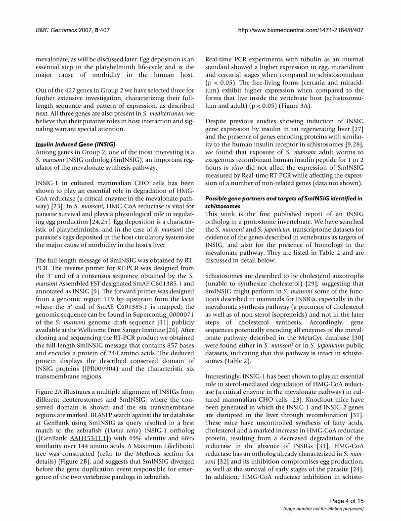
Figure 2A illustrates a multiple alignment of INSIGs fromdifferent deuterostomes and SmINSIG, where the con-served domain is shown and the six transmembraneregions are marked. BLASTP search against the nr databaseat GenBank using SmINSIG as query resulted in a bestmatch to the zebrafish (Danio rerio) INSIG-1 ortholog([GenBank: AAH45341.1]) with 49% identity and 68%similarity over 144 amino acids. A Maximum Likelihoodtree was constructed (refer to the Methods section fordetails) (Figure 2B), and suggests that SmINSIG divergedbefore the gene duplication event responsible for emer-gence of the two vertebrate paralogs in zebrafish.
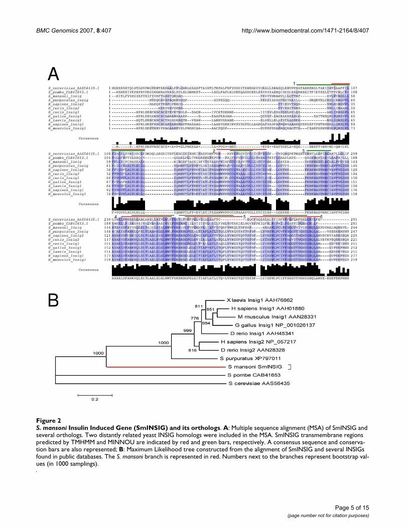
Real-time PCR experiments with tubulin as an internalstandard showed a higher expression in egg, miracidiumand cercarial stages when compared to schistosomulum(p < 0.05). The free-living forms (cercaria and miracid-ium) exhibit higher expression when compared to theforms that live inside the vertebrate host (schistosomu-lum and adult) (p < 0.05) (Figure 3A).
Despite previous studies showing induction of INSIGgene expression by insulin in rat regenerating liver [27]and the presence of genes encoding proteins with similar-ity to the human insulin receptor in schistosomes [9,28],we found that exposure of S. mansoni adult worms toexogenous recombinant human insulin peptide for 1 or 2hours in vitro did not affect the expression of SmINSIGmeasured by Real-time RT-PCR while affecting the expres-sion of a number of non-related genes (data not shown).
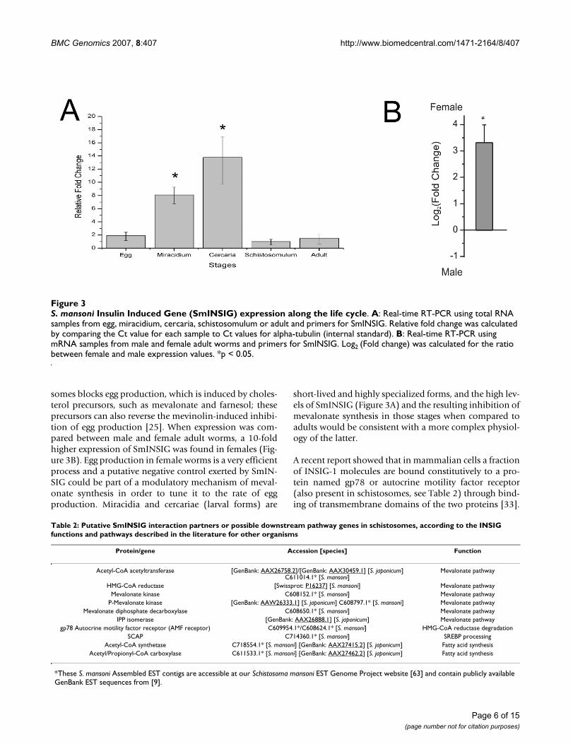
Possible gene partners and targets of SmINSIG identified in schistosomesThis work is the first published report of an INSIGortholog in a protostome invertebrate. We have searchedthe S. mansoni and S. japonicum transcriptome datasets forevidence of the genes described in vertebrates as targets ofINSIG, and also for the presence of homologs in themevalonate pathway. They are listed in Table 2 and arediscussed in detail below.
Schistosomes are described to be cholesterol auxotrophs(unable to synthesize cholesterol) [29], suggesting thatSmINSIG might perform in S. mansoni some of the func-tions described in mammals for INSIGs, especially in themevalonate synthesis pathway (a precursor of cholesterolas well as of non-sterol isoprenoids) and not in the latersteps of cholesterol synthesis. Accordingly, genesequences potentially encoding all enzymes of the meval-onate pathway described in the MetaCyc database [30]were found either in S. mansoni or in S. japonicum publicdatasets, indicating that this pathway is intact in schisto-somes (Table 2).
Interestingly, INSIG-1 has been shown to play an essentialrole in sterol-mediated degradation of HMG-CoA reduct-ase (a critical enzyme in the mevalonate pathway) in cul-tured mammalian CHO cells [23]. Knockout mice havebeen generated in which the INSIG-1 and INSIG-2 genesare disrupted in the liver through recombination [31].These mice have uncontrolled synthesis of fatty acids,cholesterol and a marked increase in HMG-CoA reductaseprotein, resulting from a decreased degradation of thereductase in the absence of INSIGs [31]. HMG-CoAreductase has an ortholog already characterized in S. man-soni [32] and its inhibition compromises egg production,as well as the survival of early stages of the parasite [24].In addition, HMG-CoA reductase inhibition in schisto-
Page 4 of 15(page number not for citation purposes)
BMC Genomics 2007, 8:407 http://www.biomedcentral.com/1471-2164/8/407
Page 5 of 15(page number not for citation purposes)
S. mansoni Insulin Induced Gene (SmINSIG) and its orthologsFigure 2S. mansoni Insulin Induced Gene (SmINSIG) and its orthologs. A: Multiple sequence alignment (MSA) of SmINSIG and several orthologs. Two distantly related yeast INSIG homologs were included in the MSA. SmINSIG transmembrane regions predicted by TMHMM and MINNOU are indicated by red and green bars, respectively. A consensus sequence and conserva-tion bars are also represented; B: Maximum Likelihood tree constructed from the alignment of SmINSIG and several INSIGs found in public databases. The S. mansoni branch is represented in red. Numbers next to the branches represent bootstrap val-ues (in 1000 samplings).
BMC Genomics 2007, 8:407 http://www.biomedcentral.com/1471-2164/8/407
somes blocks egg production, which is induced by choles-terol precursors, such as mevalonate and farnesol; theseprecursors can also reverse the mevinolin-induced inhibi-tion of egg production [25]. When expression was com-pared between male and female adult worms, a 10-foldhigher expression of SmINSIG was found in females (Fig-ure 3B). Egg production in female worms is a very efficientprocess and a putative negative control exerted by SmIN-SIG could be part of a modulatory mechanism of meval-onate synthesis in order to tune it to the rate of eggproduction. Miracidia and cercariae (larval forms) are
short-lived and highly specialized forms, and the high lev-els of SmINSIG (Figure 3A) and the resulting inhibition ofmevalonate synthesis in those stages when compared toadults would be consistent with a more complex physiol-ogy of the latter.
A recent report showed that in mammalian cells a fractionof INSIG-1 molecules are bound constitutively to a pro-tein named gp78 or autocrine motility factor receptor(also present in schistosomes, see Table 2) through bind-ing of transmembrane domains of the two proteins [33].
S. mansoni Insulin Induced Gene (SmINSIG) expression along the life cycleFigure 3S. mansoni Insulin Induced Gene (SmINSIG) expression along the life cycle. A: Real-time RT-PCR using total RNA samples from egg, miracidium, cercaria, schistosomulum or adult and primers for SmINSIG. Relative fold change was calculated by comparing the Ct value for each sample to Ct values for alpha-tubulin (internal standard). B: Real-time RT-PCR using mRNA samples from male and female adult worms and primers for SmINSIG. Log2 (Fold change) was calculated for the ratio between female and male expression values. *p < 0.05.
Table 2: Putative SmINSIG interaction partners or possible downstream pathway genes in schistosomes, according to the INSIG functions and pathways described in the literature for other organisms
Protein/gene Accession [species] Function
Acetyl-CoA acetyltransferase [GenBank: AAX26758.2]/[GenBank: AAX30459.1] [S. japonicum] C611014.1* [S. mansoni]
Mevalonate pathway
HMG-CoA reductase [Swissprot: P16237] [S. mansoni] Mevalonate pathwayMevalonate kinase C608152.1* [S. mansoni] Mevalonate pathway
P-Mevalonate kinase [GenBank: AAW26333.1] [S. japonicum] C608797.1* [S. mansoni] Mevalonate pathwayMevalonate diphosphate decarboxylase C608650.1* [S. mansoni] Mevalonate pathway
IPP isomerase [GenBank: AAX26888.1] [S. japonicum] Mevalonate pathwaygp78 Autocrine motility factor receptor (AMF receptor) C609954.1*/C608624.1* [S. mansoni] HMG-CoA reductase degradation
SCAP C714360.1* [S. mansoni] SREBP processingAcetyl-CoA synthetase C718554.1* [S. mansoni] [GenBank: AAX27415.2] [S. japonicum] Fatty acid synthesis
Acetyl/Propionyl-CoA carboxylase C611533.1* [S. mansoni] [GenBank: AAX27462.2] [S. japonicum] Fatty acid synthesis
*These S. mansoni Assembled EST contigs are accessible at our Schistosoma mansoni EST Genome Project website [63] and contain publicly available GenBank EST sequences from [9].
Page 6 of 15(page number not for citation purposes)
BMC Genomics 2007, 8:407 http://www.biomedcentral.com/1471-2164/8/407
Evidence indicates that this sterol-triggered reductase/INSIG-1/gp78 complex is essential for ubiquitination anddegradation of the reductase [33]. Based on the presenceof orthologs in the S. mansoni transcriptome database, thisevolutionarily conserved complex could also occur inschistosomes.
In line with the known S. mansoni auxotrophy for choles-terol, we could not identify either in the S. mansoni or inthe S. japonicum transcriptomes any of the enzymes specif-ically involved in cholesterol synthesis from mevalonate.Interestingly, we found a S. mansoni EST with similarity toa short partial segment of M. musculus SCAP ortholog(Table 2); SCAP has been described in mammals as aSREBP cleavage-activating protein [34]. However, wefound no gene fragments that would encode SCAP's puta-tive partner SREBP, the Sterol Regulatory Element BindingProtein involved in regulation of cholesterol biosynthesisin mammalian cells. In the absence of any evident SREBPor cholesterol synthesis enzymes it is likely that the puta-tive protein with partial similarity to SCAP might have adifferent role in S. mansoni.
Interfering with the mevalonate pathway in schistosomescould be a promising novel drug target approach becauseof the known adverse clinical consequences of eggs to thepatients and of the biological importance of egg deposi-tion in the parasite's life cycle.
VasohibinAnother gene from Group 2 that we have selected for fur-ther scrutiny is represented by S. mansoni Assembled EST(SmAE) C605907.1. Its consensus sequence had good(56%) similarity to vasohibin-like proteins (recentlynamed vasohibin 2).
Vasohibins constitute a recently described family ofendothelium-derived angiogenesis inhibitors in humans[35,36]. The presence of a Vasohibin ortholog in schisto-somes might suggest a potential angiogenesis inhibitionprocess in the host, mediated by schistosomes' molecules.Complete sequencing of clone MA3-9999U-M294-F04-U.B, the longest clone in SmAE cluster C605907.1,showed a transcript with 975 bases (Figure 4A) that wenamed SmVASL for S. mansoni Vasohibin-like gene.BLASTP comparison to Swiss-Prot/TrEMBL showed thebest match (48% identity and 58% similarity over 90amino acids) to vasohibin 2 ([Swissprot: Q86V25] orFLJ12505 protein), a recently described human vaso-hibin-like protein [36]. The amino portion of SmVASLdeduced protein (starting at Leu4) did align to Leu142 ofhuman vasohibin 2, thus suggesting that SmVASL repre-sented a partial sequence of the S. mansoni ortholog.
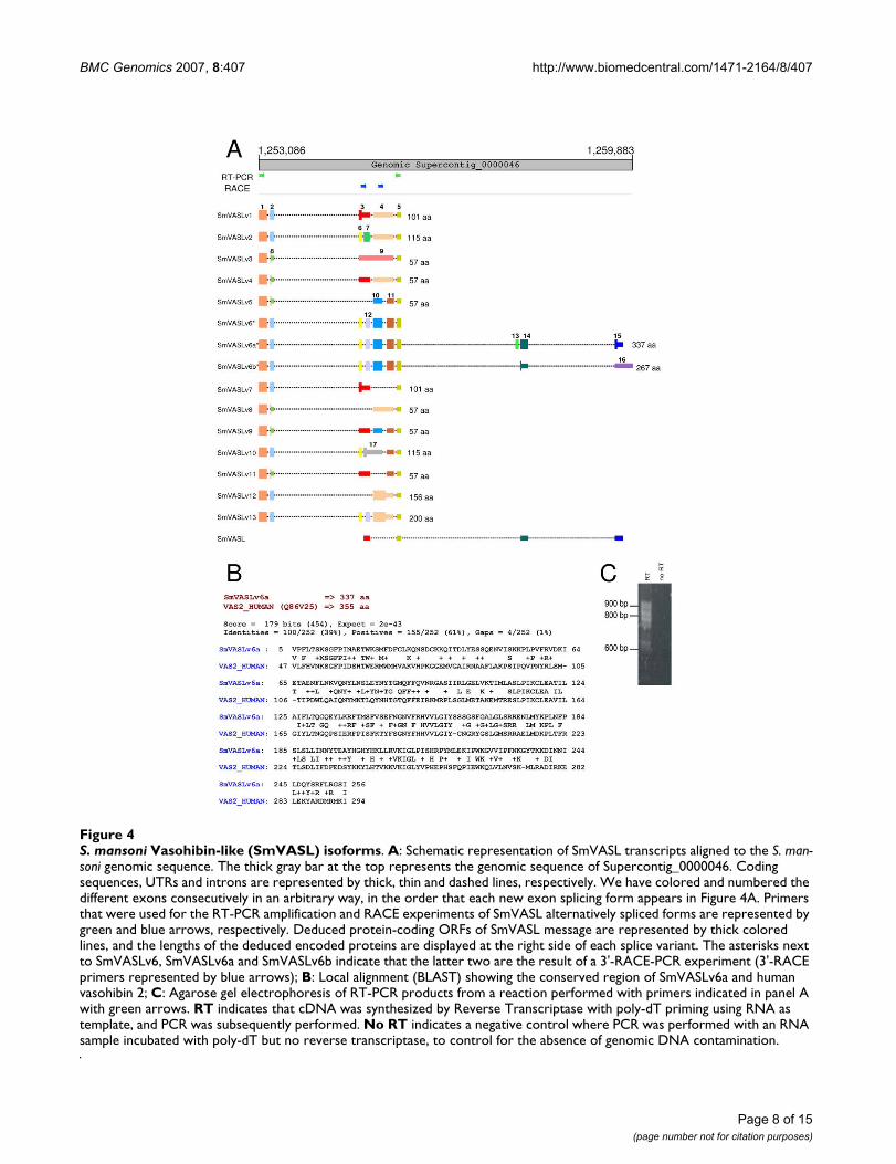
To search for possible additional portions of SmVASL, thesequence of the original clone was mapped to the draftsequence of the S. mansoni genome [11]. We found thatSmVASL maps with splicing to Supercontig _0000046,from base 1,255,010 to 1,259,701 (Figure 4A). In agenomic region of ~2,000 bases upstream from theSmVASL gene we found a sequence apparently encoding apeptide that displays similarity to vasohibin-like proteins(not shown). Next, we designed a forward and a reverseprimer based on the sequence of this region, as indicatedin Figure 4A (green arrows). These primers were used in aRT-PCR reaction, which resulted in a complex profile ofamplification with one band of ~650 bp and a smearbetween 750–900 bp (Figure 4C). No amplicon wasobtained in a parallel reaction without reverse transcrip-tion, to control for the absence of DNA contamination,indicating that the diverse range of products (Figure 4C)was derived from bona fide mRNA messages. This resultsuggested multiple alternatively spliced forms at the 5'end of full-length SmVASL.
The RT-PCR amplification products were cloned andsequenced. A total of approximately 300 clones weresequenced from both ends, disclosing fourteen differentisoforms (SmVASLv1-13, with two SmVASLv6 isoforms).Mapping of these isoforms to the S. mansoni genomicsequence confirmed that the different clones are a result ofalternative splicing (Figure 4A). Variation is caused by anumber of alternative splicing events, such as 5'-deletion,exon skipping, and junction of two exons by intron reten-tion. Two different lengths for the second exon wereobserved: exon 2 with 79 bp for variants SmVASLv1, 2, 6a,6b, 7, 10, 12 and 13; and exon 8 with 65 bp, resultingfrom a 5'-deletion in exon 2, for SmVASLv3, 4, 5, 8, 9 and11 (Figure 4A). The latter splicing form caused an earlystop, and the resulting isoforms encode a putative short57 amino acid protein. The isoforms have different 3'-UTR ends, which may be related to different stability ofthe messages.
Exon skipping was observed in SmVASLv5, 7, 8, 11 and 12(Figure 4A). Intron retention was detected in isoformsSmVASLv1, 2, 4, 12 and 13, where exon 4 is the junctionof exons 10 and 11 (that are present for example in iso-form 5). Intron retention was also detected in SmVASLv1,4, 7, 9 and 11, where exon 3 is the junction of exons 6 and7. Finally, the intron retention observed in SmVASLv3results from junction of exons 6, 7, 10 and 11.
We have identified seven different polypeptides that areencoded by these variants, representing different deducedprotein isoforms (Figure 4A). As noted earlier, isoformsSmVASLv3, 4, 5, 8, 9 and 11 encode the same 57 aminoacid protein. Due to the absence of a stop codon in theSmVASLv6 sequence in the segment amplified by RT-PCR,
Page 7 of 15(page number not for citation purposes)
BMC Genomics 2007, 8:407 http://www.biomedcentral.com/1471-2164/8/407
Page 8 of 15(page number not for citation purposes)
S. mansoni Vasohibin-like (SmVASL) isoformsFigure 4S. mansoni Vasohibin-like (SmVASL) isoforms. A: Schematic representation of SmVASL transcripts aligned to the S. man-soni genomic sequence. The thick gray bar at the top represents the genomic sequence of Supercontig_0000046. Coding sequences, UTRs and introns are represented by thick, thin and dashed lines, respectively. We have colored and numbered the different exons consecutively in an arbitrary way, in the order that each new exon splicing form appears in Figure 4A. Primers that were used for the RT-PCR amplification and RACE experiments of SmVASL alternatively spliced forms are represented by green and blue arrows, respectively. Deduced protein-coding ORFs of SmVASL message are represented by thick colored lines, and the lengths of the deduced encoded proteins are displayed at the right side of each splice variant. The asterisks next to SmVASLv6, SmVASLv6a and SmVASLv6b indicate that the latter two are the result of a 3'-RACE-PCR experiment (3'-RACE primers represented by blue arrows); B: Local alignment (BLAST) showing the conserved region of SmVASLv6a and human vasohibin 2; C: Agarose gel electrophoresis of RT-PCR products from a reaction performed with primers indicated in panel A with green arrows. RT indicates that cDNA was synthesized by Reverse Transcriptase with poly-dT priming using RNA as template, and PCR was subsequently performed. No RT indicates a negative control where PCR was performed with an RNA sample incubated with poly-dT but no reverse transcriptase, to control for the absence of genomic DNA contamination.
BMC Genomics 2007, 8:407 http://www.biomedcentral.com/1471-2164/8/407
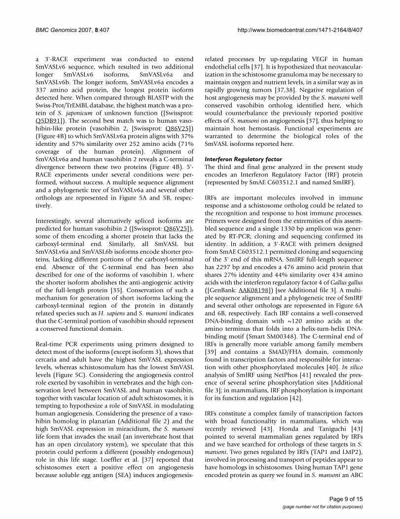
a 3'-RACE experiment was conducted to extendSmVASLv6 sequence, which resulted in two additionallonger SmVASLv6 isoforms, SmVASLv6a andSmVASLv6b. The longer isoform, SmVASLv6a encodes a337 amino acid protein, the longest protein isoformdetected here. When compared through BLASTP with theSwiss-Prot/TrEMBL database, the highest match was a pro-tein of S. japonicum of unknown function ([Swissprot:Q5DB91]). The second best match was to human vaso-hibin-like protein (vasohibin 2, [Swissprot: Q86V25])(Figure 4B) to which SmVASLv6a protein aligns with 37%identity and 57% similarity over 252 amino acids (71%coverage of the human protein). Alignment ofSmVASLv6a and human vasohibin 2 reveals a C-terminaldivergence between these two proteins (Figure 4B). 5'-RACE experiments under several conditions were per-formed, without success. A multiple sequence alignmentand a phylogenetic tree of SmVASLv6a and several otherorthologs are represented in Figure 5A and 5B, respec-tively.
Interestingly, several alternatively spliced isoforms arepredicted for human vasohibin 2 ([Swissprot: Q86V25]),some of them encoding a shorter protein that lacks thecarboxyl-terminal end. Similarly, all SmVASL butSmVASLv6a and SmVASL6b isoforms encode shorter pro-teins, lacking different portions of the carboxyl-terminalend. Absence of the C-terminal end has been alsodescribed for one of the isoforms of vasohibin 1, wherethe shorter isoform abolishes the anti-angiogenic activityof the full-length protein [35]. Conservation of such amechanism for generation of short isoforms lacking thecarboxyl-terminal region of the protein in distantlyrelated species such as H. sapiens and S. mansoni indicatesthat the C-terminal portion of vasohibin should representa conserved functional domain.
Real-time PCR experiments using primers designed todetect most of the isoforms (except isoform 3), shows thatcercaria and adult have the highest SmVASL expressionlevels, whereas schistosomulum has the lowest SmVASLlevels (Figure 5C). Considering the angiogenesis controlrole exerted by vasohibin in vertebrates and the high con-servation level between SmVASL and human vasohibin,together with vascular location of adult schistosomes, it istempting to hypothesize a role of SmVASL in modulatinghuman angiogenesis. Considering the presence of a vaso-hibin homolog in planarian (Additional file 2) and thehigh SmVASL expression in miracidium, the S. mansonilife form that invades the snail (an invertebrate host thathas an open circulatory system), we speculate that thisprotein could perform a different (possibly endogenous)role in this life stage. Loeffler et al. [37] reported thatschistosomes exert a positive effect on angiogenesisbecause soluble egg antigen (SEA) induces angiogenesis-
related processes by up-regulating VEGF in humanendothelial cells [37]. It is hypothesized that neovascular-ization in the schistosome granuloma may be necessary tomaintain oxygen and nutrient levels, in a similar way as inrapidly growing tumors [37,38]. Negative regulation ofhost angiogenesis may be provided by the S. mansoni wellconserved vasohibin ortholog identified here, whichwould counterbalance the previously reported positiveeffects of S. mansoni on angiogenesis [37], thus helping tomaintain host hemostasis. Functional experiments arewarranted to determine the biological roles of theSmVASL isoforms reported here.
Interferon Regulatory factorThe third and final gene analyzed in the present studyencodes an Interferon Regulatory Factor (IRF) protein(represented by SmAE C603512.1 and named SmIRF).
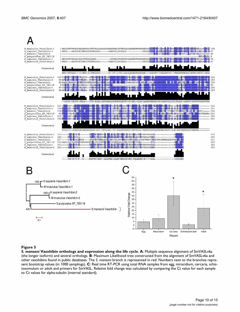
IRFs are important molecules involved in immuneresponse and a schistosome ortholog could be related tothe recognition and response to host immune processes.Primers were designed from the extremities of this assem-bled sequence and a single 1330 bp amplicon was gener-ated by RT-PCR; cloning and sequencing confirmed itsidentity. In addition, a 3'-RACE with primers designedfrom SmAE C603512.1 permitted cloning and sequencingof the 3' end of this mRNA. SmIRF full-length sequencehas 2297 bp and encodes a 476 amino acid protein thatshares 27% identity and 44% similarity over 434 aminoacids with the interferon regulatory factor 4 of Gallus gallus([GenBank: AAK08198]) [see Additional file 3]. A multi-ple sequence alignment and a phylogenetic tree of SmIRFand several other orthologs are represented in Figure 6Aand 6B, respectively. Each IRF contains a well-conservedDNA-binding domain with ~120 amino acids at theamino terminus that folds into a helix-turn-helix DNA-binding motif (Smart SM00348). The C-terminal end ofIRFs is generally more variable among family members[39] and contains a SMAD/FHA domain, commonlyfound in transcription factors and responsible for interac-tion with other phosphorylated molecules [40]. In silicoanalysis of SmIRF using NetPhos [41] revealed the pres-ence of several serine phosphorylation sites [Additionalfile 3]; in mammalians, IRF phosphorylation is importantfor its function and regulation [42].
IRFs constitute a complex family of transcription factorswith broad functionality in mammalians, which wasrecently reviewed [43]. Honda and Taniguchi [43]pointed to several mammalian genes regulated by IRFsand we have searched for orthologs of these targets in S.mansoni. Two genes regulated by IRFs (TAP1 and LMP2),involved in processing and transport of peptides appear tohave homologs in schistosomes. Using human TAP1 geneencoded protein as query we found in S. mansoni an ABC
Page 9 of 15(page number not for citation purposes)
BMC Genomics 2007, 8:407 http://www.biomedcentral.com/1471-2164/8/407
Page 10 of 15(page number not for citation purposes)
S. mansoni Vasohibin orthologs and expression along the life cycleFigure 5S. mansoni Vasohibin orthologs and expression along the life cycle. A: Multiple sequence alignment of SmVASLv6a (the longer isoform) and several orthologs. B: Maximum Likelihood tree constructed from the alignment of SmVASLv6a and other vasohibins found in public databases. The S. mansoni branch is represented in red. Numbers next to the branches repre-sent bootstrap values (in 1000 samplings). C: Real time RT-PCR using total RNA samples from egg, miracidium, cercaria, schis-tosomulum or adult and primers for SmVASL. Relative fold change was calculated by comparing the Ct value for each sample to Ct values for alpha-tubulin (internal standard).
BMC Genomics 2007, 8:407 http://www.biomedcentral.com/1471-2164/8/407
transporter gene, SMDR1 [GenBank: AAA66476.1] (35%identity and 61% coverage) that has been previouslydescribed as possible transporter of peptides [44]. Whenusing human LMP2 ([GenBank: CAA60784]) as query, wefound a schistosome ortholog in our database(C602473.1, with 55% identity and 88% coverage).Human LMP2 encodes a proteasome subunit and replacesthe Y subunit in the proteasome structure upon IFN sign-aling and this change results in a different proteasomeproteolytic activity [45]. Functional proteasomes wererecently shown to be essential for schistosome develop-ment in the vertebrate host [46]. IRFs also regulate cas-
pase-1 expression [43]. We found a caspase in S. mansonithat has higher similarity with caspase-3 than with cas-pase-1 (44% identity and 85% coverage); in mammaliansboth caspases are involved with apoptosis control; how-ever, the apoptosis pathway appears to be incomplete inthe parasite [9].
Besides the inherent capacity of IRFs to function as tran-scription factors, they selectively bind to a group of pro-teins, namely immunophilins. Interaction between IRF-4and immunophilin FKBP52 in mammalians results in aconformational change, abolishing DNA-binding activity
S. mansoni Interferon Regulatory Factor (SmIRF)Figure 6S. mansoni Interferon Regulatory Factor (SmIRF). A: Multiple sequence alignment of SmIRF and several orthologs. For displaying purposes, only the conserved region is represented; B: Maximum Likelihood tree constructed from the alignment of SmIRF and several IRFs found in public databases. The S. mansoni branch is represented in red. Numbers next to the branches represent bootstrap values (in 1000 samplings).
Page 11 of 15(page number not for citation purposes)
BMC Genomics 2007, 8:407 http://www.biomedcentral.com/1471-2164/8/407
and partial IRF-4 proteolysis [47]. One immunophilinnamed p50 [GenBank: AAA69867.1] was already identi-fied in schistosomes, having a high similarity with verte-brate FKBP immunophilins [48,49].
The IRF interactions are highly complex and not fully elu-cidated, even in mammalians, with new interactions andfunctionalities being recently reported [43]. In this workwe have pointed to the existence in schistosomes of somepossible players. Other experiments (e.g. protein-proteininteraction) are warranted to identify functions and otherpossible SmIRF interaction partners. It would be interest-ing to demonstrate if SmIRF is involved in schistosomestress responses (viral infections, for example), or in host-parasite interaction, being part of the pathway that inter-plays with host immune molecules.
ConclusionAccording to our analysis, the S. mansoni genes describedin this work are homologs of genes present in Deuterosto-mia, but absent in Ecdysozoa organisms. Given the spe-cific functions of these genes in Deuterostomia, especiallyin mammals, we envisage the possibility of co-optation ofthe schistosome ortholog in interaction and adaptation tothe host environment. The evolutionary history leading toconservation of such set of genes in these two distantlyrelated groups is not totally clear yet; more sequenceinformation from organisms of related phyla (especiallyfrom other Lophotrocozoa) should help throw light onthis problem. The three genes characterized in detail inthis work might be involved in mevalonate synthesis (eggproduction), angiogenesis control (host invasion) andimmune response (interplay with host defense mole-cules).
In S. mediterranea the orthologs of these genes may per-form ancestral non-parasitic roles. In this respect, a recentreport [50] revealed the presence of orthologs of thyroidhormone receptor (TR) in S. mansoni, S. japonicum and S.mediterranea and demonstrated that TRs in platy-helminths are highly conserved not only in sequence sim-ilarity, but also in gene organization, protein-proteininteraction and in DNA-binding ability [50]; TR was pre-viously believed to be an innovation of chordates as thegenomes of insects and nematodes do not contain TRgenes. Although the functions of TRs in invertebrates arenot fully understood, phylogenetic analysis showed thatthe TR ortholog likely originated from a common ances-tor of the Bilateria [50]. Both the results of Wu et al. [50]and ours suggest the presence in a common ancestor ofgenes previously thought to have arisen only later in evo-lution. We hypothesize that during Schistosoma evolutionthe three genes discussed in the present report might havebeen co-opted to perform functions that are crucial for theparasite-host interplay. Regardless of the actual history,
we believe the genes discussed here have characteristicsthat make them good candidates for further investigationas potential drug targets.
MethodsDatasetsS. mansoni EST sequences generated by our group [9] wereused as the query dataset of S. mansoni sequences in thiswork. Local BLAST [51] databases were formatted with allthe sequences available from arthropods, nematodes anddeuterostomes in the non redundant (nr) nucleotide sec-tion of GenBank database (updated as of May/2007) andwere used as subject in BLAST searches. In addition, all thepublicly available nucleotide sequences from non modelarthropods, nematodes and from the planarian S. mediter-ranea were used. The planarian dataset we used is com-posed of all the publicly available 171,483 nucleotidesequences (97,901 CoreNucleotide and 73,582 ESTs, onMay 2007) together with the assembled ESTs obtained bya large scale EST sequencing project [12]. The planariandataset was further supplemented with the EST assemblydata provided by the authors (10,485 assembledsequences; 6,488 contigs and 3,997 singlets) [12]. Inorder to reduce the EST sequence redundancy and conse-quently the time complexity of the process, the sequenceswere processed using CD-HIT [52] prior to database for-matting. After identifying the S. mansoni genes of interest,we have considered their downstream effectors describedin the literature for vertebrates and we searched for thepresence of orthologs which could act as molecular part-ners or members of downstream pathways in S. mansoni.When any of these orthologs were missing in the S. man-soni GenBank database the S. japonicum sequences werealternatively searched.
The S. mansoni genome sequence partial assembly (v. 3) atthe Wellcome Trust Sanger Institute [26] was used.
Similarity searchesWe used the BLASTX and TBLASTX programs [51] tosearch for S. mansoni (a platyhelminth) translatedsequences that have considerable similarity to proteinsand translated ESTs from deuterostomes, nematodes andarthropods. Perl scripts together with the Zerg parser [53]and BioPerl [54] were used to perform and process BLASTsearches.
BLAST alignments with bit scores higher than 100 andlower than 50 were considered as true matches and non-significant hits, respectively. Bit scores were used to facili-tate cross-database comparisons. Hits with intermediarybit scores (between 50 and 100) were manually inspected,basically to eliminate hits derived from unmasked lowcomplexity regions where matches occur through a single
Page 12 of 15(page number not for citation purposes)
BMC Genomics 2007, 8:407 http://www.biomedcentral.com/1471-2164/8/407
amino acid at repeated intervals. Overall, the sequencesincluded in Additional File 2 have scores higher than 80.
The S. mansoni genes without matches and Schistosomaspecific genes were filtered out. The resulting genes werefurther assigned to one of the following groups (Table 1and Additional file 1) when a given gene was detected inat least one species belonging to the corresponding clade:(1) present in S. mansoni, deuterostomes, arthropods andnematodes (all groups); (2) present in S. mansoni anddeuterostomes, but not in arthropods and nematodes; (3)present in S. mansoni and arthropods, but not in deuteros-tomes and nematodes; (4) present in S. mansoni and nem-atodes, but not in arthropods and deuterostomes); (5)present in S. mansoni, arthropods and nematodes, but notin deuterostomes; (6) present in S. mansoni, arthropodsand deuterostomes, but not in nematodes; (7) present inS. mansoni, nematodes and deuterostomes, but not inarthropods.
To evaluate the significance of the gene gains/losses undereach hypothesis, we have generated 100,000 boot-strapped samples and performed the Wilcoxon test. Sim-ulations and the significance test were performed in the Renvironment.
TMHMM [55] and MINNOU [56] were used to predicttransmembrane domains, NetPhos [41] to identify possi-ble phosphorylation sites, with SignalP 3.0 [57] to predictsignal peptides, and with InterProScan [58] to predictconserved domains.
Phylogenetic analysisMultiple Sequence Alignments in the paper were gener-ated using MUSCLE [59] and edited using JalView [60].Curated alignments were then used to generate MaximumLikelihood phylogenetic trees with PhyML package [61].Significance of the results was estimated by building 1000bootstrapped trees. The NEWICK files generated byPhyML were then displayed with MEGA [62] producingthe trees displayed in the results section.
Cloning proceduresmRNA was obtained from adult parasites conserved inRNALater (Ambion, Austin, TX, USA) by extraction of tis-sue with MACs mRNA isolation kits (Miltenyi Biotec, Ber-gisch Gladbach, Germany). 200 ng of mRNA were treatedwith 5 U of RQ1 RNAse-free DNAse (Promega, Madison,WI, USA) for 30 min at 37°C. Reverse transcription wasperformed with oligo dT primers using the protocol ofSuperscript first strand system for RT-PCR (Invitrogen,Carlsbad, CA, USA). PCR was performed with AdvantageII (BD Biosciences, Palo Alto, CA, USA) with the buffersupplied by the manufacturer, and 200 nM of each spe-cific primer using the following cycling program: 95°C for
1 min plus 35 cycles each at 95°C for 30 s, 55°C for 30 s,and 68°C for 3 min, followed by a final extension at 68°Cfor 3 min. The products were analyzed in 1.2% agarose geland cloned in pGEM-T vector (Promega) for furthersequencing. Primers are listed in Additional file 4.
Rapid amplification of cDNA ends (RACE)mRNA was obtained from adult parasites conserved inRNALater (Ambion) by extraction of tissue with MACsmRNA isolation kits (Miltenyi Biotec). 200 ng of mRNAwas used for reverse transcription using the protocol ofthe 3'RACE system kit for rapid amplification of cDNAends (Invitrogen) and specific primers. To perform 3'RACE of vasohibin gene, 3 µg of total RNA from miracid-ium was used. Reverse transcription was performed using1 µl of Super Scrip III (Invitrogen) at 65°C for 5 min,55°C for 50 min and 85°C for 5 min.
PCR reaction was performed with Advantage II polymer-ase (BD Biosciences) with buffer supplied by the manu-facturer, 200 µM dNTPs and 200 nM of each primer usingthe following cycling program: 95°C for 1 min plus 35cycles each at 95°C for 30 s, 55°C for 30 s, 68°C for 3min, followed by a final extension at 68°C for 3 min. Theproducts were analyzed in 1.2% agarose gel and cloned inpGem-T vector (Promega) for further sequencing. Primersare listed in Additional file 4.
Real-time RT-PCR proceduresmRNA was obtained from male or female adult parasitesconserved in RNALater (Ambion) by extraction of tissuewith MACs mRNA isolation kits (Miltenyi Biotec). 200 ngof mRNA were treated with RQ1 RNAse-free DNAse(Promega), using 1 U in 10 µl of reaction, for 30 min at37°C. The DNAse was inactivated at 65°C for 10 min.
Three micrograms of total RNA from each of five stageswas treated with RQ1 RNAse-free DNAse (Promega),using 4 U in 10 µl of reaction, for 1 hour at 37°C. Theresulting products were reverse transcribed with randomhexamer primers using the protocol of Superscript firststrand system for RT-PCR (Invitrogen). Control reactionswithout addition of reverse transcriptase were run in par-allel, and were used as templates for PCR negative control,to control for the absence of possible DNA contaminants.Primers for real-time RT-PCR (listed in Additional file 4)were designed using the Primer Express program version2.0.0 (Applied Biosystems, Foster City, CA, USA) withdefault parameters. Real-time RT-PCR reactions were per-formed using SYBR Green PCR master mix (Applied Bio-systems) and the specific primers in a GenAmp 5700Sequence Detection System (Applied Biosystems). P-valuewas calculated using Student's t-test with 95% confidenceinterval.
Page 13 of 15(page number not for citation purposes)
BMC Genomics 2007, 8:407 http://www.biomedcentral.com/1471-2164/8/407
EMBL sequence depositionAll sequences determined in this work were deposited atEMBL under the following numbers: SmINSIG, [EMBL:AM493258]; SmIRF, [EMBL: AM493259]; SmVASL iso-forms, [EMBL: AM493260 – AM493273].
Authors' contributionsTMV conceived the study and carried out the computa-tional analysis. RdeM, GTA and KCP performed the wet-lab experiments. TMV and RdeM analyzed the data. TMV,RdeM, JCS and SVA participated in the discussion ofresults and drafting of the manuscript. SVA coordinatedand supervised the project. All authors read and approvedthe final manuscript.
Additional material
AcknowledgementsSpecial thanks are due to Robson Francisco de Souza for the insightful dis-cussions and Dr. Phillip Newmark for kindly sharing the planarian EST assembly. We also thank Renato Alvarenga for technical help in sequencing and Fundação de Amparo a Pesquisa do Estado de São Paulo, FAPESP and
Conselho Nacional de Desenvolvimento Científico e Tecnológico, CNPq for financial support.
References1. Engels D, Chitsulo L, Montresor A, Savioli L: The global epidemi-
ological situation of schistosomiasis and new approaches tocontrol and research. Acta Trop 2002, 82:139-146.
2. WHO: TDR Strategic Direction for Research: Schistosomiasis Geneve.:World Health Organization; 2002.
3. Hotez PJ, Ferris MT: The antipoverty vaccines. Vaccine 2006,24:5787-5799.
4. King CL: Initiation and regulation of disease in schistosomia-sis. In Schistosomiasis Edited by: Mahmoud AAF. London: ImperialCollege Press; 2001:213-264.
5. Knobloch J, Kunz W, Grevelding CG: Herbimycin A suppressesmitotic activity and egg production of female Schistosomamansoni. Int J Parasitol 2006, 36:1261-1272.
6. Pearce EJ: Priming of the immune response by schistosomeeggs. Parasite Immunol 2005, 27:265-270.
7. Dunne D, Mountford A: Resistance to infection in humans andanimal models. In Schistosomiasis Edited by: Mahmoud AAF. Lon-don: Imperial College Press; 2001:133-211.
8. Salzet M, Capron A, Stefano GB: Molecular crosstalk in host-par-asite relationships: schistosome- and leech-host interactions.Parasitol Today 2000, 16:536-540.
9. Verjovski-Almeida S, DeMarco R, Martins EA, Guimaraes PE, OjopiEP, Paquola AC, Piazza JP, Nishiyama MY Jr, Kitajima JP, Adamson RE,et al.: Transcriptome analysis of the acoelomate human par-asite Schistosoma mansoni. Nat Genet 2003, 35:148-157.
10. Hu W, Yan Q, Shen DK, Liu F, Zhu ZD, Song HD, Xu XR, Wang ZJ,Rong YP, Zeng LC, et al.: Evolutionary and biomedical implica-tions of a Schistosoma japonicum complementary DNAresource. Nat Genet 2003, 35:139-147.
11. Wilson RA, Ashton PD, Braschi S, Dillon GP, Berriman M, Ivens A:'Oming in on schistosomes: prospects and limitations forpost-genomics. Trends Parasitol 2007, 23:14-20.
12. Zayas RM, Hernandez A, Habermann B, Wang Y, Stary JM, NewmarkPA: The planarian Schmidtea mediterranea as a model forepigenetic germ cell specification: analysis of ESTs from thehermaphroditic strain. Proc Natl Acad Sci USA 2005,102:18491-18496.
13. Hausdorf B: Early evolution of the bilateria. Syst Biol 2000,49:130-142.
14. Jenner RA: Evolution of animal body plans: the role of meta-zoan phylogeny at the interface between pattern and proc-ess. Evol Dev 2000, 2:208-221.
15. Philippe H, Lartillot N, Brinkmann H: Multigene analyses of bilat-erian animals corroborate the monophyly of Ecdysozoa,Lophotrochozoa, and Protostomia. Mol Biol Evol 2005,22:1246-1253.
16. Philippe H, Telford MJ: Large-scale sequencing and the new ani-mal phylogeny. Trends Ecol Evol 2006.
17. Jones M, Blaxter M: Evolutionary biology: animal roots andshoots. Nature 2005, 434:1076-1077.
18. Wolf YI, Rogozin IB, Koonin EV: Coelomata and not Ecdysozoa:evidence from genome-wide phylogenetic analysis. GenomeRes 2004, 14:29-36.
19. Delsuc F, Brinkmann H, Philippe H: Phylogenomics and thereconstruction of the tree of life. Nat Rev Genet 2005, 6:361-375.
20. Roy SW, Gilbert W: Resolution of a deep animal divergence bythe pattern of intron conservation. Proc Natl Acad Sci USA 2005,102:4403-4408.
21. Dopazo H, Dopazo J: Genome-scale evidence of the nematode-arthropod clade. Genome Biol 2005, 6:R41.
22. Ciccarelli FD, Doerks T, von Mering C, Creevey CJ, Snel B, Bork P:Toward automatic reconstruction of a highly resolved treeof life. Science 2006, 311:1283-1287.
23. Sever N, Yang T, Brown MS, Goldstein JL, DeBose-Boyd RA: Accel-erated degradation of HMG CoA reductase mediated bybinding of insig-1 to its sterol-sensing domain. Mol Cell 2003,11:25-33.
24. Chen GZ, Foster L, Bennett JL: Antischistosomal action ofmevinolin: evidence that 3-hydroxy-methylglutaryl-coen-zyme A reductase activity in Schistosoma mansoni is vital for
Additional file 1Schematic representation of the relationships between S. mansoni and three different clades and indication of the several groups that result from the presence or absence of S. mansoni genes among the organisms of each of the three clades.Click here for file[http://www.biomedcentral.com/content/supplementary/1471-2164-8-407-S1.pdf]
Additional file 2List of group 2 genes (conserved in schistosomes and deuterostomes, but lost in nematodes and arthropods). This table provides full statistics about query and hit coverage, identity and similarity percentages and annota-tions. Column "P" represents the presence (1) or absence (0) of a planar-ian homolog. The first four entries are for the three genes that were further characterized in this work.Click here for file[http://www.biomedcentral.com/content/supplementary/1471-2164-8-407-S2.xls]
Additional file 3Conserved domains in SmIRF. A: BLAST analysis against the SWISS-PROT database. A conserved N-terminal DNA-binding domain in SmIRF can be easily detected. B: In silico analysis of SmIRF using Net-Phos revealed the presence of several Serine phosphorylation sites, impor-tant for IRF function and regulation.Click here for file[http://www.biomedcentral.com/content/supplementary/1471-2164-8-407-S3.pdf]
Additional file 4Primers used in the Real-time RT-PCR and RACE experiments.Click here for file[http://www.biomedcentral.com/content/supplementary/1471-2164-8-407-S4.pdf]
Page 14 of 15(page number not for citation purposes)
BMC Genomics 2007, 8:407 http://www.biomedcentral.com/1471-2164/8/407
parasite survival. Naunyn Schmiedebergs Arch Pharmacol 1990,342:477-482.
25. Vandewaa EA, Mills G, Chen GZ, Foster LA, Bennett JL: Physiolog-ical role of HMG-CoA reductase in regulating egg produc-tion by Schistosoma mansoni. Am J Physiol 1989, 257:R618-R625.
26. Wellcome Trust Sanger Institute S. mansoni genome project[http://www.sanger.ac.uk/Projects/S_mansoni/]
27. Peng Y, Schwarz EJ, Lazar MA, Genin A, Spinner NB, Taub R: Clon-ing, human chromosomal assignment, and adipose andhepatic expression of the CL-6/INSIG1 gene. Genomics 1997,43:278-284.
28. Khayath N, Vicogne J, Ahier A, Benyounes A, Konrad C, Trolet J, Vis-cogliosi E, Brehm K, Dissous C: Diversification of the insulinreceptor family in the helminth parasite Schistosoma man-soni. Febs J 2007, 274:659-676.
29. Meyer F, Meyer H, Bueding E: Lipid metabolism in the parasiticand free-living flatworms, Schistosoma mansoni and Dugesiadorotocephala. Biochim Biophys Acta 1970, 210:257-266.
30. Caspi R, Foerster H, Fulcher CA, Hopkinson R, Ingraham J, Kaipa P,Krummenacker M, Paley S, Pick J, Rhee SY, et al.: MetaCyc: a mul-tiorganism database of metabolic pathways and enzymes.Nucleic Acids Res 2006, 34:D511-516.
31. Engelking LJ, Liang G, Hammer RE, Takaishi K, Kuriyama H, Evers BM,Li WP, Horton JD, Goldstein JL, Brown MS: Schoenheimer effectexplained – feedback regulation of cholesterol synthesis inmice mediated by Insig proteins. J Clin Invest 2005,115:2489-2498.
32. Rajkovic A, Simonsen JN, Davis RE, Rottman FM: Molecular cloningand sequence analysis of 3-hydroxy-3-methylglutaryl-coen-zyme A reductase from the human parasite Schistosomamansoni. Proc Natl Acad Sci USA 1989, 86:8217-8221.
33. Song BL, Sever N, DeBose-Boyd RA: Gp78, a membrane-anchored ubiquitin ligase, associates with Insig-1 and couplessterol-regulated ubiquitination to degradation of HMG CoAreductase. Mol Cell 2005, 19:829-840.
34. Brown MS, Ye J, Rawson RB, Goldstein JL: Regulated intramem-brane proteolysis: a control mechanism conserved from bac-teria to humans. Cell 2000, 100:391-398.
35. Shimizu K, Watanabe K, Yamashita H, Abe M, Yoshimatsu H, Ohta H,Sonoda H, Sato Y: Gene regulation of a novel angiogenesisinhibitor, vasohibin, in endothelial cells. Biochem Biophys ResCommun 2005, 327:700-706.
36. Shibuya T, Watanabe K, Yamashita H, Shimizu K, Miyashita H, Abe M,Moriya T, Ohta H, Sonoda H, Shimosegawa T, et al.: Isolation andcharacterization of vasohibin-2 as a homologue of VEGF-inducible endothelium-derived angiogenesis inhibitor vaso-hibin. Arterioscler Thromb Vasc Biol 2006, 26:1051-1057.
37. Loeffler DA, Lundy SK, Singh KP, Gerard HC, Hudson AP, Boros DL:Soluble egg antigens from Schistosoma mansoni induce angio-genesis-related processes by up-regulating vascularendothelial growth factor in human endothelial cells. J InfectDis 2002, 185:1650-1656.
38. Kumar R, Yoneda J, Bucana CD, Fidler IJ: Regulation of distinctsteps of angiogenesis by different angiogenic molecules. Int JOncol 1998, 12:749-757.
39. Mamane Y, Heylbroeck C, Genin P, Algarte M, Servant MJ, LePage C,DeLuca C, Kwon H, Lin R, Hiscott J: Interferon regulatory fac-tors: the next generation. Gene 1999, 237:1-14.
40. Durocher D, Taylor IA, Sarbassova D, Haire LF, Westcott SL, JacksonSP, Smerdon SJ, Yaffe MB: The molecular basis of FHAdomain:phosphopeptide binding specificity and implicationsfor phospho-dependent signaling mechanisms. Mol Cell 2000,6:1169-1182.
41. Blom N, Gammeltoft S, Brunak S: Sequence and structure-basedprediction of eukaryotic protein phosphorylation sites. J MolBiol 1999, 294:1351-1362.
42. Caillaud A, Hovanessian AG, Levy DE, Marie IJ: Regulatory serineresidues mediate phosphorylation-dependent and phosphor-ylation-independent activation of interferon regulatory fac-tor 7. J Biol Chem 2005, 280:17671-17677.
43. Honda K, Taniguchi T: IRFs: master regulators of signalling byToll-like receptors and cytosolic pattern-recognition recep-tors. Nat Rev Immunol 2006, 6:644-658.
44. Bosch IB, Wang ZX, Tao LF, Shoemaker CB: Two Schistosomamansoni cDNAs encoding ATP-binding cassette (ABC) fam-ily proteins. Mol Biochem Parasitol 1994, 65:351-356.
45. Gaczynska M, Goldberg AL, Tanaka K, Hendil KB, Rock KL: Protea-some subunits X and Y alter peptidase activities in oppositeways to the interferon-gamma-induced subunits LMP2 andLMP7. J Biol Chem 1996, 271:17275-17280.
46. Guerra-Sa R, Castro-Borges W, Evangelista EA, Kettelhut IC, Rod-rigues V: Schistosoma mansoni: functional proteasomes arerequired for development in the vertebrate host. Exp Parasitol2005, 109:228-236.
47. Mamane Y, Sharma S, Petropoulos L, Lin R, Hiscott J: Posttransla-tional regulation of IRF-4 activity by the immunophilinFKBP52. Immunity 2000, 12:129-140.
48. Kiang D, Karim AM, LoVerde PT: Cloning the gene encodingSchistosoma mansoni p50, an immunophilin. Gene 1996,170:137-140.
49. Osman A, Kiang D, Lo Verde PT, Karim AM: Schistosoma mansoni:characterization of p50, an immunophilin. Exp Parasitol 1995,80:550-559.
50. Wu W, Niles EG, Loverde PT: Thyroid hormone receptororthologues from invertebrate species with emphasis onSchistosoma mansoni. BMC Evol Biol 2007, 7:150.
51. Altschul SF, Madden TL, Schaffer AA, Zhang J, Zhang Z, Miller W, Lip-man DJ: Gapped BLAST and PSI-BLAST: a new generation ofprotein database search programs. Nucleic Acids Res 1997,25:3389-3402.
52. Li W, Godzik A: Cd-hit: a fast program for clustering and com-paring large sets of protein or nucleotide sequences. Bioinfor-matics 2006, 22:1658-1659.
53. Paquola ACM, Machado AA, Reis EM, da Silva AM, Verjovski-AlmeidaS: Zerg: a very fast BLAST parser library. Bioinformatics 2003,19:1035-1036.
54. Stajich JE, Block D, Boulez K, Brenner SE, Chervitz SA, Dagdigian C,Fuellen G, Gilbert JG, Korf I, Lapp H, et al.: The Bioperl toolkit:Perl modules for the life sciences. Genome Res 2002,12:1611-1618.
55. Sonnhammer EL, von Heijne G, Krogh A: A hidden Markov modelfor predicting transmembrane helices in protein sequences.Proc Int Conf Intell Syst Mol Biol 1998, 6:175-182.
56. Cao B, Porollo A, Adamczak R, Jarrell M, Meller J: Enhanced recog-nition of protein transmembrane domains with prediction-based structural profiles. Bioinformatics 2006, 22:303-309.
57. Bendtsen JD, Jensen LJ, Blom N, Von Heijne G, Brunak S: Feature-based prediction of non-classical and leaderless proteinsecretion. Protein Eng Des Sel 2004, 17:349-356.
58. Quevillon E, Silventoinen V, Pillai S, Harte N, Mulder N, Apweiler R,Lopez R: InterProScan: protein domains identifier. NucleicAcids Res 2005, 33:W116-120.
59. Edgar RC: MUSCLE: multiple sequence alignment with highaccuracy and high throughput. Nucleic Acids Res 2004,32:1792-1797.
60. Clamp M, Cuff J, Searle SM, Barton GJ: The Jalview Java alignmenteditor. Bioinformatics 2004, 20:426-427.
61. Guindon S, Gascuel O: A simple, fast, and accurate algorithmto estimate large phylogenies by maximum likelihood. SystBiol 2003, 52:696-704.
62. Kumar S, Tamura K, Nei M: MEGA3: Integrated software forMolecular Evolutionary Genetics Analysis and sequencealignment. Brief Bioinform 2004, 5:150-163.
63. Schistosoma mansoni EST Genome Project [http://bioinfo.iq.usp.br/schisto/]
Page 15 of 15(page number not for citation purposes)
Copyright © 2022 FDOKUMEN