ANALYSIS OF ELECTRON ENERGY-LOSS SPECTRA AND ...
175
ANALYSIS OF ELECTRON ENERGY-LOSS SPECTRA AND IMAGES Analyse van electronen energie-verlies spectra en beelden Proefschrift ter verkrijging van de graad van doctor aan de Erasmus Universiteit Rotterdam op gezag van de Rector Magnificus Prof. Dr. C .J. Rijnvos en volgens het besluit van het College van Decanen. De openbare verdediging zal plaatsvinden op Woensdag 30 juni 1993 om 11.45 uur door Cornelia Wilhelmina Johanna Sorber geboren te Dordrecht
-
Upload
khangminh22 -
Category
Documents
-
view
3 -
download
0
Transcript of ANALYSIS OF ELECTRON ENERGY-LOSS SPECTRA AND ...

ANALYSIS OF ELECTRON ENERGY-LOSS
SPECTRA AND IMAGES
Analyse van electronen energie-verlies
spectra en beelden
Proefschrift
ter verkrijging van de graad van doctor
aan de Erasmus Universiteit Rotterdam
op gezag van de Rector Magnificus
Prof. Dr. C .J. Rijnvos
en volgens het besluit van het College van Decanen.
De openbare verdediging zal plaatsvinden op
Woensdag 30 juni 1993 om 11.45 uur
door
Cornelia Wilhelmina Johanna Sorber
geboren te Dordrecht

Promotie-commissie
Promotor:
Co-promotor:
Overige !eden:
Prof. Dr. J.F. Jongkind
Dr. W. C. de Bruijn
Prof. Dr. E.S. Gelsema Prof. Dr. F. T. Bosman Prof. Dr. N. Bonnet
Dit proefschrift werd bewerkt binnen de vakgroep Pathologische Anatomie en de vakgroep Celbiologie van de Faculteit der Geneeskuude en Gezondheidswetenschappen, Erasmus Universiteit Rotterdam.
'fJ' Gedrukt door: Drukkerij Haveka B.V., Alblasserdam

Always look on the bright side of life.
Monty Python's Life of .Brian, 1979
Voor mijn ouders, Oma,
Wilma en Peter


Chapter 1 General introduction
Chapter 2 Materials and methods
SPECTRAL ANALYSIS
Chapter 3 I. Optimization of the background-fit
Chapter 4 2. The application of Bio-standards
IMAGE ANALYSIS
Chapter 5 I. Morphometrical analysis with electron spectroscopic imaging of non-element related images
Chapter 6 2. Morphometrical- and qualitative analysis of electron energy-loss images
Chapter 7 3. Concentration measurements in electron energy-loss images
Chapter 8 4. The use of multivariate statistical analysis for the analysis of electron energy-loss images
Chapter 9 General discussion
List of abbreviations
References
List of publications
Summary I Samenvatting
Acknowledgements
Curriculum vitae
Contents
1
17
27
49
67
87
101
117
139
145
149
161
165
169
170


CHAPTER 1
GENERAL INTRODUCTION

Chapter 1
CHAPTER 1
GENERAL INTRODUCTION
INTRODUCTION
Analytical techniques are widely used to detect the presence of chemical elements in biological and biomedical tissue. At a cellular level, this information can also provide an impression of the state of functioning of a cell.
Electron microscopy is one of the analytical techniques that can be used for the analysis of biological specimens at an ultrastructural level. Two types of data can be acquired: morphological and chemical data. Recent developments in instrumentation allow a combination of -in situ- high resolution morphometrical analysis with chemical elemental analysis [Egerton (1986)].
Among the electron microscopical techniques that allow to combine morphometrical and chemical analyses is electron energy-loss spectroscopy (EELS). With this kind of analysis the chemical data are acquired either as a spectrum to be related to the analyzed area in the image or as a chemical elemental distribution image to be related to the morphological information.
In electron microscopical analysis of cellular material, the following issues need to be addressed: •I. Identification of the chemical element(s), which may be characteristic for the
cellular material. •2. Localization of the chemical element(s) in the specimen. The localization may
be confined to certain cell-organelles. •3. Quantification of the concentration of the chemical element(s) in the biological
material. Positive identification may fail for the following reasons: •1. The element is present in the living cell but was lost as a consequence of the
treatment of the specimen. •2. The instrumental technique used may not be adequate for detection.
Amongst the many analytical techniques, which are in principle available, only a few include a combination of a sensitive -in situ- analysis of chemical elements
2

General Introduction
with the high spatial resolution as present in electron microscopy. EELS analysis can be used for both identification and quantification of chemical elements in the biological items of interest, at high spatial resolution.
In practice, quantitative EELS information may be obtained in two ways:
(a) Quantitative Spectral Analysis (QSA); In this type of analysis, an electron energy-loss (EEL) spectrum of the analyzed area of the specimen is recorded and the ionization events are detected as edges which are related to the different elements. Subsequently, the edges are extracted and used for further element quantification, in relation to the total mass of the analyzed area.
(b) Quantitative Image Analysis (QIA); For this type of analysis, several electron spectroscopic images (ESI) are recorded around one specific ionization edge. As with QSA, but now pixel-wise, the ionization event is extracted and allows 2-dimensional spatial element-distribution images to be acquired. The extraction procedures used for this analysis are similar to those used with QSA, being applied on each sequence of corresponding pixels in the images. This type of analysis offers an additional possibility of morphometry. Images which contain only the element under investigation are related to total mass images to acquire element concentration-distribution images. When more elements are present these element-distribution images can be combined to show their correlation
with each other. These two techniques will be discussed in more detail in paragraphs 1.3 and 1-4.
Numerous examples can be found in which the analysis of a specific chemical
element plays an important role in the investigation of a disease or in the metabolism of a cell: •1. Localization and identification of calcium [Arsenault & Ottensmeyer (1983);
Kortje eta!. (1990a, 1990b); Probst (1986)]. •2. Localization of conglomerations of iron in hepatic lysosomes of patients with
idiopathic haemosiderosis [Cleton et al. (1986, 1989), Ringeling et a!. (1989, 1990)].
•3. Storage of metabolites in cells from patients with certain lysosomal or cytoplasmic enzyme-deficiencies can occur because such metabolites fail in further degradation. The nature of such material, sometimes disclosed by a chemical element, which can accumulate in certain celltypes or organelles, can give information about the kind of enzyme-deficiency.
•4. Also the activity of enzymes can be determined by the precipitation of their
3

Chapter 1
reaction-products (e.g. phosphate by cerimn (capture ion) in the case of acid phosphatase, and sulphate by barimn in the case of the enzyme aryl-sulphatase) [Van Dort et al. (1989), Sorber et al. (1990b)].
•5. Intracellular NaK-determination for the diagnosis of cystic fibrosis [Roomans et al. (1980)].
The application of -in situ- EELS analysis will be used to show the validity of this analytical method. In the next paragraphs the analytical potentials of energy filtering transmission electron microscopy (EFTEM) will be shown.
' '
Eo Elastic Scattering
' '
'
' '
' . /Inelastic
/ Scattering
E-iiE 0
Eo
Zero Loss
Figure 1.1, Schematic view of the possible interactions of the incident electrons with an atom of the specimen. A large part of the incident electrons escapes from any reaction at all (zero-loss electrons). Those which have reacted with the atomic nucleus undergo virtually no loss of energy and are deflected over large angles (elastically scattered electrons). Incident electrons which have reacted with shell-electrons of the atom, are subject to an energy-loss (dE) and are scattered over small angles (inelastically scattered electrons) while the shell-electrons are excited. Inelastically scattered electrons are used for EELS-analysis.
4

General Introduction
ENERGY FILTERING TRANSMISSION ELECTRON MICROSCOPY
Interactions in the electron microscope In the electron microscope, fast-moving gun-derived electrons with energy Eo
may interact, in a variety of ways, with the atoms of the (biological) material because of electrostatic (Coulomb) forces. A large proportion of the electrons (±90%, depending on the thickness of the section), escapes from any interaction and are registered beyond the specimen as zero-loss electrons. However, a small part of the beam-electrons are scattered as a result of the interactions with the atoms of the specimen (Fig. 1.1).
The elastically scattered electrons, which have interacted with the atomic nucleus, undergo virtually no loss of energy and can be deflected in a large angle. The elastically scattered electrons that are not flltered out by a diaphragm can be
found in the zero-loss peak of the EEL-spectrum. The inelastically scattered electrons, which have interfered with the shell
electrons of the atoms in the specimen, are subject to an energy-loss (LI.E) and are deflected over a small angle, while the shell electrons are excited (the atom is ionized). Populations of such electrons are registered beyond the specimen as element specific energy-loss electrons.
Inelastical scattering The inner-shell electrons can only be excited if they absorb an amount of
energy greater than their original binding energy. Because the total energy is
conserved at each collision, the fast beam-electrons lose an equal amount of
energy. The outer-shell electrons can also undergo single-electron excitation. The
energy needed for this transition generally is only 5-50 eV. Particularly in organic compounds not all of the valence electrons return to their original state. The permanent disruption of chemical bonds is described as radiation (ionization)
damage. In addition to the outer-shell single-electron mode of excitation, inelastic
scattering may involve electrons of many atoms of the solid. This collective effect is known as a plasma resonance (an oscillation of the valence-electron density). According to quantum theory, the excitation can also be described in terms of the creation of a pseudo particle, the plasmon [Egerton (1986)].
5

Chapter 1
e· BEAM
ANULAR e· DETECTOR -
e· ELASTICALLY BACKSCATIERED
e· MIRROR
e· SPECTROMETER
X-RAY DETECTOR
SPECIMEN
INELASTICALLY SCATfERED e·
OBJ. DIAPHRAGM
DIFFRACTION PLANE
1sT INTERMEDIATE IMAGE
2ND INTERMEDIATE IMAGE
3RD INTERMEDrATE IMAGE
e· ENERGY DISPERSIVE PLANE <Jo==d€!!®~@o== e· ENERGY SPECTRUM
ENERGY SELECTING SLIT
Figure 1.2, In this cross-section of a hypothetical microscope, EELS and EPMA are combined. Above the centrally placed specimen the incident electrons
produce back-scattered electrons which can be used for image formation and X-rays for elemental analysis. In the area below the
specimen the inelastically scattered electrons that have passed through the (objective) diaphragm are guided through the electron spectrometer to the energy-dispersive plane, where the total energy-loss spectrum is. An energy-selecting slit allows element specific-parts of the energy-loss spectrum to pass for spectrum or image recording.
6

General Introduction
ES-Image Mode EM902 EEL-Spectrum Mode
Optical components Conjugated planes Conjugated planes Diaphragms
! •
Objootlvo ""~ [
Speelman
6 1st Projector system]
1st Intermediate Image ----§
IZZZl·; IZZZl
~ lmogo '"'""'' - ',' [>.··.
..... ,,. ... , ~ . ~ r? ' -. ·_-_· ml"o [ ~ U .• :<;: "'" iTb ~z
Achromatic Image_ ~ ...- " ·.. : exit plane ·, :
IZZZl :, I2Z2l
2nd Projector system~
Final Image IZZ2l :: L2ZZl
1st Diffraction pattern
'Crossover' plane
Energy dispersive
plane
riii'iTl Final c...:::..:...;- - -· Spectrum
IZl Filter entrance diaphragm
IZl Electrostatic mirror entrance diaphragm
0 Energy selecting slit diaphragm
IZl PMT entrance diaphragm
Figure 1.3, Cross-section of the Zeiss EM902 column, showing the varions optical components, diaphragms and conjugated planes for spectra and images.
The return of the shell-electron from its excited state causes its excess energy
to be emitted in the form of specific element-related X-rays or as kinetic energy of
another atomic electron (Auger emission). The amount of X-rays of a specific
energy or Auger-electrons can be used for qualitative and quantitative chemical
analyses of the irradiated material (electron-probe microanalysis (EPMA) or Auger
spectroscopy) (Fig. 1.2).
An alternative way of evaluation of the irradiated material, is electron energy
loss spectroscopy, whicb examines the element-specific amount of energy-loss. The
inelastically scattered electrons, which have lost element-specific energy, bear the
information about the spatial chemical composition of the irradiated specimen.
Recording of the energy-loss In a conventional transmission electron microscope (CTEM) the beam of
electrons is directed into a high-resolution electron spectrometer which separates
the electrons according to their kinetic energy and produces an electron energy-loss
7

Chapter 1
spectrum showing the scattered intensity as a function of the decrease in kinetic energy of the fast electrons, in electron volts (eV).
There are various techniques for the differentiation of electrons according to their energy-losses by using internal magnetic fields in a spectrometer inside the microscope column such as the electron-magnetic prism/mirror/prism combination in the projector lens system of a transmission electron microscope wltich is present in the Zeiss EM902 (Fig. 1.3) [Castaing and Henry (1962), Henkehnan and
Ottensmeyer (1974), Castaing (1975), Egerton et al. (1975), Andrew et al (1978), Egle et al. (1984)] or the omega-filter, wltich is well-suited for use with a highvoltage CTEM [Rose and Plies (1974), Krahl et al. (1978), Zanchi et al. (1975, 1977, 1978, 1982), Pejas and Rose (1978)]. Outside the microscope column one can use a sector magnet or a Wien-filter, which employs both magnetic and electrostatic fields.
In tltis thesis an electron-magnetic prism/mirror/prism instrument is used in wltich the electrons are first deflected in the magnetic field of the prism, then reflected by the electrostatic field of the mirror and again deflected in the magnetic
prism. As the deflection of the electrons in a magnetic field is proportional to the square root of their energy, the spectrometer disperses the electrons in the energy dispersive plane according to their energy (E0, E0-Ll.E;).
The Zeiss EM902, which was used during this study, contains an imaging electron-energy spectrometer, which allows high resolution imaging with energy selected (mono-energetic=mono-chromatic) electrons. This technique is called "Electron Spectroscopic hnaging (ESI)" with which a spatial resolution of less than 0.5 mu can be acltieved. Tltis energy selection is performed by the introduction of a slit in the energydispersive plane wltich is conjugate with the backfocal plane of the objective lens.
When there is no slit in the energy-dispersive plane of the spectrometer, both the unscattered-, the low-angle elastically scattered- and the inelastically scattered electrons are imaged in the final image plane (global lmaghag (Fig. 1.4a)). The
information content of these images in tltis case is comparable with that of a conventional transmission electron microscope. As these electrons have varying energies (Eo, E0-Ll.E;) and hence varying wavelengths, they are focused in the EM in different sites and impair the image quality. When a slitdiaphragm is introduced into the energy-dispersive plane, it can be positioned in such a way that the inelastically scattered electrons are stopped by the slit and only the unscattered electrons reach the fmal image plane. With biological sections tltis results in image-
8

General Introduction
contrast enhancement (Zero-loss imaging, Fig. 1.4b).
Biological materials and most embedding materials consist chiefly of light elements which do not scatter electrons elastically very effectively. As a result the contrast of biological specimens in conventional brightfield imaging is very low. Only by staining with heavy metals, a stronger elastica] scattering can be achieved which enhances the cell structures with which the stain-molecules have reacted. Heavy metal staining, however, tends to produce small clusters of metal. Consequently, the resolution is impaired and is generally limited to about 2 nrn.
~
' '
Eo
Eo, Eo -liE
a Global
Eo specimen
E0, E0-liE
~ mirror prism
Eo-liE , ~-slit
Eo
b Zero-loss
~
Eo +liE
Eo+liE, Eo
_,_ ' Eo
~ ' c ESI
Figure 1.4, Three imaging modes in the electron microscope: (a) Global imaging in which all electrons take part; (b) Zero-loss imaging in which only zero
loss electrons are used by positioning a slit in the energy-dispersive plane to filter out all other electrons; (c) Electron spectroscopic imaging (ESI) in which only inelastically scattered electrons, which have lost a small amount of energy, are used by increasing the energy of the incident beam with exactly this energy-loss.
9

Chapter 1
With ESI-imaging, heavy metal staining can be omitted. In that case the energy of
the incident beam is increased with a specific energy AE, which causes the electrons which have undergone inelastic scattering with that specific energy-loss to
be projected on the optical axis and on the detector, resulting in high-contrast
images (ESI imaging, Fig. 1.4c). In that case the initial energy of the beam is increased with the energy of interest, so that only the electrons which have lost that
specific energy will be visible on the screen.
In addition to energy-selected imaging, the energy content of the slitdiaphragm
can be converted to an energy spectrum by projecting the electrons on a
photomultiplier. By continuously changing the energy of the incident beam, a
spectrum of the irradiated area in the specimen is recorded, in which the number of
electrons is registered against the energy-loss (Fig. 1.5). As an alternative, the
spectrum can be projected into the tv-camera and recorded as an image, thus
obtaining a parallel recorded spectrum.
The electron energy-loss spectrum Around 0 e V, a high intensity peak is visible of the electrons that do not
interact with the specimen and of the low-angle elastically scattered electrons. This
peak is called the zero-loss peak (see Fig. 1.5).
From about 5-50 eV plasmon peaks can be visible. In this low-loss region
electrons ltave interacted with atomic electrons of the outer-shells. The energy
levels of these outer-shell or valence electrons are not clearly defined. Several
energy-loss mechanisms can be distinguished [Van Puymbroeck J. (1992),
Newburry (1986), Goodhew and Humphreys (1988), Loretto (1984)]: volume plasmons, single-electron excitation, excitons, radiation losses, surface plasmons.
At higher energy-losses (in the high-loss region), features, which represent inner-shell excitations, appear at element-specific energy-losses on a continuously
decreasing slope of aspecific energy-losses. They take the form of edges rather than peaks, the inner-shell intensity rising rapidly and then falling more slowly with
increasing energy-loss. The sharp rise occurs at the ionization threshold, which is
approximately the binding energy of the corresponding atomic shell. When viewed in greater detail, both the plasmon peaks and the ionization edges possess a fme
structure (white lines) which reflects the crystallographic or energy-band structure of the specimen.
10

General Introduction
Zero-loss High-loss l Low-loss IE
C-edge PIE
100
Fe-edge
80
* ~ >- 60 LlG -'iii c Q) -.E 40
',A*E·r 20 '
It LlG
0
0 100 300 400 600 Energy-loss (eV)
wr w/J
Figure 1.5, Example of a partially interrupted EEL-spectrum of a siderosome containing iron. A zero-loss peak and two edges (carbon (C) and iron (Fe)) are observed. The continuum under the Fe-edge is extrapolated according to a power-law model. PIE is the pre-ionization edge region, IE is the ionization-edge region. r is the part of the PIE used for the continuum estimation . .6. is the part of the IE used for quantification of the element. AG is an optional gain in the intensity of the signal. W is
the width of a region. IL is the integrated element-related signal. In is the integrated extrapolated continuum signal. 11 is the integrated total
signal.
II

Chapter 1
The tails of the ionization edges form a second continuous slope on top of the first one. To separate the edges from the preceding continuous slope, a mathematical procedure must be performed. Since in different parts of the spectrum, different edges may influence the continuous decreasing slope, this mathematical equation is not the same for the whole spectrum, which makes it necessary to calculate this equation for each part of the spectrum separately.
When an energy-loss spectrum is recorded from a sufficiently thin region of the specimen, each spectral feature corresponds to a different single excitation process. In thicker samples, there is a reasonable probability that a transmitted electron will be inelastically scattered more than once, giving a total energy-loss which is the sum of the individual energy-losses. In the case of plasmon scattering, the result is a series of peaks at multiples of the plasmon energy. The multiple scattering peaks have appreciable intensity if the specimen thickness approaches or exceeds the mean free path, which is the average distance between scattering events.
hmer-shell scattering occurs at comparatively low probability and therefore has a mean free path which is long compared to the section thickness. In ultrathin sectioned tissues the probability that a fast moving electron produces more than one ioner-shell excitation is therefore considered to be negligible. However, an electron which has undergone ioner-shell scattering may (with fair probability) also cause outer-shell excitation. This "mixed" inelastic scattering again involves an energyloss which is the sum of the two separate energy-losses and results in a broad peak above the ionization threshold, displaced from the threshold by approximately the plasmon-peak energy (± 30 eV). In Fig. 1.5 such a plasmon peak can be seen beyond the carbon-edge (C).
ANALYSIS OF THE ELECTRON ENERGY-LOSS SPECTRUM
Separation of the background Historically, quantitative spectral EELS-analysis has predominantly
concentrated on the curve-fitting procedures of the continuum in the pre-ionizationedge (PIE) region to acquire, after extrapolation beyond the ionization edge (IE), the core-loss (IK, IL, etc) and background (Is) integrals as defmed by Egerton [Egerton (1986)] (Fig. 1.5). K, L, M are the shells of the atom-electrons that are excited. r is the part of the PIE-region used for continuum estimation, A is the part of the IE-region used for quantification of the edge.
12

General Introduction
In EEL-spectra, the background is assumed to follow an inverse power law:
where !(E) is the intensity at energy-loss E, and A and r are constants. Several background-fitting methods have been proposed: I. Egerton's two-area method; 2. Bevington's Steepest Descent method; 3. Log-log transformation; 4. Simplex-optimization, which is a computational strategy to efficiently locate the
optimum of a function of multiple parameters. The four methods mentioned above will be discussed in chapter 3. Critical examination of these methods will show which methods are best suitable for the analysis of EEL-spectra.
Concentration measurements A relative elemental concentration (R~) can be acquired by relating the spectral
net-intensity signal IK,L,M,etc to the total number of transmitted electrons 1,. Usually it suffices to take for I, the integrated intensity in the zero-loss plus lowloss region of the spectrum (0-1 00 e V). This region contains about 99% of all transmitted electrons, as given by Egerton (1986):
R • IK,L,M,<t/~,b.) x I,(~,ll).o(~,b.)
(1.2)
where K,L,M is the shell of the atom-electron which was excited by the beamelectron, a is the partial ionization cross section, which is the probability that an incident electron causes edge-phenomena, {3 is the aperture in the objective lens back-focal plane and ll denotes the region of the spectrum used for quantification. An alternative formulation of eq. 1.2 may be used in which IB is the background integral, assuming that the background is the same for all items of interest in one section:
(1.3)
hereby avoiding the acquisition in the zero-loss + low-loss region which causes various instrumental problems.
13

Chapter 1
Specimen thickness In quantitative analysis, many parameters, reflecting instrument-settings and/or
properties of analytical procedures may influence the results obtained, such as the
size of the hole in the objective lens diaphragm (/3), the number of integrations for
image-acquisitions and the width of the r- and Li.-region (chapter 3).
An important factor is the thickness of the specimen. Increase of the section
thickness (t) increases the probability of an interaction with the specimen. As
mentioned before, when the thickness exceeds the mirtirnum free pathlength (Ax),
the probability of more than one interaction per electron increases, which may lead to a false interpretation of the results. Therefore, especially for quantitative
analyses, thickness measurements must be performed. The thickness (t) of the
specimen can be calculated according to Leapman et al. (1984a) and Malis et at
(1988):
(1.4)
where Ax is the total inelastic mean free path for the collection angle {3 and width
of the integration region (Li.), !0 is the number of the electrons which have not lost
energy (zero-loss) and I, is the total number of electrons (chapter 3).
ANALYSIS OF ELECTRON ENERGY-LOSS .IMAGES
Separation of the background In imaging mode, several images are recorded around the ionization edge.
Through each sequence of corresponding pixels in the images before the edge, as
with spectra, a background-equation is fitted, which is extrapolated and used to
separate the background from the edge-information in the post-edge image. Tltis
yields a net-intensity image containing only the chemical element of interest. Several background estimation methods will be examined (chapter 7):
I. PIE-method.
2. Linear fitting. 3. Logarithmic fitting.
The results will be compared with the results obtained from spectra.
In some cases, however, these methods cannot be used (e.g. when the background does not follow an inverse power law). Therefore multivariate statistical analysis
14

General Introduction
(MSA) is examioed as an alternative method for EELS-analysis, sioce this method does not depend on parameters to fit a background but iostead analyses the
ioformation io a set of images. This ioformation can be divided io element related ioformation (edge-ioformation) and non-element related ioformation such as thickness-fluctuations and noise (chapter 8).
Integration of chemical and morphometric image analysis For iotegrated chemical and morphometric image analysis, attention has to be
paid to the morphometric determioation of area or area-fractions io elementdistribution images, to monitor volume-changes io the chemical elemental distributions in the objects of ioterest io patient material (chapter 5, 6). To distioguish Rx-values acquired from spectra from those acquired from images, these values are marked by superscripts S and I, respectively: 5Rx or 1Rx.
BIO-ST ANDARDS
The application of standards for the determioation of unknown concentrations io cell organelles and tissue is a well known practice io X-ray microanalysis [Roomans & Shelburne (1980), De Bruijn (198la-b), De Bruijn & Cleton-Soeteman (1985), De Bruijn & Van Miert (1988)]. Pure-element thio-film standards and standards made from polyvinyl pyrolidone (PVP) films have been proposed for EELS analysis [Shuman & Somlyo (1987), Leapman & Omberg (1988)]. The use
of the Chelex100-type ion-exchange bead as a standard for EELS will be investigated in this thesis. Such standards will be termed 'Bio-standards'. To determine the usefulness of the Chelex100-beads as a Bio-standard, several sections will be measured with the quantitative spectral analysis module (QSA), as described in chapter 2, to obtain the interbead and intrabead variation in elemental concentration.
These Bio-standards were also used to test various instrumental conditions and the performance of a computational procedure and to obtain absolute element concentrations in tissue.
15

Chapter 1
16

CHAPTER2
MATERIALS AND METHODS
17

Chapter 2
CHAPTER2
MATERIALS AND METHODS
SPECTRUM ACQUISITION
The spectrum acquisition chain The instrument used for this investigation is a Zeiss EM902 transmission
microscope with an integrated electron energy filter according to Castaing/Henry/Ottensmeyer. Several acquisition chains are connected to the
EM902 transmission electron microscope (Zeiss, Oberkochen, FRG) as shown in Fig. 2.1.
The spectrum acquisition chain leads to an Olivetti M280 personal computer, which also controls the spectrum control unit, the stage control unit and the microscope control unit. This chain consists of the following essential components: •1. Photo Multiplier (PMT) (in Zeiss EM902), •2. Amplifier (lx, !Ox, IOOx) (Central Research Workshop, Erasmus University,
Rotterdam), •3. Digitizer (PC ADDA-12 card, FPC-010, QP-computer), •4. IBM compatible personal computer (Olivetti M280).
The EEL-spectra serially acquired from the EM902 are electron intensity recordings of consecutive channels in the range of 0-2000 e V with minimum nominal step increments of 0.5 eV. The energy resolution depends on the magnification and the objective-lens diaphragm, and is limited to a minimum of I e V. The setting of the spectrometer is software-controlled by a Quantitative Spectrum Analysis module (QSA) on the personal computer. The electron intensity in an energy-loss channel is measured by the photo-multiplier (PMT). This analog signal can be further amplified (lOx or IOOx) by an additional amplifier depending on the part of the spectrum which is being recorded. The first part of the spectrum (0-200 e V) needs no amplification, while the middle part (200-400 e V) needs a I Ox amplification and the last part ( 400-2000 e V) a IOOx amplification. After this optional second amplification, the analog signal is digitized by the PC ADDA-12 card ( 12-bit deep).
18

Spec. apert 2
Slit
Flu.
Zeiss EM902
Materials & Methods
Freeze Frame
amplifier
M280
Figure 2 .. 1, Schematic representation of the spectrum- and image-acquisition routes connecting the Zeiss EM902 to the Olivetti M280 computer, and to the IBAS 2000 image analyzer.
A pre-chosen energy band (e.g. 200 eV wide) is seemingly moved over the energyselecting slit, actually by changing the high tension (e.g. from 80,750 to 80,950 eV) over a constant (but adjustable) time interval. Only elements present in the irradiated area with energy-losses within that band appear, generally with an intensity edge on a steadily decreasing continuum of inelastically scattered electrons
The quantitative spectral analysis module (QSA) This module is developed by the author. It is written in Turbo Pascal 4.0 and
is organized as follows: Before the actual spectrum acquisition takes place, the instrumental conditions
are optimized with the use of an intensity calibration module by monitoring the signal from one channel of interest (channel with the highest intensity in the
spectrum) on the screen while the microscope-settings are being changed.
19

Chapter 2
·:· l>;M:'~?r'~"""'~;:.w, ,~..1<4:4.11',1(~ ·1')--i'-----------'--------------'
" c
I ce994.els IX
" ' "
" lO
" lri':
" ' ~0
" 10
Figure 2.2, The four steps of spectrum processing by the QSA-program. (a) The recorded unprocessed Ce-standard spectrum. (b) The first derivative to find the ionization edge. The dotted lioe D represents the detection-lhnit
for the edges, which is n*tT (u = standard deviation of baseline, n = a constant). (c) The fitted background indicated by the hatched area. (d) The result of the subtraction of the fitted background from the original spectrum.
20

Materials & Methods
The stage control unit is used to pre-select places of ioterest io the sections at a
beam iotensity which is kept as low as possible to reduce radiation damage. Their X-Y-coordinates are stored before spectral acquisition at all pre-selected places
takes place io one run. Spectrum recordiog is started after specifyiog io the maio-QSA-menu the start
and fioal eV-values and a set of parameters. While the spectrum data are recorded, the spectrum is on-lioe displayed on the screen. Each channel may be recorded several times to reduce noise. The mean value per channel is stored on disk. The EM902 information such as magnification, analyzed area, etc, is retrieved from the microscope control unit and is stored io front of the spectrum-data. The spectrum can then be evaluated immediately or off-lioe.
In Fig. 2.2, the processiog of a Cerium-spectrum (Fig. 2.2a) is shown. The first derivative of the recorded spectrum (Fig. 2.2b) is used for the determioation of the start of the ionization-edge. Next, the background-curve is fitted through a part of the PIE (r-range) and is extrapolated beyond the IE (Fig. 2.2c). The fitted curve is then subtracted from the origioal spectrum leadiog to the ratio-value Rc,
(Fig. 2.2d).
IMAGE ACQUISITION
Image acquisition chain Analogue images are recorded on photographic emulsion (SO 163, Kodak, The
Hague, NL) or are transferred to an image analyzer (!BAS 2000, Zeiss/Kontron, Oberkochen, FRG) by a TV camera for digitization and further analysis, processiog or storage by normal routioe (Zeiss/Kontron) software. To keep the camera-gaio settings constant duriog acquisition of a series of images, a camera light controller (CLC) has been iotroduced io the image-pathway.
Unfortunately, the dynamic range of the TV -camera was not sufficient to allow
the acquisition of both ESI- and zero-loss images with the same camera-settiogs, which is needed for R:-measurements. Therefore, an optical filter was ioserted io front of the camera which is used duriog the acquisition of the zero-loss image(s) with the same camera-settings as the ESI-images. The optical filter set, positioned io the C-mount that couples tbe TV -camera to tbe microscope column, consists of a light-tight flat box io which the round filters enclosed io a black carrier can be moved backwards and forwards. The filters are Kodak calibrated grey-value filters.
21

Chapter 2
Three optical densities are at present available (D = 5, 4, 3). The optical filter and
camera light controller were used from chapter 7 onwards.
EM902-derived energy filtered transmission electron microscopy (EFTEM) allows the acquisition of various ESI images. The three most commonly used types of images used for ultrathin-sectioned material are: •I global images (Fig. 1.4a), in which all electrons passing the objective aperture
are used for image formation and no energy-selecting slit is installed in the energy dispersive plane in front of the second projector lens system.
•2 Zero-loss filtered images (Fig. 1.4b), in which primary electrons passing the objective aperture are used and the inelastically and elastically scattered electrons are excluded from image formation by the introduction of the energyselecting slit(± 20 eV).
•3 Electron spectroscopic images (Fig. 1.4c), which are formed by selecting energy-losses with an energy-selecting slit of ± 20 eV. For ultrathin sections (± 80 nm), ~E = 250 eV is usually chosen since aspecific energy-losses give
a high contrast image. After the Carbon edge (284 eV) this information will be "drowned" in the relatively high carbon-information.
The quantitative image analysis module (QIA) To obtain images around the various ionization edges of the elements involved
(Th, Fe, Ba, Ce, P, S), as a rule, 100 real-time TV-images are acquired, digitized (512 x 512 pixels), averaged and stored on disk (Fig. 2.3a). To eliminate, for morphological purposes, an inhomogeneous image illumination, a 100 times integrated, out-of focus image of the EPON background is also acquired and subtracted from the original image. After this "shading correction", an eight bit deep grey-value frequency histogram of the image is obtained (Fig. 2.3b). The first derivative of this histogram is used to acquire crossovers between the Gaussian curves for image segmentation (Fig. 2.3c). Subsequently, guided by the results of this segmentation, a binary image of the items of interest, e.g. nucleus or cytoplasm, is obtained (Fig. 2.3d). When necessary, dilatation and/or erosion is used to complete image processing (e.g. to remove remnants of adjacent cells from an image or to convert chromatin area to nuclear area). To eliminate unwanted (noise) remnants in the picture, the IBAS function SCRAP is used.
22

Materials & Methods
freq
1
!00 ··> ~re:yu; lue
d_froq
1
b
Figure 2.3, An example of image analysis as performed on a granulocyte: (a) hnage
to be segmented (aE=250 eV); (b) Grey-value frequency bistogram; (c)
First derivative; (d) Binary image of the population with grey-value ;;,
125. The vertical lines in the histogram represent cross-overs between
the different grey-value populations in the histogram.
23

Chapter 2
Binary element-related images are given pseudo colours for better presentation
or, for example to visually emphasize co-localization (chapter 5-6). Co-localization,
defmed by a simultaneous occurrence of pixels at the same place in two images, is established by superposition of an ESI and an element-distribution image, or
alternatively, of two or more element-distribution images, by the Boolean operator OR and adapting the pseudo colours accordingly.
The resultant digital images are recorded from the !BAS-screen by a camera
(FreezeFrame videorecorder, Polaroid) loaded with colour fihn (Ektachrome, Kodak).
By comparing the number of pixels inside the items of interest with the total
number in a cell or frame, the relative area (in pixel%) may be obtained. Images of
a grating replica, acquired under the same conditions, are used to convert relative
to real areas or real lengths. Because the grating replica could not be used with
magnifications higher than 50,000, "1" nm gold-particles are used for calibration at
higher magnifications. The calibration of "I" nm gold particles was performed by
first calibrating a large population of "10" nm colloidal gold particles with a
grating replica at 50,000 x magnification. Subsequently, a sample from the same "10" nm population was added to the cells bearing the "I" nm particles. The "10" nm particles served then as an internal reference.
MATERIALS
The materials to be analyzed in the experiments are ultrathin sections of: •a EPON-embedded Bio-standards loaded with the element of interest (Fe. Ca.
24
Ce, etc).
The use of sectionable Bio-standards for quantitative X-ray microanalysis
(XRMA) and electron energy-loss spectroscopic analysis (EELS) has been
advocated by us earlier for a variety of reasons [De Bruijn (198la-b, 1985); De
Bruijn et at. (1983); De Bruijn & Cleton-Soeteman (1985); Cleton et at.
(1986); Sorber et at. (1990b, 199la-b)]. These Bio-standards:
•I can be loaded with a variety of elements;
•2 have a known externally determined mean element concentration; •3 can aid in the conversion of relative to absolute element concentrations
when co-embedded with cells or tissue containing the "unknown" concentration of that element;

Materials & Methods
•4 can also be used as test-objects for instrumental variables, since both in EPMA and EELS, the mean R,-ratio measured over several cross-sections is relatively constant ( = R, value).
•b Mouse virus-infected cell lines, which have reacted before embedding with a nominal 1 nm colloidal gold probe, directly conjugated with an anti-body (IC5) directed against viral-coat proteins;
•c Human vaginal epithelial cells which have reacted with colloidal thorium to visualize acid mucopolysaccharide negatively charged ligands [Van der Meijden
eta!. (1988); Groot (1981); Miiller (1906)], •d Rat iron-loaded liver parenchymal cells [Mostert et al. (1989); Ringeling eta!.
(1991); Cleton et al. (1986, 1989)],
•e Human liver siderosomes. •f Fibroblasts. •g Mouse peritoneal resident macrophages cell populations
Integrated morphometric, multi-element analyses are performed with mouse peritoneal resident macrophage populations, treated with three simultaneously performed cytochemical reactions to detect different enzyme activities within one cell. Enzyme-related element-precipitations expected to be detected,
differentiated and measured are: •1 barium and sulphur from aryl sulphatase (AS) activity in rough
endoplasmic reticulum (RER) stacks and lysosomes, •2 cerium and phosphor from acid phosphatase (AcPase) activity in
lysosomal precipitates, •3 platinum from platinum di-amino-benzidine (DAB) complexes which are
formed by endogenous peroxidase (PO) activity in macrophages, granulocytes and erythrocytes (see for cytochemical procedures [Van Dort et al. (1987, 1989); De Bruijn et al. (1986, 1987)]).
The biological specimens to be investigated are only glutaraldehyde-fixed. After ethanol dehydration the tissue is embedded routinely in EPON. In some cases, prior to embedding, dry Bio-standard-beads loaded with the element of interest are sprinkled around the cube of tissue. The small drop of EPON is first polymerized before the bulk of the capsule is completely filled and polymerized. The wet-sectioned, ultrathin sections are collected on unfilrned 400 mesh copper grids and used for morphometrical determinations, without any additional section
staining. The morphometric procedures are applied to images from one of the
25

Chapter 2
aforementioned imaging modes (global, zero-loss, ESI).
26

CHAPTER 3
SPECTRAL ANALYSIS 1 OPTIMIZATION OF THE BACKGROUND-FIT
27

Chapter 3
CHAPTER3
SPECTRAL ANALYSIS 1
OPTIMIZATION OF THE BACKGROUND-FIT
1bis chapter has been published in Journal of Microscopy 162 (1991), p. 23-42.
Quantitative analysis of electron energy-loss spectra from ultrathin-sectioned biological material. /. Optimization of the background-fit with the use of Riostandards.
Sorber C.W.J., Ketelaars G.A.M., Gelsema E.S., Jongkind J.F. and De Bruijn W.C.
SUMMARY
A computer program for quantitative spectral analysis (QSA) is proposed for the elemental analysis of biological material by electron energy-loss spectroscopy (EELS) in a conventional transmission electron microscope (CTEM), the Zeiss EM902. Bio-standards are used to test the performance of this program. The application of a Simplex optimization method for curve-fitting is proposed to separate the ionization edge from the background. Making use of Ce-, Ca- and FeBio-standards, this method is compared to Egerton's well-known 2-Area method,
the LogLog method and the Steepest descent method.
INTRODUCTION
Background-fitting in the electron energy-loss spectrum Historically, quantitative spectral EELS-analysis has predominantly been
concentrated on the curve-fitting procedures at the pre-ionization-edge (PIE) to acquire, after extrapolation beyond the ionization edge (IE), the core-loss (IK, IL,
etc) and background (IB) integrals as defined by Egerton [Egerton (1986)] (Fig.
28

Spectral analysis 1
1.5). Several backgroWld-fitting methods have been proposed to acquire a good fit for eq. 1.1:
•I. Bevington proposed a linear least square fitting procedure with or without taking the logarithm of the data [Bevington (1969)] and recently Leapman &
Ornberg elaborated on the same procedure [Leapman & Ornberg (1988)] (LogLog-method).
•2. Egerton proposed the 2-Area-method as a faster method for backgroWld-fitting [Egerton (1986)]. In this method the part of the PIE-range used for the fit (Prange) is divided into two parts of equal width. The intensity-ratios of these two parts are used to calculate A and r for the fit.
•3. Lin and Brown proposed improvements on the 2-Area-method for low edges or backgrounds that are highly influenced by other edges just in front of them, to make the method more accurate [Lin & Brown (1987)].
•4. Colliex proposed to use the Steepest descent-method of Bevington [Colliex et al. (1981)].
•5. Other valuable contributions are from : Leapman & Swyt (1988), Joy & Maher (1980), Aho & Rez (1985), Steele et al. (1985), Reichelt & Engel (1984),
Shuman & Somlyo (1987), Bentley et al. (1982), Berger et al. (1982). When curve-fitting procedures in ultrathin sectioned biological material are applied, analysis of elements with their ionization edge beyond the Carbon-edge is influenced by the dominant presence of that Carbon-edge. To fit a function through this background, only a small part of the post-Carbon-edge may be usable as in the case of calcium of which the edge lies only 30 e V beyond the Carbon-edge. Therefore we propose the application of another method, namely : •6. The Simplex-method [Burton & Nickless (1987); Yarbo & Deming (1974);
Shavers et al. (1979)]. Simplex optimization is a computational strategy to efficiently locate the optimum of a function of multiple parameters. It originates from the field of Operations Research [Dantzig (1683)], has since Wldergone various modifications and now exists in a number of variants [Spendley et al. (1962); Neider & Mead (1965)]. A tutorial description of the "Modified Simplex Method" and of its use in Chemometrics may be foWld in [Burton & Nickless (1987); Morgan et al. (1990)]. ·
In order to make this thesis as much as possible self-contained, the Simplex method, in a two-dimensional situation, as is the case in the present application, will be briefly reviewed.
29

Chapter 3
Rio-Standards Several sections of Chelex100 Bio-standards were measured with the QSA
module to obtain the inter-bead and the intra-bead variation in elemental
concentration. The spectra of such Bio-standards can be used to test various instrumental conditions or the performance of a computational procedure. In this study sectioned Bio-standards were used with the following purposes : •1. To show the usefulness of Bio-standards as a constant testing object. •2. To test a computer-program for EELS spectral analysis under various
instrumental conditions. •3. To calibrate the spectrum. •4. To compare several background-fitting procedures. •5. To compare spectra of the Bio-standard H+ -matrix to spectra of aldehyde-fixed
cell-matrices. •6. To use Ca- and Fe-containing Bio-standards to determine "unknown" elemental
concentrations.
The Simplex optimization procedure Initially, three pairs of parameter values for A and r of eq. 1.1 are chosen such
that the three points in (A, r)-parameter space form a triangle (the Simplex), scaled in both directions by judiciously chosen step sizes. At these three initial points, a goodness function G is calculated, which in the present application is the sum of squares of deviations of the parametric curve from the spectrum as measured, where the summation extends over all points in the r -region. The technique then relies on the iterative displacement of the Simplex in parameter space, such that the goodness function is optimized. In the Modified Simplex Method, this displacement from one iteration to the other is governed by a set of rules, based on the relative merits of the three parameter combinations defming the current Simplex vertices. These merits, based on the goodness function G are ranked as best (B), next best (N) and worst (W) (Fig. 3 .I). The next Simplex is formed by retaining the points B and N and by using the point M halfway between B and N as a reflection centre for W. In the Basic Simplex Method, the reflection point W0 is used as the third vertex (together with B and N) in the next Simplex (Fig. 3.1). This process is iteratively repeated until a preset criterion (to) on the goodness function G is met (Fig. 3.2).
30

N~Wo . .
. ' B
N
w
Basic rule
·~ No
Alternative
Wo< N
\ N
~. w
Deceleration 2
Spectral analysis 1
N
I I
iJ--- B
Acceleration
N\\2 I I .·
I ." I .
v--- B
w
Deceleration 1
Figure 3.1, lllustration of the various rules governing the Simplex displacement between iterations. The Simplex in the current iteration is shown in dotted lines, the new Simplex is hatched. B is the best, N is the next best, W is the worst combination for A and r. The middle of vertex B-N is indicated by M.
The Modified Simplex Method used here brings some refmements to the basic
principle, in order to accelerate the process in regions far from the optimum
(expansion) and to decelerate in the region of the optimum (contraction). The
"alternative" rule prevents the process from preliminary oscillations.
Acceleration: If W0 is the best vertex in the new Simplex, then in addition the
point W1 (MW1 = 2 * MW0) is also evaluated. If W1 is still the best, W1 is taken
31

Chapter 3
as the third vertex before the next iteration is started. If not, W0 is retained.
Deceleration 1: If W0 is the next best in the new Simplex, the point W2 (MW0 = 2 * MW2) is evaluated. If it is better than W0 , W2 is taken as the third vertex in the next Siroplex. Otherwise, the deceleration fails.
Deceleration 2: If W0 is the worst vertex in the new Siroplex, then according to the basic rule it would be reflected, returning to W and the process would start to oscillate. In order to prevent this, the point W3 (WM = 2 * WW3) is evaluated. If W 3 is better than W, it is taken as the third Siroplex vertex; if not, the deceleration fails.
Alternative: In cases of a failing deceleration, the original point W is retained, N
is discarded and the third new Siroplex vertex is N0 , the reflection of N.
In Fig. 3.2 part of a theoretical Siroplex-pathway is shown using the rules mentioned above. The dotted lines are lines of equal goodness G in the parameter space. The optiroum is marked by "*". The start-Siroplex (I) is first reflected following the acceleration rule forming Siroplex (2) and quickly finds its way to the optiroum following the rules described above. As mentioned earlier, for the Simplex-method the basis for evaluation is the goodness function G and the
iterative process is discontinued if a vertex is found for which the value of G is better than a preset threshold t,. In the experiroents to be described in the following sections, the influence of to on the final result has been investigated.
The 2-Area method relies on the relation between only two roughly estimated areas of the energy-loss specttum for the estiroation of the parameters A and r, whereas the Siroplex-method uses every available datapoint of the spectrum for the extrapolation into the A-region. Therefore the Siroplex method may be expected to be more accurate than the 2-area method. This will be verified in the following sections, where experiroents with different types of spectra will be described.
MATERIALS AND METHODS
Cheler00-based-Bio-standards
In this study it was examined whether the ultrathin-sectioned Bio-standards may
32

A
i A ...
opt
;
~
;
-------
Spectral analysis 1
-----
.... ----, ' ...-- ...,_ I
,-"',....--,--,I . ·;-:.I..·* I I
~ ~ ' ~ ~
/ / . / I ~ ;
;
;
;
; ;
/
; ;
--+ r
; ;
Figure 3.2, Example of a Simplex-pathway. The parameter-combinations (A, r) are changed until the optimum (*) is reached. The dotted lines are lines of equal goodness G in parameter space. See text for an explanation of the various rnles applied here.
be used as a means of standardizing some of the instrumental conditions in relation to the implementation of a computer program for EELS analysis and as a means to compare the various spectrum characteristics, such as the R,-value, A and r of the background-fit.
Experiments •1 Accuracy of R,-determination
A Bio-standard loaded with Iron and another loaded with Calcium were used to determine the accuracy of the relative concentration (R,) for both the 2-Areamethod and the Simplex-method. Several such Bio-standard sections in one ultrathin section were measured to obtain the inter-bead and the intra-bead covariation of elemental concentration
33

Chapter 3
•2 Influence of instrumental conditions on the sensitivity of the measurements
To monitor the influence of instrumental conditions on the acquired mean R,values of the Bio-standards, relative concentrations obtained with objective-lens
diaphragms of 30-60-901'm (00 .d, corresponding with aperture variations of 5-20-30 mrad, were compared with respect to the relative concentration. The
measurements were done in sequence from 30-60-901'm and from 90-60-301'm
respectively, at the same sites and under the same conditions.
•3 Energy-scale calibration To calibrate the energy-loss scale, a spectrum which contained two edges
(Cerium and Carbon) was recorded. The distance between the two edges was
obtained from the spectrum and compared to the known value.
•4 Comparison of the Simplex-method to tbree conventional methods applied
to the same set of spectra
To fit a function through the background one must first determine the r-range
which is available for the fit (Fig. 1.5). Four methods were compared: The 2-Area method, the Steepest descent method, the LogLog-method and the Simplex method.
To compare the fitting-methods, the A, r and R,-values were calculated at a
constant £>-range of 50 e V while the width of the r -range was deliberately changed
from 5 to 100 eV, for all four methods. Further the maximum error (lo) which is
allowed during the Simplex-fit was changed in order to obtain the best result. The
Simplex-fitting results of one spectrum were compared for to-values of JO·', J0-·2, 104 , 10·', JO·', respectively and compared to those from the other methods. Similar comparisons were performed on recorded Fe- and Ca-spectra. To establish that the
QSA-program was able to perform the requested fitting with sufficient accuracy, an
artificial test-spectrum was made simulating the function !(E) = 450 * E4·5
, which was assumed to approach the function for an Fe-spectrum in biological material.
The aim was to fmd the minimal r-value for a reliable calculation of the A and r
values and to check whether the R,-value from then on remained constant.
34

0.40
0.30
~ 0.20
0.10
0.00
30 11m
Spectral analysis 1
[Ill Simplex
Fe 02-Area
[[Z] Simplex ca ~2-Area
so 11m
Diaphragm diameter
Figure 3.3, The mean R,-ratio of 25 spectra acquired at three different objectivelens diaphragms for Ca-spectra and Fe-spectra. The vertical har represents the standard deviation of the 25 values obtained at each diaphragm setting.
RESULTS
•1 Accuracy of R,. -determination The inter-bead and the intra-bead covariation of the elemental concentration in
the Iron and Calcium Bio-standards were both 12%-20% (n=20, Simplex-method).
•2 Influence of instrumental conditions on the sensitivity of the measurements To monitor the influence of instrumental conditions, the acquired mean R,
values of the Bio-standards obtained with objective-lens diaphragms of 30-60-90/'m
(QJ0 _c.) corresponding with aperture variations of 5-20-30 mrad, were compared. The measurements were done in sequence from 30-60-90/'m and from 90-60-30/'m
respectively to reduce the influence of radiation damage, at the same sites and
under the same conditions.
35

Chapter 3
30
25
20
a: z 15 "'
10
5
0 30 IJffi 60 IJffi
Diaphragm diameter
Fe
Ca
[Ill Simplex
02-Area
[OSimplex
lJ2-Area
90 IJffi
Figure 3.4, The Signal-to-noise ratio (SNR) of the 25 Ca- and Fe-spectra as shown
in Fig. 3.3. Two curve-fitting-methods are applied to the same spectra: the Simplex- and the 2-Area-method. !rc,=25 eV; rp,=SO eV; .:1.=50). The SNR is calculated according to Egerton (1986, p.494) (h=14).
The results are given in Fig. 3.3 for Calcium and Iron. The highest mean Rc,-value
was found with a 301'm diaphragm. The Rc, -values tend to decrease with increasing
objective-lens diaphragm. The Rc,-values of the Simplex-method were significantly
higher than those of the 2-Area-method. Since the Ca-PlE is greatly influenced by
the C-edge and therefore has a strong curvature, the 2-Area-method induces a large
error because it takes only three data-points in consideration, while the Simplex uses all the data-points. The Rp,-values of the Simplex-method were not
significantly higher than those of the 2-Area method. The Rp,-values for the
different diaphragms were not significantly different (p>0.05).
In Fig. 3.4, the signal to noise-ratios (SNR) are given for 25 such spectra. SNR is
calculated according to Egerton (1986) (h=l4).
36

0.16
0.14
0.12
0.10
... !::!,0.08 a:
0.06
0.04
0.02
0.00 11 t2 t3
Time
Spectral analysis 1
EZl Simplex [S'J 2-Area
t4 t5
Figure 3.5, The mean Rca-values obtained from 10 analyzed areas in one section of a calcium-containing standard (6.13 wt%) during 5 consecutive analyses. Two curve-fitting methods are applied to the same spectra: the Simplex- aod the two-area method. r = 25 eV; d = 50 eV; 0o.L. = 90 l'ffio
In Fig. 3.5, 10 sectioned beads in one section of a Calcium Bio-standard are measured in 5 time sequences to show the influence of mass loss, expressed as the Rc,-ratio change, for both the Simplex- and the 2-Area-method (r=25 eV; 11=50 e V; QJ0 _L. = 901'm). The radiation damage is visible after five measurements as a reduction in the mean R,,-value. The electron dose was 1.6 * 10·11 Cis I'm' (magnification= 30.000x) [Cantow (1991)].
•3 Energy-scale calibration To calibrate the energy-loss scale, a spectrum with both cerium- and carbon
edges was recorded (Fig. 3.6). The distance between the two edges was compared to the known distance (C : 284 eV, Ce : 883 eV; distance = 599 eV). The measured distance was 602 e V which gives a difference of 0.5% (QJ0 _L. = 901'm).
37

Chapter 3
IX I tot992.els
A
2(1
1(1
1r)O Z\11) ;:(lr) ~0(1 S(rr)
~I)
b Irx 3S
;::) A
l: 10
iS
i(r
IX A
A
·1
Figure 3.6, Ce- and C-edges in one spectrum for energy-scale calibration. (a) The extrapolation of tbe PIE of carbon; (b) The subtracted C-spectrum; (c) The extrapolation of the PIE of cerium; (d) The subtracted Ce-spectrum (d). The deviation of the energy-loss scale was 0.5% (0o.L. = 90 !<ID).
38

a 100
1:1. 90
A 80 70 60 so '0
r:100 lO
600
diX 10 _b
A
I:t. A
r:1a0
I~X
A
-
-·S
·10 ,_
·"
80 70 so so 10 lO
!00
so - d '0 -
lO -
!0-
10-
0 -.
..
Spectral analysis 1
test.els
i!O iiO iiO i80 700 720 m 7!0 710
"""!J
test.els
!20 i'O iiO !80 700 7!0 m 7iO 780
\
Figure 3.7, R,-value versus width of the r-range for the Simplex-method (!,=10-6),
the 2-Area-method, the LogLog-method and the Steepest descentmethod. Test spectrum (A=450; r=4.5); (a-d): processing of the spectrum as described in chapter 2.
39

Chapter 3
0.21 e
0.20
0.20
)(
0::
0.20
.,. 0.20
i.:
0.20 0
475.00 'f
10 20 30 40 50
wr<eV)
· · ·· 2-Area
-simplex
- -Loglog
~Steep. !)., ··. : ...... ·:
60 70
· ·· 2-Area
-simplex
- -Loglog
-steep. D.
I 425.00L_ __ J_ __ ~----L----L--~----~---L--
o 10 20 30 40 50 60 70
W['(eV)
40

Spectral analysis 1
4.70 g · · · 2-Area
-simplex
4.60 --Log log
~steep. D.
~ 4.50
4.40
wr (eV)
Figure 3.7 (continued), (e): Simple_:<-method compared to the two-area-method, the LogLog-method and the Steepest descent-method. R, is shown as a
function of W r; (f-g) The variation in A (t) and the variation in r (g) as a function of r.
Similarly, the distance of the Ce-N4 ,5 to Carbon was calibrated. From this spectrum also the gain-factors could be checked. The standard-deviation of the gain-factors was< 3%.
•4 Comparison of the Simplex-method to three conventional methods applied to the same set of spectra Several spectra were processed various times while the r -raoge was varied.
The relation between the width of the r-raoge aod the measured Rx-value was plotted. First we used ao artificial spectrum "Test.els" following ao inverse power law with koown constants A aod r (Fig. 3.7):
l(E)=450•E-45
41

Chapter 3
lO
fe048.els ])! IS
A
!0
IS
r:100
500 6!0 m iiO 610 700 720 710 760 710
-b A -dlX
A
--I"; ~~~~""}t').')J{'"~"W"" ' "!..!JJI' '" .. ..... IF ' " ' -- -., ·v -· -·I
lO
fe048.els IX 2S
A
20
IS
r:100
600 620 610 660 610 700 720 710 760 710
lrY. d
10-A
s - ~ 0 - ... ..._..._..,.-~----...--........ .. .............
Figure 3.8, RF,-value as a function of the width of the r-range for the two-Areamethod, the LogLog method, the Steepest descent method and the Simplex-method (tG = 10'6}. Iron spectrum; (a-d}: processing of the Fespectrum as described in chapter 2 using the Simplex-method for the continuum-fitting.
42

Spectral analysis 1
o.3s e · · · 2-Area
-simplex
0.34 -·Log log
-steep. D.
0.33 :,
-;-lL
it 0.32
0.31
0.30L_ __ _L ____ L_ __ _L __ ~ ____ _L __ ~ ____ _L_
0 10 20 30 40 50 60 70
Wr<ev) Figure 3.8 (continued), (e): Simplex-method (tG = ro·') compared to the two
area-method, the LogLog method and the Steepest descent method. The RFe is shown as a function of W r·
The results of the r-variations (t.-range = 50 eV), shown in Fig. 3.7e-g, demonstrate that, with the Simplex method and the LogLog method, for r ~ 15 eV the input-values were found. The Steepest descent method frequently shows
deviations from these input values. The 2-Area method tends to deviate from the input values with increasing Wr. This experiment was repeated for spectra of Fe
and Ca-Bio-standards leading to a similar set of data (Fig. 3.8 and 3.9). For the Fe-spectrum of Fig. 3.8, Rp,-values for different settings of ta are shown in Fig. 3 .I 0. The differences between Ia = 104
, 10·6 , 10-8 are too small to be visible in Fig. 3.10. For that reason only theta of I0-2 and 104 are shown. As may be seen in Fig. 3 .10, ta must be 104 or smaller to obtain a good fit.
43

Chapter 3
100 a
IY. !0
A 10
70
iO
r:199 so
l!O
!0
diX 15 -b -
A 10 --
0 ............
'V',
·S -·1 o-·I s-
184 )- c 100
IX !O -A 10 -
70
iO
r: 100 so
-~
--
A
l!O
10- d lSlO!S!0-1S-10-
llO liO m liO l10 liO l!O '
"~A' """"""'" . ... A .. ......... """"" "'::;.;;".::.;.;;"::"" v' "' "'""" ..
rl\ I
1111111111111111 lllllllllllllllll!lllllllllllltluu
I I I I llO liO l!O liO l70 lSO l!O
;: """"'~--r·L. ·~
ca507. els
100 110
................. ................ v
ca507.els I
.........._
I I 100 110
-·~--------------------------------------------~ Figure 3.9, Rc.-value as a function of the width of the r-range for the 2-Area
method, the LogLog method, the Steepest descent method and the Simplex-method (tG = 10"6
), Calcimn spectrum; (a-d): processing of the Ca-spectrum as described in chapter 2 using the Simplex-method for continuum~ fitting.
44

Spectral analysis 1
o.3o 8
0.25
.... !:!. 0.20 0::
0.15-
' '
· ·· 2-Area
-simplex
--Log log
-steep. D .
0.10 L_-------'---------'-----
0 10 20
wr (eV)
Figure 3.9 (continued) (e) Simplex-method (tG ; 10.6) compared to the 2-Area-method, the LogLog method and the Steepest descent method. The Rc, is shown as a function of W r·
DISCUSSION
In EEL-spectra, for curve-fitting of the background in front of a given edge, an inverse power law of the form !(E) ; A * E" is applied. This works adequately for edges that are widely separated in energy-loss ( > 100 eV ). However, in biological specimens, edges often are too close together to fit the background properly. Furthermore, core-losses and plasmon-losses influence the fit e.g. as for the carbon edge at 284 eV and the calcium edge at 346 eV. So a fitting method is required that can use every channel available. Like the LogLog-method, the Simplex method fulfils this criterion. Moreover, from the comparison with the testspectrum (Fig. 3. 7) we can conclude that : I. The accuracy of estimates of R, A and r of the Simplex-method and LogLog
method are within a range of 0.5 % after about 15-20 eV (to ; JO·').
45

Chapter 3
0.50
_0.40 ,f C[
0.30
" ' . " \. I • :' \
I • \ / '
\I : ~ '\ : ...... ~'-"""~' -:.:-:...-,;.'"-~:.."..:.';...-,..;•'-'-'---"'::-:::-r-7"
,• ,•
-- 2-Area
··Simplex tG=10'2
·4 -Simplex tG=10
0.20L-----~--~----~----_L ____ _L ____ _L __
0 10 20 30 40 50 60
Gamma (eV)
Figure 3.10, lllustration of the influence of the value of ~ on the Shnplexmethod using Fig. 3.8. For comparison, the results of the twoArea-method are shown as well.
2. For r -ranges larger than 20 e V, the values acquired after application of the Simplex-method are virtually independent of the width of the r -range, while
for the 2-Area-method both A, r and Rx tend to fluctuate. 3. The Steepest descent method frequently tends to deviate from the optimum
combination for A and r, leading to inaccurate estimations for the continuum.
4. The 2-Area method tends to deviate from the input values with increasing Wr. From the fits of the real curves of Fe (Fig. 3.8) and Ca (Fig. 3.9) we can conclude
that: 3. The A, r (not shown) and Rx values are less constant than those calculated for
the test-spectrum. 4. For Iron the minimum r-range used in the Simplex-fit is slightly larger than
for the test-spectrum, while for Calcium the maximum possible r-range does
not (yet) lead to a constant value for Rca• A and r (A and r are not shown). 5. When to is made smaller, Rx, A and r values become constant after a shorter
r-range (Fig. 3.10). 6. The overall conclusion is that the Simplex-method is comparable to the LogLog
46

Spectral analysis 1
method and gives better results than the 2-Area-method and the Steepest descent-method. For some spectra the difference is marginal, for others it is
significant. 7. An additional improvement is that the accuracy can be improved by
diminishing Ia, although at the cost of increasing processing-time.
For these experiments the Bio-standards were good test-objects. We can conclude from the experiments performed with these Bio-standards that : a. Radiation damage is present after 5 sets of measurements, but is reasonably
under control (±20 %). b. To average out the influence of radiation damage, the spectra were recorded
from 30-60-90/'m and from 90-60-30/'m. The radiation damage itself is not dependent on the size of the objective-lens diaphragm since this diaphragm is situated under the specimen. Another solution for radiation damage is to take different spots in the same bead for each objective-lens diaphragm or to take different beads for each diaphragm.
c. By varying the diameter of the objective-lens diaphragm, the 301'm diaphragm gave the highest Rc.-value. An explanation for this might be that a spectrum recorded with a 301'm diaphragm has a higher resolution than a spectrum recorded with a 60 or 901'm diaphragm, leading to a steeper descent of the
edges in front of the edge of interest such as is the case with calcium. A steeper descent leads to a steeper extrapolation beyond the IE which leaves more signal being addicted to the It.. The measured r-values for Ca are 4.98 ±
0.19 and 3.99 ± 0.20 for 0o.L. =30 and 90 I'm respectively and for Fe 4.31 ± 0.31 and 4.88 ± 0.36 for 0o.L. =30 and 90 I'm respectively. This could be the reason for the change in Rca-values with a varying objective-lens diaphragm while Rp0-is hardly influenced at all. This steep descent induces a larger standard deviation which is why we reconunend the 90 I'm diaphragm. Another reason for this recommendation is the fact that with a 90 I'm diaphragm the electron-intensity is the highest.
d. The calibration of the e V -scale using a spectrum containing both carbon and cerium showed a difference of0.5%.
e. Bio-standards can be used to compare background-fitting-methods. f. For a set of sectioned-beads, Rca and Rp0 are more or less constant although
the inter-bead variation and intra-bead variation in elemental concentration are 12%-20%. A large contribution to this variation comes from the concentration
47

Chapter 3
covariation within and between the iodividual beads sioce the standard deviation of the concentration io the Bio-standard itself has been reported to be 12-16% [De Bruijn (1981); De Bruijn & C!eton (1985); Blaioeau et al.
(1987)]. g. The beads and their ultrathin sections can be used to compare results with
different iostruments. Ahnost every process requires the optimization of a system"s response (caiied the dependent variable) as a function of several experimental factors (the iodependent variables). The use of Chelex-based Bio-standards allows system response optimization. The Simplex optimization is a way to select the optimal settiogs among a set of iodependent parameters. In our experiments there are two such parameters, i.e. A and r. For these parameters the optimal combination is formed
for a mioiruwn least square value tolerated by the specified value of Ia· The general priociple of this process is illustrated io Fig. 3.1. When the r-range is constant e.g. 50 eV, the optimal fit is obtaioed and accordiogly Rx is established. In these experiments an additional parameter was iocluded by varyiog the r-range from 5 eV to 100 eV (if available !). In this way the mioirual width of the r-region for a consistent fit can be established. In a similar way the results of the 2-Area method were processed by stepwise iocreasiog the r-region by 2 eV.
Recently some articles related to (EELS) signal optimisation have appeared [Pun et al. (1985); Burton & Nickless (1987); Shwnan & Somlyo (1987); Trebbia (1988); Leapman & Swyt (1988)]. In these papers exceiient surveys of the various statistical approaches may be found. Although a Simplex optimisation is mentioned by Fiori et al. (1981) and Leapman et at. (1984) for X-ray spectra, the paper of Burton & Nickless (1987) prompted us to apply it to EEL-spectra. We had not the
iotention to prove the superiority of one of the four chosen methods, but to fmd quality criteria of good use io practice.
48

CHAPTER4
SPECTRAL ANALYSIS 2 THE APPLICATION OF RIO-STANDARDS
49

Spectral analysis 2
CHAPTER4
SPECTRAL ANALYSIS 2
The application of Rio-standards for quantitative analysis.
This chapter has been published in Journal of Microscopy 162 (1991), p. 43-54.
Quantitative analysis of electron energy-loss spectra from ultrathin-sectioned biological material. I. The application of Rio-standards for quantitative analysis.
Sorber C.W.J., Ketelaars G.A.M., Gelsema E.S., Jongkind J.F. and De Bruijn
W.C.
SUMMARY
Electron energy-loss spectroscopy (EELS) has been applied to determine
elemental concentrations in a true biological situation, viz. ultrathin sectioned cells
and tissues. Chelex100-based Ca- and Fe-Bio-standards are used for elemental
quantification to establish iron and calcium concentrations. These Bio-standards as
well as the biological materials are treated in a standard EM -procedure such that
"known" and "unknown" sites are localized in one ultrathin section.
Uncertainties and variabilities present in the equations that calculate the
concentration in the "unknown" site by comparing Simplex-fitted EEL-spectra from
Bio-standards with those from tissue, are outlined in two examples. With the use of
a H+ -Bio-standard, the matrix composition of such biological cell material is
analyzed, leading to values, which approach each other closely. Quantitative EELS,
using Chelex100-based Bio-standards, is advocated.
INTRODUCTION
Electron energy-loss spectroscopical (EELS) analysis of elements in ultrathin
LX-112 (EPON) sections containing single cells and/or tissue is rather complicated.
50

Three categories of elements can be analyzed: •a water-soluble ions,
•b water-insoluble crystalline materials, •c water-insoluble elements bound to a proteinaceous matrix
Spectral analysis 2
In ultrathin sectioned EPON-embedded material, containing endogenous or exogenous elements, EELS analysis is restricted to those of type •b and •c.
The role of Bio-standards as a means to standardize instrumental (EELS) conditions and its use as a freely exchangeable specimen of known composition with an objectively externally-determined element concentration has been shown in the previous chapter [Sorber et al. (199la)]. In this chapter we will elaborate upon the role of Bio-standards to determine, by quantitative EELS analysis, the
"unknown" elemental concentration in a cell, by comparing its spectral characteristics with that of the "known" concentration of the element in the ultrathin sectioned Bio-standard present in the same section. Three questions will be answered: •I Does the matrix composition of the H+ -Bio-standard, containing the elements
C, 0, N and H, have a sufficient similarity to the matrix composition of a
"Cell"? •2 Is it possible to use the iron-containing Bio-standard to determine the iron
concentration in co-embedded ferritin particles? How close is this acquired value, to the mean biochemical ferritin-iron value?
•3 Is it is possible to measure the calcium concentration in proximal tubule cells containing calcium containing stone primordia using a calcium containing Bin
standard? How does this result deviate when small pure calcium oxalate monohydrate crystals are analyzed in a similar way?
Theoretical considerations For quantitative EELS analysis the number of atoms of element x (Nx) per unit
area is defmed by:
(4.1)
In wltich IK, IL, etc. is the edge intensity, IT is the total intensity, "x is the "partial" cross-section of element x, {3 is the aperture present during spectrum acquisition and L1 is the width of the integration region. For a deflnition of IK and
I1 see Fig. 1.5.
51

Chapter 4
Similarly, when a second element (y) is present m the irradiated area, its number of atoms is proportional to:
(4.2)
In which ay is the "partial" cross-section of element y. When absolute values of N are not required, the ratio of two elements in the same irradiated area can be found according to:
Nx IK(x)"a,(J3,A)
NY IK(y)"ax(f3,A) (4.3)
In the situation in which two sites with different numbers of the same element
x (Nx,l and Nx,il in one ultrathin section within two, equally sized analyzed areas, are compared, the two numbers are related as follows:
Nx 1 /K(l).J1\2}(J3,A)
Nx;;. 1K(2J.l1\1J(J3,A) (4.4)
When one of the sites is the location of a standard with a known externally
determined number of element x, (e.g. Nx 2), the unknown number of elements
(Nx,Jl can be found according to:
N x,l (4.5)
When two elements are present in one irradiated area, there is only one IT value.
In this case, strictly speaking IT(!) is different from IT(Z)· Nevertheless, it is worthwhile to investigate whether, when measured in practice, this difference
between IT(!) and IT(Z) has such a crucial influence on the fmal result that elintination is forbidden. Especially since in our instrument, like in other instruments with a serial detection system, IT has to be measured in a second run, or with a different detector and different gain factor, at the same time. Theoretically, parallel detection might elintinate this problem.
If the mean concentration (C) of the loaded Bio-standard with element x within the analyzed area is determined with neutron activation in wt%, a conversion into atoms/nm2 (N) is needed:
52

Spectral analysis 2
(4.6)
where t is the thiclmess of the specimen, p is the specific density of the Bio
standard, NA is Avogadro's number and A is the atomic mass of the element x. The thickness (t) of the specimen can then be calculated according to Leapman et at. (1984a):
(4.7)
where Am is the total inelastic mean free path for the collection angle {3 and width of the integration region (Li.). The method to determine p-values of Bio-standards (though of a different type of bead material) have been outlined before [De Bruijn (198la, 198lb)].
The application of eq. 4.5 assumes that in both analyzed areas (1 and 2) the element is homogeneously distributed. If that is not the case, like in our experiments with the pea-seed ferritin particles, the concentration of the unlmown biological material can still be calculated following eq. 4.5, but for the conversion to atoms per ferritin core, the number of cores counted in the analyzed area must be acquired and the parameters of eqs. 4.6 and 4.7 must be calculated. This value may then be compared with the biochemical value. Due to the large number of uncertainties in the latter calculations as discussed before [Egerton (1986); Sorber et at. (1990a)] we prefer, for the time being, to postpone this conversion from wt%
to Npe until more information about the parameters of eqs. 4.6 and 4.7 (p, t and the precise analyzed area) is collected.
Another aspect in quantitative EELS analysis of biological material is related to the dominant presence of matrix elements in addition to the element of interest. For the background integral Is an equation analogous to Eq. 4.1 can be written:
(4.8)
Ny represents the total number of matrix atoms per unit area and as is the averaged matrix cross-section for all EELS-background contributions. In principle, Ny and as values from element-containing sites inside the cell are unlmown.
Among the Bio-standards available there is also the so called H+ -matrix material, in which all cationic counter ions are replaced by H+. Such unloaded
53

Chapter 4
material can be co-embedded witb aldehyde-fixed single cells to compare tbe
background-differences in tbe EEL-spectra.
MATERIALS AND METHODS
The Bin-standards used in our experiments were unloaded (H+ -Bin-standard),
or loaded witb counter iron ions (Fe-Bio-standard) or calcium (Ca-Bin-standard). Properties of tbese Bin-standards are given in Table 4.1.
Unstained ultrathin sections of tbe objects of interest witb in tbeir periphery tbe (un)loaded Bin-standards were examined witb a Zeiss 902 electron microscope witb
a 90 I-'m objective lens-diaphragm.
Composition {atomic fraction)
Bio- 1x 1 o·31 M Density Concentration
Standard H c N 0 Fe
lkDa) lg/cm31 of counterions Ca
(mean± 1 SDI
H+-chelex 450 526 5 10 7.15 1.06
Fe-chelex 442 516 5 19 18 8.02 1 .18 12.9 ± 1.0
Ca-chelex 444 519 5 19 13 7.57 1.07 6.13± 1.0
Table 4.1, Properties of the Rio-standards. Concentration of the counter ions were measured with neutron activation (NAA) and reveals the maximum obtainable loading of Bio-standards. M = molecular weight.
Figure 4.1, (Opposite page) Carbon-spectrum (above) with extrapolated background (below) from the cytoplasm of a peritoneal macrophage (a), from the matrix of a red blood cell (b), and from the matrix of a H+ -Riostandard (c). (f = 85 eV, a= 25 eV, I])= 90 I'm).
54

Spectral analysis 2
10
" I Macr99a.els IX
" ' 10
lO
10
r:m 10
llO '" "' 120
" Jri'l. " '
so 10
lO
lO
10 a
10
IX " 50
' 10
lO
" r:l90 10
" Irl. " '
50
" lO
" 10 b
., 10
I hplus994.els
IX 60
' " 10
lO
20
r:1ee 10
bY. ,. ..
' 50 .. lO
55

Chapter 4
150
100
50
Macrophage (n=5) RBC (n=4)
Figure 4.2, Mean relative carbon concentrations in macrophages, red blood cells (RBC) and H+ -Rio-standards. The vertical har represents the standard deviation.
EXPERIMENTS
Comparing matrix composition of the Bio-standard with a "Cell" To determine the similarity of the Bio-standard with biological material, H+
Rio-standards were co-embedded with a glutaraldehyde-fixed cell population from a rat peritoneal cavity. To compare the matrices, spectra containing the carbon-edge were recorded in (a) macrophages (b) red blood cells and (c) H+ -Rio-standards, all localized in one cross-section.
Iron measurements For a comparison of EEL-spectra obtained from two different concentrations of
the same element, Fe-Bio-standards, with 12,9 wt% iron, were embedded in EPON together with pea-seed ferritin which contains a mean biochemically measured
56

Spectral analysis 2
concentration of 1850 atoms Fe/ferritin core. Spectra from the Fe-Bio-standards and the ferritin were recorded from an analyzed area of 24.103 nm2 in the iron range with gain factor 100 in one ultrathin section under the same conditions.
In a second run, under the same conditions but with gain factor I , IT-spectra were recorded from the same locations as the iron-spectra.
0 ...._.0.1pm
Calcium measurements
Figure 4.3, Electron micrograph of pea-seed ferritin cores. The ferritin cores are not homogeneously distributed. For EELS measurements, parts of the specimen were chosen where the cores were almost homogeneously distributed in the analyzed area. The circle represents the analyzed area (24.1W nm2) •
For this experiment, kidney tissue was used from rats in which calcium oxalate stones were induced by a diet of 0,8 v/v% ethylene glycol plus I wt% anunoniumchloride in their drinking water for eight days. The kidneys were perfusion fixed with 1,5 wt% glutaraldehyde in 0,1 M cacodylate/HCI buffer of pH 7,4 and 1 mm3 blocks of the cortex were embedded in EPON together with CaBio-standards, with 6,13 wt% calcium. Earlier investigation of kidney tissue postfixed with osmium tetroxide revealed not only crystal ghosts in the tubular lumens but also small primordial crystal ghosts (referred to as stone-primordia) in the tubular cells [De Bruijn et al., 1990]. Therefore in only aldehyde fixed material from one section, three consecutive spectra from an analyzed area of 24.103 nm2 were recorded in the calcium range in Ca-Bio-standards, stone-primordia and crystals.
57

Chapter 4
97 15 fe507.els
IX A
10
r:100
600 620 610 660 6SO 100 120 7\0 160 7SO
A
·1
88 )0
fe511.els IX 25
A 20
15
r:100 10
15
lrx A 10
Figure 4.4, Iron spectrum with extrapolated background (above) and after background subtraction (below), from ferritin cores (a) and a Fe-Biostandard (b), containing the Fe L2,3 edge, both recorded under the
same conditions.
58

Spectral analysis 2
" a IX !~
I ca615.els
.. "
"l---,----,----,-----,-----,-----,----,----,--,--liD !SO ~0 UO llO JIO JSO JIO 100 m
... ,,--------------------
IX b I ca693.els
"
"
210 rn zso m Jzo ~o m m 100 ~~
IX " "''1-:--------------------
c ca672.els
"
"
..
210 ill UO lOO llO JIO JSO JIO 100 120
Figure 4.5, Spectra in the carbon (C) and calcimn (Ca) range from a Ca-Biostandard (a), a stone primordimn (b) and a crystal (c), all recorded under the same conditions. The crystal has the highest relative calcium concentration, but the carbon edge outline differs from that of the Ca· Bio~standard and stone-primordium.
59

Chapter 4
50
40
;:
:!.30 c: .e "§ E ~ 20 c: 0
" cl. (J
10
COM
NAA
Biostandard COM Stone-prim. Crystal
.Figure 4.6, Calcium concentrations of stone primordia and crystals, compared to the 6.13 wt% calcium concentration (as measured with neutron activation (NAA)) of Ca-Bio-standards which is used in eq. 4.5 as the
known, externally detennined, concentration (Nx,z) and compared to the 2 7.43 wt% calcium concentration of pure calcium oxalate monohydrate (COM). The influence of It on the calcium concentrations of the stone-primordium and the crystal is shown: calculations without I, <Real lead to higher concentrations than calculations with I, (~a).
Specimen thickness The section-thickness measurements using eq. 4. 7 with a mean free pathlength
value AEPON = 112.4 nm [Colliex (1981); Malis et a!. (1988)], leads to too low values for the actual specimen thickness ( s I 0 nm). Therefore we had to question the correctness of this EPON free pathlength value [Sorber et a!. (1991b)]. 11/10 values were obtained from sections containing EPON embedded Bio-standards on a fmder grid. The finder grid was then placed (sections down) in a drop of EPON
(containing 1 wt% Ruthenium Red) on top of a flattened polished, pyramidalshaped piece of polymerised EPON. The ribbon of sections was aligned over the
60

Spectral analysis 2
top of the pyramid such, that the first grid square measured before was over the top of the pyramid, leaving the remainder of the non-section bearing grid in the top of the pyramid. The selected grid square was subsequently sectioned perpendicularly, leaving the cross-section of the section plus the support film to be
found in this second section. Due to the presence of the Ruthenimn Red in the added EPON, the original section had a different contrast. images were subsequently transferred to the !BAS 2000 where calibrated thickness measurements were performed for both the EPON and the film.
The obtained value for 1\EPON was used to examine the influence of the section thickness on the obtained relative concentration (Rxl· ~-values of various ultrathin sectioned Bio-standards containing Fe, Co, and Ca, were compared and their values were plotted against estimates of the section thickness.
RESULTS Comparing matrix composition of the Bio-standard with a "Cell"
One of the criteria of a good standard is that the organic matrix in the unloaded state has the same composition as the tissue matrices. In Fig. 4.1 spectra are shown in the carbon-range, measured in the cytoplasm of a macrophage (a), matrix of a
red blood cell (b) and in the matrix from a H+ -Bio-standard (c). All spectra have a similar outline. In the spectrum from the red blood cell, a small nitrogen edge is seen at 402 eV. Relative concentration (Rc) values of carbon are given in Fig. 4.2, revealing that macrophage and red blood cell contain about the same quantity of carbon whereas the H+ -Bio-standard material has less carbon.
Iron measurements Pea-seed ferritin molecules are composed of a hydrophillic mantle protein and
an electron-dense iron core of approximately 6 nm diameter, which is clearly visible in the electron microscope (Fig. 4.3). The cores are not homogeneously distributed; for EELS measurements parts of the specimen were chosen where the cores were almost homogenously distributed in the analyzed area.
Fig. 4.4 shows spectra in the Fe L2 3 range from ferritin cores (Fig. 4.4a-b) and a Fe-Bio-standard (Fig. 4.4c-d) (12,9 wt%). Because the conditious for both measurements were the same, it is obvious that the Fe-Bio-standard has a higher concentration of iron than the ferritin. The results of multiple measurements are presented in Table 4.2, where mean relative concentrations are shown.
61

Chapter 4
Material
Pea-seed ferritin (n = 5) Fe- Sic-standard In= 6)
0.103 ± 0.02
0.331 ± 0.05 0.79 ± 0.29 6.04 ± 1.64
Table 4.2, RFe and R;,-values (calculated according to eqs. 1.2 and 1.3) with their standard deviations (SD) measured with the QSA program on Simplexfitted curves (see chapter 3).
Using eq. 4.5, Rp, of the measured ferritin iron concentration in the analyzed area is I, 87 wt%. Assuming that it is allowed to eliminate the differences between ly of the ferritin and the Fe-Bio-standard and that there is a linear relation between
the elemental concentration and R:, the concentration of the ferritin in the analyzed
area (Rpe) is 4,01 wt%. Differences in ferritin-core area and Fe-Bio-standard area are not taken into account.
Calcium measurements Fig. 4.5 shows EEL-spectra from a Ca-Bio-standard (a), a stone-primordium
(b) and a crystal (c), respectively. The spectra were measured between 220 and 420 eV, and contain the carbon K-edge in addition to the calcium L2 redge. The crystal has the highest relative calcium concentration but this spectrum has a different outline, compared to that of the Ca-Bio-standard and stone-primordium.
In Fig. 4.6 the concentrations (in wt%) of the two measured biological objects are compared to the concentration of the Ca-Bio-Standard, which contains 6,13 wt% calcium, as measured with neutron activation. In order to determine the
influence of differences in ly on the concentration, both Rca as R2a are presented. Calculations without ly (Real lead to higher concentrations than calculations with
Iy (R2al·
0.217 24.4 ± 0.53 46.4 ± 4.0 6.5 ± 1.5
Table 4.3, Comparison of the thickness in the original (1) section (eq. 4.7, l\EPON
= 112.4 nm) with that of the re-embedded (2) section, leading to the conclusion that l\EPON is 209.1 nm in this case. N=lO.
62

Spectral analysis 2
Ax 0.30 ·•·ca
• -+-co 0.25 • • ....,_Fe ... • 0.20
... . --------~~-----,
... ... .... \ ',, . 0.15
',+ ,, ... ,+
0.10 "' • "' . A 6 ·1>.· .. A . . A#:. •'~~
A 0.05 "' '
0.00 0 40 80 120 160
Section thickness (nm}
Figure 4. 7, R,-values from various ultrathin sectioned Bio-standards related to the
section thickness in run. Calcium concentration measurements are insensitive to the section-thickness. The constancy of the mean Rx-values of iron and cobalt concentration measurements are influenced by the section thickness (thickness > 100 mn).
Specimen thickness Table 4.3 shows that the examined sections were in fact approximately twice as
thick as the calculations obtained from the original sections with f..EPON = 112.4 nm. This led to the new value of 209.1 nm for the mean free pathlength of EPON.
The results obtained from Bio-standards with varying section-thicknesses are shown in Fig. 4.7. It is noticed that for "routine" ultrathin sections the Rx values are constant within certain limits. The Rx-values start to decrease beyond a certain
section thickness, which value depends on the chemical element exanrined. There. seems to be a relation between the Z-nurnber of the element loaded and the deviation point from constancy. R.-values from ultrathin sections of Ca Biostandards were relatively insensitive for section-thickness increase, whereas those
from iron Bio-standards and Cobalt Bio-standards were sensitive for this increase.
63

Chapter 4
DISCUSSION
Quantitative EELS analysis is becoming a weii known technique, usually
applied to elemental foils or sputtered elements on carbon fihns [Leapman et aL, 1984; Lin & Brown, 1987]. Elemental quantification in biological tissue still gives
rise to problems due to the low concentration of elements, difficulties in estimating
the mean cross-section, the electron mean free paths etc. Estimating biological
concentrations requires a good standard. In this chapter Chelex100-based Bin
standards are proposed. These Bin-standards can be loaded with various counter
ions, wltich are homogeneously distributed among and within the bead; with EELS
analysis some inter- and intra-bead variations are measured [Sorber et al. (1991a,
1991b)].
In theory, the Bin-standards resemble biological tissue by the common presence
of H, C, 0 and N in their matrix. EEL-spectra in the carbon range from unloaded
Bin-standards and ceiis, indicate that the number of C-atoms in the analyzed area of
the H+ -Bin-standard is less than in the ceiis. Bin-standards give us the opportunity to measure biological concentrations by
EELS analysis, provided that several conditions and parameters (Eq. 4.7) are
known:
• I Because the Bin-standards and tissue are localized in one ultrathin section, one could assume that lr for both subjects is equal. The calcium measurements
however prove that lr from the Ca-Bin-standard differs from that in the tissue.
Calculations for the concentrations without lr (R,j lead to higher
concentrations than calculations with lr (R;) (Fig. 4.6). As the iron
measurements show the same result (Table 4.2), elimination of lr is not
recommended.
•2 Specific density (p) depends on the atomic composition, wltich for biological
material is often unknown. For the Bin-standards used here, we have calculated the atomic composition, from which weight and specific density were derived
(Table 4. 1). Measuring p by determining mean volume, number and weight of a sample of Bin-standards (as performed by De Bruijn (1981a, 198Ib)) stiii has
to be performed. •3 Specimen thickness (t) has to be measured, assuming that the thickness of a
cross-section of loaded Bin-standards and biological tissue is equal for ail parts
of the section. For the iron measurements we have recorded several lrii0 spectra of the embedding medium EPON (data not shown). Calculations
64

Spectral analysis 2
according to eq. 4.7, with an inelastic mean free path (1-.m) of 112,4 nm for EPON at an accelerating voltage of 80 keV (as calculated from data from
Colliex et at. (1984)), resulted in unacceptable t values. When specimen
thickness was calculated according to Malis et at. (1988), twas found to be 20
nm. In subsequent experiments t was measured by re-embedding the specimen
and variations in t between cross-sectioned beads and surrounding EPON were
established. A new value was found for AEPON: 209.1 nm, which was confirmed by measurements of R. Door et at. (1991).
It was demonstrated in fig. 4. 7 that there is a relation between Rx and the
section thickness when the thickness exceeds the mean free pathlength for the
element under investigation.
•4 The last parameter necessary to determine the total number of atoms of an
element is the size of the analyzed area. We have calculated this for each magnification according to Bihr et at. (1988). This value is not verified yet,
nevertheless we have used this value to determine the total number of ferritin
cores in the analyzed area by a counting procedure as outlined before for other
ferritin [(Sorber et at. (1990b)]. Despite these uncertainties, we have followed eqs. 4.6 and 4.7 to determine the
only "unknown" parameter: the section thickness of the Fe-Bio-standard/ferritin
specimen using all calculated and measured values (p, M, IK, IT; see Table 4.1 and
4.2). This resulted, in order to obtain a concentration of 1850 Fe-atoms/ferritin
core, in a specimen thickness of 74 nm when R;e-values were used and 31 nm
when RFe-values are used, which are values of the same order of magnitude.
Therefore we suggest that eq. 4.6, applied to measure Bio-standards and biological
tissue with EELS, can be used and, instead of the number of atoms, also weight
percentages can be calculated.
To determine the calcium concentrations in stone primordia and crystals, the
proposed eq. 4.6 in wt% was used. This resulted in 21,56 (R~a) or 23,31 (Real
wt% calcium for primordia and in 30,27 (R~a) or 42,58 (Real wt% calcium for crystals. It remains uncertain whether it is allowed to compare the (R~.)-values from the crystal with those from the Ca-Bio-standard, since a crystal has a different
specific density (the specific density of calciumoxalate monohydrate is 2,2 g.cm·3l and it is not proved that there is a linear relation between R~a and the calciumconcentration. However, the calcium concentration in pure calciumoxalate monohydrate is 27,43 wt%, which is about the measured R~.-value (30.27).
When eq. 4.6 in wt% is used, one has to consider that the calculated
65

Chapter 4
concentration is in wt% of the analyzed area. This is no problem if the object of interest covers the total analyzed area homogeneously, like the stone primordium, but it gives difficulties in the case of ferritin, where the cores are iohomogeneously distributed over a part of the analyzed area.
The introduction of Bio-standards with an externally determined, known concentration in the same ultrathin section as biological material, eliminates most of the risks and the need for precautions, mentioned by Trebbia (1988). Moreover, such ultrathin sectioned Bio-standards open the possibility for unbiased comparisons of results obtained by various instruments, as both ultrathin sections of the material and the Bio-standard material itself are exchangeable. In addition, this material may be used as a means to fmd the minimal detectable signal and hence, the minimal detectable mass, as we have shown before for X-ray microanalysis [De Bruijn
(198la, 198lb)]. The presence of the H+ -Bio-standard offers, in addition to its role as a material
with a certain similarity to the cell matrix, the possibility to perform the subtraction procedure as proposed previously by Berger & Pennycook (1982) and recently by Shuman & Somlyo (1987) and Leapman & Swyt (1988) to improve the detection
sensitivity with EELS. Although some of the parameters to calculate the concentrations accurately
need to be confmned by direct measurements, the data presented here indicate that the loaded Bio-standards allow measurements of elemental concentrations in tissue
by EELS analysis.
66

CHAPTER 5
IMAGE ANALYSIS 1 MORPHOMETRICAL ANALYSIS WITH ELECTRON
SPECTROSCOPIC IMAGING OF NON-ELEMENT RELATED IMAGES
67

Chapter 5
CHAPTERS
IMAGE ANALYSIS 1 Morphometrical analysis with electron spectroscopic imaging of non-element
related images.
This chapter has been published in Ultramicroscopy 32 (1990), p. 55-68.
Quantitative energy-filtered image analysis in cytochemistry I. Morphometric analysis of contrast related images.
Sorber C.W.J .• DeJong A.A.W., Den Breejen N.J. and De Bruijn W.C. (1990)
SUMMARY
A combination of energy filtered electron microscopy (EFTEM) and an image analyzing system (IBAS 2000) is used for morphometric analyses of cells and (reaction) products. Image contrast is objectively established and segmentation is based upon intrinsic contrasts, in ultrathin sections.
Cross-sectioned platinum-stained erythrocytes are used as a model to determine optimal conditions for a reliable determination of contrast, area and perimeter. The influence of the following parameters is investigated: • The objective-lens diaphragm diameter • Three most frequently used contrast modes obtainable by electron
spectroscopical imaging (ESI) in a Zeiss EM902 transmission electron microscope (Global, Zero loss (LI.E = 0 eV) and LI.E = 250 eV) (chapter 2).
• The number of image integrations (lx- 250x) acquired by real time video. A thresholding procedure is proposed for objective segmentation of such
contrast-related images and applied to measure the area fraction of nuclear chromatin and the diameter of nominal I nm colloidal gold particles.
68

Image analysis 1
INTRODUCTION
When integrated cytochemical and morphometric image analysis is used to monitor the increase or decrease in product volume and elemental concentration in cells, attention has to be paid to: • Morphometry, to determine area or area fractions, which are related to the
requested volume fraction. • The determination of elemental concentrations in the items of interest. • The integration of morphometric information and concentration.
In this chapter only the morphometry will be discussed. The recently introduced energy filtered electron transmission microscopy
(EFTEM) [Adamson-Sharpe & Ottensmeyer (1984); Arsenault & Ottensmeyer (1983, 1984); Hezel et al. (1987); Reimer et al. (1991); Cleton et al. (1989)] certainly has not yet proved to overcome all mentioned limitations, but its electron spectroscopical imaging potentials [Hezel et al. (1986, 1987)] prompted us to investigate its application for a morphometric analysis using:
• ESI/TEM images. • EELS/TEM elemental distribution images.
• ESI/TEM and EELS/TEM image integration. This chapter will be restricted to ESI/TEM images. Items of interest in ESI
images will be morphometrically analyzed based upon their intrinsic contrast (i.e. the difference in greyvalues). Morphometric analysis of such ESI images, after transfer to an image analyzer, will be performed in a type of post-acquisition
digitization. Attention will be focused upon two questions: •1 How are (objectively determined) image contrasts from the two additional
contrast modes (Zero loss, and LI.E = 250 eV) present in the Zeiss EM902 related to the "classical" global contrast mode present in conventional TEM instruments.
•2 How can ESI contrast variations influence area and perimeter determinations. The area and perimeter of platinum-stained erythrocyte cross-sections will be compared, following application of: • The three aforementioned contrast modes • Different objective-lens diaphragm diameters • Different image-integration cycles for noise reduction.
A procedure for objective image segmentation will be proposed to deterntine areas and area fractions. This procedure will be applied to measure the diameter of
69

Chapter 5
nominal 1 nm colloidal gold particles, and chromatin/cell area fractions. In the next chapter, EELS/TEM element-distribution images will be morphometrically analyzed, and such images will be integrated with ESI/TEM images.
Image
Global
Zero-loss
t.E = 250 eV
30 pm
38 54 54
b.GV at aperture
60 pm
51
95
90 pm
31 51
101
Table 5.1, Contrast related to image modes and objective-lens diaphragm
diameter. One single observation.
I GLOBAL EPON
1
50 100 lOO FIRST miUWUE
t-df
1 f-
I I ""' -.., I I
r-...
fa 50 100 150 lOO --> ~reyu;rue
70

Image analysis 1
GFE'.'UfiLUE HETOGFHH
I ZERO LOSS
1
50 100
dl
1
50 100 150 200 • ·> ~reyvi I ue
freq
1
d_lreq
1
50 100 150 200 • ·> ~reyv; lue
Figure 5.1, Greyvalue frequency histogram and its first derivative of an erythrocyte taken in the global image mode (a, opposite page), in the Zero-loss
image mode (b) and in the <ill = 250 e V ESI mode (c). All images are recorded with a 90 I'm objective diaphragm. Note the increase in contrast, defined as the distance between peak maxima (.l.GV). In the ESI image (<ill = 250 e V) the increased contrast between erythrocyte
and EPON is reversed.
71

Chapter 5
RESULTS
Image contrast The influence of three different objective-lens diaphragms diameters (30, 60
and 90 JLm, giving apertures of 5, 10, 20 mrad), and of three imaging modes
(global, Zero loss, ESI at Ll.E = 250 eV) on the image contrast is first objectively established.
As a model, images acquired from the same, platinum-containing, cross
sectioned erythrocyte, surrounded by EPON, are used. One single image of each
condition is acquired and analyzed. From this set of 9 images, the greyvalue
frequency histograms and their first derivatives are acquired. Three such
histograms are shown in Fig. 5 .I. The corresponding images are acquired with a
90 P-m objective-lens diaphragm and 100 images are integrated. The image
conditions are: global (Fig. 5.la), Zero loss (Fig. 5.1b) and Ll.E = 250 eV (Fig.
5.lc). The greyvalues vary from 0 (black) to 255 (white). Two crossover types can
be noticed in the first derivatives:
•a from positive to negative, indicating the top of the peaks and •b from negative to positive, indicating the separation between the two
distributions. When we use the first set of crossovers •a, the contrast between two
populations can be defined as the distance between two peaks on the greyvalue
scale (Ll.GV). Changes in contrast can now objectively be determined. Contrast
changes as measured in the various imaging modes and with various objective
apertures at 80 ke V are shown in Table 5 .I.
From Table 5 .I we may conclude that: • the global image contrast increases with diaphragm diameter reduction;
• the relatively higher Zero-loss image contrast is less influenced by diaphragm
diameter variation; • in the Ll.E = 250 eV-images, the (inverted) contrast increases with larger
diaphragm diameters. When the experiments are repeated (n=5) with the same erythrocyte under the
same imaging conditions, the instrumental contrast variations, expressed as a
coefficient of variance (C.V. = Standard Deviation/mean value x 100%), appeared
to be smaller than or equal to I% (Table 5.2).
72

Image analysis 1
Image Integration llGV cv
Zero-loss 100x ln~5J 59.6 1.0%
Table 5~2, Contrast as measured by repeated observations of the same cell with a 30 I'm aperture (at 80 ke V).
The influence of the variation in the number of the integrated images (lx as compared to 200x) upon the contrast is shown in Fig. 5.2-5.3. In Fig. 5.2, greyvalue tracings are shown along a line crossing the erythrocyte. In these images the variation in greyvalues between a minimum and maximum are indicated for both the EPON surroundings and the erythrocyte. In Fig. 5 .3, greyvalue histograms of the total images are given. It can be noticed
that the contrast is only slightly influenced by tltis integration procedure. In Table 5.3, the greyvalues from single images as compared to the mean of the values of 5x, 25x, 50x IOOx and 250x are shown. The influence of the various objective-lens apertures upon the contrast of global images at 50 and 80 keV acceleration voltage is investigated. It is noticed that: • The mean contrast is slightly decreased when images are integrated. • The reproducibility is slightly changed by multiple image integrations, (C.V. =
1.5% as compared to < 1 %) . • The contrast is increased at lower acceleration voltage.
• An increase in diaphragm-diameter causes a decrease in Ll.GV.
Voltage
Ike VI
80 50
50
Integration
5x - 250x 5x- 250x
1x
30 11m
llGV cv
35.3 1.5% 49.8 0.9 % 51.0
60 11m
llGV cv
29.8 1.3% 46.8 0.9 % 47.0
90 11m
llGV cv
26.2 1.5%
47.8 0.9%
48.0
Table 5.3, Contrast related to the microscope acceleration voltage, integration frequency and diaphragm diameter (global imaging).
73

Chapter 5
Image 30 Jim 60 Jim 90 Jim
AGV cv AGV cv AGV cv
Global 50 43 40
Zero-loss 63.8 < 1 % 60.8 < % 64.6 <1% AE = 250 eV 116.0 1.7% 128.0 < 1 % 132.5 6.9%
Table 5.4, Contrast as measured by multiple observations of the same erythrocyte in the Zero-loss (.iE = 0 eV) or ESI mode (.iE = 250 eV) as compared to one single observation under global image conditions.
In Table 5.4, for a different erythrocyte cross-section, the same relation is shown for the Zero-loss and Ll.E = 250 eV images (100 x integrated and frequent (n=5) observations) as compared to a single global image observation: • contrast is uninfluenced by multiple Zero-loss observations;
• contrast is highly variable by multiple liE = 250 e V observations; • the same trends, as shown in Table 5 .I, for single observations are present.
Area When the crossovers from negative to positive are used to separate the two
distributions, the areas occupied by each population can be defmed as the number of pixels in each distribution/total pixel number, x 100. (Area in pixel%).
Areas of the cross-section from one single erythrocyte (shown before in Table 5.1) are compared by changing either the objective aperture or the imaging mode. Table 5.5 shows the influences on area measurements: • the calculated erythrocyte area is largest (16. 8 pixel %) when measured with
the smallest (30 I'm) diaphragm and smallest (16.1 pixel%) when using the 60
I'm diaphragm. At this magnification I pixel % = 232.1 nm2; • when the smallest diaphragm is installed, the measured area is independent of
the imaging mode; • with the largest diaphragm installed, the measured area does depend on the
imaging mode.
74

GREY VALUE 255
a
e 100 200 300
255
m
min +---------11> a
b
100 280
1x
400
200x
11GV
400
Image analysis 1
500
PO lilTS
PO lilTS
m ax n mi
Figure 5.2, Contrast tracing through a platinum contaiuing erythrocyte used for the
contrast experiments shown in Fig. 5.1. Greyvalue fluctuations are indicated between minimal and maximal values for both the erythrocyte and the EPON surroundings. LI.GV = distance between the two greyvalue populations, a is the cross-section through the erythrocyte. Imaging conditions: Zero-loss image ( LI.E = 0 eV), 90 p,m objective diaphragm (lx (a) and 200x (b) integration).
75

Chapter 5
treq
1
Figure 5.3, Greyvalue frequency histogram and its I"Irst derivative from the same erythrocyte as shown in Fig. 5.2. Note the virtual equality of the contrast between Fig. 5.3a {lx) and Fig. 5.3b {200x), and the improved separation between the erythrocyte and EPON populations upon
integration.
76

Image analysis 1
Image Area erythrocyte (pixel %)
30 pm 60 pm 90 pm
Global 16.8 16.7
Zero-loss 16.8 16.2 16.5
l'.E = 250 eV 16.8 16.1 16.4
Table 5.5, Area as measured in the three image modes and with various objective diaphragm diameters. One single observation.
The influence of integration frequency and objective aperture on tbe
erythrocyte area (measured in global images) is shown in Table 5.6:
• tbe area is significantly increased when images are integrated; • tbe differences in area induced by tbe diaphragm diameter variations are, at
least for tbese global images, not significant. (I pixel % = 120.2 nm2). In Figs. 5 .2-3, it can be observed tbat tbe number of integrations improves tbe
separation of tbe two populations.
In Table 5. 7, tbe influence of tbe same parameters on tbe area measured in tbe Zero-loss and LiE = 250 eV images of tbe same erythrocyte is shown:
• Differences in area, among tbe multiple measurements (n=5) are small
(CV < I%);
• Differences in area between tbe various objective apertures for tbe Zero-loss
images are small, tbough significant;
Image
Global
Global
Integration
5x- 250x
1x
30 pm 60 pm
Area cv Area cv
37.12 1.1% 38.74 < % 36.40 34.30
90 pm
Area
37.54 31.90
CV
< %
Table 5.6, Area related to image integration frequency in global images. The acquired area differences are not significant, among the various diaphragms using multiple integrations (p > 0.05).
77

Chapter 5
Image
Global Zero-loss
LIE= 250 eV
LIE
0 0
0 250 250 250
Integration 30 jJm 60 jJm 90 jJm
100x 1 OOx
100x
Aperture
(JJm)
30 60 90 30 60 90
----------------------------------Area
28.20 28.32 25.35
cv Area
27.90 < 1 % 28.04 < 1 % 24.80
CV Area
< 1 % 28.42
< 1 % 28.00
cv
< 1 %
<1%
Significance p
Zero-loss LIE = 250 eV
30 pm 60 pm 90 JJm 30 pm 60 pm 90 pm
< 0.05 > 0.05 < 0.05 < 0.05 < 0.05 < 0.05
> 0.05 < 0.05 < 0.05 <0.05 <0.05 <0.05
<0.05 <0.05 <0.05 < 0.05 < 0.05 < 0.05
Table 5.7, Area detennined from multiple (n=S) observations of tbe same cell under Zero-loss and ESI conditions compared to a single global mode observation. The majority of the areas acquired are significantly different (p < 0.05) both among the image modes and the diaphragms selected; p is the significance between area and diaphragm diameter.
• Differences in area between the various apertures for the LI.E = 250 e V images
are significant.
• Differences in area measured with Zero-loss or LI.E = 250 e V, are significant
for each aperture (except for the diaphragms of 30 I'm versus 90 I'm).
• The single measurement (n =I) of the global image shows the same trends as in
Table 5.5. (I pix% = 120.2 nm2).
Perimeter Variations of the erythrocyte's relative perimeter as a function of the same
parameters are given in Table 5.8 for global images:
• The perimeter is highly influenced by the differences in integration;
• The perimeter decreases with increasing integrations and stabilizes after about
50 X (C.V.= 2- 4%);
78

Image analysis 1
Image
Global
Global
Integration 30 pm
Perim.
50x- 250x 1390
1x 2392
cv
.8%
60 pm
Perim.
1515
3125
CV
3.5%
Perim.
1467
2788
90 pm
cv
2.2%
Table 5.8, Perimeter related to integration frequency and diaphragm diameter for the global image mode.
• The perimeter differences for different apertures and the image mode are significant (except for the diaphragms of 60 I'm versus 90 I'm).
In conclusion: • A 1 OOx image integration seems to be a fair compromise between speed and
noise reduction for both area and perimeter. • Different objective apertures induce significant area differences. • Different image modes (Zero-loss or t.E = 250 e V) induce small though
significant variations in the areas measured. At increased t.E values, the reduction in beam intensity favours the choice of the 90 I'm objective-lens diaphragm. As a consequence, a 90 I'm objective-lens diaphragm has been used in two experiments performed to measure items of interest based upon their intrinsic contrast and after application of the proposed objective segmentation procedure:
In Table 5.9, results are collected from measurements of 1 nm colloidal gold particles. From the same microscope fields, Zero-loss images are acquired at two different magnifications to monitor the accuracy of the measurements. For both magnifications the mean diameter, the number of pixels/particle and the shape factor is calculated.
M Dcircle Shape-(J n Npix/part lnml factor
225,000x 1.67 0.26 48 25 0.57
400,000x 1.32 0.35 25 50 0.38
Table 5.9, Colloidal gold particle diameter (nominal 1 nm) measured at two magnifications.
79

Chapter 5
In Fig. 5.4, and colour plate Fig. 5.5a-b, images and a histogram are shown from an experiment to obtain the area fraction of the chromatin in an unstained nucleated mouse peritoneal macrophage.
The greyvalue frequency histogram of the LI.E = 250 e V -image shown in Fig. 5.4a is given in Fig 5.4b. In this greyvalue histogram three populations are present: •I EPON. •2 Cytoplasm.
•3 Nuclear chromatin.
The nuclear-chromatin population has the highest greyvalue. To separate these three populations, crossovers (type b) between three distributions are found by the first derivative, obtained from the histogram (Fig. 5.4c). The images are made
binary by two thresholds positioned at the two selected crossovers, thus segmenting the image in cell/EPON (Fig. 5.5a) and nuclear chromatin/cytoplasm (Fig. 5.5b). Binary images are used to calculate relative cell and nuclear chromatin areas, and hence the area fraction of the nuclear chromatin (0.17). Alternatively, when a dilatation step is used "to fJll-in" the holes in the nucleus, the area fraction nucleus/cell is obtained (0.22). Prior to measuring, we used an erosion step to remove parts of the neighbouring cells in the corners.
DISCUSSION
Segmentation of electron microscopic images is intimately related to the image contrast. When intrinsic cell organelles are to be separated and staining has to be avoided, instrumental contrast manipulation is the first choice for optimal results. The answer to this problem in dedicated-STEM instruments was ratio contrast [Shuman et al. (1986)]. Zero-loss and EST-contrast has become an alternative in TEM instruments [Hezel et al. (1988); De Bruijn et al. (1989); Peachy et al.
(1988); Trinick & Berriman (1987)]. The announced contrast improvements [Hezel et al. (1988)] are comrrmed in this chapter by objective measurements. The influence of the image mode, the integration frequency and objective aperture upon the two morphometric parameters area and perimeter is considered.
However, the nature of the intrinsic contrast is enclosed in the chemical composition of cellular material visualized. The endogenous contrast between the nuclear chromatin and the cytoplasm as shown at LI.E = 250 e V might be induced
80

Image analysis 1
by several possible factors: • The spectral position of the phosphorus L1 ,2 edge below the carbon K edge at
Ll.E = 286 eV. • The chromatin versus cytoplasmic phosphorus concentration.
• The section thickness. Moreover, Carlemahn et al. (1984), indicated that the difference in hydrogen
concentration might play a dominant role. Section thickness has not successfully been determined in our material. Methods to determine objectively layer/section thickness have become available [Berriman & Leonard (196); Leapman et al. (1984a); Liu & Brown (1987); Malis et al. (1988)]. However, these methods need
adaptation for extremely thin, unfllmed sections of biological material due to the virtual absence of a low loss edge.
Once an averaged, shading-corrected ESI image is acquired, objective threshold determination is the next step. Conversion of the image into greyvalue histograms is generally advocated [De Bruijn & Van Miert (1988); Russ & Russ (1984, 1987); Gundersen (1986)]. The first derivative-based method we used is reproducible and fast, but sometimes requires a (median or moving boxcar filter) smoothing step to eliminate additional unwanted minor crossovers [De Bruijn & Van Miert (1988)]. Other methods proposed [Malis & Cheng (1988); Russ & Russ (1984, 1987); Gundersen (1986); Johannsen & Bille (1982); Verbeek et al. (1988); Meyer (1981)] which may give more precise results, still have to be investigated in the future.
Once the images are segmented, relative areas (pixel %) or area ratios can directly be measured, or relative areas can be converted to real areas. Limitations
encountered with our procedure are: • When greyvalues of items of interest are present in low numbers and hence a
small peak is present, it is hard to detect the crossovers when the peaks overlap in the greyvalue histogram. A solution for this problem can be a larger initial magnification or a post-magnification step in the !BAS, or a segmentation procedure based upon the "background" population alone (mean + (3-5) x
standard deviation). • When the contrast between the item of interest and its surroundings is too
small, two peaks will overlap, which makes it impossible to separate
populations completely. • When images are noisy, pixels which do not belong to the item of interest are
segmented as well.
81

Chapter 5
I
1
so
dl
1
so
!00
Figure 5.4, (a) Unstained ultratbin sectioned mouse peritoneal macrophage. hnaging condition
AE = 250 e V and 90 I'm objective diaphragm, showing the intrinsic contrast
in the bright nuclear chromatin. Bar = 1 ,.m. (b) Greyvalue frequency histogram and first derivative of the same macrophage, showing three populations, EPON/ cytoplasm/ nuclear chromatin.
EP = EPON, MA = Cytoplasm, NU = nucleus. The binary images are shown in the colour plate Fig. 5.5a-b.
NU
1!0 200 ··> jreyu! lue w;r DEHIJA!IIJE
100 150
There might also be a difference in tbe degree of confidence in tbe delineation of tbe items of interest present in tbe fmal image [Trebbia (1989)]. When in our
first derivative-based method, tbe peaks are completely separated (Fig. 5 .1) tbe
82

Image analysis 1
degree of confidence of segmentation is unimpaired. Confidence is challenged when two curves overlap and the crossover indicates the lowest value in between the two (Fig. 5.4, cytoplasm/EPON).
Another aspect is the degree of accuracy of the acquired area's, induced by (the choice of) the number of pixels per particle for the area determination of very small particles, as demonstrated by our measurements of the colloidal-gold particles shown in Table 5. 9. Theoretical aspects of this kind of inaccuracies are well known [Young (1988)].
When staining is permitted (performed) the choice of stain is important. It has to be realized that most of the staining strategies in the past intended to increase
elastic scatter. Recently Reimer et at. [Reimer et at. (1988); Reimer (1991); Reimer & Ross-Messemer (1990a, 1990b)] have calculated the gain in contrast
between global and Zero-loss images using, among other examples, 100% evaporated platinum films on pure carbon films. In this paper we used platinum containing erythrocytes for similar contrast experiments. From such erythrocytes, from the same EPON block as we used here, we have calculated before with EPMA a platinum weight percentage of 46% [De Bruijn et at. (1979).
Once an optimal contrast condition is obtained, noise reduction and shading correction are the next steps to consider. Although these steps are vital for a good fmal result, we just followed the (Zeiss/IBAS/Kontron) instructions as described and selected initially an averaging of 100 images. The noise/contrast aspect related to segmentation is outlined in Fig. 5.2, by a greyvalue cross-section through the erythrocyte used for the contrast experiments after lx and 200 x averaging. The horizontal lines indicate the mean (residual) greyvalue in each population (EPON/erythrocyte). The residual texture (noise or knife marks) in each population
is indicated by the original tracing. The width of the erythrocyte (a) as determined from this cross-section depends on whether the highest (max) or the lowest points (min) of the noise curves are interconnected. Various solutions to this problem have been proposed, like linear and non-linear Laplace filtering [Russ & Russ (1984); Gundersen (1986)].
Recently, Verbeek et at. (1988) proposed the use of a min/max filter, which located (a) halfway between (Li.GV) and midway between the (min) and (max) noise curves at that height. Our decisions for segmentation in noise-reduced images are in principle similar (halfway between the two peaks). It has to be realised that our choice to use erythrocyte cross-sections as an object for segmentation reduced the risk of loosing high resolution information due to image integration. When high
83

Chapter 5
resolution aspects in the images are to be expected and have to be preserved other strategies have to be chosen [Trebbia (1989)].
The use of a 90 p,m objective-lens diaphragm during ESI analysis is chosen for
brightness reasons, but includes the risk of significantly different area values to be
obtained. This brings us to the semantic question: which value will represent the
true value. Future experiments on specimens with externally verifiable values might
bring the answer to the question about the degree of systemic error which is present in our (mainly global) observations.
Figure 5.5, Binary image of a mouse peritoneal macrophage {a) (<lGV > 135) and of the nuclear chromatin (<lGV > 175) and the cytoplasm (135 < <lGV < 175) of the same cell (h), objectively segmented according to the greyvalue histogram in Fig. 5.4b-c. Pseudo-colour
image. Bar = 1 I'm. Figures. 5.5c-h Pseudo-colour images described in chapter 6.
84
(c) Phosphorus semi net-intensity element-distribution image of a lysosome, in which three lysosomal enzymes will be differentiated; (d) Superposition of the cerium and phosphorus leading to a new white colour at the places where the two images coincide; (e) Cerium
semi net-intensity element-distribution image of the same lysosome; (l) Superposition of cerium and barium net-intensity images showing the virtual absence of the new (yellow) colour at the places of coincidence; (g) Barium semi net-intensity element-distribution image of the same lysosome; (h) Superposition of three net-intensity element-distribution images, showing the slight coincidence of bariwn and phosphorus as a new colour at a few places. Bar valid for Figs. 5.5c-5.5h = 550 nm.

Image analysis 1

Chapter 5
In conclusion the following statements can be made:
I. Image contrast analysis. • The first derivative of the greyvalue histogram of ESI images allows an
objective detennination of the image-contrast values present in 46 wt% platinum stained erythrocytes and 100% colloidal-gold particles.
• The contrast improvement of the two EFTEM imaging modes used in the Zeiss EM902 (Zero-loss and LI.E = 250 e V) are objectively established.
II. Morphometric analysis. • A method, based upon the first derivative of the image greyvalue histogram is
also used for objective segmentation. By that procedure, cell components can be segmented by their intrinsic contrast.
• The success of objective segmentation is limited by small intrinsic contrast between the item of interest and its surroundings, the low occurrence of the item of interest and the high noise in the original image.
• It is possible to get area fractions from ESI-images from unstained, only aldehyde-fixed cells by energy-filtering with LI.E = 250 eV electrons when conditions •I and •2 are fulfilled.
• Significantly different area and perimeter values are obtained with various objective-lens diaphragms and image modes.
86

Chapter 6
CHAP1ER6
IMAGE ANALYSIS 2 Morphometricai- and qnalitative analysis of electron energy-loss images.
This chapter has been published in Ultramicroscopy 32 (1990), p. 69-79.
Quantitative energy-filtered image analysis in cytochemistry II. Morphometric analysis of element-distribution images.
Sorber C.W.J., Van Dart J.B., Ringeling P.C., Cleton-Soeteman M.l. and De Bruijn W.C.
SUMMARY
A combination of energy-filtered transmission electron microscopy (EFTEM) and an image-analysis system (IBAS-2000) is used for a morphometric analysis of chemical reaction products in cells. Electron energy-loss spectroscopic elementdistribution images are acquired from cytochemical reaction products in a variety of cellular objects: (I) colloidal thorium particles in extra-cellular coat material, (2) iron-containing ferritin particles in liver parenchymal cells, (3) barium-containing reaction products in endoplasmic reticulum stacks, (4) elements present in lysosomal cerium- and barium-containing precipitates
connected with acid phosphatase (AcPase) or aryl sulphatase (AS) enzyme activity. Areas or area fractions are determined from such element distribution images
by application of an objective image segmentation method. By superposition of two or more element-distribution images, mutual element relations are qualitatively established in lysosomal cerium- and barium-containing precipitates connected with acid phosphatase (AcPase) or aryl sulphatase (AS) enzyme activity.
By comparing electron spectroscopic (ES!) with element-distribution images, the mutual contrast per element relations are quantitatively investigated. The
88

Image analysis 2
obtained gain in resolution in such electron energy-loss spectroscopic elementdistribution images will be explained and discussed.
INTRODUCTION
To monitor, for integrated cytochemical and morphometric image analysis, changes in cytochemical product volume in patient material, attention has to be paid to the morphometric determination of area or area fractions in elementdistribution images. In addition to the qualitative information about mutual element relations, the relation of the area(s) occupied by the element(s) to the total area of the item of interest has to be established.
Energy filtered transmission electron microscopy (EFTEM) [Adamson-Sharpe & Ottensmeyer (1981); Arsenault & Ottensmeyer (1983, 1984); Peachy et al.
(1986); Trinick & Berriman (1987); Zaluzec (1985); Hezel et at. (1986); Hezel et at. (1987)] potentially allows morphometric analysis of element-distribution images and image integration of element-distribution images to those defined by contrast.
In this chapter net-intensity element-distribution images will be
morphometrically analyzed with the use of an image analyzer (IBAS 2000, Zeiss/ Kontron), in a type of post-acquisition digitization. Element net-intensity image acquisition has been used before in TEM [Adamson-Sharpe & Ottensmeyer (1981); Arsenault & Ottensmeyer (1983,1984); Peachy et al. (1986), Trinick & Berriman
(1987), Hezel et at. (1986), Hezel et at. (1987)] and STEM [Zaluzec (1985);
Sorber et at. (1990a); Colliex et at. (1984); Colliex (1985); Colliex et at. (1986)]. Quantification of such images is considered to be rather complicated, because spectrum deconvolution is still a subject of investigation [Pun et at. (1984); Egerton (1986)]. In this chapter a two-window method is used as proposed before by Arsenault & Ottensmeyer (1983), Ottensmeyer (1984), which leads to semi-netintensity element-distribution images. Programs for multi-window subtraction procedures (chapter 7-8) were not installed in our computer at the time of investigation. Morphometric analysis of such semi net-intensity element-distribution images will be performed by an objective segmentation method similar to that proposed for the ESI images (chapter 5).
Co-localization of elements introduced by multiple cytochentical reactions will be investigated by super-position of senti-net-intensity images [Arsenault &
Ottensmeyer (1984); Colliex et at. (1984)]. Finally, the area of a lysosome
89

Chapter 6
(Ll.E = 250 eV) will be determined and, by combining ESI images with elementdistribution images, the sum-area of all elements enclosed will be compared with the area acquired by contrast.
The anticipated gain io chemical spatial resolution of EFTEM over electron probe microanalysis (EPMA) [Colliex (1985)] will be demonstrated and the improvement of the morphometric accuracy obtaioed by EFTEM (as compared to EPMA) will be discussed.
Figure 6.1, Colloidal thorium particles reacted with acid mucopolysaccharides in the cell coat from human vaginal epithelial cells. (a) The pre-ionization edge image (PIE); B indicates a bacterium present in that habitat; (b)
The ionization-edge image (IE) at dE = 88 eV. In the inset in (a) a spectrmn of the colloidal thorium product is shown plus the width of the thorium window. In (b) the area is indicated in which the diameter
of the colloidal thorium particles is measured at high magnification.
90

Image analysis 2
RESULTS
Morphometric analysis is divided in two main parts: - single net-intensity element-distribution images and,
-multiple net-intensity element-distribution images. For morphometric analysis of single element-distribution images, three
examples will be described. The particle area/diameters will be measured in binary images from: (a) monomorphic colloidal-thorium particles in a cell coat at the cell surface. Whereas the area fractions will be determined for two different situations: (b) for monomorphic particles present in cells in a tissue, (c) for heteromorphic particles present in single cells.
Monomorphic particles at a cell surface In Fig. 6.1, the reaction of the acid mucopolysaccharide ligands in the cell coat
with positively charged colloidal thorium particles is shown. In the PIE image (Fig. 6.1a) no colloidal particles are observed. In the IE image (Fig. 6.1b) the presence of the colloidal particles is evident. In the inset of Fig. 6.1a, a spectrum of the extra-cellular colloidal thorium particles is shown. The absence of sulphur is indicated; the presence of carbon is shown. In Fig. 6.1b, the area in which the particles are measured at higher magnification is surrounded by a rectangle. The mean particle diameter measured from the binary net-intensity thorium images was 2.44 + 0.33 nm (n= 65). The mean minimum diameter of the oval particles was
1.60 nm. The shape factor was 0.64, morphometric accuracy: 25 pixels/ particle.
Monomorphic particles in cells in a tissue In Fig. 6.2, ferritin particles present in rat liver parenchymal cells are shown.
Fig. 6.2a shows the acquired net-intensity iron-distribution image of the lysosomal and cytoplasmic area. Fig. 6.2b shows part of a spectrum, between 680-740 eV, from the iron-containing lysosome. The 20 e V wide energy bands used for the PIE
and IE images are indicated. Fig. 6.2c shows the zero-loss image from the same site. In the square areas in Figs. 6.2a and 6.2c, single iron cores are marked. To determine the area fraction of ferritin-particles in iron-loaded rat liver parenchymal cytoplasm, iron net-intensity images are acquired from randomly chosen cytoplasmic areas at constant magnification. In that case the frame area is the reference area. Cytoplasmic ferritin particles are initially assumed to be sufficiently
91

Chapter 6
separated to be measured individually. We used the first derivative- of the grey
value frequency histogram for objective segmentation as described in chapter 5. In the cytoplasmic areas the total area of the ferritin iron-core particles is measured.
As the total area of the frame is known, the ferritin iron-core area fraction can be
determined (2.758 x 10⁻³). In addition, the number of iron-core particles per
cytoplasmic area is determined with the IBAS program COUNT. The mean ferritin iron-core particle area is subsequently established by dividing the total particle area
by the number of particles. Assuming the cytoplasmic iron cores to be spherical,
the averaged mean core diameter is calculated from 19 (0.54 l'm2) cytoplasmic fields, leading to a mean value for ferritin iron-cores of 5.93 ± 0.46 nm
(morphometric accuracy = 54 pixels/Fe-core, shape factor = 0.8139, n = 2400
particles).
b
680
Fe
710 energy (eV)
c
Figure 6.2, Cytoplasmic (square) and lysosomal ferritin particles in a rat liver parenchymal celi. (a) The iron net-intensity image; (b) Spectnnn of a lysosomal ferritin containing area. The PIE (I I f) and IE ( "') windows are indicated. The circle represents the analyzed area to scale. (c) The zero-loss hnage. The square areas in (a) and (c) indicate identical cytoplasmic ferritin iron-cores. Bar = 270 mn.
92

Image analysis 2
Figure 6.3, Barium precipitates in the endoplasmic reticnlnm stacks of a monse peritoneal macrophage. (a) The barium net-intensity hnage. (b) The binary hnage after objective segmentation. Bar = 150 nm.
Irregularly shaped particles
Barium precipitates in intracellnlar rough endoplasmic reticulum stacks are
measured in a similar way as described for ferritin iron cores. The net-intensity barium image is shown in Fig. 6.3a. The segmented binary hnage is shown in Fig. 6.3b. In the binary image the total barium-containing area is determined. Then the total area cross-section of the cell is determined (see colour plate in chapter 5, Fig. 5.5a) and the area fraction of this precipitate is calcnlated (1.5 x w-3).
The morphometric analysis of mnltiple element semi-net-intensity images is shown in Figs. 6.4-6.5. In Fig. 6.4a, part of a spectrum (AE = 750 to 950 eV) from a lysosomal area (232 nm2) containing barium and cerium is shown. Much smaller edges have been obtained from phosphorus between AE = 100 e V and 200 eV (Fig. 6.4b), whereas a snlphur edge failed to show up clearly. In Figs. 6.5a-6.5h, a lysosomal aryl sulphatase-derived barium sulphate precipitate is used to demonstrate that when in a spectrum a clear ionization edge is present, (IE)(PIE) subtraction procedures result in a bright image, (Figs. 6.5d, 6.5f, 6.5h) comparable to the ESI image at AE = 250 eV (Fig. 6.5b).
93

Chapter 6
a
' '
Ce
95
lr I
eV
Figure 6.4, Spectra from a multiple ele!nent-containing lysosomal precipitate. In (a)
the presence of barium and cerium is indicated and the PIE { ==) and IE (Ill) windows. In (b) the phosphorus edges are shown plus the cerinm
N4,5-edges and the phosphorus PIE (I I I) and IE ( =) windows.
Figure 6.5, (Opposite page) Multiple images from the same mouse peritoneal
macrophage lysosome in which it will be shown that subtraction of the PIE image (d) from the IE image (I) results in a bright image (barium net-intensity image (h)) of which the intensity is comparable with the Lffi = 250 eV ESI-image (b), provided clear edges are present in the
spectrum (see Fig. 6.4a). When in the spectrum edges are absent, like for sulphur (see Fig. 6.4b), IE-PIE subtraction (e, c) gives a resultant image with black 11holes 11
, which is comparable with the zero-loss image (a). liar = 550 mn. (c) PIE-image of sulphur Lffi = 144 eV + 20 eV; (d) PIE-image of barium Lffi = 761 eV + 20 eV; (e) IE-image of
sulphur Lffi = 184 eV + 20 eV; (I) IE-image of barium Lffi = 801 eV + 20 eV; (g) Difference image (IE-PIE) of sulphur showing the absence of any semi net-intensity sulphnr as black holes; (h) Difference image (IE
PIE) of barium showing the presence of semi net-intensity of barium as
white particles.
94

Image analysis 2
95

Chapter 6
When in the spectrum the edge is very low and riding on a steep continuum (e.g. the sulphur peak at L\.E = 164.8 eV) the resultant image after a similar subtraction operation (Figs. 6.5c, 6.5e, 6.5g) shows black holes, comparable to the particles in the zero-loss image (Fig. 6.5a).
Spectral information (Fig. 6.4a-b) indicated already the co-occurrence of AcPase-related cerium phosphate and AS-related barium in the analyzed lysosomal area. From all three elements involved (barium, cerium and phosphorus) elementdistribution images are acquired as described above.
From each element-distribution image, binary images are obtained by greylevel histogram-based segmentation. These images are collected in a colour plate in chapter 5, Figs. 5.5c-h. ln Figs. 5.5c, 5.5e, 5.5g, binary distribution images are shown from the individual elements in pseudo colours. To demonstrate presence or
absence of co-localization, we used superposition of two images and new bright pseudo colours (white, yellow) were attached to the overlapping pixels (Figs. 5.5d, 5.5f, 5.5h) In this particular case, cerium and phosphorus were expected to be found in register, whereas barium (and sulphur), preferably should be located at a different location. In Figs. 5 .5c and 5 .5e, cerium and phosphorus are depicted separately. In Fig. 5 .5d, these images are combined visualizing both elements in the original calories, and showing in white, the pixels, where the two elements coincided. In Fig. 5.5g, the presence of barium is shown. In Fig. 5.5f, cerium and barium images are superimposed showing the virtual lack of coincidence by the
absence of the yellow colour. ln Fig. 5.5h, cerium, phosphorus and barium are superimposed to show some additional (undesired) coincidence between barium and
phosphorus. Morphometric analysis, also results in area fractions from these irregular
shaped lysosomal element-containing particles (chapter 5, Figs. 5.5a-b). By converting relative area into real area, two particle areas are obtained from the same lysosome. ESI-image segmentation (Fig. 6.5b) results in an area of 23,480 nm2 The area calculated by summation of all element binary images (Fig. 5.5h) is 23,020 nm2, Jacking 460 nm2 ( = ± 1.5 %). As an element-distribution image of the peroxidase-related platinum-containing poly DAB reaction product is not acquired, theoretically the missing area could be occupied by this reaction product.
96

Image analysis 2
DISCUSSION
A two-population distribution is obtained in the greyvalue histogram of element-distribution images which similarly can be used for an objective segmentation procedure. Limitations encountered with our segmentation procedure based upon contrast, described before for ESI images, are not different from the ones encountered for element-distribution images. Recently Trebbia (1989) indicated, that the degree of confidence about the absence or presence of the items of interest in fmal images is determined by the signal to noise ratio (SNR) (see also Egerton ( 1986)). He warned not to easily accept the presence of elements at sites where they are expected. Trebbia considers the two-window acquisition procedure
for net-intensity element-related images followed in this chapter, inappropriate. However, when edge-intensities are high (as in cytochemical precipitates) and the IE-window in the energy spectrum is at a large distance from the origin (Fe = 708 eV, Ba = 781 eV, Ce = 883 eV) subtraction of the PIE image from the IE image may be acceptable. We realize that this aspect is more complicated and needs further investigation, hence we call our resultant images "semi-net-intensity images", to indicate our awareness of the restriction. Correct procedures for quantification of EEL spectra have been outlined [Pun et al. (1984); Egerton (1986); Joy & Maher (1981)].
The two-window method becomes critical when the element edges are located on the steep flank at the low-loss side of the spectrum (thorium 0 4,5 = 88 eV, phosphorus L2,3 = 132 eV, sulphur ~.3 = 165 eV). However, only sulphur failed to appear, while phosphorus and thorium were clearly detected. However, the subtraction procedure at the phosphorus edge (and probably also the sulphur edge) suffers from the cerium and barium N 4.5 edges at 105-125 eV and 100-120 eV, respectively. So the confidence level of our phosphorus distribution image is low. Our (disputable) subtraction procedure may lead to an under-estimation of the area. 'This might be a reason why in the examples chosen (viz. cerium/phosphorus) the localization does not completely coincide. In the case of barium and cerium the obtained separation might be caused by this underestimation.
The high spatial resolution in EELS analysis reported earlier [Adamson-Sharpe & Ottensmeyer (1981); Arsenault & Ottensmeyer (1983, 1984); Zaluzec (1985); Ottensmeyer (1984)] has been confmned. In the element semi net-intensity images shown here (Fig. 6.lb, Th: 2.4 urn; Figs. 6.2a-c, Fe: 5.9 mn; in chapter 5, Figs. 5.5c-5.5h, Ce, Ba and P: < 10 mn), the spatial element-related resolution is better
97

Chapter 6
than that obtained earlier with EPMA from similar material [Van Dart et al. (1989); De Bruijn et al. (1987)]. This improvement is not only related to EELS as
an analytical tool or to the microscope system to which it is attached, but is predominantly attributable to the acquisition systems employed: TEM versus STEM. Recently the following instrumental magnitudes have been formulated for high resolution STEM/EPMA [De Bruijn et al. (1987)], which determine the interpixel distance (IPD) at the specimen level and hence the resolution:
d r IP D =d -:-:'"'~-=
sp '' MNF (6.1)
In this equation dsp and dsc are distances on specimen and screen respectively, M is the magnification, N is the number of pixels/points (per line) and F and F* are reduction factors when part of the screen (F) or fewer than I 024 points (F*) are used. For STEM records four lines per particle were proposed to be acceptable as a minimum, resulting in a resolution of:
dnrin =4 IP D sp (6.2)
Accordingly, 16 pixels/particle determines the minimal accuracy.
There are no theoretical lintitations to obtaining high spatial resolution images with STEM systems, when a sufficiently intense field emission source with I nm spot size can be used. But in practice EFTEM images can be acquired at much lower magnification without sacrificing spatial resolution due to the employed postacquisition image-analyzer system. For years, ferritin particles have been used as a test specimen [Zaluzec (1985)]. Our failure to acquire iron net-intensity images from such particles in situ by EPMA/STEM analysis [De Bruijn et al. 1987)] conld be attributed to the spot size/maximal intensity which can be obtained in practice with the LaB6 cathode. Our ES!/TEM and (colloidal) particle-area analyses in element-distribution images provide arguments in favour of the combination of EFTEM with a post-acquisition analyzing system, especially when the aspect of morphometric accuracy is taken into account (25 or 50 pixels/particle). However, Young calculated for the digitization of a perfect circle, analyzed in a similar way, a percentage absolute area error of 7-8% for such low sampling densities [Young (1988)].
Additional gain by EFTEM acquisition can be found in the total acquisition time per digitized image, as compared to the time needed to acquire the EPMA/
98

Image analysis 2
STEM- images. The acquisition of a STEM/EPMA full screen 256 x 256 image
using a dwell time of I sec. per pixel/point requires a total acquisition time of
18.2 h [De Bruijn et al. (1987)]. One (100 x integrated) shading corrected, 512 x 512 full screen, TEM image takes four acquisitions of about lOs each.
Several images can be taken in a short time from the same specimen, as long as computer image-storage capacity is not limited. However, differences in total electron dose favour the use of STEM instruments. On the other hand, the time needed to calculate true EFTEM element concentration images might take several hours, although in that case such calculations can be performed off-line (chapter 7-8).
An additional advantage of EFTEM analyses might be the reduction of the influence of beam-broadening, by the absence of a (tilted) support film. On the
other hand, element EELS quantification has just started [Pun et al. (1984); De Bruijn & Van Miert (1988); Liu & Brown (1987)]. Chelex100-based Bio-standards (Bio-Rad/Polaron, Heme! Hempstead, Hertherfordshire, U.K.) used extensively already for EPMA analysis [Arsenault & Ottensmeyer (1983); De Bruijn et al.
(1987); Moster! et al. (1989); De Bruijn (198la, 198lb); De Bruijn (1984); Roos & Barnard (1984)] have been shown to be as successfully applicable for EELS
analysis [De Bruijn et al. (1989); Van Dart et al. (1988)].
In conclusion, the following statements can be made: •I Application of a two-window subtraction procedure results in semi-net
intensity images, if the ionization edge is far beyond the low-loss region and sufficiently high (barium, cerium, thorium). If that is not the case (sulphur),
the procedure breaks down. For phosphorus, in spite of the positive results, the degree of confidence of such net-intensity element distribution images is low, due to the presence of a cerium edge in the PIE window of the phosphor edge.
•2 Morphometric analysis of such semi-net-intensity element-distribution images results, after objective image segmentation, in binary element- related images
from which areas or area fractions can be determined. •3 The spatial resolution obtained in such element -distribution images from
regularly and irregularly shaped particles is shown to be between 1.5 and 5. 9
nm. •4 Superposition of pseudo-coloured images of multiple-element-containing
precipitates is used to visualize the presence (cerium and phosphorus) or absence (cerium and barium) of co-localization.
99

Chapter 6
•5 The gain in accuracy following the application of image analysis with the combination Zeiss EM902/!BAS 2000, as compared to EPMA/STEM, is related to the differences in digitization of the two systems: during nnage acquisition (EPMA/STEM) or after image acquisition.
•6 As a consequence of these differences (•1-•5), the use of the Zeiss EM902/IBAS 2000 combination will result in: a high element-related spatial resolution at low magnification, multiple images that can be acquired in a relatively short time, a situation in which shading correction and noise reduction can be incorporated in the acquisition,
a reduction of beam broadening due to the use of thin sections and the absence of a supporting fthn, and improvement of geometric accuracy due to absence of specimen tilt.
100

CHAPTER 7
IMAGE ANALYSIS 3 CONCENTRATION MEASUREMENTS IN ELECTRON
ENERGY-LOSS IMAGES
101

Chapter 7
CHAPTER 7
IMAGE ANALYSIS 3
Concentration measurements in electron energy-loss images
SUMMARY
For the quantification of the concentration of the various elements in an ultrathin section several basic steps have to be performed.
Since relative elemental concentrations CRx) to be calculated are based upon the
data beyond the edge, a reliable determination of the ionization edge is a
prerequisite. Several methods to determine the ionization edge objectively in EELspectra are investigated. Furthermore, an analysis of the influence of the location (Ea) and width (W al of the Ll.-region (the region beyond the edge used for quantitative analysis) on the acquired Rx-value in Bio-standards is presented.
Based upon these results, experiments to determine Rx-values in images are described. Several methods (Two-area, linear and power-law) to calculate the background image (IB) are investigated. The power-law method gives the best results.
Subsequently several aspects to acquire concentration distribution images are considered.
Relative concentrations as obtained from images were compared with Rx-values as obtained from spectra. To obtain image results comparable to spectral results, the ionization edge images are to be recorded with a 15 eV slit-width at least 10 eV
beyond the ionisation edge.
INTRODUCTION
Several basic steps have to be performed to allow quantification of the concentration of the various elements in ultrathin sections. Much effort has been put in the development and implementation of various methods for concentration measurements. In this chapter the following problems will be addressed: •I Stability of the position of the ionisation edge (Em).
102

Image analysis 3
800
600 -2
400
~ 200
0
-200 IEexp. IEFe
-400 690 700 710 720 730 740
Energy-loss (eV)
Figure 7.1, Three methods for determining the position of the ionization edge Cml using the first derivative of the EEL-spectrum. Jl1,,: The ionization edge position; IEexp.: The expected ionization edge position; D: Preset noise-dependent detection level.
•2 Dependence of the relative iron-concentration in spectra (5RxJ on position (E.,) and width (W .,) of the Ll.-region.
•3 Determination of the relative iron-concentrations in images eRp,). •4 Calculating element-distribution images using element specific energy-loss.
images and zero-loss images. •5 Comparison of the 1Rp, and 5Rp, values from siderosomes and Fe-Bio
standards.
•I For quantitative analysis, many parameters, reflecting instrument-settings and/or properties of analytical procedures may influence the results obtained. One of the parameters is the location of the ionization edge. Both in quantitative spectral analysis (QSA) and quantitative image analysis (QIA) the ionization-edge position (E11iJ plays an important role, so its determination must be reliable. To determine
103

Chapter 7
the ionization edge objectively, three methods were tested, all involving the calculation of the first derivative of the spectrum (Fig. 7.1). The edge is then identified at the energy-loss where: •a The first derivative crosses a preset noise-limit (D) which is usually 3a of the
base-line. •b The first derivative reaches its maximum value. •c The first derivative becomes negative beyond its maximum.
The last two methods do not depend on a preset noise level whereas the first one does. With all three methods a constant has to be subtracted to obtain Em, since the methods do not determine the edge itself but a position just beyond the
edge. In most cases procedure •a is used. Instrumental drift of the ionization edge over time may cause false
interpretation of the obtained results. Multiple spectra were taken over a time period of two hours to examine whether a correction for instrumental drift has to be performed to allow identification of the edges. Recalibration of the energy-loss scale between the spectral acquisition is a possible correction-method for this drift. Zero-loss correction, however, may cause problems of instrumental nature (overflow). Therefore, the possibility of the use of the C-edge for calibration purposes (chapter 3) will also be investigated using method •a.
•2 Another set of instrumental settings to be investigated is the width (W) and position (E) of the fitting-region (f) and of the region used for the analysis (L\.).
The Rx·values, acquired from homogeneously-loaded ultrathin-sectioned Biostandards have been shown to be constant within the beads and between beads ( ± a standard deviation of 12-20% (chapter 3)). The constant value from sectioned Biostandards was acquired with a fitting- and integration-region of 50 e V each (W r/W A = 50150). In some cases the r -region was restricted to a width of 30 e V
e.g. for calcium (Wr/W A = 30/50). Very recently, the new developments in relation to EELS-spectroscopy and -
imaging have been reviewed in retrospect [Reimer & Ross-Messemer (1991); Krivanek et al. (1991)] and in prospect [Stobbs & Boothroyd (1991)]. For qualitative image analysis with the Zeiss EM902, the use of 10-15 eV-wide slits is advocated. In most cases the ionization edge image is recorded directly beyond Em but other positions can be chosen. The acquisition of images with 50 e V wide slits has been considered to be undesirable (e.g. because of chromatic abberation [Kruit & Shuman (1985); Titchmarsh & Malis (1989)]). The influence of the position and
104

Image analysis 3
width of the slit on the value of R, was experimentally determined by quantitative
spectrum analysis. Since Q!A is QSA repeated many times, the results are valid for
QIA as well.
•3 For years the quantitative detennination of the element concentration has been
performed by the acquisition of spectra from an analyzed area in an image. Much
attention has been paid to the mathematical aspects of the fitting of an appropriate
function through the continuum of an EEL-spectrum [Egerton (1986); Pun et al.
(1985); Sorber et al. (199la); De Bruijn et al. (1991)]. In most cases it is assumed
that the continuum of EELS-spectra is described by an inverse power law model
(eq. 1.1). Similarly, several background estimation techniques can be applied to
image-analysis. Thus, a number of images in the pre-ionisation edge (PIE) region
(lprn) is acquired to estimate a background-image (Is) by extrapolation beyond the ionization edge. Such a background-image is then subtracted from the ionization
edge image (Iml to yield a net-intensity element-distribution image (Ir.J. The average grey-value of all pixels in the images was used as an equivalent of
the integral of the images. The net-intensity image value (Ir.J is then related to the
IE-image value (IL + 18) in order to obtain an estimate of 1R,:
(7.1)
Three alternatives of background-estimation were investigated (Fig. 7.2), one
using a single PIE-image as the background-image (a), the others calculating the
background-image as an extrapolation of several PIE-images either linearly (b) or
according to an inverse power-law (c). The results of these three methods were
compared using the area-fractions and 1R,-values. The slitwidth initially used was
15 eV.
•4 R;-measurements have always been a problem with serial EELS, both for
spectra and images. The high intensity-differences between the ESI-region and zero-loss region cause instrumental overflows. To solve this problem for images,
attenuation filters were introduced in the optical pathway to the camera when
recording zero-loss images with ESI-settings (chapter 2). Element concentration
images were calculated as the ratio of the resultant net-intensity image and the
zero-loss image.
105

Chapter 7
r---+ 35
Fe-ed9e
' 30 . '
25
10 -.LOG
5 PIE, PIE
6 PIE
5 PIE
4 PIE, PIE
2 PIE
1
OL__R_J_LL_ti__ll~_LL_~Lili~-L--~~--
590 610 630 650 670 690 710 730 750 770 790
Energy-loss (eV)
LIN
Figure 7 .2, illustration of three possible methods to estimate the background image (I B): Using the PIE1-image as background-image, linear (LIN) and logarithmical (LOG) extrapolation of the continuum. PIE1,2,0 : preionization edge images; IL: the element-related part of the ionizationedge image; !8: the background part of the ionization-edge image; r: part of the spectrum used for continuum estimation; .:1.: part of the spectrmn used for quantification of the element; W A: width of the A.
region; E 4 : energy-loss position in the .&.-region.
•5 Rx values obtained with QSA and QIA from the same analyzed area (1.54 p.m2
at a 30,000x magnification in the Zeiss EM902) were compared using Bio
standards.
MATERIALS AND METHODS
The EEL-spectra acquisition chain and the image acquisition chain have been
106

Image analysis 3
described in chapter 2.
•I The three methods to determine the position of the ionization edge EIE mentioned in the introduction were investigated using two series of 13 spectra each. For each of the two series, a standard deviation of the Em-values, as obtained using the three methods, was determined. The first derivative of an original Fespectrum, obtained by convolution with the kernel [1,-1], is shown in Fig. 7.1.
For the determination of the IE-edge stability, two series of 13 spectra each were recorded over one hour and over two hours, respectively.
•2 Experiments to determine the influence of the position and width of the integration region on the calculated R,-values in spectra were performed using three Bio-standards (Fe, Ca and Ce). To investigate the effect of the width Sw c,
and position sEc, on sRx, 4 different values for sw c, were selected (5, 10, 15 and 20 eV) and sEc, was incremented with one eV from E708 onwards. For each position 5R, was detennined automatically. The computer program first estimates the background using the Simplex method [Sorber et al. (1991a); De Bruijn et al. (1991)] with a r-region of 50 eV, then it calculates 5R..
•3 To determine 1RFe with three different background-estimation methods, two ionization edge images of an iron Bio-standard were examined (713 eV and 728 eV). Slit width = 15 eV. Two PIE-images were recorded (648 eV, 688 eV).
•4 For the detennination of iron concentration-distribution images, 8 energyfiltered images were serially acquired over a 100 eV range between 638-738 eV, at 20 eV intervals. Subsequently an attenuation filter of O.D. =5 is introduced in the light path to the TV -camera and with the same camera setting two images in the zero-loss region are recorded (0 and 20 e V). After extrapolation of the continuum using a linear fitting algorithm calculated from the 6 pre-edge images, Is-images of the 718 eV and 738 eV images were calculated and divided by the zero-loss images to obtain 1R:-images. This was done both for a siderosome and a Bio-standard. The results from three alternatives were investigated (1718/10, 1738 /10 and (171 8 + 1738)/(Io + Izo)).
•5 Ultrathin sections from haemosiderin-containing siderosomes in liver parenchymal cells, co-embedded with iron Bio-standards, were used to compare
107

Chapter 7
relative concentrations as obtained with spectra and images. First 4 images (W Ll =
15 eV) were acquired (658, 678, 698 and 718 eV) of five siderosomes and five
Bio-standards. Next several spectra of the analyzed area were recorded and
analyzed, keeping the conditions of the microscope as constant as possible.
RESULTS
•I The results of the three methods to obtain an estimate of Em applied to iron
Bio-standard spectra show that for the noise dependent method •a, the
reproducibility as expressed by the standard deviation, is similar to the reproducibility obtained using the noise independent methods 2 and 3 (4.3 eV). In
an additional study the standard deviation of Em of 200 randomly chosen
previously recorded Fe-spectra was found to be 4.3 eV with a mean of 708.9 eV, (using method I for the edge-detennination).
Recalibration of the energy scale using the C-edge is possible. However, the
standard deviation (3.5 eV) did not improve significantly.
The stability test of the ionization edge position showed that the estimate of Em
decreases with the duration of the experiment (calculated over a period of two
hours) the drift being of the order of -0.06 eV/min.
•2 Results of the experiments to test the influence of the location and width of the
integration-zone (~) on the detennination of the 5Rp0-values are illustrated in Fig. 7.3 for the iron Bio-standards. Fig. 7.3 displays the 5Rp
0-value as a fimction of the
distance of the lower limit of the ~-region from the real edge (ELl- Em). Four
curves are acquired with different widths of the integration-zone (W"' = 5, 15, 30 and 50 eV, respectively). From these results it is established that:
(1) With increasing 5ELl beyond the IE, the 5Rx value becomes more stable. ELl
Em must be at least 15 eV, using a slitwidth larger than 15 eV.
(2) With higher values of 5w Ll• the 5Rx becomes stable at lower 5E"'-values than
with small values of 5W fl·
The same behaviour is to be expected for images. To obtain reproducible results
while recording images, the ionization-edge image should be acquired at least 10
eV beyond the ionization edge.
108

• ~
0.40-r
0.35 ., ' ' ' I
a: 0.30 •
0.25
' '
' '
Image analysis 3
-- 5 eV
-15 ev -so ev
-------------------------·
0.20~--~----+---------+---------+---~----
0 10 20 30 40 50 60 70
Figure 7.3, Influence of the position (8E,,) and width (8W ,) of the ll.-zone on 8RFe in Rio-standard spectra. E.a.: energy-loss position in the &.-zone, Em: ionization edge position. 8W" = 5, 15 and 30 eV. The solid horizontal
line is the 8RFe-value as obtained with Wr = W" = 50 eV.
Method
LIE = 713 eV LIE = 728 eV
PIE
'Rx
0.10 0.17
Area 'Rx (.um 2 )
1.54 0.16 1.57 0.20
Linear Logarithmical
Area 'Rx Area (Jlm21 tvm 21
1.58 0.13 1.46 1.58 0.19 1.57
Table 7.1, Results of the comparison of the three metbods of background estimation as illustrated in Figs. 7.2 and 7.4. The 1~ and the area fractions with respect to the frame area.
109

Chapter 7
•• • "' . .
Figure 7 .4, Comparison of the results of the different background correction methods of Fig. 7.2 as performed on an iron-containing siderosome: Pill2-image (a), Pillrimage (b), ill-image (c). Resultant images and binary images of: the logaritinulcal method (d, g), linear method (e, h), Pill-method (f, i). Segmentation was performed as described in chapter two.
•3 For the estimation of the background-image (IB), three methods were investigated (Fig. 7.2). The resulting images are collected in Fig. 7.4. The use of a single PIE-image (Fig. 7 .4b) to be subtracted from the IE-image (Fig. 7 .4c) clearly
gives an over-estimation of the background-image (Fig. 7 .4f, i) and thus an under-
110

Image analysis 3
estimation of the iron concentration. Much information is lost especially at the boundaries of the siderosome. The linear method preserves more of the iron in the siderosome but also shows extra "iron" around the siderosome (Fig. 7.4e, 7.4h).
The method gives a slight under-estimation of the background image resulting in a surplus of iron. Results of the power-law method are intermediate (Fig. 7.4d, 7.4g). These results are in accordance with the expectations as shown in Fig. 7.2. A comparison of the resulting iron area-fractions and the 1Rpe·values (Table 7.1), confirms this observation. Since the background is supposed to follow an inverse power-law, the values acquired by the power-law method are considered to be close to the true area and 1RFe· Similar results were obtained at the higher Ed
position (Table 7.1, 728 eV). The 1RFe and area-fractions of 728 eV are slightly higher than for 713 eV (which is closer to the ionization edge).
•4 The results of the experiment to obtain element-concentration distribution images using one zero-loss image is shown in Fig. 7.5. After extrapolation of the
continuum and subtraction of the background-image thus obtained, the resultant element image is divided by the acquired zero-loss image. The greyvalues in this Fe-concentration distribution image are divided into three regions, representing three different iron concentrations. The mean greyvalue of the centre of the Biostandard was used for calibration. In the siderosome, areas were observed with a concentration three times as high as this Fe-Bio-standard. The final mean concentration of the analyzed area in the siderosome may be calculated according to:
(7.2)
in which N stands for the number of atoms in respectively the siderosome (sid) and the Bio-standard (st). In Table 7.2 the area-fractions of the different greyvalue-regions are shown.
•5 Five iron-containing Bio-standards and five siderosomes, present in the same section were analyzed both with spectra (using the Simplex method, chapter 3) and with images (using the power-law method). The values for 1Rpe and 8RFe displayed in Fig. 7 .6, show the relation between five individual measurements of 1RFe and 8RFe· 8RFe is systematically larger than 1Rpe but the variation is quite high.
Ill

Chapter 7
Figure 7.5, Element-concentration distribution images as obtained by dividing an iron net-intensity image by the zero-loss image. (a) Rim of a Fe-Biostandard. (h) Siderosome. The different grey shades represent different iron-concentrations (darkgrey > lightgrey > white). The black spots at the periphery of the image is the non-iron containing background. The ferritin-cores (white) surrounding the siderosome have approximately the same iron-concentration as the Rio-standard.
DISCUSSION
From these experiments it is concluded:
•1 In QSA several methods may be used to detect the ionisation edge objectively.
There was no difference in performance between the three implemented methods.
We used the noise dependent method (1) to determine EIE and obtained a
reproducibility of 4 e V and an accuracy of 1.4 e V, with the usual zero-calibration.
The method for calibrating the energy scale (at the zero-loss position) may be
replaced by a calibration procedure relative to the carbon edge. Although for
c~Fe-calibration, the reproducibility was not improved (3.5 eV), the accuracy was slightly better (0.6 eV) and more importantly tbis method allows intermediate calibration without an intensity overflow of the photomultiplier (PMT). The drift
calculated over time is negative, in absolute value not exceeding 0.1 eV/min, which is within the specifications of the Zeiss microscope (0.2 eV/min).
112

Image analysis 3
Area (;~m 2 )
II Ill IV Calculation Object black white lightgrey darkgrey
Nett(718 eVI Siderosome 16.3 61.4 22.2 0.1 Z.L. Bio-standard 90.7 8.7 0.4 0.2
Nett(738 eV) Siderosome 11 .1 40.4 46.2 2.3
Z.L. Sic-standard 69.9 27.6 1.9 0.6
Nett(718 eVI + Nett(738 eV) Siderosome 8.9 37.4 49.2 4.5
Z.L. + L.L. Sic-standard 74.8 23.2 1.4 0.6
Table 7 .2, Area-fractions of increasing iron-concentrations I-IV using the two IEimages separately and combined. Nett: nett iron-distribution image; Z.L.: zero-loss image (0 eV); L.L: low-loss image (20 eV).
•2 From an analysis of the spectra, varying the ll-range in position and width,
with a constant r -region of 50 e V, we can conclude that a constant 8Rx value
(within 10%) is obtained for W 11 ;;, 15 eV at E11 ;;, Em + 10 eV. The 10 eV
region beyond the EIE may be used as a guard region because it eliminates the influence of the white lines and the ionisation edge variation and drift. It is to be
expected that other edge-types (delayed-, H-type) have different optima.
The QSA-experiments suggest that for the acquisition of images, slits must be positioned 10 to 20 eV beyond EIE, with a slit width ;;, 15 eV. Under such
conditions relatively stable 1Rx-values may be obtained. Assuming the same
behaviour for images, the ionization-edge image should be recorded 10 eV or more
from the actual ionization-edge.
•3 In QIA, element-related images can be co-localized with structure-related
images from the same area. It has been shown that the morphometric parameters
(area, perimeter, etc) can be determined objectively and reproducibly in QIA [Chapter 5-6; Zondervan et al. (1989); Ringeling et al. (1991)]. For concentration
measurements in images several problems still have to be solved. Among these are
the correct determination of 1Rx and 1~, as equivalents of the 8Rx and 8R; in
113

Chapter 7
0.35
0.30
0.25
.,0.20 u..
a:
0.15
0.10
0.05 Siderosomes
1 2 3 4 5 1
_,_ 'R ,, * 'R ,,
Bio-standards
2 3 4 5
Figure 7.6, Spectral relative concentrations (5RF,) compared to the ones obtained with images (1RF,) of 5 Rio-standards and 5 siderosomes in liver parenchymal cells.
QSA, respectively.
To extrapolate the continuum and obtain the background-image beyond the ionization-edge, the power-law model is to be preferred over the PIE- and linear extrapolation methods, considering that the power law function is generally accepted as a good model for the background-fit. However, another possible method [frebbia and Bonnet (1990); Trebbia and Mary (1990)] will be investigated in the next chapter.
•4 The determination of 1R: has been a problem until now. The optical filter which has been inserted allows to record images in the element-specific region and the zero-loss region under the same instrumental conditions. This makes it possible
to obtain 1~ with images by using eq. 7 .2. The validity of the application of eq.
7.2 is based on the assumption that t,1 = t,id• "st = "sid and that there is a linear relationship between concentration and R:.
114

Image analysis 3
The presence of an outer rim in the Bio-standard with a slightly different iron concentration as shown in Fig. 7.5 is not a common observation in cross-sectioned Bio-standard beads in ultrathin sections. In most cases the more homogeneous central parts of the beads are used. Because of the inter- and intra-bead concentration variations, detected before by using spectra, a mean iron concentration value from several such bead images has to be obtained for a more
reliable estimation of the iron concentration in the unknown siderosome. More than one IE-image may be used for concentration measurements. However at
the cost of different results. Great care must be taken in thresholding the different concentrations. A standard procedure must be maintained to allow combination of the information as obtained from several objects.
•5 The differences in Fig. 7.6 between the mean values of 1RFe and 5Rpe from the siderosomes can partially be explained by the differences in electron detectors for image and spectral analysis. Also the projected circular spot from the analyzed area is completely recorded through the PMT entrance diaphragm, while the TV camera's light optical (post) magnification makes that only a part of this spot is recorded. This may introduce a difference in analyzed area. Also the positions may not have been exactly the same because of instrumental drift. In the acquisition protocol used, first all the images were recorded at the ten positions and next the spectra were recorded at approximately the same positions. Further investigations have to be performed to determine optimal conditions to be able to compare 1Rx to SRX.
115

Chapter 7
116

CHAPTER 8
IMAGE ANALYSIS 4 THE USE OF MULTIVARIATE STATISTICAL
ANALYSIS FOR QUANTIFICATION OF EELS-IMAGES
117

Chapter 8
CHAPTER 8
IMAGE ANALYSIS 4 Multivariate statistics as a tool for element analysis.
This chapter has been submitted to Journal of Cellbiology
Multivariate Statistical Analysis as a tool for element quantification
Sorber C.W.J., Bonnet N., Trebbia P., Gelsema E.S., Jongkind J.F., Beckers A.L.D. and De Bruijn W.C.
SUMMARY
In order to obtain information about the accuracy of the analysis of EELSimages, multivariate statistical analysis (MSA) is used as a tool to investigate the various effects which in combination are responsible for the image-formation. Sets of energy-loss related TEM-images, recorded with the Zeiss EM902, were analyzed with multivariate statistical and conventional methods (power-law model).
MSA is a very sensitive method which makes separation of the different sources of information in a set of images possible. Only when two or more different effects happen in exactly the same images, the effects cannot be clearly separated. Shading-correction is not necessary since this is accounted for in the average grey-value proftle. Reduction of the number of images does not have a relevant influence on the siderosome area. Side-effects such as thin spots in the EPON which appeared on the first factorial axis in the analyzed set have less influence in reduced sets. Therefore, a reduction of the number of images to six is recommended, to be able to read the different sources of information from the factorial axes. This made analysis of calcium images possible while using four images in the IE-region and only two image in the PIE-region.
118

Image analysis 4
INTRODUCTION
The aim of the analysis of EEL-spectra and images is to separate the element
specific excitation signal IK, L, M• at a specific energy-loss E, from the non-specific continuum-information !8 , on which the characteristic excitation signal is superimposed (chapter I, Fig. 1.5). Therefore, spectral data and images taken in front of the element-specific energy-loss, are analyzed in order to fmd a mathematical expression that gives a good description of the continuum in that region, in order to allow extrapolation beyond the edge. A power-law relation is considered to be valid in the region in front of the edge. For certain spectral regions, however, it is known that such a generalized expression will not lead to an
appropriate fit [Sorber et al. (1991a)]. In some cases, due to the co-occurrence of edges, only short regions are available for the analysis. Especially for imageanalysis this may hamper the application of a sound procedure for continuumestimation. The limited number of images in front of the ionization edge, the influence of plasmons in that region, the steepness of the slope of the continuum and moreover the noise will make the analysis difficult both for spectra and for images [Egerton (1986), Bonnet et al. (1988), Trebbia & Bonnet (1990), Sorber et al (1990a, 1990b)]. In this chapter we will concentrate on image-analysis. The proposed fitting and extrapolation procedures have to be applied to each of the 5122 pixels, in a set of corresponding images reflecting the spectral information (see Figs. 7.2 and 7.4). In the case of f(E)=A.E-', the parameters A and rare not the same for the entire
image due to mass-thickness fluctuations. Both parameters must therefore be calculated for each pixel separately.
However, this parametrical procedure has some limitations [Hannequin &
Bonnet (1988)]: • Even though A and r are calculated for each pixel separately, this does not solve all problems related to mass-thickness fluctuations. With thick specimens e.g., multiple scattering results in a convolution of the information from the characteristic energy-loss and multiple plasmon energy-losses. Since correct deconvolution is not possible with restricted data available along the energy-loss scale, great care must be taken in interpreting the results obtained by a procedure wltich neglects this aspect. • The choice of an "a priori" model (power-law) for the extrapolation to the background image under the ionization edge image may introduce a systematic bias
119

Chapter 8
in the results when few images are used for the estimation of the parameters of the model.
In order to fmd a solution to the problem of extracting the JK L M""image information, Hannequin and Bonnet [Hannequin & Bonnet (1988); Bonnet &
Hannequin (1989)] were among the first to apply MSA to images recorded with a STEM in order to acquire the element related net-intensity image. This was further examined by Trebbia & Bonnet (1990) and Trebbia & Mory (1990). In a similar way, sets of TEM-images, recorded with the Zeiss EM902 were analyzed using MSA. The results were compared to conventional procedures, based on a powerlaw model.
Multivariate Statistical Analysis is a non-parametric procedure to separate the significant element-related information from the redundant information (continuum). The contribution of each of the eigenfunctions to the original images may be calculated. It is a statistical analytical procedure, looking at different effects that co-occur in a data-set. Tills is modelled as an eigenvalue-problem, which allows the separation of these different sources of information mixed in each image of the original data-set. These different sources of information appear in the calculations as eigenvalues. Once the different information sources are extracted from the set of composed images, each image in the original data-set may be reconstructed while discarding some of the eigenfunctions. This makes it possible to filter and interpolate (or extrapolate) the data [Hannequin & Bonnet (1988); Bonnet & Hannequin (1989); Trebbia & Bonnet (1990); Trebbia & Mory (1990), Bonnet & Trebbia (1993); Gelsema et al. (1982, 1992a, 1992b);]. In this chapter the identification of the factorial axis (eigenvector) which contains the elementinformation will be the main goal of the analysis presented here.
As compared to the conventional procedure, MSA has one fundamental limitation. The ability of the method to detect an element in the images depends on the presence of at least two different sets of pixels which react differently in the set of images, in this case pixels containing element-information and pixels containing only background information. If the element covers the whole image, all pixels will correspond in the same way both in front of and beyond the edge and MSA will not detect the element [Gelsema et al. (1992a, 1992b)].
Several parameters for the analysis of the data with MSA will be varied in order to test their iofluence on the frnal results. These parameters include the application of shading-correction and the number of images to be analyzed.
120

Image analysis 4
Multivariate Statistical Analysis (MSA) The goal of MSA in this application is to extract the significant information
contained in a dataset of N x P greyvalues (where P is the number of pixels per image and N is the number of images). For this purpose a contingency-table of greyvalues is formed containing N rows and P columns. The formalism then
calculates an "average greyvalue profile" onto which possible effects are superimposed as orthogonal modulations. Processes that affect each of the acquired images in the same way, such as the gradual decrease in continuum intensity, shading and influences of inhomogeneous specimen thickness are accounted for in
this "average greyvalue profile" [Gelsema et al. (1992a, 1992b)]. However, imagecomponents that are not the same in all the images such as noise, acquisition artifacts and more importantly the contribution of the chemical elements of which the features are included in the analyzed set of images, may be separated from this "average greyvalue profile".
A new Q-dimensional space (Q :o:;N-1) can be defmed in which the factorial axes are representatives of the "effects" which cause each individual profile to deviate from the "average greyvalue profile". If two or more of such effects are independent, in the sense that they manifest themselves in different images and/or in different regions of the images, these effects will be represented on different
factorial axes. These factorial axes are the eigenvectors of a symmetric matrix which can be composed from the original data matrix. The factorial axes are computed as the directions of maximum projected variance under the constraint of mutual orthogonality.
In principle, each original image gives a contribution to each of the factorial axes. Conversely, each original image may be exactly reconstructed as a weighted sum of the factorial contributions, superimposed on the "average greyvalue profile" of the data-set. Once the factorial axis which results from the presence of the element has been identified, its contribution to the post-edge image(s) may be calculated. This factorial contribution shows the pixels in the image-set that
contributed to this factorial axis, i.e. the pixels with element content. This results in an image with a bimodal intensity histogram: one peak for the non-element pixels and one peak for the element pixels. According to this histogram the image may be segmented, thus immediately yielding the chemical element-map.
121

Chapter 8
Figure 11.1, (a) Zero-loss image of a siderosome. The light features to the left of the siderosome are tlrin spots in the embedding material. (b) Contrast
sensitive darldield-bnage of the same siderosome recorded jnst in front of the carbon-edge (250 eV).
MATERIALS AND METHODS
Experiments Unstained ultrathin sections from aldehyde-fixed liver tissue from patients with
an established haemochromatosis, were co-embedded with Fe-Bio-standards. For the experiments sets of images were recorded and analyzed. The sets
included structure-sensitive images acquired at Li.E=O eV and Li.E=250 eV. The
protocol for the recording of the images has been described in chapter 2. During recording of the images, the camera-gain, KV and black-level was locked on the intensity of the first energy-loss image. Each image was stored on disk, directly after its acquisition.
The processing of the recorded images was done on a Compaq deskpro 386/25 and a Commodore 386-SX with the progranunes CARTO for conventional processing [Bonnet et al. (1988)] and AFC2 for multivariate statistics processing [Hannequin & Bonnet (1988); Bonnet & Hannequin (1989); Trebbia & Bonnet (1990); Trebbia & Mory (1990)]. In the latter case, the maximum number of factorial axes that can be calculated is equal to N-1, where N is the number of images being analyzed. Usually the main information is visible on the first factorial
122

Image analysis 4
axis, the higher factorial axes containing information from other sources. Each of the original images may be projected onto any of the factorial axes.
Projection onto the axis resulting from the presence of the element to be detected will show a clear separation of the pre-edge and the post-edge images. Such projections may therefore serve to identify the "element axis" .Projection onto the other factorial axes will not show such a difference.
The following experiments were performed: •I A set of 16 images of a siderosome was recorded from 605 e V to 755 e V with a slit-width of 10 eV at a magnification of 7,000x. Images 1-10 (605-695 eV) were in the pre-ionization-edge region, images 11-16 (705-755 eV) were in the ionization-edge region. In addition, for the purpose of shading-correction, at each energy-loss an out-of-focus image of the EPON was recorded. •2 The number of images of set •I was reduced to test the influence of the number of images used in the analysis on the results. •3 Other subsets of set •I were analyzed using only one ionization edge image (image 12). These sets were analyzed both with MSA and the power-law method. •4 Additionally acquired sets with a 30,000x magnification of another siderosome are used to demonstrate some acquisition artifacts that were encountered. •5 After analysis of the previous sets, a set of six calcium images was acquired at a magnification of 12,000x. Two images were acquired in front of the calcium ionization-edge, making sure that the carbon K-edge was not included, and four images were acquired beyond the edge.
RESULTS
The zero-loss image of the image-set of the first three experiments is shown in Fig. 8.la and the contrast-sensitive dark-field image (250 eV) is shown in Fig. 8.1b. •I In Fig. 8.2a-f, the projections on the first three factorial axes for the original set (a, c, e) and for the shading-corrected set (b, d, f) are plotted as a function of the energy-loss at which the images were recorded. Likewise, pixel profiles may be projected onto any of the factorial axes, the projections being rearranged as so called factorial projection-images (Fig. 8.3a-f). Both without and with shading correction, the element information (iron in the siderosome) was visible on the first factorial axis (Fig. 8.2a-b, 8.3a-b).
123

Chapter 8
:::11. a
0.06.
-0.06 +-~-----~-~----600 620 640 660 680 700 720 740
Energy-Loss (eV}
0.10! c 0.08--1
' 0.06 __;
0.04 ~
:::·1·' ·0.02
-0.04
-o.oe"'---~----~-----eoo e2o 640 eeo eso ?oo 120 740
Energy Loss (eV)
o."je 0.08
0.00
::1~ +>"~.---~='~~~·~7"~--~~ 0.00 v
-0.02
-0.04
-o.oe+---------~-:~-::~ 600 620 640 660 680 700 720 740
Energy-Loss (eV)
owl b 0.08
0.06 '
0.04--:
0.02.
0.00
-0.02
600 620 640 660 680 700 720 740
Energy Loss (eV)
owld o.osl 0.06--;
~~~~~~--~~~~~==-=-::I' o.ool- V c;:::::::;;: -..._
-0.02
-0.04
-o.oe"--------------~ 600 620 640 66{) 680 700 720 740
Energy Loss (eV)
o.w~- f 0.""
0.00
0.0<
~=·'- ~ ,, G "
::+--r---r-. ~ -=~::~= 600 620 640 660 680 700 720 740
Energy Loss (eV)
Figure 8.2, Projections onto the first three calculated axes of the image-set, before (a,c,e) and after (b,d,l) shading correction, plotted versus energy-loss of
the images. The element-information is visible on the frrst axes as a steep rise in the projection between 680-710 eV.
124

Image analysis 4
Figure 8.3, The first three factorial projection-images of the original set (a, c, e) and the shading-corrected set (b, d, 1). The element-information is considered to be visible in the projection-image of the frrst factorial axes (a, b). The second factorial projection-image of the shadingcorrected set shows a halo (d). In the third factorial projection-image of the shading-corrected set a frequency-pattern is observed (1).
125

Chapter 8
In the cytoplasm other iron-containing features are observed. The higher factorial axes are considered to represent effects from other sources (Fig. 8.2c-f, 8.3c-f). In the second factorial projection-image of the shading-corrected set a kind of halo around the centre can be seen (Fig. 8.3d). This may be caused by the shadingcorrection, which may have been too strong. The third factorial projection-image of the shading-corrected set shows an interference-pattern. After closer examination of the acquisition chain the camera light controller was found to produce a line that
moved over the image with a certain frequency during the acquisition of the images. The shading-correction resulted in an interference-pattern of the image of the siderosome and the out-of-focus EPON image.
After MSA-analysis, the first factorial projection-image was used as an element-distribution image, from which the area of the siderosome and the total
element area were obtained. To obtain binary images the value zero was used as a separator. In Fig. 8.4 an
example is given of a greyvalue-histogram of the projection image containing the iron-information (Fig. 8.4a) and of a net-iron distribution image as obtained with the power-law method (Fig. 8.4b). The pixels left of the zero-line in Fig. 8.4a are black in the projection-image and the pixels right of the zero-line are white in the binary image (Fig. 8.3a).
Variance (%) Area (pm 2)
Images Figure Shading
N Carr.
FA FA FA Sid. Total 1 2 3-15
1-1 6 8.2-3 a, c, No 16 87 2 11 9.50 11.02
e; 8.5a
1-1 6 8.2-3 b, d, Yes 16 87 2 11 9.73 10.77
f; 8.5a
Table 8.1, The areas and variance percentages of the experiments comparing the original set and the shading-corrected set, hnages 1-10 are in the PIEregion and hnage 11-16 are in the IE-region. N is the number of
analyzed images, FA is factorial axis, SD is the standard deviation.
126

12
10
"' , " 8 ~ 00 ~ 0 .c; t::. 6 >-u c ~ ~ 4 0'" ~ u:
2
0
30
25
"' , ~ 20 00 ~ 0 .c;
l::=.-15 >-u c ~
5- i 0 ~ u.
5
a
-100
b
-50 0
Greyvalue
0-~--.---,---.--
-40 -30 -20 -10 0
Greyvalue
Image analysis 4
50 100
10 20 30 40
Figure 8.4, Greyvalue frequency histograms of a factorial projection image containing the iron information (a) and of the net iron-distribution image as obtained with the power-law method (b). Iu the factorial projection image, 0 is used for separation of the pixels contributing to the factorial axis and the non-contributing pixels. In the net iron·
distribution image, separation of the distributions was performed as described in chapter 5.
127

Chapter 8
Figure 8.5, (a) Superposition of the projection-images onto the frrst factorial axis of Fig. 8.3. The original set (Fig. 8.3a) is shown as black and the shadingcorrected set (Fig. 8.3b) is shown as white. The overlap after superposition is grey. (b) Superposition of the projection-image onto the
first factorial axis of the image-set containing images 1-10 and 12 (shown as black) and the element net-distribution image of the powerlaw method (shown as black). The overlap after superposition is grey.
The iofluence of the shading-correction is frrst considered. The binary first factorial projection-images of the original set (black, Fig. 8.3a) and of the shadingcorrected set (white, Fig. 8.3b) are combined in Fig. 8.5a. The overlap of the two images is di~played in darkgrey. The area of the siderosome is somewhat larger after application of shading-correction. In the areas occupied by the small features around the siderosome, small differences can be seen especially at the periphery of the image.
In our experiments, when higher instnnnental magnifications are used, the shading-component of the images plays a more important role (Fig. 8.6). As a consequence, the frrst factorial axis may contain this shading information (Fig. 8.6a), while the element-information has moved to the second factorial axis (Fig. 8.6b).
128

0.10
0.05
-0.05
·0·"-""c'-~~-~~-~-~~soo 620 640 660 680 700 720 740
Energy Loss {eV}
0.10
0.08
0.06
0.04
Image analysis 4
600 620 640 660 680 700 720 740
Energy Loss {eV)
Figure 8.6, The influence of shading at higher magnifications (30,000x). The shading-effect is visible on the first factorial axis and the element· information on the second factorial axis.
The results of the morphometric analysis of these sets of images are shown in Table 8.1. Ouly the percentages of variance of the first two factorial axes are shown. The higher factorial axes of the data-set have ouly a very small contribution to the image-set ( 11 % divided over 13 factorial axes). The siderosome area is slightly larger after application of a shading-correction.
In the following experiments, the original set without shading-correction will be
used.
129

Chapter 8
0.10] a
:::j 0.04i 0.02
-0.02
-0.04
-o.oeL---~-~----~-~---aoo s2o 540 sea sao ?oo 120 740
0.10 c 0.08
0.06
j 0.04
Energy Loss (eV)
I 0.02
ll. 0.00 +----------+------
-0.04...;
-o.oac_-~~-~---~-~--aoo 520 640 sao sao ?oo 120 740
0.10]8
'·"'
j 0.04
Energy Loss (eV)
I ::::+----------,L ____ _ -0.02
-0.04
-o.oe+--~~-~-~-~-~~-eoo e2o 640 sao sao ?oo 120 740
Energy Loss (eV)
0.10 b '·"' '·"'
0.02,
I ./\ ~ '·"'+-I --.,.,~~-'~v---~ -0.02
-0.04
-0.06·1-----~--,-,-,-,-
600 820 640 660 680 700 720 740
Energy Loss (eV)
'"jd 0.08
0.00
~ '"'I i 002
ll. 0.00 fo· ==~~--~="--'lc-;o'"----0.02 v -0.04
-0.06 -'----------~---600 620 640 660 680 700 720 740
Energy Loss (eV)
0.10] f o.oa .
0.06
'·"' 0.02
o.oo t--------oo--""--',.-.,-----0.02
-0.04
-o.os+--~-~-~------~-eoo e2o 640 560 sao ?oo 120 74oO
Energy Loss (eV)
Figure 8. 7, The influence of reduction in the number of analyzed images of Fig. 8.2. Factorial axes 1 (lefthand side) and axes 2 (righthand side) are shown; a-b, when images 7-16 are used (N = 10); c-d, when images 1-13 are used (N=13); e-f, when images 7-13 are used (N=7).
!30

Image analysis 4
Variance (%) Area (J1m 2)
Images Figure mo. N FA FA FA Siderosome Total leV) 2 ;;o3
1-1 6 8.2-8.3 1 00/60 16 87 2 11 9.50 11.02 1-1 5 100/50 15 87 2 11 9.45 10.78 1-14 100/40 14 84 2 14 9.54 10.95 1-1 3 8.7a,b 1 00/30 13 83 3 14 9.56 10.96
4-16 70/60 13 88 2 10 9.45 10.71 6-16 50/60 11 88 2 10 9.46 10.64 7-16 8.7c,d 40/60 10 88 2 10 9.46 10.60 8-16 30/60 9 86 3 11 9.52 10.67 9-16 20/60 8 80 5 15 9.47 10.63 10-16 10/60 7 79 6 15 9.47 10.63
7-13 8. 7e,f, 8.8b 40/30 7 88 4 8 9.46 10.75 8-13 30/30 6 82 11 7 9.54 10.93 9-12 20/20 4 78 14 8 9.51 10.69 1 0-11 10/10 2 100 9.55 10.84
Table 8.2, The areas and variance percentages of the experiments with reduction of the nnmber of images to be analyzed, Images 1-10 are in the PIEregion and images 11-16 are in the IE-region. N is the number of analyzed images, FA is factorial axis, SD is the standard deviation.
•2 Subsets of the original set described above were analyzed by reducing the number of images to be analyzed. The results are shown in Table 8.2 and Fig. 8.7. When the number of images to be analyzed is reduced, the detected areas do not differ significantly (:5 I%). The variance explained by the factorial axes of Fig. 8.7 and the measured areas of the siderosome are shown in Table 8.2. In the case
of fewer images beyond the edge, the variance on the first factorial axis is quite stable. When the number of images in the pre-edge region is reduced, the variance is only decreased when fewer than three images remain. The area of the siderosome is approximately the same as for the complete set. When both in front of and beyond the edge the number of images is reduced no relevant changes are observed in the siderosome area. Evidently when only two images are used, all
variance is explained by the first factorial axis and there is no way to separate different effects.
131

Chapter 8
Figure 8.8, (a) Binary projection-image of the firSt factorial axis of the original set (N=l6). (b) Binary projection-image of the first factorial axis of the set
containing images 7-13 (N=7). (c) Binary net-intensity elementdistribution image of the power-law model.
•3 Another subset containing images 1-10 and 12 was analyzed both with MSA and conventional methods (power-law model). The number of images to be analyzed was also varied in this subset. The Fe-containing areas were calculated and compared. In Fig. 8.8c the binary resultant image of the power-law method is displayed. The grey-value histogram of the enhanced resultant images of the power-law shows three peaks (Fig. 8.4b). The last two peaks, corresponding to two different elemental concentrations, are used for the element area calculations. The first peak contains the background-pixels. The results are shown in Table 8.3. Reduction of the number of images or analyzing the even or uneven numbered images does not change the results significantly. However when only five PIEimages are used, the siderosome area is reduced when using MSA. Furthermore the power-law analysis yields an increased siderosome area as compared to MSA and its results are more stable. The areas of the resultant images of the set containing images l-10 and 12 of the fust projected factorial axis (black) and the power-law model (white) are compared in Fig. 8.5b. The overlap of the two images is displayed in grey. The areas of the power-law model are slightly larger than the ones of the MSA-method.
132

Image analysis 4
Variance (%) Area lvm 21
Siderosome Total
PIE- r N FA FA FA MSA Power MSA Power
images leVI 2 <:3 law law
1-10 100 11 74 5 21 9.60 9.86 11.50 11.35 1-10 odd 100 6 87 5 8 9.59 9.86 11.46 11.38 1-10 even 100 6 88 4 8 9.59 9.86 11 .46 11.35 4-10 70 8 76 7 16 9.61 9.87 11.40 11.37 7-10 40 5 88 4 8 9.54 9.87 11 . 1 5 11.00
Table 8.3, The areas and variance percentages of the sets of images analyzed with both MSA and the power-law method. Only one IE-image is nsed (image 12) wbile the nmnber of images in the PIE-region (images 1-10) is varied. N is the nnmber of analyzed images, FA is factorial axis, SD is the standard deviation.
•1-3 Some of the image sequences in •1, •2, •3 showed some features at the periphery of the image. Comparison with the zero-loss image and the contrast
sensitive darkfield images showed that these features were thin spots in the embedding material. These thin spots do not show up when the power-law method is used (Fig. 8.8c). Reduction of the number of images to be analyzed also reduces the effect in the thin spots (Fig. 8.8b).
The projection image of the first factorial axis of the original set shows a
combination of the iron information and the effect that takes place in the thin spots. The projection image of the second factorial axis (Fig. 8.3b) shows the thin spots almost solitary. However, note that the effect on the second axis, which happens only in the thin spots, happens at almost the same energy-losses as the elementeffect takes place on the first factorial axis. This may be the reason why they are not completely separated on the first axis.
•4 Some influences of the acquisition procedures which were not directly visible in the recorded sets of images, remained undetected with the conventional methods. In Fig. 8.9, a combination of the first two factorial axes is shown of an experiment in which 5 images were recorded in one run before storage to disk to reduce the acquisition time. The radiation damage during the time of storage had proceeded such that this was recorded as a deviation from the average grey-value profile.
133

Chapter 8
Tirree groups of 5 points can clearly be separated in the figure. ln the experiments in this chapter each image was stored on disk immediately after its acquisition to keep the influence of the radiation-damage constant. The radiation damage is then accounted for in the average grey-value profile.
0.03
C\1 0.01
~ ~ 0.00
u.. -O.Q1
-0.02
-o.os+--~-~-~---l--~-~--0.12 -0.09 -0.06 -o.03 0 0.03 0.06
Factorlal Axis 1
Figure 8.9, Combination of the first two factorial axes of a set of 15 images. The images were recorded in sets of 5 before storage on disk. Three groups of data
points are observed due to the influence of radiation damage during storage
time.
•5 The first two factorial axes of the calcium experiment are shown in Fig. 8.10 a-b. The first factorial projection-image shows a perfect calcium-distribution (Fig. 8.10c) while the second factorial projection-image (Fig. 8.10d) shows noise. The variance percentage of the element-axis was only 60% which is less than with the iron images but the number of pixels contributing to this axis were far less than with the iron experiments. For comparison, the zero-loss image and the contrastsensitive darkfield-image at 250 eV are shown in Fig. 8.10e and Fig. 8.10f respectively.
134

Image analysis 4
o.oa a 0.03
b 0.02 0.02
0.01 O.Q1
o.oot-----1---------- 0.00
-O.Q1 -0.01
-0.02 -0.02
-o.oa-1----~-~--~--~-~ -0.03 325 335 345 355 365 375 325 335 345 355 365 375
Energy Loss (eV) Energy Loss (eV}
Figure 11.10, Projections of the image-promes onto factorial axis 1 (a) and 2 (b) and the projections of the pixel sequences on both axes (c and d respectively) of the calcium-experiment. For comparison the zero-loss image (e) and the contrast-sensitive darklield image (!) are also shown. Only six images were used, two in front of the calcium-edge and four beyond the edge. The calcium-distribution is clearly visible in the f'rrst
factorial projection-image (a).
135

Chapter 8
DISCUSSION
From the experiments we may conclude that MSA is a very sensitive procedure which allows us to separate different sources of information in a set of images. After separation of these sources the individual information-streams have to be interpreted to allow the information of interest to be extracted.
The aim of a correct acquisition-procedure is to show the chemical element information as the main source of information. The remainder of the informationstreams such as noise, visible on the higher factorial axes, can then be disregarded.
MSA was able to clearly separate all the different information-streams and reveal their influence on the iroages. Projection of the pixel sequences onto the factorial axis with the element information, provided the net element-distribution image.
•I After application of shading-correction, it was observed that the calculated siderosome-area of the shading-corrected set is slightly larger than that of the original set but this increase is only small. Some features at the periphery of the image do not show up in the shading-corrected set. Because of the halo of the second projection-image we may assume that the shading-correction bas been too strong. This might also lead to an increase in the detected siderosome area because the periphery of the siderosome is also visible in this second projection-image. A sbading -correction should not be necessary since this is accounted for in the average greyvalue profile and it may introduce other effects on the factorial axes (Fig. 8.3d). However, there are indications that when higher instrumental magnifications are used, the shading-component of the images plays a more important role (Fig. 8. 6). As a consequence, the first factorial axis may contain this sbading information (Fig. 8.6a), while the element-information is recorded on the second factorial axis (Fig. 8.6b). But even then shading-correction is not necessary because the second factorial axis can now be used for quantification.
•2 Reduction of the number of iroages to be analyzed does not bave a significant influence on the results. The calculated areas of the siderosome are quite stable both for MSA and the power-law model. This makes a reduction of the number of images possible which will make the acquisition and processing time shorter and hence reduce the influence of radiation damage.
136

Image analysis 4
•3 From the comparison of MSA of the original set with the conventional method we may conclude that the power-law model gives a slightly higher area-fraction, especially at the borders of the features. This may be due to the fact that the segmentation of the conventional logaritbmical procedure was based on the crossovers of the three visible contributions (Fig. 8.4b), and the calculated siderosome areas were based on the two element peaks, while during MSA analysis only part
of the middle contribution, which was divided by the zero-line, was used. The duration of the analysis is approximately the same for the power-law model and MSA.
•1-3 Some effect is detected in the thin spots in the EPON which seems to occur at the same energy-loss as the edge-effect in the iron-containing parts. It has been noticed before in EELS-analysis that side-effects appear in thin spots or holes [Kortje et al, 1993]. These effects may be caused by some sort of scattering, the exact nature of which still has to be established. It may be caused by X -rays which cause the fluorescent screen to illuminate. The fact that the effects appear in the same images as the edge-effect seems to point to a certain relation between the two effects. These thin spots in the EPON are less prominent when a shading-correction is performed or when the number of images is reduced. Further investigations have to be performed to explain these effects.
The original data-set can be reconstructed using only one factorial axis, e.g.
factorial axis I (element-information). The effects of noise and other redundant information is reduced in the reconstructed set (not shown). This set can then be analyzed with the power-law method thus using MSA as a preprocessing step.
•4 More acquisition- and analysis-conditions such as the instrumental magnification, radiation-doses and slit-widths, may be examined to further improve
the results.
•5 Iron was a relatively easy element to analyze, with a large available region for. continuum-estimation. It was very interesting to perform the same analysis for a more complicated element such as calcium with the large carbon-edge just in front. Great care was taken to avoid the carbon-edge, so only two pre-edge images were used with four ionization-edge images. MSA detects differences in the images so,
137

Chapter 8
in contrast to the power-law method, the use of only one pre-edge image is in principle sufficient. From the first factorial projection images we may conclude that MSA also performed satisfactorily with the calcium-set.
It has to be realized that by using MSA, sofar only the presence/absence of the element is established. It remains to be determioed whether the intensity can be used for concentration measurements. Recently Gelserna et al. (1992) have demonstrated that this may be the case.
In short we may conclude that MSA may be used to separate the different sources of information in a set of images. Only when two or more different effects happen in exactly the same images, the effects cannot be separated. Shadingcorrection is not necessary since this is accounted for in the average grey-value profile. Reduction of the number of images does not have a significant influence on the detected siderosome area. Side-effects such as thin spots in the EPON which appeared on the first factorial axis in the analyzed set have less influence in reduced sets. Therefore, a reduction of the number of images is possible. The use of six images is recommended to be able to read the different sources of
information from the factorial axes.
138

CHAPTER 9
GENERAL DISCUSSION
139

Chapter 9
CHAPTER9
GENERAL DISCUSSION
The aim of the work described in this thesis was to obtain procedures that allow the localization aod quaotification of chemical elements in biological materials with electron energy-loss spectroscopy aod electron spectroscopic imaging. Several problems had to be solved to allow elemental quaotification in biological tissue.
For elemental aoalysis, first of all the presence of the elements had to be established. Therefore, the chemical elements were first identified by recording electron energy-loss spectra (chapter 3, 4). One of the first steps was to find the ionization-edge, for which the first derivative of the spectrum was used (chapter 2, 7). If the chemical element was present, the next step was to separate the ionization-edge from the continuum on which it is riding, for which a power-law function was fitted through the continuum preceding the ionization-edge:
(1.1)
By subtracting this continuum from the original spectrum, the edge was separated. To optimize the continuum-fit of eq. 1.1 (the dependent variable) as a function
of several experimental factors (the independent variables: A aod r) the Simplexmethod [Burton & Nickless (1987); Yarbo & Deming (1974); Shavers et a!. (1979)] was proposed in this thesis. This method was compared to the two-area method [Egerton, (1986)], which is a very inaccurate method for continuum estimation, the LogLog method [Bevington (1969)] aod the Steepest descent method [Colliex (1981)] aod the Simplex-method aod the LogLog-method proved to be superior. Comparison with the Steepest descent-method as proposed by Colliex (1981) learned that the Steepest descent-method tends to deviate from the optimal combination for A aod r, while the LogLog-method as proposed by Bevington performed as adequately as the Simplex-method (chapter 3). Other methods have been tested afterwards by Vao Puymbroeck (1992), leading to the conclusion that different regions (zero-loss, low-loss, high-loss) need different continuumestimation algorithms. Special attention has to be paid to elements with core-losses
140

General discnssion
and plasmon-losses of other elements just in front of their ionization-edge, which
will influence the continuum-fit (e.g. carbon ~ redge (284 eV) and the calcium K-edge (346 eV)).
For quantification of the element, the ~-ratio as defined by Egerton (1986) may be used, which involves the measurement of all electrons that irradiate the
specimen and pass the objective diaphragm:
R • IK,L,M,.,/P.Ll) x l,(P,Ll).cr(fj,Ll)
(1.2)
For this purpose, the first 100 eV in the energy-loss spectrum is used which approximates this value. However, due to instrumental problems (dynamic range of
the electron-detectors), Ik and I, could not be measured during the same analysis
and another ratio (~)was used in this thesis (chapter 3, 4):
R IK,L,M,,"<P.Ll)
X IK,L,M,<t/P,Ll.)+Ie(fj,Ll) (1.3)
Spectra allow only global measurement of the elements present in the analyzed
area. Especially when only a small area is occupied by the chemical element, spectral analysis is very insensitive. In contrast to this, images give detailed
information of the exact location of these elements and the analysis of chemical
elements in images does not depend on the components in the rest of the image.
However, image-analysis does induce other problems to be solved such as shading
and the segmentation of the elemental area. At least two forms of shading were visible in the acquired images: additive- and multiplicative shading. The standard
shading-correction procedure of the !BAS 2000 corrects for additive shading. An
adequate shading-correction procedure should account for both the additive and the
multiplicative component. However, when the extrapolated continuum-image is
subtracted from the ionization-edge image, the additive shading is automatically accounted for in the resulting net-intensity image. When in the final procedure the
net-intensity image is divided by the zero-loss image, the multiplicative component is also accounted for (chapter 7, 8).
Segmentation of the various contributions in an image as performed in this
thesis is based on the cross-over point of the first derivative of the greyvalue
141

Chapter 9
histogram as described in chapter 2. This method, which is based on the total greyvalue-distribution in the image to be analyzed, works adequately for the experiments shown in this thesis. In addition to these qualitative aspects, quantitative element distribution images are obtained with ESI. By recording several images in the pre-ionization edge region and one or more images in the post-ionization edge region, the continuum can be fitted pixel-wise and extrapolated just like in spectra.
Once the chemical elemental areas have been segmented, morphometrical analysis may be performed on semi net-intensity element-distribution images (chapter 6). Co-localization of several chemical elements was easily demonstrated with ESI. However, the results of chapter 6, as obtained with the two-window method, in which a PIE-image is subtracted from the IE-image, are highly disputable. Later experiments with more PIE-images, using the power-law method, learned that the phosphorus distribution image suffered from the influence of the Ce N4 ,edge. The edges of phosphorus and cerium were lying so close together that they could not be separated. On the other hand, now it was possible to obtain sulphur distribution images. The localization of the sulphur coincided exactly with the barium distribution image.
It has been shown that calculations according to a power-law relation can be performed resulting in reliable quantitative net-intensity distribution images. The influence of the chosen fitting-procedure upon the final images is shown in chapter 7 (Fig. 7.4). However, these images still suffer from the presence of noise and other "useless" image elements. Therefore, the use of "Multivariate Statistical Analysis (MSA)" [Hannequin & Bonnet (1989), Bonnet & Hannequin (1988), Trebbia & Bonnet (1991), Trebbia & Mary (1991), Gelsema et al. (1992)] was investigated for the analysis of electron spectroscopic images (chapter 8). MSA is a sensitive method which allows in principle the separation of the elementinformation in a set of images, from the "useless" information, such as shading and radiation damage. Compared to the power-law model, the same results were obtained with MSA regarding the element-area. However, MSA was able to reduce the influence of artifacts induced during acquisition and analysis. This may influence the minimum detection limit, which has to be investigated further in future experiments. Moreover, since MSA makes reduction of the number of PIEimages possible, the calcium-signal in a set of images could clearly be separated,
142

General discnssion
which has always been a problem in biological material because of the carbon K
edge just in front of the calcium-edge. Only when different events happened in the
same images, MSA was not able to separate these events completely. The source of
the event that happened in the thin spots in the embedding material, which
coincided with the element-effect still has to be investigated.
Safar the images have only been used to demonstrate the presence and
localization of the chemical elements, but concentration measurements still had to be performed. Like for spectra, R:-images should be calculated. Instrumental
problems have been solved in this case by introducing the optical calibrated attenuation filters in the image-pathway (chapter 2). The problems with ~measurements, may be solved for spectra by recording parallel spectra with the TV-camera (pEELS). However, energy-loss calibration is rather difficult in this
case, especially since an aberration was observed at the edges of the tv-camera
images.
Our initial efforts to compare the quantitative spectral results to the quantitative
image results were not very successful. Comparison of EEL-spectra and -images
led to higher Rx-values for spectra. This may be explained by the higher resolution
for the spectra because of the smaller slitwidth during acquisition (SW slit= 1 e V, 1W,u1= 15 eV) and the difference in analyzed area. As mentioned before, the
localization of the chemical elements is more important than the overall
concentration in the analyzed area. Therefore, future experiments should
concentrate on image-analysis. Spectra may be used for quick identification of the
elements, because of their higher resolution and their faster processing.
During acquisition of spectra and images of the elements as tested in this thesis
several problems were encountered. The acquisition conditions have to be
optimized in order to obtain optimal results after analysis. Several problems were
investigated:
• Radiation damage In the microscope, during acquisition, the section will suffer from radiation
damage, which also causes some elements to evaporate. Therefore, sections should
only be analyzed once and further experiments should be performed on a different section of the same specimen (chapter 5, 6). In addition, the acquisition of the
spectra and images must be kept as short as possible and the time-lapse between
143

Chapter 9
two successive acquisitions has to be kept constaot (chapter 8).
• Section thickness EELS and ESI of ultrathin sectioned material also have their iostrwnental
limitations. The section thickness has to be below the mean free pathlength for the
electrons, dependiog on the element (chapter 1). The section-thickness (t) was
measured by re-embeddiog the specimen and variations io thickness io between
cross-sectioned Bio-staodard beads and surroundiog EPON were established. The
results obtaioed confirmed the observations of Door et at. (1990) that the ioelastic
mean free pathlength (Ain) for EPON at an acceleratiog voltage of 80 ke V is 209 .I nm. When plotting Rx agaiost section thickness (t), a lioear relationship is
observed. Only when the section thickness exceeds '-in• is this linear relationship
not longer valid (chapter 5).
• Specimen treatment Most elements (Ca, Fe, Ba, Ce, P, S), that are present in the original specimen
survive the classical treatment of the specimen to allow observation in the EM
(fixation, embedding, sectioning), depending on the solubility and attachment to the
organic matrix. Other elements (N a, K) are washed out of the specimen or are
evaporated by the beam-electrons during the acquisition (Bo). Therefore, it is vital to treat the specimen carefully and to know the consequences of the treatment for
the elemental composition of the specimen. Cryo-fixation may solve this problem
(see also chapter 6).
The overall conclusion of this thesis is that EELS is a method which allows
element-analysis on a cellular level by spectral analysis. It can easily identify
chemical elements in biological materials. Element distribution-images are easily
and quickly obtained. Moreover, analysis by the ESI-techoique gives good quality
high-resolution contrast images from unstained thin sections. To minimize the
relative intensity of the background, EELS and ESI require a very thin specimen
( ,; 200 nm for 80 ke V incident energy).
Results as obtained with MSA are comparable to the results as obtained with the power-law method. In addition, MSA was able to retrieve the calcium
information from a set of calcium-images. The power -law method failed in this case, since not enough images in front of the Ca-edge are available for the continuum-fit. It is to be expected that other elements such as phosphorus which
encounter similar problems with the power-law method may also adequately be
analyzed with MSA.
144

ABBREVIATIONS
145

p
A
A
B CTEM
E
Eo EDX
EELS
EFTEM
EM EPMA
EPON
ESI
G eV
IBAS
res IE I(E)
Is I K,L,M
I,
Aperture of the objective-lens back-focal plane
region of the spectrum used for quantification
Energy-loss i
Region of the spectrum used for background-estimation
Minimum free pathlength of element x
Specific density
Partial ionization cross-section, the probability that an incident electron
causes edge-phenomena
Averaged matrix cross-sections for all EELS-background contributions
parameter for background calculation (conventional method)
Atomic mass
Best fit in simplex-procedure
Conventional Transmission Electron Microscope
Energy-loss
Initial energy of the beam
Energy Dispersive X-ray microanalysis
Electron Energy Loss Spectroscopy
Energy Filtered Transmission Electron Microscopy
Electron Microscope
Electron Probe MicroAnalysis
LX -112 plastic embedding material
Electron Spectroscopic Imaging
Goodness function for the simplex -procedure
Electron Volt
lntegriert Bild Analyse System (Integrated Image Analysis System)
Antibody directed against viral-coat proteins
Ionization Edge
Intensity at energy-loss E
Background integral
Core-loss integral
All electrons in the spectrum (usually all electrons from 0-100 eV which
contains 98% of all electrons)
M Molecular weight
MSA Multivariate Statistical Analysis
N Next best fit in simplex-procedure
NA Avogadro's number
146

NAA
Nx
NT PIE PMT
PVP
QIA
QSA
r
~ 'Rx, SRx
STEM
t
TEM
to w X
Abbreviations
Neutron activation analysis
Number of atoms of element x
Total number of matrix atoms per unit area
Pre Ionization Edge
Photomultiplier
Polyvinyl pyrolidone
Quantitative Image Analysis
Quantitative Spectral Analysis
parameter for background calculation (conventional method)
Relative element concentration using eq. 2
Relative element concentration using eq. 3 in images (I) and spectra (S)
Scanning Transmission Electron Microscope
Thickness of the specimen
Transmission Electron Microscope
preset criterion on the goodness function G in the simplex-procedure
Worst fit in simplex-procedure
Chemical element
147

148

REFERENCES
149

Adamson-Sharpe K.M. and Ottensmeyer F.P. (1981) Spatial resolution and detection sensitivity in micro-analysis by electron energy loss selected imaging. J.
Microsc. 122: 309-314.
Abn C. and Rez P. (1985) Inner shell edge proftles in electron energy loss spectroscopy. Ultramicroscopy 17: 105-116.
Andrew J.W., Ottensmeyer F.P. and Martell E. (1978) An improved magnetic prism
design for a transmission electron microscope energy ftlter. Electron
Microscopy/1978 <Jh Int. Cong. 1, Ed. by J.M. Sturgess, Microscopical Society of Canada, Toronto: 536-537.
Arsenault A.L. and Ottensmeyer F.P. (1983) Quantitative spatial distributions of calcium, phosphorus and sulphur in calcifying epiphysis by high resolution spectroscopic imaging. Proc. nat. Acad. Sci. U.S.A. 80: 1322-1326.
Arsenault A.L. and Ottensmeyer F.P. (1984) Stereoscopic representation of complex overlapping elemental maps in electron spectroscopic images. J. Microsc.
133: 69-72.
Bentley J, Lebman G.L. and Sklad P.S. (1982) Improved background fitting for electron energy loss spectra. Proc. I (jh International congress on electron
microscopy 1: 5 85-5 86.
Berger S.D. and Pennycook S.J. (1982) Detection of nitrogen at {100} platelets in
diamond. Nature Vol. 298: 635-637.
Berriman J. and Leonard K.R. (1986) Methods for specimen thickness determination in electron microscopy. Ultramicroscopy 19: 349-366.
Bevington P.R. (1969) Data reduction and error analysis for the physical sciences.
McGraw-Hill book company, New York, San Francisco, St. Louis, Toronto, London and Sydney: p. 246-254.
Bihr J., Rilk A. and Benner G. (1988) Angle of acceptance and energy resolution in the imaging electron energy-loss spectrometer of the EM 902. Inst. Phys.
Conf. Ser. No 93 1. Tutorials, Instrumentation and Techniques, Ed. by P.J. Goodhew & H.G. Dickinson, Institute of Physics, Bristol and Philadelphia,
Chapter 2: 159-160 Blaineau S., Julliard A.K., Amsellem J. and Nicaise G. (1987) Quantitative X-ray
microanalysis of calcium with the Camebax-TEM system in frozen, freezesubstituted and resin-embedded tissue sections. Histochemistry 81: 545-555.
Bonnet N., Colliex C., Mary C and Tence M. (1988) Developments in processing image sequences for elemental mapping. Scanning Microsc. Suppl. 2, Ed. by 0. Johari, SEM Inc., AMF O'Hare, Chicago, Ill.: 351-364.
150

References
Bonnet N. and Hannequin P. (1989) Chemical mapping and multivariate statistical
analysis (prospect) Ultramicroscopy 28: 248-251.
Bonnet N., Simova E. and Thomas X. (1991) Application of multivariate statistical
analysis to time dependent spectroscopy. Microsc. Microanal. Microstruct. 2:
129-142. Bonnet N. and Trebbia P. (1993) Multi-dimensional data analysis and processing in
electron-induced microanalysis. In press. Borland L. and Van Heel M. (1990) Classification of image data in conjugate
representation spaces. J. Opt. Soc. Am. A. 4: 601-610.
Burton K.W.C. and Nickless G. (1987) Optimisation via Simplex. Part I. Background, defmitions and a simple application. Chemometrics and
Intelligent Laboratory Systems 1: 135-149.
Carlemalm E., Acetarin J.D., Villiger W., Colliex C. and Kellenberger E. (1982) Heavy metal containing surroundings provide much more "negative" contrast
by Z-imaging in STEM than with conventional modes. J. Ultrastruct. Res. 80: 339-343.
Castaing R. and Henry L. (1962) Filtrage magnetique des vitesses en microscopie
electronique. C.R. Acad. Sci. Paris B255: 76-78.
Castaing R. (1975) Energy filtering in electron microscopy and electron diffraction.
Physical Aspects of Electron Microscopy and Microbeam Analysis, Ed. by B. Siegel, Wiley, New-York: 287-301.
Cleton M.I., Roelofs J.M., Blok-Van Hoek C.J.G. and De Bruijn W.C. (1986) Integrated image and X-ray microanalysis of hepatic lysosomes in a patient
with idiopathic hemosiderosis before and after treatment by phlebotomy.
Scanning Electron Microsc. 1986/ffi, Ed. by 0. Johari, SEM Inc., A.M.F.
O'Hare, Chicago, Ill.: 999-1006.
Cleton M.I., Moster! L.J., Sorber C.W.J., de Jong A.A.W., De Jeu-Jaspers C.M.H. and De Bruijn W.C. (1989) Effect of phlebotomy on the ferritin iron
content in the rat liver as determined morphometrically with the use of
electron energy-loss spectroscopy. Cell and Tissue Res. 256: 601-605.
Colliex C., Jeanguillaume C., Trebbia P. (1981) Quantitative local microanalysis with EELS. Microprobe Analysis of Biological Systems, Ed. by T .E.
Hutchinson and A.P. Somlyo, Academic Press, New York: 251-271.
Colliex C., Jeanguillaume C. and Mory C. (1984) Unconventional modes for STEM imaging of biological structures. J. Ultrastr. Res. 88: 177-206
Colliex C. (1985) An illustrated review on various factors governing the high spatial
151

resolution capabilities in EELS microanalysis. Ultramicroscopy 18: 131-150.
Colliex C., Manoubi T. and Krivanek O.L. (1986) EELS in the electron microscope: a review of present trends. J. Electron Microsc. 35/4: 307-313.
Dantzig G.B. (1963) Linear programming and extensions. Princeton University Press, Princeton, NJ.
De Bruijn W.C., Schellens J.P.M., Van Buitenen J.M.H. and VanderMeulen J. (1979) X-ray microanalysis of colloidal-gold labelled lysosomes in rat liver sinusoidal cells after incubation with acid phosphatase activity. Histochemistry 66: 137-148.
De Bruijn W.C. (1981) Ion exchange beads as standards for X- ray microanalysis of biological tissue. Beitr. Elektronemikrosk. Direktabb. Obeifl. 16: 369-372.
De Bruijn W.C. (1981) Ideal standards for quantitative X-ray microanalysis of biological specimens. Scanning Electron Microscopy 1981/Il, Ed. by 0. Johari, SEM lnc., A.M.F. O'Hare, Chicago, ill: 357-367.
De Bruijn W.C. and Zeelen J.Ph. (1983) Combined image and X-ray microanalysis of biological material. Beitr. Elektronenmikrosk. Direktabb. Obeifl. 16: 385-
388.
De Bruijn W.C. (1984) Standards for quantitative X-ray microanalysis of biological specimens. Les Editions de Physique. Colloque C2, suppl. 2; Tome 45, Les Editions de Physique, Toulouse, France: 469.
De Bruijn W.C. (1985) integration of X-ray microanalysis and morphometry of biological material. Scanning Electron Microscopy 1985/Il, Ed. by 0. Johari, SEM lnc., A.M.F. O'Hare, Chigaco, ill: 697-712.
De Bruijn W.C. and Cleton-Soeteman M.I. (1985) Application of chelex standard beads in integrated morphometrical and X-ray micro-analysis. Scanning Electron Microscopy 1985/Il, Ed. by 0. Johari, SEM lnc., A.M.F. O'Hare, Chigaco, ill: 715-729.
De Bruijn W.C., Van der Meulen J., Brederoo P. and Daems W.Th. (1986) PIstaining of peroxidase reaction products at the ultrastructural level Histochemistry 84: 492-500.
De Bruijn W.C., Ketelaars D., Vermeij M., Cao L.C., Boeve E. and SchrOder F.H. (1987) Image analysis and X-ray microanalysis in cytochemistry. Scanning Microsc. 1987/4, Ed. by 0. Johari, SEM lnc., A.M.F. O'Hare, Chigaco, Ill:
1651-1667. De Bruijn W.C. and Van Miert M.P.C. (1988) Extraneous background-correction
program for matrix bound multiple poiot X-ray microanalysis. Scanning
152

References
Microsc. 1988/2, Ed. by 0. Johari, SEM Inc., A.M.F. O'Hare, Chigaco, ill:
319-322.
De Bruijn W.C., Sorber C.W.J., De Jong A.A.W. and Cleton-Soeternan M.I.
(1989) Quantitative image analysis with EELS and EPMA in cytochentistry.
Proc .. 1st Eur. Workshop. Europ. Microbeam Analysis Soc., p 191.
De Bruijn W.C. and Sorber C.W.J. (1990) The application of Rio-standards for
electron energy-loss analysis of biological materials. Proc. XJfh Intern.
Congress for Electron Microscopy Seattle 2, San Francisco Press Inc.: 68-69.
Bruijn W.C., Ketelaars D., Gelsema E. and Sorber L. (1991) Comparison of the
simplex method with several other methods for background-fitting for electron
energy loss spectral quantification of biological materials. Microsc.
Microanal. Microstruct. 2: 281-291. Door, R., Frosch D. and Martin R. (1991) Estimation of section thickness and
quantification of iron standards with EELS. J. Microsc. 162: 15-22.
Drechsler M. and Cantow H.-J. (1991) EELS data acquisition, processing and
display for the Zeiss CEM 902 based on Lotus 1-2-3°: application examples
from a biological system and inorganic transition metal compounds. J.
Microsc. 162: 61-76.
Egerton R.F., Philip J.G., Turner P.S. and Whelan M.J. (1975) Modification of a transmission electron microscope to give energy-loss spectra and energy
selected images and diffraction patterns. J. Phys. E. 8: 1033-1037.
Egerton R.F. (1986) Energy Loss-Spectroscopy in the electron microscope. Plenum
Press, New York and London.
Egerton R.F. and Crozier P.A. (1987) A compact parallel-recording detector for
EELS. J. Microsc. 148: 157-166. Eg1e W., Rilk A., Bihr J. and Menzel M. (1984) Microanalysis in the EM 902:
Tests on a new TEM for ESI and EELS. 42"d Ann. Proc. Electron Microsc.
Soc. A. 1, Ed. by G.W. Bailey, San Francisco Press, San Francisco: 566-567.
Engel A. and Reichelt R. (1984) Imaging of biological structures with the scanning
transmission electron microscope. J. Ultrastruct. Res. 88: 105-120.
Fiori C.E., Swyt C.R. and Gorlen K.E. (1981) Applications of the top-hat digital
filter to a non-linear spectral unravelling procedure in energy-dispersive X-ray
microanalysis. Proceedings of the Microbeam Analysis Society, Ed. by R.
Geiss: 320. Gelsema E.S., Queiros C. and Timmers T. (1982) The formalism of correspondence
analysis as a means to describe object samples. Proc. lfh ICFR, Milnchen:
153
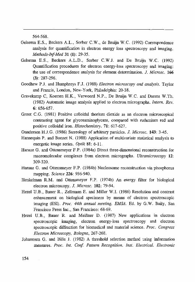
564-568.
Gelsema E.S., Beckers A.L., Sorber C.W., de Bruijn W.C. (1992) Correspondence
analysis for quantification in electron energy loss spectroscopy and imaging. Methods-Irif-Med 31 (1): 29-35.
Gelsema E.S., Beckers A.L.D., Sorber C.W.J. and De Bruijn W.C. (1992) Quantification procedures for electron energy-loss spectroscopy and imaging:
the use of correspondence analysis for element deterroination. J. Microsc. 166 (3): 287-296.
Goodhew P.J. and Humphreys F.J. (1988) Electron microscopy and analysis. Taylor
and Francis, London, New-York, Philadelphia: 20-38.
Gravekamp C, Koerten H.K., Verwoerd N.P., De Bruijn W.C. and Daerns W.Th.
(1982) Automatic image analysis applied to electron micrographs. Intern. Rev.
6: 656-657.
Groot C.G. (1981) Positive colloidal thorium dioxide as an electron microscopical
contrasting agent for glycosamiooglycans, compared with ruthenium red and positive colloidal iron. Histochemistry. 71: 617-627.
Gundersen H.J.G. (1986) Stereology of arbitrary particles. J. Microsc. 143: 3-45.
Hannequin P. and Bonnet N. (1988) Application of multivariate statistical analysis to
energetic image series. Optik 81: 6-1!.
Harauz G. and Ottensmeyer F.P. (1984a) Direct three-dimensional reconstruction for
macromolecnlar complexes from electron micrographs. Ultramicroscopy 12:
309-320.
Harauz G. and Ottensmeyer F.P. (1984b) Nucleosome reconstruction via phosphorus
mapping. Science 226: 936-940.
Henkehnan R.M. and Ottensmeyer F.P. (1974b) An energy filter for biological
electron microscopy. J. Microsc. 102: 79-94.
Hezel U.B., Bauer R., Zellmann E. and Miller W.l. (1986) Resolution and contrast enhancement on biological specimens by means of electron spectroscopic
imaging (ESI). Proc. 44th annual meeting, EMSA. Ed. by G. W. Baily, San
Francisco Press Inc., San Francisco: 68-69. Hezel U.B., Bauer R. and MeiBner D. (1987) New applications in electron
spectroscopic imaging, electron energy-loss spectroscopy and electron spectroscopic diffraction for biomedical and material science. Proc. Congress
Electron Microscopy, Bologna, 267-268. Johannsen G. and Bille J. (1982) A threshold selection method using information
measures. Proc. Int. Con[. Pattern Recognition. Inst. Electrical. Electronic
!54

References
Engineers Inc. l(Computer Soc. Press. Miinchen): 140-142.
Joy D.C. and Maher D.M. (1980) Electron energy loss spectroscopy : detectable limits for elemental analysis. Ultramicroscopy 5: 333-342.
Joy D.C. and Maher M. (1981) The quantification of electron energy-loss spectra. J.
Microsc. 124: 37-48.
Kortje K.H., Freihiifer D. and Rahmann H. (1990) Cytochemical localization of calcium in the central nervous system of vertebrates. Ultramicroscopy 32: 12-17.
Kor1je K.H., Kortje D. and Rahmann H. (1990) The application of energy-fl.ltering electron microscopy for the cytochemical localization of Ca2+ -ATPase activity
in synaptic terminals. J. Microsc. 162: 105-114. Kortje K.H. (1993) personal communications Krahl D., Herrmann K.-H. and Kunath W. (1978) Electron optical experiments with
a magnetic imaging filter. Electron Microscopy/1987 9th 1nt. Cong. Ed. by J.M. Sturgess, Microscopical Society of Canada, Toronto: 42-43.
Krivanek O.L., Gubbens A.J. and Dellby N. (1991) Developments in EELS instrumentation for spectroscopy and imaging. Microsc. Microanal. Microstruct. 2: 315-332.
Kruit P. and Shuman H. (1985) The influence of objective lens aberrations in energy-loss spectrometry. Ultramicroscopy 17: 263-268.
Leapman, R.D., Fiori, C.E. & Swyt, C.R. (1984a) Mass thickness determination by electron energy loss for quantitative X-ray microanalysis in biology. J.
Micros c. 133: 239-253.
Leapman R.D., Fiori C.E., Gorlen K.E., Gibson C.C. and Swyt C.R. (1984b) Combined elemental and STEM imaging under computer control.
Ultramicroscopy 12: 281-292. Leapman R.D., Gorlen K.E. and Swyt R.E. (1985) Digital processing of electron
energy-loss spectra and images. Scanning Electron Microscopy 1985/I, Ed. by 0. Johari, SEM, AMF O'Hare, Chicago. Ill.: 1-13.
Leapman R.D. and Ornberg R.L. (1988) Quantitative electron energy loss spectroscopy in biology. Ultramicroscopy 24: 251-268.
Leapman R.D. and Swyt C.R. (1988) Separation of overlapping core edges in electron energy loss spectra by multiple-least-squares fitting. Ultramicroscopy 26: 393-404.
Liu D.R. and Brown L.M. (1987) Influence of some practical factors on background extrapolation in EELS quantification. J. Microsc. 147: 37-49.
155

Loretto M.H. (1984) Electron beam analysis of materials Chapman and Hall Ltd., London, chapter 2.
Malis T., Cheng S.C. and Egerton R.F. (1988) EELS Log-ratio technique for specimen-thickness measurement in the TEM. J. Electr. Microsc. Techn. 8: 193-200.
Meyer F. (1981) Cytology automation with mathematical morphology. Bioi. Cell. 41: 1-6.
Morgan E., Burton K. and Nickless G. (1990) Optimization using the modified
Simplex method. Chemometrics and Intelligent Laboratory Systems 7: 209-222.
Mostert L.J., Cleton M.I., De Bruijn W.C., Koster J.F. and Van Eijk H. G. (1989)
Studies on ferritin in rat liver and spleen during repeated phlebotomy. Int. J. Biochem. 21: 39-47.
Miiller A. (1906) Bemerkungen iiber das Hydrosols des Thoriumoxydhydrats. Ber.
Deutsch. Chem. Ges. 93: 2857-2859. Neider J.A. and Mead R. (1965) A Simplex method for function minimisation.
Computer Journal 7: 308-313. Newburry D.E. (1986) Principles of analytical electron microscopy. Ed. by D.C.
Joy, A.D. Romig Jr. and J.I. Goldstein, Plenum Press, New-York: 1-27. Ottensmeyer P.F. (1984) Electron spectroscopic imaging: parallel energy-filtering
and microanalysis in the fixed-beam electron microscope. J. Ultrastruct. Res.
88: 121-134. Peachy L.D., Heath J.P. and Lamprecht G. (1986) The use of an energy-filtering
electron microscope to reduce chromatic aberration in images of thick biological specimens. Proc. 44th annual meeting, EMSA. Ed. by G.W. Baily, San Francisco Press Inc., San Francisco: 88-91.
Pejas W. and Rose H. (1978) Outline of an imaging magnetic energy filter free of second-order aberrations. Electron Microscopy/1978 9th Int. Cong., Ed. by J .M. Sturgess, Microscopical Society of Canada, Toronto: 44-45.
Probst W. (1986) Ultrastructural localization of calcium in the CNS of vertebrates. Histochemistry 85: 231-239.
Pun T., Ellis J.R. and Eden M. (1984) Optimized acquisition parameters and statistical detection limits in quantitative EELS. J. Microsc. 135: 295-316.
Pun T., Ellis J.R. and Eden M. (1985) Weighted least squares estimation of background in EELS imaging. J. Microsc. 137: 93-100.
Rademakers L.H.P.M., Koningsberger J.C., Sorber C.W.J., Baart de Ia Faille H.,
156

References
Van Hattum J. and Marx J.J.M. (1993) Accwnulation of iron in erythroblasts
of patients with erythropoietic protoporphyria. European Journal of Clinical
Investigation 23: 130-138.
Reichelt R. and Engel A. (1984) Monte carlo calculations of elastic and inelastic
electron scattering in biological and plastic materials. Ultramicroscopy 13:
279-294.
Reimer L., Fromm I. and Rennekamp R. (1988) Operation modes of electron spectroscopic imaging and electron energy-loss spectroscopy on a transmission
electron microscope. Ultramicroscopy 24: 339-354.
Reimer L. and Ross-Messemer M. (1990a) Contrast in the electron spectroscopic
imaging mode of a TEM. II. Z-ratio, structure-sensitive and phase contrast. J.
Microsc. 159: 143-160.
Reimer L. and Ross-Messemer M. (1990b) Contrast in the electron spectroscopic
imaging mode of a TEM I. Influence of zero-loss filtering on a scattering
contrast. J. Microsc. 155: 169-182.
Reimer L. (1991) Energy-Filtering Transmission Electron Microscopy. Advances in
electronics and electron physics 81: 43-126. Reimer L., Rennekamp R., Fromm I. and Langenfeld M. (1991) Contrast in the
electron spectroscopic imaging mode of a TEM. IV. Thick specimens imaged
by the most-probable energy loss. J. Microsc. 162: 3-14.
Ringeling P.L., Cleton M.I., Kroos M.J. Sorber C.W.J., De Bruijn W.C., Harrison
P.M. and Van Eijk H.H. (1989) Lysosomal and cytosolic ferritins. A biochemical and electron-spectroscopic study. Bioi. Metals 2: 114-121.
Ringeling P.L., Cleton M.I., Huijskes-Heins M.I.E., Seip M.J.E., De Bruijn W.C.
and Van Eijk H. G. (1990) Analysis of iron-containing compounds in different compartments of the rat-liver after iron loadiog. Bioi. Metals 3: 176-182.
Roomans G.M. and Shelburne J.D. (1980) Basic methods io biological X-ray
microanalysis io. Scanning Electron Microsc. ][[, Ed. by 0. Johari, SEM Inc.
A.M.F. O'Hare, Chigaco, Ill.: 309-321.
Roos N. and Barnard T. (1984) Aminoplastic standards for quantitative X-ray microanalysis of thin sections of plastic-embedded biological material.
UUramicroscopy 15: 277-286.
Rose H. and Plies E. (1974) Entwurf eioes fehlerarmen magoetishen EnergieAnalysators. Optik 40: 336-341.
Russ J.C. and Russ J.C. (1984) Image processing io a general purpose computer. J.
Microsc. 135: 89-102.
157

Russ J.C. and Russ J.C. (1987) Automatic discrimination of features in grey-scale images. J. Microsc. 148: 263-277.
Shavers C.L., Parsons M.L. and Deming S.N. (1979) Simplex optimization of chemical systems. J. Chern. Ed. 56: 307-309.
Shuman H. and Somlyo A.P. (1982) Energy-fJ.ltered transmission electron microscopy of ferritin. Proc. Nat. Acad. Sci. USA 79: 106-107.
Shuman H., Chang C.F., and Somlyo A.P. (1986) Elemental imaging and resolution
in energy-fJ.ltered conventional electron microscopy. Ultramicroscopy 19: 121-133.
Shuman H. and Somlyo A.P. (1987) Electron energy loss analysis of near-traceelement concentrations of calcium. Ultramicroscopy 21: 23-32.
Somlyo A.P. and Shuman H. (1982) Electron probe and electron energy Joss analysis in biology. Ultramicroscopy 8: 219-233.
Somlyo A.P. (1984) Compositional mapping in biology: X-rays and electrons. J.
Ultrastruct. Res. 88: 135-142. Sorber C.W.J., DeJong A.A.W., Den Breejen N.J. and De Bruijn W.C. (1990a)
Quantitative energy-filtered image analysis in cytochemistry I. Morphometric
analysis of contrast related images. Ultramicroscopy 32: 55-68. Sorber C.W.J., Van Dort J.B. Ringeling P.C., Cleton-Soeteman M.I. and De Bruijn
W.C. (1990b) Quantitative energy-fJ.ltered image analysis in cytochemistry II. Morphometric analysis of element-distribution images. Ultramicroscopy 32:
69-79. Sorber C.W.J., Ketelaars G.A.M., Gelsema E.S., Jongkind J.F. and De Bruijn
W.C. (199la) Quantitative analysis of electron energy-Joss spectra from ultrathin-sectioned biological material. I. Optimization of the background-fit with the use of Bio-standards. J. Microsc. 162: 23-42.
Sorber C.W.J., Ketelaars G.A.M., Gelsema E.S., Jongkind J.F. and De Bruijn W.C. (!99lb) Quantitative analysis of electron energy-loss spectra from ultrathin-sectioned biological material. I. The application of Bio-standards for quantitative analysis. J. Microsc. 162: 43-54.
Spendley W., Hext G.R. and Himsworth F.R. (1962) Sequential application of Simplex designs in optimisation and evolutionary operations. Technometrics 4:
441-461 Steele J.D., Titchmarsh J.M., Chapman J.N. and Paterson J.H. (1985) A single
stage process for quantifying electron energy loss spectra. Ultramicroscopy 17: 273-276.
!58

References
Stobbs W.M. and Boothroyd C.B. (1991) Approaches for energy loss and energy filtered imaging in TEM in relation to the materials problems to be solved. Microsc. Microanal. Microstruct. 2: 333-350.
Titchmarsh J.M. and Malis T.F. (1989) On the effect of objective lens chromatic aberration on quantitative electron-energy-loss spectroscopy (EELS). Ultramicroscopy 28: 277-282.
Trebbia P. (1988) Unbiased method for signal estimation in electron energy loss spectroscopy, concentration measurements and detection limits in quantitative microanalysis: methods and programs. Ultramicroscopy 24: 399-408.
Trebbia P. (1989) Proc. 1st Europ. Workshop. Europ. Microbeam Analysis Soc.:
107.
Trebbia P. and Bonnet N. (1990). EELS elemental mapping with unconventional methods. I Theoretical bases: image analysis with multivariate statistics and entropy concepts. Ultramicroscopy 34: 165-178.
Trebbia P. and Mory C. (1990) EELS elemental mapping with unconventional methods II. Applications to biological specimens. Ultramicroscopy 34: 179-203.
Trebbia P. (1990) EELS elemental mapping with unconventional methods I. Theoretical basis: image analysis with multivariate statistics and entropy concepts. Ultramicroscopy 36: 165-178.
Trinick J. and Berriman J. (1987) Zero-loss electron microscopy with the Zeiss EM902. Ultramicroscopy 21: 393-398.
Vander Meijden W., Koerten H.K., Van Mourik W. and De Bruijn W.C. (1988) Descriptive light and electron microscopy of normal and clue cell-positive discharge. Gynecol. obstet. Invest. 25: 47-57.
Van Dort J.B., Zeelen J.Ph. and de Bruijn W.C. (1987) An improved procedure for the X-ray microanalysis of acid phosphatase activity in lysosomes. Histochemistry 87: 71-77.
Van Dart J.B., Ketelaars G.A.M., De Jong A.A.W. and De Bruijn W.C. (1988) Electron energy-loss spectroscopy in ultrastructoral cytochemistry. Inst. Phys.
Con[. Ser. 93, York: 577-578. Van Dart J.B., Ketelaars G.A.M., Daems W. Th. and De Bruijn W.C. (1989)
Ultrastructural electron probe X-ray microanalytical reaction product identification of three different enzymes in the same mouse resident peritoneal macrophage. Histochemistry 92: 243-253.
Van Puymbroeck J. (1992) Electron energy-loss spectroscopy: Development of
159

methodology and software for the acquisition and processing of spectra.
Thesis. Verbeek P.W., Vrooman H.A., Van Vliet L.J. (1988) Low-level image processing
by Max-Min filters. Signal Processing 15, 249-258.
Verbucken A.H., van Grieken E., Poulas G.J. and De Bruijn W.C. (1984)
Embedded ion exchange beads as standards, for laser microprobe mass
analysis of biological specimens. Analytical Chemistry 56: 1362-1370.
Yarbo L.A. and Deming S.N. (1974) Selection and preprocessing of factors for
simplex optimization. Anal. Chim. Acta 73: 391-398.
Young, I.T. (1988) Sampling density and quantitative microscopy. Anal. quant. Cytol. histol. 10/4: 269-275.
Zaluzec N.J. (1985) Digital filters for application to data analysis in electron energy
loss spectroscopy. Ultramicroscopy 18: 185-190.
Zanchi G., Perez J.-P. and Sevely J. (1975) Adaptation of a magnetic filtering
device on a one megavolt electron microscope. Optik 43: 495-501.
Zanchi G., Sevely J. and Jouffrey B. (1977a) An energy filter for high voltage
electron microscopy. J. Microsc. Spectrosc. Electron. 2: 95-104.
Zanchi G., Sevely J. and Jouffrey B. (1977b) Second-order image aberration of a
one megavolt magnetic filter. Optik 48: 173-192.
Zanchi G. Kihn Y. and Sevely J. (1982) On aberration effects in the chromatic plane
of the OMEGA filter. Optik 60: 427-436.
Zondervan P.E., de Jong A., Sorber C.W.J., Kok L.P., De Bruijn W.C. and Van der Kwast Th.H. (1988) Microwave-stimulated incubation in immunoelectron
microscopy: a quantitative study. Histochemical J. 20: 359-364.
160

LIST OF
PUBLICATIONS
161

Publications
LIST OF PUBLICATIONS
Sorber C.W.J., DeJong A.A.W., Den Breejen N.J. and De Bruijn W.C. (1990) Quantitative energy-filtered image analysis in cytochemistry I. Morphometric analysis of contrast related images. Ultramicroscopy 32, p. 55-68.
Sorber C.W.J., Van Dort J.B. Ringeling P.C., Cleton-Soeteman M.I. and De Bruijn W.C. (1990) Quantitative energy-filtered image analysis in cytochemistry II. Morphometric analysis of element-distribution images. Ultramicroscopy 32, p. 69-79.
Sorber C.W.J., Ketelaars G.A.M., Gelsema E.S., Jongkind J.F. and De Bruijn W.C. (1991) Quantitative analysis of electron energy-loss spectra from ultrathin-sectioned biological material. I. Optimization of the background-fit with the use of Bio-standards. J. Microsc. 162, p. 23-42.
Sorber C.W.J., Ketelaars G.A.M., Gelsema E.S., Jongkind J.F. and De Bruijn W.C. (1991) Quantitative analysis of electron energy-loss spectra from ultrathin-sectioned biological material. I. The application of Bio-standards for quantitative analysis. J. Microsc. 162, p. 43-54.
Sorber C.W.J., Ketelaars G.A.M., Beckers A.L.D. and De Bruijn W.C. (1991) Proc. znd Europ. Workshop European Microbeam Analysis Soc., p. 76.
Sorber C.W.J., Bonnet N, Trebbia P., Gelsema E.S., Jongkind J.F., Beckers A.L.D. and De Bruijn W.C. (1993) Multivariate Statistical Analysis as a tool for EELS-analysis: submitted to J. Microsc.
162

Publications
OTHER PUBLICATIONS
Cleton M.I., Mostert L.J., Sorber C.W.J., de Jong A.A.W., De Jeu-Jaspers C.M.H. and De Bruijn W.C. (1989) Effect of phlebotomy on the ferritin iron content in the rat liver as determined morphometrically with the use of electron energy-loss spectroscopy. Cell and Tissue Res. 256: 601-605.
De Bruijn W.C., Sorber C.W.J., De Jong A.A.W. and Cleton-Soeteman M.l. (1989) Quantitative image analysis with EELS and EPMA in cytochemistry. Proc .. 1st Eur. Workshop. Europ. Microbeam Analysis Soc.: 191.
De Bruijn W.C. and Sorber C.W.J. (1990) The application of Bio-standards for electron energy-loss analysis of biological materials. Proc. Xlfh Intern.
Congress for Electron Microscopy Seattle 2, San Francisco Press Inc.: 68-69.
De Bruijn W.C., Ketelaars D., Gelsema E. and Sorber L. (1991) Comparison of the simplex method with several other methods for background-fitting for electron energy loss spectral quantification of biological materials. Microsc.
Microanal. Microstruct. 2: 281-291.
De Bruijn W.C., Ketelaars G.A.M., Jongkind J.F. and Sorber C.W.J. (1991) Proc. 2nd Europ. Workshop European Microbeam Analysis Soc.: 40.
De Bruijn W.C., Sorber C.W.J., Trommelen-Ketelaars G.A.M., Jongkind J.F., Beckers A.L.D. and Gelsema E.S. (1993) Electron energy-loss spectroscopy and electron probe X-ray microanalysis of biological material: a comparative quantitative analysis of electron microscopical images. In: X-ray Microanalysis
in biology: Experimental techniques and applications. Edited by Sigee D.C, Morgan A.J., Summer A.T and Warley A.: p. 81-98.
De Bruijn W.C., Sorber C.W.J., Gelsema E.S., Beckers A.L.D., Jongkind J.F. (1993) Energy-ffitered transmission electron microscopy of biological specimens Scanning Microscopy: in press.
!63

Gelsema E.S., Beckers A.L., Sorber C.W. and de Bruijn W.C. (1992) Correspondence analysis for quantification in electron energy loss spectroscopy and imaging. Methods-Inf-Med 31 (1): 29-35
Gelsema E.S., Beckers A.L.D., Sorber C.W.J. and De Bruijn W.C. (1992) Quantification procedures for electron energy-loss spectroscopy and imaging: the use of correspondence analysis for element deterroination. J. Microsc. 166 (3): 287-296.
Rademakers L.H.P.M., Koningsberger J.C., Sorber C.W.J., Baart de Ia Faille H., Van Hattum J. and Marx J.J.M. (1993) Accumulation of iron in erythroblasts of patients with erythropoietic protoporphyria. European Journal
of Clinical Investigation 23: 130-138.
Ringeling P.L., Cleton M.I., Kroos M.J. Sorber C.W.J., De Bruijn W.C., Harrison P.M. and Van Eijk H.H. (1989) Lysosomal and cytosolic ferritios. A biochemical and electron-spectroscopic study. Bioi. Metals 2: 114-121.
Willemsen R., Briinken R., Sorber C.W.J., Hoogeveen A.T., Wisselaar H.A., Van Dongen J.M. and Reuser A.J.J. (1991) A quantitative immunoelectron microscopic study on soluble, membrane-asscociated and membrane-bound lysosomal enzymes in human intestioal epithelial cells. Histochemical Journal
23: 467-473.
Zondervan P.E., de Jong A., Sorber C.W.J., Kok L.P., De Bruijn W.C. and Van der Kwast Th.H. (1988) Microwave-stimulated incubation in immunoelectron microscopy: a quantitative study. Histochemical J. 20: 359-364.
164

SUMMARY
SAMENV ATTING
165

SUMMARY
Several methods are proposed for the elemental analysis of biological
material with the use of electron energy-loss spectroscopy (EELS) and electron
spectroscopic imaging (ESI) in a Zeiss EM902.
For spectra, the Simplex-method was tested and compared to Egerton's 2-area method, the Steepest descent method and the Loglog method. The Simplex
method and the Loglog method performed best. The Simplex method was then used
for the analysis of biological specimens containing calcium and iron. The results of
the analysis of a test-spectrum, an iron-spectrum and a calcium-spectrum taken
with various widths of the r -region were compared. It was observed that calcium is
a hard element for EELS-analysis because of the carbon K-edge just in front of its
ionization-edge. Ca- and Fe-Bio-standards, which were co-embedded with the
specimens, were used to obtain elemental concentrations in the specimen.
In combination with the Kontron IBAS 2000, morphometrical image analysis
was performed on cells and their reaction products. The influence of several acquisition factors was analyzed (objective-lens diaphragm diameter, image mode,
number of integrations during image acquisition). Segmentation is applied using the
cross-overs of the frrst derivative of the greyvalue distribution histogram of the image. Co-localization of several chemical elements could be demonstrated by
comparing the net-intensity distribution images of the respective elements.
In images, several methods were tested for the separation of the element
information from the continuum. The power-law method gives the best results. The
influence of the width of the integration-region on the obtained relative
concentration was tested. Since the power-law method did not overcome all problems encountered
with image-analysis (Calcium), Multivariate statistical analysis (MSA) was
exanrined as an alternative. The latter method performed very well and was able to analyze calcium images. MSA also reduces the influence of other sources of
information in the images such as shading and radiation damage.
166

Summary I Samenvatting
SAMENVATTING
Verschillende methoden worden onderzocht voor de element analyse van biologisch materiaal met behulp van "electronen energie-verlies spectroscopie" (EELS) en "electronen spectroscopische afbeelding" (ESI) in een Zeiss EM902.
Voor spectra werd de Simplex-methode getest en vergeleken met Egerton's 2-oppervlakken methode, de steilste pad methode en de Loglog methode. De Simplex methode en de Loglog methode gaven de beste resultaten. De Simplexmethode werd vervolgens gebruikt voor de analyse van biologische preparaten die calcium en ijzer bevatten. De resultaten van de analyse van een test-spectnnn, een ijzer-spectnnn en een calcium-spectnnn, verkregen met verschillende groottes voor het r -gebied, werden vergeleken. Hieruit blijkt onder andere dat calcium een erg moeilijk element is door de koolstof-piek net voor zijn ionisatie-kant. Calcium en ijzer Bio-standaarden, die bij de coupe waren ingebed, werden gebruikt om element concentraties te bepalen.
In combinatie met de Kontron IBAS 2000, werden cellen en hun reactieprodukten morfologische geanalyseerd. De invloed van verschillende factoren werd bepaald (diametergrootte van het object-lens diafragma, afbeeld mode, aantal integraties bij beeldopname). Voor de segmentatie van de beeld-objecten werd gebruik gemaakt van de 1 e afgeleide van het grijswaarde frequentie-histogram van het beeld. Co-localisatie van verscheidene chemische elementen kon worden aangetoond door de netto-intensiteits verdelings beelden van de respectievelijke
elementen te vergelijken. In beelden werden verschillende methodes getest om de element-informatie
te scheiden van het continufun. De "Power-law" methode gaf de beste resultaten. De invloed van de breedte van de .:1-zone op de verkregen relatieve concentratie werd getest.
Aangezien de "Power-law" methode niet aile problemen kon oplossen die in beeld-analyse werden ondervonden (calcium) werd het gebruik van "Multivariate Statistische Analyse" (MSA) als alternatief onderzocht. MSA gaf zeer goede resultaten en maakte het ook mogelijk om calcium beelden te analyseren. MSA reduceerde ook de invloed van andere informatie-bronnen uit de beelden zoals "shading" en stralingsschade.
167


Acknowledgements
Toen ik vijf jaar geleden op de afdeling pathologie stage liep had ik nooit kunnen dromen dat ik in 1993 een promotie-onderzoek zou afronden. Dit is vooral mogelijk gemaakt door het vertrouwen en de steun van mijn co-promotor, Dr. Wim de Bruijn en mijn promotor, Prof. Dr. Hans Jongkind, waarvoor ik ze zeer dankbaar ben. Samen met Prof. Dr. Edzard Gelsema zorgden ze door hun heel verschillende invalshoeken voor een unieke combinatie om problemen te benaderen. Aile drie heel hartelijk bedankt hiervoor en voor het kritisch lezen van het manuscript. De overige !eden van de leescommissie wil ik oak nog bedanken: Prof. Dr. Fre Bosman voor de gastvrijheid op de afdeling pathologie en voor het "automatiseren" van de afdeling waardoor ik vee! extra ervaring maar oak vee! extra werk kreeg, en Prof. Dr. Noel Bonnet voor zijn gastvrijheid tijdens mijn verblijf in Reims (Noel, thank you for your hospitality during my stay in Reims).
De afdeling electronen-microscopie ben ik zeer dankbaar voor de oergezellige tijd tijdens de afgelopen vijf jaar. Piet van der Hen!, Ton de Jong, Peter van Run, Annemiek van den Broek, Theo van der Schaft, Pieter Derkx en Alex Nigg, ik zal jullie missen! Vooral de discussies tijdens de "werkbesprekingen". Drie collega's hadden al eerder afscheid genomen van de afdeling pathologie maar Paula Delfos, Diane Trommelen en Elly Visser oak jullie zal ik heel erg missen, vooral de uitjes tussen de middag naar de stad.
Ton de Jong, dank voor aile electronen microscopische lessen die je me met vee! geduld en een uitgestreken gezicht hebt bijgebracht (labjassen, veiligheidsbrillen, mondkapjes en handschoenen om een filament te vervangen ???).
Frank van der Paune en Paula Delfos bedankt voor de fotografie. lk weet dat ik soms erg lastig was maar de foto' s I grafieken zijn prachtig.
Piet van der Hen! (mijn paranymphomaan, je was als een vader voor de EM-afdeling) en Diane Trommelen bedankt voor het paranymphen.
De feestcommissie, sorry, het personeels-fonds, bedankt voor de gezellige planningen, vergaderingen, feesten en labdagen. Monica Seijbel, Josiane Godschalk, Huib de Bruin, Hein Sleddens en Frank van der Paune het was altijd heel gezellig.
De stagiaires van de afgelopen vijf jaar wil ik ook nag bedanken: Jolanda Krijnen, Nettie de Breejen, Loek Timmermans, Mariella Ansems, Coert-Jan van Mook, Robert Brtinken, Ellen Raaymakers, Kees de Crom en Bart Willemse.
En natuurlijk ben ik mijn onders zeer dankbaar voor hun constante steun tijdens de laatste 28 jaar.
Verder hebben nag vele mensen bijgedragen aan dit proefschrift en aangezien ik niet iedereen bij naam kan noemen wil ik hierbij iedereen bedanken die op enigerlei wijze heeft bijgedragen aan dit proefschrift of aan de gezellige sfeer tijdens de afgelopen 5 jaar.
169

CURRICULUM VITAE
Lianne Sorber werd geboren op 27 december 1964 te Dordrecht. Na het eindexamen VWO in juli 1983 aan het Thomas More College te Oudenbosch heeft zij een jaar Biologie gestudeerd aan de Katholieke Universiteit te Nijmegen. Aangezien deze studie niet voldeed aan haar verwachtingen heeft zij deze studie afgebroken en vervolgens heeft zij van augustus 1984 tot juli 1988 de opleiding Hogere Laboratorium Informatica en Automatisering gevolgd aan het toenmalige Dr Struycken Instituut te Etten-Leur. Na haar stage op de afdeling Pathologische Anatornie van de Erasmus Universiteit Rotterdam in het 4° jaar van deze opleiding heeft zij van october 1988 tot december 1992 haar promotie onderzoek verricht als AIO (assistent in op1eiding) op de afdeling E1ectronen Microscopie van de Pathologische Anatornie met als resultaat dit proefschrift. Hiervan werden drie maanden doorgebracht aan de INSERM-unit van de Universiteit te Retms (Frankrijk). V anaf 1 juni 1993 zal zij werkzaam zijn bij Unilever in Sharnbrook, Engeland.
170