Points fattening on P1× P1 and symbolic powers of bi-homogeneous ideals
Upload
khangminh22Category
view
1download
0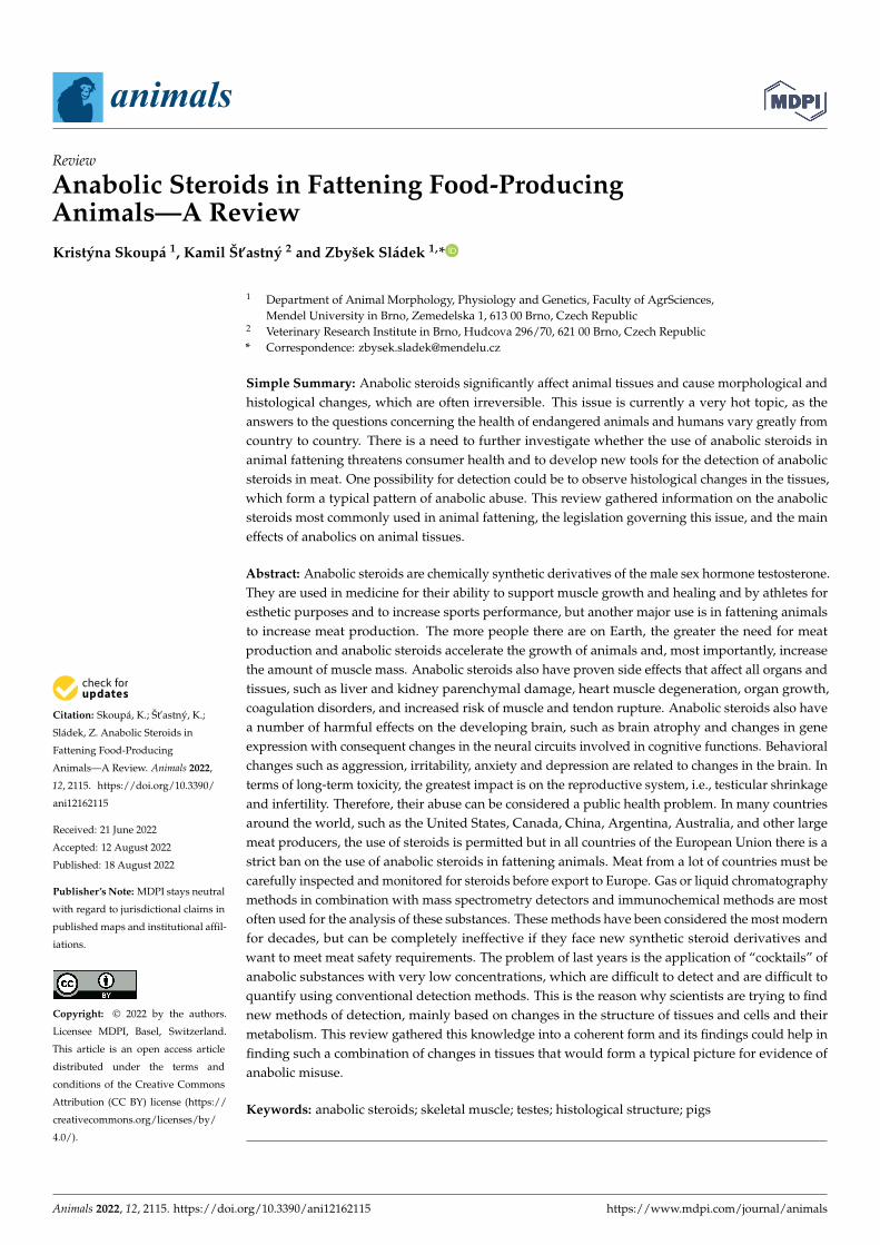
Citation: Skoupá, K.; Št’astný, K.;
Sládek, Z. Anabolic Steroids in
Fattening Food-Producing
Animals—A Review. Animals 2022,
12, 2115. https://doi.org/10.3390/
ani12162115
Received: 21 June 2022
Accepted: 12 August 2022
Published: 18 August 2022
Publisher’s Note: MDPI stays neutral
with regard to jurisdictional claims in
published maps and institutional affil-
iations.
Copyright: © 2022 by the authors.
Licensee MDPI, Basel, Switzerland.
This article is an open access article
distributed under the terms and
conditions of the Creative Commons
Attribution (CC BY) license (https://
creativecommons.org/licenses/by/
4.0/).
animals
Review
Anabolic Steroids in Fattening Food-ProducingAnimals—A ReviewKristýna Skoupá 1, Kamil Št’astný 2 and Zbyšek Sládek 1,*
1 Department of Animal Morphology, Physiology and Genetics, Faculty of AgrSciences,Mendel University in Brno, Zemedelska 1, 613 00 Brno, Czech Republic
2 Veterinary Research Institute in Brno, Hudcova 296/70, 621 00 Brno, Czech Republic* Correspondence: [email protected]
Simple Summary: Anabolic steroids significantly affect animal tissues and cause morphological andhistological changes, which are often irreversible. This issue is currently a very hot topic, as theanswers to the questions concerning the health of endangered animals and humans vary greatly fromcountry to country. There is a need to further investigate whether the use of anabolic steroids inanimal fattening threatens consumer health and to develop new tools for the detection of anabolicsteroids in meat. One possibility for detection could be to observe histological changes in the tissues,which form a typical pattern of anabolic abuse. This review gathered information on the anabolicsteroids most commonly used in animal fattening, the legislation governing this issue, and the maineffects of anabolics on animal tissues.
Abstract: Anabolic steroids are chemically synthetic derivatives of the male sex hormone testosterone.They are used in medicine for their ability to support muscle growth and healing and by athletes foresthetic purposes and to increase sports performance, but another major use is in fattening animalsto increase meat production. The more people there are on Earth, the greater the need for meatproduction and anabolic steroids accelerate the growth of animals and, most importantly, increasethe amount of muscle mass. Anabolic steroids also have proven side effects that affect all organs andtissues, such as liver and kidney parenchymal damage, heart muscle degeneration, organ growth,coagulation disorders, and increased risk of muscle and tendon rupture. Anabolic steroids also havea number of harmful effects on the developing brain, such as brain atrophy and changes in geneexpression with consequent changes in the neural circuits involved in cognitive functions. Behavioralchanges such as aggression, irritability, anxiety and depression are related to changes in the brain. Interms of long-term toxicity, the greatest impact is on the reproductive system, i.e., testicular shrinkageand infertility. Therefore, their abuse can be considered a public health problem. In many countriesaround the world, such as the United States, Canada, China, Argentina, Australia, and other largemeat producers, the use of steroids is permitted but in all countries of the European Union there is astrict ban on the use of anabolic steroids in fattening animals. Meat from a lot of countries must becarefully inspected and monitored for steroids before export to Europe. Gas or liquid chromatographymethods in combination with mass spectrometry detectors and immunochemical methods are mostoften used for the analysis of these substances. These methods have been considered the most modernfor decades, but can be completely ineffective if they face new synthetic steroid derivatives andwant to meet meat safety requirements. The problem of last years is the application of “cocktails” ofanabolic substances with very low concentrations, which are difficult to detect and are difficult toquantify using conventional detection methods. This is the reason why scientists are trying to findnew methods of detection, mainly based on changes in the structure of tissues and cells and theirmetabolism. This review gathered this knowledge into a coherent form and its findings could help infinding such a combination of changes in tissues that would form a typical picture for evidence ofanabolic misuse.
Keywords: anabolic steroids; skeletal muscle; testes; histological structure; pigs
Animals 2022, 12, 2115. https://doi.org/10.3390/ani12162115 https://www.mdpi.com/journal/animals

Animals 2022, 12, 2115 2 of 16
1. Introduction
Steroid hormones are generally complex lipophilic molecules synthesized from theprecursor cholesterol molecule in endocrine cells in the adrenal cortex, testes, ovaries, andplacenta. They are mostly formed as steroid hormone precursors and only after stimulationof the secreting cells of the above-mentioned organs are they converted into active hormonesand penetrate from the parent cell by simple diffusion as their intracellular concentrationincreases. The hypothalamo–pituitary axis is then the superior system for their productionand control of endocrine glands by means of tropical hormones. Steroid hormones act onthe paracrine and endocrine pathways, where they are released by endocrine cells into theinterstitial space and specifically affect cells in the immediate vicinity. In the body, they playa crucial role in regulating and communicating across cells, tissues, and organs throughoutan individual’s life [1,2].
The mechanisms of action of individual steroid hormones differ significantly fromeach other, especially depending on their physical and chemical properties. Hydrophilichormones act primarily on the cell surface, where they bind to receptors in the plasmamembrane. On the contrary, hydrophobic steroid hormones bind to free plasma proteinsand are able to diffuse freely across cell membranes and thus activate specific intracellularhormone receptors. The effects of hormones can then be generally divided into long-termand short-term effects. Long-term effects are mediated by high-affinity intracellular receptorproteins that are localized in specific target tissues for each steroid hormone. The interactionof the steroid–receptor complex with hormone-responsive genes leads to tissue-specificexpression of proteins that either directly or indirectly generate the body’s biologicalresponses attributed to steroid hormones. This mode of cellular action is generally referredto as the genomic effect of hormones, is slow, and occurs with a time delay of hours oreven days [3]. On the other hand, non-genomic action is any mode of action in which genetranscription is not performed directly. These are mostly short-term and rapid reactions thatinvolve specific G protein-coupled receptors located on the cell membrane. They containmany second messengers, including cAMP and diacylglycerol, kinases, and ion flux. Short-term side effects include sexual and reproductive disturbances, fluid retention, increasedhunger, general loss of energy, and development or worsening of infections, and severeacne is often reported in humans. Short-term side effects are reversible in most cases [2,4].
Synthetic steroid hormones or anabolic androgenic steroids (AAS) are chemicallysynthetic derivatives of the male sex hormone testosterone or its structural modifications.Testosterone occurs naturally in the body (especially in males, but to a lesser extent infemales) and is involved in the regulation of a large number of physiological processessuch as metabolism, development, and regulation of reproductive processes, increasingsexual libido and bone and muscle metabolism. They have a significant anabolic effecton protein synthesis, including muscle tissue, and are therefore applied to animals andhumans to rapidly increase muscle mass [5,6]. Steroid hormones can indirectly increasemuscle growth through effects on cortisol or by increasing circulating levels of IGF-I topromote long bone growth or directly by affecting the muscle satellite cell population.Some observations suggest an indirect effect through changes in the balance of endogenoushormones. Trenbolone has been reported to increase plasma levels of growth hormoneand/or insulin; these hormones are known to stimulate the transport of amino acids acrossthe cell membrane [7,8]. This has the net effect of greater protein synthesis and reducedprotein degradation, resulting in increased anabolism of muscle tissue due to larger maturebody size and a more active population of satellite cells. Increased proliferative activityof satellite cells should increase muscle growth rate [9]. In addition to anabolic function,they also have androgenic effects because they affect the growth and function of the malereproductive organs. For natural testosterone, both effects are more or less in balance; theindividual synthetic preparations differ in their balance between anabolic and androgenicactivity. Alkylation of C17, esterification of the hydroxyl group on this carbon, and otherchemical modifications seek to increase the anabolic effect and stabilize the molecule, butnone of the available anabolic steroids are purely anabolic [5,6,10].

Animals 2022, 12, 2115 3 of 16
Anabolic steroids have often been used in livestock fattening around the world fortheir ability to accelerate muscle growth and thereby speed up and ensure fattening andprofit from meat sold. Currently, some countries outside the EU approve the use of anumber of preparations based on steroid hormones for fattening cattle and sheep. No typeof anabolic is approved to promote growth in dairy cows, pigs, or poultry. It is estimatedthat 80%–90% of cattle in fattening outside the EU are treated with at least one type ofgrowth-promoting anabolic agent. After their possible negative impact on human healthbegan to be investigated, some states began to limit their use and defined it in their legisla-tion [6]. There are still very few studies investigating the anabolic effect directly in farmanimals; most studies are conducted on laboratory animals such as mice and rats, or theeffects of steroids are investigated in human medicine [5,6]. It turns out that the use of AASalso brings economic and environmental benefits too. Webb et al. determined the environ-mental and economic impacts of cattle raised with different levels of growth-promotingtechnologies, non-hormonally treated and implanted. The implanted technology reducedthe carbon footprint by 8%, energy consumption by 6%, water consumption by 4%, andreactive nitrogen losses by 8% [11]. A study by Capper et al. evaluated the hypothesis that theuse of steroid implants in Brazilian beef cattle would reduce resource use, greenhouse gasemissions, and economic costs of production, thereby improving ecological and economicsustainability. The use of implants reduced GHG emissions for 1.0 × 10⁶ kg HCW of beefby 15.8%. The 6.13% increase in kg of HCW beef produced generates a cost reduction of3.76% and an increase in the return on invested capital of 4.14% on average [12]. Anotherstudy conducted on cattle shows that the use of anabolic implants reduces water consump-tion by 9.85%, the need for sown land by 9.58%, greenhouse gas emissions by 7.5%, fossilfuel consumption by 4.98%, and animal feed costs by 7.55% [13].
The purpose of the review was to summarize information on legislative measures forthe use of anabolic steroids, the most frequently used or abused anabolic steroids, and theeffect of anabolic steroids on the tissues of farm animals.
2. Legislative Measures for the Use of AAS2.1. History of the Use of Anabolic Steroids
The effects of steroid hormones have been used unknowingly by farmers since ancienttimes. They noticed increased domestication of animals after neutering and also bettermeat quality in neutered animals. In ancient Egypt, as early as 800 AD, extract of testicleswas used as an aphrodisiac [14,15]. In 1786, the first targeted transplantation of a cock’stesticles was performed, during which it was found that the animal’s secondary sexualcharacteristics were affected [16,17]. Subsequent experiments in 1849 described changesin appearance and behaviour of cock chickens after castration, and a substance producedby the testes, which is transported in the body by the blood to the target tissues it affects,was described by Arnold Berthold [18,19]. This substance was later identified as the sexhormone testosterone. The anabolic effect of synthetic hormones was first described byCharles Kochakian in 1935 and mentioned the possible use of these substances in tissueregeneration and growth stimulation [15]. From 1937, clinical trials using testosteronederivatives were conducted, and from the 1940s, these substances began to be used byhumans as doping agents in sports. In 1948, Dinusson, Andrews, and Beeson conductedthe first experiment to support cattle growth with a synthetic estrogen derivative [20].In 1953, a synthetic ester of estrogen-diethylstilbestrol was first added to cattle feed [21].The development of anabolic steroid preparations later became a major topic of researchand the pharmaceutical industry. AAS have been investigated for possible use in thetreatment of a wide range of diseases, such as muscle and wound healing, support in thetreatment of osteoporosis, and severe burns, where they speed up tissue regeneration byup to several days. They are prescribed after surgery and radiation therapy and for thetreatment of anemia, but their use is currently declining and they are used only after othertreatment options have been exhausted. The use of anabolic steroids is also associated witha wide range of negative side effects described above, and, therefore, with any therapy,

Animals 2022, 12, 2115 4 of 16
it is necessary to consider whether their beneficial effect on health sufficiently outweighsthe often severe negative effects [22]. In animal production, they took their place in therole of growth stimulants in fattening. Unfortunately, they have been and still are misusedin both food-producing animals and by athletes. It also contributes to the fact that thereis little information and published texts that 100% confirm the side effects for the finalconsumers of meat containing residues of anabolic substances that cannot be neutralized.However, many studies based on epidemiological studies draw attention to the relationshipof hormonal residues in food with cancer [14,15,23–25].
2.2. Current Situation in Europe
The use of anabolic steroids to promote growth in food-producing animals has beenbanned since 1961 in the Netherlands, since 1962 in Belgium, and since 1973 in all Beneluxcountries. In all countries of the European Union there has been a strict ban on the use ofhormonal or thyrostatic substances and growth-promoting beta-agonists since 1988. Theonly exceptions are some approved veterinary medicines that can be used in defined indica-tions. It is therefore prohibited to use anabolic substances for non-therapeutic purposes inlivestock farming, as well as to import meat from animals treated with these substances intothe EU market, as an increasing number of studies have demonstrated the possible adverseeffects of their residues in meat products on human health. In the past, these rules havelimited meat imports from the United States, Canada, Argentina, Australia, Asia, and SouthAfrica, where anabolic steroids are permitted as part of livestock feed and medicines. Atpresent, the import of meat from these countries into the EU is allowed, under the conditionof certification that the animals have not been given anabolic substances. The relevantinternational organizations (FAO, WHO, OIE, Codex) therefore had to set up programs tomonitor these substances. The ban on the use of hormones is laid out in Council Directive96/22/EC [26]. Measures requiring EU countries to monitor these substances and theirresidues in animals and products of animal origin have so far been laid out in Directive96/23/EC, which will be replaced by Regulation (EU) 2017/625 of the European Parlia-ment and of the Council from December 2022 [27,28]. Directive (EU) 2017/625 appliesto food control and introduces new national surveillance programs for the monitoring ofresidues of prohibited substances. For example, the acceptable levels of α-testosterone andβ-testosterone are set by the FDA at 1.54–2.62 and 1.06–1.56 µg/kg, and the permissiblelimit for trenbolone is 10 µg/kg in liver and 2 µg/kg in muscle [29]. Meat, milk, eggs, andhoney are monitored, and blood, urine, and milk samples are taken from live animals onfarms and from carcasses at slaughterhouses. The use of veterinary medicinal productsis governed by Council Regulation 2377/90/EC [30,31], which details a procedure forthe establishment of maximum residue limits (MRLs) of veterinary medicinal products infoodstuffs of animal origin. The European Medicines Agency decides whether a specifiedmaximum residue limit should be applied to a certain food and lays out rules regardingthe conditions for calculating this residue limit. In 2021, a new Directive (EU) 2021/808was issued, which sets out conservative analytical methods for detecting a few AAS withthe well-known organic structure of their molecules [32]. However, it is limited by currentknowledge and technical capabilities and therefore cannot cover the detection of newlysynthesized AAS and the black market; in large part, producing these new compounds isstill a few steps ahead.
In 1999, 2000, and 2002, the Scientific Committee on Veterinary Measures Relatingto Public Health (SCVPH) conducted extensive studies on the possible adverse effects ofsteroid hormone residues in beef and meat products on human health, and some hormoneshave been declared carcinogens. In the light of these conclusions, Directive 2003/74/ECwas adopted with a view to a permanently ban the use of hormones in livestock farming.In 2007, the European Food Safety Authority (EFSA) provided evidence that all hormonesbanned in the EU may have endocrine, developmental, immunological, neurobiological,genotoxic, and carcinogenic effects, especially in risk groups such as children [33].

Animals 2022, 12, 2115 5 of 16
Although screening anabolic steroids in pigs and cattle reveals very few positive casesin EU countries, their misuse cannot be ruled out. Steroid hormones are legally producedfor therapeutic purposes and can then be diverted to illegal applications [34].
2.3. Current Situation in the Rest of the World
In contrast to strict bans in the EU, in countries outside Europe, anabolic steroids canbe used legally as fattening aids because national authorities have recognized their use asrisk-free or, in some cases, have only defined their use.
In the United States, the Food and Drug Administration (FDA) has approved a num-ber of steroid hormonal preparations for fattening cattle and sheep since the 1950s [35].Permitted substances include natural testosterone, estrogen, progesterone, and some oftheir synthetic derivatives. All information on FDA-approved products that can be usedas hormonal implants can be found in the Code of Federal Regulations (CFR). All thesesubstances are applied in order to increase the growth rate of animals and provide higherefficiency of feed conversion into meat. New substances are always approved by the FDAonly after studies have shown that food from such treated animals is safe for consumersand does not harm the treated animal or the environment. Based on scientific data, theFDA sets limits on the content of hormones in meat. A safe level for human consumptionis one which, based on extensive scientific studies, is not expected to have any harmfuleffect on humans. Hormones are most commonly applied as skin implants from the backof an animal’s ear, and unless otherwise specified, only one implant is administered to theanimal at each stage of growth. The ears are removed at slaughter and are therefore notused for consumption. The implants are intended for cattle and sheep only and are freelyavailable in the United States. It is estimated that up to 90% of cattle for fattening haveat least one anabolic implant, mostly combined implants. No implant type is approvedfor accelerating the growth of dairy cows, pigs, or poultry [36]. More than 20 states in theUnited States that use hormone implants report that their use has reduced greenhousegas emissions and water and energy consumption in beef production compared with beefgrown without growth-promoting hormones [37,38].
Similar laws apply in Canada, where limited amounts of steroid hormones have onlybeen used in fattening cattle since the 1960s [39].
Argentina allows the use of testosterone as well as the synthetic substances zeranoland trenbolone acetate in the fattening of cattle and sheep. In Brazil, since 1991, the use ofnatural and artificial substances for the purpose of animal growth as well as their import,production, and marketing have been prohibited [40].
In China, the first version of the regulation governing the administration of veterinarymedicines, including hormonal products, was issued in 1987 [41,42]. Major amendmentswere made in 2001 and the current version of the Regulation on the administration ofveterinary medicines in China was adopted in 2004. Over time, the Ministry of Agricultureof China (MOA) added supporting regulations as measures for the registration of veterinarymedicinal products [40–44], Including administrative measures on over-the-counter veteri-nary medicinal products and standards for the use of veterinary medicinal products in beefand dairy cattle, pigs, sheep, hens, and rabbits [45]. Since 1999, the MOA has introducedmandatory monitoring and control of the use of veterinary products, including some AAS,each year, and in 2002 issued the latest standards for MRLs for veterinary medicines usedin food-producing animals. Most current MRLs are inspired by and derived from EU andUS standards, but these standards are often more stringent than those in China. Comparedwith the USA and European countries, the residue limits of veterinary drugs in China areupdated very slowly. Appropriate standards should be speeded up, especially for thosemedicines that have not yet been included in current standards, in particular antibioticsand large amounts of anabolic hormones [42,46].
In Japan, regulations are stricter and residue limits for veterinary drugs are set similarlyto the United States. Anabolic hormone residues must be below the MRL; otherwise, theyare considered a potential risk to human health [42,47]. However, studies often indicate

Animals 2022, 12, 2115 6 of 16
that little or no attention is paid to monitoring the misuse of anabolic steroids in fatteningfarms on small businesses and farms, and therefore steroid hormone abuse may still beencountered in some Asian countries, e.g., Bangladesh and Oman [48,49].
In Australia, the use of steroid hormonal products is partially permitted and hasbeen approved since the mid-1970s. Only Tasmania banned their use in 2000 [47]. The useof hormonal products is overseen by the Australian Pesticides and Veterinary MedicineAuthority (APVMA), which states that the use of hormonal growth promoters is safewhen applied according to the label instructions. Australia also has a hormone-free food-producing animals program that produces meat for the European market [50,51].
In South Africa, some growth promoters, such as testosterone, estradiol, progesterone,zearanol, and trenbolone acetate, have been approved to promote growth in cattle since1947 [52]. However, most African countries have laws regulating the use of growing promotersin food-producing animals, but do not have competent authorities to carry out risk analysesof residues of these substances, mainly due to a lack of trained staff or a lack of financialresources [52,53].
3. The Most Commonly Used or Misused Anabolic Steroids3.1. Boldenon
Boldenone, a 1(2)-dehydrogenated analogue of testosterone, differs chemically fromtestosterone by a double bond between C1 and C2. It is characterized by high anabolicand low androgenic activity and due to its effects muscles grow slower but is of betterquality than most testosterone derivatives [54–56]. It is approved as a veterinary drug inmany countries, especially for the treatment of horses, calves, and lambs [57,58]. The useof boldenone is banned by humans and food-producing animals and for doping in race-horses. The most commonly used derivative is Boldenone Undecylenate, which is appliedintramuscularly by injection. Under more recent IFHA and FEI laws, steroids can only beadministered to a horse under certain therapeutic conditions and the horse must not com-pete for at least 60 days afterwards [54]. Boldenone has been classified by the InternationalAgency for Research on Cancer as a possible carcinogen, with a higher carcinogenicityindex than other used anabolics [54,59]. Boldenone has often been detected in a numberof biological samples in various EU countries—Italy [54,59,60], Belgium [54,61], and theNetherlands [60]. However, boldenone and its metabolites are naturally produced steroidsby farm animals and are excreted in large amounts in faeces. In addition, boldenonehas been shown to be formed from plant fat phytosterols in feed [57,62,63]. Nielen et al.,2004 [64] showed that the presence of 17β-Boldenone conjugates at any level in the urine ofcalves is evidence of illegal application. Urine must be collected without faecal contamina-tion to avoid erroneous results. Around the year 2000, the frequent use of boldenone incattle fattening and doping in horses was thus proven [54,59,60]. New studies from 2022prove that when using ultrasensitive detection methods, we can always detect boldenonein low concentrations in horse urine samples, and for anti-doping tests at horse races theexact spectrum of biomarkers must be used [65]. In stallions, only testosterone and 17β-boldenone are generally considered to be endogenous. However, the ‘semi’-endogenouspresence of 17β-boldenone and related compounds, for example in mares and geldings,is a complicating factor in doping control. The IFHA thus abandoned the zero-tolerancepolicy for stallions and a threshold value for free and conjugated boldenone of 15 ng/mLwas established. Despite this threshold for stallions, the presence of 17β-boldenone in theurine of mares or geldings is still prohibited [54,66].
3.2. Chlortestosteron
Chlortestosterone, clostebol, is a 4-chlorinated derivative of testosterone. Chlorinationprevents the steroid from being converted to dihydrotestosterone and at the same timemakes it unable to convert to estrogen. Its use is prohibited in humans and animals, withthe exception of some approved veterinary drugs that can be used in specific indications,mainly for dermatological use [67,68]. In the past, it was used mainly for fattening cattle

Animals 2022, 12, 2115 7 of 16
and to increase the performance of racehorses. After 1990, a large number of positive resultsfor 4-chlortestosterone acetate were reported in France and Belgium [69], and thereforeefforts began in Europe to develop more accurate and sensitive detection methods [70,71].However, it is still used for fattening cattle in China and Japan [72].
3.3. Nandrolon
Nandrolone, 19-nortestosterone, is an anabolic steroid with a chemical structure similarto testosterone. Due to the lack of a methyl group at C19, it has a better binding affinity forandrogen receptors and an increased rate of onset of anabolic activity, and thus also fastereffects on muscle growth. Nandrolone and trenbolone have the highest anabolic:androgenicratio of all AAS. It is most often used in the form of esters, especially as nandrolone decanoateand nandrolone phenylpropionate [73]. Illegal use of nandrolone and its esters in livestockfattening as growth promoters has often been reported in the EU in the past [60,74,75], asnandrolone has long been considered only a synthetic AAS. Its natural form was first con-firmed in veterinary plasma medicine in horses in 1980 [76] and subsequently endogenousnandrolone was also confirmed in human medicine in 1984 [77]. 17β-19-nortestosteronewas later shown to occur naturally in the urine of boars [78], bulls [79] and sheep, cows,sows, and mares during pregnancy [43,59], often at relatively high concentrations, andevidence of nandrolone abuse is not easy to find [79]. Due to its properties, nandrolone isalso often part of anabolic cocktails, so the methods for its detection must be constantlyimproved [80].
3.4. Stanozolol
Stanozolol is a synthetic 17α-alkylated derivative of 5α-dihydrotestosterone. It waspreviously widely used in human and veterinary medicine, especially for its very high ratioof anabolic: androgenic activity, which means that it has anabolic activity with minimalandrogenic undesirable effects [81]. Currently, it is only used in domestic animals, especiallydogs, to increase muscle mass and stimulate the appetite of exhausted animals or in thetreatment of tracheal collapse [82]. It is not used in livestock for human consumption [83,84].It is considered a controlled substance, but is gradually being withdrawn from circulationin a large number of countries. In the past, its frequent misuse in animal fattening has beenfound across European countries. Most of these illegal applications were discovered around1999, the same year as laboratory methods rapidly improved [85,86]. After administration,stanozolol is metabolized very rapidly, so urinary levels of the parent compound are verylow. Thus, the level of urinary excretion is also low, i.e., 3%–5% of the total administeredamount, and stanozolol can be detected in urine for 2–3 days. Its detection in urine istherefore almost impossible [87]. Improvements in stanozolol residue analyses are nowbeing achieved by screening for its marker metabolite 16β-hydroxystanozolol [86,88].
3.5. Trenbolon
Trenbolone is an androgenic anabolic derivative of nandrolone, specifically nandrolonewith two added double bonds in the steroid nucleus. It is most often used in the form ofesters, especially as trenbolone acetate (TBA) and trenbolone enanthate (TBE). TBA is avery strong anabolic steroid with strong anabolic effects and therefore has great potentialfor use in fattening. A number of side effects of its use have been described, includingaggression, increased blood pressure and cholesterol levels, skin rashes, negative effectson the thyroid gland, decreased sexual function, and testicular atrophy. Most of theseeffects are irreversible [89]. TBA is licensed as a growth stimulant for cattle in severalcountries around the world, such as the United States, Australia and New Zealand, whichare major meat exporters [36,90,91]. However, this causes global problems, as meat fromthese countries often demonstrates a positive TBA above the MRL (10 µg/kg in liver and2 µg/kg in muscle), even though the meat has been certified as hormone-free [92,93]. In2000, an extensive inspection of beef in supermarkets imported from Australia and theUSA was carried out in Indonesia, and up to half of the samples contained trenbolone

Animals 2022, 12, 2115 8 of 16
residues [94]. The study by El Shaid et al. [95] reported similar results from analyses ofsausages and burgers in Egypt, where, again, up to half of the samples were positivefor trenbolone.
4. Effect of Anabolic Steroids on Tissues
Regular intake of anabolic steroids causes changes in the organization at genetic,metabolomic, and structural levels directly in animal tissues. Increased protein synthesis,fat loss, and a better sensory profile of final meat products are the targeted changes forwhich anabolic steroids are used in animal fattening. Like natural hormones, syntheticsteroids imitate hormonal functions and interfere with the endocrine system of animals.In addition to endocrine changes, steroids have been shown to have carcinogenic, im-munotoxic, mutagenic, and teratogenic effects, and the changes are often irreversible. Therisks always depend on the age, sex, and individual tolerance of the animals to thesesubstances [96,97]. As the use of steroid substances in fattening animals poses a potentialrisk to consumers, their presence needs to be monitored and new detection methods de-veloped to prevent the prevalence of public health risks. Many anabolic substances thatcan theoretically be misused in meat production have not been studied for toxic effects onhumans yet [98,99].
4.1. Effect of Anabolic Steroids on Skeletal Muscle
Skeletal muscle is the most economically important tissue in animal production andthe application of anabolic steroids is mostly targeted at it. The increase in body parametersis best processed in beef cattle after the use of steroidal implants. Implanting increasedaverage daily gain by 21% and improved feed efficiency by 11% in feedlot cattle. In addition,carcass weight was increased by 7% due to implanting. Moreover, a 5% increase was alsoreported in ribeye size, a 7% decrease in fat cover, a 5% decrease in marbling score, anda 17% decrease in percent of carcasses grading [7]. A study by Perry et al. compared theeffects of steroid implants on performance and carcass composition in Holstein, Angus,and Angus x Simmental steers. Daily gain was increased by 17%, 26%, and 21% in Holstein,Angus, and crossbred steers, respectively. The implant increased total daily protein and fatgain by 23% [100].
Most studies describing the effect of anabolics on muscle mass have been performedin animal models such as mice or rats, and from livestock in cattle or sheep [101]. Very fewstudies have been performed on pig models, although in many European countries pork isconsumed to a much greater extent than beef and lamb [102].
What the studies agree on is that AASs increase muscle hypertrophy and proteinsynthesis, metabolize fat stores, and enhance the response when AASs are combined withstrength activity. Thus, in general, the application of steroids leads to an increase in thegrowth rate of lean mass. At the histological level in muscle, the diameter and area ofmuscle fibers increase and there is a significant increase in the proportion of fast low-oxidation fibers [103–105]. Body weight gain is increased through nitrogen retention andnet protein gain without any changes in the digestibility of nitrogen intake. The amount ofprotein production can then be measured by comparing the amount of nitrogen suppliedwith the amount of nitrogen in the animal waste. Anabolic hormones therefore acceleratenitrogen retention in the body. However, high doses of AAS can cause degenerativechanges in muscle that can lead to loss of function and permanent damage, such as musclefiber breakdown, cell infiltration, vacuolation, swelling, and mitochondrial damage [105].The hypertrophy of these multinucleated muscle fibers is accompanied by an increasein the number of myonuclei, with the primary source of new myonuclei being activatedand incorporated satellite cells otherwise located outside the muscle fibers. Satellite cellsalso need to maintain their reserve fund to be able to self-renew. The increased numberof satellite cells in the growing muscle causes an increased capacity for skeletal musclegrowth [106] and also increases the ability to regenerate muscles. It is demonstrated thatAAS supplementation after injury in mice increases the number of proliferating satellite cells

Animals 2022, 12, 2115 9 of 16
and the cross-sectional area of the fibers in the regenerating muscle [107,108]. In addition,the results of new studies show that a common feature of the effects of various growthhormones on muscle cell proliferation and differentiation is the activation of the polyaminebiosynthesis pathway [106]. In older animals, the AAS-induced muscle growth processesare slower because most of the remaining satellite cells become silent and no longer multiplyor differentiate unless stimulated by injury or strength training [109]. However, recentresearch [110] shows that faster muscle (re) growth in older animals may be induced byprevious AAS use. Myonuclei, acquired during anabolic-induced hypertrophy, are at leastpartially preserved, and later muscle growth after AAS stimulation is then up to twice asfast. This AAS-influenced “muscle memory” hypothesis is based mainly on data obtainedfrom rodent experiments, and due to differences in muscle formation between species, thetransfer of these results to livestock is ambiguous and difficult [110,111]. A more preciseidentification of the molecular mechanism by which AAS improves livestock muscle growthefficiency is necessary to develop more effective strategies to improve meat production, butthe exact mechanisms are currently unknown.
Our preliminary results from experiments on pigs agree with the results of the previ-ously mentioned studies. After the application of testosterone and nandrolone decanolate,there were changes in the muscle of the pigs in the form of an increase in the diameter andarea of the muscle fibers and a higher amount of connective tissue (endomysium) betweenthe muscle fibers (Figure 1). Whether the number of nuclei is a statistically significant pa-rameter is still under study, as the counting of nuclei in histological sections is not accurate,as they are randomly distributed along the fiber.
Animals 2022, 12, x FOR PEER REVIEW 9 of 17
muscle fiber breakdown, cell infiltration, vacuolation, swelling, and mitochondrial dam-age [105]. The hypertrophy of these multinucleated muscle fibers is accompanied by an increase in the number of myonuclei, with the primary source of new myonuclei being activated and incorporated satellite cells otherwise located outside the muscle fibers. Sat-ellite cells also need to maintain their reserve fund to be able to self-renew. The increased number of satellite cells in the growing muscle causes an increased capacity for skeletal muscle growth [106] and also increases the ability to regenerate muscles. It is demon-strated that AAS supplementation after injury in mice increases the number of proliferat-ing satellite cells and the cross-sectional area of the fibers in the regenerating muscle [107,108]. In addition, the results of new studies show that a common feature of the effects of various growth hormones on muscle cell proliferation and differentiation is the activa-tion of the polyamine biosynthesis pathway [106]. In older animals, the AAS-induced muscle growth processes are slower because most of the remaining satellite cells become silent and no longer multiply or differentiate unless stimulated by injury or strength train-ing [109]. However, recent research [110] shows that faster muscle (re) growth in older animals may be induced by previous AAS use. Myonuclei, acquired during anabolic-in-duced hypertrophy, are at least partially preserved, and later muscle growth after AAS stimulation is then up to twice as fast. This AAS-influenced “muscle memory” hypothesis is based mainly on data obtained from rodent experiments, and due to differences in mus-cle formation between species, the transfer of these results to livestock is ambiguous and difficult [110,111]. A more precise identification of the molecular mechanism by which AAS improves livestock muscle growth efficiency is necessary to develop more effective strategies to improve meat production, but the exact mechanisms are currently unknown.
Our preliminary results from experiments on pigs agree with the results of the pre-viously mentioned studies. After the application of testosterone and nandrolone decano-late, there were changes in the muscle of the pigs in the form of an increase in the diameter and area of the muscle fibers and a higher amount of connective tissue (endomysium) between the muscle fibers (Figure 1). Whether the number of nuclei is a statistically sig-nificant parameter is still under study, as the counting of nuclei in histological sections is not accurate, as they are randomly distributed along the fiber.
Figure 1. Histological section of muscle fibers of pig from the control group without anabolic ster-oids (C), from the group after testosterone administration (T), and from the group after administra-tion of nandrolone (N). After the application of AAS, there was an increase in the diameter and area of muscle fibers and a greater amount of endomysium between muscle fibers in the muscle of pigs. With nandrolone, the steroid effect was even stronger (unpublished data).
4.2. Effect of Anabolic Steroids on the Male Reproductive System The AAS affects the genitals, especially in males, in a completely different way than
the muscles. Thus, they do not cause their growth, but numerous studies have shown that AASs have a strong toxic and degenerative impact [108,112–114]. The application of ana-bolics has an impact mainly on the testicles, where it causes disturbances of
Figure 1. Histological section of muscle fibers of pig from the control group without anabolic steroids(C), from the group after testosterone administration (T), and from the group after administrationof nandrolone (N). After the application of AAS, there was an increase in the diameter and area ofmuscle fibers and a greater amount of endomysium between muscle fibers in the muscle of pigs.With nandrolone, the steroid effect was even stronger (unpublished data).
4.2. Effect of Anabolic Steroids on the Male Reproductive System
The AAS affects the genitals, especially in males, in a completely different way than themuscles. Thus, they do not cause their growth, but numerous studies have shown that AASshave a strong toxic and degenerative impact [108,112–114]. The application of anabolicshas an impact mainly on the testicles, where it causes disturbances of spermatogenesis,epithelial degeneration, and overall disorganization of the anatomical and histologicalstructure. The membranes of the seminiferous tubules are deformed, the layers of thegerminal epithelium inside the tubules are reduced, and the number and mobility ofsperm are reduced [113]. This loss of germ cells causes mitosis and meiosis to stop, andhigh doses of AAS cause degeneration and decreased Sertoli cell support function. As aresult, the layers of sperm cells are reduced to one layer and the sperm cells begin todegenerate [112]. Interstitial spaces expand, necrotic Leydig cell numbers increase, andoverall anabolic uptake has an inhibitory effect on the hypothalamic–pituitary testicularaxis, leading to a reduction in natural testosterone production. There is also damage to the

Animals 2022, 12, 2115 10 of 16
additional gonads and enlargement of the prostate [114,115]. Ultimately, AAS has similareffects to chemical castration. Several studies suggest a positive relationship between AASapplication and infertility and carcinogenesis progression. Commonly used hormones, suchas nandrolone and stanozolol, can potentially induce the progression of various cancers,such as Leydig cell tumors, through many pathways [109,116]. Approximately six weeksafter cessation of anabolic hormone therapy in a mouse study, a gradual improvement inthe structural condition of the testis was observed, but there was no complete recovery offunction [114,117].
Our preliminary results from experiments on pigs show a reduction in the germinalepithelium in the seminiferous tubules after application of nadrolon to complete destructionof the epithelium and the formation of a fibrous network from the ligament inside thelumen of the canals, reminiscent of prepubertal structures. There was a decrease in theincidence of mature spermatids and spermatozoa. The seminiferous tubules were spacedapart. Leydig cells deformations, wrinkling, and degeneration occurred (Figure 2). Thus,reduced testosterone production can be expected, leading to reduced fertility or evencomplete sterility of individuals.
Animals 2022, 12, x FOR PEER REVIEW 10 of 17
spermatogenesis, epithelial degeneration, and overall disorganization of the anatomical and histological structure. The membranes of the seminiferous tubules are deformed, the layers of the germinal epithelium inside the tubules are reduced, and the number and mobility of sperm are reduced [113]. This loss of germ cells causes mitosis and meiosis to stop, and high doses of AAS cause degeneration and decreased Sertoli cell support func-tion. As a result, the layers of sperm cells are reduced to one layer and the sperm cells begin to degenerate [112]. Interstitial spaces expand, necrotic Leydig cell numbers in-crease, and overall anabolic uptake has an inhibitory effect on the hypothalamic–pituitary testicular axis, leading to a reduction in natural testosterone production. There is also damage to the additional gonads and enlargement of the prostate [114,115]. Ultimately, AAS has similar effects to chemical castration. Several studies suggest a positive relation-ship between AAS application and infertility and carcinogenesis progression. Commonly used hormones, such as nandrolone and stanozolol, can potentially induce the progres-sion of various cancers, such as Leydig cell tumors, through many pathways [109,116]. Approximately six weeks after cessation of anabolic hormone therapy in a mouse study, a gradual improvement in the structural condition of the testis was observed, but there was no complete recovery of function [114,117].
Our preliminary results from experiments on pigs show a reduction in the germinal epithelium in the seminiferous tubules after application of nadrolon to complete destruc-tion of the epithelium and the formation of a fibrous network from the ligament inside the lumen of the canals, reminiscent of prepubertal structures. There was a decrease in the incidence of mature spermatids and spermatozoa. The seminiferous tubules were spaced apart. Leydig cells deformations, wrinkling, and degeneration occurred (Figure 2). Thus, reduced testosterone production can be expected, leading to reduced fertility or even com-plete sterility of individuals.
Figure 2. Histological section through the seminiferous tubules of pig testes from the control group without anabolic steroids (C), from the group after testosterone administration (T), and from the group after administration of nandrolone (N). A reduction in the germinal epithelium in the semi-niferous tubules after testosterone, the destruction of the epithelium, and the formation of a fibrous network from the ligament inside the lumen of the canals after nandrolone application are evident (unpublished data).
4.3. Effect of Anabolic Steroids on Other Tissues In addition to the above-mentioned tissues, in which the manifestation of AAS is the
most intensively studied, anabolics also affect other tissues in the body. These are mostly significant degenerative changes and disorganization of the histological structure. A neg-ative effect on the heart muscle, kidneys, liver, and bones is described [114]. The results of numerous studies, especially in rats, agree that long-term use of AAS causes patholog-ical cardiac hypertrophy that persists and even increases after treatment. After higher doses of synthetic steroids, such as nandrolone decanolate, vasocongestion, muscle fiber retraction, massive dilaceration, and muscle fiber rupture occur in the heart tissue.
Figure 2. Histological section through the seminiferous tubules of pig testes from the control groupwithout anabolic steroids (C), from the group after testosterone administration (T), and from thegroup after administration of nandrolone (N). A reduction in the germinal epithelium in the semi-niferous tubules after testosterone, the destruction of the epithelium, and the formation of a fibrousnetwork from the ligament inside the lumen of the canals after nandrolone application are evident(unpublished data).
4.3. Effect of Anabolic Steroids on Other Tissues
In addition to the above-mentioned tissues, in which the manifestation of AAS is themost intensively studied, anabolics also affect other tissues in the body. These are mostlysignificant degenerative changes and disorganization of the histological structure. A nega-tive effect on the heart muscle, kidneys, liver, and bones is described [114]. The results ofnumerous studies, especially in rats, agree that long-term use of AAS causes pathologicalcardiac hypertrophy that persists and even increases after treatment. After higher doses ofsynthetic steroids, such as nandrolone decanolate, vasocongestion, muscle fiber retraction,massive dilaceration, and muscle fiber rupture occur in the heart tissue. Reduced pumpingefficiency of the heart is reported [109,114,118]. These pathophysiological cardiac effectscan be linked to the fact that androgen receptors in cardiomyocytes allow steroids to affectthe physiology of the heart [114].
Persistent changes even after discontinuation of AAS treatment are also described inthe kidneys. After application of testosterone esters and nandrolone decanoate, swellingand weight gain occur in the kidneys. On the contrary, there is atrophy and deformation ofthe glomeruli and cracking of the glomerular walls. Damage to the urinary tubules, rupture,vacuolar epithelial degeneration of the proximal coiled tubules, and thickening of the basallamina in the distal coiled tubules are visible. The tubules move away from each other andbleeding occurs between them due to overloading of the renal vessels [114,115,119,120]. Af-

Animals 2022, 12, 2115 11 of 16
ter long-term application, lesions and large necrotic areas form in the urinary tubules after afew weeks [114]. These findings suggest the possibility of chronic kidney damage after AASapplication, which may lead to progressive kidney failure. The study by Cho et al. [121]indicated that AAS could be a risk factor for the development and progression of renal cellcarcinoma. These changes in renal structure also correlate with the finding of frequent renaldisorders in bodybuilders who have used high doses of AAS. Hartung et al. 2001 [122]stated that renal biopsy reveals nephrosclerosis and severe kidney lesions in bodybuilders.
In the liver, in contrast to the kidneys, the greatest weight gain was observed at lowdoses of nandrolone decanolate. At higher doses there was only a slight increase, but therewas a sharp increase in the amount of liver enzymes. There were disorders of bile formationand drainage, dilation of blood vessels, and their ruptures [120,123].
AAS and androgens are generally involved in bone growth and maintenance of bonehomeostasis [108]. Studies show that anabolics do not affect bone quality, but speed up thestrengthening process and increase bone callus formation after injury [124]. Souza et al. [125]described that after administration of nandrolone decanolate, there was no increase infemoral weight or length and there was no difference in relation to the diameter of theepiphysis and the diaphysis. Marchi et al. [126] found that nandrolone stimulated bonemarrow production and it could be used in the treatment of aplastic anemia. In sheep, thebeneficial effect of stanozol on articular cartilage regeneration in femorotibial osteoarthritishas been confirmed [127]. The positive effect of stanozol on joints is also described byathletes and, the study by Falanga et al. [128] disclosed that stanozol increases collagensynthesis. However, this effect has not been described with other anabolic steroids.
5. Conclusions
Anabolic steroids significantly affect animal tissues and cause morphological andhistological changes, which are often irreversible. This issue is currently a very hot topic,as the answers to questions about endangered animal and human health vary greatly fromone country to another. Whether the use of AAS in animal fattening threatens the health ofconsumers needs to be further studied and new tools for detecting AAS in meat need to bedeveloped. One of the detection options could be the observation of histological changes inthe tissues that form the typical picture of AAS misuse.
Author Contributions: Conceptualization, K.Š. and Z.S.; methodology, K.Š., Z.S. and K.S.; investiga-tion, K.Š.; data curation, K.S.; writing—original draft preparation, K.S., K.Š. and Z.S.; writing—reviewand editing, K.S. and Z.S.; supervision, Z.S.; project administration, K.Š.; funding acquisition, K.Š. Allauthors have read and agreed to the published version of the manuscript.
Funding: This research was funded by National Agency for Agricultural Research project “Metabolomicsof steroid hormones with a significant anabolic effect as a basis for new analytical control methodolo-gies designed to demonstrate the practices of abuse of prohibited substances in fattening livestock”,grant number QK1910311.
Institutional Review Board Statement: The study was performed in compliance with Act No. 246/1992Coll. of the Czech National Council for the protection of animals against cruelty and with the agree-ment of the Branch Commission for Animal Welfare of the Ministry of Agriculture of the CzechRepublic (permission no. MZe 17214).
Informed Consent Statement: Not applicable.
Data Availability Statement: Not applicable.
Acknowledgments: The author team thanks the National Agency for Agricultural Research forfinancial support of grant number QK1910311.
Conflicts of Interest: The authors declare no conflict of interest.
References1. Medvei, V.C. A History of Endocrinology; MTP Press: Hingham, MA, USA, 1982.2. Cole, T.J.; Short, K.L.; Hooper, S.B. The science of steroids. Semin. Fetal Neonatal Med. 2019, 24, 170–175. [CrossRef] [PubMed]

Animals 2022, 12, 2115 12 of 16
3. Echeverria, P.C.; Picard, D. Molecular chaperones, essential partners of steroid hormone receptors for activity and mobility.Biochim. Biophys. Acta Mol. Cell Res. 2010, 1803, 641–649. [CrossRef] [PubMed]
4. Lösel, R.; Wehling, M. Nongenomic actions of steroid hormones. Nat. Rev. Mol. Cell Biol. 2003, 4, 46–55. [CrossRef] [PubMed]5. Kuhn, C.M. Anabolic Steroids. Recent Prog. Horm. Res. 2002, 57, 411–434. [CrossRef] [PubMed]6. Kreutzer, K.V.; Turk, J.R.; Casteel, S.W. Clinical Biochemistry in Toxicology. In Clinical Biochemistry of Domestic Animals, 6th ed.;
Kaneko, J.J., Harvey, J.W., Bruss, M.L., Eds.; Academic Press: Cambridge, MA, USA, 2008; pp. 821–838.7. Johnson, B.J.; White, M.E.; Hathaway, M.R.; Christians, C.J.; Dayton, W.R. Effect of a combined trenbolone acetate and estradiol
implant on steady-state IGF-I mRNA con-centrations in the liver of wethers and the longissimus muscle of steers. J. Anim. Sci.1998, 76, 491–497. [CrossRef] [PubMed]
8. Becker, C.h.; Riedmaier, I.; Reiter, M.; Tichopad, A.; Pflaffl, M.W.; Meyer, H.H.D. Effect of trenbolone acetate plus estradiol ontranscriptional regulation of metabolism path-ways in bovine liver. Horm. Mol. Biol. Clin. Investig. 2010, 2, 257–265.
9. Smith, Z.K.; Johnson, B.J. Mechanisms of steroidal implants to improve beef cattle growth: A review. J. Appl. Anim. Res. 2020, 48,133–141. [CrossRef]
10. Yoshida, E.M.; Erb, S.R.; Scudamore, C.H.; Owen, D.A. Severe Cholestasis and Jaundice Secondary to an Esterified Testosterone, aNon-C17 Alkylated Anabolic Steroid. J. Clin. Gastroenterol. 1994, 18, 268–269. [CrossRef]
11. Webb, M.J.; Pendell, D.L.; Harty, A.A.; Salverson, R.R.; Rotz, C.A.; Underwood, K.R.; Olson, K.C.; Blair, A.D. Influence of GrowthPromoting Technologies on Animal Per-formance, Production, Economics, Environmental Impacts and Carcass Characteristics ofBeef. Meat Muscle Biol. 2017, 1, 23–24. [CrossRef]
12. Capper, J.L.; De Carvalho, T.B.; Hancock, A.S.; Filho, O.G.S.; Odeyemi, I.; Bartram, D.J. Modeling the effects of steroid implant useon the environmental and economic sus-tainability of Brazilian beef production. Transl. Anim. Sci. 2021, 5, txab144. [CrossRef][PubMed]
13. Capper, J.L. The environmental and economic impact of steroid implant and be-ta-adrenergic agonist use within U.S. beefproduction. In Proceedings of the ADSA-ASAS Joint Annual Meeting, Indianapolis, IN, USA, 8–12 July January 2013.
14. Dotson, J.L.; Brown, R.T. The History of the Development of Anabolic-Androgenic Steroids. Pediatric Clin. N. Am. 2007, 54,761–769. [CrossRef] [PubMed]
15. Fourcroy, J. History of androgens and anabolic steroids: Use, abuse, and identification. J. Urol. 2010, 183, e433. [CrossRef]16. Zhao, S.; Zhu, W.; Xue, S.; Han, D. Testicular defense systems: Immune privilege and innate immunity. Cell. Mol. Immunol. 2014,
11, 428–437. [CrossRef] [PubMed]17. Nieschlag, E.; Nieschlag, S. Testosterone deficiency: A historical perspective. Asian J. Androl. 2014, 16, 161–168. [CrossRef]
[PubMed]18. Soma, K.K. Testosterone and Aggression: Berthold, Birds and Beyond. J. Neuroendocrinol. 2006, 18, 543–551. [CrossRef] [PubMed]19. Lukas, S.E. Current perspectives on anabolic-androgenic steroid abuse. Trends Pharmacol. Sci. 1993, 14, 61–68. [CrossRef]20. Dinusson, W.E.; Andrews, F.N.; Beenson, W.M. The effects of stilbestrol, testosterone, thyroid alteration and spaying on the
growth and fattening of beef heifers. J. Anim. Sci. 1950, 9, 321–330. [CrossRef] [PubMed]21. Ronquillo, M.G.; Hernandez, J.C.A. Antibiotic and synthetic growth promoters in animal diets: Review of impact and analytical
methods. Food Control. 2017, 72, 255–267. [CrossRef]22. Yeh, S.; Lovitt, S.; Schuster, M.W. Usage of megestrol acetate in the treatment of ano-rexia-cachexia syndrome in the elderly. J.
Nutr. Health Aging 2009, 13, 448–454. [CrossRef]23. Verbeke, R. Senstitive multi-residue method for detection of anabolics in urine and in tissues of slaughtered animals. J. Chromatogr.
A 1979, 177, 69–84. [CrossRef]24. Brunetti, A.; Manfioletti, G. Hormone receptorsand breast cancer. Front. Endocrinol. 2019, 10, 205. [CrossRef] [PubMed]25. Reig, M.; Toldrá, F. Veterinary drug residuesin meat: Concerns and rapid methods for detection. Meat Sci. 2008, 78, 60–67.
[CrossRef] [PubMed]26. Official Journal of the European Union, L12523/05/1996. Council Directive 96/22/EC of 29 April 1996 Concerning the Prohibition on
the Use in Stockfarming of Certain Substances Having a Hormonal or Thyrostatic Action and of Beta-Agonists, and Repealing Directives81/602/EEC, 88/146/EEC and 88/299/EEC; European Commission: Brussels, Belgium, 1996.
27. Official Journal of the European Union, L125, 23/05/1996. Council Directive 96/23/EC of 29 April 1996 on Measures to MonitorCertain Substances and Residues Thereof in Live Animals and Animal Products and Repealing Directives 85/358/EEC and 86/469/EEC andDecision 89/187/EEC and 91/664/EEC; European Commission: Brussels, Belgium, 1996.
28. Official Journal of the European Union, L 95/1. Council Regulation (EU) 2017/625 Of The European Parliament And Of The Councilof 15 March 2017 on Official Controls and Other Official Activities Performed to Ensure the Application of Food and Feed, Law, Rules onAnimal Health and Welfare, Plant Health and Plant Protection Products, Amending; European Commission: Brussels, Belgium, 2017.
29. Nachman, K.E.; Smith, T.J. Hormone use in food animal production:assessing potential dietary exposures and breast cancer risk.Curr. Environ. Health Rep. 2015, 2, 1–14. [CrossRef] [PubMed]
30. Official Journal of the European Union, L22418 August 1990. Council Regulation 2377/90/EC of 26 June 1990 Laying Down aCommunity Procedure for the Establishment of Maximum Residue Limits of Veterinary Medicinal Products in Foodstuffs of Animal Origin;European Commission: Brussels, Belgium, 1990.
31. Official Journal of the European Communities L221. Commission Decision (2002/657/EC) of 12 August 2002; European Commission:Brussels, Belgium, 2002.

Animals 2022, 12, 2115 13 of 16
32. Official Journal of the European Union, L 180/84. Commission Implementing Regulation (EU) 2021/808 of 22 March 2021 on thePerformance of Analytical Methods for Residues of Pharmacologically Active Substances Used in Food-Producing Animals and on theInterpretation of Results as well as on the Methods to be Used for Sampling and Repealing Decisions 2002/657/EC and 98/179/EC; EuropeanCommission: Brussels, Belgium, 2021.
33. Official Journal of the European Union, L 262/17. Directive 2003/74/EC of The Europe-An Parliament and of the Council of 22 September2003 Amending Council Directive 96/22/EC Concerning the Prohibition on the Use in Stockfarming of Certain Substances Having aHormonal or Thyrostatic Action and of Beta-Agonists; European Commission: Brussels, Belgium, 2003.
34. Karg, H.; Vogt, K. Control of Hormone Treatment in Animals and Residues in Meat—Regulatory Aspects and Approaches inMethodology. J. Assoc. Off. Anal. Chem. 2020, 61, 1201–1208. [CrossRef]
35. National Research Council (US) Committee to Study the Human Health Effects of Subtherapeutic Antibiotic Use in Animal Feeds.The Effects on Human Health of Sub-Therapeutic Use of Antimicrobials in Animal Feeds; National Academies Press (US): Washington,DC, USA, 1980.
36. FOOD AND DRUG ADMINISTRATION: Steroid Hormone Implants Used for Growth in Food-Producing Animals [Online]. Avail-able online: https://www.fda.gov/animal-veterinary/product-safety-information/steroid-hormone-implants-used-growth-food-producing-animals (accessed on 13 March 2022).
37. Stackhouse, K.R.; Rotz, C.A.; Oltjen, J.W.; Mitloehner, F.M. Growth promoting technologies reduce the carbon footprint, ammoniaemissions, and costs of California beef production systems. J. Anim. Sci. 2012, 90, 4656–4665. [CrossRef] [PubMed]
38. Webb, M.J.; Block, J.J.; Harty, A.A.; Salverson, R.R.; Daly, R.F.; Jaeger, J.R.; Underwood, K.R.; Funston, R.N.; Pendell, D.P.; Rotz,C.A.; et al. Cattle and carcass performance, and life cycle assessment of production systems utilizing additive combinations ofgrowth promotant technologies. Transl. Anim. Sci. 2020, 4, txaa216. [CrossRef]
39. CANADIAN ANIMAL HEALTH INSTITUTE: Hormones [Online]. Available online: https://www.cahi-icsa.ca/hormones(accessed on 14 March 2022).
40. Sundlof, S.F. Drug and chemical residues in livestock. Vet. Clin. N. Am. Food Anim. Pract. 1989, 5, 411–449. [CrossRef]41. Yongmin, B. The Challenges for Food Safety in China. China Perspect. 2004, 53. [CrossRef]42. Zeng, Z.; Yang, F.; Wang, L. Veterinary Drug Residues in China: Science, Technology, Management and Regulation. In Food Safety
in China; John Wiley & Sons Ltd.: Hoboken, NJ, USA, 2017; pp. 219–235.43. MOA. Regulation on Administration of Veterinary Drugs. 2004. Available online: http://www.gov.cn/gongbao/content/2004
/content_62760.htm (accessed on 26 April 2022).44. MOA. Measures for Registration of Veterinary Drugs. 2012. Available online: http://www.moa.gov.cn/fwllm/zxbs/xzxk/bszl/
syj/201204/t20120426_2612402.htm (accessed on 13 March 2022).45. Wu, H.; Weng, C. Certified Veterinarian and Certified Veterinarian System. Chin. J. Vet. Drug 2010, 55, 29–33.46. Wang, X.; Liu, Y.; Su, Y.; Yang, J.; Bian, K.; Wang, Z.; He, L. High-throughput screening and confirmation of 22 banned veterinary
drugs in feedstuffs using LC-MS/MS and high-resolution Orbitrap mass spectrometry. J. Agric. Food Chem. 2014, 62, 516–527.[CrossRef]
47. Passantino, A. Steroid Hormones in Food Producing Animals. In A Bird’s-Eye View of Veterinary Medicine; Perez-Marin, C.C., Ed.;InTech: Córdoba, Spain, 2012; ISBN 978-953-51-0031-7.
48. Aslam, M.H.; Hashem, M.A.; Hossain, M.M.; Islam, M.S.; Rana, M.S.; Habibullah, M. Present status on the use of anabolic steroidsand feed additives in small scale cattle fattening in Bangladesh. Progress. Agric. 2012, 23, 1–13. [CrossRef]
49. Al-Amri, I.; Kadim, I.T.; Alkindi, A.; Hamaed, A.; Al-Magbali, R.; Khalaf, S.; Al-Hosni, K.; Mabood, F. Determination of residues ofpesticides, anabolic steroids, antibiotics, and antibacterial compounds in meat products in Oman by liquid chromatography/massspectrometry and enzyme-linked immunosorbent assay. Vet. World 2021, 14, 709–720. [CrossRef] [PubMed]
50. APVMA. Australian Pesticides and Veterinary Medicines Authority. Substances Not Permitted for Use on Food-ProducingAnimals in Australia. Available online: https://apvma.gov.au/node/11626 (accessed on 30 April 2022).
51. Aroeira, C.N.; Feddern, V.; Gressler, V.; Contreras-Castillo, C.F.; Hopkins, D.L. Growth Promoters in Cattle and Pigs: A Review ofLegislation and Implications for Human Health. Food Rev. Int. 2021, 38, 1–23. [CrossRef]
52. Patrick, S.M.; Aneck-Hahn, N.H.; Wyk, S.V.; Van Zilj, M.C.; Huma, M.; Jager, C.H. Veterinary growth promoters in cattlefeedlot runoff: Estrogenic activity and potential effects on the rat male reproductive systém. Environ. Sci. Pollut. Res. 2020, 27,13939–13948. [CrossRef] [PubMed]
53. Mitema, E.S. Improved management of drugs, hormones and pesticides in Africa. Onderstepoort J. Vet. Res. 2009, 76, 155–159.[CrossRef]
54. De Brabander, H.F.; Poelmans, S.; Schilt, R.; Stephany, R.W.; Le Bizec, B.; Draisci, R.; Sterk, S.S.; Ginkel, L.A.; Courtheyn, D.;Van Hoof, N.; et al. Presence and metabolism of the anabolic steroid boldenone in various animal species: A review. Food Addit.Contam. 2004, 21, 515–525. [CrossRef]
55. Behairy, A.; Mohamed, W.A.M.; Ebraheim, L.L.M.; Soliman, M.M.; Abd-Elhakim, Y.M.; El-Sharkawy, N.I.; Saber, T.M.; El Deib,M.M. Boldenone Undecylenate-Mediated Hepatorenal Impairment by Oxidative Damage and Dysregulation of Heat ShockProtein 90 and Androgen Receptors Expressions: Vitamin C Preventive Role. Front. Pharmacol. 2021, 12, 651497. [CrossRef]
56. Saber, T.M.; Omran, B.H.F.; El Deib, M.M.; El-Sharkawy, N.I.; Metwally, M.M.M.; Abd-Elhakim, Y.M. Early postmortembiochemical, histological, and immunohistochemical alterations in skeletal muscles of rats exposed to boldenone undecylenate:Forensic implication. J. Forensic Leg. Med. 2021, 83, 102248. [CrossRef]

Animals 2022, 12, 2115 14 of 16
57. Elmajdoub, A.; Garbaj, A.; Abolghait, S.; El-Mahmoudy, A. Evaluation of boldenone as a growth promoter in broilers: Safety andmeat quality aspects. J. Food Drug Anal. 2016, 24, 284–292. [CrossRef]
58. Aly, M.A.S.; El-Shamarka, M.E.; Soliman, T.N.; Elgabry, M.A.E. Protective effect of nanoencapsulated curcumin against boldenone-induced testicular toxicity and oxidative stress in male albino rats. Egypt. Pharm. J. 2021, 20, 72–81.
59. Rossi, C.A.S.; Arioli, F.; Bassini, A.; Chiesa, L.M.; Dell’orto, V.; Montana, M.; Pompa, G. Evidence for false-positive results forboldenone testing of veal urine due to faecal cross-contamination during sampling. Food Addit. Contam. 2004, 21, 756–762.
60. Groot, M.J.; Lasaroms, J.J.P.; Bennekom, E.O.; Meijer, T.; Vinyeta, E.; Klis, J.D.; Nielen, M.W. Illegal treatment of barrows withnandrolone ester: Effect on growth, histology and residue levels in urine and hair. Food Addit. Contam. Part A 2012, 29, 727–735.[CrossRef]
61. De Wasch, K.; Poelmans, S.; Verslycke, T.; Janssen, C.; Van Hoof, N.; De Brabander, H.F. Alternative to vertebrate animalexperiments in the study of metabolism of illegal growth promotors and veterinary drugs. Anal. Chim. Acta 2002, 473, 59–69.[CrossRef]
62. Gallina, G.; Ferretti, G.; Merlanti, R.; Civitareale, C.; Capolongo, F.; Draisci, R.; Montesissa, C. Boldenone, Boldione, and MilkReplacers in the Diet of Veal Calves: The Effects of Phytosterol Content on the Urinary Excretion of Boldenone Metabolites. J.Agric. Food Chem. 2007, 55, 8275–8283. [CrossRef] [PubMed]
63. Verheyden, K.; Noppe, H.; Zorn, H.; Van Immerssel, F.; Bussche, J.V.; Wille, K.; Bekaert, K.; Janssen, C.R.; De Brabander, H.F.;Vanhaecke, L. Endogenous boldenone-formation in cattle: Alternative invertebrate organisms to elucidate the enzymatic pathwayand the potential role of edible fungi on cattle’s feed. J. Steroid Biochem. Mol. Biol. 2010, 119, 161–170. [CrossRef]
64. Nielen, M.W.F.; Rutgers, P.; Bennekom, E.O.; Lasaroms, J.J.P.; Rhinj, J.A. Confirmatory analysis of 17β-boldenone, 17α-boldenoneand androsta-1,4-diene-3,17-dione in bovine urine, faeces, feed and skin swab samples by liquid chromatography–electrosprayionisation tandem mass spektrometry. J. Chromatogr. B 2004, 801, 273–283. [CrossRef]
65. Viljanto, M.; Kaabia, Z.; Taylor, P.; Muir, T.; Habershon-butcher, J.; Bailly-Chouriberry, L.; Scarth, J. Differentiation of boldenoneadministration from ex vivo transformation in the urine of castrated male horses. Drug Test. Anal. 2022, 14, 887–901. [CrossRef][PubMed]
66. Decloedt, A.; Van Landschoot, A.; Vanhaecke, L. Mass Spectrometry for the Detection of Endogenous Steroids and Steroid Abusein (Race) Horses and Human Athletes. In Mass Spectrometry; InTech: London, UK, 2016. [CrossRef]
67. Leund, G.N.W.; Ho, E.N.M.; Leung, D.K.K.; Tang, F.P.W.; Wan, T.S.M.; Yeung, J.H.K.; Wong, H.N.C. Metabolic Studies of ClostebolAcetate in Horses. Chromatographia 2005, 61, 397.
68. Rahnema, C.D.; Crosnoe, L.E.; Kim, E.D. Designer steroids–over-the-counter supplements and their androgenic component:Review of an increasing problem. Andrology 2015, 3, 150–155. [CrossRef]
69. Leyssens, L.; Royackers, E.; Gielen, B.; Missotten, M.; Schoofs, J.; Czech, J.; Noben, J.P.; Hendriks, L.; Raus, J. Metabolites of4-chlorotestosterone acetate in cattle urine as diagnostic markers for its illegal use. J. Chromatogr. B 1994, 654, 43–54. [CrossRef]
70. Le Bizec, B.; Montrade, M.; Monteau, F.; Gaudin, I.; Andre, F. 4-Chlorotestosterone acetate metabolites in cattle after intramuscularand oral administrations. Clin. Chem. 1998, 44, 973–984. [CrossRef] [PubMed]
71. Crabbe, P.; Meyer, U.J.; Zhi, Z.; Pieraccini, G.; O’keeffe, M.; Van Peteghem, C. Screening of Clostebol and its Metabolites in BovineUrine with ELISA and Comparison with GC-MS Results in an Interlaboratory Study. J. Anal. Toxicol. 2003, 27, 213–220. [CrossRef]
72. Jiafeng, Y.; Decheng, S.; Xiaoyong, L.; Guangyu, L.; Min, B.S. Multiresidue determina-tion of 19 anabolic steroids in animal oilusing enhanced matrix removal lipid cleanup and ultrahigh performance liquid chromatography-tandem mass spectrometry.Anal. Ical Methods 2021, 13, 2374–2383. [CrossRef] [PubMed]
73. Pan, M.M.; Kovac, J.R. Beyond testosterone cypionate: Evidence behind the use of nandrolone in male health and wellness. Transl.Androl. Urol. 2016, 5, 213–219. [CrossRef]
74. Sauer, M.J.; Samuels, T.P.W.; Howells, L.G.; Seymour, M.A.; Nedderman, A.; Houghton, E.; Bellworthy, S.J.; Andersons, S.;Coldham, N.G. Residues and metabolism of 19-nortestosterone laureate in steers. Analyst 1998, 123, 2653–2660. [CrossRef]
75. Rosegger, J.; Schmerold, I.; Ahmed, S.; Schuch, R.; Eppinger, G.; Steiner, S.; Baumgartner, W.; Armstrong, H.; Schauberger, G.;Mcevoy, J.D.G.; et al. Natural occurrence and elimination of 19-nortestosterone in sheep: Pregnant ewes, male and female lambsbefore and after treatment. Vet. Med. Austria 2009, 96, 171–183.
76. Houghton, E.; Dumasia, M.C. Studies related to the metabolism of anabolic steroids in the horse: The identification of some16-oxygenated metabolites of 19- nortestosterone. Xenobiotica 1980, 10, 381–390. [CrossRef] [PubMed]
77. Dehennin, L.; Silberzahn, P.; Reiffsteck, A.; Zwain, I. 19-norandrostenedione and 19-nortestosterone in human and equinefollicular fluid incidence on the accuracy of radioimmunoassay of some androgens. Pathol. Biol. 1984, 32, 828–829.
78. Poelmans, S.; De Wasch, K.; Noppe, H.; Van Hoof, N.; Van Cruchten, S.; Le Bizec, B.; Deceuninck, Y.; Sterk, S.; Van Rossum, H.J.;Hoffman, M.K.; et al. Endogenous occurrence of some anabolic steroids in swine matrices. Food Addit. Contam. 2005, 22, 808–815.[CrossRef] [PubMed]
79. Scarth, J.; Akre, C.; Ginkel, L.; Le Bizec, B.; De Brabander, H.; Korth, W.; Points, J.; Teale, P.; Kay, J. Presence and metabolism ofendogenous androgenic-anabolic steroid hormones in meat-producing animals: A review. Food Addit. Contam. 2009, 26, 640–671.[CrossRef]
80. Ouzia, S.; Royer, A.; Pezzolato, M.; Benedetto, A.; Biasibetti, E.; Guitton, Y.; Le Bizec, B.; Bozetta, E.; Dervilly, G. Nandroloneand estradiol biomarkers identification in bovine urine applying a liquid chromatography high-resolution mass spectrometrymetabolomics approach. Drug Testind Anal. 2021, 14, 879–886. [CrossRef] [PubMed]

Animals 2022, 12, 2115 15 of 16
81. National Center for Biotechnology Information. PubChem Compound Summary for CID 25249, Stanozolol. 2022. Availableonline: https://pubchem.ncbi.nlm.nih.gov/compound/Stanozolol (accessed on 22 March 2022).
82. Adamama-Moraitou, K.K.; Pardali, D.; Athanasiou, L.V.; Prassinos, N.N.; Kritsepi, M.; Rallis, T.S. Conservative Management ofCanine Tracheal Collapse with Stanozolol: A Double Blinded, Placebo Control Clinical Trial. Int. J. Immunopathol. Pharmacol. 2011,24, 111–118. [CrossRef] [PubMed]
83. Martins, M.C.; Peffers, M.J.; Lee, K.; Rubios-Martines, L.M. Effects of stanozolol on normal and IL-1β-stimulated equinechondrocytes in vitro. BMC Vet. Res. 2018, 14, 103.
84. Salmani, S.; Rastabi, H.I.; Tabatabaei, S.R.F.; Rezaee, A.; Gooraninejad, S.; Mosallanejad, B. The Effects of Stanozolol andNandrolone Decanolate Hormones on Erytropoetin and Testosteron Serum Concentrations in Dogs. Iran. J. Vet. Med. 2021, 15,325–334.
85. Ferchaud, V.; Bizec, B.; Montrade, M.P.; Maume, D.; Monteau, F.; André, F. Gas chromatographic–mass spectrometric identificationof main metabolites of stanozolol in cattle after oral and subcutaneous administration. J. Chromatogr. B 1997, 695, 269–277.[CrossRef]
86. Courtheyn, D.; Bizec, B.; Brambilla, G.; De Brabander, H.F.; Cobbaert, E.; Van De Wiele, M.; Vercammen, J.; Wasch, K. Recentdevelopments in the use and abuse of growth promoters. Anal. Chim. Acta 2002, 473, 71–82. [CrossRef]
87. Tsitsimpikou, C.h.; Tsarouhas, K.; Spandidos, D.A.; Tsatsakis, A.M. Detection of stanozolol in the urine of athletes at a pg level:The possibility of passive exposure. Biomed. Rep. 2016, 5, 665–666. [CrossRef] [PubMed]
88. Poelmans, S.; Wasch, K.; De Brabander, H.F.; Wiele, M.V.; Courtheyn, D.; Ginkel, L.A.; Sterk, S.S.; Delehaut, P.; Dubois, M.; Schilt,R.; et al. Analytical possibilities for the detection of stanozolol and its metabolites. Anal. Chim. Acta 2002, 473, 39–47. [CrossRef]
89. Post, L.O.; Bataller, N.; Parkhie, M.; Keller, W.C. Regulatory Toxicology. In Clinical Veterinary Toxicology; Mosby: MarylandHeights, MO, USA, 2004; pp. 28–45.
90. Saicic, S.; Spiric, A.; Jankovic, S.; Dordevic, M. A trenbolone acetate/estradiol combination in feedlot simmental bulls: Meatquality and withdrawal time of trenbolone. Acta Vet. 2000, 50, 137–146.
91. Kolok, A.S.; Ali, J.M.; Rogan, E.G.; Bartelt-Hunt, S.L. The Fate of Synthetic and Endogenous Hormones Used in the US Beef andDairy Industries and the Potential for Human Exposure. Curr. Environ. Health Rep. 2018, 5, 225–232. [CrossRef] [PubMed]
92. Qaid, M.M.; Abdoun, K.A. Safety and concerns of hormonal application in farm animal production: A review. J. Appl. Anim. Res.2022, 50, 426–439. [CrossRef]
93. Munawaroh, I.S.; Rahayu, P. Determination of trenbolone acetate hormone residue on imported beef meat and imported beefliver at slaughterhouse and cold storage. In Proceedings of the International Seminar on Livestock Production and VeterinaryTechnology, Bogor, Indonesia, 6–7 September 2021; p. 31.
94. Widiastuti, R.; Murdiati, T.B.; Yuningsih. Residu of 17-beta-trenbolon on imported calf meat and liver distributed in Jakarta(Indonesia). In Proceedings of the Seminar Nasional Peternakan dan Veteriner, Bogor, Indonesia, 18–19 September 2000.
95. El Shahid, E.Y.M.A.; El Shater, M.A.; Hasan, M.A.; Ibrahim, M.H. Chemical residues in burger and sausage meat products. BenhaVet. Med. J. 2021, 40, 161–164.
96. Hirpessa, B.B.; Ulusoy, B.H.; Hecer, C. Hormones and Hormonal Anabolics: Residues in Animal Source Food, Potential PublicHealth Impacts, and Methods of Analysis. J. Food Qual. 2020, 2020, 5065386. [CrossRef]
97. Nazli, B.; Olgun, E.O.; Çakir, B.; Demirci, M. An analytical study to determine prohibited anabolic residues in red meat tissueusing LC-MS/MS system. Food Sci. Technol. 2022, 42. [CrossRef]
98. Pleadin, J.; Samardžija, M. Hormonally active substances in the food chain from farm animals to consumers. Vet. Stanica 2019, 50,501–512.
99. Benedetto, A.; Pezzolato, M.; Biasibetti, E.; Bozzetta, E. Omics applications in the fight against abuse of anabolic substances incattle: Challenges, perspectives and opportunities. Curr. Opin. Food Sci. 2021, 40, 112–120. [CrossRef]
100. Perry, T.C.; Fox, D.G.; Beermann, D.H. Effect of an implant of trenbolone acetate and estradiol on growth, feed efficiency, andcarcass composition of Holstein and beef steers. J. Anim. Sci. 1992, 69, 4696–4702. [CrossRef] [PubMed]
101. Clancy, M.J.; Lester, J.M.; Roche, J.F. The Effects of Anabolic Agents and Breed on the Fibers of the Longissimus Muscle of MaleCattle. J. Anim. Sci. 1986, 63, 83–91. [CrossRef] [PubMed]
102. Guyomarda, H.; Bouamra-Mechemacheb, Z.; Chatellierc, V.; Delabyd, L.; Dé-tang-Dessendree, C.; Peyraudf, J.L.; Réquillartb,V. Review: Why and how to regulate animal production and consumption: The case of the European Union. Animal 2021, 15,100283. [CrossRef] [PubMed]
103. Kellermeier, J.D.; Tittor, A.W.; Brooks, J.C.; Galyean, M.L.; Yates, D.A.; Hutcheson, J.P.; Nichols, W.T.; Streeter, M.N.; Johnson, B.J.;Miller, M.F. Effects of zilpaterol hydrochloride with or without an estrogen-trenbolone acetate terminal implant on carcass traits,retail cutout, tenderness, and muscle fiber diameter in finishing steers. J. Anim. Sci. 2009, 87, 3702–3711. [CrossRef] [PubMed]
104. Fontana, K.; Campos, G.E.R.; Staron, R.S.; Cruz-Höfling, M.A. Effects of Anabolic Steroids and High-Intensity Aerobic Exerciseon Skeletal Muscle of Transgenic Mice. PLoS ONE. 2013, 8, e80909. [CrossRef] [PubMed]
105. Elgendy, H.; Alhawary, A.; El-Shahat, M.; Ali, A. Effect of Anabolic Steroids on the Cardiac and Skeletal Muscles of Adult MaleRats. Int. J. Clin. Dev. Anat. 2018, 4, 1–14. [CrossRef]
106. Reichhardt, C.C.; Ahmadpour, A.; Christensen, R.G.; Ineck, N.E.; Murdoch, G.K.; Thornton, K.J. Understanding the influence oftrenbolone acetate and polyamines on proliferation of bovine satellite cells. Domest. Anim. Endocrinol. 2021, 74, 106479. [CrossRef][PubMed]

Animals 2022, 12, 2115 16 of 16
107. Velders, M.; Diel, P. How Sex Hormones Promote Skeletal Muscle Regeneration. Sports Med. 2013, 43, 1089–1100. [CrossRef][PubMed]
108. Carson, J.A.; Manolagas, S.C. Effects of sex steroids on bones and muscles: Similarities, parallels, and putative interactions inhealth and disease. Bone 2015, 80, 67–78. [CrossRef]
109. Kim, J.T.; Roberts, K.; Dunlap, G.; Perry, R.; Washington, T.; Wolchok, J.C. Nandrolone supplementation does not improvefunctional recovery in an aged animal model of volumetric muscle loss injury. J. Tissue Eng. Regen. Med. 2022, 16, 367–379.[CrossRef]
110. Snijders, T.; Aussieker, T.; Holwerda, A.; Parise, G.; Van Loon, L.J.C.; Verdijk, L.B. The concept of skeletal muscle memory:Evidence from animal and human studies. Acta Physiol. 2020, 229, e13465. [CrossRef] [PubMed]
111. Egner, I.M.; Brussgaard, J.C.; Eftestøl, E.; Gundersen, K. A cellular memory mechanism aids overload hypertrophy in muscle longafter an episodic exposure to anabolic steroids. J. Physiol. 2013, 591, 6221–6230. [CrossRef] [PubMed]
112. Hijazi, M.M.; Azmi, M.A.; Hussain, A.; Naqvi, S.N.H.; Perveen, R.; Hijazi, S. Androgenic Anabolic Steroidal-Based Effects on theMorphology of Testicular Structures of Albino Rats. Pak. J. Zool. 2012, 44, 1529–1537.
113. Tousson, E.; El-Moghazy, M.; Massoud, A.; Akel, A. Histopathological and Immunohistochemical Changes in the Testes of RabbitsAfter Injection With the Growth Promoter Boldenone. Reprod. Sci. 2012, 19, 253–259. [CrossRef]
114. Kahal, A.; Allem, R. Reversible effects of anabolic steroid abuse on cyto-architectures of the heart, kidneys and testis in adultmale mice. Biomed. Pharmacother. 2018, 106, 917–922. [CrossRef]
115. Yarrow, J.F.; Conover, C.F.; McCoy, S.C.; Lipinska, J.A.; Santillana, C.A.; Hance, J.M.; Cannady, D.F.; VanPelt, T.D.; Sanchez, J.;Conrad, B.P.; et al. 17β-Hydroxyestra-4,9,11-trien-3-one (trenbolone) exhibits tissue selective anabolic activity: Effects on muscle,bone, adiposity, hemoglobin, and prostate. Am. J. Physiol. Endocrinol. Metab. 2011, 300, E650–E660. [CrossRef]
116. Salerno, M.; Cascio, O.; Bertozzi, G.; Sessa, F.; Messina, A.; Monda, V.; Cipolloni, L.; Biondi, A.; Daniele, A.; Pomara, C. Anabolicandrogenic steroids and carcinogenicity focusing on Leydig cell: A literature review. Oncotarget 2018, 9, 19415–19426. [CrossRef]
117. Kumar, P.; Lindberg, L.; Thirkill, T.L.; Ji, J.W.; Martsching, L.; Douglas, G.C. The MUC1 Extracellular Domain Subunit Is Found inNuclear Speckles and Associates with Spliceosomes. PLoS ONE. 2012, 7, e42712. [CrossRef]
118. Seara, F.A.C.; Barbosa, R.A.Q.; Oliveira, D.F.; Silva, D.L.S.G.; Carvalho, A.B.; Ferreira, A.C.F.; Nascimento, J.H.M.; Olivares, E.L.Administration of anabolic steroid during adolescence induces long-term cardiac hypertrophy and increases susceptibility toischemia/reperfusion injury in adult Wistar rats. Mol. Biol. 2017, 171, 34–42. [CrossRef] [PubMed]
119. Aljeboori, K.H.; Majhool, A.B. Pathological and Immunological changes induced in male rats treated with therapeutic doses ofsustanon. Al-Anbar J. Vet. Sci. 2017, 10, 52–57.
120. Mohammed, A.R.S.; Al-Galad, G.M.; Abd-Elgayd, A.A.; Mwaheb, M.A.; Al-Hamboly, H.M. Effect of Nandrolone Decanoate(Anabolic Steroid) on the Liver and Kidney of Male Albino Rats and the Role of Antioxidant (Antox-Silymarin) as AdjuvantTherapy. J. Drug Metab. Toxicol. 2017, 8, 224. [CrossRef]
121. Cho, J.; Izumi, K.; Huang, C.h.K. Androgen and Androgen Receptor in Kidney Cancer. Nephrol. Open J. 2015, 1, e7–e8. [CrossRef]122. Hartung, R.; Gerth, J.; Fünfstück, R.; Gröne, H.J.; Stein, G. End-stage renal disease in a bodybuilder: A multifactorial process or
simply doping? Nephrol. Dial. Transplant. 2001, 16, 163–165. [CrossRef]123. Navarro, V.J.; Khan, I.; Björnsson, E.; Seeff, L.B.; Serrano, J.; Hoofnagle, J.H. Liver injury from herbal and dietary supplements.
Hepatology 2017, 65, 363–373. [CrossRef]124. Guimarães, A.P.F.G.M.; Butezloff, M.M.; Zamarioli, A.; Assa, J.P.M.; Volpon, J.B. Nandrolone decanoate appears to increase bone
callus formation in young adult rats after a complete femoral fracture. Acta Cirúrgica Bras. 2017, 32, 924–934. [CrossRef] [PubMed]125. Souza, D.B.; Brasil, F.B.; Marchon, R.G.; Félix-Patrício, B. Effects of nandrolone decanoate on femur morphology. Experimental
study. Acta Cirúrgica Bras. 2021, 36, e360507. [CrossRef]126. Marchi, P.N.; Sueur Vieira, A.N.L.; Ribeiro, J.F.A.; Geraldes, S.S.G.; Ramos, P.R.R.; Malchert, A.; Guimarães-Okamoto, P.T.C. Use
of Nandrolone Decanoate in Treatment of Pure Red Cell Aplasia Secondary to Diclofenac Administration: A Case Report. Top.Companion Anim. Med. 2017, 32, 44–47. [CrossRef] [PubMed]
127. Spadari, A.; Romagnoli, N.; Predieri, P.G.; Borghetti, P.; Cantoni, A.M.; Corradi, A. Effects of intraarticular treatment withstanozolol on synovial membrane and cartilage in an ovine model of osteoarthritis. Res. Vet. Sci. 2013, 3, 379–387. [CrossRef][PubMed]
128. Falanga, V.; Greenberg, A.S.; Zhou, L.; Ochoa, S.M.; Roberts, A.B.; Falabella, A.; Yamaguchi, Y. Stimulation of Collagen Synthesisby the Anabolic Steroid Stanozolol. J. Investig. Dermatol. 1998, 6, 1193–1197. [CrossRef] [PubMed]
Copyright © 2022 FDOKUMEN