In vitro oocyte maturation in the sea bass: effects of hCG, pituitary extract and steroids
17
Journal of Fish Biology (1999) 55, 9–25 Article No. jfbi.1999.0964, available online at http://www.idealibrary.com on In vitro oocyte maturation in the sea bass: effects of hCG, pituitary extract and steroids L. A. S*, J. F. A*, M. C*, J. C², D. E. K‡ S. Z*§ *Instituto de Acuicultura de Torre de la Sal (Consejo Superior de Investigaciones Cientı ´ficas), Torre de la Sal, 12595 Castello ´n, Spain; ²Division of Cell Biology, German Cancer Research Center, Im Neuenheimer Feld 280, D-69120 Heidelberg, Germany and ‡Department of Animal and Plant Sciences, University of Sheffield, Sheffield S10 2TN, U.K. (Received 25 November 1998, Accepted 27 February 1999) Exposure of sea bass Dicentrarchus labrax oocytes to 25, 50, 100, and 300 IU ml "1 of human chorionic gonadotropin (hCG) resulted in a dose-dependent increase (ED 50 =60 IU ml "1 ) in the maximal maturation and volume response rate over a 120-h incubation period. Oocytes responded in a similar dose-dependent manner to graded doses of homologous sea bass pituitary extract (ED 50 =3·3 #10 "2 PE ml "1 ). The response latency of sea bass oocytes to endogenous and exogenous gonadotropins was similar (t 1/2 z45 and 48 h, respectively). Both 17Æ,20-dihydroxy-4-pregnen-3-one (17,20P) and 17Æ,20,21-trihydroxy-4-pregnen-3-one (20S) stimulated maturation in a dose- and time-dependent manner with t 1/2 values less than those of hCG (15 h and 7·5 h at 100 ng ml "1 , respectively); 17Æ,20Æ-dehydroxy-4-pregnen-3- one (17,20ÆP) was ineffective. 17,20P was more potent at all doses tested. The stimulatory actions of gonadotropin and maturation-inducing steroid (MIS) involved both transcription and translation since maturation was significantly inhibited in the presence of actinomycin D and cycloheximide. Moreover, pituitary extract and hCG stimulated release of higher levels of 17,20P in a dose- and time-dependent manner in follicle incubations as compared to 20S suggesting that this steroid may in fact be the MIS in this species. ? 1999 The Fisheries Society of the British Isles Key words: oocyte maturation; maturation-inducing steroid; gonadotropin; sea bass. INTRODUCTION Oocyte maturation in many teleosts is induced by the sequential activation of three major mediators: gonadotropin(s) from the pituitary, maturation-inducing steroid (MIS) from the follicle, and maturation promoting factor (MPF) activated in the ooplasm (Nagahama, 1987; Nagahama et al., 1993; Thomas, 1994). While MPFs are universal, both gonadotropins and MISs are species specific. In teleosts, as in higher vertebrates, final oocyte maturation occurs prior to ovulation and consists of the migration and breakdown of the germinal vesicle (GVBD), chromosome condensation, and formation of the first polar body (Goetz, 1983; Thomas, 1994). Final maturation culminates in ovulation or the discharge of the oocyte from the enveloping follicular tissue into the ovarian lumen. Several cytoplasmic processes are also evident. The lipid droplets and §Author to whom correspondence should be addressed: Tel.: +34 964 319500; fax: +34 964 319509; email: [email protected] 9 0022–1112/99/070009+17 $30.00/0 ? 1999 The Fisheries Society of the British Isles
-
Upload
independent -
Category
Documents
-
view
3 -
download
0
Transcript of In vitro oocyte maturation in the sea bass: effects of hCG, pituitary extract and steroids

Journal of Fish Biology (1999) 55, 9–25Article No. jfbi.1999.0964, available online at http://www.idealibrary.com on
In vitro oocyte maturation in the sea bass: effects of hCG,pituitary extract and steroids
L. A. S*, J. F. A*, M. C*, J. C†,D. E. K‡ S. Z*§
*Instituto de Acuicultura de Torre de la Sal (Consejo Superior de InvestigacionesCientıficas), Torre de la Sal, 12595 Castellon, Spain; †Division of Cell Biology, GermanCancer Research Center, Im Neuenheimer Feld 280, D-69120 Heidelberg, Germany and‡Department of Animal and Plant Sciences, University of Sheffield, Sheffield S10 2TN,
U.K.
(Received 25 November 1998, Accepted 27 February 1999)
Exposure of sea bass Dicentrarchus labrax oocytes to 25, 50, 100, and 300 IU ml"1 of humanchorionic gonadotropin (hCG) resulted in a dose-dependent increase (ED50=60 IU ml"1) inthe maximal maturation and volume response rate over a 120-h incubation period. Oocytesresponded in a similar dose-dependent manner to graded doses of homologous sea basspituitary extract (ED50=3·3#10"2 PE ml"1). The response latency of sea bass oocytes toendogenous and exogenous gonadotropins was similar (t1/2z45 and 48 h, respectively). Both17á,20â-dihydroxy-4-pregnen-3-one (17,20âP) and 17á,20â,21-trihydroxy-4-pregnen-3-one(20âS) stimulated maturation in a dose- and time-dependent manner with t1/2 values less thanthose of hCG (15 h and 7·5 h at 100 ng ml"1, respectively); 17á,20á-dehydroxy-4-pregnen-3-one (17,20áP) was ineffective. 17,20âP was more potent at all doses tested. The stimulatoryactions of gonadotropin and maturation-inducing steroid (MIS) involved both transcriptionand translation since maturation was significantly inhibited in the presence of actinomycin Dand cycloheximide. Moreover, pituitary extract and hCG stimulated release of higher levels of17,20âP in a dose- and time-dependent manner in follicle incubations as compared to 20âSsuggesting that this steroid may in fact be the MIS in this species.
? 1999 The Fisheries Society of the British Isles
Key words: oocyte maturation; maturation-inducing steroid; gonadotropin; sea bass.
§Author to whom correspondence should be addressed: Tel.: +34 964 319500; fax: +34 964 319509;email: [email protected]
INTRODUCTION
Oocyte maturation in many teleosts is induced by the sequential activation ofthree major mediators: gonadotropin(s) from the pituitary, maturation-inducingsteroid (MIS) from the follicle, and maturation promoting factor (MPF)activated in the ooplasm (Nagahama, 1987; Nagahama et al., 1993; Thomas,1994). While MPFs are universal, both gonadotropins and MISs are speciesspecific.
In teleosts, as in higher vertebrates, final oocyte maturation occurs prior toovulation and consists of the migration and breakdown of the germinal vesicle(GVBD), chromosome condensation, and formation of the first polar body(Goetz, 1983; Thomas, 1994). Final maturation culminates in ovulation or thedischarge of the oocyte from the enveloping follicular tissue into the ovarianlumen. Several cytoplasmic processes are also evident. The lipid droplets and
9
0022–1112/99/070009+17 $30.00/0 ? 1999 The Fisheries Society of the British Isles

10 . . .
yolk globules coalesce to a degree which varies among species (Goetz, 1983).Globally, in advanced teleosts such as the striped bass Morone saxatilis(Walbaum), lipid coalescence results in the formation of one major droplet(Goetz, 1983; King V et al., 1994b), while the oocytes of less-advanced teleosts[e.g. the rainbow trout, Oncorhynchus mykiss (Walbaum)] exhibit relatively lesslipid coalescence resulting in large numbers of droplets (Goetz, 1983). Consist-ent with this view, teleosts in an intermediate evolutionary position, such as thethree-spined stickleback Gasterosteus aculeatus L., have an intermediate degreeof coalescence (Wallace & Selman, 1981; Selman & Wallace, 1989). The yolkglobules of the oocytes fuse during final oocyte maturation, increasing thetranslucency of the ooplasm (Wallace & Selman, 1981), and hydration enlargesthe oocyte significantly. In this regard, investigators have reported three- tofour-fold volume increases in vivo (Fulton, 1898; Kuo et al., 1974) and similarincreases in vitro in teleost oocytes (Hirose, 1976; Wallace & Selman, 1981;Goetz, 1983; Selman & Wallace, 1989; Wallace et al., 1993; Cerda et al., 1996).The increases in oocyte volume correlated with increases in total K+ (Watanabe& Kuo, 1986; Wallace et al., 1992) and with protein dephosphorylation (Craik,1982; Craik & Harvey, 1982, 1984). Although many of the cellular andmolecular mechanisms responsible for oocyte hydration need to be elucidated,the cytoplasmic processes described above are reliable indicators of maturationin teleost oocytes (Hay et al., 1987).
Several studies have determined the efficiency of various pituitary andgonadotropin preparations to induce oocyte maturation in vivo (Pickford & Atz,1957; Chadhuri, 1976) and in vitro (Goetz, 1983) in many species. In somespecies, homologous pituitary preparations are ineffective in inducing oocytematuration in vitro due to unsuitable incubation conditions (Goetz, 1983; Patino,1990). Gonadotropins reach target follicular cells in vivo by means of capillarybeds located in the theca layer surrounding the follicle. However, in vitro,gonadotropins must cross the surface epithelium of the follicle which may beimpermeable to them under inappropriate culture conditions. Media compos-ition, pH, and length of incubation are crucial for successful gonadotropinresponsiveness of cultured oocytes (Patino & Thomas, 1990).
Although several studies describe the regulation of oocyte maturation in vitroin salmonid and non-salmonid species, very little is known about oocytematuration in the European sea bass Dicentrarchus labrax L., an importantmarine species for both basic and applied research in many European countries,and a representative of advanced teleosts. Thus, the objectives of this study wereto develop an in vitro system which would provide a means of examining oocytematurational processes in the sea bass. The volume increase that accompaniesmaturation processes was used as a partial indicator of oocyte maturation inaddition to verification of coalescence of lipid droplets and ooplasm translucencyor yolk clarification. Appropriate culture conditions were determined so thatminimal atresia or degeneration of vitellogenic, preovulatory oocytes occurredand gonadotropin responsiveness was maintained. The ability of homologoussea bass pituitary extract to induce oocyte maturation was examined in additionto human chorionic gonadotropin (hCG), a structurally related variant ofpituitary gonadotropins found in mammals yet effective in other species of fishboth in vivo and in vitro (Pickford & Atz, 1957; Kuo et al., 1974; Zohar &

11
Gordin, 1979; Wallace & Selman, 1980; Barnabe & Barnabe-Quet, 1985; Zanuyet al., 1986; Canario & Scott, 1990; Patino & Thomas, 1990; Berlinsky &Specker, 1991; Kagawa et al., 1994). The effects of various known MISsand their production by follicles was examined in response to gonadotropicstimulation. Finally, this study also investigated whether the stimulatory actionsof gonadotropin and possible MISs were dependent on transcription andtranslation.
MATERIALS AND METHODS
CHEMICALS AND HORMONESUnless otherwise stated, all chemicals and dry media were purchased from Sigma (St
Louis, MO, U.S.A.). Sea bass pituitary extract was prepared by homogenizing thepituitaries of five adult females (harvested in November; 22·9–3·0 mg wet weight) in seabass Ringers (SBR; Sorbera et al., 1996) and 0·25% acetic acid. SBR consisted of (inm): 130 NaCl; 5·0 KCl; 1·0 Na2HPO4; 1·0 NaHCO3; 1·0 MgSO4; 2·0 CaCl2; 25·0 Hepes(N-[2-Hydroxyethyl]piperazine-N*-[2-ethanesulfonic acid]); 5·0 glucose; 1·0 Na+ pyruate,and was titrated to 7·4 with NaOH. The pituitary homogenate was centrifuged at2800 rpm for 10 min and the supernatant stored in 250-ìl aliquots at "20) C until usewhen it was then diluted to the appropriate concentrations expressed as pituitaryequivalents (PE) ml"1, with SBR. All procedures were performed on ice. LyophilizedhCG (2500 IU, Laboratorios Serono, S.A., Madrid) was reconstituted to the appropriateconcentrations (based on in vivo studies; Zohar & Gordin, 1979; Zanuy & Carrillo, 1984)in SBR upon use. Actinomycin D and cycloheximide were also dissolved in media at thetime of use. 17á,20â-dihydroxy-4-pregnen-3-one (17,20âP), 17á,20â-dihydroxy-4-pregnen-3-one (17,20áP), and 17á,20â,21-trihydroxy-4-pregnen-3-one (20âS; Steraloids,Wilton, NH, U.S.A.) were dissolved in ethanol. The final volume of ethanol in follicleincubations did not exceed 0·1% and at this concentration did not effect basal orstimulated maturation.
ANIMALS AND HARVESTING OF FOLLICLE-ENCLOSING OOCYTESMature female sea bass (3–6 years of age) were held in 2000-l tanks with flow-through
sea water, maintained under natural conditions of photoperiod and temperature, and feda standard, natural diet consisting of bogue (Boops boops L.) administered three times aweek supplemented with squid (Loligo vulgaris) once per week. During the breedingseason (January through March), fish were either killed with an overdose of MS-222(3-aminobenzoic acid ethyl ester, methanesulphonate salt) and ovaries removed bylaparotomy, or fish were anaesthetized (100 mg l"1 MS-222) and small fragments ofovary were obtained by cannulation. The cannulation procedure involved insertingpolyethylene tubing into the oviduct for a distance of c. 5 cm (depending on the length ofthe ovary) from the cloaca or to the mid-portion of the ovary and the oocytes with intactfollicles were sucked out orally as the cannula was withdrawn.
ISOLATION AND STAGING CRITERIA OF OOCYTESAll procedures were performed on ice. Oocytes with intact follicles were separated
from the ovarian tissue by gentle agitation and trituration and if necessary, withwatchmaker’s forceps and placed in SBR. Oocytes were characterized into four stagestaking into account preliminary results where oocytes were chemically cleared using aclearing solution (Pankhurst, 1985; King V et al., 1994b) of ethanol : formalin : aceticacid (6 : 3 : 1 v/v) and examined under a dissecting microscope equipped with an ocularmicrometer to observe the position of the germinal vesicle. Oocytes were classified asfollows. In stage I, vitellogenic and postvitellogenic oocytes 500–800 ìm in diameter(Zanuy & Carrillo, 1984; Mayer et al., 1988, 1990; Berlinsky & Specker, 1991) with anopaque cytoplasm and no indication of yolk clarification [Fig. 1(a)], the yolk vesicle

12 . . .
occupied the entire ooplasm and the appearance was uniform. Stage II were larger andincluded two types of oocytes with intact follicles. In some, the clarity of the ooplasmwas obscured progressively from the periphery towards the centre resulting in a granularappearance, and, in some instances, the germinal vesicle could be identified in the centreor migrated partially toward the periphery of the oocyte. In others, although thegerminal vesicle had not migrated yet, a distinct clearing from the periphery of the oocytetowards the centre was observed. These oocytes were considered to be in the initial stageof final maturation [Fig. 1(b)]. Stage III oocytes had intact follicles in which GVBD andyolk clarification had occurred but lipid droplet coalescence was not yet complete [Fig.1(c)]. Stage IV oocytes consisted of fully mature oocytes including both transparent,preovulatory oocytes, which had undergone oil droplet coalescence, hydration, andGVBD, and ovulated oocytes, which had ruptured from the follicle [Fig. 1(d)]. Onlystage I oocytes were selected for incubation in this study.
FOLLICLE-ENCLOSED OOCYTE CULTUREPreliminary experiments showed no significant difference in maturation responses for
follicle-enclosed oocytes cultured in 50–100% Leibovitz (L-15) medium. Therefore, a75% dilution was chosen for subsequent experiments. For each preparation, stage Ioocytes with intact follicles were harvested from three to four females and pooled. Todetermine the optimum conditions, follicles were placed in a 24-well plastic dish (20follicles/well; Falcon 3047, Becton Dickinson, Lincoln Park, NJ, U.S.A.) containing1·0 ml well"1 of either 75% L-15 medium with glutamine and added Hepes (25 m)titrated to pH 7·4 or SBR with or without added gonadotropin. Gentamicin (0·1 mgml"1) was added to all media. Oocytes were then incubated at 18, 25, or 30) C inhumidified air for up to 120 h, according to each experimental paradigm. Three to 10replicate incubations were made for each treatment and control culture at each timepoint; each experiment was repeated at least three times. Oocytes were scored formaturation at c. 20-h intervals at which time media were changed. Atretic or degener-ating follicles (as described by Mayer et al., 1988) were noted for calculation of the percent of cumulative atresia and then removed and not included in the determination of percent final maturation, which included stage IV oocytes.
F. 1. Photomicrograph of sea bass oocytes with intact follicles during the course of maturation [(a)–(d)]until ovulation in vitro. (a) Stage I: postvitellogenic, meiotically arrested, prematuration oocytewith no indication of yolk clarification; opaque but uniform ooplasm. (b) Stage II: maturingoocytes in the process of germinal vesicle migration with a stippled or granular ooplasm indicatingyolk clarification (arrowhead) or with distinct yolk clarification occurring at the periphery, prior togerminal vesicle migration (arrow). (c) Stage III: maturing oocyte exhibiting yolk clarification andGVBD but incomplete lipid droplet clarification. (d) Stage IV: fully mature, ovulated oocyte. Bar,500 ìm.
MEASUREMENT OF MIS FROM INCUBATION MEDIAThroughout incubation experiments, media were collected at various time points and
stored at "80) C prior to measurement of MIS content. MIS from media was measuredby specific EIAs for 17,20âP, 20âS, and 17,20áP based on the method described byCuisset et al. (1994) for 11-ketotestosterone. Full details of the method and EIA
13
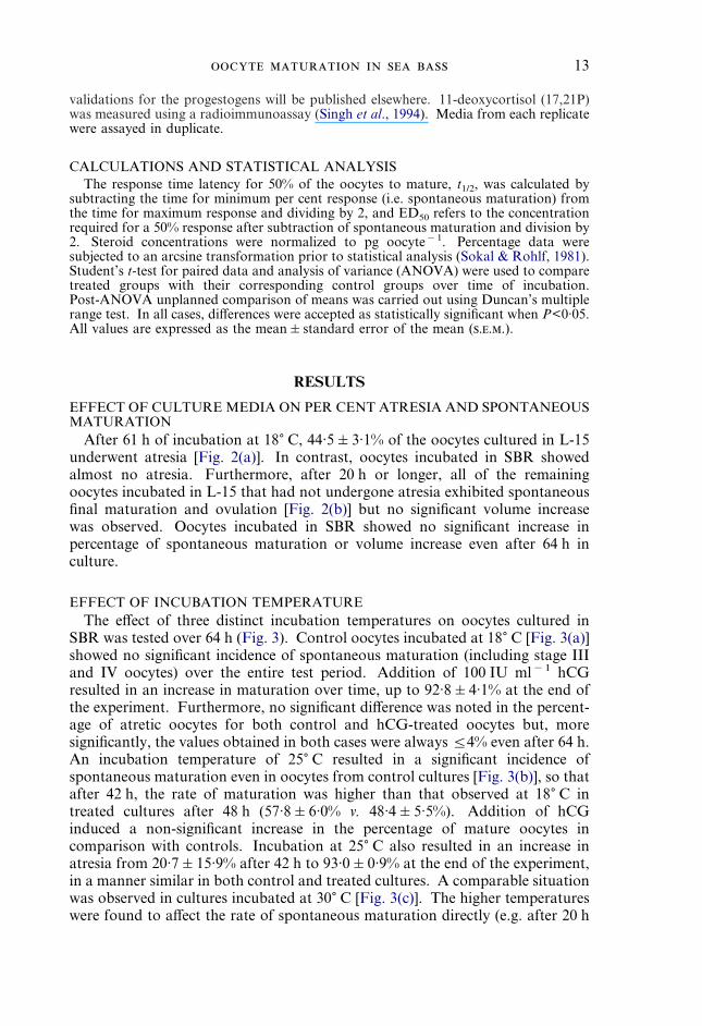
validations for the progestogens will be published elsewhere. 11-deoxycortisol (17,21P)was measured using a radioimmunoassay (Singh et al., 1994). Media from each replicatewere assayed in duplicate.
CALCULATIONS AND STATISTICAL ANALYSISThe response time latency for 50% of the oocytes to mature, t1/2, was calculated by
subtracting the time for minimum per cent response (i.e. spontaneous maturation) fromthe time for maximum response and dividing by 2, and ED50 refers to the concentrationrequired for a 50% response after subtraction of spontaneous maturation and division by2. Steroid concentrations were normalized to pg oocyte"1. Percentage data weresubjected to an arcsine transformation prior to statistical analysis (Sokal & Rohlf, 1981).Student’s t-test for paired data and analysis of variance (ANOVA) were used to comparetreated groups with their corresponding control groups over time of incubation.Post-ANOVA unplanned comparison of means was carried out using Duncan’s multiplerange test. In all cases, differences were accepted as statistically significant when P<0·05.All values are expressed as the mean&standard error of the mean (...).
RESULTS
EFFECT OF CULTURE MEDIA ON PER CENT ATRESIA AND SPONTANEOUSMATURATION
After 61 h of incubation at 18) C, 44·5&3·1% of the oocytes cultured in L-15underwent atresia [Fig. 2(a)]. In contrast, oocytes incubated in SBR showedalmost no atresia. Furthermore, after 20 h or longer, all of the remainingoocytes incubated in L-15 that had not undergone atresia exhibited spontaneousfinal maturation and ovulation [Fig. 2(b)] but no significant volume increasewas observed. Oocytes incubated in SBR showed no significant increase inpercentage of spontaneous maturation or volume increase even after 64 h inculture.
EFFECT OF INCUBATION TEMPERATUREThe effect of three distinct incubation temperatures on oocytes cultured in
SBR was tested over 64 h (Fig. 3). Control oocytes incubated at 18) C [Fig. 3(a)]showed no significant incidence of spontaneous maturation (including stage IIIand IV oocytes) over the entire test period. Addition of 100 IU ml"1 hCGresulted in an increase in maturation over time, up to 92·8&4·1% at the end ofthe experiment. Furthermore, no significant difference was noted in the percent-age of atretic oocytes for both control and hCG-treated oocytes but, moresignificantly, the values obtained in both cases were always ¦4% even after 64 h.An incubation temperature of 25) C resulted in a significant incidence ofspontaneous maturation even in oocytes from control cultures [Fig. 3(b)], so thatafter 42 h, the rate of maturation was higher than that observed at 18) C intreated cultures after 48 h (57·8&6·0% v. 48·4&5·5%). Addition of hCGinduced a non-significant increase in the percentage of mature oocytes incomparison with controls. Incubation at 25) C also resulted in an increase inatresia from 20·7&15·9% after 42 h to 93·0&0·9% at the end of the experiment,in a manner similar in both control and treated cultures. A comparable situationwas observed in cultures incubated at 30) C [Fig. 3(c)]. The higher temperatureswere found to affect the rate of spontaneous maturation directly (e.g. after 20 h

14 . . .
at 18, 25 and 30) C, spontaneous maturation in control cultures was 2·9&1·9,28·5&6·7 and 42·9&6·8%, respectively). As judged from these results, SBR wasconsidered an appropriate medium and 18) C a suitable temperature for the invitro culture of sea bass oocytes and both conditions were used in the subsequentexperiments.
70
100
00
Time (h)
% M
atu
rati
on
50
80
60
40
20
10 20 30 40 60
(b) * * *
70
100
00
% A
tres
ia50
80
60
40
20
10 20 30 40 60
(a)
**
*
F. 2. Effects of culture media on per cent atresia (a) and spontaneous maturation (b) of sea bass follicleenclosed oocytes in vitro. Data expressed as mean&.. from three experiments each with10 replicate determinations. Each replicate consisted of 20 oocytes. (-) Oocytes incubated in75% L-15 medium. (,) Oocytes incubated in SBR. *Indicates significant differences atP<0·001.
EFFECTS OF hCG ON FINAL OOCYTE MATURATIONIn order to characterize the response of sea bass oocytes to a mammalian
gonadotropin, graded doses of hCG were used and the effects examined overtime. Gonadotropin-induced final maturation was time- and dose-dependent[Fig. 4(a)]. Control oocytes showed no significant percentage of spontaneousmaturation over time. Incubation with 100 IU hCG ml"1 provoked an averagemaximal maturation response rate of 86·9% (t1/2250 h). The highest dose of300 IU ml"1 stimulated a maximum maturation response smaller than that of100 IU ml"1. There was a longer response latency with 50 IU hCG ml"1
(t1/2=100 h), producing an average maximal response rate of 60·4% over the

15
100
00
Time (h)
80
60
40
20
20 40 60
(c) 100
0
80
60
40
20
100
00
% M
atu
rati
on
80
60
40
20
20 40 60
(b) 100
0
80
60
40
20
100
00
80
60
40
20
20 40 60
(a) 100
0
80
60
40
20
% A
tres
ia
*
F. 3. Effects of incubation temperature on the rates of oocyte maturation and atresia in sea bass follicleenclosed oocytes incubated in SBR in vitro. Data expressed as mean&.. from three experiments(three to five replicate determinations) with 20 oocytes in each treatment. Oocytes were incubatedin the presence (+hCG, solid bars/circles) and absence ("hCG, open bars/circles) of 100 IU hCGml"1 at (a) 18) C, (b) 25) C, (c) 30) C. *Indicates significant differences at P<0·05.
120-h incubation period. Incubation with 25 IU hCG ml"1 induced only aslight but significant response rate of 13·9%, a 2·3-fold increase over controlvalues after 59 h. Essentially the same rate was maintained for the remainder ofthe incubation period. The lowest dose of 10 IU hCG ml"1 had no significanteffect on maturation.
Gonadotropin-induced maturation was also verified by observing changes infollicular volume (data not shown). Control follicles exhibited no significantchange in volume throughout the incubation period but incubation with 100 IUhCG ml"1 produced a 270·6% average increase in follicular volume. Incubationwith 50 IU hCG ml"1 induced a 241·2% volume increase in oocytes over timewhile 10 or 25 IU hCG ml"1 had no significant effect.

16 . . .
70
100
00
% M
atu
rati
on
50
80
60
40
20
10 20 30 40 60
PE ml–1
00.0010.010.11.0
hCG
a
b
c
d(b)
120
100
00 100
80
60
40
20
20 40 60 80
102550100300
0
ab
c
e(a)
d
Time (h)
F. 4. Time courses of per cent maturation induced by different concentrations of (a) hCG (10, 25, 50,100, or 300 IU hCG ml"1) and (b) logarithmic dilutions of pituitary extract (0·001, 0·01, 0·1,1·0 PE ml"1) extract or 100 IU ml"1 hCG in sea bass oocytes incubated at 18) C in SBR in vitro.Each point represents the mean&.. from four experiments of the percent of maturation of 20oocytes in replicates of four for each treatment. Different letters indicate significant differences atP<0·05.
EFFECT OF PITUITARY EXTRACT ON OOCYTE MATURATION ANDVOLUME
In addition to examining the effects of an exogenous gonadotropin on oocytematuration, endogenous gonadotropins were also investigated. The relativepotency of sea bass pituitary extract (0·001–1·0 PE ml"1) and an effective doseof hCG (100 IU ml"1) were compared in oocytes incubated in SBR at 18) C for69 h [Fig. 4(b)]. No significant maturation rate was observed in control oocytesand the lowest dose of pituitary extract was unable to induce maturation over theentire test period. By 45 h, all oocytes incubated with the remaining concen-trations of pituitary extract were significantly different from the controlsalthough not different from each other. After this time, clear dose-dependentresponses were observed. At the end of the experiment, 1·0 PE ml"1 induced amaturation rate of 89% comparable to the 90% rate elicited by 100 IU hCGml"1. In addition, both treatments had a similar response latency (t1/2248 h v.45 h for hCG and PE, respectively). No significant difference was observedbetween these two treatments. Incubation with 0·1 and 0·01 PE ml"1 resulted inmaturation rates of 64·0% (t1/2255 h) and 34·5%, respectively. No significantvolume changes were observed in control oocytes or those treated with 0·001 PE

17
ml"1. However, both 100 IU hCG ml"1 and 1·0 PE ml"1 elicited c. 190%increase in oocyte volume, whereas oocytes incubated with 0·1 and 0·01 PE ml"1
responded with a 178 and 133% volume increase, respectively (data notshown).
% M
atu
rati
on
Time (h)
100
100
00 60
80
60
40
20
20 40 80
b(c)
100
100
00 60
80
60
40
20
20 40 80
b
c(b)
100
100
00 60
80
60
40
20
20 40 80
b
(a)
a
b
c
a
Steroid (ng ml–1)
0.11.0101001000
0
a
F. 5. Time courses of per cent maturation induced by graded doses (ng ml"1 of 17,20âP (a), 20âS (b),and 17,20áP (c) and hCG (– – –,– – –, 100 IU ml"1) in sea bass oocytes incubated at 18) C inSBR in vitro. Each point represents the mean&.. from four assays of the per cent of maturationor volume of 20 oocytes in replicates of three to four for each treatment. Different letters indicatesignificant differences at 72–96 h of incubation at P<0·05.
EFFECTS OF 17,20âP, 20âS, AND 17,20áP ON OOCYTE MATURATIONAddition of 17,20âP significantly stimulated maturation in a time- and
dose-dependent manner with 100 and 1000 ng ml"1 producing a maximumresponse of 90–94% comparable with that of hCG [Fig. 5(a)]. However, theresponse time latency for the higher doses of 17,20âP were shorter than that ofhCG (10 and 15 h v. 50 h for 1000, 100, and 100 IU hCG ml"1, respectively).20âS [Fig. 5(b)] also exhibited dose- and time-dependent stimulation of matu-ration with response time latencies shorter than that of hCG. However, 20âS

18 . . .
was less potent than 17,20âP exhibiting a maximum maturational response ofonly 50–55%. 17,20áP was ineffective in stimulating maturation [Fig. 5(c)].
TRANSCRIPTION AND TRANSLATION ACTIONGonadotropin- and 17,20âP-induced maturation was significantly blocked in
the presence of 1 ìg ml"1 actinomycin D, a transcription inhibitor (Fig. 6).Furthermore, cycloheximide (1 ìg ml"1), a translation blocker, was also foundto block gonadotropin- and MIS-induced maturation to basal levels. Similarresults were observed with 20âS (data not shown).
MIS PRODUCTION FROM INCUBATIONS OF FOLLICLE-ENCLOSEDOOCYTES
Homologous pituitary extract stimulated production of 17,20âP at 12 h in adose-dependent manner following the pattern of maturation observed in thepresence and absence of pituitary extract at 12 h and 24 h [Fig. 7(a)]. Maximumlevels of 8·58&2·32 and 10·56&4·35 pg oocyte"1 were detected in response to1·0 pituitary (PE ml"1) and hCG (100 IU ml"1), respectively, at 12 h. Theproduction of 17,20âP was also time-dependent with a gradual decrease in MISdetected as time of incubation increased (data not shown). 20âS was alsodetected in incubations [Fig. 7(b)]. However, levels were two-fold lower than17,20âP in response to the same doses of PE and hCG. 17,21P and 17,20áP werealso detected in incubation media, but levels remained constant regardless oftreatment or incubation time (1·1&0·05 ng oocyte"1 and 8·2&0·08 pgoocyte"1 for 17,21P and 17,20áP, respectively; data not shown).
100
0
Treatment
% M
atu
rati
on
80
60
40
20
Basal
Control+ actinomycin+ cycloheximide
hCG 17,20βP
* *
F. 6. Effects of actinomycin D (1 ìg ml"1) and cycloheximide (1 ìg ml"1) on hCG- (100 IU ml"1) and17,20âP-stimulated maturation in follicle-enclosed oocytes after 72 h of incubation at 18) C in SBRin vitro. Each point represents the mean&.. from three experiments of the per cent of maturationfrom 10 oocytes in replicates of three for each treatment. *Indicates significant differences atP<0·001.
DISCUSSIONThe optimal conditions for the in vitro culture of sea bass follicle-enclosed
oocytes were established and included incubation in humidified air at 18) C in

19
PE (pituitary ml–1)
hCG
16
00 0.1
8
6
4
2
0.001 0.01 1.0
(b)100
0
80
60
40
20
14
12
10
20βS
(pg
ooc
yte–1
)
% M
atu
rati
on
16
00 0.1
8
6
4
2
0.001 0.01 1.0
(a)100
0
80
60
40
20
14
12
1017
,20β
P (
pg o
ocyt
e–1)
Maturation at 12 h + PEMaturation at 24 h + PEMaturation at 24 h – PE
Maturation at 12 h + PEMaturation at 24 h + PEMaturation at 24 h – PE
hCG
F. 7. The effects of homologous pituitary extract (PE ml"1) or hCG (100 IU ml"1) on 17,20âP (a) and20âS (b) production (pg oocyte"1) after 12 h of incubation at 18) C of follicle-enclosed oocytes inSBR (bars). Maturation at 12 and 24 h in the presence of pituitary extract and at 24 h in theabsence of pituitary extract are also plotted. Data expressed at mean&.. from duplicatedeterminations of replicates.
medium consisting of SBR (pH 7·5). These conditions not only preventedspontaneous maturation of oocytes, but also rendered oocytes fully responsive togonadotropin action. The spawning season of this teleost occurs betweenJanuary and March in the central Mediterranean, but photoperiod and temper-ature manipulation allow for the availability of viable oocytes (stage I) fromOctober to May (Zanuy et al., 1986; Carrillo et al., 1995). Photoperiodmanipulation together with the in vitro system described here, will enable thestudy of sea bass oocyte maturation during most of the year in another advancedmarine teleost, thus complementing other existing in vitro systems of oocyteculture for other species.
The sea bass oocytes responded to gonadotropins in a manner similar to whathas been described for other species in vitro. Since hCG is capable of inducingoocyte maturation and spawning in the sea bass in vivo (Zanuy & Carrillo, 1984;Zanuy et al., 1986) and hCG is effective in other teleost species both in vivo andin vitro (Pickford & Atz, 1957; Zohar & Gordin, 1979; Wallace & Selman, 1980;Berlinsky & Specker, 1991; King V et al., 1994a; Zhu et al., 1994), hCG was

20 . . .
tested, in addition to homologous pituitary extract, as a possible maturation-inducing agent in vitro. The response latency of sea bass oocytes to endogenousand exogenous gonadotropins was similar (t1/2z45 and 48 h, respectively) andcomparable with that observed in other marine teleosts. In addition, the actionof several putative MISs on maturation was examined. Both 17,20âP and 20âSwere produced not only by follicle-enclosed oocytes in response togonadotropins in a dose- and time-dependent manner, but were also capable ofinducing maturation, although 17,20âP was more potent and present at higherlevels in incubation media in response to the same gonadotropin doses ascompared to 20âS, implying that oocytes were more responsive to the formerMIS. Response time latencies for the MISs were much shorter than those ofgonadotropins at maximal response doses tested (t1/2=10 v. 48 h, respectively).17,20áP was ineffective. It has been demonstrated in vitro in several speciesthat gonadotropin-stimulated final maturation has a longer response latencythan steroid-induced maturation. For example, in Fundulus heteroclitus L.,while steroid-induced maturation can be complete within 20 h of incubation,gonadotropin-induced maturation proceeded more slowly and was completewithin c. 48 h (Wallace & Selman, 1980; Petrino et al., 1993), since in vitrogonadotropins do not act directly on the oocyte to induce resumption of meiosisbut instead stimulate steroidogenesis in the granulosa cells.
Maturation processes induced by gonadotropins and MIS depended on bothgene expression and protein synthesis, since actinomycin D and cycloheximideblocked inductional responses in this species. These results are in agreement withpreviously reported studies in the yellow perch Perca flavescens (Mitchill) andamago salmon Oncorhynchus rhodurus (Gunther). Studies have shown that theseinhibitors suppress the production of 17,20âP from the follicular layers of theoocyte, therefore inhibiting GVBD and hence maturation (Theofan & Goetz,1981; Nagahama & Adachi, 1985; Patino & Thomas, 1990).
The MIS has been identified as 17,20âP in the rainbow trout and several otherteleosts (Scott et al., 1982; Nagahama et al., 1983; Young et al., 1986) and as20âS in the Atlantic croaker Micropogonias undulatus L. and the spotted seatrout Cinoscion nebulosus L. (Thomas, 1994). Both 17,20âP and 20âS have beenimplicated as inducers of oocyte maturation in the striped bass, a close relative ofthe sea bass (Berlinsky & Specker, 1991; King V et al., 1994a,b), although recentevidence from binding studies strongly implicates 20âS as the MIS in this species(King V et al., 1997). Since plasma 17,20âP did not appear to change duringovulation in sea bass (Prat et al., 1990), 20âS was suggested as a possible MIS inthis species. The present results, however, suggest that both 17,20âP and 20âScan be potential MIS. In addition to 17,20âP and 20âS, the plasma of ovulatingsea bass also contains both conjugated and reduced metabolites of these steroids(Scott et al., 1990). We did not, however, measure these metabolites which haveno known physiological activity in the ovary and in any case vary in a similarmanner to that of the free unreduced steroids. In the Indian catfish,gonadotropins first stimulate the interrenals which in turn produce 17,21P, thematurational steroid responsible for induction of oocyte maturation at the levelof the ovary (Thomas, 1994); 17,21P production by sea bass follicles was notaffected by gonadotropins, suggesting that 17,21P is not a candidate for MIS inthis species. In addition to the three established mediators of oocyte maturation,

21
rat ovarian GnRH can modulate oocyte maturation directly by inducing meiosis(Hillensjo & Lemaire, 1980; Erickson et al., 1994). Although GnRH bindingsites have been identified in the goldfish Carassius auratus L. ovary (Habibi &Pati, 1993), a role for intraovarian GnRH in the regulation of sea bass and otherteleost oocyte maturation remains to be determined. Further studies arenecessary to identify conclusively the sea bass MIS and the mode ofgonadotropin action in the stimulation of oocyte maturation. Future studiesshould include measurement of conjugates for 17,20âP and 20âS since they aredetected in high concentration in vivo (Thomas, 1988; Scott et al., 1990) and theyare more likely to be produced in vitro due to the static conditions. These studieswill be possible with the present in vitro system.
Most species for which oocyte in vitro systems have been developed havesynchronous ovaries and/or exhibit distinct profiles of gonadotropin secretionwhich increase throughout the maturation stages with a possible surge prior toovulation, as in cyprinids (Crim et al., 1975; Stacey et al., 1979). This increase ingonadotropin results in a priming effect which enables oocytes to becomeresponsive to MIS and therefore undergo final maturation (Zhu et al., 1989;Kagawa et al., 1994). In contrast, the sea bass ovary contains group-synchronous clutches of oocytes (Mayer et al., 1990). This suggests that the levelof gonadotropins in such animals may remain relatively high and constantthroughout the breeding season (Wallace & Selman, 1980; Navas et al., 1998).Thus, the oocytes from a species with group-synchronous ovaries exhibit acrucial property in that maturing oocytes have the ability to acquire responsive-ness to endogenous gonadotropins and MIS so that maturation may commencewhile oocytes in earlier stages of development are not affected and vitellogenesisproceeds unaffected. The change in the response of follicular cells to a stimuluscould be due to an alteration in the number of receptors for gonadotropins or toinduction of different cellular signalling transducer molecules. For example, inmammals the ability of FSH to activate adenylate cyclase is not related to thenumber of FSH receptors present (Richards, 1994). Differential responsivenesscould also be due to desensitization of the gonadotropin receptors, as has beensuggested for the black sea bass Centropristis striata (L.) (Cerda et al., 1997).However, elucidation of this alteration in oocyte responsiveness remains to beexamined in the sea bass. In this regard, maximal maturation in response toexogenous and endogenous gonadotropins ranged between 86% and 90%, butmaturation never reached 100% even in the longest incubation periods assayed.In the sea bass, after spawning, 2–10% of postvitellogenic oocytes do not maturebut degenerate or undergo atresia (Mayer et al., 1988). The oocytes that did notmature in vitro may reflect the situation in vivo. However, it is possible that someoocytes chosen for incubation were vitellogenic oocytes that were improperlystaged as postvitellogenic. If this were the case, then the oocytes may not yet becompetent to respond to gonadotropins. This could be due in part to a reducednumber of gonadotropin receptors, inactivation of appropriate signal transduc-tion pathways or lack of prior exposure to sufficient levels of gonadotropins at akey stage of development (Richards, 1994).
This work was supported by CICYT MAR96-1859 to M.C., a postdoctoral fellowshipfrom the Spanish government to L.A.S., and a predoctoral fellowship from the regional

22 . . .
government of Valencia, Spain (Generalitat Valenciana) to J.F.A. Support from theBritish Council and the Spanish Ministry of Education and Culture under the frameworkof a Spanish–British Integrated Actions (HB97-235) to M.C. and D.E.K. is alsogratefully acknowledged.
References
Barnabe, G. & Barnabe-Quet, R. (1985). Advancement et amelioration de la ponteinduite chez le loup Dicentrarchus labrax (L.) a l’aide d’un analogue de LHRHinjecte. Aquaculture 49, 125–132.
Berlinsky, D. L. & Specker, J. (1991). Changes in gonadal hormones during oocytedevelopment in the striped bass, Morone saxatilis. Fish Physiology andBiochemistry 9, 51–62.
Canario, A. V. M. & Scott, A. P. (1990). Effects of steroids and human chorionicgonadotropin on in vitro oocyte final maturation in two marine flatfish: the dabLimanda limanda, and the plaice, Pleuronectes platessa. General and ComparativeEndocrinology 77, 161–176.
Carrillo, M., Zanuy, S., Prat, F., Cerda, J., Ramos, J., Mananos, E. & Bromage, N.(1995). Sea bass (Dicentrarchus labrax). In Broodstock Management and Egg andLarval Quality (Bromage, N. R. & Roberts, R. J., eds), pp. 138–168. Oxford:Blackwell Science.
Cerda, J., Selman, K., Hiao, S.-M. & Wallace, R. A. (1996). Observations on oocytematuration and hydration in vitro in the black sea bass, Centropristis striata(Serranidae). Aquatic Living Resources 9, 325–335.
Cerda, J., Selman, K., Hiao, S.-M. & Wallace, R. A. (1997). Evidence for the differentialregulation of ovarian follicle responsiveness to human chorionic gonadotropin invitro in a serranid teleost, Centropristis striata. Aquaculture 159, 143–157.
Chadhuri, H. (1976). Use of hormones in induced spawning of carps. Journal of theFisheries Research Board of Canada 33, 940–947.
Craik, J. C. A. (1982). Levels of phosphoprotein in the eggs and ovaries of some fishspecies. Comparative Biochemistry and Physiology 72B, 507–512.
Craik, J. C. A. & Harvey, S. M. (1982). Phosphorus metabolism and water uptakeduring final maturation of ovaries of teleosts with pelagic and demersal eggs.Marine Biology 90, 285–289.
Craik, J. C. A. & Harvey, S. M. (1984). Biochemical changes occurring during finalmaturation of eggs of some marine and freshwater teleosts. Journal of Fish Biology24, 599–610.
Crim, L. W., Watts, E. G. & Evans, D. M. (1975). The plasma gonadotropin profileduring sexual maturation in a variety of salmonid fishes. General and ComparativeEndocrinology 27, 62–70.
Cuisset, B., Pradelles, P., Kime, D. E., Kuhn, E. R., Babin, P. & Le Menn, F. (1994).Enzyme immunoassay for 11-ketotestosterone using acethylcholinesterase as label.Application to the measurement of 11-ketotestosterone in plasma of Siberiansturgeon. Comparative Biochemistry and Physiology 108C, 229–241.
Erickson, G. F., Li, D., Sadrkhanloo, R., Liu, X.-J., Shimasaki, S. & Ling, N. (1994).Extrapituitary actions of gonadotropin-releasing hormone: stimulation of insulin-like growth factor-binding protein-4 and atresia. Endocrinology 134, 1365–1372.
Fulton, T. W. (1898). On the growth and maturation of the ovarian eggs of teleosteanfishes. Report of the Fisheries Board of Scotland 3, 88–134.
Goetz, F. W. (1983). Hormonal control of oocyte final maturation and ovulation infishes. In Fish Physiology (Hoar, W. S., Randall, D. J. & Donaldson, E. M., eds),Vol. IX, Part B, pp. 117–170. New York: Academic Press.
Habibi, H. R. & Pati, D. (1993). Extrapituitary gonadotropin-releasing hormone(GnRH) binding sites in goldfish. Fish Physiology and Biochemistry 11, 43–49.

23
Hay, D. E., Outram, D. N., McKeown, B. A. & Hurlburt, M. (1987). Ovariandevelopment and oocyte diameter as maturation criteria in pacific herring (Clupeaharengus pallasi). Canadian Journal of Fisheries and Aquatic Sciences 45, 1496–1502.
Hillensjo, T. & Lemaire, W. J. (1980). Gonadotropin-releasing hormone agoniststimulates meiotic maturation of follicle-enclosed rat oocytes in vitro. Nature 287,145–146.
Hirose, K. (1976). Endocrine control of ovulation in medaka (Oryzias latipes) and ayu(Plecoglossus altivelis). Journal of the Fisheries Research Board of Canada 33,989–994.
Kagawa, H., Tanaka, H., Okuzawa, K. & Hirose, K. (1994). Development ofmaturational competence of oocytes of red seabream, Pagrus major, after humanchorionic gonadotropin treatment in vitro requires RNA and protein synthesis.General and Comparative Endocrinology 94, 199–206.
King V, W., Thomas, P. & Sullivan, C. V. (1994a). Hormonal regulation of finalmaturation of striped bass oocytes in vitro. General and Comparative Endocrinol-ogy 96, 223–233.
King V, W., Thomas, P., Harrell, R. M., Hodson, R. G. & Sullivan, C. V. (1994b).Plasma levels of gonadal steroids during final oocyte maturation of striped bass,Morone saxatilis L. General and Comparative Endocrinology 95, 178–191.
King V, W., Ghosh, S., Thomas, P. & Sullivan, C. V. (1997). A receptor for the oocytematuration-inducing hormone 17á,20â,21-trihydroxy-4-pregnen-3-one on ovarianmembranes of striped bass. Biology of Reproduction 56, 266–271.
Kuo, C.-M., Nash, C. E. & Shehadeh, Z. H. (1974). A procedural guide to inducespawning in grey mullet (Mugil cephalus L.). Aquaculture 3, 1–14.
Mayer, I., Shackley, S. E. & Ryland, J. S. (1988). Aspects of the reproductive biology ofthe bass, Dicentrarchus labrax L. I. An histological and histochemical study ofoocyte development. Journal of Fish Biology 33, 609–622.
Mayer, I., Shackley, S. E. & Witthames, P. R. (1990). Aspects of the reproductivebiology of the bass, Dicentrarchus labrax L. II. Fecundity and pattern of oocytedevelopment. Journal of Fish Biology 36, 141–148.
Nagahama, Y. (1987). 17á,20â-Dihydroxy-4-pregnen-3-one: a teleost maturation-inducing hormone. Development, Growth and Differentiation 29, 1–12.
Nagahama, Y. & Adachi, S. (1985). Identification of maturation-inducing steroid in ateleost, the amago salmon (Oncorhynchus rhodurus). Developmental Biology 109,428–435.
Nagahama, Y., Hirose, K., Young, G., Adachi, S., Suzuki, K. & Tamaoki, B. (1983).Relative in vitro effectiveness of 17á,20â-dihydroxy-4-pregnen-3-one and otherpregnene derivatives on germinal vesicle breakdown in oocytes of ayu (Plecoglos-sus altivelis), amago salmon (Oncorhynchus rhodurus), rainbow trout (Salmogairdneri) and goldfish (Carassius auratus). General and Comparative Endocrinol-ogy 51, 15–23.
Nagahama, Y., Yoshikuni, M., Yamashita, M., Sakai, N. & Tanaka, M. (1993).Molecular endocrinology of oocyte growth and maturation in fish. Fish Physiol-ogy and Biochemistry 11, 3–14.
Navas, J. M., Mananos, E., Thrush, M., Ramos, J., Zanuy, S., Carrillo, M., Zohar, Y. &Bromage, N. (1998). Effect of dietary lipid composition on vitellogenin, 17â-estradiol and gonadotropin plasma levels and spawning performance in captive seabass (Dicentrarchus labrax L.). Aquaculture 165, 65–79.
Pankhurst, N. W. (1985). Final maturation and ovulation of oocytes of the goldeye,Hiodon alosoides (Rafinesque) in vitro. Canadian Journal of Zoology 63, 1003–1009.
Patino, R. & Thomas, P. (1990). Induction of maturation of Atlantic croaker oocytes by17á,20â,21-trihydroxy-4-pregnen-3-one in vitro: consideration of some biologicaland experimental variables. Journal of Experimental Zoology 255, 97–109.
Petrino, T. R., Lin, Y.-W. P., Netherton, J. C., Powell, D. H. & Wallace, R. A. (1993).Steroidogenesis in Fundulus heteroclitus V: Purification, characterization, and
24 . . .
metabolism of 17á,20â-dihydroxy-4-pregnen-3-one by intact follicles and its role inoocyte maturation. General and Comparative Endocrinology 92, 1–15.
Pickford, G. E. & Atz, J. W. (1957). The Physiology of the Pituitary Gland of Fishes.New York: New York Zoological Society.
Prat, F., Zanuy, S., Carrillo, M., de Mones, A. & Fostier, A. (1990). Seasonal changes inplasma levels of gonadal steroids of sea bass, Dicentrarchus labrax L. General andComparative Endocrinology 78, 361–373.
Richards, J. S. (1994). Hormonal control of gene expression in the ovary. EndocrineReviews 15, 725–751.
Scott, A. P., Sheldrick, E. L. & Flint, A. P. F. (1982). Measurement of 17á,20â-dihydroxy-4-pregnen-3-one in plasma of trout (Salmo gairdneri Richardson):Seasonal changes and response to salmon pituitary extract. General andComparative Endocrinology 46, 444–451.
Scott, A. P., Canario, A. V. M. & Prat, F. (1990). Radioimmunoassay of ovariansteroids in plasmas of ovulating female sea bass (Dicentrarchus labrax). Generaland Comparative Endocrinology 78, 299–302.
Selman, K. & Wallace, R. A. (1989). Cellular aspects of oocyte growth in teleosts.Zoological Science 6, 211–231.
Singh, P. B., Kime, D. E., Epler, P. & Chyb, J. (1994). Impact ofã-hexachlorocyclohexane exposure on plasma gonadotropin levels and in vitrostimulation of gonadal steroid production by carp hypophysial homogenate inCarassius auratus. Journal of Fish Biology 44, 195–204.
Sokal, R. & Rohlf, J. (1981). Biometry. The Principles and Practice of Statistics inBiological Research. 2nd edn. New York: W. H. Freeman..
Sorbera, L. A., Mylonas, C. C., Zanuy, S., Carrillo, M. & Zohar, Y. (1996). Sustainedadministration of GnRH increases milt volume without altering sperm counts inthe sea bass (Dicentrarchus labrax). Journal of Experimental Zoology 276,361–368.
Stacey, N. E., Cook, A. F. & Peter, R. E. (1979). Ovulatory surge of gonadotropinin the goldfish, Carassius auratus. General and Comparative Endocrinology 37,246–249.
Theofan, G. & Goetz, F. W. (1981). The in vitro effects of transcriptional andtranslational protein synthesis inhibitors on final maturation and ovulation ofyellow perch (Perca flavescens) oocytes. Comparative Biochemistry and Physiology69, 557–561.
Thomas, P. (1988). Changes in plasma levels of maturation-inducing steroids in severalperciform fishes during induced ovulation. American Zoologist 28, 53A.
Thomas, P. (1994). Hormonal control of final oocyte maturation in sciaenid fishes. InPerspectives in Comparative Endocrinology (Davey, K. G., Peter, R. E. & Tobe, S.S., eds), pp. 619–625. Ottawa: National Research Council of Canada.
Wallace, R. A. & Selman, K. (1980). Oogenesis in Fundulus heteroclitus. II. Thetransition from vitellogenesis into maturation. General and ComparativeEndocrinology 42, 345–354.
Wallace, R. A. & Selman, K. (1981). Cellular and dynamic aspects of oocyte growth inteleosts. American Zoologist 21, 325–343.
Wallace, R. A., Greeley, M. S. & McPherson, R. (1992). Analytical and experimentalstudies on the relationship between Na+ and K+, and water uptake during volumeincreases associated with Fundulus oocyte maturation in vitro. Journal of Com-parative Physiology B 162, 241–248.
Wallace, R. A., Boyle, S. M., Grier, H. J., Selman, K. & Petrino, T. R. (1993).Preliminary observations on oocyte maturation and other aspects of reproductivebiology in captive female snook, Centropomus undecimalis. Aquaculture 116,257–273.
Watanabe, W. O. & Kuo, C.-M. (1986). Water and ion balance in hydrating oocytes ofthe grey mullet, Mugil cephalus (L.), during hormone-induced final maturation.Journal of Fish Biology 28, 425–437.
25
Young, G., Adachi, S. & Nagahama, Y. (1986). Role of ovarian thecal and granulosalayers in gonadotropin-induced synthesis of a salmonid maturation-inducingsubstance (17á,20â-dihydroxy-4-pregnen-3-one). Developmental Biology 118, 1–8.
Zanuy, S. & Carrillo, M. (1984). La salinite: un moyen pour retarder la ponte du bar. InL’Aquaculture du Bar et des Sparides (Barnabe, G. & Billard, R., eds), pp. 73–80.Paris: INRA Publications.
Zanuy, S., Carrillo, M. & Ruiz, F. (1986). Delayed gametogenesis and spawning of seabass (Dicentrarchus labrax L.) kept under different photoperiod and temperatureregimes. Fish Physiology and Biochemistry 2, 53–63.
Zhu, Y., Aida, K., Furukawa, K. & Hanyu, I. (1989). Development of sensitivity tomaturation-inducing steroids and gonadotropins in the oocytes of the tobinumeri-dragonet, Repomucenus beniteguri, Callionymidae (Teleostei). General andComparative Endocrinology 76, 250–260.
Zhu, Y., Kobayashi, M., Furukawa, K. & Aida, K. (1994). Gonadotropin developssensitivity to maturation-inducing steroid in the oocytes of daily spawning teleosts,tobinumeri-dragonet Repomucenus beniteguri and Kisu Sillago japonica. FisheriesScience 60, 541–545.
Zohar, Y. & Gordin, H. (1979). Spawning kinetics in the gilthead sea-bream, Sparusaurata L. after low doses of human chorionic gonadotropin. Journal of FishBiology 15, 665–670.