An unrecognized extracellular function for an intracellular adapter protein released from the...
6
An unrecognized extracellular function for an intracellular adapter protein released from the cytoplasm into the tumor microenvironment Paul J. Mintz a,1 , Marina Cardo ´ -Vila a , Michael G. Ozawa a , Amin Hajitou a,2 , Roberto Rangel a , Liliana Guzman-Rojas a , Dawn R. Christianson a , Marco A. Arap a,3 , Ricardo J. Giordano a , Glauco R. Souza a , Jeffrey Easley a,4 , Ahmad Salameh a,b , Salvatore Oliviero b , Ricardo R. Brentani c , Erkki Koivunen a , Wadih Arap a,5 , and Renata Pasqualini a,5 a The David H. Koch Center, University of Texas M. D. Anderson Cancer Center, Houston, TX 77030; b Department of Molecular Biology, University of Siena, Siena 53100, Italy; and c Office of the President, A. C. Camargo Hospital, Sa ˜ o Paulo, SP 01509, Brazil Communicated by Richard L. Sidman, Harvard Medical School, Boston, MA, August 13, 2008 (received for review May 18, 2008) Mammalian cell membranes provide an interface between the intra- cellular and extracellular compartments. It is currently thought that cytoplasmic signaling adapter proteins play no functional role within the extracellular tumor environment. Here, by selecting combinatorial random peptide libraries in tumor-bearing mice, we uncovered a direct, specific, and functional interaction between CRKL, an adapter protein [with Src homology 2 (SH2)- and SH3-containing domains], and the plexin-semaphorin-integrin domain of 1 integrin in the extracellular milieu. Through assays in vitro, in cellulo, and in vivo, we show that this unconventional and as yet unrecognized protein– protein interaction between a regulatory integrin domain (rather than a ligand-binding one) and an intracellular adapter (acting out- side of the cells) triggers an alternative integrin-mediated cascade for cell growth and survival. Based on these data, here we propose that a secreted form of the SH3/SH2 adaptor protein CRKL may act as a growth-promoting factor driving tumorigenesis and may lead to the development of cancer therapeutics targeting secreted CRKL. cancer CrkL integrin phage display C ell membranes have evolved as a tight and compartmentalized, but dynamic, interface between intracellular and extracellular contents (1, 2). To maintain such homeostasis, transmembrane receptors mediate bidirectional signaling across the cell surface through a complex spatial and temporal organization (3, 4). Thus, protein location in signal transduction is central to specificity of cellular responses (2, 5, 6). For example, cell surface receptors such as integrins undergo conformational changes elicited through li- gand binding to enable a cross-talk with signal transduction cas- cades such as the MAPK pathways; conventional integrin ligands include ECM proteins that recognize integrin extracellular domains and cytoskeletal proteins that interact with intracellular domains (3–4, 7–9). To gain insight into signal transduction across cell membranes in cancer, we set out to identify functional protein interactions in a tumor xenograft model; we reasoned that a combinatorial approach (10–14) in vivo might provide clues by emulating ligand–receptor binding in the context of the tumor microenvironment. Here, we show a specific interaction between the intracellular signaling protein CRKL and a regulatory (rather than ligand-binding) 1 integrin extracellular domain. Surprisingly, we found that CRKL targets the plexin-semaphorin-integrin (PSI) domain of 1 integrin chain located outside of the cell and promotes cell growth and survival. These results indicate an unrecognized integrin-mediated outside-in function for intracellular mediators, such as Src homol- ogy 2 (SH2)- and SH3-containing proteins, in activating prolifer- ative pathways. Results Combinatorial Selection in Vivo Yields Tumor-Homing Peptides. We administered a phage library (12, 14, 15) i.v. into nu/nu (nude) mice bearing human DU145-derived prostate cancer xenografts and recovered tumors after 24-h circulation. We recovered an enriched population of tumor-targeting phage (Fig. 1A) and individual phage clones (Fig. 1B). The dominant peptide (YRCTLNSPFFWED- MTHECHA) was functionally characterized; we evaluated its tumor-targeting specificity in vivo in tumor-bearing mice. After i.v. administration of YRCTLNSPFFWEDMTHECHA-phage, we observed marked homing to tumors (insertless phage served as a negative control) with barely detectable phage localization in several control organs (Fig. 1C). We also in vitro-targeted DU145 cells with an aqueous-to-organic phase separation assay (16) and a phage-based immunof luorescence assay (Kaposi Sarcoma cells; KS1767); consistently, YRCTLNSPFFWEDMTHECHA-phage bound to tumor cell surfaces to a greater extent than negative control phage (Fig. 1 D and E). We next evaluated the internal- ization of the peptide by fusing a proapoptotic motif (13, 17–19) to the tumor-homing sequence. We found targeted cell death relative to controls (Fig. 1 F and G), indicating that YRCTLNSPFFWED- MTHECHA mediates ligand-directed internalization. These re- sults show that YRCTLNSPFFWEDMTHECHA targets tumor cells and enables internalization. A Tumor-Homing Peptide Sequence Mimics a Regulatory Integrin Extracellular Domain. To determine whether the peptide sequence mimics a native protein, we performed a similarity search of YRCTLNSPFFWEDMTHECHA and other selected peptide se- quences. By using BLAST followed by protein alignment, we found that the peptides resembled sequences present on 1 integrin. Unexpectedly, the dominant sequence YRCTLNSPFFWED- MTHECHA had similarity to the PSI domain (residues 26–78) of the 1 integrin; moreover, we found that other selected peptides also appeared within the same region. We then asked whether the Author contributions: P.J.M., M.C.-V., M.G.O., R.R.B., E.K., W.A., and R.P. designed research; P.J.M., M.C.-V., A.H., R.R., L.G.-R., D.R.C., M.A.A., R.J.G., G.R.S., J.E., A.S., and S.O. performed research; P.J.M., M.C.-V., M.G.O., A.H., R.R., L.G.-R., D.R.C., M.A.A., R.J.G., G.R.S., J.E., A.S., S.O., R.R.B., E.K., W.A., and R.P. analyzed data; and P.J.M., M.C.-V., M.G.O., W.A., and R.P. wrote the paper. The authors declare no conflict of interest. Freely available online through the PNAS open access option. 1 Present address: Department of Surgery, Imperial College London, London W12 0NN, United Kingdom. 2 Present address: Department of Gene Therapy, Imperial College London, London W2 1PG, United Kingdom. 3 Present address: Department of Urology, University of Sa ˜ o Paulo, Sa ˜ o Paulo, SP 01065, Brazil. 4 Present address: Undergraduate Program, Massachusetts Institute of Technology, Cam- bridge, MA 02139. 5 To whom correspondence may be addressed. E-mail: [email protected] or [email protected]. This article contains supporting information online at www.pnas.org/cgi/content/full/ 0807543105/DCSupplemental. © 2009 by The National Academy of Sciences of the USA 2182–2187 PNAS February 17, 2009 vol. 106 no. 7 www.pnas.orgcgidoi10.1073pnas.0807543105
-
Upload
independent -
Category
Documents
-
view
0 -
download
0
Transcript of An unrecognized extracellular function for an intracellular adapter protein released from the...
An unrecognized extracellular function for anintracellular adapter protein released from thecytoplasm into the tumor microenvironmentPaul J. Mintza,1, Marina Cardo-Vilaa, Michael G. Ozawaa, Amin Hajitoua,2, Roberto Rangela, Liliana Guzman-Rojasa,Dawn R. Christiansona, Marco A. Arapa,3, Ricardo J. Giordanoa, Glauco R. Souzaa, Jeffrey Easleya,4, Ahmad Salameha,b,Salvatore Olivierob, Ricardo R. Brentanic, Erkki Koivunena, Wadih Arapa,5, and Renata Pasqualinia,5
aThe David H. Koch Center, University of Texas M. D. Anderson Cancer Center, Houston, TX 77030; bDepartment of Molecular Biology, University of Siena,Siena 53100, Italy; and cOffice of the President, A. C. Camargo Hospital, Sao Paulo, SP 01509, Brazil
Communicated by Richard L. Sidman, Harvard Medical School, Boston, MA, August 13, 2008 (received for review May 18, 2008)
Mammalian cell membranes provide an interface between the intra-cellular and extracellular compartments. It is currently thought thatcytoplasmic signaling adapter proteins play no functional role withinthe extracellular tumor environment. Here, by selecting combinatorialrandom peptide libraries in tumor-bearing mice, we uncovered adirect, specific, and functional interaction between CRKL, an adapterprotein [with Src homology 2 (SH2)- and SH3-containing domains],and the plexin-semaphorin-integrin domain of �1 integrin in theextracellular milieu. Through assays in vitro, in cellulo, and in vivo, weshow that this unconventional and as yet unrecognized protein–protein interaction between a regulatory integrin domain (ratherthan a ligand-binding one) and an intracellular adapter (acting out-side of the cells) triggers an alternative integrin-mediated cascade forcell growth and survival. Based on these data, here we propose thata secreted form of the SH3/SH2 adaptor protein CRKL may act as agrowth-promoting factor driving tumorigenesis and may lead to thedevelopment of cancer therapeutics targeting secreted CRKL.
cancer � CrkL � integrin � phage display
Cell membranes have evolved as a tight and compartmentalized,but dynamic, interface between intracellular and extracellular
contents (1, 2). To maintain such homeostasis, transmembranereceptors mediate bidirectional signaling across the cell surfacethrough a complex spatial and temporal organization (3, 4). Thus,protein location in signal transduction is central to specificity ofcellular responses (2, 5, 6). For example, cell surface receptors suchas integrins undergo conformational changes elicited through li-gand binding to enable a cross-talk with signal transduction cas-cades such as the MAPK pathways; conventional integrin ligandsinclude ECM proteins that recognize integrin extracellular domainsand cytoskeletal proteins that interact with intracellular domains(3–4, 7–9).
To gain insight into signal transduction across cell membranes incancer, we set out to identify functional protein interactions in atumor xenograft model; we reasoned that a combinatorial approach(10–14) in vivo might provide clues by emulating ligand–receptorbinding in the context of the tumor microenvironment. Here, weshow a specific interaction between the intracellular signalingprotein CRKL and a regulatory (rather than ligand-binding) �1integrin extracellular domain. Surprisingly, we found that CRKLtargets the plexin-semaphorin-integrin (PSI) domain of �1 integrinchain located outside of the cell and promotes cell growth andsurvival. These results indicate an unrecognized integrin-mediatedoutside-in function for intracellular mediators, such as Src homol-ogy 2 (SH2)- and SH3-containing proteins, in activating prolifer-ative pathways.
ResultsCombinatorial Selection in Vivo Yields Tumor-Homing Peptides. Weadministered a phage library (12, 14, 15) i.v. into nu/nu (nude) micebearing human DU145-derived prostate cancer xenografts and
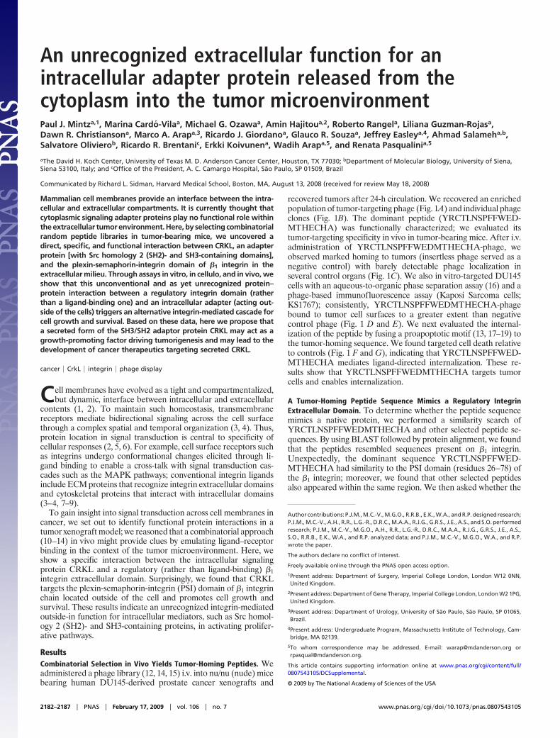
recovered tumors after 24-h circulation. We recovered an enrichedpopulation of tumor-targeting phage (Fig. 1A) and individual phageclones (Fig. 1B). The dominant peptide (YRCTLNSPFFWED-MTHECHA) was functionally characterized; we evaluated itstumor-targeting specificity in vivo in tumor-bearing mice. After i.v.administration of YRCTLNSPFFWEDMTHECHA-phage, weobserved marked homing to tumors (insertless phage served as anegative control) with barely detectable phage localization inseveral control organs (Fig. 1C). We also in vitro-targeted DU145cells with an aqueous-to-organic phase separation assay (16) and aphage-based immunofluorescence assay (Kaposi Sarcoma cells;KS1767); consistently, YRCTLNSPFFWEDMTHECHA-phagebound to tumor cell surfaces to a greater extent than negativecontrol phage (Fig. 1 D and E). We next evaluated the internal-ization of the peptide by fusing a proapoptotic motif (13, 17–19) tothe tumor-homing sequence. We found targeted cell death relativeto controls (Fig. 1 F and G), indicating that YRCTLNSPFFWED-MTHECHA mediates ligand-directed internalization. These re-sults show that YRCTLNSPFFWEDMTHECHA targets tumorcells and enables internalization.
A Tumor-Homing Peptide Sequence Mimics a Regulatory IntegrinExtracellular Domain. To determine whether the peptide sequencemimics a native protein, we performed a similarity search ofYRCTLNSPFFWEDMTHECHA and other selected peptide se-quences. By using BLAST followed by protein alignment, we foundthat the peptides resembled sequences present on �1 integrin.Unexpectedly, the dominant sequence YRCTLNSPFFWED-MTHECHA had similarity to the PSI domain (residues 26–78) ofthe �1 integrin; moreover, we found that other selected peptidesalso appeared within the same region. We then asked whether the
Author contributions: P.J.M., M.C.-V., M.G.O., R.R.B., E.K., W.A., and R.P. designed research;P.J.M., M.C.-V., A.H., R.R., L.G.-R., D.R.C., M.A.A., R.J.G., G.R.S., J.E., A.S., and S.O. performedresearch; P.J.M., M.C.-V., M.G.O., A.H., R.R., L.G.-R., D.R.C., M.A.A., R.J.G., G.R.S., J.E., A.S.,S.O., R.R.B., E.K., W.A., and R.P. analyzed data; and P.J.M., M.C.-V., M.G.O., W.A., and R.P.wrote the paper.
The authors declare no conflict of interest.
Freely available online through the PNAS open access option.
1Present address: Department of Surgery, Imperial College London, London W12 0NN,United Kingdom.
2Present address: Department of Gene Therapy, Imperial College London, London W2 1PG,United Kingdom.
3Present address: Department of Urology, University of Sao Paulo, Sao Paulo, SP 01065,Brazil.
4Present address: Undergraduate Program, Massachusetts Institute of Technology, Cam-bridge, MA 02139.
5To whom correspondence may be addressed. E-mail: [email protected] [email protected].
This article contains supporting information online at www.pnas.org/cgi/content/full/0807543105/DCSupplemental.
© 2009 by The National Academy of Sciences of the USA
2182–2187 � PNAS � February 17, 2009 � vol. 106 � no. 7 www.pnas.org�cgi�doi�10.1073�pnas.0807543105
similarity of the peptide YRCTLNSPFFWEDMTHECHA wasspecific for the PSI domain of the �1 integrin or common to otherknown integrin � chains. After fit analysis and molecular modeling,we concluded that the homology between YRCTLNSPFFWED-MTHECHA and the PSI domain of �1 integrin was indeed the bestalignment (Fig. S1 A–C).
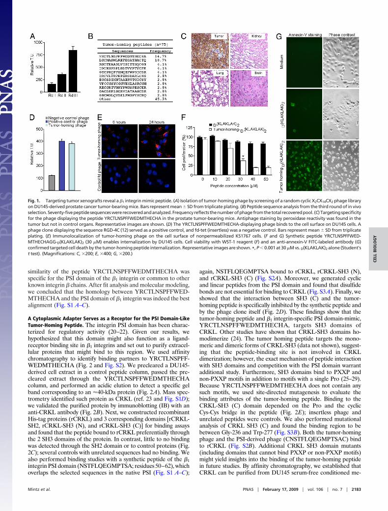
A Cytoplasmic Adapter Serves as a Receptor for the PSI Domain-LikeTumor-Homing Peptide. The integrin PSI domain has been charac-terized for regulatory activity (20–22). Given our results, wehypothesized that this domain might also function as a ligand-receptor binding site in �1 integrins and set out to purify extracel-lular proteins that might bind to this region. We used affinitychromatography to identify binding partners to YRCTLNSPFF-WEDMTHECHA (Fig. 2 and Fig. S2). We precleared a DU145-derived cell extract in a control peptide column, passed the pre-cleared extract through the YRCTLNSPFFWEDMTHECHAcolumn, and performed an acidic elution to detect a specific gelband corresponding to an �40-kDa protein (Fig. 2A). Mass spec-trometry identified such protein as CRKL (ref. 23 and Fig. S1D);we validated the purified protein by immunoblotting (IB) with ananti-CRKL antibody (Fig. 2B). Next, we constructed recombinantHis-tag proteins (rCRKL) and 3 corresponding domains [rCRKL-SH2, rCRKL-SH3 (N), and rCRKL-SH3 (C)] for binding assaysand found that the peptide bound to rCRKL preferentially throughthe 2 SH3 domains of the protein. In contrast, little to no bindingwas detected through the SH2 domain or to control proteins (Fig.2C); several controls with unrelated sequences had no binding. Wealso performed binding studies with a synthetic peptide of the �1integrin PSI domain (NSTFLQEGMPTSA; residues 50–62), whichoverlaps the selected sequences in the native PSI (Fig. S1 A–C);
again, NSTFLQEGMPTSA bound to rCRKL, rCRKL-SH3 (N),and rCRKL-SH3 (C) (Fig. S2A). Moreover, we generated cyclicand linear peptides from the PSI domain and found that disulfidebonds are not essential for binding to CRKL (Fig. S3A). Finally, weshowed that the interaction between SH3 (C) and the tumor-homing peptide is specifically inhibited by the synthetic peptide andby the phage clone itself (Fig. 2D). These findings show that thetumor-homing peptide and �1 integrin-specific PSI domain-mimic,YRCTLNSPFFWEDMTHECHA, targets SH3 domains ofCRKL. Other studies have shown that CRKL-SH3 domains ho-modimerize (24). The tumor homing peptide targets the mono-meric and dimeric forms of CRKL-SH3 (data not shown), suggest-ing that the peptide-binding site is not involved in CRKLdimerization; however, the exact mechanism of peptide interactionwith SH3 domains and competition with the PSI domain warrantadditional study. Furthermore, SH3 domains bind to PXXP andnon-PXXP motifs in addition to motifs with a single Pro (25–29).Because YRCTLNSPFFWEDMTHECHA does not contain anysuch motifs, we used site-directed mutagenesis to evaluate thebinding attributes of the tumor-homing peptide. Binding to theCRKL-SH3 (C) domain depended on the Pro and the cyclicCys-Cys bridge in the peptide (Fig. 2E); insertless phage andunrelated peptides were controls. We also performed mutationalanalysis of CRKL SH3 (C) and found the binding region to bebetween Gly-236 and Trp-277 (Fig. S3B). Both the tumor-homingphage and the PSI-derived phage (CNSTFLQEGMPTSAC) bindto rCRKL (Fig. S2B). Additional CRKL SH3 domain mutants(including domains that cannot bind PXXP or non-PXXP motifs)might yield insights into the binding of the tumor-homing peptidein future studies. By affinity chromatography, we established thatCRKL can be purified from DU145 serum-free conditioned me-
Fig. 1. Targeting tumor xenografts reveal a �1 integrin mimic peptide. (A) Isolation of tumor-homing phage by screening of a random cyclic X2CX14CX2 phage libraryon DU145-derived prostate cancer tumor-bearing mice. Bars represent mean � SD from triplicate plating. (B) Peptide sequence analysis from the third round of in vivoselection.Seventy-fivepeptidesequenceswererecoveredandanalyzed.Frequency reflects thenumberofphagefromthetotal recoveredpool. (C) Targetingspecificityfor the phage displaying the peptide YRCTLNSPFFWEDMTHECHA in the prostate tumor-bearing mice. Antiphage staining by peroxidase reactivity was found in thetumor but not in control organs. Representative images are shown. (D) The YRCTLNSPFFWEDMTHECHA-displaying phage binds to the cell surface on DU145 cells. Aphage clone displaying the sequence RGD-4C (12) served as a positive control, and fd-tet (insertless) was a negative control. Bars represent mean � SD from triplicateplating. (E) Immunolocalization of tumor-homing phage on the cell surface of nonpermeabilized KS1767 cells. (F and G) Synthetic peptide YRCTLNSPFFWED-MTHECHAGG-D(KLAKLAK)2 (30 �M) enables internalization by DU145 cells. Cell viability with WST-1 reagent (F) and an anti-annexin-V FITC-labeled antibody (G)confirmed targeted cell death by the tumor-homing peptide internalization. Representative images are shown. *, P � 0.001 at 30 �M vs. D(KLAKLAK)2 alone (Student’st test). (Magnifications: C, �200; E, �400; G, �200.)
Mintz et al. PNAS � February 17, 2009 � vol. 106 � no. 7 � 2183
CELL
BIO
LOG
Y
dium and that a control column with a mutant tumor-homingpeptide no longer bound to CRKL at detectable levels (Fig. S2C).
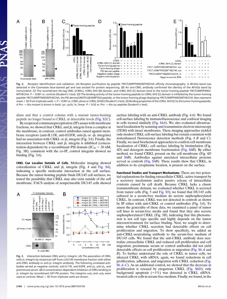
By reciprocal coimmunoprecipitation (IP) assays with membranefractions, we showed that CRKL and �1 integrin form a complex atthe membrane; in contrast, control antibodies raised against mem-brane receptors (anti-IL11R, anti-EGFR, anti-�3 or -�5 integrins)had no association with CRKL or �1 integrin (Fig. 3A). Finally, theinteraction between CRKL and �1 integrin is inhibited (concen-tration-dependent) by a recombinant PSI domain (IC50 � 20 nM;Fig. 3B); consistent with the co-IP, control integrins showed nobinding (Fig. 3A).
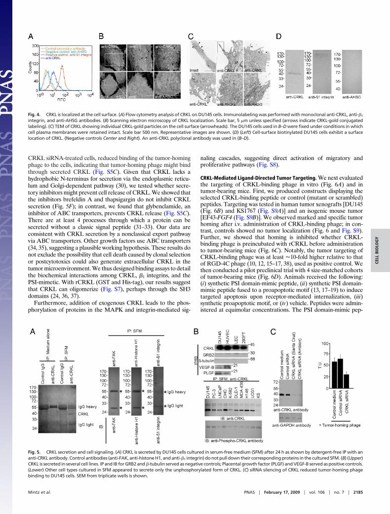
CRKL Can Localize Outside of Cells. Molecular imaging showedcolocalization of CRKL and �1 integrin (Fig. 4 and Fig. S4),indicating a specific molecular interaction at the cell surface.Because the tumor-homing peptide binds DU145 cell surfaces, wetested the possibility that CRKL may also exist outside of the cellmembrane. FACS analysis of nonpermeable DU145 cells showed
surface labeling with an anti-CRKL antibody (Fig. 4A). We foundcell-surface labeling by immunofluorescence and confocal imagingin cells treated similarly (Fig. S4A). We also evaluated ultrastruc-tural localization by scanning and transmission electron microscopy(TEM) with intact membranes. These imaging approaches yieldedonly modest CRKL cell-surface labeling but remain consistent withnonenhanced fluorescence detection methods (Fig. 4 B and C).Finally, we used biochemical approaches to confirm cell membranelocalization of CRKL: cell surface labeling by biotinylation (Fig.4D) and detergent membrane fractionation (Fig. S4B). By eithermethod, we found CRKL present on the cell membrane (Fig. 4Dand S4B). Antibodies against unrelated intracellular proteinsserved as controls (Fig. S4B). These results show that CRKL, inaddition to its cytoplasmic location, is present on the surface.
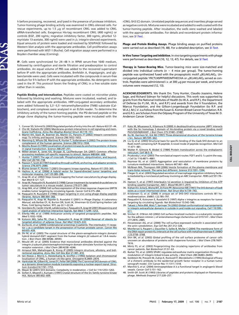
Functional Studies and Transport Mechanisms. There are two poten-tial explanations for finding extracellular CRKL: active transport bya secretory mechanism and/or passive release of intracellularcontents caused by cell death. Because CRKL lacks a classictransmembrane domain, we evaluated whether CRKL is secretedfrom tumor cells (Fig. 5 and Fig. S5); we found that DU145 cellscultured in a serum-free medium do secrete unphosphorylatedCRKL. In contrast, CRKL was not detected in controls as shownby IP either with anti-CRKL or control antibodies (Fig. 5A). Toassess the generality of these data, we examined a panel of tumorcell lines in serum-free media and found that they also secreteunphosphorylated CRKL (Fig. 5B), indicating that this phenome-non is not cell type specific and highly depends on the tumormicroenvironment for surface binding. Next, we sought to deter-mine whether CRKL secretion had detectable effects on cellproliferation and migration. To show specificity, we added ananti-CRKL-neutralizing antibody to the serum-free medium ofDU145 cells. We found that the anti-CRKL antibody does neu-tralize extracellular CRKL and reduced cell proliferation and cellmigration; preimmune serum or control antibodies did not yielddetectable effects on cell proliferation or migration (Fig. S6 A andB). To further understand the role of CRKL in tumor cells, wesilenced CRKL with siRNA; again, we found reductions in cellproliferation, adhesion, and migration with CRKL reduction (Fig.S6 A–C). As an additional control, we showed that the decrease inproliferation is rescued by exogenous CRKL (Fig. S6D); onlybackground apoptosis (�1%) was detected in CRKL siRNA-treated cells or cells in serum-free medium. Finally, we found, in the
Fig. 2. Receptor identification and validation. (A) Receptor purification by peptide YRCTLNSPFFWEDMTHECHA affinity chromatography. A 40-kDa band wasdetected in the Coomassie blue-stained gel and was excised for protein sequencing. (B) An anti-CRKL antibody confirmed the identity of the 40-kDa band byimmunoblot. (C) The recombinant His-tag CRKL (rCRKL), rCRKL-SH3 (N) domain, and rCRKL-SH3 (C) domain bind to the tumor-homing peptide YRCTLNSPFFWED-MTHECHA. P � 0.001 vs. controls (Student’s t test). (D) The binding activity of the tumor-homing peptide to rCRKL-SH3 (C) domain is inhibited by the tumor-homingpeptide YRCTLNSPFFWEDMTHECHA, the PSI-derived (NSTFLQEGMPTSA) peptide, or the tumor-homing phage displaying YRCTLNSPFFWEDMTHECHA. Bars representmean�SD from triplicate wells. *, P�0.001 vs. rCRKL alone or rCRKL-SH3(C) (Student’s t test). (E) Binding properties of the rCRKL-SH3 (C) to the tumor-homing peptide.A Pro3 Ala mutant is shown in bold. cyc, cyclic; ln, linear. P � 0.02 vs. Pro3 Ala cyc peptide (Student’s t test).
Fig. 3. Interaction between CRKL and �1 integrin. (A) The association of CRKLwith �1 integrin by reciprocal coIP from a DU145 membrane fraction with eitheranti-CRKL antibody or anti-�1 integrin antibody. The following unrelated anti-bodies served as negative controls: anti-IL11R, anti-EGFR, anti-�3, anti-�5, andpreimmune serum. (B) A concentration-dependent inhibition of CRKL binding to�1 integrin by recombinant GST-PSI protein. The integrins �v�3 and �v�5 wereused as controls. Mean � SD from triplicate wells are shown.
2184 � www.pnas.org�cgi�doi�10.1073�pnas.0807543105 Mintz et al.
CRKL siRNA-treated cells, reduced binding of the tumor-homingphage to the cells, indicating that tumor-homing phage might bindthrough secreted CRKL (Fig. S5C). Given that CRKL lacks ahydrophobic N-terminus for secretion via the endoplasmic reticu-lum and Golgi-dependent pathway (30), we tested whether secre-tory inhibitors might prevent cell release of CRKL. We showed thatthe inhibitors brefeldin A and thapsigargin do not inhibit CRKLsecretion (Fig. 5F); in contrast, we found that glybenclamide, aninhibitor of ABC transporters, prevents CRKL release (Fig. S5C).There are at least 4 processes through which a protein can besecreted without a classic signal peptide (31–33). Our data areconsistent with CRKL secretion by a nonclassical export pathwayvia ABC transporters. Other growth factors use ABC transporters(34, 35), suggesting a plausible working hypothesis. These results donot exclude the possibility that cell death caused by clonal selectionor postcytotoxics could also generate extracellular CRKL in thetumor microenvironment. We thus designed binding assays to detailthe biochemical interactions among CRKL, �1 integrins, and thePSI-mimetic. With rCRKL (GST and His-tag), our results suggestthat CRKL can oligomerize (Fig. S7), perhaps through the SH3domains (24, 36, 37).
Furthermore, addition of exogenous CRKL leads to the phos-phorylation of proteins in the MAPK and integrin-mediated sig-
naling cascades, suggesting direct activation of migratory andproliferative pathways (Fig. S8).
CRKL-Mediated Ligand-Directed Tumor Targeting. We next evaluatedthe targeting of CRKL-binding phage in vitro (Fig. 6A) and intumor-bearing mice. First, we produced constructs displaying theselected CRKL-binding peptide or control (mutant or scrambled)peptides. Targeting was tested in human tumor xenografts [DU145(Fig. 6B) and KS1767 (Fig. S9A)] and an isogenic mouse tumor[EF43-FGF4 (Fig. S9B)]. We observed marked and specific tumorhoming after i.v. administration of CRKL-binding phage; in con-trast, controls showed no tumor localization (Fig. 6 and Fig. S9).Further, we showed that homing is inhibited whether CRKL-binding phage is preincubated with rCRKL before administrationto tumor-bearing mice (Fig. 6C). Notably, the tumor targeting ofCRKL-binding phage was at least �10-fold higher relative to thatof RGD-4C phage (10, 12, 15–17, 38), used as positive control. Wethen conducted a pilot preclinical trial with 4 size-matched cohortsof tumor-bearing mice (Fig. 6D). Animals received the following:(i) synthetic PSI domain-mimic peptide, (ii) synthetic PSI domain-mimic peptide fused to a proapoptotic motif (13, 17–19) to inducetargeted apoptosis upon receptor-mediated internalization, (iii)synthetic proapoptotic motif, or (iv) vehicle. Peptides were admin-istered at equimolar concentrations. The PSI domain-mimic pep-
Fig. 4. CRKL is localized at the cell surface. (A) Flow cytometry analysis of CRKL on DU145 cells. Immunolabeling was performed with monoclonal anti-CRKL, anti-�1
integrin, and anti-AHSG antibodies. (B) Scanning electron microscopy of CRKL localization. Scale bar, 5 �m unless specified (arrows indicate CRKL-gold conjugatedlabeling). (C) TEM of CRKL showing individual CRKL-gold particles on the cell surface (arrowheads). The DU145 cells used in B–D were fixed under conditions in whichcell plasma membranes were retained intact. Scale bar 500 nm. Representative images are shown. (D) (Left) Cell-surface biotinylated DU145 cells exhibit a surfacelocation of CRKL. (Negative controls Center and Right). An anti-CRKL polyclonal antibody was used in (B–D).
Fig. 5. CRKL secretion and cell signaling. (A) CRKL is secreted by DU145 cells cultured in serum-free medium (SFM) after 24 h as shown by detergent-free IP with ananti-CRKL antibody. Control antibodies (anti-FAK, anti-histone H1, and anti-�1 integrin) do not pull down their corresponding proteins in the cultured SFM. (B) (Upper)CRKL is secreted in several cell lines. IP and IB for GRB2 and �-tubulin served as negative controls; Placental growth factor (PLGF) and VEGF-B served as positive controls.(Lower) Other cell types cultured in SFM appeared to secrete only the unphosphorylated form of CRKL. (C) siRNA silencing of CRKL reduced tumor-homing phagebinding to DU145 cells. SEM from triplicate wells is shown.
Mintz et al. PNAS � February 17, 2009 � vol. 106 � no. 7 � 2185
CELL
BIO
LOG
Y
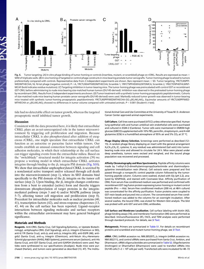
tide had no detectable effect on tumor growth, whereas the targetedproapoptotic motif inhibited tumor growth.
DiscussionConsistent with the data presented here, it is likely that extracellularCRKL plays an as-yet-unrecognized role in the tumor microenvi-ronment by triggering cell proliferation and migration. Becauseintracellular CRKL is also phosphorylated after addition of exog-enous rCRKL, one might speculate that extracellular CRKL canfunction as an autocrine or paracrine factor within tumors. Ourresults establish an unusual connection between signaling and celladhesion molecules, in which their relationship at the cell surfacecan trigger signaling events from the extracellular milieu. Based onthe ‘‘switchblade’’ structural model for integrin activation (39) wepropose a working model in which extracellular CRKL activatesintegrins through binding to the �1 integrin PSI domain (Fig. S10).We show that intracellular unphosphorylated CRKL is secreted bya nonclassical active transport and/or released through cell deathinto the microenvironment (step 1), where its SH3 domains bindspecifically to the PSI domain of the �1 integrin on the tumor cellsurface (step 2). Upon binding, the �1 integrin changes conforma-tion from a bent to extended (active) form and thereby triggersdownstream phosphorylation of target proteins in the integrin-mediated pathway (steps 3 and 4) and/or MAPK pathway (steps5–7), affecting tumor cell migration and proliferation (step 8).Precedent for intracellular molecules such as nuclear proteins (40,41), transcription factors (42), and stress-response chaperones (13,43, 44) on the cell surface has been reported. Ligand–receptorinteractions between signaling molecules and surface receptorswithin the extracellular environment may have general biologicalsignificance.
Materials and MethodsReagents. Anti-CRKL (Santa Cruz, Cell Signaling,Epitomics, or Upstate Biotech-nology), antiphospho-CRKL (Cell Signaling), anti-�1 integrin (Chemicon or BD),anti-IL11R (Santa Cruz), anti-�3 and anti-�5 integrins (45), anti-EGFR (46), anti-grb2 (Santa Cruz), anti-�6 integrin (Chemicon), anti-fetuin A/�2-Heremans-Schmid glycoprotein (AHSG; R&D Systems), preimmune serum (Jackson), anti-His(Santa Cruz), anti-GST (Santa Cruz), and anti-GAPDH (Ambion) were used. Pep-tides were synthesized to our specifications (AnaSpec). Nude mice were pur-chased (Harlan), and tumors were generated as described (13, 47). The Institu-
tional Animal Care and Use Committee at the University of Texas M. D. AndersonCancer Center approved animal experiments.
Cell Culture. Cell lines were purchased (ATCC) unless otherwise specified. Humanlung epithelial cells and human umbilical vein endothelial cells were purchasedand cultured in EGM-2 (Cambrex). Tumor cells were maintained in DMEM-highglucose (GIBCO) supplemented with 10% FBS, penicillin, streptomycin, and 4 mMglutamine (ICN) in a humidified atmosphere of 95% air and 5% CO2 at 37 °C.
Phage Display Library Selection. Screenings were performed as described (12–15). A random phage library displaying an insert with the general arrangementX2CX12CX2 (C, cysteine; X, any residue) was administered (tail vein) into tumor-bearing nude mice and allowed to circulate for 24 h. Mice were placed underdeep anesthesia, tumors were excised and weighed, and the bound phagepopulation was recovered and processed.
Affinity Chromatography and Mass Spectrometry. Peptide affinity columns weremade by 1-ethyl-3-(3-dimethylaminopropyl)carboiimide and diaminodipro-pylamine immobilization resin (Pierce). Cell extracts were prepared and firstpassed through a nonspecific control peptide column followed by the tumor-homing peptide column. Columns were washed, eluted with Gly (pH 2.2), ana-lyzed by SDS/PAGE, and stained with Coomassie blue. Affinity purification ofCRKL from serum-free conditioned medium was performed and confirmed withrecombinantGST-tagfusionproteinexpressingtumor-homingormutantcontrolpeptide (Pro3 Ala). Serum-free conditioned medium (200 mL at 48-h culture)was concentrated for the affinity purification. Fusion proteins were coupled toGST-resin beads and poured into a column. The concentrated serum-free condi-tioned medium was added to the columns for an overnight incubation. Afterseveral washes, the bound CRKL was eluted for Western blot analysis. The blotwas probed with anti-GST and anti-CRKL antibodies.
Cell Surface and Membrane Localization. Cell surface labeling with biotin (42),phage binding assays (16), and membrane fractionation (44) were performed asdescribed. Immunofluorescence (IF), FACS, and TEM analyses were performedthrough standard protocols. For details, see SI Text.
Mutagenesis. Primers are summarized in Table S1. For details on recombinantproteins and scrambled and mutant tumor-homing phage, see SI Text.
siRNA. CRKL (mRNA accession no. NM�005207), �1 integrin (mRNA accession no.NM�002211), and control siRNAs were purchased from Santa Cruz, Ambion, andDharmacon. siRNA oligonucleotides are summarized in Table S2. Oligofectamine(Invitrogen) or DharmaFect (Dharmacon) were used to transfect siRNAs intoDU145 cells (1–2 � 105 cells per well). Transfected cells were incubated for 48–72
Fig. 6. Tumor targeting. (A) In vitro phage binding of tumor-homing or controls (insertless, mutant, or scrambled) phage on rCRKL. Results are expressed as mean �SEM of triplicate wells. (B) In vivo homing of targeted or control phage constructs in mice bearing prostate tumor xenografts. Tumor-homing phage localized to tumorspreferentially compared with controls. Representative data from 2 independent experiments are shown. Bars represent mean � SD. Tumor targeting, YRCTLNSPFF-WEDMTHECHA; fd, fd-tet phage (negative control); P-�A, YRCTLNSAFFWEDMTHECHA; Scramble 1, YRFCTSPFHEWHLENTDMCA; Scramble 2, YRECTDSPHEFHLWNT-MCAF (bold indicates residue mutations). (C) Targeting inhibition in tumor-bearing mice. The tumor-homing phage was preincubated with control GST or recombinantGST-CRKL before administering to nude mice bearing size-matched human tumors (DU145-derived). Inhibition was observed in the pretreated tumor-homing phagebyrecombinantCRKL.Results from2 independentexperimentsare shown. (D) Tumor treatmentwithasynthetic tumor-homingproapoptoticpeptidomimetic.Cohortsof size-matched nude mice bearing human prostate cancer xenografts (DU145-derived) were used. Markedly reduced tumor growth was observed in tumor-bearingmice treated with synthetic tumor-homing proapoptotic peptidomimetic YRCTLNSPFFWEDMTHECHA-GG-D(KLAKLAK)2. Equimolar amounts of YRCTLNSPFFWED-MTHECHA or D(KLAKLAK)2 showed no differences in tumor volume compared with untreated animals. P � 0.001 (Student’s t test).
2186 � www.pnas.org�cgi�doi�10.1073�pnas.0807543105 Mintz et al.
h before processing, recovered, and lysed in the presence of protease inhibitors.Tumor-homing phage binding activity was examined in CRKL-silenced cells. Forrescue experiments, up to 1.5 �g of recombinant CRKL was added to CRKLsiRNA-transfected cells. Exogenous His-tag recombinant CRKL (400 ng/mL) orcontrols (EGF, 200 ng/mL; migration inhibitory factor, 300 ng/mL; phorbol 12-myristate 13-acetate, 300 ng/ml) were used in �1 integrin-silenced experiments.Equal amounts of protein were loaded and resolved by SDS/PAGE followed byWestern blot analysis with the appropriate antibodies. Cell proliferation assayswere performed with WST-1 (Roche). Cell migration assays were performed in aBoyden chamber assay (Corning).
IP. Cells were synchronized for 24–48 h in RPMI serum-free 1640 medium,followed by centrifugation and sterile filtration and preabsorption to controlantibodies. An equal volume of PBS was added to the recovered supernatantbefore IP with the appropriate antibodies. Brefeldin A, thapsigargin, and gly-benclamide were used. Cells were incubated with the compounds in serum-freemedium for 9 h before IP with the appropriate antibodies. No detergents wereused in the IP. This protocol favors the finding of CRKL in a free soluble staterather than in vesicles.
Peptide Binding and Internalization. Peptides were coated on microtiter platesfollowed by blocking and washing. Mixtures were incubated, washed, and la-beled with the appropriate antibodies. HRP-conjugated secondary antibodieswere added followed by 3,3�–5,5�–tetramethylbenzidine (TMB) substrate (Cal-biochem), and complexes were analyzed in an ELISA reader. To determine theinhibitory activity of the tumor-homing peptide, the PSI-derived peptide or thephage clone displaying the tumor-homing peptide were incubated with the
rCRKL-SH3 (C) domain. Unrelated peptide sequences and insertless phage servedasnegativecontrols.Mixtureswere incubatedandaddedtowells coatedwiththetumor-homing peptide. After incubation, the wells were washed and labeledwith the appropriate antibodies. For details and recombinant protein informa-tion, see SI Text.
Phage and Protein Binding Assays. Phage binding assays on purified proteinswere carried out as described (19, 48). For a detailed description, see SI Text.
In Vivo Tumor Targeting and Inhibition. In vivo targeting experiments with phagewere performed as described (10, 12, 13, 47). For details, see SI Text.
Therapy in Tumor-Bearing Mice. Tumor-bearing mice were size-matched anddivided into individual cohorts (n � 4 mice per group). The tumor-homingpeptide was synthesized fused with the proapoptotic motif D(KLAKLAK)2. Un-conjugated peptide YRCTLNSPFFWEDMTHECHA or D(KLAKLAK)2 served as con-trols. Peptides were administered i.v. at 300 �g per mouse per week, and tumorvolumes were measured (12, 13).
ACKNOWLEDGMENTS. We thank Drs. Tony Hunter, Claudio Joazeiro, HeleneSage, and Richard Sidman for helpful discussions. This work was supported bygrants from the National Institutes of Health (to W.A. and R.P.), the Departmentof Defense (to P.J.M., W.A., and R.P.) and awards from the V Foundation, theMarcus Foundation, and the Gillson-Longenbaugh Foundation (to R.P. andW.A.). M.C.V. is a Fellow from the Susan G. Komen Breast Cancer Foundation. R.R.and G.R.S. are Scholars from the Odyssey Program of the University of Texas M. D.Anderson Cancer Center.
1. Conner SD, Schmid SL (2003) Regulated portals of entry into the cell. Nature 422:37–44.2. Cho W, Stahelin RV (2005) Membrane–protein interactions in cell signaling and mem-
brane trafficking. Annu Rev Biophys Biomol Struct 34:119–151.3. Martin KH, Slack JK, Boerner SA, Martin CC, Parsons JT (2002) Integrin connections
map: To infinity and beyond. Science 296:1652–1653.4. Manning G, Whyte DB, Martinez R, Hunter T, Sudarsanam S (2002) The protein kinase
complement of the human genome. Science 298:1912–1934.5. Mochly-Rosen D (1995) Localization of protein kinases by anchoring proteins: A theme
in signal transduction. Science 268:247–251.6. Miller-Jensen K, Janes KA, Brugge JS, Lauffenburger DA (2007) Common effector
processing mediates cell-specific responses to stimuli. Nature 448:604–608.7. Hunter T (2007) The age of cross-talk: Phosphorylation, ubiquitination, and beyond.
Mol Cell 28:730–738.8. Pawson T, Scott JD (1997) Signaling through scaffold, anchoring, and adaptor proteins.
Science 278:2075–2080.9. Blume-Jensen P, Hunter T (2001) Oncogenic kinase signaling. Nature 411:355–365.
10. Hajitou A, et al. (2006) A hybrid vector for ligand-directed tumor targeting andmolecular imaging. Cell 125:385–398.
11. Arap W, et al. (2002) Steps toward mapping the human vasculature by phage display.Nat Med 8:121–127.
12. Arap W, Pasqualini R, Ruoslahti E (1998) Cancer treatment by targeted drug delivery totumor vasculature in a mouse model. Science 279:377–380.
13. Arap MA, et al. (2004) Cell surface expression of the stress response chaperone GRP78enables tumor targeting by circulating ligands. Cancer Cell 6:275–284.
14. Pasqualini R, Ruoslahti E (1996) Organ targeting in vivo using phage display peptidelibraries. Nature 380:364–366.
15. Pasqualini R, Arap W, Rajotte D, Ruoslahti E (2001) in Phage Display: A LaboratoryManual, eds Barbas CF, III, Burton DR, Scott JK, Silverman GJ (Cold Spring Harbor LabPress, Cold Spring Harbor, NY), pp 22.1–22.24.
16. Giordano RJ, Cardo-Vila M, Lahdenranta J, Pasqualini R, Arap W (2001) Biopanning andrapid analysis of selective interactive ligands. Nat Med 7:1249–1253.
17. Ellerby HM, et al. (1999) Anticancer activity of targeted proapoptotic peptides. NatMed 5:1032–1038.
18. Kolonin MG, Saha PK, Chan L, Pasqualini R, Arap W (2004) Reversal of obesity bytargeted ablation of adipose tissue. Nat Med 10:625–632.
19. Zurita AJ, et al. (2004) Combinatorial screenings in patients: The interleukin-11 recep-tor � as a candidate target in the progression of human prostate cancer. Cancer Res64:435–439.
20. Shi M, et al. (2005) The crystal structure of the plexin-semaphorin-integrin domain/hybrid domain/I-EGF1 segment from the human integrin �2 subunit at 1.8-Å resolu-tion. J Biol Chem 280:30586–30593.
21. Mould AP, et al. (2005) Evidence that monoclonal antibodies directed against theintegrin � subunit plexin/semaphorin/integrin domain stimulate function by inducingreceptor extension. J Biol Chem 280:4238–4246.
22. Arnaout MA, Mahalingam B, Xiong JP (2005) Integrin structure, allostery, and bidi-rectional signaling. Annu Rev Cell Dev Biol 21:381–410.
23. ten Hoeve J, Morris C, Heisterkamp N, Groffen J (1993) Isolation and chromosomallocalization of CRKL, a human crk-like gene. Oncogene 8:2469–2474.
24. Harkiolaki M, Gilbert RJ, Jones EY, Feller SM (2006) The C-terminal SH3 domain of CRKLas a dynamic dimerization module transiently exposing a nuclear export signal. Struc-ture (London)14:1741–1753.
25. Mayer BJ (2001) SH3 domains: Complexity in moderation. J Cell Sci 114:1253–1263.26. Sicheri F, Moarefi I, Kuriyan J (1997) Crystal structure of the Src family tyrosine kinase
Hck. Nature 385:602–609.
27. Kato M, Miyazawa K, Kitamura N (2000) A deubiquitinating enzyme UBPY interactswith the Src homology 3 domain of Hrs-binding protein via a novel binding motifPX(V/I)(D/N)RXXKP. J Biol Chem 275:37481–37487.
28. Xu W, Harrison SC, Eck MJ (1997) Three-dimensional structure of the tyrosine kinasec-Src. Nature 385:595–602.
29. Liu Q, et al. (2003) Structural basis for specific binding of the Gads SH3 domain to anRxxK motif-containing SLP-76 peptide: A novel mode of peptide recognition. Mol Cell11:471–481.
30. Walter P, Gilmore R, Blobel G (1984) Protein translocation across the endoplasmicreticulum. Cell 38:5–8.
31. Prudovsky I, et al. (2003) The nonclassical export routes: FGF1 and IL-1� point the way.J Cell Sci 116:4871–4881.
32. Reynwar BJ, et al. (2007) Aggregation and vesiculation of membrane proteins bycurvature-mediated interactions. Nature 447:461–464.
33. McNiven MA, Thompson HM (2006) Vesicle formation at the plasma membrane andtrans-Golgi network: The same but different. Science 313:1591–1594.
34. Flieger O, et al. (2003) Regulated secretion of macrophage migration inhibitory factoris mediated by a nonclassical pathway involving an ABC transporter. FEBS Lett 551:78–86.
35. Hamon Y, et al. (1997) Interleukin-1� secretion is impaired by inhibitors of the ATPbinding cassette transporter, ABC1. Blood 90:2911–2915.
36. Kishan KV, Scita G, Wong WT, Di Fiore PP, Newcomer ME (1997) The SH3 domain of Eps8exists as a novel intertwined dimer. Nat Struct Biol 4:739–743.
37. Kristensen O, et al. (2006) A unique set of SH3–SH3 interactions controls IB1 ho-modimerization. EMBO J 22:785–797.
38. Pasqualini R, Koivunen E, Ruoslahti E (1997) Alpha v integrins as receptors for tumortargeting by circulating ligands. Nat Biotechnol 15:542–546.
39. Takagi J, Petre BM, Walz T, Springer TA (2002) Global conformational rearrangementsin integrin extracellular domains in outside-in and inside-out signaling. Cell 110:599–611.
40. Sinclair JF, O’Brien AD (2002) Cell surface-localized nucleolin is a eukaryotic receptorfor the adhesin intimin-� of enterohemorrhagic Escherichia coli O157:H7. J Biol Chem277:2876–2885.
41. Hovanessian AG, et al. (2000) The cell-surface-expressed nucleolin is associated withthe actin cytoskeleton. Exp Cell Res 261:312–328.
42. Monferran S, Paupert J, Dauvillier S, Salles B, Muller C (2004) The membrane form ofthe DNA repair protein Ku interacts at the cell surface with metalloproteinase 9. EMBOJ 23:3758–3768.
43. Shin BK, et al. (2003) Global profiling of the cell surface proteome of cancer cellsuncovers an abundance of proteins with chaperone function. J Biol Chem 278:7607–7616.
44. Mintz PJ, et al. (2003) Fingerprinting the circulating repertoire of antibodies fromcancer patients. Nat Biotechnol 21:57–63.
45. Barker TH, et al. (2005) SPARC regulates extracellular matrix organization through itsmodulation of integrin-linked kinase activity. J Biol Chem 280:36483–36493.
46. Goldstein NI, Prewett M, Zuklys K, Rockwell P, Mendelsohn J (1995) Biological efficacyof a chimeric antibody to the epidermal growth factor receptor in a human tumorxenograft model. Clin Cancer Res 11:1311–1318.
47. Marchio S, et al. (2004) Aminopeptidase A is a functional target in angiogenic bloodvessels. Cancer Cell 5:151–162.
48. Smith GP, Scott JK (1993) Libraries of peptides and proteins displayed on filamentousphage. Methods Enzymol 217:228–257.
Mintz et al. PNAS � February 17, 2009 � vol. 106 � no. 7 � 2187
CELL
BIO
LOG
Y