An evaluation of the clinical and cost-effectiveness of pulmonary artery catheters in patient...
154
An evaluation of the clinical and cost-effectiveness of pulmonary artery catheters in patient management in intensive care: a systematic review and a randomised controlled trial S Harvey, K Stevens, D Harrison, D Young, W Brampton, C McCabe, M Singer and K Rowan Health Technology Assessment 2006; Vol. 10: No. 29 HTA Health Technology Assessment NHS R&D HTA Programme August 2006
Transcript of An evaluation of the clinical and cost-effectiveness of pulmonary artery catheters in patient...

Health Technology Assessm
ent 2006;Vol. 10: No. 29
Pulmonary artery catheters in patient m
anagement in intensive care
An evaluation of the clinical and cost-effectiveness of pulmonary arterycatheters in patient management inintensive care: a systematic review and a randomised controlled trial
S Harvey, K Stevens, D Harrison, D Young, W Brampton, C McCabe, M Singer and K Rowan
Health Technology Assessment 2006; Vol. 10: No. 29
HTAHealth Technology AssessmentNHS R&D HTA Programme
The National Coordinating Centre for Health Technology Assessment,Mailpoint 728, Boldrewood,University of Southampton,Southampton, SO16 7PX, UK.Fax: +44 (0) 23 8059 5639 Email: [email protected]://www.hta.ac.uk ISSN 1366-5278
FeedbackThe HTA Programme and the authors would like to know
your views about this report.
The Correspondence Page on the HTA website(http://www.hta.ac.uk) is a convenient way to publish
your comments. If you prefer, you can send your comments to the address below, telling us whether you would like
us to transfer them to the website.
We look forward to hearing from you.
August 2006
Copyright notice
© Queen's Printer and Controller of HMSO 2006 HTA reports may be freely reproduced for the purposes of private research and study and may be included in professional journals provided that suitable acknowledgement is made and the reproduction is not associated with any form of advertising Violations should be reported to [email protected] Applications for commercial reproduction should be addressed to HMSO, The Copyright Unit, St Clements House, 2–16 Colegate, Norwich NR3 1BQ

How to obtain copies of this and other HTA Programme reports.An electronic version of this publication, in Adobe Acrobat format, is available for downloading free ofcharge for personal use from the HTA website (http://www.hta.ac.uk). A fully searchable CD-ROM isalso available (see below).
Printed copies of HTA monographs cost £20 each (post and packing free in the UK) to both public andprivate sector purchasers from our Despatch Agents.
Non-UK purchasers will have to pay a small fee for post and packing. For European countries the cost is£2 per monograph and for the rest of the world £3 per monograph.
You can order HTA monographs from our Despatch Agents:
– fax (with credit card or official purchase order) – post (with credit card or official purchase order or cheque)– phone during office hours (credit card only).
Additionally the HTA website allows you either to pay securely by credit card or to print out yourorder and then post or fax it.
Contact details are as follows:HTA Despatch Email: [email protected]/o Direct Mail Works Ltd Tel: 02392 492 0004 Oakwood Business Centre Fax: 02392 478 555Downley, HAVANT PO9 2NP, UK Fax from outside the UK: +44 2392 478 555
NHS libraries can subscribe free of charge. Public libraries can subscribe at a very reduced cost of £100 for each volume (normally comprising 30–40 titles). The commercial subscription rate is £300 per volume. Please see our website for details. Subscriptions can only be purchased for the current orforthcoming volume.
Payment methods
Paying by chequeIf you pay by cheque, the cheque must be in pounds sterling, made payable to Direct Mail Works Ltdand drawn on a bank with a UK address.
Paying by credit cardThe following cards are accepted by phone, fax, post or via the website ordering pages: Delta, Eurocard,Mastercard, Solo, Switch and Visa. We advise against sending credit card details in a plain email.
Paying by official purchase orderYou can post or fax these, but they must be from public bodies (i.e. NHS or universities) within the UK.We cannot at present accept purchase orders from commercial companies or from outside the UK.
How do I get a copy of HTA on CD?
Please use the form on the HTA website (www.hta.ac.uk/htacd.htm). Or contact Direct Mail Works (seecontact details above) by email, post, fax or phone. HTA on CD is currently free of charge worldwide.
The website also provides information about the HTA Programme and lists the membership of the variouscommittees.
HTA

An evaluation of the clinical and cost-effectiveness of pulmonary arterycatheters in patient management inintensive care: a systematic review and a randomised controlled trial
S Harvey,1* K Stevens,2 D Harrison,1 D Young,3
W Brampton,4 C McCabe,2 M Singer5 and K Rowan1
1 Intensive Care National Audit and Research Centre, London, UK2 Health Economics and Decision Science, ScHARR, University of
Sheffield, UK3 Oxford Radcliffe Hospitals NHS Trust, UK4 Aberdeen Royal Infirmary NHS Grampian, UK5 Department of Medicine and Wolfson Institute of Biomedical Research,
University College London, UK
* Corresponding author
Declared competing interests of authors: M Singer has undertaken consultancy workand received research funds from Deltex Medical, manufacturers of the CardioQoesophageal Doppler device.
Published August 2006
This report should be referenced as follows:
Harvey S, Stevens K, Harrison D, Young D, Brampton W, McCabe C, et al. An evaluationof the clinical and cost-effectiveness of pulmonary artery catheters in patient managementin intensive care: a systematic review and a randomised controlled trial. Health Technol Assess 2006;10(29).
Health Technology Assessment is indexed and abstracted in Index Medicus/MEDLINE,Excerpta Medica/EMBASE and Science Citation Index Expanded (SciSearch®) and Current Contents®/Clinical Medicine.

NHS R&D HTA Programme
The research findings from the NHS R&D Health Technology Assessment (HTA) Programme directlyinfluence key decision-making bodies such as the National Institute for Health and Clinical
Excellence (NICE) and the National Screening Committee (NSC) who rely on HTA outputs to help raisestandards of care. HTA findings also help to improve the quality of the service in the NHS indirectly inthat they form a key component of the ‘National Knowledge Service’ that is being developed to improvethe evidence of clinical practice throughout the NHS.
The HTA Programme was set up in 1993. Its role is to ensure that high-quality research information onthe costs, effectiveness and broader impact of health technologies is produced in the most efficient wayfor those who use, manage and provide care in the NHS. ‘Health technologies’ are broadly defined toinclude all interventions used to promote health, prevent and treat disease, and improve rehabilitationand long-term care, rather than settings of care.
The HTA Programme commissions research only on topics where it has identified key gaps in theevidence needed by the NHS. Suggestions for topics are actively sought from people working in theNHS, the public, service-users groups and professional bodies such as Royal Colleges and NHS Trusts.
Research suggestions are carefully considered by panels of independent experts (including service users)whose advice results in a ranked list of recommended research priorities. The HTA Programme thencommissions the research team best suited to undertake the work, in the manner most appropriate to findthe relevant answers. Some projects may take only months, others need several years to answer theresearch questions adequately. They may involve synthesising existing evidence or conducting a trial toproduce new evidence where none currently exists.
Additionally, through its Technology Assessment Report (TAR) call-off contract, the HTA Programme isable to commission bespoke reports, principally for NICE, but also for other policy customers, such as aNational Clinical Director. TARs bring together evidence on key aspects of the use of specifictechnologies and usually have to be completed within a short time period.
Criteria for inclusion in the HTA monograph seriesReports are published in the HTA monograph series if (1) they have resulted from work commissionedfor the HTA Programme, and (2) they are of a sufficiently high scientific quality as assessed by the refereesand editors.
Reviews in Health Technology Assessment are termed ‘systematic’ when the account of the search,appraisal and synthesis methods (to minimise biases and random errors) would, in theory, permit thereplication of the review by others.
The research reported in this monograph was commissioned by the HTA Programme as project number97/08/03. The contractual start date was in January 2000. The draft report began editorial review inDecember 2004 and was accepted for publication in January 2006. As the funder, by devising acommissioning brief, the HTA Programme specified the research question and study design. The authorshave been wholly responsible for all data collection, analysis and interpretation, and for writing up theirwork. The HTA editors and publisher have tried to ensure the accuracy of the authors’ report and wouldlike to thank the referees for their constructive comments on the draft document. However, they do notaccept liability for damages or losses arising from material published in this report.
The views expressed in this publication are those of the authors and not necessarily those of the HTA Programme or the Department of Health.
Editor-in-Chief: Professor Tom WalleySeries Editors: Dr Aileen Clarke, Dr Peter Davidson, Dr Chris Hyde,
Dr John Powell, Dr Rob Riemsma and Dr Ken SteinManaging Editors: Sally Bailey and Sarah Llewellyn Lloyd
ISSN 1366-5278
© Queen’s Printer and Controller of HMSO 2006
This monograph may be freely reproduced for the purposes of private research and study and may be included in professional journals providedthat suitable acknowledgement is made and the reproduction is not associated with any form of advertising.
Applications for commercial reproduction should be addressed to NCCHTA, Mailpoint 728, Boldrewood, University of Southampton, Southampton, SO16 7PX, UK.
Published by Gray Publishing, Tunbridge Wells, Kent, on behalf of NCCHTA.Printed on acid-free paper in the UK by St Edmundsbury Press Ltd, Bury St Edmunds, Suffolk. G

Objectives: To evaluate the clinical and cost-effectiveness of managing critically ill patients in adult,general intensive care with or without pulmonaryartery catheters (PACs).Design: An open, multi-centre, randomised controlledtrial with economic evaluation (cost–utility and cost-effectiveness analysis).Setting: The setting was general (mixedmedical/surgical) intensive care units (ICUs) in the UKadmitting adults.Participants: Adult patients in participating ICUsdeemed by the responsible treating clinician to requiremanagement with a PAC.Interventions: These were insertion of a PAC andsubsequent clinical management, at the discretion ofthe responsible treating clinicians, using data derivedfrom the PAC. The control group were managedwithout a PAC but with the option of using alternativecardiac output monitoring devices. Main outcome measures: The main outcomemeasure was hospital mortality. Secondary outcomemeasures were length of stay in the ICU, length of stayin an acute hospital and organ-days of support in theICU. For the economic evaluation, the main outcomemeasure was quality-adjusted life-years (QALYs) andthe secondary outcome measure was hospital mortality. Results: Sixty-five ICUs in the UK participated. Ofthese, 43 (66%) used alternative cardiac outputmonitoring devices in control group patients. A total of1263 patients were identified as being eligible for thetrial. Of these, 1041 (82.4%) were randomised andallocated to management with (n = 519) or without (n = 522) a PAC. There were no losses to follow-up.
However, 27 patients (13 in the PAC group and 14 inthe control group) were withdrawn from the trialbecause either the patient withdrew consent onrecovering mental competency or the relativeswithdrew agreement following randomisation. Data on1014 patients were included in the analysis. Participantsin the two groups had similar baseline characteristics.There was no difference in hospital mortality forpatients managed with (68.4%) or without (65.7%) aPAC. The adjusted hazard ratio (PAC versus no PAC)was 1.09 [95% confidence interval (CI) 0.94 to 1.27].There was no difference in the median length of stay inICU, the median length of stay in an acute hospital ormean organ-days of support in ICU between the twogroups. The economic evaluation found that theexpected cost per QALY gained from the withdrawal ofPAC was £2985. The expected cost per life gainedfrom the withdrawal of PAC was £22,038. Conclusions: Clinical management of critically illpatients with a PAC, as currently practised in the UK,neither improves hospital survival for adult, generalintensive care patients nor reduces length of stay inhospital. The lack of demonstrable benefit from adevice previously believed to be beneficial could beexplained by statistical chance, by misinterpretation ofPAC-derived data, by ineffective treatment strategiesbased on data correctly interpreted using the currentparadigm or by subsequent inaction following insertionof the device. It is also possible that detailed data onhaemodynamics, however used, cannot modify thedisease process sufficiently to influence diseaseoutcome. The economic evaluation, using decisionanalysis techniques rather than conventional hypothesis
Health Technology Assessment 2006; Vol. 10: No. 29
iii
© Queen’s Printer and Controller of HMSO 2006. All rights reserved.
Abstract
An evaluation of the clinical and cost-effectiveness of pulmonaryartery catheters in patient management in intensive care: a systematic review and a randomised controlled trial
S Harvey,1* K Stevens,2 D Harrison,1 D Young,3 W Brampton,4 C McCabe,2
M Singer5 and K Rowan1
1 Intensive Care National Audit and Research Centre, London, UK2 Health Economics and Decision Science, ScHARR, University of Sheffield, UK3 Oxford Radcliffe Hospitals NHS Trust, UK4 Aberdeen Royal Infirmary NHS Grampian, UK5 Department of Medicine and Wolfson Institute of Biomedical Research, University College London, UK* Corresponding author

testing, suggests that the withdrawal of the PAC fromroutine clinical practice in the NHS would beconsidered cost-effective in the current decision-making climate, and might result in lives or life-yearsbeing saved at modest cost. With the declining use ofPACs in the UK and the findings of this report
indicating no overall benefit from management with aPAC, it should now be possible to examineprotocolised management with a PAC in selectedgroups of critically ill patients against appropriatecontrols, something that was difficult while PACs werethe considered standard of care.
Abstract
iv

Health Technology Assessment 2006; Vol. 10: No. 29
v
List of abbreviations .................................. vii
Executive summary .................................... ix
1 Introduction ............................................... 1
2 Systematic review ...................................... 3Objective ..................................................... 3Criteria for considering studies ................. 3Methods ...................................................... 3Results ........................................................ 4Discussion ................................................... 17Conclusions ................................................ 20
3 Randomised controlled trial to evaluate the clinical effectiveness of pulmonary artery catheters in patient management in intensive care ......................................... 21Aim ............................................................. 21Design and development of the trial protocol ...................................................... 21Methods ...................................................... 21Results ........................................................ 26Discussion ................................................... 33
4 Economic evaluation of the incremental cost-effectiveness of withdrawing pulmonary artery catheters from routine use in intensive care .................................. 37Introduction ............................................... 37Methods ...................................................... 37Results ........................................................ 38Discussion ................................................... 39
5 Overall conclusions .................................... 43
6 Recommendations for future research ..... 45
Acknowledgements .................................... 47
References .................................................. 49
Appendix 1 Cardiovascular and cardiac output monitoring devices ......................... 55
Appendix 2 Search strategy for the systematic review ........................................ 59
Appendix 3 Data collection form for systematic review ........................................ 61
Appendix 4 Patient information sheet ...... 65
Appendix 5 Patient consent form ............. 67
Appendix 6 Relative’s information sheet ........................................................... 69
Appendix 7 Relative’s form ....................... 71
Appendix 8 Patient information sheet forretrospective consent .................................. 73
Appendix 9 Patient incapacity to provideconsent form .............................................. 75
Appendix 10 Data collection forms for the RCT ............................................................ 77
Appendix 11 Augmented Care Period (ACP) data .................................................. 93
Appendix 12 Sepsis-related Organ FailureAssessment (SOFA) ..................................... 95
Appendix 13 Acute Physiology And Chronic Health Evaluation II (APACHE II) ............................................... 97
Appendix 14 Data collection forms for theeconomic evaluation ................................... 99
Health Technology Assessment reportspublished to date ....................................... 135
Health Technology Assessment Programme ................................................ 147
Contents


ACP Augmented Care Period
APACHE II Acute Physiology And ChronicHealth Evaluation version II
APS acute physiology score
ASA American Society ofAnesthesiologists
CEA cost-effectiveness analysis
CEAC cost-effectiveness acceptabilitycurve
CI confidence interval
CMP Case Mix Programme
CPAP continuous positive airwaypressure
CUA cost–utility analysis
CVP central venous pressure
HDU high-dependency unit
ICNARC Intensive Care National Auditand Research Centre
ICU intensive care unit
IQR inter-quartile range
LOS length of stay
LREC Local Research EthicsCommittee
MREC Multi-centre Research EthicsCommittee
NICE National Institute for Healthand Clinical Excellence
OR odds ratio
PAC pulmonary artery catheter
PACU post-anaesthesia care unit
QALY quality-adjusted life-year
RCT randomised controlled trial
SCCM Society of Critical CareMedicine
SD standard deviation
SEM standard error of mean
SOFA Sepsis-related Organ FailureAssessment
Health Technology Assessment 2006; Vol. 10: No. 29
vii
© Queen’s Printer and Controller of HMSO 2006. All rights reserved.
List of abbreviations
All abbreviations that have been used in this report are listed here unless the abbreviation is well known (e.g. NHS), or it has been used only once, or it is a non-standard abbreviation used only in figures/tables/appendices in which case the abbreviation is defined in the figure legend or at the end of the table.


Health Technology Assessment 2006; Vol. 10: No. 29
ix
© Queen’s Printer and Controller of HMSO 2006. All rights reserved.
IntroductionBackgroundBedside pulmonary artery catheterisation graduallybecame a standard of care for critically ill patientsfollowing its introduction three decades ago. Thisadoption into mainstream practice occurredwithout any evaluation of either its clinical or cost-effectiveness. The ongoing, long-standing debateabout the clinical effectiveness of pulmonary arterycatheters (PACs) was rekindled in 1996 followingthe publication of a large, non-randomised, risk-adjusted study. The study suggested that patientshad an increased odds of dying, within 30 days ofadmission to an intensive care unit (ICU), if a PACwas used [odds ratio (OR) 1.24, 95% confidenceinterval (CI) 1.03 to 1.49] and that PACs increasedthe use of resources. In addition, acontemporaneous, systematic review revealed thatthat there was very little evidence from randomisedcontrolled trials (RCTs) to support the clinicalmanagement of critically ill patients with PACs. Weundertook and updated the systematic review toconsider the need for an RCT.
Summary of systematic reviewObjectiveThe objective was to search systematically for, andcombine, all the evidence from RCTs relating tothe effect of the clinical management of criticallyill patients with a PAC both on mortality and onthe costs of care.
Inclusion criteriaAll RCTs, with or without blinding, were includedwhere adult patients were randomised to bemanaged with or without (control) a PAC, the PACwas inserted in an ICU or during a surgicalprocedure leading to ICU admission, and eithermortality, length of stay in ICU or hospital or thecosts of care had been measured. There was norestriction on language. Studies were excluded if aPAC was placed solely for organ support prior toorgan donation in patients declared brain deadfollowing brain-stem death testing.
Outcome measuresThe primary outcome measure was hospitalmortality and the secondary outcome measures
were length of stay in ICU and hospital and thecosts of care.
Search strategyThe following electronic databases were searched(initially to June 2001, then updated to November2003): Cochrane Central Register of ControlledTrials, MEDLINE, EMBASE and CINAHL.Conference abstracts from the four majorEuropean and North American annual critical careconferences were hand-searched from 1995 to2001. Reference lists of previous reviews, and bothrelevant and potentially relevant studies identifiedfrom the searches, were checked. Both experts incritical care and manufacturers of PACs werecontacted for relevant literature.
Identification of studiesCitations were checked, with respect to theinclusion criteria, by four reviewers working inpairs and the final included studies were agreed byconsensus between all reviewers.
Assessment of methodological qualityIncluded studies were assessed for possible sourcesof bias as recommended by the UK CochraneCentre – no ‘quality’ scale was used.
AnalysisSeparate meta-analyses were undertaken,combining data from studies that had includedpatient populations with similar characteristics. Aweighted OR was calculated across studies using arandom-effects model (Cochrane statisticalpackage RevMan version 4.2.7). All analyses werebased on the results reported on the intention-to-treat principle.
ResultsFrom all searches (to November 2003), 3282discrete citations were identified. Of these, fullpaper copies were obtained and reviewed for 39and, of these, 11 studies were eligible for inclusion.These fell broadly into two groups: studies ofgeneral intensive care patients (n = 3); and studiesof high-risk surgery patients (n = 8). The lattergroup could be further subdivided into those thatdid (n = 5) or did not (n = 3) include preoperativeoptimisation as part of the intervention. Only two
Executive summary

x
studies were multi-centre (both identified from theupdated search). All remaining studies were small,under-powered and in one or two centres. Potentialproblems of bias existed for four studies, eitherbecause the randomisation and concealmentmethods were inadequate or because there was ahigh crossover rate from the control to theintervention group. Mortality statistics variedacross studies but none reported a statisticallysignificant difference between those managed withor without a PAC. Data pooled for the studies ofgeneral intensive care patients (n = 3) yielded anOR of 0.97 (95% CI 0.74 to 1.26) on comparingPAC with no PAC. In the studies of high-risksurgery patients, for studies that did not includepreoperative optimisation (n = 3) the pooled ORwas 1.10 (95% CI 0.13 to 9.06) and for those thatdid 0.98 (95% CI 0.72 to 1.33). No studiesreported differences in ICU or hospital length ofstay. Four studies, conducted in the USA and usinghospital charges as a measure of costs of care,indicated that costs were generally higher forpatients managed with a PAC, but these resultswere not statistically significant.
ConclusionEvidence from RCTs to support the clinicalmanagement of critically ill patients with PACs isscant. Initial searching of the literature (to June2001) identified only one small RCT of generalintensive care patients. This study wasdiscontinued prematurely. The remaining sevenstudies were of high-risk surgery patients. Hencethe initial review indicated a clear need for a largemulti-centre RCT. Updated searching after theRCT had commenced revealed no conclusiveevidence.
ObjectivesThe objectives were to evaluate the clinical and cost-effectiveness of managing critically ill patients inadult, general intensive care with or without PACs.
DesignThe study was an open, multi-centre, RCT witheconomic evaluation (cost–utility and cost-effectiveness analysis).
SettingThe setting was general (mixed medical/surgical)ICUs in the UK admitting adults.
ParticipantsThe participants in the trial were all adult patientsin participating ICUs deemed by the responsibletreating clinician to require management with aPAC unless: less than 16 years of age; admitted toICU electively for preoperative optimisation priorto surgery; a PAC already in place on admission toICU; had previously been entered into the RCT;or dead using brain-stem death criteria and a PAC being placed for organ support prior todonation.
InterventionsThese were insertion of a PAC and subsequentclinical management, at the discretion of theresponsible treating clinicians, using data derivedfrom the PAC. The control group were managedwithout a PAC but with the option of usingalternative cardiac output monitoring devices.
Outcome measuresThe main outcome measure was hospital mortality.Secondary outcome measures were length of stayin the ICU, length of stay in an acute hospital andorgan-days of support in the ICU. For theeconomic evaluation, the main outcome measurewas quality-adjusted life-years (QALYs) and thesecondary outcome measure was hospitalmortality.
ResultsSixty-five ICUs in the UK participated. Of these,43 (66%) used alternative cardiac outputmonitoring devices in control group patients. Atotal of 1263 patients were identified as beingeligible for the trial. Of these, 1041 (82.4%) wererandomised and allocated to management with (n = 519) or without (n = 522) a PAC. There wereno losses to follow-up. However, 27 patients (13 inthe PAC group and 14 in the control group) werewithdrawn from the trial because either thepatient withdrew consent on recovering mentalcompetency or the relatives withdrew agreementfollowing randomisation. Data on 1014 patientswere included in the analysis. Participants in thetwo groups had similar baseline characteristics.There was no difference in hospital mortality forpatients managed with (68.4%) or without (65.7%)a PAC. The adjusted hazard ratio (PAC versus noPAC) was 1.09 (95% CI 0.94 to 1.27). There was
Executive summary

no difference in the median length of stay in ICU,the median length of stay in an acute hospital ormean organ-days of support in ICU between thetwo groups. The economic evaluation found thatthe expected cost per QALY gained from thewithdrawal of PAC was £2985. The expected costper life gained from the withdrawal of PAC was£22,038.
ConclusionsClinical management of critically ill patients with aPAC, as currently practised in the UK, neitherimproves hospital survival for adult, generalintensive care patients nor reduces length of stayin hospital. The lack of demonstrable benefit froma device previously believed to be beneficial couldbe explained by statistical chance, bymisinterpretation of PAC-derived data, byineffective treatment strategies based on datacorrectly interpreted using the current paradigmor by subsequent inaction following insertion ofthe device. It is also possible that detailed data onhaemodynamics, however used, cannot modify thedisease process sufficiently to influence outcome.The economic evaluation, using decision analysistechniques rather than conventional hypothesistesting, suggests that the withdrawal of the PACfrom routine clinical practice in the NHS would be
considered cost-effective in the current decision-making climate, and might result in lives or life-years being saved at modest cost.
Future researchThe use of PACs is declining in the UK,predominately because other, less invasivetechnologies for measuring cardiac output are nowbecoming available. As it is unclear whetherderiving detailed haemodynamic data, from a PACor from any other means, affects outcome incritically ill patients, these new devices must besubjected to proper evaluation. Ideally, it needs tobe determined whether the lack of effectivenessseen in this study is unique to PACs or is a ‘classeffect’ of all haemodynamic monitors measuringcardiac output. This study examined theeffectiveness of clinical management of critically illpatients with a PAC (a package of pulmonaryartery catheterisation and subsequentunprotocolised management) in a heterogeneouspopulation of critically ill patients. By indicatingno overall benefit from management with a PAC,it should now be possible to examine protocolisedmanagement with a PAC in selected groups ofcritically ill patients against appropriate controls,something that was difficult while PACs were theconsidered standard of care.
Health Technology Assessment 2006; Vol. 10: No. 29
xi
© Queen’s Printer and Controller of HMSO 2006. All rights reserved.


Pulmonary artery catheterisation was firstintroduced into medicine in 19441 and was
used initially to assess the severity of mitral valvedisease. The pulmonary artery catheter (PAC) wasinserted into the venous circulation, usually viaone of the large veins in the groin, arm, neck orchest, and advanced through the chambers of theright side of the heart and into the pulmonaryartery. It was then advanced until its tiptemporarily occluded the branch of the pulmonaryartery in which it sat. This interruption of forwardflow allowed pressure measured at the catheter tipto reflect filling pressures in the left side of theheart, at the end of the occluded pulmonaryvessel. The degree of elevation of this pressure wasa measure of the severity of mitral valve disease.The procedure was largely confined to cardiaccatheterisation laboratories. The introduction ofthe balloon flotation catheter in the 1970s allowedinsertion of these catheters at the bedside withoutthe need for X-ray guidance to position thecatheter correctly in the pulmonary artery.2 Thischanged what was primarily a one-off diagnosticprocedure into a monitoring technique withcontinuous or intermittent measurement anddisplay of pressures in the right atrium, rightventricle, pulmonary artery and left atrium.Nearly all types of PAC are also capable ofmeasuring blood flow through the heart, that is,the cardiac output.
The technique gradually became accepted as thegold standard method to measure cardiac outputand other haemodynamic variables. The conceptthat detailed knowledge of haemodynamicvariables in critically ill patients would translate toa survival advantage was widespread, and so thePAC became a standard of care without evaluationof either its clinical or cost-effectiveness. PACs arenow usually used to monitor patients with severecardiac or respiratory failure. They are used toguide treatment with fluids and inotropic (cardiacstimulant) drugs during the most severe phase ofthe illness. Most are fitted with devices to measurecardiac output and are often connected tomonitors that display not only the primaryvariables (pressures and cardiac output) but alsosecondary derived measures of adequacy of thecirculation such as resistance to blood flow. Theyare introduced into the circulation, generally via
one of the large veins in the neck or chest, andusually stay in place for a few days. The PAC andother devices available to measure cardiac outputare described in detail in Appendix 1.
With the possible exception of electronic foetalmonitoring, no monitoring device has polarisedopinion as much as the PAC.3–7 Proponents arguefor its unique ability to allow accuratemeasurement of cardiac output and otherhaemodynamic variables, allowing improveddiagnosis and management of circulatoryinstability.6,7 Critics point to complicationsassociated with its insertion and use,3–5,8,9
inaccuracies in measurement, poor interpretationof data10–12 and the lack of positive outcomebenefits with suggestions of increased mortalityfrom retrospective analyses.13,14
In 1996, a comprehensive review of all availablecomparative and randomised clinical trialsinvolving pulmonary artery catheterisation waspublished.15 Of 34 published studies reviewed,only one was considered ‘level 1’ evidence.16
This showed no benefit for treatment aimed atachieving ‘goal’ values for haemodynamicvariables (goal-directed therapy) in a mixedintensive care population. The remaining studieswere equally split between those showing nodifference or worsened outcome with the PAC andthose showing a benefit. Thus there was no clearindication that the PAC improves outcome.Furthermore, many of these studies involving thePAC were trials of preoperative optimisation,which involves an overall package of enhancedcare on an intensive care unit (ICU), of which thePAC is only one component, or trials of goal-directed therapy where outcome may be primarilydetermined by other components of thealgorithm, such as blood transfusion. Pragmaticstudies of PAC use following admission to theICU, the commonest clinical situation, werevirtually non-existent; only one randomisedcontrolled trial (RCT) was identified, which wasdiscontinued prematurely because of poorrecruitment and which had a cross-over rate fromthe control to treatment group of nearly 50%.17 Inthis trial, of 148 eligible patients in twoparticipating hospitals, only 33 (22.3%) wererecruited. Of the 17 patients allocated to the
Health Technology Assessment 2006; Vol. 10: No. 29
1
© Queen’s Printer and Controller of HMSO 2006. All rights reserved.
Chapter 1
Introduction

control group (not to be managed with a PAC),eight had a PAC inserted following randomisation.Ethical concerns were the most frequently citedreason for not recruiting eligible patients.
This long-standing debate about the clinicaleffectiveness of PACs was rekindled with thepublication of a large, non-randomised, risk-adjusted study by Connors and colleagues in1996,18 which suggested an increased odds of 30-day mortality [odds ratio (OR) 1.24, 95%confidence interval (CI) 1.03 to 1.49] in patientsmanaged with a PAC during the first 24 hoursfollowing admission to ICU, and increasedutilisation of resources. The media coverage in theUSA19–21 that followed publication of the study ledto a formal press release from the Society ofCritical Care Medicine (SCCM)22 challenging theconclusions of the study, predominantly because itwas a non-randomised comparison. In December1996, the SCCM convened a multidisciplinaryConsensus Conference on the PAC. A ConsensusStatement was published in June 1997.7 Ingeneral, the statement identified that the level ofpublished evidence to support the use of PAC was paltry and, scientifically, very poor. However,the statement supported the continuing use of PACs.
Although widely discussed, the ConsensusStatement did not help clarify the indications for a PAC. It was, in scientific terms, an unsystematic,narrative review.23 The potential for biasedselection of the conference participants was notaddressed and, unlike the review cited above, theselection and review of the evidence were notbased on any defined criteria. The ConsensusStatement relied on ‘expert opinion’ for responseand the method of consensus for that response
was, therefore, of paramount importance. Explicitscientific methods exist for reaching consensus, forexample, nominal group or Delphi techniques,24
but none were used.
The Connors and colleagues’ study also provokednumerous editorials in scientific journalsconcerning the clinical and cost-effectiveness ofthe PAC.5,25,26 In the UK, the correspondence thatfollowed27–29 the editorial published in the BMJ26
suggested there was considerable equipoiseamongst UK clinicians. In 1997, MacKirdy andcolleagues30 conducted a similar risk adjustedcomparison of patients managed with and withouta PAC using Scottish data, and reported similarresults to Connors and colleagues.
This led the Intensive Care National Audit andResearch Centre (ICNARC) to respond to the‘Consultation to identify National Health ServiceResearch and Development Priorities’ in October1996 by identifying the PAC as a technologyurgently requiring evaluation.
The proposed study needed not only to addressthe clinical and cost-effectiveness of the PAC ascurrently used in the NHS but also to address thecriticisms levelled at previous studies. Therefore,the study consisted of three distinct activities:
1. a systematic review of the evidence on the PACto inform the final design of the subsequentRCT
2. a multi-centre, open, RCT to evaluate theclinical effectiveness of PACs in patientmanagement in adult, general intensive care
3. an economic evaluation of the cost-effectivenessof PACs in patient management in adult,general intensive care.
Introduction
2

ObjectiveThe objective was to search systematically for andcombine all the evidence from RCTs evaluatingthe effect of management of intensive care patientswith PACs on mortality and the costs of care.
Criteria for considering studiesAll RCTs, with or without blinding, in whichpatients were allocated to be managed with a PAC(of any type) in one arm or to be managed withouta PAC in another (control) arm were considered.Additional criteria for the selection of studies werethat:
● More than 50% of the participants in the trialwere adult (16 years of age and above).
● The PAC was placed in a critical care unit orwas placed during a surgical procedure leadingto admission to a critical care unit [a criticalcare unit was defined as either an ICU, a high-dependency unit (HDU), a post-anaesthesiacare unit (PACU) or a service specific criticalcare unit].
● One or more of the following outcomes hadbeen measured: mortality (ICU, 28-day, 30-day,hospital); length of stay in ICU; length of stayin hospital; or costs of care.
Studies were excluded if there were participantswho had been declared brain dead using brainstem death criteria, where a PAC was being placedsolely for organ support prior to donation.
Primary and secondary outcomemeasuresThe primary outcome measure was hospitalmortality. The secondary outcome measures werelength of stay in ICU, length of stay in hospitaland costs of care.
MethodsSearch strategyPrevious reviewsWe reviewed the studies cited in a previouslypublished review.15
Electronic searchesWe searched the Cochrane Central register ofControlled Trials (CENTRAL) in the CochraneLibrary, issue 4, 2001, MEDLINE (all records toJune 2001), EMBASE (all records to June 2001),CINAHL (all records to June 2001) and SIGLE(all records to June 2001). The search strategyused the optimum terms recommended by theCochrane Collaboration to identify RCTscombined with terms to identify studies involvingPACs (Appendix 2) and was adapted according tothe database searched. There was no restriction onthe language of published studies. At the end of2003, the search described above was updated toinclude all records to November 2003.
Manual searchesConference abstracts from the four majorEuropean and North American annual critical careconferences, run by the European Society ofIntensive Care Medicine, the SCCM, the AmericanThoracic Society and the Erasme Hospital, FreeUniversity of Brussels, were searched from 1995 to2001.
SnowballingThe reference lists of potentially relevant citations,identified from the electronic searches, and theincluded studies were checked for further relevantstudies. The reference lists of any systematic ornarrative reviews identified from the searches werealso checked.
ExpertsKey people in the field of critical care werecontacted, including clinicians and otherresearchers. The final list of identified studies wascirculated to delegates at a PAC-Man StudyCollaborators’ Meeting with a request forinformation on any missed studies.
IndustryManufacturers of the PAC were contacted.
Identification of studies The citations generated from the database searcheswere divided between two pairs of reviewers,working independently. The titles and abstracts ofthe citations were screened for potentially relevantstudies. The full texts of all potentially relevant
Health Technology Assessment 2006; Vol. 10: No. 29
3
© Queen’s Printer and Controller of HMSO 2006. All rights reserved.
Chapter 2
Systematic review

citations were obtained, divided between the twopairs of reviewers and assessed independently byeach reviewer for inclusion. The final selection ofstudies for inclusion was made by consensusbetween the four reviewers.
Data extractionThe full-text paper of each included study wasreviewed by two reviewers independently and thefollowing data were extracted using the datacollection form (Appendix 3):
● general information, including title, leadauthor, journal, publication details and name ofreviewer
● study characteristics, including verification ofstudy eligibility, characteristics of the studypopulation, methodological quality of the study,interventions and outcomes
● outcome measures and results, including lengthof follow-up, drop-outs and measures of effect.
Data were double-checked and entered intoRevMan 4.2.7, a software program distributed bythe Cochrane Collaboration to record the resultsof systematic reviews and perform meta-analyses.
Assessment of methodological qualityMethodological quality of the trials was assessed asrecommended by the UK Cochrane Centre.31
Possible sources of bias, selection bias,performance bias, attrition bias and detection biaswere described. Scales for measuring the validityor ‘quality’ of trials exist, but were not used for thecurrent review.
Data synthesis The aims, methods and outcome measures ofinterest (mortality, length of stay in ICU andhospital and costs of care) were summarised foreach included study. Mortality was expressed asabsolute numbers and percentages and lengths ofstay were expressed as mean, median and range,for survivors and non-survivors separately, wherereported. Results on costs of care were expressedin a range of measures.
Patients admitted to ICU are a heterogeneousgroup in terms of diagnosis, prognosis andresource utilisation. This heterogeneity exists bothbetween patients within a single ICU and betweenthe case mix of patients admitted to individualICUs, and it would be inappropriate to combinedata from studies of different patient populationsinto one meta-analysis. Therefore, separate meta-analyses were undertaken combining data fromstudies that had included patient populations with
similar characteristics. Studies that had includedother interventions in addition to the PAC werealso combined in a separate meta-analysis. Forstudies that had two PAC intervention groups, thetwo groups were combined. The outcome measureof interest was hospital mortality; however, if thiswas not reported, the mortality at the point closestto hospital discharge was used. A weighted OR wascalculated across studies using a random effectsmodel in the Cochrane statistical package RevManversion 4.2.7. All analyses were based on theintention-to-treat principle.
ResultsA total of 3282 discrete citations were identifiedfrom the database searches, manual searches,snowballing and contact with experts. Afterscreening by title and then abstract, full papercopies were obtained for 39 citations. Of these, 11 studies were identified from both the originaland updated searches17,32–41 (Figure 1). The 28citations42–69 that were excluded following fullpaper review are listed in Table 1 with the reasonsfor exclusion.
Description of included studiesThe 11 studies that met the inclusioncriteria17,32–41 and were included in the review fellbroadly into two groups as follows:
1. First were studies of general intensive carepatients, where patients were randomised tomanagement with a PAC, or managementwithout, following admission to the ICU. Threestudies (Table 2) were identified. One was a two-centre trial in Canada,17 one was a single-centre trial in the UK32 and one was a multi-centre trial (36 hospitals) in France.33
2. Second were studies of high-risk surgerypatients, which can be further subdivided intothose that did not include preoperativeoptimisation as part of the intervention34–36
and those that did.37–41 Eight studies wereidentified (Table 2). Seven were single-centretrials in the USA,34–40 and one was a multi-centre trial (19 hospitals) in Canada.41
Methodological quality of studiesSelection biasFive of the studies17,32,33,39,41 clearly used adequaterandomisation and concealment schemes (Table 2).One study34 did not use adequate concealment,although the two groups of patients were fairlywell balanced at baseline for numbers of patients,age and American Society of Anesthesiologists
Systematic review
4

Health Technology Assessment 2006; Vol. 10: No. 29
5
© Queen’s Printer and Controller of HMSO 2006. All rights reserved.
Citations excluded on title andabstract alone, n = 3243
Studies excluded after full textevaluation (see Table 1), n = 28
Studies excluded from meta-analyses (but still included innarrative synthesis), n = 0
Total number of citationsn = 3282 (following removal of duplicates)
Studies retrieved for moredetailed evaluation, n = 39
Relevant studies included insystematic review, n = 11
Studies included in meta-analysis:Meta-analysis 1, n = 3Meta-analysis 2, n = 3Meta-analysis 3, n = 5
FIGURE 1 Flow diagram of study selection process
TABLE 1 Studies excluded following full paper review
Study Reason for exclusion
Schultz et al., 198542 Not all patients assigned to the control group were transferred to ICU or HDU following surgeryOrlando et al.,198543 Conference abstract onlySenagore et al., 198744 Not an RCT comparing management with and without a PACRaybin, 198945 LetterTuman et al., 198946 Not an RCT Eyer et al., 199047 Not an RCT comparing management with and without a PACShoemaker et al., 199048 Patients were randomly allocated in the second part of the study only. In addition, there were no
clear data on mortality in the two groupsMermel et al., 199149 Not an RCTCobb et al., 199250 Not an RCT comparing management with and without a PACMitchell et al., 199251 Not an RCT comparing management with and without a PACBach et al., 199252 Not an RCT comparing management with and without a PACBach et al., 199253 Not an RCT comparing management with and without a PACSola and Bender, 199354 Review articleYu et al., 199355 Not an RCT comparing management with and without a PACKearns, 199356 Summary of a previously reported RCT50
Boyd et al., 199357 Not an RCT comparing management with and without a PACYu et al., 199558 Not an RCT comparing management with and without a PACBoldt et al., 199559 Not an RCT comparing management with and without a PACBrazzi et al., 199560 Not an RCT comparing management with and without a PACHolmes et al., 199761 Not an RCTZiegler et al., 199762 Not an RCT comparing management with and without a PACLatour-Perez and Not an RCT
Calvo-Embuena, 199863
Cohen et al., 199864 Not an RCT comparing management with and without a PACStewart et al., 199865 Not an RCTGirbes et al., 199966 Study end-point was the commencement of surgeryWilson et al., 199967 Not all patients assigned to the control group were transferred to ICU or HDU following surgeryBarone et al., 200168 Review and meta-analysisBonazzi et al., 200269 Patients assigned to the control group were not transferred to ICU or HDU following surgery

Systematic review
6 TA
BLE
2D
escr
iptio
n of
incl
uded
RCT
s
Stud
yM
ain
aim
Sele
ctio
n cr
iter
iaO
utco
mes
A p
riori
Tota
l To
tal
Ran
dom
isat
ion/
allo
cati
on
sam
ple
size
el
igib
le,
rand
omis
ed,
proc
edur
eca
lcul
atio
n?n
n
Stud
ies
of g
ener
al I
CU
pat
ient
s: m
anag
emen
t w
ith
a PA
C v
ersu
s m
anag
emen
t w
itho
ut a
PA
C
cont
inue
d
Guy
att,
1991
17To
inve
stig
ate
the
impa
ct o
f rig
ht h
eart
cath
eter
isatio
n on
phys
iolo
gica
l sta
tus
and
stay
in t
he IC
U
Entr
y cr
iter
ia:
Ass
isted
ven
tilat
ion;
hypo
tens
ion
with
CVP
≥
10 c
m H
2O; o
ligur
ia w
ithC
VP ≥
10 c
m H
2O; o
ligur
iaw
ith h
ypox
aem
ia; h
ypox
aem
iaan
d C
VP <
10 c
m H
2O;
phys
icia
n be
lieve
d pa
tient
mig
ht b
enef
it fr
om a
PA
C
Excl
usio
n cr
iter
ia:
PAC
eth
ical
ly c
ontr
aind
icat
ed;
PAC
an
ethi
cal i
mpe
rativ
e;PA
C p
lace
d pr
e-op
erat
ivel
y fo
rin
trao
pera
tive
mon
itorin
g;or
gan
tran
spla
nt s
urge
ry;
rece
ivin
g hi
gh fr
eque
ncy
jet
vent
ilatio
n; c
onse
nt fr
om a
clos
e re
lativ
e no
t ob
tain
ed
Mor
talit
y (n
ot d
efin
ed),
ICU
LO
S
Mai
n ou
tcom
e no
tst
ated
No
148
33C
ompu
ter-
gene
rate
d ra
ndom
num
bers
sto
red
in s
eque
ntia
llym
arke
d en
velo
pes,
whi
ch w
ere
chec
ked
daily
Rhod
es e
t al
.,20
0232
To c
ompa
re s
urvi
val a
ndcl
inic
al o
utco
mes
of
criti
cally
ill p
atie
nts
man
aged
with
a P
AC
com
pare
d w
ith t
hose
man
aged
with
out
Entr
y cr
iter
ia:
Circ
ulat
ory
shoc
k-he
art
rate
>10
0 be
ats/
min
, sys
tolic
BP
<10
0 m
mH
g, u
nres
pons
ive
to50
0 m
l flu
id c
halle
nge;
olig
uria
-ur
ine
outp
ut <
0.5
ml/k
g/h
desp
ite 5
00 m
l flu
id c
halle
nge;
requ
irem
ent
for
vaso
activ
ein
fusio
n; n
eed
for
mec
hani
cal
vent
ilatio
n
Excl
usio
n cr
iter
ia:
less
tha
n 18
yea
rs o
f age
;ad
mitt
ed t
o IC
U fo
rpr
eope
rativ
e op
timisa
tion
Pri
mar
y28
-day
mor
talit
y an
dm
orbi
dity
Also
rep
orte
d:IC
U L
OS
Hos
pita
l LO
S
Noa
202
202
(1 p
atie
ntw
ithdr
awn
follo
win
gra
ndom
isatio
n)
Com
pute
r-ge
nera
ted
rand
omnu
mbe
rs s
tore
d in
sea
led
enve
lope
s

Health Technology Assessment 2006; Vol. 10: No. 29
7
© Queen’s Printer and Controller of HMSO 2006. All rights reserved.
TA
BLE
2D
escr
iptio
n of
incl
uded
RCT
s (c
ont’d
)
Stud
yM
ain
aim
Sele
ctio
n cr
iter
iaO
utco
mes
A p
riori
Tota
l To
tal
Ran
dom
isat
ion/
allo
cati
on
sam
ple
size
el
igib
le,
rand
omis
ed,
proc
edur
eca
lcul
atio
n?n
n
Stud
ies
of h
igh-
risk
sur
gery
pat
ient
s: m
anag
emen
t w
ith
a PA
C v
ersu
s m
anag
emen
t w
itho
ut a
PA
C
cont
inue
d
Rich
ard
et a
l.,20
0333
To c
ompa
re 2
8-da
ym
orta
lity
in p
atie
nts
man
aged
with
and
with
out
a PA
C
Entr
y cr
iter
ia:
Circ
ulat
ory
shoc
k (a
ccor
ding
to d
efin
ed c
riter
ia);
acut
ere
spira
tory
failu
re >
24 h
ours
Excl
usio
n cr
iter
ia:
Less
tha
n 18
yea
rs o
ld;
card
ioge
nic
shoc
k; p
late
lets
<10
,000
; pat
ient
not
com
mitt
ed t
o fu
ll tr
eatm
ent;
mor
ibun
d; e
nrol
led
in a
noth
erRC
T
Pri
mar
y28
-day
mor
talit
y
Seco
ndar
y14
-day
mor
talit
y 90
-day
mor
talit
yIC
U L
OS
Hos
pita
l LO
SM
orbi
dity
(def
ined
)
90%
pow
erto
det
ect
10%
diffe
renc
e in
mor
talit
y.n
= 1
000
Not
repo
rted
681
(5 p
atie
nts
with
draw
nfo
llow
ing
rand
omisa
tion)
24-h
our
cent
ral t
elep
hone
rand
omisa
tion
serv
ice
Isaa
cson
et
al.,
1990
34To
det
erm
ine
whe
ther
mon
itorin
g w
ith P
AC
or
a C
VP c
athe
ter
resu
ltsin
a d
iffer
ence
in p
atie
ntou
tcom
e
Entr
y cr
iter
ia:
Elec
tive
abdo
min
al a
ortic
reco
nstr
uctiv
e su
rger
y
Excl
usio
n cr
iter
ia:
Seve
re, i
nope
rabl
e co
rona
ryar
tery
dise
ase;
cor
pul
mon
ale;
unco
mpe
nsat
ed c
onge
stiv
ehe
art
failu
re; d
ocum
ente
dca
rdio
myo
path
y; p
oor
left
vent
ricul
ar fu
nctio
n (e
ject
ion
frac
tion
<40
%);
sym
ptom
atic
valv
ular
dise
ase;
ren
al fa
ilure
(blo
od u
rea
nitr
ogen
>60
);se
vere
res
tric
tive/
obst
ruct
ive
pulm
onar
y di
seas
e (v
ital
capa
city
<50
% o
f pre
dict
edvo
lum
e); d
eclin
ed c
onse
nt;
emer
genc
y ao
rtic
pro
cedu
res
Intr
aope
rativ
e an
dpo
stop
erat
ive
fluid
requ
irem
ents
Perio
pera
tive
mor
bidi
ty(d
efin
ed)
Even
ts r
elat
ed t
o ei
ther
the
CVP
cat
hete
r or
PAC
ICU
LO
SH
ospi
tal L
OS
Cos
ts o
f car
e
Also
rep
orte
d: H
ospi
tal
mor
talit
y
Mai
n ou
tcom
e no
tst
ated
No
151
102
Top
card
tak
en fr
om m
arke
d,sh
uffle
d ca
rds
plac
ed fa
ce d
own
in a
file

Systematic review
8 TA
BLE
2D
escr
iptio
n of
incl
uded
RCT
s (c
ont’d
)
Stud
yM
ain
aim
Sele
ctio
n cr
iter
iaO
utco
mes
A p
riori
Tota
l To
tal
Ran
dom
isat
ion/
allo
cati
on
sam
ple
size
el
igib
le,
rand
omis
ed,
proc
edur
eca
lcul
atio
n?n
n
cont
inue
d
Joyc
e et
al.,
1990
35To
tes
t th
e hy
poth
esis
that
cen
tral
haem
odyn
amic
mon
itorin
g is
not
nece
ssar
y in
pot
entia
lly‘lo
w-r
isk’ p
atie
nts
unde
rgoi
ng a
bdom
inal
aort
ic s
urge
ry
Entr
y cr
iter
ia:
Elec
tive
infr
a-re
nal a
ortic
reco
nstr
uctiv
e su
rger
y
Excl
usio
n cr
iter
ia:
Uns
tabl
e an
gina
; rec
ent
myo
card
ial i
nfar
ctio
n (≤
6 m
onth
s); l
eft
vent
ricul
arej
ectio
n fr
actio
n (L
VEF)
<50
%
Perio
pera
tive
haem
odyn
amic
s, fl
uid
and
card
iac
drug
adm
inist
ratio
n,op
erat
ion
time
and
clam
p tim
ePo
stop
erat
ive
rena
lfu
nctio
nPo
stop
erat
ive
vent
ilatio
nLi
ne c
ompl
icat
ions
ICU
LO
SH
ospi
tal L
OS
30-d
ay p
ost-
oper
ativ
em
orta
lity
Mai
n ou
tcom
e no
tst
ated
No
Not
cle
ar40
‘Sea
led
enve
lope
tec
hniq
ue’.
No
othe
r de
tails
giv
en
Pear
son
et a
l.,19
8936
To d
eter
min
e if
the
use
of P
AC
com
pare
d w
itha
CVP
cat
hete
r is
asso
ciat
ed w
ithde
crea
sed
mor
bidi
ty,
mor
talit
y or
cos
ts
Entr
y cr
iter
ia:
Sche
dule
d fo
r el
ectiv
e ca
rdia
csu
rger
y
Excl
usio
n cr
iter
ia:
Non
e re
port
ed
ICU
mor
talit
y m
orbi
dity
(not
def
ined
),co
sts
of c
are
Also
rep
orte
d:IC
U L
OS
Mai
n ou
tcom
e no
tst
ated
No
Not
cle
ar22
6Ta
ble
of r
ando
m n
umbe
rs. N
oot
her
deta
ils g
iven

Health Technology Assessment 2006; Vol. 10: No. 29
9
© Queen’s Printer and Controller of HMSO 2006. All rights reserved.
TA
BLE
2D
escr
iptio
n of
incl
uded
RCT
s
Stud
yM
ain
aim
Sele
ctio
n cr
iter
iaO
utco
mes
A p
riori
Tota
l To
tal
Ran
dom
isat
ion/
allo
cati
on
sam
ple
size
el
igib
le,
rand
omis
ed,
proc
edur
eca
lcul
atio
n?n
n
Stud
ies
of h
igh-
risk
sur
gery
pat
ient
s: m
anag
emen
t w
ith
a PA
C +
pre
oper
ativ
e op
tim
isat
ion
vers
us m
anag
emen
t w
itho
ut a
PA
C +
usu
al p
reop
erat
ive
care
cont
inue
d
Bend
er e
t al
.,19
9737
To d
eter
min
e w
heth
erpl
acem
ent
of a
PA
Cw
ith o
ptim
isatio
n of
haem
odyn
amic
s re
sults
in o
utco
me
impr
ovem
ent
afte
rel
ectiv
e va
scul
ar s
urge
ry
Entr
y cr
iter
ia:
Sche
dule
d fo
r el
ectiv
ein
frar
enal
aor
tic r
econ
stru
ctio
nor
low
er li
mb
reva
scul
arisa
tion
(by
one
surg
eon)
Excl
usio
n cr
iter
ia:
Ant
icip
ated
nee
d be
fore
surg
ery
for
supr
aren
al o
rsu
prac
oelia
c cl
ampi
ng;
myo
card
ial i
nfar
ctio
n w
ithin
3m
onth
s or
inad
equa
tely
cont
rolle
d an
gina
; poo
rlyco
mpe
nsat
ed c
onge
stiv
e he
art
failu
re; c
oron
ary
arte
ry b
ypas
ssu
rger
y w
ithin
6 w
eeks
;sy
mpt
omat
ic a
ortic
/mitr
alva
lvul
ar d
iseas
e
Adv
erse
out
com
es(d
efin
ed) i
nclu
ding
30
-day
mor
talit
y
Mai
n ou
tcom
e no
tst
ated
Nob
104
104
Not
des
crib
ed
Berla
uk e
t al
.,19
9138
To t
est
the
hypo
thes
isth
at o
ptim
ising
haem
odyn
amic
s us
ing
aPA
C w
ould
impr
ove
outc
ome
in p
atie
nts
unde
rgoi
ng li
mb-
salv
age
arte
rial s
urge
ry
Entr
y cr
iter
ia:
Sche
dule
d to
rec
eive
an
in s
ituve
in g
raft
bypa
ss fo
r lo
wer
limb
vasc
ular
insu
ffici
ency
Excl
usio
n cr
iter
ia:
Myo
card
ial i
nfar
ctio
n w
ithin
3
mon
ths;
cor
onar
y ar
tery
bypa
ss g
raft
with
in 6
wee
ks;
unco
mpe
nsat
ed c
onge
stiv
ehe
art
failu
re; s
ever
e va
lvul
ardi
seas
e; u
nsta
ble
angi
na
Pri
mar
yC
ardi
ovas
cula
rco
mpl
icat
ions
, e.g
.co
nges
tive
card
iac
failu
re, a
rrht
hym
ia,
myo
card
ial i
nfar
ctio
n
Seco
ndar
yIm
med
iate
post
oper
ativ
e gr
aft
thro
mbo
sis a
nd a
dver
sein
trao
pera
tive
even
ts
Also
rep
orte
d: M
orta
lity
(not
def
ined
)
No
Not
cle
ar89
Rand
om n
umbe
r ge
nera
tor.
Patie
nts
ente
red
cons
ecut
ivel
y in
orde
r of
app
eara
nce
on t
hesu
rgic
al s
ched
ule.
No
othe
rde
tails
giv
en

Systematic review
10 TA
BLE
2D
escr
iptio
n of
incl
uded
RCT
s (c
ont’d
)
Stud
yM
ain
aim
Sele
ctio
n cr
iter
iaO
utco
mes
A p
riori
Tota
l To
tal
Ran
dom
isat
ion/
allo
cati
on
sam
ple
size
el
igib
le,
rand
omis
ed,
proc
edur
eca
lcul
atio
n?n
n
cont
inue
d
Sand
ham
et
al.,
2003
41To
com
pare
goa
l-di
rect
ed t
hera
py g
uide
dby
a P
AC
with
sta
ndar
dca
re w
ithou
t th
e us
e of
a PA
C in
hig
h-ris
ksu
rgic
al p
atie
nts
Entr
y cr
iter
ia:
Age
≥60
yea
rs; A
SA c
lass
III o
rIV
risk
; sch
edul
ed fo
rur
gent
/ele
ctiv
e m
ajor
abdo
min
al, t
hora
cic,
vas
cula
ror
ort
hopa
edic
sur
gery
Pri
mar
yH
ospi
tal m
orta
lity
Seco
ndar
y6-
mon
th m
orta
lity
12-m
onth
mor
talit
yH
ospi
tal m
orbi
dity
(def
ined
)
90%
pow
erto
dist
ingu
ishbe
twee
nho
spita
lm
orta
lity
rate
sof
10
and
15%
in t
hetw
o gr
oups
3803
1994
Com
pute
r-ge
nera
ted
sequ
ence
conc
eale
d in
sea
led,
opa
que,
cons
ecut
ivel
y nu
mbe
red
enve
lope
s. S
trat
ified
acc
ordi
ngto
typ
e of
sur
gery
, ASA
cla
ss a
ndbl
ocke
d ac
cord
ing
to c
entr
e
Shoe
mak
er
et a
l.,19
8839
To c
ompa
re o
utco
mes
of p
atie
nts
who
seth
erap
eutic
goa
ls w
ere
to m
aint
ain
norm
alha
emod
ynam
ic v
alue
sw
ith t
hose
who
se g
oals
wer
e to
att
ain
supr
anor
mal
val
ues
empi
rical
ly o
bser
ved
incr
itica
lly il
lpo
stop
erat
ive
surv
ivor
s
Entr
y cr
iter
ia:
Patie
nts
with
one
or
mor
e hi
ghof
11
high
-risk
crit
eria
prev
ious
ly d
efin
ed a
ndas
soci
ated
with
a m
orta
lity
rate
clo
se t
o 30
%
Excl
usio
n cr
iter
ia:
Non
e re
port
ed
Mor
talit
y an
d m
orbi
dity
(not
def
ined
)
Mai
n ou
tcom
e no
tst
ated
No
146
91C
ards
arr
ange
d ac
cord
ing
tora
ndom
num
bers
tab
les,
by
anou
tsid
e pe
rson
, pla
ced
in s
eale
d,op
aque
env
elop
es, o
pene
d in
sequ
ence

Health Technology Assessment 2006; Vol. 10: No. 29
11
© Queen’s Printer and Controller of HMSO 2006. All rights reserved.
TA
BLE
2D
escr
iptio
n of
incl
uded
RCT
s (c
ont’d
)
Stud
yM
ain
aim
Sele
ctio
n cr
iter
iaO
utco
mes
A p
riori
Tota
l To
tal
Ran
dom
isat
ion/
allo
cati
on
sam
ple
size
el
igib
le,
rand
omis
ed,
proc
edur
eca
lcul
atio
n?n
n
BP, b
lood
pre
ssur
e; C
VP c
athe
ter,
cent
ral v
enou
s pr
essu
re c
athe
ter
(see
app
endi
x 1
for
desc
riptio
n); L
OS,
leng
th o
f sta
y.
aA
utho
rs n
oted
tha
t as
the
re w
ere
no p
rosp
ectiv
e da
ta o
n w
hich
to
perf
orm
a p
ower
cal
cula
tion,
the
stu
dy w
as d
esig
ned
as a
pilo
t w
ith t
he in
tent
ion
to r
ecru
it 20
0 pa
tient
s so
tha
tfu
ture
tria
ls co
uld
be a
ppro
pria
tely
pow
ered
.b
Aut
hors
not
ed t
hat
the
stud
y as
orig
inal
ly d
esig
ned
was
exp
ecte
d to
sho
w a
sig
nific
ant
decr
ease
in c
ompl
icat
ions
afte
r 20
0 pa
tient
s w
ere
enro
lled.
Vale
ntin
e et
al.,
1998
40To
det
erm
ine
whe
ther
the
mor
bidi
ty a
ndm
orta
lity
rate
s of
aor
ticsu
rger
y ar
e re
duce
d by
preo
pera
tive
haem
odyn
amic
optim
isatio
n an
dpe
riope
rativ
em
onito
ring
with
a P
AC
Entr
y cr
iter
ia:
Elec
tive
abdo
min
al a
ortic
reco
nstr
uctio
n.
Excl
usio
n cr
iter
ia:
Myo
card
ial i
nfar
ctio
n w
ithin
3
mon
ths;
cor
onar
y ar
tery
bypa
ss s
urge
ry w
ithin
6 w
eeks
;se
vere
aor
tic/m
itral
val
vedi
seas
e; u
nsta
ble
angi
na/r
ecen
tch
ange
in a
ngin
a sy
mpt
oms;
clin
ical
ly o
vert
con
gest
ive
card
iac
failu
re; a
dvan
ced
chro
nic
rena
l ins
uffic
ienc
y;re
do a
ortic
ope
ratio
ns;
addi
tiona
l pro
cedu
res,
e.g
.re
nal a
rter
y by
pass
gra
fting
perf
orm
ed
Adv
erse
pos
tope
rativ
eev
ents
(def
ined
)D
urat
ion
of v
entil
atio
nIC
U L
OS
Hos
pita
l LO
SH
ospi
tal m
orta
lity
Mai
n ou
tcom
e no
tst
ated
No
126
120
Seal
ed e
nvel
opes
. No
othe
rde
tails
giv
en

(ASA) classification. Two studies35,40 used sealedenvelopes but gave no further details; both studiesreported that the patient characteristics at baselinewere similar in the two treatment groups, althoughone35 did not present data in the paper. Inaddition, this study also included a non-randomised group of patients (n = 11), althoughno information was given as to how these patientswere selected. All data from the two randomisedgroups were compared with the non-randomisedgroup in the analysis. One study38 reported that arandom number generator was used and thatpatients were first randomised into two groups.One group was allocated to PAC group 1 and inthe other patients were further randomised intotwo more groups and allocated to either PACgroup 2 or the control group. This meant that thethree groups were not balanced; 45 patients wereallocated to PAC group 1, 23 to PAC group 2 and21 to the control group. There were alsodifferences between the groups with regard to pastmedical history of angina, congestive cardiacfailure and hypertension. One study used arandomisation and concealment scheme that wasclearly inadequate.36 In this study, a table ofrandom numbers was used to allocate patients toone of three groups: ‘standard’ PAC group,‘continuous mixed-venous oxygen monitoring’PAC group or control group. However, the authorsreported that ‘ethical considerations’ (which werenot described) led to the reassignment of somepatients allocated to the control group (based onthe patient’s hospital number). Forty-six (62%)control patients were reassigned to be monitoredusing one or other of the two PACs under study.These patients were analysed as separate groups.Thus there were five study groups with an unevendistribution of patients. The patient characteristicsof the five groups at baseline were not reported.There was one study37 in which the randomisationand allocation procedure was not described and,although the two groups were similar for age andsex, a slightly higher proportion of patients in thePAC group required aortic surgery (53%) or had apast history of hypertension (53%) compared withthe control group (38% and 30%, respectively).
Performance biasThe intervention under study – management witha PAC – meant that it was not possible to blindstudy investigators (or participants) to theassigned treatment group. However, except forone study,36 there was no indication of systematicdifferences in the care provided to participantsother than the intervention under study. Thestudy by Pearson and colleagues36 evaluated twotypes of PAC, one of which allowed continuous
monitoring of mixed venous oxygen saturation.The authors reported that prior tocommencement of the trial, the ICU nurses andhouse-staff physicians underwent specific in-service training in mixed venous oxygensaturation monitoring. Staff were not informedthat the purpose of the study was a cost–benefitanalysis, but were rather encouraged to utilise theinformation provided by the mixed venous oxygenPAC to reduce the number of cardiac output andlaboratory determinations.
Attrition biasFor all studies, the numbers of patients withdrawnfollowing randomisation was low (Table 3).However, for two studies,17,39 there was a high rateof crossover from the control group (Table 3).Guyatt17 reported that of the 17 patients allocatedto the control group, eight (47%) weresubsequently managed with a PAC and Shoemakerand colleagues39 reported that 17 (57%) patientsallocated to the control group were subsequentlymanaged with a PAC during the postoperativeperiod.
Detection biasAs noted previously, the nature of the interventionunder study meant that blinding was not possible.There was no evidence of systematic differencesbetween the treatment groups with regard tooutcome assessment, although Valentine andcolleagues40 reported that two patients in thecontrol group developed profound shock soonafter induction of anaesthesia and needed to bemanaged with a PAC. The operations werecancelled and the patients transported to the ICUfor observation. Placement of a PAC wasconsidered a study end-point in both cases. Thesepatients were included in the analysis ofcomplications on the basis of intention-to-treat butwere not included in the outcome analysis.
MortalityOverall, only three studies34,39,41 reported hospitalmortality. The remainder either reported 28-day mortality,32,33 30-day mortality35,37 or ICUmortality.36 There were three studies17,38,39 thatreported mortality but did not specify the type ofmortality statistic.
Studies of general ICU patients (n = 3)Two studies32,33 reported 28-day mortality (Table 3). Neither study detected a statisticallysignificant difference between the two treatmentgroups. The other study by Guyatt17 did notspecify the mortality time point but reported ahigher mortality rate in the PAC group (63%)
Systematic review
12

Health Technology Assessment 2006; Vol. 10: No. 29
13
© Queen’s Printer and Controller of HMSO 2006. All rights reserved.
TA
BLE
3M
ain
resu
lts o
f inc
lude
d RC
Ts
Stud
yTr
eatm
ent
allo
cati
onW
ithd
raw
n af
ter
Cro
ssov
ers
Mai
n re
sult
sp
(95%
CI)
rand
omis
atio
n, n
Stud
ies
of g
ener
al I
CU
pat
ient
s: m
anag
emen
t w
ith
a PA
C v
ersu
s m
anag
emen
t w
itho
ut a
PA
C
Stud
ies
of h
igh-
risk
sur
gery
pat
ient
s: m
anag
emen
t w
ith
a PA
C v
ersu
s m
anag
emen
t w
itho
ut a
PA
C
cont
inue
d
Guy
att,
1991
17PA
C, n
= 1
6:PA
C p
lace
d an
d us
ed a
t th
e di
scre
tion
of t
heat
tend
ing
phys
icia
n (n
o pr
otoc
ol)
Con
trol
, n=
17:
Usu
al c
are
but
man
aged
with
out
a PA
C
0PA
C: 0
Con
trol
: 8M
orta
lity
(not
spe
cifie
d)PA
C: 1
0/16
(63%
)C
ontr
ol: 9
/17
(53%
)
ICU
LO
S, m
ean
days
(sur
vivo
rs)
PAC
: 10.
3C
ontr
ol: 8
.1
0.58
(–43
.1 t
o 24
.0%
)
0.58
(–
10.2
to
5.8)
Rich
ard
et a
l.,20
0333
PAC
, n=
335
:PA
C p
lace
d an
d us
ed a
t th
e di
scre
tion
ofat
tend
ing
clin
icia
n (n
o pr
otoc
ol)
Con
trol
, n=
341
Stan
dard
car
e w
ithou
t a
PAC
5PA
C: 6
Con
trol
: 15
28-d
ay m
orta
lity
PAC
: 199
/335
(59.
4%)
Con
trol
: 208
/341
(61%
)
Rhod
eset
al.,
2002
32PA
C,n
= 9
6:PA
C p
lace
d an
d us
ed a
t th
e di
scre
tion
ofat
tend
ing
clin
icia
n (n
o pr
otoc
ol)
Con
trol
, n=
105
:U
sual
car
e w
ithou
t a
PAC
or
any
othe
r fo
rm o
fca
rdia
c ou
tput
mon
itorin
g
1PA
C: 0
Con
trol
: 1(w
ithdr
awn
from
stud
y an
d no
tin
clud
ed in
ana
lyse
s)
28-d
ay m
orta
lity
PAC
: 46/
96 (4
7.9%
)C
ontr
ol: 5
0/10
5 (4
7.6%
)
ICU
LO
S, m
edia
n (I
QR
) da
ysSu
rviv
ors:
PAC
: 10
(2, 1
4)C
ontr
ol: 6
(2, 1
3)
Hos
pita
l LO
S, m
edia
n (I
QR
) da
ysSu
rviv
ors:
PAC
: 29
(15,
54)
Con
trol
: 25(
15, 5
3)
>0.
99(–
13 t
o 14
%)
0.27
(–2.
4 to
7.5
)
0.81
(–17
to
18)
Isaa
cson
et
al.,
1990
34PA
C, n
= 4
9:PA
C p
lace
d
Con
trol
, n=
53:
CVP
cat
hete
r pl
aced
0PA
C: 0
Con
trol
: 1H
ospi
tal m
orta
lity
PAC
: 1/4
9 (2
.0%
)C
ontr
ol: 0
/53
ICU
LO
S, m
ean
(SD
) da
ysPA
C: 2
.7 (2
.6)
Con
trol
: 2.1
(1.0
)
Hos
pita
l LO
S, m
ean
(SD
) da
ysPA
C: 1
0.2
(8.4
)C
ontr
ol: 9
.4 (6
.8)
0.13
0.60
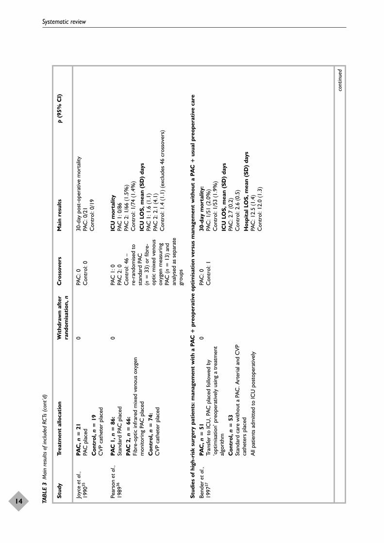
Systematic review
14 TA
BLE
3M
ain
resu
lts o
f inc
lude
d RC
Ts (
cont
’d)
Stud
yTr
eatm
ent
allo
cati
onW
ithd
raw
n af
ter
Cro
ssov
ers
Mai
n re
sult
sp
(95%
CI)
rand
omis
atio
n, n
Stud
ies
of h
igh-
risk
sur
gery
pat
ient
s: m
anag
emen
t w
ith
a PA
C +
pre
oper
ativ
e op
tim
isat
ion
vers
us m
anag
emen
t w
itho
ut a
PA
C +
usu
al p
reop
erat
ive
care
cont
inue
d
Joyc
e et
al.,
1990
35PA
C, n
= 2
1PA
C p
lace
d
Con
trol
, n=
19
CVP
cat
hete
r pl
aced
0PA
C: 0
Con
trol
: 030
-day
pos
t-op
erat
ive
mor
talit
yPA
C: 0
/21
Con
trol
: 0/1
9
Pear
son
et a
l.,19
8936
PAC
1, n
= 8
6:St
anda
rd P
AC
pla
ced
PAC
2, n
= 6
6:Fi
bre-
optic
infr
ared
mix
ed v
enou
s ox
ygen
mon
itorin
g PA
C p
lace
d
Con
trol
, n=
74:
CVP
cat
hete
r pl
aced
0PA
C 1
: 0PA
C 2
: 0C
ontr
ol: 4
6 –
re-r
ando
mise
d to
stan
dard
PA
C
(n=
33)
or
fibre
-op
tic m
ixed
ven
ous
oxyg
en m
easu
ring
PAC
(n=
13)
and
anal
ysed
as
sepa
rate
grou
ps
ICU
mor
talit
yPA
C 1
: 0/8
6PA
C 2
: 1/6
6 (1
.5%
)C
ontr
ol: 1
/74
(1.4
%)
ICU
LO
S, m
ean
(SD
) da
ysPA
C 1
: 1.6
(1.1
)PA
C 2
: 2.1
(4.1
)C
ontr
ol: 1
.4 (1
.1) (
excl
udes
46
cros
sove
rs)
Bend
er e
t al
.,19
9737
PAC
, n=
51
Tran
sfer
to
ICU
, PA
C p
lace
d fo
llow
ed b
y‘o
ptim
isatio
n’ p
reop
erat
ivel
y us
ing
a tr
eatm
ent
algo
rithm
Con
trol
, n =
53
Stan
dard
car
e w
ithou
t a
PAC
. Art
eria
l and
CVP
cath
eter
s pl
aced
All
patie
nts
adm
itted
to
ICU
pos
tope
rativ
ely
0PA
C: 0
Con
trol
: 130
-day
mor
talit
y:PA
C: 1
/51
(2.0
%)
Con
trol
: 1/5
3 (1
.9%
)
ICU
LO
S, m
ean
(SD
) da
ysPA
C: 2
.7 (0
.2)
Con
trol
: 2.6
(0.5
)
Hos
pita
l LO
S, m
ean
(SD
) da
ysPA
C: 1
2.5
(1.4
)C
ontr
ol: 1
2.0
(1.3
)

Health Technology Assessment 2006; Vol. 10: No. 29
15
© Queen’s Printer and Controller of HMSO 2006. All rights reserved.
TA
BLE
3M
ain
resu
lts o
f inc
lude
d RC
Ts (
cont
’d)
Stud
yTr
eatm
ent
allo
cati
onW
ithd
raw
n af
ter
Cro
ssov
ers
Mai
n re
sult
sp
(95%
CI)
rand
omis
atio
n, n
cont
inue
d
Berla
uk e
t al
.,19
9138
PAC
1, n
= 4
5Tr
ansf
er t
o IC
U, P
AC
pla
ced
follo
wed
by
‘tune
-up
’ tre
atm
ent
(usin
g pr
edef
ined
end
-poi
nts)
at
leas
t 12
h p
reop
erat
ivel
y
PAC
2, n
= 2
3Tr
ansf
er t
o an
aest
hetic
hol
ding
are
a, P
AC
plac
ed fo
llow
ed b
y ‘tu
ne-u
p’ t
reat
men
t (u
sing
pred
efin
ed e
nd p
oint
s) a
t le
ast
3 h
preo
pera
tivel
y.
Con
trol
, n=
21
Usu
al c
are
with
out
a PA
C. A
rter
ial a
nd C
VPca
thet
ers
plac
ed
All
patie
nts
adm
itted
to
ICU
pos
tope
rativ
ely
0PA
C 1
: 0PA
C 2
: 0C
ontr
ol: 1
Mor
talit
y (n
ot s
peci
fied)
PAC
1: 1
/45
(2.2
%)
PAC
2: 0
/23
Con
trol
: 2/2
1 (9
.5%
)
ICU
LO
S, m
ean
(SD
) da
ysPA
C 1
: 3.5
(2.0
)PA
C 2
: 2.5
(1.3
)C
ontr
ol: 2
.6 (2
.1)
Hos
pita
l LO
S, m
ean
(SD
) da
ysPA
C 1
: 19.
4 (1
1.6)
PAC
2: 1
8 (1
2.0)
Con
trol
: 15.
4 (7
.5)
0.08
(PA
C 1
and
PA
C2
vs c
ontr
ol)
Sand
ham
et
al.,
2003
41PA
C, n
= 9
97PA
C p
lace
d pr
ior
to s
urge
ry, f
ollo
wed
by
trea
tmen
t di
rect
ed t
o pr
edef
ined
phy
siolo
gica
lgo
als
Con
trol
, n=
997
Stan
dard
car
e w
ithou
t a
PAC
. Pla
cem
ent
ofC
VP c
athe
ter
perm
itted
All
patie
nts
adm
itted
to
ICU
pos
tope
rativ
ely
0PA
C: 5
8C
ontr
ol: 5
2H
ospi
tal m
orta
lity
PAC
: 78/
997
(7.8
%)
Con
trol
: 77/
997
(7.7
%)
Hos
pita
l LO
S, m
edia
n (I
QR
) da
ysPA
C: 1
0 (7
–15)
Con
trol
: 10
(7–1
5)
0.93
0.41
Shoe
mak
er
et a
l.,19
8839
PAC
con
trol
, n=
30
Tran
sfer
to
ICU
. PA
C p
lace
d fo
llow
ed b
yst
anda
rd m
anag
emen
t to
ach
ieve
nor
mal
val
ues
of h
aem
odyn
amic
and
oxy
gen
tran
spor
tva
riabl
es
PAC
pro
toco
l, n
= 2
8Tr
ansf
er t
o IC
U. P
AC
pla
ced
follo
wed
by
trea
tmen
t to
ach
ieve
sup
ra-n
orm
alha
emod
ynam
ic a
nd o
xyge
n tr
ansp
ort
valu
es
Con
trol
, n=
30
CVP
cat
hete
r pl
aced
. Sta
ndar
d ca
re
All
patie
nts
adm
itted
to
ICU
pos
tope
rativ
ely
3PA
C c
ontr
ol: 0
PAC
pro
toco
l: 0
Con
trol
: 17
Post
oper
ativ
e m
orta
lity
(not
spe
cifie
d)PA
C c
ontr
ol: 1
0/30
(33%
)PA
C p
roto
col:
1/28
(3.6
%)
Con
trol
: 7/3
0 (2
3%)
ICU
LO
S, m
ean
(SD
) da
ysPA
C c
ontr
ol: 1
5.8
(3.1
)PA
C p
roto
col:
10.2
(1.6
)C
ontr
ol: 1
1.5
(1.7
)
Hos
pita
l LO
S, m
ean
(SD
) da
ysPA
C c
ontr
ol: 2
5.2
(3.4
)PA
C p
roto
col:
19.3
(2.4
)C
ontr
ol: 2
2.2
(2.8
)
<0.
01 (P
AC
pro
toco
lvs
PA
C c
ontr
ol)
<0.
10 (P
AC
pro
toco
lvs
con
trol
)<
0.20
(PA
C c
ontr
ol v
sco
ntro
l)
<0.
05 (P
AC
pro
toco
lvs
PA
C c
ontr
ol)
Not
giv
en

Systematic review
16 TA
BLE
3M
ain
resu
lts o
f inc
lude
d RC
Ts (
cont
’d)
Stud
yTr
eatm
ent
allo
cati
onW
ithd
raw
n af
ter
Cro
ssov
ers
Mai
n re
sult
sp
(95%
CI)
rand
omis
atio
n, n
CVP
cat
hete
r, ce
ntra
l ven
ous
pres
sure
cat
hete
r (s
ee A
ppen
dix
1 fo
r de
scrip
tion)
; IQ
R, in
ter-
quar
tile
rang
e; L
OS,
leng
th o
f sta
y.
Vale
ntin
e et
al.,
1998
40PA
C, n
= 6
0Tr
ansf
er t
o IC
U, P
AC
pla
ced
follo
wed
by
‘tune
-up
’ tre
atm
ent
(usin
g pr
edef
ined
end
-poi
nts
used
by
Berla
uk e
t al
.38) a
t le
ast
14 h
preo
pera
tivel
y
Con
trol
, n=
60
Not
tra
nsfe
rred
to
ICU
, CVP
cat
hete
r pl
aced
and
no s
peci
fic p
reop
erat
ive
trea
tmen
t
All
patie
nts
adm
itted
to
ICU
pos
tope
rativ
ely
2PA
C: 0
Con
trol
: 2 (e
xclu
ded
from
out
com
ean
alys
is)
Hos
pita
l mor
talit
yPA
C: 3
/60
(5.0
%)
Con
trol
: 1/6
0 (1
.7%
)
ICU
LO
S (p
osto
pera
tive
),
mea
n (S
D)
days
PAC
: 8 (1
)C
ontr
ol: 7
(1)
Hos
pita
l LO
S, m
ean
(SD
) da
ysPA
C: 1
3 (2
)C
ontr
ol: 1
3 (2
)
Repo
rted
as
not
signi
fican
t
Repo
rted
as
not
signi
fican
t
Repo
rted
as
not
signi
fican
t
Stud
yTr
eatm
ent
n/N
Con
trol
n/N
OR
(ra
ndom
)95
% C
IW
eigh
t%
OR
(ra
ndom
)95
% C
I
Guy
att 1
99117
Rhod
es e
t al.
2002
32
Rich
ard
et a
l. 20
0333
Tota
l (95
% C
I)To
tal e
vent
s: 2
55 (T
reat
men
t), 2
67 (C
ontr
ol)
Test
for
hete
roge
neity
: �2
= 0
.43,
df =
2 (p
= 0
.81)
, I2
= 0
%Te
st fo
r ov
eral
l effe
ct z
= 0
.24
(p =
0.8
1)
10/1
646
/96
199/
335
447
9/17
50/1
0520
8/34
1
463
3.62
22.7
673
.62
100.
00
1.48
1.01
0.94
0.97
(0.3
7 to
5.9
5)(0
.58
to 1
.76)
(0.6
9 to
1.2
7)
(0.7
4 to
1.2
6)
0.1
Favo
urs
trea
tmen
tFa
vour
s co
ntro
l0.
25
100.
51
2
FIG
UR
E 2
Met
a-an
alys
is 1:
man
agem
ent
with
a P
AC v
ersu
s m
anag
emen
t w
ithou
t a
PAC:
stu
dies
of g
ener
al IC
U p
atie
nts

compared with the control group (53%) based onintention-to-treat (Table 3). However, of the 17patients allocated to the control group, eight weresubsequently managed with a PAC and, of these,seven (88%) died.
Data from the three studies (910 patients) werepooled to give an OR of 0.97 (95% CI 0.74 to1.26), comparing management with a PAC tomanagement without a PAC, based on intention-to-treat (Figure 2).
Studies of high-risk surgery patients that did notinclude preoperative optimisation as part of theintervention (n = 3)One study34 reported hospital mortality. In thisstudy, one death occurred in the PAC groupcompared with none in the control group (Table 3).Joyce and colleagues35 examined 30-day mortalityand reported no deaths in either treatment group(Table 3). Pearson and colleagues36 reported nodifference in ICU mortality between the groups.Overall, there were two deaths, one in thecontinuous mixed venous oxygen monitoring PACgroup and one in the ‘control group’ who werereassigned following randomisation to bemanaged with a standard PAC (Table 3).
Data from the three studies (368 patients) werepooled to give an OR of 1.10 (95% CI 0.13 to9.06) comparing management with a PAC tomanagement without a PAC, based on intention-to-treat (Figure 3). One study36 had two PACgroups, which were combined for the pooledanalysis.
Studies of high-risk surgery patients that includedpreoperative optimisation as part of theintervention (n = 5)Both Valentine and colleagues40 and Sandham andcolleagues41 reported no significant difference inhospital mortality between the treatment groups(Table 3). Bender and colleagues37 reported nodifference in 30-day mortality (Table 3). The otherstudies38,39 did not specify the type of mortalitystatistic. Of these, one38 reported no difference inmortality, whereas the other39 reported asignificant difference in mortality between the PACplus protocol group and the PAC plus standardcare group (p < 0.01). However, there was nosignificant difference in mortality between thecontrol group and either the PAC plus protocolgroup or the PAC plus standard care group (Table 3).
Data from the five studies (2395 patients) werepooled to give an OR of 0.98 (95% CI 0.72 to
1.33) comparing management with a PAC pluspreoperative optimisation with usual preoperativecare and management without a PAC, based onintention-to-treat. Two studies38,39 had two PACgroups, which were combined for the pooledanalysis (Figure 4).
Length of stay in ICUSix studies17,34,36,37–39 reported the mean length ofstay in ICU for survivors and non-survivorscombined. Two studies reported either the mean17
or the median32 length of stay in ICU for survivorsonly. Details, including p-values (if reported), aregiven in Table 3. All but one of the studies39
reported no significant difference in ICU length ofstay between the groups. Of note, control groupdata from Pearson and colleagues’ study36 excludethe 46 patients who were reassigned followingrandomisation. The mean and standard deviation(SD) values for these patients were reportedseparately, as follows: reassigned to managementwith standard PAC (n = 33), 2.8 (5.0) days in ICU;reassigned to management with mixed venousoxygen PAC (n = 13), 2.6 (3.8) days in ICU.Shoemaker and colleagues39 reported that patientsin the PAC plus standard care group spentsignificantly longer in the ICU than patients inthe PAC plus protocol group (p < 0.05) (Table 3).
Length of stay in hospitalFive studies34,37–40 reported the mean and one41
reported the median length of stay in hospital forsurvivors and non-survivors combined. Rhodesand colleagues32 reported the median length ofstay for survivors only. None of the studies found asignificant difference between the groups (Table 3).
CostFour studies reported costs of care.34,36,38,40 Thesestudies were all conducted in the USA and usedhospital charges as a measure of costs of care(Table 4). None of the studies reported significantdifferences in the cost of care for patients in thePAC group compared with patients in the controlgroup. In addition to the total hospital charges,Isaacson and colleagues34 reported professionalfees charged by the anaesthesiologists per patientin each group and found that fees weresignificantly higher per patient in the PAC groupthan the control group (p = 0.0001), as would beexpected in a fee-for-service system (Table 4).
DiscussionEleven trials17,32–41 were identified comparingpatients in intensive care managed with and
Health Technology Assessment 2006; Vol. 10: No. 29
17
© Queen’s Printer and Controller of HMSO 2006. All rights reserved.

Systematic review
18
Stud
yTr
eatm
ent
n/N
Con
trol
n/N
OR
(ra
ndom
)95
% C
IW
eigh
t%
OR
(ra
ndom
)95
% C
I
Pear
son
et a
l. 98
936
Isaa
cson
et a
l. 19
9034
Joyc
e et
al.
1990
35
Tota
l (95
% C
I)To
tal e
vent
s: 2
(Tre
atm
ent)
, 1 (C
ontr
ol)
Test
for
hete
roge
neity
: �2
= 0
.79,
df =
1 (p
= 0
.37)
, I2
= 0
%Te
st fo
r ov
eral
l effe
ct z
= 0
.09
(p =
0.9
3)
1/15
21/
49
0/21
22
2
1/74
0/53
0/19
1
46
57.2
542
.75
100.
00
0.48
3.31
1.10
(0.0
3 to
7.8
4)(0
.13
to 8
3.17
)N
ot e
stim
able
(0.1
3 to
9.0
6)
0.1
Favo
urs
trea
tmen
tFa
vour
s co
ntro
l0.
25
100.
51
2
FIG
UR
E 3
Met
a-an
alys
is 2:
man
agem
ent
with
a P
AC v
ersu
s m
anag
emen
t w
ithou
t a
PAC:
stu
dies
of h
igh-
risk
surg
ery
patie
nt
Stud
yTr
eatm
ent
n/N
Con
trol
n/N
OR
(ra
ndom
)95
% C
IW
eigh
t%
OR
(ra
ndom
)95
% C
I
Shoe
mak
er e
t al.
1988
39
Berla
uk e
t al.
1991
38
Bend
er e
t al.
1997
37
Vale
ntin
e et
al.
1998
40
Sand
ham
et a
l. 20
0341
Tota
l (95
% C
I)To
tal e
vent
s: 9
4 (T
reat
men
t), 8
8 (C
ontr
ol)
Test
for
hete
roge
neity
: �2
= 3
.60,
df =
4 (p
= 0
.46)
, I2
= 0
%Te
st fo
r ov
eral
l effe
ct z
= 0
.12
(p =
0.9
0)
11/5
8 1
/68
1/5
1 3
/60
78/
997
12
34
7/30
2/21
1/53
1/60
77/9
97
116
1
8.18
1.56
1.20
1.79
87.2
8
100.
00
0.77
0.14
1.04
3.11
1.01
0.98
(0.2
6 to
2.2
4)(0
.01
to 1
.65)
(0.0
6 to
17.
08)
(0.3
1 to
30.
73)
(0.7
3 to
1.4
1)
(0.7
2 to
1.3
3)
0.1
Favo
urs
trea
tmen
tFa
vour
s co
ntro
l0.
25
100.
51
2
FIG
UR
E 4
Met
a-an
alys
is 3:
man
agem
ent
with
PAC
+ p
reop
erat
ive o
ptim
isatio
n ve
rsus
man
agem
ent
with
out
PAC
+ u
sual
pre
oper
ative
car
e: s
tudi
es o
f hig
h-ris
k su
rger
y pa
tient
s

without a PAC. Of these, only three investigatedthe clinical effectiveness of PACs in themanagement of general intensive carepatients.17,32,33 The remaining eight studies wereof high-risk surgical patients and, of these, fivewere trials investigating whether preoperativeoptimisation improves patient outcome.37–41
Hence placement of a PAC was part of anenhanced package of care that also includedadmission to ICU preoperatively and‘optimisation’ of haemodynamic variables topredetermined goals prior to surgery. Overall,regardless of the patient population studied orinclusion of additional interventions, there was noimprovement in patient outcomes as a result ofmanagement with a PAC.
Our results are similar to those of previous reviewsinvestigating the effectiveness of PACs.15,68 Cooperand colleagues15 included both randomised andnon-randomised studies of critically ill patients inintensive care but restricted their selection ofstudies to those published in English and thosewhere PAC-derived haemodynamic data were usedto alter therapy. They found that there was verylittle evidence from RCTs to support the use of thePAC in clinical practice. Barone and colleagues68
restricted inclusion of studies in their review toRCTs published in English that described specifictherapeutic goals and that were confined tovascular surgery patients. Four papers wereidentified and included in the meta-analysis,
which showed no difference between patientsmanaged with a PAC and those managed without.For the current review, we restricted inclusion ofstudies to those that had used an RCT design.However, we minimised exclusion criteria; forexample, there was no restriction on language orachieving specific therapeutic goals based on PAC-derived data. This was to ensure all RCTscomparing management with and without a PACinvolving critically ill patients in intensive carewere included in the review. As a result, the studiesidentified were of different ICU populations withdifferent outcomes and testing differenthypotheses. Hence it was not possible to combinedata from all studies into one meta-analysis.Therefore, three separate meta-analyses wereconducted to investigate the impact ofmanagement with a PAC on mortality in differentpatient populations.
The majority of studies identified in this reviewwere relatively small and were designed toinvestigate perioperative monitoring, with orwithout preoperative optimisation ofhaemodynamics, in patients undergoing high-risksurgery. The main barrier to an effectiveevaluation of PACs in the management of generalintensive care patients has been the lack ofequipoise amongst intensive care clinicians, whichwas one of the main reasons for eligible patientsbeing excluded from the Canadian trial.17 Ourinitial search of the literature (to June 2001)
Health Technology Assessment 2006; Vol. 10: No. 29
19
© Queen’s Printer and Controller of HMSO 2006. All rights reserved.
TABLE 4 Main results of cost evaluations
Study Measure Main resultsa
Studies of high-risk surgery patients: management with a PAC versus management without a PACIsaacson et al., 199034 Total hospital charges per patient, mean (SD) PAC: $16,680 (9,108)
Control: $15,813 (9,028)Anaesthesiologist’s fee per patient, mean (SD) PAC: $1,739 (225)
Control: $1,551 (252)
Pearson et al., 198936 Total costs (billed to patient), mean (SD) PAC 1: $855.51 (231)PAC 2: $1128.38 (759)Control: $591.19 (68)b
Studies of high-risk surgery patients: management with a PAC + preoperative optimisation versusmanagement without a PAC + usual pre-operative careBerlauk et al., 199138 Total hospital charge, mean (SD) PAC 1: $29,102 (13,207)
PAC 2: $23,770 (12,418)Control: $23,386 (12,303)
Shoemaker et al., 198839 Hospital charges, average (not specified) PAC control: $37,335PAC protocol: $27, 665Control: $30,748
a Costs in US$.b The costs given for the control group excluded the 46 patients who reassigned after randomisation, which were as
follows; reassigned to management with standard PAC (n = 33): mean total cost, $986.38 (578); reassigned tomanagement with mixed venous oxygen PAC (n = 13): mean total cost, $1126.38 (382).

revealed that this was the only RCT of generalintensive care patients that had been conducted. It was unfortunately discontinued prematurelybecause of poor recruitment of patients. Theremaining seven studies identified were RCTsinvolving high-risk surgery patients.
Publication of the non-randomised study byConnors and colleagues in 1996,18 suggesting thatmanagement using a PAC may be harmful topatients, has, it would seem, established clinicalequipoise within the critical care community. Sincethen, in addition to the trial reported below, twomore trials involving general intensive carepatients have been published and were identifiedin the updated literature searches in 2003. Thefirst was a single-centre trial in the UK32 whichrecruited 201 patients with haemodynamic shockand was intended as a pilot study to inform futureRCTs. The second was a large, multi-centre, RCT,conducted in France, which recruited 676 patientswith acute respiratory distress syndrome andshock.33 However, it was not sufficiently poweredto detect a 10% absolute difference in 28-daymortality. In addition, another RCT involvinghigh-risk surgery patients has also been publishedby Sandham and colleagues,41 which is the largestRCT to date investigating the effectiveness ofpreoperative optimisation using a PAC.
Few studies have included a cost-effectivenessevaluation as part of an assessment of PACs. Weidentified four studies,34,36,38,39 all conducted inhospitals in the USA, that included a cost
component. These were all based on the totalhospital costs charged to patients. One of theweaknesses of this approach is that specific chargesvary across hospitals; for example, patients may ormay not be charged for the cost of dailymonitoring using a PAC. In general, higher totalcosts were reported for patients managed withPACs compared with those managed without,although these were not statistically significant.
ConclusionsThe initial search of the literature revealed thatthere was very little RCT evidence on theeffectiveness of PACs in clinical practice. One RCTinvestigating the management of general intensivecare patients using a PAC and seven studiesinvestigating management of high-risk surgerypatients were identified. These were all small,mainly single-centre studies. In addition, althoughsome studies included an evaluation of costs ofcare, again these were small studies, which wereconducted in the USA and were based on hospitalcharges. This review identified a clear need for alarge, multi-centre RCT of the clinical and cost-effectiveness of PACs in the management ofpatients in general intensive care. The currentRCT was therefore designed to address a numberof issues, including the range of patients managedin intensive care with a PAC and the lack ofconsensus within the critical care communityregarding management protocols. The trial isdescribed in detail in Chapter 3.
Systematic review
20

AimThe aim of the RCT was to test the hypothesisthat hospital mortality is decreased in critically illpatients in adult ICUs who are managed with aPAC compared with those who are not.
Design and development of thetrial protocolThere were a number of potential problems thathad to be addressed in the design of the trial. Animportant issue was a lack of consensus amongcritical care clinicians as to the indications forinsertion of a PAC and how patients should besubsequently managed following its placement,including administration of fluids and vasoactivedrugs and target levels for blood pressure andcardiac output. Also, PACs were widely used inICUs in the UK across a range of patients and bydoctors and nurses with varying levels of expertise.Given the lack of consensus, it was feltinappropriate to introduce a management protocolfor the trial, particularly as it would have becomean evaluation of the PAC plus the protocol ratherthan the PAC itself. Furthermore, the results wouldnot have been generalisable outside the patientgroup treated according to the protocol. Therefore,a multi-centre, pragmatic RCT design was adoptedto investigate the effectiveness of PACs as they arecurrently used in the UK. In brief, a study designwas chosen to answer the question, ‘What would bethe effect on hospital mortality of removing PACsfrom clinical use?’
The UK critical care community, in addition tointernational experts, were consulted and asked toprovide input into the development of the trialprotocol. In 2000, representatives from all general,adult ICUs in the UK were invited to aCollaborators’ Meeting to discuss the draft trialprotocol. One of the main talking points of themeeting was whether or not less invasivemonitoring devices should be allowed as analternative to the PAC. Such devices have become
increasingly popular and a large proportion ofICUs were now using them, either as analternative or as an adjunct to the PAC. At least50% of clinicians argued that to participate in thetrial, they would have to be allowed the option touse alternative devices to measure cardiac outputin patients allocated to the control group, that is,those not to be managed with a PAC. In otherwords, the PAC itself would be ‘on trial’. Othersargued that the trial should be an evaluation ofthe PAC versus no other form of cardiac outputmonitoring, thus examining the impact ofmonitoring cardiac output. These views wereincorporated into the design of the trial, andparticipating ICUs elected to be in one of twostrata as follows:
● stratum A: no option to use alternative devicesto measure cardiac output in control grouppatients
● stratum B: option to use alternative devices tomeasure cardiac output in control grouppatients.
The alternative devices available to monitor thecardiovascular system and measure cardiac outputare described in Appendix 1. At the start of thetrial, it was anticipated that most ICUs usingdevices other than the PAC to measure cardiacoutput would be using pulse contour or trans-oesophageal Doppler devices. The use of centralvenous pressure (CVP) catheters is ubiquitous inintensive care patients and was permitted in anunrestricted manner for all patients in the trial.Echocardiography is a technique used fordiagnostic rather than monitoring purposes andwas also permitted in an unrestricted manner inthe trial.
MethodsThe final trial protocol was approved by theLondon Multi-centre Research Ethics Committee(MREC) and each participating hospital’s LocalResearch Ethics Committee (LREC).
Health Technology Assessment 2006; Vol. 10: No. 29
21
© Queen’s Printer and Controller of HMSO 2006. All rights reserved.
Chapter 3
Randomised controlled trial to evaluate the clinical effectiveness of pulmonary artery catheters in
patient management in intensive care

ICU recruitmentA database of all adult general ICUs in the UKhas been constructed by ICNARC using TheDirectory of Critical Care (formerly The Directoryof Emergency and Special Care Units) producedand published by CMA Medical Data, and throughdirect contact with ICUs. A postal invitation toparticipate in the trial was sent to the ClinicalDirector at every general, adult ICU listed in thisdatabase (n = 263). Follow-up letters were sent tonon-responders. ICUs that expressed an interestin participating were contacted by telephone andvisited by a member of the study team. Aprerequisite for an ICU to participate in the trialwas that all consultants should agree to include alleligible patients and to abide by randomisation tominimise selective enrolment and crossover ofpatients.
A Local Investigator, who was responsible for theconduct of the trial locally, was identified at eachICU. Another contact, usually a nurse or a datacollection clerk, was also identified to act as anadditional link between the ICU and the Trial Coordinating Centre. Once approval had beenobtained from the relevant LREC and thehospital’s Research and Development Department,one of the Trial Research Nurses visited the ICU.The visit typically involved a presentation to thestaff describing the background, aims andmethods of the trial followed by discussion andquestions. The Trial Research Nurse checked thatall trial materials were on site and went throughthe enrolment and randomisation procedures andthe data collection forms with the ICU staff.Follow-up visits were arranged as necessary tomonitor progress, maintain awareness of the trialand encourage ongoing participation.
NewslettersDuring the course of the trial, monthly newsletterswere sent to all ICUs in the UK to keep theminformed of progress, to encourage participationand to remind Local Investigators to enrol alleligible patients.
Collaborators’ MeetingsDuring the patient recruitment phase of the trial,representatives from all participating ICUs wereinvited to two Collaborators’ Meetings. These wereto keep collaborators informed of study progress,to encourage continued participation and toprovide an opportunity to discuss any issues withthe conduct of the trial locally. An additionalCollaborators’ Meeting was convened to presentthe results of the PAC-Man Study torepresentatives from participating ICUs.
Support costsThe ICUs were offered no financial incentives totake part in the trial but were reimbursed £15 perpatient recruited as a contribution towards theadministration costs of the trial.
Trial participantsThe trial population comprised general intensivecare patients fulfilling the eligibility criteriaoutlined below.
Inclusion and exclusion criteriaPatients were eligible for inclusion in the trial ifthey were intensive care patients deemed by theresponsible treating clinician to requiremanagement with a PAC. Patients were excluded ifthey were less than 16 years of age, were admittedto the ICU electively prior to surgery forpreoperative optimisation, had a PAC already in situon admission to the ICU, had previously beenentered into the trial or had been declared braindead following brain stem death testing, with a PACbeing placed for organ support prior to donation.
Patient consentWhere possible, patients were asked to give theirinformed consent prior to randomisation. TheLocal Investigator discussed the trial with thepatient and gave them an information sheet toread (Appendix 4). The patient then signed aconsent form indicating their agreement to takepart (Appendix 5). However, in most cases this wasnot possible because PACs are frequently used inthe most severely ill patients who have alteredconsciousness. In these cases, where possible, theLocal Investigator discussed the trial with thepatient’s relatives and gave them an informationsheet explaining that although they could notprovide consent on behalf of the patient, theycould offer an opinion as to whether the patientwould object to taking part in medical research(Appendix 6). The relative was then asked toprovide signed assent in the form of a writtenagreement, prior to randomisation (Appendix 7).If the relatives could not be contacted, the patientwas randomised and signed assent was obtainedfrom the relative as soon as possible, retrospectively.If the patient regained mental competency, thepatient was informed of their enrolment into thetrial and given an information sheet to read(Appendix 8). The patient was then asked to signa consent form indicating agreement to the use oftheir data in the trial analysis (Appendix 5).
Randomisation and allocationRandomisation was carried out via an independent,central, 24-hour telephone randomisation service.
Randomised controlled trial to evaluate the clinical effectiveness of pulmonary artery catheters
22

A study investigator was available 24 hours perday to answer questions relating to enrolment andrandomisation. To randomise a patient, the LocalInvestigator telephoned the randomisation centreand provided basic identification details,confirmed the patient’s eligibility for the trial andprovided data for minimisation. Patients weregiven a unique study number and were assignedon a 1:1 allocation to be managed either with orwithout a PAC. Treatment was assigned by theminimisation method to balance across thefollowing four factors: individual ICU; age group(16–44 years, 45–64 years, 65 years and above);the presumptive clinical syndrome (acuterespiratory failure, multi-organ dysfunction,decompensated heart failure, other clinicalsyndrome); and surgical status (elective surgical,emergency surgical, non-surgical).
Patients allocated to, according to local practice,be managed with a PAC had the catheter placed assoon as possible following randomisation. ThePAC remained in place for as long as theresponsible treating clinician considered necessary.Patients allocated to the control group weremanaged without a PAC with the option to usealternative cardiac output monitoring technologiesin ICUs that elected to be in stratum B. In bothgroups, clinical management followingrandomisation was at the discretion of theresponsible treating clinician. Participating ICUsmaintained a screening log of eligible patients notenrolled into the trial, recording the date ofeligibility for the trial, date of birth, sex andreason for exclusion.
Sample size calculationThe original sample size calculation was based ondata kindly supplied by the Scottish Intensive CareSociety Audit Group (personal communication) in1999 from their database containing details of allpatients admitted to Scottish ICUs. The datasuggested a hospital mortality of 50% for patientsmanaged with a PAC. Since it was felt that a 5%change in hospital mortality would represent aclinically important finding, we wished to be ableto detect a reduction in hospital mortality from55%, for patients not managed with a PAC, to 50%(with the potential risk of a rise to 60%) based ona 5% level of statistical significance for a two-sidedtest. It was calculated that a sample of 4184patients (2092 in each arm) would have 90%power to detect a difference of this size. Weallowed for a 4% non-compliance rate in the PACgroup and 8% in the control group and a 5% lossto follow-up, which increased the required samplesize to 5673.
At the point where we estimated our originalsample size, approximately 18% of admissions toUK ICUs were being managed with a pulmonaryartery catheter.70 However, the results of theConnors and colleagues’ study (39% increasedodds in hospital mortality associated withmanagement with a PAC)18 caused a precipitousreduction in the use of PACs (recent unpublisheddata suggest that approximately 3% of admissionsare now managed with a PAC) and a rapid uptakeof less invasive cardiac output monitoringtechnologies. The consequence of this was slowpatient recruitment to the trial. In response to this,the Trial Steering Committee agreed that the TrialStatistician should examine the characteristics(severity of illness) and actual hospital mortality ofcontrol group patients recruited at that point (n = 147). These results, combined with anecdotalevidence from clinicians, strongly suggested thatclinical practice for managing patients with PACshad changed with their use being restricted tofewer and more severely ill patients. Indeed, theactual hospital mortality for these patients wasmuch higher, 69% (95% CI 61 to 77%) than thatoriginally incorporated (50%) into the sample sizecalculation. Despite this change in clinical practice,the hypothesis being tested in the RCT remainedvalid. The Trial Steering Committee decided torecalculate the sample size, increasing the clinicallyimportant difference from 5 to 10%. This decisionwas based on the changes in clinical practice, thehigher actual hospital mortality and pragmatism,that is, an achievable sample size. The reduction inrelative hospital mortality thus changed from 10 to14.5%. It was calculated that a sample of 992patients (496 in each arm) would have 90% powerto detect a 10% change in hospital mortality basedon a 5% level of significance for a two-sided test.We allowed for a 4% non-compliance rate in thePAC group and 8% in the control group, whichincreased the sample size to 1281 patients. Sincethe follow-up ended at hospital discharge, allowinga 5% loss to follow-up was considered unnecessary.We felt confident that the results of the PAC-ManStudy would remain meaningful in light of theresults of Connors and colleagues’ study.18
Changes to the trial protocol There were two amendments to the original trialprotocol approved by the MREC on 12 March2001. The first amendment, on 27 September2002, reflected the change to the sample sizecalculation and is described above. The secondamendment dealt with the issue of patients whoregained consciousness following their criticalillness but did not regain mental competency to beable to provide informed consent. The exclusion
Health Technology Assessment 2006; Vol. 10: No. 29
23
© Queen’s Printer and Controller of HMSO 2006. All rights reserved.

criteria (described above) were amended in orderto exclude patients admitted to the ICU where thespecific reasons for admission, or othercircumstances, suggested that they might later beunable to give consent on recovery; for example,mental handicap or learning difficulties. Thepatient consent procedure was also amended sothat for a patient who regained consciousness butdid not regain sufficient mental competency tounderstand the purpose and consequences ofparticipating in the study nor, in the opinion ofthe Local Investigator (ICU Consultant), was likelyto do so, a ‘Patient Incapacity to ProvideRetrospective Consent’ form (Appendix 9) wascompleted and a copy sent to the Chair of theLREC. Following notification to the LREC, and inthe absence of any objection from it, datacollection continued and the data were submittedfor inclusion in the trial analysis without thepatient’s consent unless or until they regainedmental competence. The patient’s next of kin,where available, were asked if they knew of anyreason why the patient would not want their datato be used in this way. If a reason was identified,the patient was excluded from the trial. TheLRECs were informed of these amendments to thetrial protocol following approval by the MREC.
DataGiven the limited resources available to ICUs forthe conduct of research, data collection for thetrial was kept to a minimum and restricted wherepossible to data that are recorded routinely inmost ICUs (Appendix 10).
At study entry, prior to randomisation, baselinedata recorded included sex, date of birth, sourceof admission (surgical/non-surgical) andpresumptive clinical syndrome. At the time ofrandomisation, the primary reason for wanting tomanage the patient with a PAC and the currentorgan monitoring/support, based on the criteriaused for the Augmented Care Period (ACP)dataset71 (Appendix 11), were collected. Inaddition, raw clinical data for the Sepsis-relatedOrgan Failure Assessment (SOFA) score72
(Appendix 12) were collected to provideinformation on the patient’s severity of illness atthe time they were being considered for a PAC.During the first 24 hours in the ICU, raw clinicaldata for the Acute Physiology And Chronic HealthEvaluation version II (APACHE II) severity scoringsystem73 (Appendix 13) were collected.
Post-randomisation use of alternative cardiacoutput monitoring devices and type of organmonitoring/support based on the criteria used for
the ACP dataset71 (Appendix 11) were collecteddaily during the patient’s stay in the originalrecruiting ICU. At discharge from the originalrecruiting ICU, the patient’s outcome (alive/dead)was recorded and the date and time of discharge ordeath were recorded. Patients were followed upuntil discharge from, or death in, an acute hospital.
For patients allocated to the PAC group,complications occurring as a direct result ofinsertion of the PAC, changes in managementwithin the first 2 hours as a direct result of PAC-derived data, duration of management with theinitial PAC and overall management with a PACwere also recorded.
Primary and secondary outcomemeasuresThe primary outcome was hospital mortality,defined as death from any cause before ultimatedischarge from an acute hospital. ICU mortality,defined as death from any cause before dischargefrom the original recruiting ICU, and mortality at 28 days post-randomisation were also recordedfor comparability with other studies. Secondaryoutcomes were the length of stay in the originalICU, total length of stay in an acute hospital and organ-days of support in the originalrecruiting ICU.
Follow-upAll patients were followed up to hospital discharge,the study end-point. Enrolment of patients into thetrial ended on 31 March 2004. Data collection,including follow-up of patients, continued until 30June 2004. Data on patients who remained in anacute hospital at this point were censored.
Data preparationAll data were double entered and validated in arelational database (Microsoft Access 2000) andwherever possible variables were given numericcodes. Data were exported to the statisticalpackage Stata 8.2 (StataCorp, College Station, TX, USA) for statistical analysis.
Data integrityAll data were checked for inconsistencies, forphysiological variables outside predeterminedranges and for missing data, which were followedup with Local Investigators.
Data analysisAll the main analyses used the intention-to-treatprinciple, considering the groups as randomised,and followed a predetermined statistical analysisplan.
Randomised controlled trial to evaluate the clinical effectiveness of pulmonary artery catheters
24

Representativeness of participating ICUsParticipating ICUs were compared with adultgeneral ICUs participating in the ICNARC CaseMix Programme (CMP), the national comparativeclinical audit of patient outcome from intensivecare.74 Currently, 73% (n = 181) of adult generalICUs in England, Wales and Northern Irelandtake part in the CMP (Scotland has its ownperformance assessment programme).Comparison was by hospital type (university,university affiliated, non-university) and size ofunit, that is, number of critical care beds.
Representativeness of participantsEligible participants and non-participants werereported according to the Consolidated Standardsof Reporting Trials (CONSORT) statement.75 Trialparticipants were compared with eligible non-participants (recorded in the screening logs atparticipating ICUs) for age and sex.
Comparability of the groupsThe two groups (PAC and control) were comparedat entry for general baseline characteristics [age,sex and stratum, (A or B)], for baselinecharacteristics at the time of admission to ICU[surgical status – medical, elective surgery,emergency surgery, APACHE II acute physiologyscore (APS), APACHE II score, risk of deathpredicted by APACHE II and confirmed infection)and for baseline characteristics at the time ofrandomisation (time in hours to randomisationpost-ICU admission, major presumptive clinicalsyndrome – acute respiratory failure, multi-organdysfunction, decompensated heart failure andother, and SOFA score). Numbers and proportionsor means with SDs were reported as appropriatein each group. In line with currentrecommendations, statistical tests comparingbaseline characteristics between the two groupswere not performed.75,76
Main analysesThe numbers of deaths in each group werecompared with Fisher’s exact test. Survival timeswere compared with Kaplan–Meier curves andtested with a log-rank test, for all patients and bystratum. For the purpose of in-hospital survivalanalyses, patients discharged from an acutehospital alive were assumed to survive until theend of the follow-up period (3 months afterpatient recruitment ended). Data from patientsstill in an acute hospital at this point werecensored. The hazard ratio for management with aPAC compared with no PAC was calculated from aCox proportional hazards model, both with andwithout adjustment for prognostic factors (age,
sex, surgical status, major presumptive clinicalsyndrome at time of randomisation, SOFA score attime of randomisation and APACHE II scoreduring the first 24 hours in ICU).
Secondary analysesHospital mortality was also assessed inpredetermined subgroups based on option or notto use alternative cardiac output monitoringtechnologies in the control group, APACHE II-predicted risk of hospital death, presumptiveclinical syndrome and historic frequency of PACuse in the participating ICU derived from the1998 Audit Commission survey.70 The subgroupswere compared by testing the interaction withtreatment effect in the adjusted Cox proportionalhazards model. For APACHE II-predicted risk ofhospital death, predicted log odds of death wereentered as a linear term in the Cox model.
A secondary analysis to account for non-compliance (PAC patients not receiving PAC) andcontamination (control patients receiving PAC) wasundertaken using the methods described by Cuzickand colleagues.77 The treatment effect in complierswas estimated as the relative risk with 95% CI.
Secondary outcomesDifferences between the two groups in the medianlength of stay in the original recruiting ICU andin an acute hospital were compared using theWilcoxon rank-sum test. A bootstrap t-test78 wasundertaken to estimate the difference in meanlength of stay in ICU and an acute hospitalbetween the two groups. Organ-days of supportwere calculated as the sum of the days ofindividual organ support. For example, onecalendar day where three individual organs weresupported was equivalent to three calendar dayswhere one individual organ was supported. Themean organ-days of support were comparedbetween the two groups using a bootstrap t-test.
Complications and management of patients inthe PAC groupComplications and changes in managementfollowing insertion of the PAC were described andthe numbers (%) tabulated. The duration ofmanagement with the initial PAC was calculatedfrom the dates and times of insertion and removal.The overall number of days of management with aPAC following randomisation was calculated fromdaily follow-up data.
Use of alternative cardiac monitoring devices The use of alternative cardiac output monitoringdevices was reported by treatment group
Health Technology Assessment 2006; Vol. 10: No. 29
25
© Queen’s Printer and Controller of HMSO 2006. All rights reserved.

(PAC and control) and by stratum, including non-compliance within strata.
An independent Data Monitoring and EthicsCommittee conducted two interim safety analysesand at both points recommended that the trialcontinue.
ResultsRecruitment of ICUsRecruitment of ICUs took place between July 2000and December 2003. Ninety-seven ICUs expressedan interest in taking part in the trial and were sentan LREC application pack. Of these, 79 ICUssought and gained approval from their LRECs.The remaining ICUs decided not to participatefor a variety of reasons, most frequently becausethere was lack of equipoise within the ICU orthere were limited resources available for researchactivities. A total of 65 ICUs recruited one or morepatients (Figure 5). Of these, 55 were in England,four in Northern Ireland, three in Scotland andthree in Wales. The characteristics of participating
ICUs were similar to those of ICUs in the CMP.Thirteen (20%) of the participating ICUs werelocated in university hospitals, eight (12.3%) inuniversity-affiliated hospitals and 44 (67.7%) innon-university hospitals (Table 5). The number ofbeds in an individual ICU ranged from four to 22beds. Ten ICUs (15.4%) had five beds or less, 40(61.5%) had between six and 10 beds, 14 (21.5%)had between 11 and 15 beds and one (1.5%) had22 beds (Table 6).
Twenty-two (34%) ICUs opted to be in stratum A and 43 (66%) in stratum B. ICUs were not asked to give reasons for their choice of stratum.Ten of the 13 ICUs located in university hospitalsopted to be in stratum B. Of the 22 ICUs instratum A, five (two located in university hospitalsand three in non-university hospitals) acquired an alternative cardiac output monitoring deviceduring the patient recruitment period and crossed over to stratum B (Table 7). Patients inthese units were considered to be in stratum A or B depending on the date on which they were randomised, that is, before or after thecrossover.
Randomised controlled trial to evaluate the clinical effectiveness of pulmonary artery catheters
26
0
10
20
30
40
50
60
70
Participating hospitals (n = 65)
No.
of p
atie
nts
enro
lled
FIGURE 5 Patient recruitment by ICU

Recruitment of trial participantsA total of 1263 patients were identified as beingeligible for participation in the trial between 15October 2001 and 31 March 2004 (Figure 6). Ofthese, 222 (17.6%) were not enrolled into the trial.The most frequently reported reason for notenrolling eligible patients were that the clinicianrefused to randomise the patient because of loss ofequipoise (49.5%) or that the relatives refused onthe patient’s behalf (21.2%) (Table 8). Theexcluded patients were similar with regard tomean age and sex to patients who were enrolledinto the trial (Table 9).
Hence, 1041 patients were randomised andallocated to either the control group (n = 522) or
to the PAC group (n = 519). Of the 522 patientsallocated to the control group, 24 (4.6%) weresubsequently managed with a PAC. In all but onecase (due to staff error), this was due to loss ofequipoise. Of the 519 patients allocated to thePAC group, 33 (6.4%) were subsequently managedwithout a PAC. Of these, insertion of the PAC wasunsuccessful in 13 (2.5%) cases, the clinicalcondition either deteriorated or improvedfollowing randomisation in 14 (2.7%) cases suchthat a PAC was considered inappropriate and in afurther six cases (1.2%) there were safety concerns(coagulopathy). There were no losses to follow-up,but 14 patients in the control group and 13 inPAC group were excluded from the trial analysisbecause either the patient withdrew consent on
Health Technology Assessment 2006; Vol. 10: No. 29
27
© Queen’s Printer and Controller of HMSO 2006. All rights reserved.
TABLE 5 Location of ICUs – hospital type
Type of hospital PAC-Man n (%) CMPa n (%)
University 13 (20.0) 41 (22.7)University-affiliated 8 (12.3) 32 (17.7)Non-university 44 (67.7) 108 (59.7)Total 65 (100) 181 (100)
a CMP, Case Mix Programme, the national comparative clinical audit of patient outcome (England, Wales and NorthernIreland).
TABLE 6 Size of ICU
Reported number of beds PAC-Man n (%) CMPa n (%)
≤ 5 10 (15.4) 34 (18.8)6–10 40 (61.5) 105 (58.0)
11–15 14 (21.5) 31 (17.1)≥ 16 1 (1.5) 11 (6.1)Total 65 (100) 181 (100)
a CMP, Case Mix Programme, the national comparative clinical audit of patient outcome (England, Wales and NorthernIreland).
TABLE 7 Characteristics of ICUs by stratum
Hospital type Stratum A n (%) Stratum Ba n (%)(n = 17) (n = 48)
University 1 (5.9) 12 (25.0)University-affiliated 3 (17.6) 5 (10.4)Non-university 13 (76.5) 31 (64.6)
Size of ICU≤ 5 beds 5 (29.4) 5 (10.4)
6–10 beds 11 (64.7) 29 (60.4)11–15 beds 1 (5.9) 13 (27.1)≥ 16 beds 0 1 (2.1)
a Includes the five ICUs that swapped stratum during the patient recruitment period.

Randomised controlled trial to evaluate the clinical effectiveness of pulmonary artery catheters
28
Identified as eligiblen = 1263
Enrolment
Randomisedn = 1041
Allocated to Control, n = 522
Received allocated intervention, n = 498
Did not receive allocated intervention, n = 24
Allocated to PAC, n = 519
Received allocated intervention, n = 486
Did not receive allocated intervention, n = 33
Loss to follow-up, n = 0 Loss to follow-up, n = 0
Excluded from analysis, n = 14
Analysed, n = 508
Allocation
Follow-up
Analysis Excluded from analysis, n = 13
Analysed, n = 506
Excludedn = 222
FIGURE 6 Patient recruitment
TABLE 8 Eligible patients excluded from the study (n = 222)
Reason for exclusion n (%)
Clinician refused, no equipoise 110 (49.5)Relative refused 47 (21.2)Relatives not approached 7 (3.2)Change in clinical conditiona 7 (3.2)Practical issueb 25 (11.3)Staff errorc 16 (7.2)No reason given 10 (4.5)
a The patient’s clinical condition either deteriorated or improved.b Lack of monitoring equipment/study materials/necessary clinical expertise, failure of randomisation service/study
recruitment suspended by unit.c Staff forgot to randomise/misunderstood study protocol.
TABLE 9 Characteristics of excluded patients
Eligible – enrolled n = 1014a Eligible – excluded n = 222b
Male, n (%) 591 (58.3) 114 (51.4)Female, n (%) 423 (41.7) 96 (43.2)Age (years), mean (SD) 65.0 (13.7) 63.9 (15.8)
a Excludes 27 patients who withdrew from the study following randomisation.b 12 patients sex not recorded, 98 patients some or all data required to calculate age not recorded.

recovery or the relatives withdrew agreementfollowing randomisation. Data from a total sampleof 1014 patients were analysed (Figure 6).
Trial participants at baseline Two-thirds (66.3%) of the participants weremedical (non-surgical) patients. Overall, the most frequent diagnoses on admission to ICUwere pneumonia (n = 203, 20.0%) and septicshock or septicaemia (n = 106, 10.5%) (Table 10).The characteristics of participants at baseline, bytreatment group, are detailed in Table 11.Participants in the two groups had similarcharacteristics, including severity of illness duringthe first 24 hours in the ICU and at the time ofrandomisation using the APACHE II score73
(Appendix 12) and SOFA score72 (Appendix 11),respectively. The median time to randomisationpost-ICU admission was also similar for the twogroups. For the PAC group, the median time fromrandomisation to device insertion was 1.7 hours(interquartile range 1.1–2.7 hours). The mostfrequently reported reason for wanting to managethe patient with a PAC was to guideinotropic/vasoactive drug treatment in a patientalready receiving these drugs, 73.5 and 70.9% ofpatients in the PAC and control groups,respectively (Table 11).
Primary outcomeAll patients were followed up until discharge from an acute hospital, except for one patientwho was still in hospital 3 months after the end of recruitment and whose data were censored atthis point. Hospital mortality was similar in thetwo groups, 65.7% in the control group and
68.4% in the PAC group (Table 12), as was in-hospital survival to 90 days (Figure 7). Thehazard ratio for PAC compared with control,adjusting for prognostic factors, was 1.09 (95% CI 0.94 to 1.27) (Table 13). There was no evidence to reject the hypothesis ofproportionality (p = 0.54). The relative risk forPAC compared with control adjusting for non-compliance and contamination77 was 1.05(95% CI 0.94 to 1.15) (Table 14). In thepredetermined subgroups, no differences intreatment effect on hospital mortality wereobserved (Table 15).
Secondary outcomesTable 16 details both the mean and median valuesfor length of stay in the original recruiting ICU,the total length of stay in an acute hospital andthe organ-days of support in the originalrecruiting ICU for survivors and non-survivors. Nodifferences were observed between the two groups.
Complications as a result of PACinsertionOne or more direct complications were reportedin 46 (9.5%) of 486 patients in whom PACinsertion was attempted. The most frequentcomplications reported were haematoma at theinsertion site (n = 17, 3.5%), arterial puncture(n = 16, 3.3%) and arrhythmias requiringtreatment within 1 hour of insertion [n = 16, 3.3%(one of which was a cardiac arrest)]. Othercomplications included pneumothorax (n = 2),haemothorax (n = 1) and retrieval of a ‘lost’insertion guide wire from the femoral vein andinferior vena cava (n = 2).
Health Technology Assessment 2006; Vol. 10: No. 29
29
© Queen’s Printer and Controller of HMSO 2006. All rights reserved.
TABLE 10 Most frequently reported diagnoses on admission to ICU for all participants
Diagnosis on admission to ICU n (%)
Pneumonia 203 (20.0)Septicaemia/septic shock 106 (10.5)Left ventricular failure/cardiogenic shock/cardiogenic pulmonary oedema 69 (6.8)Non-traumatic large bowel perforation or rupture 52 (5.1)Aortic or iliac dissection, aneurysm or rupture 48 (4.7)Acute myocardial infarction 36 (3.6)Acute renal failure 30 (3.0)Acute pancreatitis 25 (2.5)Duodenal perforation due to ulcers 19 (1.9)Non-cardiogenic pulmonary oedema (ARDS) 16 (1.6)Large bowel tumour 14 (1.4)Leaking large bowel anastomosis 14 (1.4)Bleeding duodenal ulcer, duodentitis or duodenal diverticulum 10 (1.0)Hypovolaemic shock 10 (1.0)
ARDS, acute respiratory distress syndrome.

Randomised controlled trial to evaluate the clinical effectiveness of pulmonary artery catheters
30
TABLE 11 Patients’ characteristics
Characteristic Treatment group
Control (n = 508) PAC (n = 506)
Minimised by:ICU, n (%):
A (no option of alternative cardiac output monitoring device) 107 (21.1) 105 (20.8)B (option of alternative cardiac output monitoring device) 401 (78.9) 401 (79.2)
Age (years), mean (SD) 65.3 (13.1) 64.7 (14.3)Surgical status, n (%):
Non-surgical 340 (66.9) 332 (65.6)Elective surgery 32 (6.3) 32 (6.3)Emergency surgery 136 (26.8) 142 (28.1)
Major presumptive clinical syndrome, n (%):Acute respiratory failure 66 (13.0) 68 (13.4)Multi-organ dysfunction 337 (66.3) 328 (64.8)Decompensated heart failure 56 (11.0) 55 (10.9)Other 49 (9.6) 55 (10.9)
Other:Sex, n (%):
Female 204 (40.2) 219 (43.3)Male 304 (59.8) 287 (56.7)
1st 24-hour APACHE II APS, mean (SD)a 18.0 (6.4) 17.3 (6.3)1st 24-hour APACHE II score, mean (SD)a 22.7 (6.5) 22.1 (6.6)APACHE II risk of death, median (IQR)b 0.39 (0.23–0.55) 0.37 (0.23–0.57)Likely infection on ICU admission, n (%)c 273 (53.7) 300 (59.3)Time (hours) to randomisation post-ICU admission, median (IQR)d 15.3 (4.3–34.8) 16.2 (5.8–42.0)SOFA score at time of randomisation, mean (SD) 8.6 (2.7) 8.6 (2.7)Main reason for wishing to manage patient with a PAC, n (%):
To guide inotropic/vasoactive drug treatment in a patient not yet receiving 39 (7.7) 41 (8.1)these drugs
To guide inotropic/vasoactive drug treatment in a patient already receiving 358 (70.9) 371 (73.5)these drugs
To guide fluid/diuretic/haemofiltration treatment 50 (9.9) 50 (9.9)To guide treatment of oliguria 21 (4.2) 10 (2.0)To guide treatment of a metabolic acidosis 21 (4.2) 19 (3.8)To diagnose and/or guide treatment of the cause for failure to wean from 3 (0.6) 3 (0.6)
mechanical ventilationOther diagnostic reasons 13 (2.6) 11 (2.2)
a Excludes 23 patients in the control group and 23 in the PAC group who stayed in ICU <8 h.b Excludes 44 patients in the control group and 47 in the PAC group who stayed in ICU <8 h or who had incomplete data. c Likely infection = strongly suggestive by evidence, or laboratory-confirmed infection.d Excludes 13 patients where date and/or time of randomisation and/or admission missing.
TABLE 12 Crude hospital mortality by treatment group
Treatment group Status at hospital dischargea Total
Alive, n (%) Dead, n (%)
Control 174 (34.3) 333 (65.7) 507PAC 160 (31.6) 346 (68.4) 506Total 334 (33.0) 679 (67.0) 1013
a One patient remained in hospital at the end of the follow-up period.Fisher’s exact test: p = 0.385.

Health Technology Assessment 2006; Vol. 10: No. 29
31
© Queen’s Printer and Controller of HMSO 2006. All rights reserved.
508 240 205 194 188 186 183506 232 191 179 174 168 166
Control
PAC
ControlPAC
0
0.25
0.50
0.75
1.00
p(su
rviv
al)
0 15 30 45 60 75 90Time from randomisation (days)
FIGURE 7 Kaplan–Meier survival curves for in-hospital mortality by treatment group. Patients discharged alive from hospital before 90 days assumed to still be alive at 90 days. Numbers at foot of figure are numbers at risk in each treatment group. Plot truncated at90 days. There were 12 deaths later than 90 days (5 PAC, 7 control). Log-rank test (stratified by stratum): �2(1) = 0.77, p = 0.381.
TABLE 13 Cox proportional hazards model
Model Hazard ratio (95% CI) p-ValuePAC vs control
Unadjusted 1.07 (0.92 to 1.24) 0.40Adjusteda 1.09 (0.94 to 1.27) 0.25
a Adjusted for age, sex, surgical status, major presumptive clinical syndrome, SOFA score at time of randomisation andAPACHE II score at ICU admission.
TABLE 14 Treatment effect in compliers
Model Relative risk (95% CI)PAC vs control
Unadjusted 1.04 (0.95 to 1.14)Adjusteda 1.05 (0.94 to 1.15)
a Adjusted for non-compliance and contamination using the method of Cuzick and colleagues.77

Changes in management as a directresult of PAC insertionOne or more changes in clinical managementwithin 2 hours of insertion of the PAC werereported in 389 (80.0%) of the PAC grouppatients (Table 17). The most frequently
reported changes were infusion of 200 ml or more of fluid above maintenance levels in 1 hour(n = 205, 42.2%), change in the dose of avasoactive drug of >25% (n = 211, 43.4%) andintroduction of a vasoactive drug (n = 156,32.1%).
Randomised controlled trial to evaluate the clinical effectiveness of pulmonary artery catheters
32
TABLE 15 Subgroup analyses
Subgroup Hospital mortality Adjusted hazard p-Valueratio (95% CI)
Control PAC PAC vs controln/N (%) n/N (%)
Alternative cardiac output monitoring 0.48c
Stratum A (no option to use) 71/107 (66.4) 75/105 (71.4) 1.21 (0.87–1.68)Stratum B (option to use) 262/400 (65.5) 271/401 (67.6) 1.06 (0.90–1.26)
APACHE II risk of deatha 0.92c
0 to 0.281 89/158 (56.3) 101/169 (59.8) 1.14 (0.85–1.52)0.282 to 0.499 107/158 (67.7) 108/156 (69.2) 1.08 (0.83–1.42)0.500 to 1 123/167 (73.7) 124/154 (80.5) 1.09 (0.85–1.40)
Major presumptive clinical syndrome 0.69c
Acute respiratory failure 38/66 (57.6) 48/68 (70.6) 1.39 (0.91–2.13)Multi-organ dysfunction 231/336 (68.8) 224/328 (68.3) 1.04 (0.87–1.26)Decompensated heart failure 35/56 (62.5) 39/55 (70.9) 1.07 (0.68–1.69)Other 29/49 (59.2) 35/55 (63.6) 1.13 (0.69–1.85)
PACs per admissionb 0.97c
<0.05 71/100 (71.0) 79/99 (79.8) 1.19 (0.85–1.65)0.05–0.11 62/100 (62.0) 60/96 (62.5) 1.11 (0.77–1.61)0.11–0.15 71/102 (69.6) 70/106 (66.0) 1.14 (0.81–1.60)≥ 0.15 56/90 (62.2) 63/88 (71.5) 1.25 (0.86–1.81)
a Excludes 44 patients in the control group and 47 in the PAC group who stayed <8 h in the ICU or who had incompletedata.
b Based on 782 admissions from 48 units (that participated in the PAC-Man Study) in England and Wales with historic dataon PAC use from the Audit Commission survey.70
c p-Value for test of interaction between treatment and subgroup in adjusted Cox proportional hazards model.
TABLE 16 Secondary outcomes
Outcome measure Control PAC p-Value
ICU length of stay (days)Survivors Median (IQR) 11.0 (5.7–21.0) 12.1 (6.2–22.3) 0.26a
Mean 15.7 16.8 0.43b
Non-survivors Median (IQR) 2.5 (0.8–7.2) 2.6 (0.7–8.4) 0.71a
Mean 7.1 6.8 0.90b
Hospital length of stay (days)Survivors Median (IQR) 40 (21–70) 34 (23–61) 0.43a
Mean 52.4 48.9 0.51b
Non-survivors Median (IQR) 3 (1–11) 3 (1–11) 0.90a
Mean 12.4 10.9 0.49b
Organ-days of support in ICUSurvivors Median (IQR) 19 (10–32) 19 (12–33) 0.32a
Mean 25.2 26.2 0.66b
Non-survivors Median (IQR) 8 (4–21) 9 (4–20) 0.74a
Mean 15.2 16.7 0.37b
a p-Value from Wilcoxon rank-sum test for difference in distribution.b p-Value from bootstrap t-test for difference in mean.

The first PAC remained in place for a medianduration of 1.96 days (interquartile range 1.0–3.0 days); the total number of days that a PAC was indwelling in individual patients was amedian of 3 (interquartile range 2–4) days.
Use of alternative cardiac outputmonitoring devices Table 18 details the use of alternative cardiacmonitoring devices by treatment group. Thedevices most frequently used were the oesophagealDoppler and pulse contour/indicator dilutionsystems. The use of alternative devices by stratumis detailed in Table 19. There were five patients instratum A who were managed with one or morealternative devices.
Meta-analysisData from the current RCT were combined withdata from three previous trials of general intensivecare patients identified in the systematicreview17,32,33 to give a pooled OR of 1.05 (95% CI0.87 to 1.26) (Figure 8).
DiscussionIn this trial, the largest academic-funded RCTperformed to date in UK intensive care, nostatistically significant difference in hospitalmortality was found between critically ill patientsmanaged with or without a PAC. Similarly, therewas no statistically significant difference in lengthof stay in either intensive care or hospital or inorgan-days of support between the two groups.
The pragmatic design of the PAC-Man Studyreflected the lack of consensus within the criticalcare community on specific patient managementprotocols. Although some argued strongly for astructured approach (treatment algorithms basedon haemodynamic variables), the lack of reliabledata meant that no common strategy could beagreed regarding timing or indications forcatheter insertion, selection and manipulation ofspecific drugs, fluids and support devices orhaemodynamic end-points. Furthermore, the trialoutcome would then have reflected the
Health Technology Assessment 2006; Vol. 10: No. 29
33
© Queen’s Printer and Controller of HMSO 2006. All rights reserved.
TABLE 17 Changes in management following PAC insertion (PAC group, n = 486)
Change in management n (%)
Change in dose of vasoactive or inotropic drug of more than 25% 211 (43.4)Infusion of 200 ml or more of fluid above maintenance in 1 hour 205 (42.2)Introduction of a vasoactive or inotropic drug 156 (32.1)Additional diuretic therapy 36 (7.4)Previously unscheduled haemofiltration/dialysis session 17 (3.5)Previously unscheduled imaging (other than a chest X-ray) 5 (1.0)Other change 14 (2.9)
TABLE 18 Use of alternative cardiac output monitoring devices by treatment group
Flow measurement technique Control, n (%) PAC, n (%)(n = 508) (n = 506)
Trans-oesophageal Doppler 179 (35.2) 55 (10.9)Indicator dilution 124 (24.4) 15 (3.0)Trans-thoracic Doppler 5 (1.0) 7 (1.4)Trans-thoracic bioimpedance 2 (0.4) 5 (1.0)Other flow measurement device 5 (1.0) 2 (0.4)
TABLE 19 Use of alternative cardiac output monitoring devices by stratum
Flow measurement technique Stratum A Stratum B(no option to use), n (%) (no option to use), n (%)(n = 212) (n = 802)
Trans-oesophageal Doppler 4 (1.9) 230 (28.7)Indicator dilution 2 (0.9) 137 (17.1)Trans-thoracic Doppler 1 (0.5) 11 (1.4)Trans-thoracic bioimpedance 1 (0.5) 6 (0.8)Other flow measurement device 0 7 (0.9)

combination of choice of strategy employed andPAC use rather than the use of the PAC itself,which was the primary question demanded by thefunding body, particularly in light of its potentialto cause harm as suggested by Connors andcolleagues’ study18 and a similar analysis inScotland.30 The RCT thus evaluated ‘usual’management with a PAC and, as a result, reflectedcurrent practice in ICUs within the NHS.
Allowing the option to use alternative cardiacoutput monitoring technologies in some ICUs(stratum B) also reflected ‘usual’ or ‘current’ carewithin the NHS. Adoption of these technologieshas markedly increased in recent years,79 whichwas reflected by the large number of ICUs thatopted to be in stratum B. Of these, 25% (n = 12)were university hospitals compared with 5.9%(n = 1) in stratum A. The small number ofpatients randomised into stratum A (n = 212)prevented a sufficiently powered comparison ofmanagement with a PAC against no cardiac outputmonitoring. Although there was no evidence ofinteraction between treatment effect and stratum,the statistical power of such tests is low and thereis still a need for a formal evaluation of these lessinvasive monitoring devices in the critically ill.
To maximise the external validity of the trial,minimal patient exclusion criteria were employedto allow the enrolment of the majority of patientsdeemed to require management with a PAC. Inaddition, all general, adult ICUs in the UK wereinvited to participate in the trial, of whichapproximately one quarter ultimately took part,with a representative proportion of ICUs beinglocated in university, university affiliated and non-university hospitals. External validity may also be
threatened if potential trial participants areexcluded because clinicians lack equipoise and donot invite them to participate, because ofadministrative errors, or because they decline toparticipate.80 Of the 1263 patients who wereidentified as being eligible for the trial, 222(17.6%) were excluded. Of these, nearly half(49.5%) were because the responsible treatingclinician lacked equipoise. The patient’s relativesrefused permission in 47 cases (21.2%) and inanother 23 cases (10.4%) administrative errorsmeant that the patient or their relatives were notinvited to participate. Excluded patients werecomparable to those included in the trial withregard to age and sex. However, it is not knownwhether they differed in other ways, such asseverity of illness, as these data could not becollected. Documentation of eligible patientsexcluded from the trial was at the discretion ofparticipating ICUs and may have been under-recorded. Regular contact was maintained withparticipating ICUs throughout the trial andmaintenance of the screening log was emphasisedeach time. We are therefore reasonably confidentthat the estimate of excluded patients was accurate.Other similar trials have either not reported thenumber of patients excluded33 or have reportedmuch higher exclusion rates. For example, Guyatt17
reported that of 148 eligible patients, 77.7% (n = 115) were excluded, most frequently becausethe attending physician felt that it was unethicalnot to use a PAC (n = 52). However, there was nodifference in the severity of illness between eligiblepatients excluded compared with those included.17
Sandham and colleagues41 reported that 1994patients (52.4%) of 3803 eligible patients wererandomised into their perioperative study. Theremaining 1809 patients were not enrolled because
Randomised controlled trial to evaluate the clinical effectiveness of pulmonary artery catheters
34
Study Treatmentn/N
Controln/N
OR (random)95% CI
Weight%
OR (random)95% CI
Guyatt 199117
Rhodes et al. 200232
Richard et al. 200333
Harvey et al. 2005
Total (95% CI)Total events: 601 (Treatment), 600 (Control)Test for heterogeneity: �2 = 1.09, df = 3 (p = 0.78), I2 = 0%Test for overall effect z = 0.48 (p = 0.63)
10/1646/96
199/335346/506
953
9/17 50/105208/341333/507
970
1.7911.2936.5150.41
100.00
1.481.010.941.13
1.05
(0.37 to 5.95)(0.58 to 1.76)(0.69 to 1.27)(0.87 to 1.47)
(0.87 to 1.26)
0.1Favours treatment Favours control
0.2 5 100.5 1 2
FIGURE 8 Management with a PAC versus management without a PAC: studies of general ICU patients

they declined to participate (n = 1074), there wasno ICU bed available (n = 370) or they were notinvited to participate by the clinician (n = 365).The authors did not report whether there wereimportant differences, such as severity of illness,between included and excluded patients.
To ensure high internal validity, a rigorous processof randomisation was adopted. Compliance withrandomisation was high and complete follow-up ofall patients was achieved. As a result, the twogroups were comparable for all the importantknown prognostic factors.
The PAC-Man Study is the first RCT of thisintervention with sufficient power (83%) to detecta 10% change in hospital mortality. Our results aresimilar to two smaller, pragmatic studies previouslyconducted in critically ill patients in intensive care.One was a single-centre study in the UK of 201patients with predominantly septic or cardiogenicshock.32 Mortality at 28 days was similar forpatients managed with (47.9%) and without (47.6%)a PAC. The other study was a multi-centre RCT,conducted in 36 ICUs in France, of 676 patientswith sepsis and/or acute respiratory distresssyndrome.33 The authors reported higher 28-daymortality, but again there was no difference betweenthe two groups: 59.4 versus 61% in patientsmanaged with and without a PAC, respectively.
Thus, since the publication of the non-randomisedstudy by Connors and colleagues in 1996,18 a largeamount of evidence has now been accumulatedfrom RCTs. The combined data from these trialsrefute the suggestion by Connors and colleagues18
that PAC use is related to a significant increase inmortality, but suggest that no benefit accrues fromuse either.
The generalisability of our findings to othercountries remains to be determined. The mainindication for insertion of a PAC in our trial was toguide vasoactive drug treatment (>80% ofpatients), suggesting a similar patient cohort tothat enrolled in the French multi-centre study.However, their use of the PAC in other patientgroups such as heart failure was not stated.33
The high APACHE II scores and high hospitalmortality seen in our trial population indicate thatthe most seriously ill patients were clearly beingidentified for management with a PAC. Reservingthe use of the PAC primarily for these critically illpatients with a poor prognosis may reduce itseffectiveness in improving outcome. This issupported by a meta-analysis81 showing outcome
benefit from the PAC when used to directprotocolised haemodynamic interventions in high-risk surgical patients, but not when commenced incritically ill intensive care patients with establishedmultiple organ failure and a predicted high risk ofdeath. This suggests that a different treatmentparadigm other than manipulation ofmacrocirculatory variables may need to beconsidered in such patients.82
The lack of overall benefit in these studies of PACuse in general intensive care patients could bevariously explained by statistical chance, bymisinterpretation of PAC-derived data, by correctinterpretation of data but formulation of ineffectivetreatment plans, by the whole paradigm by whichwe use haemodynamic data from PACs beingincorrect or because there is no additionaladvantage being gained from a more detailedknowledge of haemodynamics however used. Weundertook a large, well-powered trial to minimisethe possibility of a beta (Type II) statistical error,and the confirmatory results from the meta-analysismake the possibility of a statistical error even moreunlikely. We have no evidence to confirm or refutewhether technical aspects of PAC use wereundertaken correctly. We did not directly examinequality of use (data interpretation and treatmentplan formulation), although no outcome differencewas seen on comparing units with high and lowhistorical frequency of use, which was used as amarker of experience. However, previousstudies10–12 have highlighted high levels ofignorance in terms of correct waveform recognitionand data interpretation among both ICU doctorsand nurses. Iberti and colleagues administered a31-question multiple-choice examination to 496physicians practising in 13 medical facilities in theUSA and Canada,10 and a 37-question multiple-choice examination to 216 nurses attending theAmerican Association of Critical Care Nurses’National Teaching Institute conference.11 In bothstudies they found wide variations in theunderstanding of the use of the PAC. The meantest score for the physicians was 20.7 (67% correct)with an SD of 5.4 and a range of 6–31 (19–100%).Mean scores varied independently by training,frequency of use of PAC data in treatment ofpatients, frequency of inserting a PAC and whetherthe respondent’s hospital was a primary medicalschool affiliate. The mean test score for the nurseswas 16.5 (48.5% correct) with an SD of 5.7 and arange of 1–31. Test scores were significantlyassociated with years of experience in critical care,critical care registered nurse certification,responsibility for repositioning and manipulatingthe PAC, frequency of use and self-assessed
Health Technology Assessment 2006; Vol. 10: No. 29
35
© Queen’s Printer and Controller of HMSO 2006. All rights reserved.

adequacy of knowledge. Gnaegi and colleagues12
used the same tool to investigate French, Swiss andBelgian intensive care clinicians’ knowledge andreported similar results. Hence it is possible thatthe data derived from the PACs were usedincorrectly. It would be unwise, however, toattribute failure of the trial to show benefit toincorrect PAC use without further data, particularlyas clinicians cannot agree on what is ‘correct use’. A large proportion of patients did have sometreatment modification after the PAC was placed,suggesting that treatment plans were formulatedbased on the data obtained from the PAC.However, these changes did not translate to anoutcome advantage. Hence the treatment planswere either incorrect or ineffective. Assuming thatthe plans were correct according to the currentparadigm, this may suggest that the whole basis onwhich we manage macrocirculatory disturbances
may be of no benefit to the patient. No deaths weredirectly attributed to PAC insertion in our trial,although 9.5% of patients did suffer significantinsertion-related complications. We did not studylate-onset complications such as infection.
In conclusion, this multi-centre RCT hasdemonstrated that the current use of PACs in UKclinical practice does not reduce hospital mortalityin general, adult ICU patients. However, the PACis essentially a measuring/monitoring device,providing very comprehensive data on circulatoryvariables. If knowledge of these variables does notinfluence outcome, other devices that measure thesame variables using different technologies maynot produce any benefit to the patient either.These technologies therefore should come undercloser scrutiny.
Randomised controlled trial to evaluate the clinical effectiveness of pulmonary artery catheters
36
IntroductionOne of the limitations of the existing evidence onPACs, from the perspective of healthcare decision-makers, is that none of the trials of PAC use ingeneral intensive care patients previouslyincorporated an economic evaluation. The onlyeconomic evaluation of PAC use comes from aretrospective, case-controlled study by Connorsand colleagues, which found an increase inresource use associated with the use of PACs andincreased odds of 30-day mortality.18 Thelimitations of this study were discussed at length inthe literature following its publication, and it isnow generally held that this study does notprovide a sound foundation for healthcaredecision-making.3,5,25,26
This pre-existing evidence does provide someinformation on outcomes, but this has only beensubject to hypothesis testing. Effectively, doctorshave decided that the available evidence isinsufficient to support a decision to withdraw thePAC and are awaiting further information.
The objective of the economic evaluation was toidentify any difference in the expected costs andoutcomes of patients treated with and without aPAC in order to inform healthcare decision-makingprocesses, from the perspective of the NHS. As theuse of a PAC is an established intervention, ‘NoPAC’ is characterised as the new intervention forthe purposes of the economic evaluation. Theanalysis therefore gives the implications ofimplementing a new treatment in place ofstandard treatment, which is the relevant questionfor the decision-maker, that is, whether we shouldprovide critical care without using the PAC.
Classical inference analysis assesses whether thedata are consistent with rejecting the nullhypothesis of difference in costs or outcomes,using the standard error probabilities. By contrast,the economic evaluation addresses the questionthat the health service needs to answer, ‘Whatwould be the impact on costs and outcomes if the
PAC was withdrawn from use in the NHS?’Without an answer to this question, the NHS doesnot have a relevant evidence basis for its decisionregarding the future use of the PAC.
MethodsForm of economic evaluationThe primary economic evaluation is a cost–utilityanalysis (CUA), reporting the cost per quality-adjusted life-year (QALY) gained from not usingthe PAC. The secondary analysis is a cost-effectiveness analysis (CEA), and reports the costper life gained from not using the PAC.
These two forms of economic evaluation differ interms of the outcome measure that they use. CEAcan be performed on any alternatives which have acommon effect, and the result is expressed as thecost per unit of effect. CUA employs utility as ameasure of the value of a programme and themost common measure used for this is the QALY.Results are expressed as the cost per QALY.83
CUA is increasingly required by decision-makersboth in the UK and elsewhere, as it allows thecomparison of interventions across all areas ofhealth care.84,85 Hence, the analysis allows thereader to compare the cost-effectiveness ofwithdrawing PAC with other interventions and toconsider the likely attitude of healthcare decision-making bodies, such as the National Institute forHealth and Clinical Excellence (NICE),85 to theresults.
OutcomesThe primary outcome of the economic evaluationwas QALYs. We estimated the quality-adjusted lifeexpectancy for each survivor at hospital dischargebased upon the Office of National Statistics age-and sex-specific life expectancy tables86 and theEQ-5D age- and sex-specific quality of lifeweights.87 This was calculated as the sum of theEQ-5D utility value for each year of liferemaining. It is well established that mortality
Health Technology Assessment 2006; Vol. 10: No. 29
37
© Queen’s Printer and Controller of HMSO 2006. All rights reserved.
Chapter 4
Economic evaluation of the incremental cost-effectiveness of withdrawing pulmonary artery
catheters from routine use in intensive care

after hospital discharge is higher in patients whohave survived a period of intensive care than inthe general population.88 Although there arereports of variations in both the timing andabsolute value of this mortality by some diagnosticgroups, the data necessary to estimate survival inthe heterogeneous PAC-Man RCT population andincorporate them into our analyses were notavailable,88 so population estimates were used.
CostsThe perspective for the economic evaluation wasthe NHS, hence only costs incurred by the NHSwere considered. Although other costs certainlyfall on the patients and their families, and possiblysocial services, it is likely that these will be small inrelation to the NHS costs and unlikely to affect theresults of the analysis.
For each patient, the length of stay in the originalrecruiting ICU and the total length of stay in anacute hospital were recorded. Routine NHS datasources were used to obtain unit costs. The NHSreference costs for a day of intensive care and for aday of medical ward care were obtained from theNHS reference cost database 2002–3.89 Nodistinction was made between patients cared foron a medical or surgical ward during a portion oftheir stay; they were all regarded as ‘medical’patients. Cost data were obtained from ICUs inthe trial and used to validate the NHS referencecost, as an estimate of the mean cost per day ofcare in an ICU.
All ICUs participating in the trial were asked tosubmit data on the costs of their ICU for thefinancial year 2002–3 via a series of four costquestionnaires (Appendix 14). The results of thesewere used to calculate a total cost per ICU andthis was divided by the total days of care providedby that ICU during the same period, to give anestimate of the average cost per day.
The cost of each patient’s intensive care episodewas calculated as the number of days of intensivecare multiplied by the mean cost of a day ofintensive care reported in the NHS reference costdatabase. The cost of each patient’s hospital stayoutside the ICU was estimated as the mean cost ofa day on a medical ward, multiplied by thenumber of days on the ward recorded in the casereport form. The total cost for each patient wasthe sum of these two figures.
Future healthcare costsConsistent with current practice, future unrelatedhealthcare costs were not incorporated into the
economic evaluation.90 In the absence of data tothe contrary, we had to assume that future relatedcosts were negligible. We believe that this is areasonable approach as it would be difficult toestablish that healthcare usage after dischargefrom hospital was attributable to the use orabsence of a PAC, rather than the primary reasonfor admission.
Discounting As all events within the trial occurred within asingle year, there was no need to discount costs oroutcomes for the CEA. For the CUA, future QALYswere discounted at 3.5% per annum, as per therecommendations of the UK Treasury GreenBook91 and the current guidance from NICE.85
Sensitivity analysisMean costs and outcomes were calculated from thetrial data and provide sample estimates of theparameters of interest to decision-makers, thepopulation mean costs and outcomes. It is nowgood practice to address explicitly the uncertaintyregarding the true population values of theseparameters.90 It is increasingly recognised thatalthough one-way sensitivity analyses can providesome insight into the potential importance of thisuncertainty, probabilistic sensitivity analysis is themost appropriate method of incorporating theuncertainty across all the parameters into theresults of an economic evaluation.85
We used the non-parametric bootstrap to constructprobability distributions for the population meancosts and outcomes for patients managed with andwithout a PAC.92
Incremental costs and outcomes were calculatedfor each bootstrapped simulation and plotted onthe incremental cost effectiveness plane. This wasrepeated 10,000 times.93 The 10,000 simulationsare used to construct a cost-effectivenessacceptability curve.94
We report the expected cost per QALY andexpected cost per life gained from theprobabilistic sensitivity analysis.
ResultsTable 20 reports the descriptive statistics for theICU, ward and total costs for the patients in eacharm of the trial, calculated using the NHSreference costs. The average cost of a day inintensive care using 2002–3 reference costs was£1293 and the average cost from data we collected
Economic evaluation of the incremental cost-effectiveness of withdrawing pulmonary artery catheters
38

from the trial ICUs (n = 33) for the same financialyear was £1353. The mean total cost per patient is£18,612 in the PAC group compared with £19,210in the No PAC group.
Table 21 reports the outcomes for each group forQALYs and mortality. In the PAC group, the meanQALY value was 3.75, compared with 3.95 in theNo PAC group.
Figure 9 presents the scatter plots on the cost-effectiveness plane for the cost per QALY gainedand the cost per life gained from the withdrawalof the PAC, using the non-parametric bootstrapprocedure. In both cases it can be seen that themajority of simulations lie in the north-eastquadrant of the plane, which means an increase incost and increase in effect, indicating that thewithdrawal of the PAC is most likely to produce ahealth gain, but with an increase in cost.
The bootstrap analysis produced an expected costper QALY gained from the withdrawal of the PACof £2985. The associated expected cost per lifegained from the withdrawal of the PAC was£22,038.
Figures 10 and 11 are derived from the same dataas Figure 9, but they report the probability that thewithdrawal of the PAC from the NHS will be cost-effective over a range of cost-effectivenessthresholds. Figure 10 is the CEAC for QALYs andFigure 11 is the CEAC for hospital mortality.
It is worth noting that there is no cost-effectivenessthreshold at which the probability that withdrawalof the PAC is cost-effective equals one. Hence,there is some risk that the withdrawal of the PACwill do harm. However, this risk would onlydisappear if we had perfect information, which isnever the case in practice.
DiscussionRCTs to date have concluded that there is nosignificant difference in clinical outcomes betweenpatients managed with and without a PAC inintensive care. However, such research hascharacterised the uncertainty using an arbitrarythreshold, above which uncertainty is deemed tobe ‘significant’ i.e. it should influence decisions,and below which it is deemed ‘non-significant’ andtherefore should not influence decisions. It hasalso almost completely ignored questions relatingto the cost-effectiveness of PACs.
Here, we have examined the cost-effectiveness ofPAC use in managing critically ill patients from adecision science perspective. Our analysis, basedon the data collected during this high-qualityRCT, indicates that withdrawing PACs fromroutine use in the NHS is likely to produce healthgains, at a price which is considered acceptable bycurrent decision-making bodies.85 The expectedestimate of the cost per QALY gained fromwithdrawing the PAC is £2985, which comparesfavourably with many routinely providedtherapies, such as statins for the management ofcoronary heart disease.95
The cost-effectiveness of withdrawing the PAC isdriven by both the costs and the outcomes. Ourbest estimate of the risk of death (the mean) ishigher in the PAC group, that is, PAC has a
Health Technology Assessment 2006; Vol. 10: No. 29
39
© Queen’s Printer and Controller of HMSO 2006. All rights reserved.
TABLE 20 Descriptive statistics for ward, ICU and total cost (£) by group
Mean Maximum Minimum SEM
Ward costNo PAC 4,108 115,216 0a 430PAC 3,393 66,158 0a 330Intensive care costNo PAC 15,103 474,591 1,293 1,146PAC 15,219 305,186 1,293 921Total cost per patientNo PAC 19,211 474,591 1,293 1,290PAC 18,612 305,186 1,293 1,056
a The minimum ward cost of £0 reflects a number of patients who were admitted directly to ICU but did not survive.
TABLE 21 Patient outcomes by allocated group
Mortality QALYs (SEM)
No PAC 0.66 3.95 (0.27)PAC 0.68 3.75 (0.28)

Economic evaluation of the incremental cost-effectiveness of withdrawing pulmonary artery catheters
40
Incremental cost and QALYs
Cost-effectiveness plane
Incremental QALYs
2.01.51.00.50–0.5–1.0–1.5–2.0
Incr
emen
tal c
ost (
£)8000
6000
4000
2000
0
–2000
–4000
–6000
–8000
FIGURE 9 Bootstrap plots
Incremental cost and lives saved
Cost-effectiveness plane
Incremental lives saved
0.20.10–0.1–0.2
Incr
emen
tal c
ost (
£)
8000
6000
4000
2000
0
–2000
–4000
–6000
–8000

negative impact on health. At the same time, thePAC is associated with a shorter length of hospitalstay. This is potentially explained by the increasedmortality. The combined effect is that we wouldexpect withdrawal of the PAC to increase totalcosts and increase total health gain.
There are a number of limitations to this analysis.We have assumed general population lifeexpectancy after hospital discharge. Althoughthere is some evidence of excess mortality insurvivors of intensive care88 from a single-centreretrospective study, this does not provide sufficient
Health Technology Assessment 2006; Vol. 10: No. 29
41
© Queen’s Printer and Controller of HMSO 2006. All rights reserved.
Maximum willingness to pay per QALY (£)
3300 13,200 23,100 33,000 42,900 52,8000
Prob
abili
ty c
ost-
effe
ctiv
e0.8
0.7
0.6
0.5
0.4
0.3
FIGURE 10 Cost-effectiveness acceptability curve: cost per QALY
11,000 44,000 77,000 110,000 143,000 176,00000.3
0.4
0.5
0.6
0.7
0.9
Maximum willingness to pay per life saved (£)
Prob
abili
ty c
ost-
effe
ctiv
e
0.8
FIGURE 11 Cost-effectiveness acceptability curve: cost per life gained

information to adjust the patient-level lifeexpectancy predictions. Also, our analysisexamines the difference in survival between twoICU populations which are likely to be equallyaffected and, therefore, the impact of the excessgeneral mortality post-ICU is likely to be small.Similarly, it would be desirable to use health-related quality of life data from intensive caresurvivors. We failed to identify an appropriatedataset. However, given that the focus of theanalysis is on the difference between two ICUpopulations, it is likely that the impact of thisfailure upon our results would be small.
A number of factors may lead to an underestimateof the uncertainty around the cost-effectivenessratio. These are the use of general population lifeexpectancy, the use of the age- and sex-adjustedEQ-5D utility values and the use of a fixed costper day for the costs, as there will be variations inother resource use not fully captured by length ofstay. However, there is no reason to believe thatthey may impact on the expectation of the cost-effectiveness, which is the important figure for thedecision.
Within the decision analysis framework, thedecision criterion is that the expected cost per lifesaved/QALY is at or below the societal willingnessto pay for the unit of health gain. However, a cost-effectiveness analysis will rarely, if ever, completelydescribe the decision problem. Decision-makersare often interested in the degree of uncertaintyaround the cost-effectiveness estimates.85 In thiscontext, it is worth noting that mortality analysisindicates a 19% risk of harm against an 81%chance of benefit even when the willingness to payequals £200,000 per life saved, that is, the oddsare 4:1 in favour of the withdrawal of the PACleading to health gain. To conclude that there isno difference between management with a PACand management without a PAC would in effect bea decision to support the continued option to usethe PAC as per current clinical practice.
An important issue for decision-makers toconsider when choosing to withdraw funding foran established technology or not to fund a newtechnology is whether the decision is irreversible,that is, if further evidence were to demonstratethat PACs were in fact a cost-effective intervention,would it be possible to reintroduce them to theNHS. Palmer and Smith proposed that real optionpricing could be used to incorporate the value offuture opportunities foregone by making anirreversible decision.96 However, as PACs are stillin widespread use in other countries, and are used
in the NHS outside critical care, it is unlikely thatthe decision to withdraw PAC from use in UKcritical care medicine would be irreversible.97
Potentially more important than the specific resultof this trial is the demonstration of the differencein the implications of the PAC-Man Study whenthe data are analysed to inform decision-making,rather than test a hypothesis. The 5% significancelevel is a well-established convention. It adopts aconservative approach to rejecting the nullhypothesis. However, hypotheses are not decisions.Decisions directly affect the cost and outcomes ofhealthcare. It seems reasonable that decisionsshould be based upon our expectation of what willhappen rather than whether a hypothesis has beenproven subject to an arbitrary level of significance.Therefore, trials which report no statisticallysignificant difference should not be interpreted asevidence that the expected outcomes ofcomparators are equivalent.
In many areas of research, particularly laboratoryresearch and some clinical trials, hypothesistesting is the appropriate framework. However,whenever trials are intended to inform decisionsregarding clinical practice and resource allocation,decision analysis is the appropriate framework.Any other approach makes extremely inefficientuse of the evidence generated by the trial andrisks making unsupported conclusions. Theimplication of this is that Phase III trials andpragmatic trials should be analysed in a decisionanalytic framework to inform decision-making,even if a conventional hypothesis testing analysisis also performed for licensing purposes.
This issue has been well rehearsed in the healtheconomics literature94,98 and was highlighted in aneditorial in The Lancet.99 The most recent methodsguidance from NICE for England and Walesstrongly promotes the decision analysis approachto the analysis of both effectiveness and cost-effectiveness of therapies.85 There is also someevidence that the Federal Drug Administration inthe USA is interested in non-frequentist methods,to make the maximum use of available data.100
A strength of this approach is that it givescomplete information on uncertainty surroundingthe information given. Thus, where differences aregenerated from small amounts of data, they willcorrespondingly have large uncertainty. Similarly,when the amount of data is larger, the uncertaintyis lower. This avoids reliance on arbitrarythresholds such as those used in conventionalfrequentist analysis.
Economic evaluation of the incremental cost-effectiveness of withdrawing pulmonary artery catheters
42

Where now for the PAC? All large RCTs to dateassessing its effectiveness in intensive care
have found no overall advantage, and the additionof this study’s results to the meta-analysisreinforces this view. The increasing availability ofsophisticated and less invasive diagnostictechnologies such as the trans-oesophagealDoppler measurement may lead to these devicesgradually replacing the PAC in clinical use.Whether these devices can produce a survivaladvantage for patients when PACs cannot remains
unclear, although as they provide essentially thesame information as PACs it seems likely that theywill be equally ineffective. If the PAC is to beretained in clinical practice, its declining use101
and hence decreased familiarity across all strata ofmedical and nursing staff mandates regulartraining to maintain these skills during both dayand night, incurring costs that are difficult tojustify, especially as the cost-effectiveness analysissuggests that withdrawing the PAC from routineuse in the NHS is likely to produce health gains.
Health Technology Assessment 2006; Vol. 10: No. 29
43
© Queen’s Printer and Controller of HMSO 2006. All rights reserved.
Chapter 5
Overall conclusions


The PAC is still regarded as the gold standardmethod for measuring cardiac output in a
critical care setting, but whether this will still bethe case in the future remains uncertain, given thedecline in use in recent years.101 Part of thisdecline is related to the increased use of less- ornon-invasive cardiac output monitoring devices.Although these devices use different technologiesand provide different sets of additional primaryand derived haemodynamic variables, essentiallythey are all used to monitor haemodynamics. Theproblem is that we do not know whether theinformation that such devices deliver is of benefitto patients in terms of survival or other patient-centred advantages. The fact that the PAC, themost data-rich haemodynamic monitoring device,does not alter patient outcome suggests that closescrutiny of these devices as a whole is warrantedTherefore, the first area for research is to conductstudies to investigate whether monitoring cardiacoutput, using any of the available devices,improves patient outcome.
The second area for research is to determineoptimal management protocols for circulatorypressures, oxygen delivery, fluid and inotropetherapy using targeted end-points and toinvestigate the benefits of early goal-directedtherapy in a multi-centre trial.
The third area for research is to conduct efficacytrials to examine the use of the PAC in specificpatient groups using well-defined treatmentprotocols. This process has already started; theFluid and Catheter Treatment Trial (FACTT) isusing a 2 × 2 factorial study design to separate theeffect of the PAC itself and the treatmentalgorithm by comparing the use of a liberal fluidmanagement strategy with a conservative fluidmanagement strategy, and comparing the use of aPAC with the use of a CVP catheter in patientswith acute lung injury and acute respiratorydistress syndrome. At the time of writing, thismulti-centre RCT was ongoing in the USA, andaimed to enrol 1000 patients (250 patients in eachof the four groups). Another multi-centre RCT,recently completed in the USA, is the EvaluationStudy of Congestive Heart Failure and PulmonaryArtery Catheterisation Effectiveness (ESCAPE),which investigated the utility of the PAC in
patients with advanced cardiac failure.102 At thetime of writing, the full trial report had not beenpublished, although the results were presented atthe American Heart Association’s ScientificSessions 2004, where it was reported that therewas no difference in the number of deaths or inthe number days spent in hospital for patientsmanaged with a PAC compared with thosemanaged without.103 More studies are needed andperhaps the most obvious study follows on fromthe meta-analysis81 showing that there may be abenefit from presurgical treatment of high-risksurgical patients with a PAC. Althoughappropriately designed studies in this area arevital, given the short supply of ICU beds in theNHS, the costs and logistics of undertaking suchstudies may be prohibitive.
As with PACs, efficacy studies of the new cardiacoutput monitoring devices in well-defined patientgroups with clear treatment plans are urgentlyneeded to prevent the same situation that hasoccurred with the PAC of widespread use andacceptance in the absence of data on effectiveness.Multiple single-centre perioperative studies usingalternative cardiac output monitoring devices haveshown outcome benefit when the devices wereused to optimise fluid administration or targetelevated levels of oxygen delivery either during orimmediately after cardiac surgery,104–106
orthopaedic surgery107,108 or other high-risksurgery.109 The fourth area of research is toconduct similar multi-centre studies in the criticalcare environment.
Two broader research issues arise from this study.The first is how to reconcile two different resultsfrom the same study. Using conventionalhypothesis testing, the PAC-Man Study shows nobenefit if PACs are used in general intensive carepatients, but from a decision analysis perspective,it shows a probable small survival benefit if PACsare not used, and a cost per QALY that is inessence the cost of completion of care for theseadditional survivors. The hypothesis-testingapproach was the basis on which the RCT wasdesigned and powered and is the standardapproach in academic medicine. The decisionanalytic approach of the cost-effectivenessevaluation seeks to find the ‘best answer’ from the
Health Technology Assessment 2006; Vol. 10: No. 29
45
© Queen’s Printer and Controller of HMSO 2006. All rights reserved.
Chapter 6
Recommendations for future research

available data. The debate on how theseapproaches should be used to advise clinicians andhealthcare commissioners is ongoing.
The second issue relates to the evaluation ofcritical care technologies, and especiallymonitoring technologies. Monitoring devices inuse are often the result of incremental advances intechnology, rather than totally novel techniques.For example, pulse-contour devices use acombination of an old technique (pulse contouranalysis) combined with variants of other oldertechniques to allow them to be calibrated(indicator dilution cardiac output), which are all
enabled by advances in transducer andmicroprocessor technology. The devices are usuallyapproved by regulatory authorities as theyperform the same core functions as predicatedevices. There is rarely a situation where thetechnology is stable long enough to undertake anRCT. Even if clinical trials are undertaken,monitoring devices only provide data, and thepatient’s outcome depends on the actions resulting from interpretation of these data.Further work needs to be undertaken on how toevaluate not only monitoring technologies, butalso the human factors that influence theireffectiveness.
Recommendations for future research
46

We wish to thank the NHS Research andDevelopment Health Technology Assessment
Programme for funding the study, the ScottishIntensive Care Society Audit Group for providingdata to enable the original sample size calculationto be performed and the staff at participatinghospitals: Antrim Hospital, A Ferguson, PBolleddula, P Shand; Bradford Royal Infirmary, A Hughes, M Fizio; Broomfield Hospital, K Kiff, C Mitchell-Inwang; Burnley General Hospital, S Price, A Lacey; Castle Hill Hospital, S Bennett,M Howarth, J Dickson; Causeway Hospital, C Watters, K McCusker; Central MiddlesexHospital, V Mak, E Pawley; Chase Farm Hospital,N Macartney, J Weadick; Cheltenham GeneralHospital, W Brampton; Chorley and South RibbleDistrict General Hospital, M Calleja; Countess ofChester Hospital, N Robin; Craigavon Hospital, C Clarke, J McAteer; Crosshouse Hospital, C Hawksworth, S Sheddon; Derby City GeneralHospital, P Harris, K Greatorex; Derbyshire RoyalInfirmary, P Harris, K Greatorex; Diana Princessof Wales Hospital, A Asumang, G Millar; Dumfriesand Galloway Royal Infirmary, D Williams, P Jefferson; Eastbourne District General Hospital,J Cook, M Chequers; Glan Clwyd District GeneralHospital, B Tehan, L Brayshaw; Grantham andDistrict Hospital, S Jayamaha, B Udom;Homerton University Hospital, G Wray, L Tham;John Radcliffe Hospital, D Young, S Noel; Kentand Sussex Hospital, P Sigston, I Cassar; KetteringGeneral Hospital, L Hollos; Leicester RoyalInfirmary, W Russell, C Clark; Leighton Hospital,A Martin, T Schiavone; Lister Hospital, O Boomers; Luton and Dunstable Hospital, M Patten, R Whetton; Maidstone Hospital, C Jappie, G Wilkie; Middlesex Hospital, M Singer,M MacKay; Nevill Hall Hospital, C Harrison, T Watkins, A Fail; Pilgrim Hospital, M Spittal, E Chapman; Princess Alexandra Hospital, D Dutta, S Arbuthnott, J Power; Queen ElizabethII Hospital, S Bates; Queen Mary’s Hospital, D Harding, A Cook; Queen’s Hospital, P Allsop;Queen’s Medical Centre, K Girling, R Evley, R Harvey; Rochdale Infirmary, R Shaikh, C Howorth; Royal Albert Edward Infirmary, P Cobner, J Hylton, M Taylor; Royal BerkshireHospital, A Kapila, A Ronan, R Seaman; RoyalBournemouth General Hospital, M Schuster-Bruce, E Riggs; Royal Cornwall Hospital,
J Boyden, K Burt; Royal London Hospital, H Owen-Reece, G Marshall; Royal PrestonHospital, I Donaldson, L Bury, D Melling; RoyalUnited Hospital, J Nolan, J Hunt; SalisburyDistrict Hospital, D Murray, J O’Neill;Southampton General Hospital, J Pappachan, K de Courcy-Golder; St Mary’s Hospital, G Taylor,L Self; Stobhill General Hospital, D Ure; Tauntonand Somerset Hospital, A Daykin, D McCabe, S Howes; The Great Western Hospital, APickworth, K Mayell, K Holloway, S Morris; The Ipswich Hospital, A Kong; The UlsterHospital, J Trinder, L Dunlop, A Wilson; TorbayHospital, M Swart, D Golightly, S Slimm; TraffordGeneral Hospital, R Longbottom, P Langlands;University Hospital Lewisham, M Mostert, S Rowe;Walsgrave Hospital, D Watson; Watford GeneralHospital, V Page, P Martin, B Dorman;Whittington Hospital, C Hargreaves; WilliamHarvey Hospital, C Toner, K Hughes;Worcestershire Royal Hospital, N Volpe, C Gates;Worthing Hospital, R Venn, N Waterhouse, H Lane; Wrexham Maelor Hospital, L Gemmell, C Davies, G Davies; Wythenshawe Hospital, P Nightingale, D Downs, K Millward; and YorkDistrict Hospital, H Paw, C Morris.
Contribution of authorsSheila Harvey (Research Fellow, ICNARC)conducted and analysed data for the systematicreview, conducted the analysis and interpretationof data for the RCT and drafted the manuscript.Katherine Stevens (Research Associate in HealthEconomics) conducted the analysis andinterpretation of data for the economic evaluationand drafted the manuscript. David Harrison(Statistician, ICNARC) conducted the analysis andinterpretation of data for the RCT and drafted themanuscript. Duncan Young (ConsultantAnaesthetist) conducted and analysed the data forthe systematic review, interpreted the data for theRCT and assisted with drafting the manuscript.William Brampton (Consultant Anaesthetist)conducted the systematic review, interpreted thedata for the RCT and assisted with drafting themanuscript. Chris McCabe (Senior Lecturer inHealth Economics) designed the economicevaluation, analysed and interpreted the data and
Health Technology Assessment 2006; Vol. 10: No. 29
47
© Queen’s Printer and Controller of HMSO 2006. All rights reserved.
Acknowledgements

drafted the manuscript. Mervyn Singer (Professorof Critical Care Medicine) conceived and designedthe study, interpreted the data and drafted themanuscript. Kathy Rowan (Director, ICNARC)conceived and designed the study, conducted thesystematic review, interpreted the data for theRCT and drafted the manuscript.
Study managementSheila Harvey, Study Coordinator; JoanneAshcroft, Research Nurse; Carys Jones, ResearchNurse; Katherine Stevens, Research Associate inHealth Economics; Emma North, StudyAdministrator; Minesh Tailor, Data Clerk;
Dr Chris McCabe, Principal Investigator (healtheconomics); Dr Kathy Rowan, PrincipalInvestigator (non-clinical); Professor MervynSinger, Principal Investigator (clinical).
Steering groupProfessor Nick Black (Chair); Sheila Adam; Dr William Brampton; Professor Diana Elbourne;Dr David Harrison; Professor Mark Sculpher; Dr Dewi Williams; Dr Duncan Young.
Data Monitoring and Ethics CommitteeProfessor Martin Vessey (Chair); Dr John Kerr;Professor Jon Nicholl.
Acknowledgements
48
1. Cournand A, Lauson HD, Bloomfield RA, Breed ES, Baldwin EdeF. Recording of right heartpressures in man. Proc Soc Exp Biol Med 1944;55:34–36.
2. Swan H, Ganz W, Forrester J, Marcus H, Diamond G, Chonette D. Catheterization of theheart in man with use of a flow-directed balloontipped catheter. N Engl J Med 1970;283:447–51.
3. Robin ED. Death by pulmonary artery flow-directed catheter. Time for a moratorium? Chest1987;92:727–31.
4. Robin ED. Defenders of the pulmonary arterycatheter. Chest 1988;93:1059–66.
5. Dalen JE, Bone RC. Is it time to pull thepulmonary artery catheter? JAMA 1996;276:916–18.
6. Vincet JL, Dhainaut JF, Perret C, Suter P. Is thepulmonary artery catheter misused? A Europeanview. Crit Care Med 1998;26:1283–7.
7. Pulmonary Artery Catheter Consensus ConferenceParticipants. Pulmonary Artery CatheterConsensus Conference: Consensus statement. CritCare Med 1997;25:910–25.
8. Bowdle TA. Complications of invasive monitoring.Anesthesiol Clin North America 2002;20:571–88.
9. Coulter TD, Wiedmann HP. Complications ofhemodynamic monitoring. Clin Chest Med 1999;20:249–67.
10. Iberti TJ, Fischer EP, Leibowitz AB, Panacek EA,Silverstein JH, Albertson TE. A multi-center studyof physicians’ knowledge of the pulmonary arterycatheter. Pulmonary Artery Catheter Study Group.JAMA 1990;264:2928–32.
11. Iberti TJ, Daily EK, Leibowitz AB, Schecter CB,Fischer EP, Silverstein JH. Assessment of criticalcare nurses’ knowledge of the pulmonary arterycatheter. The Pulmonary Artery Catheter StudyGroup. Crit Care Med 1994;22:1674–8.
12. Gnaegi A Feihl F, Perret C. Intensive carephysicians’ insufficient knowledge of right-heartcatherization at the bedside: time to act? Crit CareMed 1997;25:213–20.
13. Gore JM, Goldberg RJ, Spodick DH, Alpert JS,Dalen JE. A community-wide assessment of the useof pulmonary artery catheters in patients withacute myocardial infarction. Chest 1987;92:721–7.
14. Zion MM, Balkin J, Rosenmann D, Goldbourt U,Reicher-Reiss H, Kaplinsky E, et al. Use of
pulmonary artery catheters in patients with acute myocardial infarction. Chest 1990;98:1331–5.
15. Cooper AB, Doig GS, Sibbald WJ. Pulmonaryartery catheters in the critically ill. Crit Care Clin1996;12:777–93.
16. Gattinoni L, Brazzi L, Pelosi P, Latini R, Togoni G,Pesenti A, et al. A trial of goal-orientatedhemodynamic therapy in critically ill patients.SvO2 Collaborative Group. N Engl J Med1995;333:1025–32.
17. Guyatt G. A randomized control trial of right-heart catheterization in critically ill patients.Ontario Intensive Care Study Group. J IntensiveCare Med 1991;6:91–5.
18. Connors AF, Speroff TS, Dawson NV, Thomas C,Harell FE, Wagner D, et al. The effectiveness ofright heart catheterization in the initial care ofcritically ill patients. SUPPORT Investigators.JAMA 1996;276:889–97.
19. Winslow R. Study questions safety and cost ofheart device. Wall Street J 1996;September 18.
20. Friend T. Heart test increases chance ofcomplications and death. USA Today 1996;September 18.
21. Associated Press. Heart monitoring system maykill some patients. Dallas Morning News 1996;September 18.
22. Hoyt JW. Society of Critical Care Medicineopposes proposed FDA moratorium on right heartcatheterization. Society of Critical Care Medicine PressRelease 1996;September 17.
23. Sibbald WJ, Keenan SP. Show me the evidence: a critical appraisal of the Pulmonary ArteryCatheter Consensus Conference and othermusings on how critical care practitioners need toimprove the way we conduct business. Crit CareMed 1997;25:2060–3.
24. Murphy MK, Black NA, Lamping DL, McKee CM,Sanderson CFB, Askham J, et al. Consensusdevelopment methods, and their use in clinicalguideline development. Health Technology Assess1998;2(3).
25. Reinhart K, Radermacher P, Sprung CL, Phelan D,Bakker J, Steltzer H. PA catheterization – quovadis? Do we have to change the current practicewith this monitoring device? Intensive Care Med1997;23:605–9.
Health Technology Assessment 2006; Vol. 10: No. 29
49
© Queen’s Printer and Controller of HMSO 2006. All rights reserved.
References

26. Soni N. Swan song for the Swan–Ganz catheter?The use of pulmonary artery catheters probablyneeds re-evaluation – but they should not bebanned. BMJ 1996;313:763–4.
27. Dexter TJ. Catheterisation is useful when used byexperienced clinicians. BMJ 1996;313:1328.
28. Lazarus R. Proposed moratorium on use ofpulmonary artery catheters is premature. BMJ1996;313:1328.
29. Konarzewski W. Pulmonary artery catheters shouldbe banned from intensive care units. BMJ 1996;313:1328.
30. MacKirdy FN, Livingston BM, Howie JC. Therelationship between the presence of pulmonaryartery catheters and case-mix adjusted outcome ofpatients admitted to Scottish Intensive Care units.Presented at Intensive Care Society ScientificMeeting, May 1997.
31. Clarke M, Oxman AD, editors. CochraneReviewers’ Handbook 4.2.0 [updated March 2003].In The Cochrane Library, Issue 2 edition. Oxford:Update Software; 2003.
32. Rhodes A, Cusack RJ, Newman PJ, Grounds M,Bennett D. A randomised, controlled trial of thepulmonary artery catheter in critically ill patients.Intensive Care Med 2002;28:256–64.
33. Richard C, Warszawski J, Anguel N, Deye N,Combes A, Barnoud D, et al. Early use of thepulmonary artery catheter and outcomes inpatients with shock and acute respiratory distresssyndrome: a randomized controlled trial. JAMA2003;290:2713–20.
34. Isaacson IJ, Lowdon JD, Berry AJ, Smith RB 3rd,Knos GB, Weitz FI, et al. The value of pulmonaryartery catheter and central venous monitoring inpatients undergoing abdominal aorticreconstructive surgery: a comparative study of twoselected, randomized groups. J Vasc Surg 1990;12:754–60.
35. Joyce W, Provan JL, Ameli FM, McEwan MM,Jelenich S, Jones DP. The role of centralhaemodynamic monitoring in abdominal aorticsurgery. A prospective randomised study. Eur J VascSurg 1990;4:633–6.
36. Pearson KS, Gomez MN, Moyers JR, Carter JG,Tinker JH. A cost/benefit analysis of randomizedinvasive monitoring for patients undergoingcardiac surgery. Anesth Analg 1989;69:336–41.
37. Bender JS, Smith-Meek MA, Jones CE. Routinepulmonary artery catheterization does not reducemorbidity and mortality of elective vascularsurgery: results of a prospective randomized trial.Ann Surg 1997;226:229–37.
38. Berlauk JF, Abrams JH, Gilmour IJ, O’Connor SR,Knighton DR, Cerra FB. Preoperativeoptimization of cardiovascular hemodynamics
improves outcome in peripheral vascular surgery.A prospective, randomized clinical trial. Ann Surg1991;214:289–99.
39. Shoemaker WC, Appel PL, Kram HB, Waxman K,Lee T-S. Prospective trial of supranormal values ofsurvivors as therapeutic goals in high-risk surgicalpatients. Chest 1988;94:1176–86.
40. Valentine RJ, Duke ML, Inman MH, Grayburn PA,Hagino RT, Kakish HB, et al. Effectiveness ofpulmonary artery catheters in aortic surgery: arandomized trial. J Vasc Surg 1998;27:203–12.
41. Sandham JD, Hull RD, Brant RF, Knox L, Pineo GF, Doig CJ, et al. A randomized, controlledtrial of the use of pulmonary-artery catheters inhigh-risk surgical patients. N Engl J Med 2003;348:5–14.
42. Schultz RJ, Whitfield GF, LaMura JJ, Raciti A,Krishnamurthy S. The role of physiologicmonitoring in patients with fractures of the hip. J Trauma 1985;25:309–16.
43. Orlando R, Nelson LD, Civetta JM. Invasive pre-operative evaluation of high risk patients. Crit Care Med 1985;13:26.
44. Senagore A, Waller JD, Bonnell BW, Bursch LR,Scholten DJ. Pulmonary artery catheterization: a prospective study of internal jugular andsubclavian approaches. Crit Care Med 1987;15:35–7.
45. Raybin DM. Pulmonary artery catheterization.Chest 1989;96:957.
46. Tuman KJ, McCarthy RJ, Spiess BD, DaValle M,Hompland SJ, Dabir R, et al. Effect of pulmonaryartery catheterization on outcome in patientsundergoing coronary artery surgery. Anesthesiology1989;70:199–206.
47. Eyer S, Brummitt C, Crossley K, Siegel R, Cerra F.Catheter-related sepsis: prospective, randomizedstudy of three methods of long-term cathetermaintenance. Crit Care Med 1990;18:1073–9.
48. Shoemaker WC, Kram HB, Appel PL, FlemingAW. The efficacy of central venous and pulmonaryartery catheters and therapy based upon them inreducing mortality and morbidity. Arch Surg 1990;125:1332–7.
49. Mermel LA, McCormick RD, Springman SR, Maki DG. The pathogenesis and epidemiology ofcatheter-related infection with pulmonary arterySwan–Ganz catheters: a prospective study utilizing molecular subtyping. Am J Med 1991;91:197S–205S.
50. Cobb DK, High KP, Sawyer RG, Sable CA, Adams RB, Lindley DA, et al. A controlled trial ofscheduled replacement of central venous andpulmonary-artery catheters. N Engl J Med 1992;327:1062–8.
References
50

51. Mitchell JP, Schuller D, Calandrino FS, Schuster DP. Improved outcome based on fluidmanagement in critically ill patients requiringpulmonary artery catheterization. Am Rev RespirDis 1992;145:990–8.
52. Bach A, Bohrer H, Geiss HK. Safety of a guidewiretechnique for replacement of pulmonary arterycatheters. J Cardiothorac Vasc Anesth 1992;6:711–4.
53. Bach A, Stubbig K, Geiss HK. Infectious risk ofreplacing venous catheters by the guide-wiretechnique. Zentralbl Hyg Umweltmed 1992;193:150–9.
54. Sola JE, Bender JS. Use of the pulmonary arterycatheter to reduce operative complications. SurgClin North Am 1993;73:253–64.
55. Yu M, Levy MM, Smith P, Takiguchi SA, Miyasaki A, Myers SA. Effect of maximizingoxygen delivery on morbidity and mortality rates in critically ill patients: a prospective,randomized, controlled study. Crit Care Med1993;21:830–8.
56. Kearns PJ. A controlled trial of scheduledreplacement of central venous and pulmonarycatheters. J Parenter Enteral Nutr 1993;17:292.
57. Boyd O, Grounds RM, Bennett ED. A randomizedclinical trial of the effect of deliberateperioperative increase of oxygen delivery onmortality in high-risk surgical patients. JAMA1993;270:2699–707.
58. Yu M, Takanishi D, Myers SA, Takiguchi SA,Severino R, Hasaniya N, et al. Frequency ofmortality and myocardial infarction duringmaximizing oxygen delivery: a prospective,randomized trial. Crit Care Med 1995;23:1025–32.
59. Boldt J, Heesen M, Muller M, Hempelmann G.Continuous monitoring of critical patients with anewly developed pulmonary arterial catheter. A cost analysis. Anaesthesist 1995;44:423–8 (inGerman).
60. Brazzi L, Gattioni L, Latini R, Tognoni G and theSvO2 Collaborative Group. The SvO2 study:general design and results of the feasibility phaseof a multicenter, randomized trial of threedifferent hemodynamic approaches and twomonitoring techniques in the treatment ofcritically ill patients. Controll Clin Trials 1995;16:74–87.
61. Holmes DR Jr, Califf RM, Van de Werf F, Berger PB, Bates ER, Simoons ML, et al.Difference in countries’ use of resources andclinical outcome for patients with cardiogenicshock after myocardial infarction: results from theGUSTO trial. Lancet 1997;349:75–8.
62. Ziegler DW, Wright JG, Choban PS, Flancbaum L.A prospective randomized trial of preoperative‘optimization’ of cardiac function in patients
undergoing elective peripheral vascular surgery.Surgery 1997;122:584–92.
63. Latour-Perez J, Calvo-Embuena R. Evaluation ofthe use of the pulmonary artery catheter inpatients with acute myocardial infarct: the PAEECstudy. Project of the Epidemiologic Analysis ofCritical Illness. Med Clin (Barc) 1998;110:721–6 (in Spanish).
64. Cohen Y, Fosse JP, Karoubi P, Reboul-Marty J,Dreyfuss D, Hoang P, et al. The ‘hands-off ’catheter and the prevention of systemic infectionsassociated with pulmonary artery catheter: aprospective study. Am J Respir Crit Care Med 1998;157:284–7.
65. Stewart RD, Psyhojos T, Lahey SJ, Levitsky S,Campos CT. Central venous catheter use in low-risk coronary artery bypass grafting. Ann ThoracSurg 1998;66:1306–11.
66. Girbes AR, Ligtenberg JJ, Sonneveld JP, Wierda JM. Prevention of hypotension afterinduction of anesthesia after preoperative tune-up.A preliminary report of the Groningen Tune-upStudy [published erratum appears: Neth J Med2000;56:33]. Neth J Med 1999;54:215–20.
67. Wilson J, Woods I, Fawcett J, Whall R, Dibb W,Morris C, et al. Reducing the risk of major electivesurgery: randomised controlled trial ofpreoperative optimisation of oxygen delivery. BMJ1999;318:1099–103.
68. Barone JE, Tucker JB, Rassias D, Corvo PR.Routine perioperative pulmonary arterycatheterization has no effect on rate ofcomplications in vascular surgery: a meta-analysis.Am Surg 2001;67:674–9.
69. Bonazzi M, Gentile F, Biasi GM, Migliavacca S,Esposti D, Cipolla M, et al. Impact of perioperativehaemodynamic monitoring on cardiac morbidityafter major vascular surgery in low risk patients. A randomised pilot trial. Eur J Vasc Surg 2002;23:445–51.
70. Audit Commission. Critical to success: the place ofefficient and effective critical care services withinthe acute hospital. URL: http://www.audit-commission.gov.uk/Products/NATIONAL-REPORT/40B50F26-ED9F-4317-A056-042B31AEA454/CriticalToSuccess.pdf. Accessed 23 May 2005.
71. NHS Executive. Intensive and high dependency caredata collection (ACP data set). London: Departmentof Health; 1997.
72. Vincent JL, Moreno R, Takala J, Willatts S, De Mendonca A, Bruining H, et al. The SOFA(Sepsis-related Organ Failure Assessment) score todescribe organ dysfunction/failure. On behalf ofthe Working Group on Sepsis-Related Problems ofthe European Society of Intensive Care Medicine.Intensive Care Med 1996;22:707–10.
Health Technology Assessment 2006; Vol. 10: No. 29
51
© Queen’s Printer and Controller of HMSO 2006. All rights reserved.

73. Knaus WA, Draper EA, Wagner DP, Zimmerman JE.APACHE II: a severity of disease classificationsystem. Crit Care Med 1985;13:818–29.
74. Rowan K. Intensive Care Society has set up acentre for national audit. BMJ 1996;313:1007–8.
75. Altman DG, Schulz KF, Moher D, Egger M,Davidoff F, Elbourne D, et al.; CONSORT GROUP(Consolidated Standards of Reporting Trials). Therevised CONSORT statement for reportingrandomized trials: explanation and elaboration.Ann Intern Med 2001;134:663–94.
76. Altman DG, Dore CJ. Baseline comparisons inrandomized clinical trials. Stat Med 1991;10:797–9.
77. Cuzick J, Edwards R, Segnan N. Adjusting fornon-compliance and contamination in randomizedclinical trials. Stat Med 1997;16:1017–29.
78. Thompson SG, Barber JA. How should cost datain pragmatic randomised trials be analysed? BMJ2000;320:1197–200.
79. Neill H, Warnock L, Hamilton R. A postal surveyto determine the variation on the use of cardiacoutput monitoring equipment in UK and Republicof Ireland intensive care units. Br J Anaesth 2004;92:616.
80. Britton A, McKee M, Black N, McPherson K,Sanderson C, Bain C. Choosing betweenrandomised and non-randomised studies: asystematic review. Health Technol Assess 1998;2(13).
81. Kern JW, Shoemaker WC. Meta-analysis ofhemodynamic optimization in high-risk patients.Crit Care Med 2002;30:1686–92.
82. Singer M, De Santis V, Vitale D, Jeffcoate W.Multiorgan failure is an adaptive, endocrine-mediated, metabolic response to overwhelmingsystemic inflammation. Lancet 2004;364:545–8.
83. Drummond MF, O’Brien B, Stoddart G, Torrance GW. Methods for the economic evaluation ofhealth care programmes. Oxford: Oxford MedicalPublications, Oxford University Press; 1997.
84. Tarn TYH, Smith MD. Pharmacoeconomicguidelines around the world. ISPOR Connections2004;10(4):5–15
85. National Institute for Clinical Excellence. Guide tothe methods of technology appraisal. London: NICE,2004.
86. Office for National Statistics (ONS). Interim lifetables 2000–2002. http://www.gad.gov.uk/Life_tables/Interim_Life_tables.htm. http://www.statistics.gov.uk/StatBase/ssdataset.asp?vlnk=7420&Pos=7&ColRank=2&Rank=1000.Accessed 15 September 2004.
87. Kind P, Hardman G, Macran S. UK PopulationNorms for EQ-5D. University of York. DiscussionPaper 172. York: University of York; 1999.
88. Wright JC, Penderleith L, Ridley SA, Long termsurvival following intensive care: sub-groupanalysis and comparison with the generalpopulation. Anaesthesia 2003;58;637–42.
89. NHS Reference costs 2002–2003.http://www.dh.gov.uk/PolicyAndGuidance/OrganisationPolicy/FinanceAndPlanning/NHSReferenceCosts/fs/en (accessed 1 September2004).
90. Gold MR, Siegel JE, Russell LB, Weinstein MC.Cost-effectiveness in health and medicine. Oxford:Oxford University Press; 1996.
91. HM Treasury. Green Book 2003. URL:http://greenbook.treasury.gov.uk/. Accessed 14September 2004.
92. Chaudhary MA, Stearns SC. Estimating confidenceintervals for cost effectiveness ratios: an examplefrom a randomised trial. Stat Med 1996;14:1447–58.
93. Briggs A, Fenn P. Confidence intervals or surfaces?Uncertainty on the cost-effectiveness plane. HealthEcon 1998;7:723–40.
94. Van Hout BA, Al MJ, Gordon GS, Rutten FF.Costs, effects and C/E ratios alongside a clinicaltrial. Health Econ 1994:3:309–19.
95. McCabe CJ, Payne JN, Pickin DM, Sampson F forNHS Centre for Reviews and Dissemination.Cholesterol and coronary heart disease: screening and treatment. Effective Health Care1998;4(1)1–16.
96. Palmer S, Smith PC. Incorporating options values into the economic evaluation of healthcaretechnologies. J Health Econ 2000;19:755–66.
97. Harvey S, Young D, Brampton W, Cooper AB,Doig G, Sibbald W, et al. Pulmonary arterycatheters for adult patients in intensive care. TheCochrane Database of Systematic Reviews 2006:Submitted for publication in Issue 3.
98. Briggs AH, O’Brien BJ.The death of costminimization analysis Health Econ 2001;10:179–84.
99. Claxton K, Sculpher M, Drummond M. A rationalframework for decision making by the NationalInstitute for Clinical Excellence (NICE). Lancet2002;360:711–15.
100. Food and Drug Administration. Can Bayesianapproaches to studying new treatment improveregulatory decision making?http://webcasts.prous.com/bayesian2004/index.asp.Accessed July 2006.
101. Scottish Intensive Care Society Audit Group. AnnualReport 2004. Scottish Intensive Care Society AuditGroup www.scottishintensivecare.org.uk/Annual_Report_2004.pdf. Accessed 23 May 2005.
References
52

102. Shah MR, O’Connor CM, Sopko G, Hasselblad V,Califf RM, Stevenson LW. Evaluation Study ofCongestive Heart Failure and Pulmonary ArteryCatheterization Effectiveness (ESCAPE): designand rationale. Am Heart J. 2001;141:528–35.
103. National Institutes of Health News. URL:http://www.nhlbi.nih.gov/new/press/04-11-09.htm.Accessed 2 September 2005.
104. Mythen MG, Webb AR. Perioperative plasmavolume expansion reduces the incidence of gutmucosal hypoperfusion during cardiac surgery.Arch Surg 1995;130:423–9.
105. McKendry M, McGloin H, Saberi D, Caudwell L,Brady AR, Singer M. Randomised controlled trialassessing the impact of a nurse delivered, flowmonitored protocol for optimisation of circulatorystatus after cardiac surgery. BMJ 2004;329:258.
106. Polonen P, Ruokonen E, Hippelainen M, PoyhonenM, Takala J. A prospective, randomized study
of goal-oriented hemodynamic therapy in cardiacsurgical patients. Anesth Analg 2000;90:1052–9.
107. Sinclair S, James S, Singer M. Intraoperativeintravascular volume optimisation and length ofhospital stay after repair of proximal femoralfracture: randomised controlled trial. BMJ 1997;315:909–12.
108. Venn R, Steele A, Richardson P, Poloniecki J,Grounds M, Newman P. Randomized controlledtrial to investigate influence of the fluid challengeon duration of hospital stay and perioperativemorbidity in patients with hip fractures. Br JAnaesth 2002;88:65–71.
109. Gan TJ, Soppitt A, Maroof M, el-Moalem H,Robertson KM, Moretti E, et al. Goal-directedintraoperative fluid administration reduces lengthof hospital stay after major surgery. Anesthesiology2002;97:820–6.
Health Technology Assessment 2006; Vol. 10: No. 29
53
© Queen’s Printer and Controller of HMSO 2006. All rights reserved.


The pulmonary artery catheter(PAC)The PAC is a multi-lumen plastic catheter between80 and 100 cm long. It has one lumen opening atthe tip of the catheter, one lumen leading to asmall balloon just back from the tip of the catheterand one lumen leading to an opening on the sideof the catheter about 30 cm from the tip. MostPACs have a thermistor (temperature sensor) fittednear the catheter tip.
The PAC is usually inserted, via an introducerdevice, into the subclavian vein or the internaljugular vein. Once the tip of the catheter is in thevein, the balloon is inflated with 1.5 ml of air. Thisincreases the cross-sectional area at the tip of thecatheter, and creates drag in the blood flowing inthe vein. The catheter is then slowly advanced andthe drag on the balloon guides the catheter tipinto the superior vena cava, through the rightatrium and right ventricle of the heart and intothe pulmonary artery. The PAC continues to beadvanced until it finally ‘wedges’ in a branch ofthe pulmonary artery, occluding the blood flowwith the balloon. The pulmonary vasculaturedownstream from the occlusion acts as a conduitand transmits the atrial pressure in the left side ofthe heart to the catheter tip. The balloon is thendeflated, and only inflated briefly to record the‘wedge’ (left atrial) pressure.
The PAC lumens are connected to pressuretransducers, which display waveforms on amonitor. The morphology of the waveforms andthe absolute pressure recorded are used toconfirm correct placement. Using the lumenleaving the side of the PAC, the atrial pressure inthe right side of the heart can be recorded. Byinflating the balloon, the atrial pressure in the leftside of the heart can be recorded from the tip ofthe catheter. With the balloon deflated, thecatheter tip lumen measures pulmonary arterypressure. Most PACs can also be used todetermine cardiac output (strictly pulmonaryblood flow) using a thermodilution technique.Most commonly this is an intermittent method
whereby cold saline, at a known temperaturebelow body temperature, is injected into the rightatrial lumen and mixes with the blood due toturbulence in the right ventricle of the heart andthe pulmonary outflow tract. The resultant fall inblood temperature is detected by the thermistorat the tip of the catheter and is a function of theflow of blood out of the right ventricle of theheart, that is, cardiac output. The higher theblood flow, the less temperature reduction isdetected. If the blood pressure and heart rate arerecorded simultaneously, a comprehensive set ofprimary and calculated variables can be generatedto describe the functioning of the cardiovascularsystem. The left and right atrial pressures give anindication of ‘preload’, the pressure distendingthe ventricles and which partially determinescardiac output. The pulmonary artery and bloodpressures are measures of ‘afterload’, theresistance to blood outflow from the ventricles.Dividing cardiac output by heart rate gives thestroke volume, a reasonably sensitive indicator ofcardiac performance. It is also possible tocalculate the hydraulic resistances of the systemicand pulmonary circulation and the workperformed by each chamber of the heart per beator per minute. If blood is sampled from the tip ofthe PAC in the pulmonary artery and from thearterial system (usually via a peripheral arterialcannula), the total oxygen consumption perminute can be calculated, along with the totaloxygen delivery to the tissues per minute. Bloodsampled variously from the right atrium, the‘wedged’ catheter tip or the pulmonary artery canbe used to diagnose various embolic diseases andintra-cardiac shunts (ventricular septal defects).Finally, the left and right atrial waveforms givesome indication of tricuspid and mitral valvedysfunction.
The main risks of pulmonary arterycatheterisation are damage or perforation tovessels during insertion, cardiac perforation,dysrhythmias, rupture of a branch of thepulmonary artery, pulmonary infarction, valvedamage and endocarditis. These are all very rarewith the exception of benign dysrhythmias.
Health Technology Assessment 2006; Vol. 10: No. 29
55
© Queen’s Printer and Controller of HMSO 2006. All rights reserved.
Appendix 1
Cardiovascular and cardiac output monitoring devices

PACs are most commonly used to determine theleft ventricular preload (left atrial pressure) andcardiac output in patients presumed to have poorcardiac function. The measurements taken are thenused to decide on the most appropriate treatmentwith fluids or inotropes to ‘optimise’ cardiacfunction. Less commonly, PACs are used todiagnose specific cardiac or pulmonary conditions.PAC use in the UK is almost wholly confined toICUs, operating theatres and some cardiac catheterlaboratories. Many variants on the ‘standard’ PAChave been developed, including devices to monitorcontinuous cardiac output and continuous mixedvenous oxygen saturation, which, when comparedwith arterial oxygen saturation, will reflect changesin both cardiac output and the whole body oxygenconsumption. Other variants include devices thatcan pace the heart, devices that can measure rightventricular ejection fraction (percentage of bloodejected from the heart with each beat) and deviceswith extra lumens for drug infusion.
Other cardiovascular/cardiacoutput monitoring devicesEchocardiography This produces an ultrasound image of the heart,either in a normal two-dimensional view or a onelinear dimensional/time format (M-mode). Thestandard echocardiogram is also known as atransthoracic echocardiogram as images of thecardiovascular system are taken through the chestwall. It allows assessment of cardiac valve function,the motion and morphology of the cardiacchambers and any abnormal communicationsbetween the left and right sides of the heart andcan be used to calculate the cardiac output and leftventricular ejection fraction. In artificiallyventilated patients, views are frequently severelylimited owing to encroachment of the lungs intothe acoustic window and the poor transmission ofultrasound through air. Echocardiography can alsobe undertaken using a trans-oesophageal probewith a pair of ultrasound transducers mounted ona modified gastroscope introduced into theoesophagus. Since the transducer is closer to theheart image, quality is improved and the lungs donot encroach. Some structures such as the aorta,pulmonary artery, heart valves and left and rightatria are better imaged using trans-oesophagealechocardiography. However, although theprocedure can be performed easily, consciouspatients may require light sedation and a localanaesthetic lubricant for the pharynx.Echocardiography is essentially an intermittentdiagnostic rather than monitoring technique.
Central venous pressure (CVP)catheter This is a device that is introduced into thesuperior vena cava usually via one the large veinsin the neck or chest and measures pressures in thesuperior vena cava or right atrium using a watermanometer or an electronic transducer attachedto a monitor. The superior vena cava pressureequates to the right atrial pressure and, in normalcircumstances, right atrial pressure is an estimateof the volume of blood in the circulation (cardiacpreload). As circulating blood volume is one of thedeterminants of cardiac output, manipulating theCVP using intravenous fluids and vasodilatordrugs allows the cardiac output to be ‘optimised’.However, right-sided heart pressures do not alwaysequate with left-sided pressures, especially in thecritically ill. Therefore, the CVP may not provide areliable index of left-ventricular preload, which isthe main determinant of cardiac output. For thisreason, CVP is considered a less than optimalmonitor of the circulation.
Trans-oesophageal DopplermeasurementTrans-oesophageal Doppler measurement ofcardiac output uses an ultrasound probe that isplaced in the mid-oesophagus via the nose ormouth and directed at the descending aorta. Asthe oesophagus and aorta are adjacent andparallel in the mid-chest, the geometricrelationship between the aorta and the probe inthe aorta is known. As a result, it is possible tomeasure the peak blood flow velocity in thedescending aorta by using the Doppler shift in thefrequency of ultrasound reflected from the bloodcells, using a transducer at the tip of the flexibleprobe. The amount of blood ejected with eachheart beat (stroke volume) is obtained bymultiplying mean stroke distance (aortic bloodflow velocity–time integral) by the aortic cross-sectional area to give blood flow volume per heartbeat in the visualised area. Cardiac output is thencalculated by multiplying the stroke volume byheart rate. The aortic cross-sectional area can bemeasured either at the bedside using trans-oesophageal echocardiography or derived from anomogram based on the patient’s age, weight andheight. Some oesophageal Doppler devices havean M-mode ultrasound transducer incorporatedinto their probe in order to measure aorticdiameter instantaneously, but viewing angle iscritical for this to be correct. Assumptions aremade about the relationship between the peakflow and the mean blood flow velocity to givestroke distance. The device does not provide anyinformation about pressures in the cardiac
Appendix 1
56

chambers, although some information can beobtained on cardiac contractility and vascularresistance from the velocity–time waveform. Thedevice actually measures descending aortic bloodflow, not true cardiac output, though these areusually linked in a fixed ratio.
Although the probe is small, it is not well toleratedby conscious patients and its use is confined tosedated or anaesthetised patients. The advantagesare that it is a simple technique and it is fairly easyto achieve adequate probe positioning. The maincomplication is trauma to the pharynx andoesophagus.
Trans-thoracic Doppler measurementTrans-thoracic Doppler measurement is similar totrans-oesophageal Doppler cardiac outputmeasurement. The ultrasound probe is placedexternally on the sternal notch (the depression atthe top of the sternum) and ascending aorticblood flow is determined from the product ofblood velocity and aortic cross-sectional area. Thedevice is less invasive than the trans-oesophagealDoppler probe, which means that it can be used inconscious patients. However, it can be difficult tomaintain a constant angle between the ascendingaorta and the probe, so it is often difficult toobtain reliable measurements. The device does notprovide any information about pressures in thecardiac chambers or the great veins, although, aswith trans-oesophageal Doppler measurement,some information can be obtained on cardiaccontractility and vascular resistance from thevelocity–time waveform.
Impedance cardiography This is a non-invasive method that monitors thecardiac output continuously without the need for ahigh degree of operator skill. The measurement ofcardiac output is based on the principle thatchanges in trans-thoracic impedance occur whenblood is ejected from the left ventricle. This effectis used to determine stroke volume from equationsutilising the electrical field size of the thorax, basethoracic impedance, fluctuation related to systoleand ventricular ejection time. A correction factorfor sex, height and weight is also introduced. Thetechnique utilises four pairs of electrodes placed inproscribed positions on the neck and thoraxconnected to a monitor which measures trans-thoracic impedance using a low-amplitude, high-frequency current applied across the electrodes.Some indirect inferences about cardiacperformance can be made from the
impedance–time waveform. It is of limited value inthe critically ill who often have very low baselinethoracic impedances due to increased lung water.The device does not measure vascular pressures.
Pulse contour methods These analyse arterial pulse waveforms forcalculation of cardiac output. Pulse waveforms arederived from a large peripheral artery such as theaxillary or femoral artery. The notion that strokevolume can be quantified from the pulse pressureassumes that the rate of blood flow from thearterial to the venous system is proportional to therate of arterial pressure decline. Trans-pulmonarythermal indicator dilution is used initially and atregular intervals to measure cardiac output inorder to calibrate the software. The calibrationinvolves injecting cold saline solution into acentral vein and measuring the temperaturechange using a thermistor on the tip the arterialcannula. The advantage of this technique is that itprovides continuous measurement of cardiacoutput and is suitable for both conscious andunconscious patients. However, it is only accuratein patients in sinus rhythm. Arterial and centralvenous pressures are also recorded, so a number ofcalculated haemodynamic variables can beobtained.
A similar technique (indicator dilution method)involves using dilute lithium chloride solution asthe indicator for calibration, with a fast-responding lithium ion-selective electrode as thesensor on the arterial side of the circulation, andthen uses pulse contour analysis for monitoringcardiac output.
Techniques to measure cardiac outputusing variants of the Fick principle These rely on the uptake of an indicator gas fromthe lungs. If the amount of gas taken up in unittime is known, and the solubility of the gas onblood is known, pulmonary blood flow can becalculated. Although acetylene, nitrous oxide andoxygen have been used in the past, the onlycommercially available device uses carbon dioxide.A small ‘dead space’ is introduced into thebreathing circuit of an artificially ventilatedpatient, causing carbon dioxide to be inhaled. Thecardiac output is then determined from thesubsequent change in end-tidal carbon dioxideconcentration. The device is automated and makesregular measurements. This device does notmeasure vascular pressures.
Health Technology Assessment 2006; Vol. 10: No. 29
57
© Queen’s Printer and Controller of HMSO 2006. All rights reserved.


The search strategy and terms were as follows:
1 ((randomized controlled trial) or (controlledclinical trial) or (clinical trial)) in PT
2 placebo*3 random*4 control*5 prospectiv*6 volunteer*7 clini*8 trial*9 (placebo* or random* or control* or
prospectiv* or volunteer* or (clini* neartrial*)) in TI,AB
10 "random-allocation"11 "double-blind-method"12 "single-blind-method"13 "random-allocation" or "double-blind-method"
or "single-blind-method"14 "randomized-controlled-trials"/all subheadings15 "follow-up-studies"/all subheadings16 "prospective-studies"/all subheadings17 explode "clinical-trials"/all subheadings18 explode "evaluation-studies"/all subheadings19 "placebos"/all subheadings20 TG=comparative-study21 "research-design"/all subheadings22 singl*23 doubl*24 tripl*
25 trebl*26 blind*27 mask*28 (singl* or doubl* or tripl* or trebl*) near
(blind* or mask*)29 TG=animal30 TG=human31 (TG=animal) not (TG=human)32 (#1 or #9 or #13 or #14 or #15 or #16 or
#17 or #18 or #19 or #20 or #21 or #28)not #31
33 pulmonary34 arter*35 flotation36 catheter*37 (pulmonary arter*) near (flotation or
catheter*)38 right?heart39 right40 heart41 swan?ganz42 swan43 ganz44 ((right?heart) or (right heart) or (swan?ganz)
or (swan ganz)) near catheter*45 "Catheterization-Swan-Ganz"/all subheadings46 (#37 or #44 or #45) and #3247 #4648 (COMMENT or EDITORIAL) in PT49 #47 not ((COMMENT or EDITORIAL) in PT)
Health Technology Assessment 2006; Vol. 10: No. 29
59
© Queen’s Printer and Controller of HMSO 2006. All rights reserved.
Appendix 2
Search strategy for the systematic review


Data Collection Form(Final version 19 Oct 2001)
PAC-Man in adult intensive care (clinical and cost effectiveness)
Study identifier Author
Year of publication Journal
Country (where study performed)
Reviewer
Funding source: government pharmaceutical privateunfunded unclear
Verification/selection of study eligibility (circle one)
Was randomised yes no unclear
Used usual care control groups yes no unclear
Involved adults (16 years of age or above) yes no unclear
Included patients admitted electively for pre-operative optimisation yes no unclear
Included patients with a PAC already in situ from elsewhere yes no unclear
Included brain dead patients with a catheter placed for organ support prior
to donation yes no unclear
Gives data on mortality yes no unclear
Gives data on morbidity yes no unclear
Gives data on complications with PAC yes no unclear
Gives data on cost of PAC Management yes no unclear
Inclusion criteria
Exclusion criteria
Health Technology Assessment 2006; Vol. 10: No. 29
61
© Queen’s Printer and Controller of HMSO 2006. All rights reserved.
Appendix 3
Data collection form for systematic review

Methods
Randomisation and allocation concealment method
A = Clearly adequate: Centralised randomisation by telephone; randomisation scheme controlled bypharmacy; numbered or coded identical containers administered sequentially; on site computer systemwhich can only be accessed after entering the characteristics of an enrolled participant; sequentiallynumbered, sealed, opaque envelopes.
B = Possibly adequate: Sealed envelopes but not sequentially numbered or opaque; list of randomnumbers read by someone entering patient into trial (open list); a trial in which the description suggestsadequate concealment, but other features are suspicious (for example: markedly unequal controls andtrial groups; stated random, but unable to obtain further details).
C = Clearly inadequate: Any allocation procedure transparent before assignment (for example: an openlist of random numbers, alteration, date of birth, day of week, case record number).
D = Not described
Average duration of follow-up:
Participants
Number of subjects screened
Total number of eligible subjects Number of subjects enrolled
Number of subjects completed trial
Setting: (circle all that apply)medical surgical cardiac
Intervention (state type of flow measurement if used)
Group 1
Group 2
Group 3
Drop-outs
Intervention Group Total no. randomised Total on assigned Reasonstreatment
All Group 1 Group 2 Group 3
Mean age
Age range or variability
Appendix 3
62

Outcomes and results
How was variability reported?a. Standard deviationb. Standard error meanc. ranged. othere. not given
How was statistical significance reported?a. confidence intervalb. p valuec. otherd. not given
Is interim outcome data available (i.e. data reported after start and before end of study) and if so,which outcome?
Changes in protocol during study:
Comments:
Anything useful in reference list yes no(if yes *)
No. Group 1 (nn = ) No. Group 2 (nn = ) No. Group 3 (nn = )
Death
Morbidities (total no.)
Morbidities (no. of patients)
Complications (total no.)
Complications (no. of patients)
Cost (+ variability)
Hosp. LOS (+ variability)
ICU LOS (+ variability)
Health Technology Assessment 2006; Vol. 10: No. 29
63
© Queen’s Printer and Controller of HMSO 2006. All rights reserved.


PATIENT INFORMATION SHEET
A Study to investigate whether the use of the Pulmonary Artery Catheter is of Benefit to Patients inIntensive Care.
Principal Local Investigator
You are being invited to take part in a research study while you are here as a patient in the intensive careunit. Before you decide, it is important for you to understand why the research is being done and what itwill involve. Please read the following information carefully and, if you wish, to discuss it with yourrelatives or friends. Ask us if there is anything that is unclear or if you would like more information.Thank you for reading this.
What is the purpose of the study?The pulmonary artery catheter is a measuring instrument which has been used in intensive care patientsfor thirty years to assess the heart’s performance. This catheter is fed into one of the big blood vesselswithin the chest where it can remain for up to several days. The measurements the catheter provides canhelp the doctors and nurses to keep track of the patient’s condition.
At present, we do not know whether or not the use of the pulmonary artery catheter provides long-termbenefit. This level of uncertainty is so high that the Department of Health have agreed to fund a nationalstudy involving more than 5000 Intensive Care patients in many centres around the UK. This willcompare the progress of patients who either receive or do not receive the pulmonary artery catheter. TheLondon MultiCentre Research Ethics Committee as well as your local Hospital Ethics Committee havereviewed and approved the study.
Why have I been chosen?At this point in time, your doctors would normally consider placing a pulmonary artery catheter into youto obtain the additional information felt necessary to guide your care.
Do I have to take part?It is up to you to decide whether or not to take part. If you do decide to take part you will be given thisinformation sheet to keep and be asked to sign a consent form. If you decide to take part, you are stillfree to withdraw at any time and without giving a reason. This will not affect the standard of care youreceive.
What will happen to me if I take part?As we do not know which way of treating patients is best, we need to make comparisons. People are putinto groups and then compared. The groups are selected by a computer that will decide on a chance basis(as if it were tossing a coin) whether you will receive the catheter or not. For those not chosen to receivethe catheter, other techniques may be used instead. Your progress will be closely followed to see whetheror not use of the catheter turns out to be beneficial.
What do I have to do?All other care will continue in the usual manner. If you are in the group that does not receive thecatheter, and the doctor in charge of your care considers that there is an overwhelming need at anysubsequent point of time to insert this device, then you will receive the catheter.
Health Technology Assessment 2006; Vol. 10: No. 29
65
© Queen’s Printer and Controller of HMSO 2006. All rights reserved.
Appendix 4
Patient information sheet

What are the possible risks and benefits of taking part?There are risks associated with the placing and the use of this catheter that may be more important thanthe information it supplies. These risks include bleeding from the blood vessels or damage to the heartvalves through which it passes, infection and abnormal heart rhythms.
On the other hand, if the catheter is not used, there may be risks resulting from the doctors not havingthe information it would have supplied. Some of this information cannot be obtained in any other way,even with alternative devices.
What if something goes wrong?This study is investigating an established procedure rather than a new technique. Indeed, half thepatients participating in this study will not receive the pulmonary artery catheter which, in normalcircumstances, would have occurred. If you are harmed by taking part in this research project, there areno special compensation arrangements. If you are harmed due to someone’s negligence, then you mayhave grounds for a legal action but you may have to pay for it. Regardless of this, if you wish to complainabout any aspect of the way you have been approached or treated during the course of this study, thenormal National Health Service complaints mechanisms will be available to you.
Will my taking part in this study be kept confidential?All information that is collected about you during the course of the research will be kept strictlyconfidential. Any information about you which leaves the hospital will have your name and addressremoved so that you cannot be recognised from it.
What will happen to the results of the research study?The study is estimated to take one year, commencing in Spring 2001. It is hoped to publish the results bywinter 2002. If you would like a copy of the published results, please contact the Principal LocalInvestigator (name given above)
Contact for further informationIf you would like further information, please feel free to contact
Dr , the consultant leading the study on this unit.
Appendix 4
66

Centre Number:
Study Number:
Patient Identification Number for this trial:
PATIENT CONSENT FORM
Title of Project: A Study to investigate whether the use of the Pulmonary Artery Catheter is of Benefitto Patients in Intensive Care
Name of Researcher:
Please initial box
1. I confirm that I have read and understand the information sheet dated
(version ……………………) for the above study and have had the opportunity to ask questions.
2. I understand that my participation is voluntary and that I am free to withdraw at any time, without giving any reason, without my medical care or legal rights being affected.
3. I understand that sections of any of my medical notes may be looked at by responsible individuals from the intensive care unit where it is relevant to my taking part in research. I give permission for these individuals to have access to my records.
4. I agree to take part in the above study.
Name of Patient Date Signature
Name of Person taking consent Date Signature(if different from researcher)
Researcher Date Signature
1 copy for patient; 1 for researcher; 1 to be kept with hospital notes
Health Technology Assessment 2006; Vol. 10: No. 29
67
© Queen’s Printer and Controller of HMSO 2006. All rights reserved.
Appendix 5
Patient consent form


RELATIVE’S INFORMATION SHEET
A Study to investigate whether the use of the Pulmonary Artery Catheter is of Benefit to Patients inIntensive Care.
Principal Local Investigator
We would like your relative to take part in a research study while he/she is a patient in this intensive careunit. Unfortunately, your relative is not well enough to be able to decide for him/herself whether or not toparticipate. Legally, you cannot provide consent on his/her behalf. However, we ask you to read thepatient’s information sheet carefully and give your opinion as to whether or not you think your relativewould have objected to taking part in this medical research.
When your relative has regained consciousness and has the ability to understand the purpose of thisstudy, we will seek his/her permission retrospectively.
If you have further questions either now or at any time subsequently, please feel
free to contact Dr , the consultant leading the study on this unit.
Thank you for your time in considering this request.
Health Technology Assessment 2006; Vol. 10: No. 29
69
© Queen’s Printer and Controller of HMSO 2006. All rights reserved.
Appendix 6
Relative’s information sheet


Centre Number:
Study Number:
Patient Identification Number for this trial:
RELATIVE’S FORMTitle of Project: A Study to investigate whether the use of the Pulmonary Artery Catheter is of Benefitto Patients in Intensive Care
Name of Researcher:
Please initial box
1. I confirm that I have read and understand the information sheet dated _______________
(version ……………………) for the above study and have had the opportunity to ask questions.
2. I understand that I cannot legally give consent for my relative to participate in the study. However, in my opinion, he/she would not have objected to taking part.
3. I understand that relevant sections of any of my relative’s medical notes may be looked at by responsible individuals involved with the study. In my opinion, he/she would not have objected to these individuals having access to his/her records.
Name of Relative Date Signature
Relationship to patient
Name of Person informing relative Date Signature(if different from researcher)
Researcher Date Signature
1 copy for patient; 1 for researcher; 1 to be kept with hospital notes
Health Technology Assessment 2006; Vol. 10: No. 29
71
© Queen’s Printer and Controller of HMSO 2006. All rights reserved.
Appendix 7
Relative’s form


RETROSPECTIVE CONSENTPATIENT INFORMATION SHEET
A study to investigate whether the use of the Pulmonary Artery Catheter is of Benefit to Patients inIntensive Care
Principal Local Investigator
During your stay in the intensive care unit you took part in a research study that is currently taking placein more than 70 intensive care units throughout the UK. It is important for you to understand why theresearch is being done and what it involves. Please read the following information carefully and, if youwish, discuss it with your relatives or friends. Ask if there is anything that is unclear or if you would likeany more information. Thank you for reading this.
What is the purpose of the study?The pulmonary artery catheter is a measuring instrument, which has been used in intensive care patientsfor thirty years to assess the heart’s performance. This catheter is fed into one of the big blood vesselswithin the chest where it can remain for up to several days. The measurements the catheter provides mayhelp doctors and nurses to keep track of the patient’s condition.
At present, we do not know whether or not the use of the pulmonary artery catheter provides long-termbenefit. The potential risks of insertion of the catheter, for example bleeding, may or may not beoutweighed by the information that it provides. This level of uncertainty is so high that the Departmentof Health have given funding for this national study involving more than 5000 Intensive Care patients inmany centres around the UK. The study will compare the progress of patients who either receive or donot receive the pulmonary artery catheter. The London Multicentre Research Ethics Committee, as wellas your Local Hospital Ethics Committee, have reviewed and approved the study.
Why was I chosen and what happened to me?During your stay in the intensive care unit doctors considered placing a pulmonary artery catheter. As wedo not know which way of treating patients is best you were entered into the study and allocated to one oftwo groups. The groups were allocated on a chance basis by a computer (as if tossing a coin) so that youhad a 50% chance of receiving a catheter or not. For those in the group not receiving a catheter othermonitoring techniques may have been used instead. All patients, whether they had a pulmonary arterycatheter placed or not, received all other care in the usual manner and both groups received carefulmonitoring. If you did not receive a catheter, the doctors who cared for you during your stay could haveinserted the device at any point if they had felt that there was an overwhelming need.
What if something had gone wrong?The study is investigating an established procedure rather than a new technique. Indeed, half thepatients who participate in the study will not receive a pulmonary artery catheter, which, in normalcircumstances, would have occurred. If you were to have been harmed by taking part in this researchproject, there are no special compensation arrangements. If you had been harmed due to someone’snegligence, then you may have grounds for legal action but you may have to pay for it. Regardless of this,if you wish to complain about any aspect of the way you have been approached or treated during thecourse of the study, the normal National Health Service complaints mechanisms is available to you.
Health Technology Assessment 2006; Vol. 10: No. 29
73
© Queen’s Printer and Controller of HMSO 2006. All rights reserved.
Appendix 8
Patient information sheet for retrospective consent

What happens now?Your remaining stay in hospital will continue as normal. There is no need for you to undergo any specialtests or investigations or for you to be inconvenienced in any way.
Why are you explaining this to me?As part of the study routine medical information was collected. If you give your permission, the collectedinformation can be used and you will be asked to sign a consent form. If you do not wish the informationto be used then you do not have to give your consent and the data collected for the study will bedestroyed. If you decide not to give permission you do not have to give a reason and the standard of careyou receive will not be affected. Information in your hospital record remains unaffected.
Will my taking part be kept confidential?All the information that has been collected about you during the course of the research study will be keptstrictly confidential. Any information about you that leaves the hospital will have your name and addressremoved so that you cannot be recognised from it.
What will happen to the results of the research study?The study is estimated to take one year and it is hoped that the results will be published by winter 2002.If you would like a copy of the published results, please contact the Principal
Local Investigator who is the consultant leading the study at the intensivecare unit.
Appendix 8
74

PATIENT INCAPACITY TO PROVIDE RETROSPECTIVE INFORMED CONSENT
(FOR THE USE OF THEIR DATA)
Project title: A Study to investigate whether the use of the Pulmonary Artery Catheter is of Benefit toPatients in Intensive Care
Patient details
Initials:
Date of birth:
Date of randomisation to PAC-Man Study:
Patient Study Number:
Please state clearly the reasons why the patient is unable to provide retrospective informed consent:
Name of clinician Signature Date
Name of Researcher Signature Date(if different)
1 copy placed in the hospital notes, 1 copy for researcher, 1 copy for the Local Research Ethics Committee
Health Technology Assessment 2006; Vol. 10: No. 29
75
© Queen’s Printer and Controller of HMSO 2006. All rights reserved.
Appendix 9
Patient incapacity to provide consent form


Health Technology Assessment 2006; Vol. 10: No. 29
77
© Queen’s Printer and Controller of HMSO 2006. All rights reserved.
Appendix 10
Data collection forms for the RCT

Appendix 10
78
Centre Number:
Patient Data Booklet — page 1
Please complete before you telephone the Randomisation Service
1. Consent obtained from patient? Yes 1 No 2
If NO, signed assent should be obtained, as soon as possible from a relative
Inclusion Criteria
2. Is the patient deemed by the clinician to require management with a pulmonary artery catheter?
Yes 1
No 2 ➔ Exclude
Exclusion Criteria
3. Is the patient less than 16 years of age? Yes 1 ➔ Exclude
No 2
4. Has the patient been admitted electively prior to surgery for pre-operative optimisation? Yes 1 ➔ Exclude
No 2
5. Does the patient have a pulmonary artery catheter already in situ? Yes 1 ➔ Exclude
No 2
6. Has the patient been entered into the PAC-Man Study previously during this hospital admission?
Yes 1 ➔ Exclude
No 2
7. Is the patient brain stem dead or currently being considered for brain stem death testing? Yes 1 ➔ Exclude
No 2
8. Date of birth / (dd/mm/yyyy) 9. Sex: Male 1 2
10.Patient’s initials
Pre-Randomisation Data Collection11. Which option below best describes the patient’s major, presumptive, clinical syndrome? (tick (✓) one box only)
a. Acute respiratory failure 1
b. Multi-organ dysfunction 2
c. Decompensated (congestive) heart failure 3
d. Other (please specify) 4
4
12. Was the patient admitted to your ICU directly from theatre/recovery in your hospital? Yes 1 No 2
If YES, was the surgery? (tick (✓) one box only)
a. Emergency (immediate surgery, where resuscitation is simultaneous with surgical treatment) 1
b. Urgent (surgery as soon as possible after resuscitation) 1
c. Scheduled (early surgery but not immediately life saving) 2
d. Elective (surgery at a time to suit both patient and surgeon) 2
If the patient is not excluded, please telephone the Randomisation ServiceTelephone: 0800 387 444
Patient Study Number:
13. Please indicate to which group the patient has been allocated by the Randomisation Service (tick (✓) one box only)
To be managed with a pulmonary artery catheter 1
Not to be managed with a pulmonary artery catheter 2
PACMAN/SE/1/901
STUDYENTRY
Person completing form:
Name (PRINT): _______________________________________ Signature: ____________________________________
Date: / / (dd/mm/yyyy) Time of randomisation: : (24 hour clock)
Female /

Health Technology Assessment 2006; Vol. 10: No. 29
79
© Queen’s Printer and Controller of HMSO 2006. All rights reserved.
At Time of Randomisation
Q14 Organs being monitored/supported at time of randomisationAdvanced respiratory system monitoring/support is indicated by one or more of the following:• Mechanical ventilatory support (excluding mask CPAP or non-invasive methods eg. mask ventilation)• Extracorporeal respiratory support
Basic respiratory system monitoring/support is indicated by one or more of the following:• More than 50% oxygen by fixed performance mask• The potential for deterioration to the point of needing advanced respiratory support• Physiotherapy to clear secretions at least two hourly, whether via a tracheostomy, minitracheostomy or in absence of an artificial airway• Patients recently extubated after a prolonged period of intubation and mechanical ventilation• Mask CPAP or non-invasive ventilation• Patients who are intubated to protect the airway but needing no ventilatory support and who are otherwise stable
Circulatory system monitoring/support is indicated by one or more of the following:• Vasoactive drugs used to support arterial pressure or cardiac output• Circulatory instability due to hypovolaemia from any cause• Patients resuscitated following cardiac arrest where intensive care is considered clinically appropriate• Intra-aortic balloon pumping
Neurological system monitoring/support is indicated by one or more of the following:• Central nervous system depression, from whatever cause, sufficient to prejudice the airway and protective reflexes• Invasive neurological monitoring eg. ICP, jugular bulb sampling
Renal system monitoring/support is indicated by:• Acute renal replacement therapy (haemodialysis, haemofiltration etc)
Q15f. Severe chronic lung disease defined by one or more of the following:
• FEV1 less than 35% predicted, or;• FEV1/VC less than 50% predicted, or;• Chronic hypercapnia (PaCO2 greater than 6.6 kPa/45 mmHg) and/or chronic hypoxaemia (PaO2 less than 7.3 kPa/55 mmHg) on FIO2 of
0.21, or;• Radiographic evidence of over-inflation or chronic interstitial infiltration, or;• Hospitalisation within the past six months for respiratory failure (PaCO2 greater than 6.6 kPa/50 mmHg or PaO2 less than 7.3 kPa/55
mmHg or SaO2 less than 88% on FIO2 of 0.21), or;• Chronic restrictive, obstructive, neuromuscular, chest wall or pulmonary vascular disease resulting in severe exercise restriction eg.
unable to climb stairs or perform household duties, secondary polycythaemia, severe pulmonary hypertension (mean pulmonary arterypressure greater than 40 mmHg) or ventilator dependency.
i. Body Mass Index = Weight in Kg/(Height in m)2
Q22 Assessment of Glasgow Coma Score• If sedated/paralysed for the whole time period in your ICU prior to randomisation, tick box• Only Glasgow Coma Scores assessed when the admission is free from the effects of sedative and/or paralysing or neuromuscular blocking
agents are valid• Assessment of the Glasgow Coma Score:
The best eye opening response4suoenatnopS
To 3dnammoc labrev To 2niap
1esnopser oN
The best motor response6dnammoc labrev syebO5niap sesilacoL4lawardhtiw noixelF
Flexion-abnormal/decorticate rigidity 3Extension/decerebrate rigidity 2
1esnopser oN
The best verbal response5sesrevnoc dna detatneirO4sesrevnoc dna detatneirosiD3sdrow etairporppanI2sdnuos elbisneherpmocnI1esnopser oN
If an admission is intubated, use clinical judgement to score verbal response as follows:
5detatneiro sraeppAResponsive but ability to converse questionable 3
1evisnopsernu yllareneG

Appendix 10
80
Centre Number:
Patient Initials:
Patient Study Number:
Patient Data Booklet — page 3
AT TIME OFRANDOMISATION
14. Please indicate the organs being monitored/supported at the time of randomisation
a. Advanced respiratory monitoring/support Yes No
b. Basic respiratory monitoring/support Yes No
c. Circulatory monitoring/support Yes No
d. Neurological monitoring/support Yes No
e. Renal monitoring/support Yes No 15. Please indicate the presence, or not, of any of the following conditions at the time of randomisation
a. Clinical evidence of left sided heart failure (elevated left atrial pressure) Yes No b. Chest x-rays showing new bilateral infiltrates consistent with pulmonary oedema
during the 48 hours prior to randomisation Yes No
c. Burns on greater than 40% of body surface area Yes No
d. Bone marrow transplantation including autologous transplantation Yes No
e. Acute myocardial infarction in the last 30 days Yes No
f. Severe chronic lung disease Yes No
g. Neuromuscular disease that precludes spontaneous ventilation Yes No
h. Vasculitis with diffuse alveolar haemorrhage (Wegener’s or similar) Yes No
i. Morbid obesity (Body Mass Index greater than 40kg m–2) Yes No
j. Pregnancy Yes No
16.
a. To guide inotropic/vasoactive drug treatment in a patient not yet receiving these drugs
b. To guide inotropic/vasoactive drug treatment in a patient already receiving these drugs
c. To guide fluid/diuretic/haemofiltration treatment
d. To guide treatment of oliguria
e. To guide treatment of a metabolic acidosis
f. To diagnose and/or guide treatment of the cause for failure to wean from mechanical ventilation
g. Other diagnostic reasons (intra-cardiac shunt, pulmonary hypertension etc)Please give the most recent ie. last recorded values prior to randomisation
17. Arterial blood gas value FIO2 .(from the same sample) PaO2 . kPa or mmHg
PaCO2 . kPa or mmHg
pH/H+ . pH or nmol l–1
Please give the most recent ie. last recorded values prior to randomisation
18. Haemoglobin (g dl–1) .
19. Platelet count (×103 mm–3)
20. Total serum bilirubin (�mol l–1)
21. Serum creatinine (�mol l–1)
22. Total Glasgow Coma Score or tick if sedated/paralysed
23. Mean arterial blood pressure (mmHg)
24. Vasopressors administered for at least one hour? Yes No If YES, please record the maximum dose in the last hour
Name of drug: Maximum dose (�g kg–1 min–1):
Name of drug: Maximum dose (�g kg–1 min–1):
Name of drug: Maximum dose (�g kg–1 min–1):
Please continue on second sheet ➔PACMAN/R/1/901

Health Technology Assessment 2006; Vol. 10: No. 29
81
© Queen’s Printer and Controller of HMSO 2006. All rights reserved.
Centre Number:
Patient Initials:
Patient Study Number:
Patient Data Booklet — page 5
AT TIME OFRANDOMISATION
(continuation sheet)
FOR PATIENTS RANDOMISED NOT TO BE MANAGED WITH A PULMONARY ARTERY CATHETER ➔
FOR PATIENTS RANDOMISED TO BE MANAGED WITH A PULMONARY ARTERY CATHETER
25.Pulmonary artery catheter inserted? Yes ➔ go to Q26
If NO, (tick (✓) one box only) No
Patient died ➔ go to Q32
Consent/Assent withdrawn ➔ complete Withdrawal From Study form (page 15)
Other ➔ complete Protocol Violation form (page 15)and then go to Q32
26. Date pulmonary artery catheter insertion completed / / (dd/mm/yyyy)
27. Time pulmonary artery catheter insertion completed : (24 hour clock)
28. Date of post-insertion chest x-ray / / (dd/mm/yyyy)
29. Time of post-insertion chest x-ray : (24 hour clock)
30. Did any of the following complications occur as a direct result of the pulmonary artery catheter being inserted?
a. Local bleeding from insertion site for more than one hour Yes No
b. Haematoma at insertion site Yes No
c. Arterial puncture Yes No
d. Arrhythmias requiring treatment within one hour of insertion Yes No
e. Pneumothorax Yes No
f. Haemothorax Yes No
g. Pericardial tamponade Yes No
h. Other (please specify) Yes No
31. Did any of the following changes in the management of the patient occur within two hours as a direct result ofthe pulmonary artery catheter being inserted?
a. Infusion of 200ml or more of fluid above maintenance in one hour Yes No
b. Introduction of a vasoactive or inotropic drug, including inhaled vasodilators (eg. NO) Yes No
c. Change in the dose of vasoactive or inotropic drug of greater than 25% Yes No
d. Previously unscheduled haemofiltration or dialysis session Yes No
e. Additional diuretic therapy Yes No
f. Previously unscheduled surgical or drainage procedure Yes No
g. Previously unscheduled imaging (other than a chest x-ray) Yes No
h. Other (please specify) Yes No
PACMAN/R/1/901
Person completing form:
Name (PRINT): _______________________________________ Signature: ____________________________________
Date: / / (dd/mm/yyyy)
go to Q32 on page 7

Appendix 10
82
Admission to ICU
The Admission to ICU form should be completed for the first 24 hours in your ICU, regardless of the day of randomisationIf the patient stays less than 24 hours, please complete for the period in your ICU
Q35 Past Medical HistoryPortal hypertension• evidence of portal hypertension is the presence of oesophageal or gastric varices demonstrated by surgery, imaging or endoscopy or the
demonstration of retrograde splenic venous flow by ultrasound• do not include gastrointestinal bleeding without evidence of portal hypertensionHepatic encephalopathy• episodes of hepatic encephalopathy, Grade 1 or greater
Grade 0 No abnormality detectedGrade 1 Slowness in cerebration, intermittent mild confusion and euphoriaGrade 2 Confused most of the time, increasing drowsinessGrade 3 Severe confusion, rousable, responds to simple commandsGrade 4 Unconscious, responds to painful stimuli
Very severe cardiovascular disease• fatigue, claudication, dyspnoea or angina at rest, where any activity increases symptoms, symptoms must be due to myocardial or peripheral
vascular disease, functionally, this patient cannot stand alone, walk slowly or dress without symptomsSevere respiratory disease• has permanent shortness of breath with light activity due to pulmonary disease, functionally, this patient is unable to work and has shortness of
breath performing most normal activities of daily living eg. walking 20 metres on level ground, walking slowly in the house, climbing one flight ofstairs, dressing or standing
Home ventilation• has used or uses home ventilation• ventilation is defined as where all or some of the breaths or a portion of the breaths (pressure support) are delivered by a mechanical device,
ventilation can be simply defined as a treatment where some or all of the energy required to increase lung volume during inspiration is supplied by amechanical device
• CPAP is excludedChronic renal replacement• currently requires chronic renal replacement therapy (either chronic haemodialysis, chronic haemofiltration or chronic peritoneal dialysis) for
irreversible renal diseaseAIDS• HIV positive with clinical complications• clinical complications include pneumocystis carinii pneumonia, Karposi’s sarcoma, lymphoma, tuberculosis and toxoplasma infection• do not include AIDS-related complex or HIV positivity aloneSteroid treatment• has received ≥0.3 mg kg–1 prednisolone or an equivalent dosage of another corticosteroid, daily for the six months prior to admission to your unitRadiotherapy• has received externally administered radiotherapy, excluding all of the following: radiotherapy for non-invasive skin tumours; enteral or parenteral
radioisotope therapy; radioactive implants; radiotherapy for prevention of heterotopic bone formationChemotherapy• has received drug treatment resulting in a lower resistance to infection• examples include drug treatment for malignancy, vasculitides, rheumatoid arthritis, inflammatory bowel disease etc.Metastatic disease• has distant (not regional lymph node) metastases, documented by surgery, imaging or biopsyLymphoma• has active lymphoma, documented by surgery, imaging or biopsyCongenital immunohumoral or cellular immune deficiency• has a documented congenital immunohumoral or congenital cellular immune deficiency state• examples include Common Variable Immunodeficiency (CVID), agammaglobulinaemia including X linked (XLA), severe combined immuno deficiency
(SCID), Chronic Granulomatous Disease, IgA deficiency, IgG deficiency, functional antibody deficiency, hyper IgE syndrome, Wiskott Aldrichsyndrome, Chronic Mucocutaneous Candidiasis (CMCC), Di George syndrome, Ataxia Telangiectasia, Leucocyte Adhesion Defect, Complementdeficiencies, C1 Esterase inhibitor deficiency, Kostmann’s syndrome
Q36 Primary reason for admission to your unit• Body system: eg. respiratory, cardiovascular, poisoning, traumatic.• Anatomical site: eg. lungs, upper airway and trachea, coronary arteries, heart valves, thoracic aorta, stomach, oesophagus, endocrine pancreas etc.• Physiological/Pathological process: eg. haemorrhage, infection, trauma, accidental intoxication or poisoning, self-intoxication or poisoning,
inflammation, obstruction etc.• Condition: eg. lung collapse or atelectasis, haemorrhage, infection, bacterial pneumonia, cervical cord injury, myxoma, trauma to aortic valve etc.
• ICNARC Coding Method: if you are using the ICNARC Coding Method, please enter the diagnostic code
Health Technology Assessment 2006; Vol. 10: No. 29
83
© Queen’s Printer and Controller of HMSO 2006. All rights reserved.
Centre Number:
Patient Initials:
Patient Study Number:
Patient Data Booklet — page 7
ADMISSION TO ICU
32. Is your ICU part of ICNARC (ie. participating in the Case Mix Programme)? Yes
No ➔ go to Q33
Don’t know ➔ go to Q33
If YES, Case Mix Programme Admission Number
Postcode: ➔ go to Q55
33. Date of admission to your ICU / / (dd/mm/yyyy)
34. Time of admission to your ICU : (24 hour clock)
35. Past medical history of one or more of the conditions listed below (✓ tick one box on each line)Evidence that condition existed or therapy received in six months prior to admission to your ICU and documented,either prior to admission or at admission to your ICU
Biopsy proven cirrhosis Yes No
Portal hypertension Yes No
Hepatic encephalopathy Yes No
Very severe cardiovascular disease Yes No
Severe respiratory disease Yes No
Home ventilation Yes No
Chronic renal replacement Yes No
AIDS Yes No
Steroid treatment (daily for six months) Yes No
Radiotherapy Yes No
Chemotherapy Yes No
Metastatic disease Yes No
Acute myelogenous/lymphocytic leukaemia or multiple myeloma Yes No
Chronic myelogenous/lymphocytic leukaemia Yes No
Lymphoma Yes No
Congenital immunohumoral or cellular immune deficiency state Yes No
36. Primary reason for admission to your unit (please describe)
______________________________________________________________________________________________________________________________________________________________________________________________________________________________________________
______________________________________________________________________________________________________________________________________________________________________________________________________________________________________________
______________________________________________________________________________________________________________________________________________________________________________________________________________________________________________
______________________________________________________________________________________________________________________________________________________________________________________________________________________________________________
______________________________________________________________________________________________________________________________________________________________________________________________________________________________________________
______________________________________________________________________________________________________________________________________________________________________________________________________________________________________________
______________________________________________________________________________________________________________________________________________________________________________________________________________________________________________
______________________________________________________________________________________________________________________________________________________________________________________________________________________________________________
Please continue on second sheet ➔
Body system:
Anatomical site:
Physiological/Pathological process:
Condition:
OR Code with ICNARC Coding Method: . . . .
PACMAN/ICUA/1/901

Appendix 10
84
Admission to ICU
The Admission to ICU form should be completed for the first 24 hours in your ICU regardless of the day of randomisationIf the patient stays less than 24 hours, please complete for the period in your ICU
Q37 & 38 Central/Non-central Temperature• Tympanic membrane, nasopharyngeal, oesophageal, rectal, pulmonary artery, bladder are considered as central temperature measurement sites
Q41 Heart Rate• For admissions who are paced, record the actual measured ventricular rate• Ventricular rates should not be recorded for any admissions during periods of iatrogenic disturbance eg. physiotherapy, turning, periods of
crying etc
Q42 & 43 Non-ventilated/Ventilated Respiratory Rate• A respiratory rate is defined as ventilated where all or some of the breaths or a portion of the breaths (pressure support) are delivered by a
mechanical device, ventilation can be simply defined as a treatment where some or all of the energy required to increase lung volumeduring inspiration is supplied by a mechanical device
• High frequency and jet ventilators, negative pressure ventilators and BIPAP are considered as ventilated• Hand ventilation by a member of your team is considered as ventilated• CPAP and ECMO are considered as not ventilated• For admissions who are ventilated, the respiratory rate should account for both ventilated and spontaneous breaths in a minute
Q45 Intubated arterial blood gas with highest FIO2• Intubated is defined as either a laryngeal mask or an endotracheal, endobronchial or tracheostomy tube in place
Q46-51 Serum sodium/potassium/creatinine/haematocrit/haemoglobin/white blood cell count• Laboratory results only, performed either in the departments of Clinical Chemistry or Haematology or in near patient testing laboratories with
formal quality control programmes in operation. For white blood cell count, the effects if steroids, inotropes and splenectomy are ignored
Q52 Urine output• No account is taken of the effect of diuretics
Q53 Assessment of Glasgow Coma Score
• Only Glasgow Coma Scores assessed when the admission is free from the effects of sedative and/or paralysing or neuromuscular blockingagents are valid
• Assessment of the Glasgow Coma Score:
The best eye opening response4suoenatnopS
To 3dnammoc labrev To 2niap
1esnopser oN
The best motor responseObeys verbal command 6
5niap sesilacoL4lawardhtiw noixelF
Flexion-abnormal/decorticate rigidity 3Extension/decerebrate rigidity 2
1esnopser oN
The best verbal response5sesrevnoc dna detatneirO4sesrevnoc dna detatneirosiD3sdrow etairporppanI2sdnuos elbisneherpmocnI1esnopser oN
If an admission is intubated, use clinical judgement to score verbal response as follows:5detatneiro sraeppA
Responsive but ability to converse questionable 31evisnopsernu yllareneG

Health Technology Assessment 2006; Vol. 10: No. 29
85
© Queen’s Printer and Controller of HMSO 2006. All rights reserved.
Centre Number:
Patient Initials:
Patient Study Number:
Patient Data Booklet — page 9
ADMISSION TO ICU(continuation sheet)
Please complete for first 24 hours in your ICU (regardless of day of randomisation)(If only one value available, record in lowest box) tsehgiHtsewoL
37. Central temperature (°C) . .38. Non-central temperature (°C) . .39. Systolic BP/paired diastolic BP (mmHg) / /
40. Diastolic BP/paired systolic BP (mmHg) / /
41. Heart rate (beats min–1)
42. Non ventilated respiratory rate ( breaths min–1)
43. Ventilated respiratory rate ( breaths min–1)
44. Arterial blood gas with lowest PaO2 Lowest PaO2 . kPa or mmHg(from the same sample) FIO2 .
PaCO2 . kPa or mmHg
pH/H+ . pH or nmol l–1
Intubated Yes No
45. Intubated arterial blood gas with highest FIO2 Highest FIO2 .(from the same sample) PaO2 . kPa or mmHg
PaCO2 . kPa or mmHg
pH/H+ . pH or nmol l–1
tsehgiHtsewoL
46. Serum sodium (mmol l–1)
47. Serum potassium (mmol l–1) . .
48. Serum creatinine (�mol l–1)
49. Haematocrit (%)
50. Haemoglobin (g dl–1) . .51. White blood cell count (×109 l–1) . .52. Urine output (ml)
Total for first 24 hours mls
Total whilst in your ICU if stay less than 24 hours mls
53. Total Glasgow Coma Score (GCS)
Sedated/paralysed for whole of first 24 hours in your ICU Yes ➔ please give most recent pre-sedation total GCS
(if less than 24 hours, for whole period in your ICU) No ➔ please give lowest total GCS
54. Infection confirmed in first 24 hours in your ICU? Yes No
If YES, (✓ tick one box only) Laboratory confirmed in first 24 hours
Strongly suggested by evidence in first 24 hours
Person completing form:
Name (PRINT): __________________________________________ Signature: ____________________________________
Date: / / (dd/mm/yyyy)
PACMAN/ICUA/1/901

Appendix 10
86
Follow-up
Day of randomisation – complete for time period from time of randomisation to 23:59Post randomisation days – complete for time period from 00:00 to 23:59
Q55 Organs being monitored/supportedAdvanced respiratory system monitoring/support is indicated by one or more of the following:• Mechanical ventilatory support (excluding mask CPAP or non-invasive methods eg mask ventilation)• Extracorporeal respiratory support
Basic respiratory system monitoring/support is indicated by one or more of the following:• More than 50% oxygen by fixed performance mask• The potential for deterioration to the point of needing advanced respiratory support• Physiotherapy to clear secretions at least two hourly, whether via a tracheostomy, minitracheostomy or in absence of an artificial airway• Patients recently extubated after a prolonged period of intubation and mechanical ventilation• Mask CPAP or non-invasive ventilation• Patients who are intubated to protect the airway but needing no ventilatory support and who are otherwise stable
Circulatory system monitoring/support is indicated by one or more of the following:• Vasoactive drugs used to support arterial pressure or cardiac output• Circulatory instability due to hypovolaemia from any cause• Patients resuscitated following cardiac arrest where intensive care is considered clinically appropriate• Intra-aortic balloon pumping
Neurological system monitoring/support is indicated by one or more of the following:• Central nervous system depression, from whatever cause, sufficient to prejudice the airway and protective reflexes• Invasive neurological monitoring eg. ICP, jugular bulb sampling
Renal system monitoring/support is indicated by:• Acute renal replacement therapy (haemodialysis, haemofiltration etc)
Indicator Dilution• Such as PiCCO, Lidco or similar

Health Technology Assessment 2006; Vol. 10: No. 29
87
© Queen’s Printer and Controller of HMSO 2006. All rights reserved.
Centre Number:
Patient Initials:
Patient Study Number:
Patient Data Booklet — page 11
FOLLOW-UP
55. Organ monitoring/support (✓ tick appropriate boxes) Day of Day 1 Day 2 Day 3 Day 4
randomisation post-randomisation post-randomisation post-randomisation post-randomisationDate:
Present Present Present Present PresentYes No Yes No Yes No Yes No Yes No
Advanced respiratory supportBasic respiratory supportCirculatory supportNeurological supportRenal supportPulmonary artery catheterTrans-oesophageal DopplerTrans-thoracic DopplerTrans-thoracic bioimpedanceIndicator dilution
Echo-cardiogram
Other flow measurement device
Day 5 Day 6 Day 7 Day 8 Day 9post- randomisation post-randomisation post-randomisation post-randomisation post-randomisation
Date:Present Present Present Present Present
Yes No Yes No Yes No Yes No Yes NoAdvanced respiratory supportBasic respiratory supportCirculatory supportNeurological supportRenal supportPulmonary artery catheterTrans-oesophageal DopplerTrans-thoracic DopplerTrans-thoracic bioimpedanceIndicator dilution
Echo-cardiogram
Other flow measurement device
Day 10 Day 11 Day 12 Day 13 Day 14post- randomisation post-randomisation post-randomisation post-randomisation post-randomisation
Date:Present Present Present Present Present
Yes No Yes No Yes No Yes No Yes NoAdvanced respiratory supportBasic respiratory supportCirculatory supportNeurological supportRenal supportPulmonary artery catheterTrans-oesophageal DopplerTrans-thoracic DopplerTrans-thoracic bioimpedanceIndicator dilution
Echo-cardiogram
Other flow measurement device
If still in your ICU after 14 days post-randomisation, please use separate continuation sheet
PACMAN/FU/1/901

Appendix 10
88
Outcome
Q62 & 63 Removal of pulmonary artery catheter• The date and time of removal of the pulmonary artery catheter should be for the first catheter that was inserted following randomisation
even if it was subsequently replaced
Consent
Is the patientdeemed by a clinician to require
management with PAC?
Is the patientable to give informed
consent?
Are next of kinavailable?
Complete Study Entry Form
Randomise and enterinto PAC-Man Study
Provide PatientInformation Sheet
Provide Relative andPatient Information Sheet
Provide Relative andPatient Information Sheet
Has signed consentbeen obtained?
Has signed assent beenobtained from next of kin?
Has retrospective signed assent beenobtained from next
of kin?
Complete Study Entry Form
Randomise and enterinto PAC-Man Study
Complete Study Entry Form
Randomise and enterinto PAC-Man Study
Exclude fromPAC-Man Study
Continue participation inPAC-Man Study
Has retrospective signed consent been obtainedfrom patient once recovered
of kin?
Withdraw fromPAC-Man Study
Yes
Yes
Yes YesYes
Yes
No
No
No
No
NoNo
No
Yes

Health Technology Assessment 2006; Vol. 10: No. 29
89
© Queen’s Printer and Controller of HMSO 2006. All rights reserved.
Centre Number:
Patient Initials:
Patient Study Number:
Patient Data Booklet — page 13
OUTCOME
56.Status at discharge from your ICU
Alive ➔ go to Q57
Dead ➔ go to Q60
57. Date of discharge from your ICU / / (dd/mm/yyyy)
58. Time of discharge from your ICU : (24 hour clock)
59. Was the patient discharged directly to another hospital? Yes No
If YES, please write the name of the hospital
60. Date of death / / (dd/mm/yyyy)
61. Time of death : (24 hour clock)
For patients randomised to be managed with a pulmonary artery catheter
62. Date first pulmonary artery catheter removed / / (dd/mm/yyyy)
63. Time first pulmonary artery catheter removed : (24 hour clock)
For patients randomised not to be managed with a pulmonary artery catheter
64. Was a pulmonary artery catheter inserted after randomisation? Yes ➔complete Protocol Violation
form on page 15
No
For ALL patientsPlease note, informed signed consent must be obtained from all surviving patients. For mostpatients this will need to be done retrospectively, once the patient has regained consciousness.
65. Status at ultimate discharge from hospital
Alive ➔ go to Q66
Dead ➔ go to Q67
66. Date of discharge from hospital / / (dd/mm/yyyy) ➔ go to Q69
67. Date of death in hospital / / (dd/mm/yyyy)
68. Time of death : (24 hour clock) ➔ go to Q69
69. Has informed signed consent been obtained from the patienteither before randomisation or retrospectively? Yes No
Person completing form:
Name (PRINT): ___________________________________________ Signature: ____________________________________
Date: / / (dd/mm/yyyy)
Once the booklet has been completed, the Top copy of pages 1–13 shouldbe removed and returned to ICNARC in the self-addressed envelope supplied
PAC/ICUO/1/901
, specify reason:If NO

Appendix 10
90
Withdrawal From Study
The Withdrawal From Study form should be completed if the patient withdraws or is withdrawn from the study at any point during theirhospital admission following randomisation
Protocol ViolationThe Protocol Violation form should be completed for any patient who is not managed according to their allocated group, either intervention(to be managed with a pulmonary artery catheter) or control (not to be managed with a pulmonary artery catheter)

Health Technology Assessment 2006; Vol. 10: No. 29
91
© Queen’s Printer and Controller of HMSO 2006. All rights reserved.
Centre Number:
Patient Initials:
Patient Study Number:
Patient Data Booklet — page 15
WITHDRAWELFROM STUDY/PROTOCOL VIOLATION
WITHDRAWAL FROM STUDYPlease complete if the patient was withdrawn from the PAC-Man Study after randomisation
Date of withdrawal from study / / (dd/mm/yyyy)
Time of withdrawal from study : (24 hour clock)
Reason for withdrawal from study (tick ✓ appropriate boxes)
Patient refused/withdrew Consent
Relative refused/withdrew Assent
Other reason (please specify)
PROTOCOL VIOLATIONFor patients randomised to be managed with a pulmonary artery catheter
Reason why the pulmonary artery catheter was NOT inserted?
For patients randomised NOT to be managed with a pulmonary artery catheterReason why the pulmonary artery catheter WAS inserted?
Person completing form:
Name (PRINT): ___________________________________________ Signature: ____________________________________
Date: / / (dd/mm/yyyy)
Please return the Top copy of this form as soon as possible by fax or mail to:PAC-Man Study
ICNARCTavistock HouseTavistock Square
London WC1H 9HR
Tel: 020 7388 2856Fax: 020 7388 3759
email: [email protected]
PAC/TWV/1/901


Organ system monitoring/support was definedas follows according to the ACP criteria:
Advanced respiratory system monitoring/supportis indicated by one or more of the following:
● mechanical ventilatory support (excluding maskcontinuous positive airway pressure (CPAP) ornon-invasive methods, such as mask ventilation)
● extracorporeal respiratory support.
Basic respiratory system monitoring/support isindicated by one or more of the following:
● more than 50% oxygen by fixed performancemask
● the potential for deterioration to the point ofneeding advanced respiratory support
● physiotherapy to clear secretions at least 2-hourly, whether via a tracheostomy,minitracheostomy or in absence of an artificialairway
● patients recently extubated after a prolongedperiod of intubation and mechanical ventilation
● mask CPAP or non-invasive ventilation● patients who are intubated to protect the airway
but needing no ventilatory support and who areotherwise stable.
Circulatory system monitoring/support isindicated by one or more of the following:
● vasoactive drugs used to support arterialpressure or cardiac output
● circulatory instability due to hypovolaemia fromany cause
● patients resuscitated following cardiac arrestwhere intensive care is considered clinicallyappropriate
● intra-aortic balloon pumping.
Neurological system monitoring/support isindicated by one or more of the following:
● central nervous system depression, fromwhatever cause, sufficient to prejudice theairway and protective reflexes
● invasive neurological monitoring, such as intra-cranial pressure monitoring, jugular bulbsampling.
Renal system monitoring/support is indicated by:
● acute renal replacement therapy (haemodialysis,haemofiltration, etc.).
ReferenceNHS Executive. Intensive and High Dependency Care DataCollection, (ACP data set). London: Department ofHealth; 1997.
Health Technology Assessment 2006; Vol. 10: No. 29
93
© Queen’s Printer and Controller of HMSO 2006. All rights reserved.
Appendix 11
Augmented Care Period (ACP) data


The sepsis-related organ failure assessment(SOFA) score was created to describe
quantitatively and objectively as possible thedegree of organ dysfunction/failure over time ingroups of patients or even in individual patients.There are two major applications of such a SOFAscore:
● To improve understanding of the naturalhistory of organ dysfunction/failure and theinterrelation between the failure of the variousorgans.
● To assess the effects of new therapies on thecourse of organ dysfunction/failure. This couldbe used to characterise patients at entry (andeven serve within the entry criteria) or toevaluate the effects of treatment.
The SOFA score is not designed to predict but todescribe a sequence of complications in thecritically ill. It assesses six organ systems (seebelow) and uses a score from 0 (normal) to 4 (mostabnormal) for each organ. The worst values foreach day are recorded.
Respiratory: Oxygenation (PaO2/FiO2) withadditional points forrespiratory support
Coagulation: Platelet count
Liver: Serum bilirubin
Cardiovascular: Mean arterial blood pressurewith additional points forincreasing doses of vasoactivedrugs
Central nervous Glasgow Coma Scoresystem:
Renal: Serum creatinine or urineoutput
ReferenceVincent JL, Moreno R, Takala J, Willatts S, De Mendonca A, Bruining H, et al. The SOFA (Sepsis-related Organ Failure Assessment) score to describeorgan dysfunction/failure. On behalf of the WorkingGroup on Sepsis-Related Problems of the EuropeanSociety of Intensive Care Medicine. Intensive Care Med1996;22:707–10.
Health Technology Assessment 2006; Vol. 10: No. 29
95
© Queen’s Printer and Controller of HMSO 2006. All rights reserved.
Appendix 12
Sepsis-related Organ Failure Assessment (SOFA)


APACHE II is a severity of disease classification system. It is a revised version of the prototype
system, APACHE. The basis for APACHE’sdevelopment was the hypothesis that the severityof acute disease (risk of death) can be measured byquantifying the degree of abnormality of multiplephysiological variables at the commencement oftreatment.
APACHE II uses a point score based upon initialvalues of 12 routine physiological measurements(see below), age and previous health status toprovide a general measure of severity of disease.An increasing score (range 0–71) was closelycorrelated with the subsequent risk of hospitaldeath for 5815 intensive care admissions from 13hospitals in the original validation study.
When APACHE II scores are combined with anaccurate description of disease, they canprognostically stratify acutely ill patients and assistinvestigators in comparing the success of new ordiffering forms of therapy.
The APACHE II severity of disease classificationsystem has three components:
● Acute physiology score (APS)The APS is determined from the most deranged(worst) physiological value, such as the lowestblood pressure or the highest respiratory rate,during the initial 24 hours after admission tothe ICU. The physiological variables aretemperature, mean arterial blood pressure,
heart rate, respiratory rate, oxygenation,arterial pH, serum sodium concentration,serum potassium concentration, serumcreatinine concentration, haematocrit, whiteblood cell count and Glasgow Coma Score.
● AgePoints are assigned for age.
● Chronic healthPoints are assigned if the patient has a historyof severe organ system insufficiency or isimmuno-compromised. More points areassigned for non-surgical or emergency surgicalpatients.
APACHE II scoreThe APACHE II score equals APS + age points +chronic health points.
APACHE II predicted risk of deathThis combines the APACHE II score, surgicalstatus and a weighting for diagnosis to produce apredicted mortality.
ReferenceKnaus WA, Draper EA, Wagner DP, Zimmerman JE.APACHE II: a severity of disease classification system.Crit Care Med 1985;13:818–29.
Health Technology Assessment 2006; Vol. 10: No. 29
97
© Queen’s Printer and Controller of HMSO 2006. All rights reserved.
Appendix 13
Acute Physiology And Chronic Health Evaluation II (APACHE II)


Health Technology Assessment 2006; Vol. 10: No. 29
99
© Queen’s Printer and Controller of HMSO 2006. All rights reserved.
Appendix 14
Data collection forms for the economic evaluation

Appendix 14
100
CAPITAL EQUIPMENTQUESTIONNAIRE
This questionnaire asks about capital equipment in your unit for the f inancial year 2002/03 (1 April 2002 to 31 March 2003)
Centre Number:
Hospital Name:
Unit type: ICU ICU/HDU (please tick (✓) appropriate)
ICU/CCU ICU/HDU/CCU
Other (describe)
PAC/CQ4/1/203
If you have any queries, please contact: Ms Katherine Stevens Tel: 0114 2220841 email: [email protected] or Mr Chris McCabe Tel: 0114 2220728 email: [email protected]

Health Technology Assessment 2006; Vol. 10: No. 29
101
© Queen’s Printer and Controller of HMSO 2006. All rights reserved.
PAC-Man Study — economic evaluation
The PAC-Man Study is a multi-centre, randomised controlled trial to evaluate the clinical and cost effectiveness of pulmonary artery catheters in patient management in intensive care. The Study is funded by the NHS R&D Health Technology Assessment Programme.
These days an economic evaluation has to form part of any randomised controlled trial. For PAC-Man, we need to do a detailed assessment of the costs of critical care in order to undertake this economic evaluation. Given that there is no validated “short method” currently available, we opted for data collection at the unit level rather than at the patient level, as we thought the latter would be an enormous burden on units. We are not using the Cost Block method, as it has never been externally validated, however, a bonus of our approach is the possibility of undertaking a validation of the Cost Block method.
What’s in it for my unit?
The economic evaluation will also provide extremely valuable information on the cost and resource structure of your unit. Your cost data will be fed back to you with comparative data from other units* and should inform discussion on unit resources within your Trust by identifying the key cost drivers and the degree of variation between you and other units.
What’s in it for critical care?
Not only will this research estimate the cost effectiveness of pulmonary artery catheters, it will also validate and inform further development of a “short method” for the economic evaluation of critical care.
So, why do we need a validated costing method for critical care? As you are aware, the NHS has become increasingly interested in identifying improvements in the population’s health that results from its activities. The reason for this interest is that resources are (and always will be) limited, and therefore, it is important to maximise the health the NHS creates within its limited budget. For better or for worse, this objective has been formalised through the creation of the National Institute of Clinical Excellence (NICE) with its commitment to consider whether the NHS should provide therapies based primarily, although not exclusively, upon how cost effective those therapies are. The cost effectiveness of therapies measures how much health (normally measured in years of life or Quality Adjusted Life Years - QALYs) a therapy produces for each pound of resource spent.
What we don’t want is a costing method imposed upon us from above...
The PAC-Man economic evaluation team, based in Sheffield, has extensive experience of supporting units in the collection of this type of resource and cost data. If your unit is finding it difficult, why not phone us and see if we can make it easier for you:
Chris McCabe tel 0114 222 0728
(e-mail: [email protected])
Katherine Stevens tel 0114 222 0841
(e-mail: [email protected])
*Please be assured that all data provided will remain strictly confidential

Appendix 14
102
Notes and guidance for completion
‘Your unit’ is the unit participating in the PAC-Man Study, i.e. the facility within which intensive care is provided. If both high dependency and intensive care are provided within the same facility, then give the costs for both. Where high dependency is provided as a separate facility, please give the costs for your unit ONLY.
All information provided will be strictly confidential and no data will be published which will allow individual units to be identified.
We will provide you with feedback of how your unit compares with other units of a similar type, but these results will only be given to you and the other units will not be identified.
The questionnaire should be completed by relevant staff and returned directly to us in the pre-addressed envelope provided.
Please give information for the financial year 2002/03, i.e. 1 April 2002 to 31 March 2003.
You should include all equipment located within your unit. We have left blank rows for you to add any equipment that is in your unit but not covered by our list, or if there is not enough room under the headings. If you do not have an item please put ‘0’ rather than leaving the cell blank.
If, for any reason, the year 2002/03 is significantly atypical, please explain why, but do not modify the numbers. If you have any questions, please do not hesitate to contact us.
Please print your answers clearly
Thank you very much for taking the time to complete this questionnaire
Please return (in the envelope provided) to: Ms Katherine StevensSheffield Health Economics GroupSchool of Health and Related ResearchThe University of SheffieldRegent Court30 Regent StreetSheffieldS1 4DA

Health Technology Assessment 2006; Vol. 10: No. 29
103
© Queen’s Printer and Controller of HMSO 2006. All rights reserved.
1. Cardiovascular/monitoring
1 2 3 4 5 6
ItemTotal
quantityModel Supplier Age
(years)
Is this equipment shared with
other units or departments? (Y/N)
If yes, what proportion of time do you use it? (%)
Patient monitors
Type:
Type:
Type:
Type:
Portable/transfer monitors
Type:
Type:
Type:
Single parameter module:
Multi parameter module:
Other monitoring modules
Type:
Central processing unit:
Portable non-invasiveblood pressure machine:
Oxygen saturation monitor:
Bedside glucose analyser:
12 Lead ECG recorder:
ICP monitor:
Blood gas analyser:
Cont’d

Appendix 14
104
1 2 3 4 5 6
ItemTotal
quantityModel Supplier
Age (years)
Is this equipment shared with
other units or departments?
(Y/N)
If yes, what proportion of time do you use it?
(%)
Other near patient blood analyser
Type:
Tympanic thermometer:
Other thermometers
Type:
Type:
Defibrillator
External pacemaker:
Cardiac ECHO machine:
Pulmonary artery catheter monitor:
Ultrasonic vessel locator:
Intra-aortic balloon pump device:
Extra corporeal circulation machine:
Trans-thoracic Dopplercardiac output monitor:
Transoesophageal Dopplercardiac output monitor:
Bioimpedance cardiac output device:
PiCCO:
Lidco:
1. Cardiovascular/monitoring (cont’d)
Cont’d

Health Technology Assessment 2006; Vol. 10: No. 29
105
© Queen’s Printer and Controller of HMSO 2006. All rights reserved.
Person completing form:
Name (PRINT): Signature:
Date: / / (dd/mm/yyyy)
1 2 3 4 5 6
ItemTotal
quantityModel Supplier
Age (years)
Is this equipment shared with
other units or departments? (Y/N)
If yes, what proportion of time do you use it? (%)
Please use this space to add any items in this section not already covered
1. Cardiovascular/monitoring (cont’d)

Appendix 14
106
2. Respiratory
1 2 3 4 5 6
ItemTotal
quantityModel Supplier
Age (years)
Is this equipment shared with
other units or departments? (Y/N)
If yes, what proportion of time do you use it? (%)
Humidifier
Type:
Type:
Type:
Suction unit:
Portable suction machine:
Oxygen flow meter:
CPAP unit:
Portable oxygen unit:
Non-invasive ventilator:
Bipap machine:
X-ray viewer:
Ventilator
Type:
Type:
Type:
Type:
Type:
Cont’d

Health Technology Assessment 2006; Vol. 10: No. 29
107
© Queen’s Printer and Controller of HMSO 2006. All rights reserved.
Person completing form:
Name (PRINT): Signature:
Date: / / (dd/mm/yyyy)
2. Respiratory (cont’d)
1 2 3 4 5 6
ItemTotal
quantityModel Supplier
Age (years)
Is this equipment shared with
other units or departments? (Y/N)
If yes, what proportion of time do you use it? (%)
Portable ventilator
Type:
Type:
Type:
Anaesthetic machine:
Nitric oxide delivery system:
Non disposable bag valve mask:
Other gas flow meters:
Entonox system:
Gas scavenging system:
Metabolic monitor:
Gas monitoring equipment:
Laryngoscope:
Please use this space to add any items in this section not already covered

Appendix 14
108
Person completing form:
Name (PRINT): Signature:
Date: / / (dd/mm/yyyy)
3. Renal
1 2 3 4 5 6
ItemTotal
quantityModel Supplier
Age (years)
Is this equipment shared with
other units or departments? (Y/N)
If yes, what proportion of time do you use it? (%)
Haemofiltration machine
Type:
Type:
Haemodialysis machine
Type:
Type:
Water purifier (fordialysis):
Activated clotting time machine:
Please use this space to add any items in this section not already covered

Health Technology Assessment 2006; Vol. 10: No. 29
109
© Queen’s Printer and Controller of HMSO 2006. All rights reserved.
Cont’d
4. IV/Nutrition
1 2 3 4 5 6
ItemTotal
quantityModel Supplier
Age (years)
Is this equipment shared with
other units or departments? (Y/N)
If yes, what proportion of time do you use it? (%)
Volumetric pumps
Type:
Type:
Type:
Syringe pumps
Type:
Type:
Type:
PCA pump
Type:
Type:
Type:
Enteral nutrition pump
Type:
Type:
Type:
Pressure infusion devices
Type:
Type:
Type:

Appendix 14
110
Person completing form:
Name (PRINT): Signature:
Date: / / (dd/mm/yyyy)
1 2 3 4 5 6
ItemTotal
quantityModel Supplier
Age (years)
Is this equipment shared with
other units or departments? (Y/N)
If yes, what proportion of time do you use it? (%)
Epidural pump
Type:
Type:
Type:
Please use this space to add any items in this section not already covered
4. IV/Nutrition (cont’d)

Health Technology Assessment 2006; Vol. 10: No. 29
111
© Queen’s Printer and Controller of HMSO 2006. All rights reserved.
5. General patient care
1 2 3 4 5 6
ItemTotal
quantityModel Supplier
Age (years)
Is this equipment shared with
other units or departments? (Y/N)
If yes, what proportion of time do you use it? (%)
Hoist:
Bed frame
Type:
Type:
Mattress
Type:
Type:
Type:
Therapy or low loss air beds
Type:
Type:
Type:
Drip stand:
Patient bedside table:
Patients chairs:
Nurse bedside trolley (for charts etc.):
Nurses’ chair:
Electric fan:
Patient warmer i.e. bair hugger:
Patient cooling system:
Cont’d

Appendix 14
112
Person completing form:
Name (PRINT): Signature:
Date: / / (dd/mm/yyyy)
1 2 3 4 5 6
ItemTotal
quantityModel Supplier
Age (years)
Is this equipment shared with
other units or departments? (Y/N)
If yes, what proportion of time do you use it? (%)
Thromboembolic prevention devices eg. foot or leg pumps:
Patient or bed scales:
Any moving/handling equipment
Type:
Type:
Type:
Type:
Please use this space to add any items in this section not already covered
5. General patient care (cont’d)

Health Technology Assessment 2006; Vol. 10: No. 29
113
© Queen’s Printer and Controller of HMSO 2006. All rights reserved.
Person completing form:
Name (PRINT): Signature:
Date: / / (dd/mm/yyyy)
6. Diagnostics
1 2 3 4 5 6
ItemTotal
quantityModel Supplier
Age (years)
Is this equipment shared with
other units or departments? (Y/N)
If yes, what proportion of time do you use it? (%)
Gastroscope:
Bronchoscope:
Endoscope:
Other Fibrescope:
Fibrescope camera:
Fibrescope monitor:
Fibrescope light source:
Fibrescope disinfection unit:
Ophthalmoscope:
Otoscope/auroscope:
Please use this space to add any items in this section not already covered

Appendix 14
114
Person completing form:
Name (PRINT): Signature:
Date: / / (dd/mm/yyyy)
7. Other
1 2 3 4 5 6
ItemTotal
quantityModel Supplier
Age (years)
Is this equipment shared with
other units or departments? (Y/N)
If yes, what proportion of time do you use it? (%)
Blood warmer:
Security camera:
Washing machine:
Dryer:
Drugs fridge:
Drugs freezer:
Angle lamp:
Trolley:
Television:
Music system/radio:
Air conditioning unit:
Ice maker:
Water cooler:
Patient transfer trolley:
Video:
Computerised information system workstation:
Computerised information system central processing unit:
Beam or pendant system:
Please use this space to add any items in this section not already covered

Health Technology Assessment 2006; Vol. 10: No. 29
115
© Queen’s Printer and Controller of HMSO 2006. All rights reserved.
Person completing form:
Name (PRINT): Signature:
Date: / / (dd/mm/yyyy)
8. Information technology/office
1 2 3 4 5 6
ItemTotal
quantityModel Supplier
Age (years)
Is this equipment shared with
other units or departments? (Y/N)
If yes, what proportion of time do you use it? (%)
Computer
Type:
Type:
Type:
Type:
Printer
Type:
Type:
Scanner:
Camera
Type:
Type:
Fax machine:
Visitors chairs:
Photocopier:
Digital projector:
Please use this space to add any items in this section not already covered

Appendix 14
116
Person completing form:
Name (PRINT): Signature:
Date: / / (dd/mm/yyyy)
9. Maintenance
Maintenance services are normally provided either by medical technicians employed by the hospital or covered by a maintenance contract. For equipment maintained under contract please give the annual cost (for 2002/03) of any maintenance contracts you have for the equipment in the unit. Please enter N/A if not applicable.
If the maintenance of your equipment is provided by medical technicians employed by the hospital, and they have not been included in your Staff Questionnaire, please give details below. If you are unsure, please call us on the telephone numbers given at the front of this booklet. Please enter N/A if not applicable.

Health Technology Assessment 2006; Vol. 10: No. 29
117
© Queen’s Printer and Controller of HMSO 2006. All rights reserved.
DIRECT PATIENT SUPPORT& OTHER MEDICAL AND
SURGICAL SERVICES QUESTIONNAIRE
This questionnaire asks about direct patient support and other medical and surgical services for your unit for the financial year 2002/03 (1 April 2002 to 31 March 2003)
Centre Number:
Hospital Name:
Unit type: ICU ICU/HDU (please tick (✓) appropriate)
ICU/CCU ICU/HDU/CCU
Other (describe)
PAC/CQ2/1/203
If you have any queries, please contact: Ms Katherine Stevens Tel: 0114 2220841 email: [email protected] or Mr Chris McCabe Tel: 0114 2220728 email: [email protected]

Appendix 14
118
Direct patient support
1. Please give 2002/03 costs for:
Drugs, parenteral nutrition and intravenous fluid £ pa
Special feeds and catering £ pa
Medical gases £ pa
Imaging, including radiology and nuclear medicine – include charges for hospital x-rays and non staff running costs for unit based equipment (excl. depreciation) £ pa
Medical and surgical supplies £ pa
Dressings £ pa
Disposable linen £ pa
Linen £ pa
Patients’ clothing £ pa
Staff uniforms £ pa
Provisions £ pa
2. EITHER: Total pathology £ pa
OR: Microbiology/virology £ pa
Biochemistry £ pa
Haematology, including blood products and cross-matching £ pa
Histopathology (including post-mortem data) £ pa
Other pathology (including cytogenetics) £ pa
Cont’d...

Health Technology Assessment 2006; Vol. 10: No. 29
119
© Queen’s Printer and Controller of HMSO 2006. All rights reserved.
Person completing form:
Name (PRINT): Signature:
Date: / / (dd/mm/yyyy)
Direct patient support cont’d
3. Please give 2002/03 costs for:
CSSD/TSSU £ pa
Dialysis fluids £ pa
Cleaning agents, fluids etc £ pa
If these items have already been included in costs listed in questions 1 and 2, please give details in the space below or enter N/A if not applicable.
4. Please list any other patient services for which the unit is charged, and the total cost for 2002/03, for example physiotherapy. Please enter N/A if not applicable
Service Total cost
£ pa
£ pa
£ pa
£ pa
£ pa
£ pa
£ pa
£ pa

Appendix 14
120
Person completing form:
Name (PRINT): Signature:
Date: / / (dd/mm/yyyy)
Other medical and surgical services not covered in the budget
1. Please list any medical and surgical services (e.g. cardiologists) supplied to the unit, but not charged to the unit for the financial year 2002/03. Please indicate how often these services were provided during the year. Please enter N/A if not applicable.
Description How often
2. Have any out-of-hours services been provided to the unit? Yes No
If Yes, please list them and how often they were provided during the year 2002/03if they have not been included elsewhere.
Description How often

Health Technology Assessment 2006; Vol. 10: No. 29
121
© Queen’s Printer and Controller of HMSO 2006. All rights reserved.
STAFF QUESTIONNAIRE
This questionnaire asks for the salary employment costs for the staff attributed to your unit for the financial year 2002/03 (1 April 2002 to 31 March 2003)
Centre Number:
Hospital Name:
Unit type: ICU ICU/HDU
(please tick (✓) appropriate)
ICU/CCU ICU/HDU/CCU
Other (describe)
PAC/CQ1/2/203
If you have any queries, please contact: Ms Katherine Stevens Tel: 0114 2220841 email: [email protected] or Mr Chris McCabe Tel: 0114 2220728 email: [email protected]

Appendix 14
122
Person completing form:
Name (PRINT): Signature:
Date: / / (dd/mm/yyyy)
Medical staff costs
Please complete for all medical staff that have sessions (including on-call) in your unit during the financial year 2002/2003, excluding locum staff.
Medical Post % WTE dedicated to the unit Annual Salary of 1 WTE Total Employment Cost*
WTE= Whole Time Equivalent Total Employment Costs – please calculate the total employment cost as the pro-rata salary cost for each post you list, including on-costs for each post.

Health Technology Assessment 2006; Vol. 10: No. 29
123
© Queen’s Printer and Controller of HMSO 2006. All rights reserved.
Person completing form:
Name (PRINT): Signature:
Date: / / (dd/mm/yyyy)
Nursing staff costs
Please complete for all nursing staff working in your unit during the financial year 2002/03, excluding agency and bank staff.
Nurse Post Grade(As grades A to I)
% WTE dedicated to the unit
Salary Total Employment Cost*
WTE= Whole Time Equivalent Total Employment Costs – please calculate the total employment cost as the pro-rata salary cost for each post you list, including on-costs for each post.

Appendix 14
124
Person completing form:
Name (PRINT): Signature:
Date: / / (dd/mm/yyyy)
Nursing staff costs(continued)
Nurse Post Grade(As grades A to I)
% WTE dedicated to the unit
Salary Total Employment Cost*
WTE= Whole Time Equivalent Total Employment Costs – please calculate the total employment cost as the pro-rata salary cost for each post you list, including on-costs for each post.

Health Technology Assessment 2006; Vol. 10: No. 29
125
© Queen’s Printer and Controller of HMSO 2006. All rights reserved.
Person completing form:
Name (PRINT): Signature:
Date: / / (dd/mm/yyyy)
Technical staff costs
Please complete for all technical staff working in your unit during the financial year 2002/2003, excluding agency and locum staff.
Name of Post Grade % WTE dedicated to the unit
Salary Total Employment Cost*
WTE= Whole Time Equivalent Total Employment Costs – please calculate the total employment cost as the pro-rata salary cost for each post you list, including on-costs for each post.

Appendix 14
126
Person completing form:
Name (PRINT): Signature:
Date: / / (dd/mm/yyyy)
Other clinical staff costs
Please complete for all other clinical staff working in your unit during the financial year 2002/03. This should include anyone that has sessions in your unit e.g. physiotherapists, dieticians, pharmacists etc.
Name of Post Grade % WTE dedicated to the unit
Salary Total Employment Cost*
WTE= Whole Time Equivalent Total Employment Costs – please calculate the total employment cost as the pro-rata salary cost for each post you list, including on-costs for each post.

Health Technology Assessment 2006; Vol. 10: No. 29
127
© Queen’s Printer and Controller of HMSO 2006. All rights reserved.
Non-clinical managerial and administrative staff costs
Please complete for all non-clinical managerial and administrative staff working in your unit during the financial year 2002/2003, excluding agency and temporary staff. Include secretarial and reception staff, audit clerks etc.
Name of Post Grade % WTE dedicated to the unit
Salary Total Employment Cost*
WTE= Whole Time Equivalent Total Employment Costs – please calculate the total employment cost as the pro-rata salary cost for each post you list, including on-costs for each post.
Person completing form:
Name (PRINT): Signature:
Date: / / (dd/mm/yyyy)

Appendix 14
128
Other staff costs
If there are any other staff costs incurred by your unit that have not already been covered in the previous categories, please give details below.
Name of Post Grade % WTE dedicated to the unit
Salary Total Employment Cost*
WTE= Whole Time Equivalent Total Employment Costs – please calculate the total employment cost as the pro-rata salary cost for each post you list, including on-costs for each post.
Thank you for your helpPlease return the completed questionnaire in the envelope supplied
Person completing form:
Name (PRINT): Signature:
Date: / / (dd/mm/yyyy)

Health Technology Assessment 2006; Vol. 10: No. 29
129
© Queen’s Printer and Controller of HMSO 2006. All rights reserved.
UNIT CHARACTERISTICS, SUPPORT SERVICES AND WORKS
QUESTIONNAIRE
This questionnaire asks about characteristics, support services and works for your unit for the financial year 2002/03 (1 April 2002 to 31 March 2003
Centre Number:
Hospital Name:
Unit type: ICU ICU/HDU (please tick (✓) appropriate)
ICU/CCU ICU/HDU/CCU
Other (describe)
PAC/CQ3/1/203
If you have any queries, please contact: Ms Katherine Stevens Tel: 0114 2220841 email: [email protected] or Mr Chris McCabe Tel: 0114 2220728 email: [email protected]

Appendix 14
130
Person completing form:
Name (PRINT): Signature:
Date: / / (dd/mm/yyyy)
Unit Characteristics
All questions are for the financial year 2002-200
1. Is the unit a purpose-built building? YES NO (i.e. was the facility built with the intention of housing an ICU)
2. Approximately how long is it since the building housing the unit was constructed or had a major refurbishment? YEARS
3. Was the building housing the unit designed for a limited period of life? YES NO
If Yes, what was its design life? YEARS
4. Is any major structural work planned for the unit in the next 12 months? YES NO If Yes, please give details

Health Technology Assessment 2006; Vol. 10: No. 29
131
© Queen’s Printer and Controller of HMSO 2006. All rights reserved.
Instructions for the following section
Does your unit (please tick (✓) one box only):
1. have ICU beds (level 3) only, i.e. there are no designated highdependency beds?
Please complete Part A for your ICU beds
2. have a policy of using beds flexibly, i.e. a variable ratio of ICU and high dependency beds?
Please complete Part A for your ICU beds and high dependency beds
3. have designated ICU beds (level 3), used for ICU patients only, and designated high dependency beds, used for high dependency patients only?
Please complete Part A for your ICU beds and Part B for your high dependency beds

Appendix 14
132
Person completing form:
Name (PRINT): Signature:
Date: / / (dd/mm/yyyy)
PART AIntensive care
1. How many beds are funded for your unit for the year 2002/03?
2. What is the total number of beds in your unit for the year 2002/03?(i.e. that ‘physically exist’)
3. What is the total number of available beds in your unit for the year 2002/03? (i.e. that can be used)
4. Where bed availability is less than the total beds in your unit (Question 2), please give reasons for this below or enter N/A if not applicable.
5. What was the total occupancy* in your unit for the year 2002/03?
PART BHigh Dependency
6. How many beds are funded for your unit for the year 2002/03?
7. What is the total number of beds in your unit for the year 2002/03?(i.e. that ‘physically exist’)
8. What is the total number of available beds in your unit for the year 2002/03? (i.e. that can be used)
9. Where bed availability is less than the total beds in your unit (Question 7), please give reasons for this below or enter N/A if not applicable.
10. What was the total occupancy* in your unit for the year 2002/03?
* Please give the mean and standard deviation of the daily proportions of the total beds occupied at 00.00 hours (midnight) each day relative to the total number of beds available

Health Technology Assessment 2006; Vol. 10: No. 29
133
© Queen’s Printer and Controller of HMSO 2006. All rights reserved.
Person completing form:
Name (PRINT): Signature:
Date: / / (dd/mm/yyyy)
P L E A S E C O M P L E T E E A C H Q U E S T I O N
Support Services and Works
Please give as accurately as possible
11. What is the floor area of the unit m2
12. What is the heated volume of the unit? m3
13. What is the cost of heat, light and power (including hot water)?(If necessary, apportion on the basis of the volume of the unit as a percentage of total hospital volume. If you prefer, give the total cost of heat, light and power.)
Electricity £ pa
Coal/Oil/Gas £ pa
OR
Total heat, light and power £ pa
14. What are the 2002/03 building maintenance costs for the unit? (If necessary, apportion on the basis of the volume of the unit as a percentage of total hospital volume) £ pa
15. What are the local authority rates for 2002/03 and other utility charges? (If necessary apportion on the basis of the volume of the unit as a percentage of total hospital volume)
General rates £ pa
Water/Sewerage rates £ pa
16. What are the support service costs for the unit for 2002/03 for:
Portering (if necessary, use bed occupancy to apportion) £ pa
Cleaning (if necessary, use area cleaned to apportion) £ pa
Laundry (if necessary, use number of pieces to apportion) £ pa
Hospital administration (if necessary, use bed occupancy to apportion) £ pa
Postage and/or telephone (if separate from administration) £ pa
Medical records (if necessary, use bed occupancy to apportion) £ pa
Transport (excluding ambulance service) £ pa
17. If ambulance transport for the unit is paid for by the hospital, please give the cost for 2002/03 £ pa
If not, please give an estimate of the number of patients arriving at the unit by ambulance in 2002/03 patients


Health Technology Assessment 2006; Vol. 10: No. 29
147
Health Technology AssessmentProgramme
Prioritisation Strategy GroupMembers
Chair,Professor Tom Walley, Director, NHS HTA Programme,Department of Pharmacology &Therapeutics,University of Liverpool
Professor Bruce Campbell,Consultant Vascular & GeneralSurgeon, Royal Devon & ExeterHospital
Dr Edmund Jessop, MedicalAdvisor, National Specialist,Commissioning Advisory Group(NSCAG), Department ofHealth, London
Professor Jon Nicholl, Director,Medical Care Research Unit,University of Sheffield, Schoolof Health and Related Research
Dr John Reynolds, ClinicalDirector, Acute GeneralMedicine SDU, RadcliffeHospital, Oxford
Dr Ron Zimmern, Director,Public Health Genetics Unit,Strangeways ResearchLaboratories, Cambridge
Director, Professor Tom Walley, Director, NHS HTA Programme,Department of Pharmacology &Therapeutics,University of Liverpool
Deputy Director, Professor Jon Nicholl,Director, Medical Care ResearchUnit, University of Sheffield,School of Health and RelatedResearch
HTA Commissioning BoardMembers
Programme Director, Professor Tom Walley, Director, NHS HTA Programme,Department of Pharmacology &Therapeutics,University of Liverpool
Chair,Professor Jon Nicholl,Director, Medical Care ResearchUnit, University of Sheffield,School of Health and RelatedResearch
Deputy Chair, Professor Jenny Hewison,Professor of Health CarePsychology, Academic Unit ofPsychiatry and BehaviouralSciences, University of LeedsSchool of Medicine
Dr Jeffrey AronsonReader in ClinicalPharmacology, Department ofClinical Pharmacology,Radcliffe Infirmary, Oxford
Professor Deborah Ashby,Professor of Medical Statistics,Department of Environmentaland Preventative Medicine,Queen Mary University ofLondon
Professor Ann Bowling,Professor of Health ServicesResearch, Primary Care andPopulation Studies,University College London
Dr Andrew Briggs, PublicHealth Career Scientist, HealthEconomics Research Centre,University of Oxford
Professor John Cairns, Professorof Health Economics, PublicHealth Policy, London School ofHygiene and Tropical Medicine,London
Professor Nicky Cullum,Director of Centre for EvidenceBased Nursing, Department ofHealth Sciences, University ofYork
Mr Jonathan Deeks, Senior Medical Statistician,Centre for Statistics inMedicine, University of Oxford
Dr Andrew Farmer, SeniorLecturer in General Practice,Department of Primary Health Care, University of Oxford
Professor Fiona J Gilbert,Professor of Radiology,Department of Radiology,University of Aberdeen
Professor Adrian Grant,Director, Health ServicesResearch Unit, University ofAberdeen
Professor F D Richard Hobbs,Professor of Primary Care &General Practice, Department ofPrimary Care & GeneralPractice, University ofBirmingham
Professor Peter Jones, Head ofDepartment, UniversityDepartment of Psychiatry,University of Cambridge
Professor Sallie Lamb, Professor of Rehabilitation,Centre for Primary Health Care, University of Warwick
Professor Stuart Logan,Director of Health & SocialCare Research, The Peninsula Medical School, Universities of Exeter &Plymouth
Dr Linda Patterson, Consultant Physician,Department of Medicine,Burnley General Hospital
Professor Ian Roberts, Professorof Epidemiology & PublicHealth, Intervention ResearchUnit, London School ofHygiene and Tropical Medicine
Professor Mark Sculpher,Professor of Health Economics,Centre for Health Economics,Institute for Research in theSocial Services, University of York
Dr Jonathan Shapiro, SeniorFellow, Health ServicesManagement Centre,Birmingham
Ms Kate Thomas,Deputy Director,Medical Care Research Unit,University of Sheffield
Ms Sue Ziebland,Research Director, DIPEx,Department of Primary HealthCare, University of Oxford,Institute of Health Sciences
Current and past membership details of all HTA ‘committees’ are available from the HTA website (www.hta.ac.uk)
© Queen’s Printer and Controller of HMSO 2006. All rights reserved.

Health Technology Assessment Programme
148
Diagnostic Technologies & Screening PanelMembers
Chair,Dr Ron Zimmern, Director ofthe Public Health Genetics Unit,Strangeways ResearchLaboratories, Cambridge
Ms Norma Armston,Lay Member, Bolton
Professor Max BachmannProfessor of Health Care Interfaces, Department of Health Policy and Practice,University of East Anglia
Professor Rudy BilousProfessor of Clinical Medicine &Consultant Physician,The Academic Centre,South Tees Hospitals NHS Trust
Dr Paul Cockcroft, Consultant MedicalMicrobiologist and ClinicalDirector of Pathology,Department of ClinicalMicrobiology, St Mary'sHospital, Portsmouth
Professor Adrian K Dixon,Professor of Radiology,University Department ofRadiology, University ofCambridge Clinical School
Dr David Elliman, Consultant Paediatrician/Hon. Senior Lecturer,Population Health Unit, Great Ormond St. Hospital,London
Professor Glyn Elwyn,Primary Medical Care Research Group,Swansea Clinical School,University of Wales Swansea
Mr Tam Fry, HonoraryChairman, Child GrowthFoundation, London
Dr Jennifer J Kurinczuk,Consultant ClinicalEpidemiologist,National PerinatalEpidemiology Unit, Oxford
Dr Susanne M Ludgate, MedicalDirector, Medicines &Healthcare Products RegulatoryAgency, London
Professor William Rosenberg,Professor of Hepatology, LiverResearch Group, University ofSouthampton
Dr Susan Schonfield, Consultantin Public Health, SpecialisedServices Commissioning NorthWest London, HillingdonPrimary Care Trust
Dr Phil Shackley, SeniorLecturer in Health Economics,School of Population andHealth Sciences, University ofNewcastle upon Tyne
Dr Margaret Somerville, PMSPublic Health Lead, PeninsulaMedical School, University ofPlymouth
Dr Graham Taylor, ScientificDirector & Senior Lecturer,Regional DNA Laboratory, TheLeeds Teaching Hospitals
Professor Lindsay WilsonTurnbull, Scientific Director,Centre for MR Investigations &YCR Professor of Radiology,University of Hull
Professor Martin J Whittle,Associate Dean for Education,Head of Department ofObstetrics and Gynaecology,University of Birmingham
Dr Dennis Wright, Consultant Biochemist &Clinical Director, Pathology & The KennedyGalton Centre, Northwick Park & St Mark’sHospitals, Harrow
Pharmaceuticals PanelMembers
Chair,Dr John Reynolds, ChairDivision A, The John RadcliffeHospital, Oxford RadcliffeHospitals NHS Trust
Professor Tony Avery, Head of Division of PrimaryCare, School of CommunityHealth Services, Division ofGeneral Practice, University ofNottingham
Ms Anne Baileff, ConsultantNurse in First Contact Care,Southampton City Primary CareTrust, University ofSouthampton
Professor Stirling Bryan,Professor of Health Economics,Health Services Management Centre,University of Birmingham
Mr Peter Cardy, ChiefExecutive, Macmillan CancerRelief, London
Professor Imti Choonara,Professor in Child Health,Academic Division of ChildHealth, University ofNottingham
Dr Robin Ferner, ConsultantPhysician and Director, WestMidlands Centre for AdverseDrug Reactions, City HospitalNHS Trust, Birmingham
Dr Karen A Fitzgerald,Consultant in PharmaceuticalPublic Health, National PublicHealth Service for Wales,Cardiff
Mrs Sharon Hart, Head of DTB Publications, Drug &Therapeutics Bulletin, London
Dr Christine Hine, Consultant inPublic Health Medicine, SouthGloucestershire Primary CareTrust
Professor Stan Kaye,Cancer Research UK Professor of Medical Oncology,Section of Medicine, The Royal Marsden Hospital,Sutton
Ms Barbara Meredith,Lay Member, Epsom
Dr Andrew Prentice, SeniorLecturer and ConsultantObstetrician & Gynaecologist,Department of Obstetrics &Gynaecology, University ofCambridge
Dr Frances Rotblat, CPMPDelegate, Medicines &Healthcare Products RegulatoryAgency, London
Professor Jan Scott, Professor of Psychological Treatments,Institute of Psychiatry,University of London
Mrs Katrina Simister, AssistantDirector New Medicines,National Prescribing Centre,Liverpool
Dr Richard Tiner, MedicalDirector, Medical Department,Association of the BritishPharmaceutical Industry,London
Dr Helen Williams,Consultant Microbiologist,Norfolk & Norwich UniversityHospital NHS Trust
Current and past membership details of all HTA ‘committees’ are available from the HTA website (www.hta.ac.uk)

Therapeutic Procedures PanelMembers
Chair, Professor Bruce Campbell,Consultant Vascular andGeneral Surgeon, Departmentof Surgery, Royal Devon &Exeter Hospital
Dr Aileen Clarke,Reader in Health ServicesResearch, Public Health &Policy Research Unit, Barts &the London School of Medicine& Dentistry, London
Dr Matthew Cooke, Reader inA&E/Department of HealthAdvisor in A&E, WarwickEmergency Care andRehabilitation, University ofWarwick
Dr Carl E Counsell, ClinicalSenior Lecturer in Neurology,Department of Medicine andTherapeutics, University ofAberdeen
Ms Amelia Curwen, ExecutiveDirector of Policy, Services andResearch, Asthma UK, London
Professor Gene Feder, Professorof Primary Care R&D,Department of General Practiceand Primary Care, Barts & theLondon, Queen Mary’s Schoolof Medicine and Dentistry,London
Professor Paul Gregg,Professor of OrthopaedicSurgical Science, Department ofGeneral Practice and PrimaryCare, South Tees Hospital NHSTrust, Middlesbrough
Ms Bec Hanley, Co-Director,TwoCan Associates,Hurstpierpoint
Ms Maryann L Hardy, Lecturer, Division ofRadiography, University ofBradford
Professor Alan Horwich,Director of Clinical R&D,Academic Department ofRadiology, The Institute ofCancer Research, London
Dr Simon de Lusignan,Senior Lecturer, Primary Care Informatics,Department of CommunityHealth Sciences,St George’s Hospital MedicalSchool, London
Professor Neil McIntosh,Edward Clark Professor of Child Life & Health,Department of Child Life &Health, University of Edinburgh
Professor James Neilson,Professor of Obstetrics andGynaecology, Department ofObstetrics and Gynaecology,University of Liverpool
Dr John C Pounsford,Consultant Physician,Directorate of Medical Services,North Bristol NHS Trust
Karen Roberts, NurseConsultant, Queen ElizabethHospital, Gateshead
Dr Vimal Sharma, ConsultantPsychiatrist/Hon. Senior Lecturer,Mental Health Resource Centre,Cheshire and Wirral PartnershipNHS Trust, Wallasey
Dr L David Smith, ConsultantCardiologist, Royal Devon &Exeter Hospital
Professor Norman Waugh,Professor of Public Health,Department of Public Health,University of Aberdeen
Health Technology Assessment 2006; Vol. 10: No. 29
149Current and past membership details of all HTA ‘committees’ are available from the HTA website (www.hta.ac.uk)

Health Technology Assessment Programme
150Current and past membership details of all HTA ‘committees’ are available from the HTA website (www.hta.ac.uk)
Expert Advisory NetworkMembers
Professor Douglas Altman,Director of CSM & CancerResearch UK Med Stat Gp,Centre for Statistics inMedicine, University of Oxford,Institute of Health Sciences,Headington, Oxford
Professor John Bond,Director, Centre for HealthServices Research, University ofNewcastle upon Tyne, School ofPopulation & Health Sciences,Newcastle upon Tyne
Mr Shaun Brogan, Chief Executive, RidgewayPrimary Care Group, Aylesbury
Mrs Stella Burnside OBE,Chief Executive, Office of theChief Executive. TrustHeadquarters, AltnagelvinHospitals Health & SocialServices Trust, Altnagelvin AreaHospital, Londonderry
Ms Tracy Bury, Project Manager, WorldConfederation for PhysicalTherapy, London
Professor Iain T Cameron,Professor of Obstetrics andGynaecology and Head of theSchool of Medicine,University of Southampton
Dr Christine Clark,Medical Writer & ConsultantPharmacist, Rossendale
Professor Collette Clifford,Professor of Nursing & Head ofResearch, School of HealthSciences, University ofBirmingham, Edgbaston,Birmingham
Professor Barry Cookson,Director, Laboratory ofHealthcare Associated Infection,Health Protection Agency,London
Professor Howard Cuckle,Professor of ReproductiveEpidemiology, Department ofPaediatrics, Obstetrics &Gynaecology, University ofLeeds
Dr Katherine Darton, Information Unit, MIND – The Mental Health Charity,London
Professor Carol Dezateux, Professor of PaediatricEpidemiology, London
Mr John Dunning,Consultant CardiothoracicSurgeon, CardiothoracicSurgical Unit, PapworthHospital NHS Trust, Cambridge
Mr Jonothan Earnshaw,Consultant Vascular Surgeon,Gloucestershire Royal Hospital,Gloucester
Professor Martin Eccles, Professor of ClinicalEffectiveness, Centre for HealthServices Research, University ofNewcastle upon Tyne
Professor Pam Enderby,Professor of CommunityRehabilitation, Institute ofGeneral Practice and PrimaryCare, University of Sheffield
Mr Leonard R Fenwick, Chief Executive, Newcastleupon Tyne Hospitals NHS Trust
Professor David Field, Professor of Neonatal Medicine,Child Health, The LeicesterRoyal Infirmary NHS Trust
Mrs Gillian Fletcher, Antenatal Teacher & Tutor andPresident, National ChildbirthTrust, Henfield
Professor Jayne Franklyn,Professor of Medicine,Department of Medicine,University of Birmingham,Queen Elizabeth Hospital,Edgbaston, Birmingham
Ms Grace Gibbs, Deputy Chief Executive,Director for Nursing, Midwifery& Clinical Support Services, West Middlesex UniversityHospital, Isleworth
Dr Neville Goodman, Consultant Anaesthetist,Southmead Hospital, Bristol
Professor Alastair Gray,Professor of Health Economics,Department of Public Health,University of Oxford
Professor Robert E Hawkins, CRC Professor and Director ofMedical Oncology, Christie CRCResearch Centre, ChristieHospital NHS Trust, Manchester
Professor Allen Hutchinson, Director of Public Health &Deputy Dean of ScHARR,Department of Public Health,University of Sheffield
Dr Duncan Keeley,General Practitioner (Dr Burch& Ptnrs), The Health Centre,Thame
Dr Donna Lamping,Research Degrees ProgrammeDirector & Reader in Psychology,Health Services Research Unit,London School of Hygiene andTropical Medicine, London
Mr George Levvy,Chief Executive, MotorNeurone Disease Association,Northampton
Professor James Lindesay,Professor of Psychiatry for theElderly, University of Leicester,Leicester General Hospital
Professor Julian Little,Professor of Human GenomeEpidemiology, Department ofEpidemiology & CommunityMedicine, University of Ottawa
Professor Rajan Madhok, Medical Director & Director ofPublic Health, Directorate ofClinical Strategy & PublicHealth, North & East Yorkshire& Northern Lincolnshire HealthAuthority, York
Professor David Mant, Professor of General Practice,Department of Primary Care,University of Oxford
Professor Alexander Markham, Director, Molecular MedicineUnit, St James’s UniversityHospital, Leeds
Dr Chris McCall, General Practitioner, TheHadleigh Practice, Castle Mullen
Professor Alistair McGuire,Professor of Health Economics,London School of Economics
Dr Peter Moore, Freelance Science Writer, Ashtead
Dr Sue Moss, Associate Director,Cancer Screening EvaluationUnit, Institute of CancerResearch, Sutton
Mrs Julietta Patnick, Director, NHS Cancer ScreeningProgrammes, Sheffield
Professor Tim Peters,Professor of Primary CareHealth Services Research,Academic Unit of PrimaryHealth Care, University ofBristol
Professor Chris Price, Visiting Chair – Oxford, ClinicalResearch, Bayer DiagnosticsEurope, Cirencester
Professor Peter Sandercock,Professor of Medical Neurology,Department of ClinicalNeurosciences, University ofEdinburgh
Dr Eamonn Sheridan,Consultant in Clinical Genetics,Genetics Department,St James’s University Hospital,Leeds
Dr Ken Stein,Senior Clinical Lecturer inPublic Health, Director,Peninsula TechnologyAssessment Group, University of Exeter
Professor Sarah Stewart-Brown, Professor of Public Health,University of Warwick, Division of Health in theCommunity Warwick MedicalSchool, LWMS, Coventry
Professor Ala Szczepura, Professor of Health ServiceResearch, Centre for HealthServices Studies, University ofWarwick
Dr Ross Taylor, Senior Lecturer, Department ofGeneral Practice and PrimaryCare, University of Aberdeen
Mrs Joan Webster, Consumer member, HTA –Expert Advisory Network

How to obtain copies of this and other HTA Programme reports.An electronic version of this publication, in Adobe Acrobat format, is available for downloading free ofcharge for personal use from the HTA website (http://www.hta.ac.uk). A fully searchable CD-ROM is alsoavailable (see below).
Printed copies of HTA monographs cost £20 each (post and packing free in the UK) to both public andprivate sector purchasers from our Despatch Agents, York Publishing Services.
Non-UK purchasers will have to pay a small fee for post and packing. For European countries the cost is£2 per monograph and for the rest of the world £3 per monograph.
You can order HTA monographs from our Despatch Agents, York Publishing Services by:
– fax (with credit card or official purchase order) – post (with credit card or official purchase order or cheque)– phone during office hours (credit card only).
Additionally the HTA website allows you either to pay securely by credit card or to print out yourorder and then post or fax it.
Contact details are as follows:York Publishing Services Email: [email protected] Box 642 Tel: 0870 1616662YORK YO31 7WX Fax: 0870 1616663UK Fax from outside the UK: +44 1904 430868
NHS libraries can subscribe free of charge. Public libraries can subscribe at a very reduced cost of £100 for each volume (normally comprising 30–40 titles). The commercial subscription rate is £300 per volume. Please contact York Publishing Services at the address above. Subscriptions can only bepurchased for the current or forthcoming volume.
Payment methods
Paying by chequeIf you pay by cheque, the cheque must be in pounds sterling, made payable to York PublishingDistribution and drawn on a bank with a UK address.
Paying by credit cardThe following cards are accepted by phone, fax, post or via the website ordering pages: Delta, Eurocard,Mastercard, Solo, Switch and Visa. We advise against sending credit card details in a plain email.
Paying by official purchase orderYou can post or fax these, but they must be from public bodies (i.e. NHS or universities) within the UK.We cannot at present accept purchase orders from commercial companies or from outside the UK.
How do I get a copy of HTA on CD?
Please use the form on the HTA website (www.hta.ac.uk/htacd.htm). Or contact York PublishingServices (see contact details above) by email, post, fax or phone. HTA on CD is currently free of chargeworldwide.
The website also provides information about the HTA Programme and lists the membership of the variouscommittees.
HTA

Health Technology Assessm
ent 2006;Vol. 10: No. 29
Pulmonary artery catheters in patient m
anagement in intensive care
An evaluation of the clinical and cost-effectiveness of pulmonary arterycatheters in patient management inintensive care: a systematic review and a randomised controlled trial
S Harvey, K Stevens, D Harrison, D Young, W Brampton, C McCabe, M Singer and K Rowan
Health Technology Assessment 2006; Vol. 10: No. 29
HTAHealth Technology AssessmentNHS R&D HTA Programme
The National Coordinating Centre for Health Technology Assessment,Mailpoint 728, Boldrewood,University of Southampton,Southampton, SO16 7PX, UK.Fax: +44 (0) 23 8059 5639 Email: [email protected]://www.hta.ac.uk ISSN 1366-5278
FeedbackThe HTA Programme and the authors would like to know
your views about this report.
The Correspondence Page on the HTA website(http://www.hta.ac.uk) is a convenient way to publish
your comments. If you prefer, you can send your comments to the address below, telling us whether you would like
us to transfer them to the website.
We look forward to hearing from you.
August 2006