Coronary artery myogenic response in a genetic model of hypertrophic cardiomyopathy
Upload
khangminh22Category
view
3download
0
Training is Medicine;
Endurance and Strength
Training in Coronary Artery
Disease and Health
Thesis for the degree philosophiae doctor
Trondheim,June 2008
Norwegian University of Science and TechnologyFaculty of MedicineDepartment of Circulation and Medical Imaging
Trine Karlsen
NTNUNorwegian University of Science and Technology
Thesis for the degree philosophiae doctor
Faculty of MedicineDepartment of Circulation and Medical Imaging
© Trine Karlsen
ISBN 978-82-471-1070-6 (printed version)ISBN 978-82-471-1071-3 (electronic version)ISSN 1503-8181
Doctoral theses at NTNU, 2008:194
Printed by NTNU-trykk
Trening er medisin:
Optimal utholdenhets og styrketrening hos koronar pasienter og friske mennesker Intervalltrening med høy aerob intensitet ved 90-95% av maksimal hjerte frekvens er mer effektiv enn kontinuerlig trening med lav til moderat intensitet for å forbedre maksimalt oksygenopptak blant friske unge menn. Maksimalt hjerteminuttvolum økte i samme omfang som maksimalt oksygenopptak, men bare i gruppene som trente høy aerob intensitets intervall trening. Høy aerob intensitets utholdenhetstrening er signifikant mer effektivt enn trening på laktatterskel (85% av maksimal hjertefrekvens) og på 70% av maksimal hjertefrekvens, for å øke maksimalt oksygenopptak og hjertets slagvolum. Økningen i maksimalt oksygenopptak korresponderer med forandringer i slagvolum, og indikerer at det eksisterer en nær sammenheng mellom disse to parametrene. Høy aerob intensitets intervall trening på 85-95% av peak hjerte frekvens gir en signifikant økning i hjertets slagvolum samt venstre ventrikkels ejeksjonsfraksjon i pasienter med koronar hjertesykdom. Høy aerob intensitets intervall trening øker hjertets peak slagvolum og venstre ventrikkels ejeksjonsfraksjon som en funksjon av økt myokard kontraktilitet og bedring i venstre ventrikkels systoliske funksjon. Intervall trening i hyperoksi (100% oksygen) gav ingen effekt utover intervall trening i normoksi (21% oksygen) hos pasienter med koronar hjertesykdom med mild til moderat iskemi. Trening i hyperoksi økte VO2peak og peak slagvolum tilsvarende som trening i normoksi. Ettersom akutt eksponering til hyperoksi ikke forbedret VO2peak konkluderes det med at koronarpasienter har en perifer oksygen begrensning for VO2peak i forkant og i etterkant av 10 uker med trening i hyperoksi. Trening av maksimal legg press med fokus på få repetisjoner med tung belastning og maksimal konsentrisk kontraksjon øker maksimal muskel styrke, kraftutviklingshastighet, og mekanisk gang effektivitet hos koronarpasienter gjennom et minimum av trening. Økning i muskelstyrke og kraftutviklingshastighet kan overføres til bedret mekanisk effektivitet under gang, tilsvarende friske menn i samme aldersgruppe. Bakgrunnen for å gjennomføre studiene var et ønske om å undersøke effekten av utholdenhetstrening med ulik intensitet og varighet, men med identisk energiforbruk. I tillegg ønsket vi å undersøke nye aspekter og mekanismer knyttet til intervalltrening med høy aerob intensitet blant hjertepasienter, samt å undersøke effekten av maksimal styrketrening på muskestyrke, reaksjonshastighet og submaksimal utholdenhet i denne pasientgruppen. Studiene i denne avhandlingen er gjennomført som kontrollerte treningsintervensjoner, men testing av blant annet utholdenhet, muskelstyrke og hjertefunksjon i forkant og etterkant av en 8-10 ukers treningsperiode.
Trine Karlsen
Institutt for Sirkulasjon og Bildediagnostikk Veiledere: Jan Hoff (hovedveileder) og Jan Helgerud (biveileder)
Finansieringskilde: Samarbeidsorganet for NTNU og Helse Midt Norge
Ovennevnte avhandling er funnet verdig til å forsvares offentlig for graden philosophiae doctor i klinisk medisin.
Disputas finner sted i Auditoriet, Medisinsk Teknisk Forskning Senter, NTNU. Onsdag 18.juni 2008, kl 12:15

1
Content PREFACE 3 DEFINITIONS 4 ABBREVIATIONS 4 SUMMARY 5 1 INTRODUCTION 6
1.1 AEROBIC ENDURANCE 6 1.2 MAXIMAL OXYGEN UPTAKE 7 1.3 VO2MAX , AGING AND INACTIVITY 7 1.4 CARDIOPULMONARY EXERCISE TESTING 8 1.5 SAFETY OF TESTING AND TRAINING 8 1.6 MORTALITY 8 1.7 COST OF TREATMENT 9 1.8 ENDURANCE TRAINING 9 1.9 ENDURANCE TRAINING IN CORONARY ARTERY DISEASE PATIENTS 9 1.10 STROKE VOLUME 11 1.11 MYOCARDIAL PERFUSION 12 1.12 ENDOTHELIAL FUNCTION 12 1.13 LIMITATIONS TO EXERCISE 13 1.14 HYPEROXIA, PERFORMANCE AND EXERCISE 13 1.15 SKELETAL MUSCULAR STRENGTH 14 1.16 STRENGTH TRAINING 15 1.17 MUSCULAR STRENGTH AND ENDURANCE PERFORMANCE 16 1.18 THE SAFETY OF STRENGTH TRAINING IN CORONARY ARTERY DISEASE 17 1.19 BLOOD VOLUME 18 1.20 QUALITY OF LIFE 18
2 OBJECTIVE 19 3 METHODOLOGY 20
3.1 OXYGEN UPTAKE 20 3.1.1 Walking efficiency 20 3.1.2 Running economy 20 3.1.3 Maximal/Peak oxygen uptake 20
3.2 CARDIAC OUTPUT AND STROKE VOLUME 21 3.3 TOTAL BLOOD- AND PLASMA VOLUME 22 3.4 CARDIOVASCULAR MAGNETIC RESONANCE 23 3.5 MAXIMAL SKELETAL MUSCLE STRENGTH 24 3.6 RATE OF FORCE DEVELOPMENT 24 3.7 TRAINING INTERVENTION 25
3.7.1 Interval training 25 3.7.2 Strength training 26
3.8 STATISTICAL ANALYSIS 27 4 SUMMARY OF RESULTS 28 5 DISCUSSION 30
5.1 IMPROVEMENTS IN MAXIMAL AEROBIC POWER 30 5.2 MYOCARDIAL CHANGES WITH TRAINING 32 5.3 DEMAND AND SUPPLY LIMITATIONS TO TRAINING 35 5.4 MAXIMAL STRENGTH TRAINING 36 5.5 WALKING EFFICIENCY AND RUNNING ECONOMY 38 5.6 BLOOD VOLUME 39 5.7 QUALITY OF LIFE 40 5.8 TRAINING IS MEDICINE 40
6 CONCLUSIONS 41 7 REFERENCES 42
2
Acknowledgements The present PhD thesis was carried out between 2004 – 2008 at the Faculty of Medicine, Department of Circulation and Medical Imaging at the Norwegian University of Science and Technology, and made possible through a research grant from the liaison committee of NTNU and the central Norway regional health authority. Contributions were also received from the Trondheim research and teaching grant, and Dr.Fürst medical laboratory’s grant for clinical chemical and clinical physiology research. First, I would like to express my gratitude to my supervisor Professor Jan Hoff for introducing me to clinical research at NTNU. His supervision, assistance and support along the path of this PhD thesis are most appreciated, as are the challenging discussions and lessons about the importance of applied exercise physiology research. My co-supervisor Professor Jan Helgerud greatly deserves gratitude and recognition for supervision, help and discussions in planning and completing the thesis over the past 4 years. I also gratefully appreciate the opportunity to learn from his knowledge in the field of cross country ski physiology. Doctor Asbjørn Støylen has been an important contributor to the work of this thesis, as the responsible cardiologist and a co-author of several publications. I am deeply thankful to Asbjørn for his involvement in patient testing, and for sharing his expertise in the field of cardiology and training. Kjetil Høydal, my fellow PhD colleague and collaborator deserves immense thanks for his perfection and expertise in cardiopulmonary testing and training, for always helping out and blessing everybody with his cheerfulness. Siri Bjørgen, my colleague and office mate has made my time as a PhD student a thriving experience. I am deeply grateful for her assistance during testing and training and her contribution as a discussion partner. A special thanks goes to Siri for recruiting me as a fellow runner in the aching adventure of the 2007 New York marathon. Great thanks also goes to the researches at the MR research centre, Skejby Hospital, Aarhus University Hospital for making cardiovascular magnetic resonance imaging possible. Thank you: W Yong Kim, Henrik Pedersen, Lau Brix, Steffen Ringgaard and Jørn Kværness at Philips Healthcare Nordic for sharing your expertise in this field. Several other researchers and colleagues have made contributions to the present thesis. I am deeply grateful to Nina Lauritsen for her contribution to maximal strength training of coronary artery disease patients, Aud Hiller for patient recruitment, Ragnhild Bach and Ingrid Arbo for assistance during blood volume measurements, Inger Skogen for her helpfulness and Vigdis Schnell Husby for testing assistance. I also would like to thank all my colleagues, students and friends at NTNU for a good working environment and support during the work of this thesis. A special thanks to my friends in the Norwegian female cross country ski team for the opportunity to apply my research into cross country skiing. I wish to thank my parents Marith and Knut for their support throughout life, and for encouraging me to become independent and appreciate the importance of focused hard work. At the present time I like to think my father is proudly smiling at me while sailing his boat in the Tir n'a noir. My talented sister Gry also deserves thanks for always asking me the difficult questions. Finally, to Jørn, the most important person in my life, thank you for who you are and for our life together. Trondheim April 2008 Trine Karlsen
3
Preface The following thesis is based upon an introduction to the field, a summary of the thesis, and the papers listed below. The work for this degree was carried out in the laboratory for exercise physiology and sports science at the Department of Circulation and Medical Imaging, The Faculty of Medicine, The Norwegian University of Science and Technology and the Department of Cardiology, University Hospital of Aarhus, an is to be concluded with the degree PhD in clinical medical research. Paper I Jan Helgerud, Kjetil Høydal, Eivind Wang, Trine Karlsen, Pål R Berg, Marius Bjerkaas, Thomas Simonsen, Cecilie S Helgesen, Nina L Hjorth, Ragnhild Bach, Jan Hoff: Aerobic High-Intensity Intervals Improve VO2max more than moderate training. Medicine in Science in Sports and Exercise 2007; 39: 665 – 671. Paper II Jan Helgerud, Trine Karlsen, W Yong Kim, Kjetil L Høydal, Asbjørn Støylen, Henrik Pedersen, Lau Brix, Steffen Ringgaard, Jørn Kværnes, Jan Hoff: How to Improve Stroke Volume in Heart Patients Paper III Trine Karlsen, Jan Hoff, Asbjørn Støylen MD, Mie Cappelen Skovholdt, Kari Guldbrandsen Aarhus, Jan Helgerud: Aerobic interval training improve VO2peak in heart patients; no additional effect from hyperoxia. Scandinavian Cardiovascular Journal. In Press Paper IV Trine Karlsen, Jan Helgerud, Asbjørn Støylen, Nina Lauritsen, Jan Hoff: Strength Training Restores Walking in Heart Patients.
4
Definitions Maximal oxygen uptake (VO2max): The highest oxygen uptake achieved during dynamic exercise with large muscle groups. VO2max is by most authors regarded as the best single measure of aerobic endurance. Peak oxygen uptake (VO2peak): The highest oxygen uptake achieved in a patient population where all the criteria for VO2max cannot be fulfilled. Mechanical work efficiency: The efficiency of skeletal muscles to transform biomechanical energy into the external work of movement. Work Economy: The ratio between oxygen cost and exercise load. Lactate threshold: The level of exercise where equilibrium between production and removal of lactate exists. Cardiac output (CO): The volume of blood ejected into the main artery by each ventricle. Normally cardiac output is expressed as litres per minute. Both ventricles eject the same amount of blood with only small fluctuations. Stroke Volume (SV): The volume of blood ejected from the ventricle into the main artery each heart beat. Normally stroke volume is calculated through dividing the cardiac output by the heart rate. Ejection fraction (EF): The percentage of the end diastolic volume ejected as stroke volume. Maximal muscular strength: A muscles maximal potential to develop force. Rate of force development: The ability to produce force per time unit. Hyperoxia: Inspiration of a gas mixture with an oxygen content exceeding ambient air.
Abbreviations VO2max Maximal oxygen uptake
VO2peak Peak oxygen uptake
a-vO2 difference Arterio-venous oxygen difference
1RM One repetition maximum
PO2 Partial pressure of oxygen
5
Summary High aerobic intensity interval training at 90-95% of maximal heart rate is more effective than
continuous training with low to moderate intensity in improving maximal oxygen uptake in
healthy young men. Maximal cardiac stroke volume was improved to a similar extent in high
aerobic intensity interval training only. It is concluded that high aerobic intensity endurance
training is significantly more effective than isocaloric training at lactate threshold (85% of
maximal heart rate) or 70% of maximal heart rate, in improving maximal oxygen uptake and
cardiac stroke volume. Improvements in maximal oxygen uptake corresponded with changes
in stroke volume, indicating a close link between the two.
High aerobic intensity interval training at 85-95% of peak heart rate significantly improves
peak cardiac stroke volume and resting left ventricular ejection fraction in coronary artery
disease patients. High aerobic intensity interval training improves peak cardiac stroke volume
and left ventricular ejection fraction in coronary artery disease patients due to increased
myocardial contractility and enhanced left ventricular systolic performance.
Hyperoxic high aerobic intensity interval training at 85-95% of peak heart rate gave no
additional effect over normoxic high aerobic intensity interval training in coronary artery
disease patients. Hyperoxic training improves VO2peak and peak stroke volume to the same
extent as ambient air training in stable coronary artery disease patients with mild to moderate
coronary ischemia. As acute hyperoxia did not increase VO2peak it is concluded that the
coronary artery disease patients showed peripheral oxygen limitations in VO2peak both before
and after 10 weeks of hyperoxic training. Hyperoxic training may thereby represent no
increase in cardiovascular shear stress.
Maximal leg press exercise focusing on few repetitions with heavy loads and maximal
concentric contractions improves maximal strength, rate of force developments and walking
mechanical efficiency in coronary artery disease patients through a minimal exercise effort.
Improved muscular strength and rate of force development translates into improved walking
mechanical efficiency returning the patients work efficiency to the levels of healthy age
matched subjects.

6
1 Introduction Maximal oxygen uptake (VO2max) has been named the prognostic variable that does not get
enough attention in cardiac medicine. This in spite of the history of epidemiology research
documenting the importance of physical training as a protecting agent for the development of
cardiovascular disease, ranging from the early Greek philosophers to the modern
epidemiology work introduced by professor Jeremy N. Morris in the 1950’s [101, 127].
Cardiovascular disease includes the diagnosis hypertension, ischemic heart disease, stroke,
arrhythmia, congestive heart disease and valvular disease. The most common form of heart
disease is coronary heart disease most often caused by atheroma and complications following
thereafter, and thrombosis in particular [197]. Coronary artery stenosis impair myocardial
blood flow leading to ischemia, reduced myocardial contractile function and may eventually
result in myocardial infarction and death [161]. The cardinal symptoms of coronary heart
disease are dyspnoea, chest pain or discomfort, cyanosis, syncope, palpitation and edema,
together with dyspnoea and fatigue at low effort [135, 197].
Cardiovascular disease is the dominant chronic disease accounting for ~50% of all deaths in
developed countries [197]. Coronary heart disease is the leading cause of death, and a major
cause of physical disability in the United States [2]. By the year 2020 cardiovascular disease
is predicted to be the number one cause of death and disability accounting for one in every
three deaths, claiming 25 million lives annually. Cardiovascular disease requires expensive
treatment, pharmaceuticals alone costing $36 billion in the United States in 2001, counting for
19% of all drug costs [197]. Despite major advances in pharmacological treatment a number
of heart failure patients suffer from dyspnoea, fatigue, reduced exercise capacity and poor
quality of life [177], and a higher prevalence of disability is reported compared with healthy
age matched individuals [138].
1.1 Aerobic endurance Aerobic endurance is defined as the ability to perform large-muscle, whole body exercise at
moderate to high intensities for extended periods of time [129]. Aerobic endurance depends
on the supply of oxygen and nutrients to the working muscles, the muscles ability to
metabolise nutrients and the removal of metabolites produced [199]. VO2max, anaerobic
threshold and work economy or efficiency (i.e. the oxygen cost to generate a given work load)
7
has been identified as the primary influencing factors to aerobic endurance performance [87,
129].
1.2 Maximal oxygen uptake Maximal oxygen uptake is in most publications regarded the single best predictor of aerobic
endurance [96], defined as the highest oxygen uptake an individual may attain during exercise
engaging large muscle groups while breathing air at sea level [129, 200]. VO2max is largely
determined by maximal cardiac output, the oxygen carrying capacity of the blood and the
oxidative capacity of the active skeletal muscle tissue [87, 129]. VO2max can be displayed as
( ) differencevO-a SVHRVO 22max ⋅⋅= ,where heart rate (HR) times stroke volume (SV)
equals cardiac output (QO) and the arterio-venous oxygen difference (a-vO2 difference) is the
difference in oxygen content in arterial and venous blood [12]. Oxygen uptake increases
linearly with increasing power output [182]. For each litre of oxygen consumed, about 5
kilocalories of energy output will be delivered, thereby a greater oxygen uptake results in a
larger aerobic energy output [199]. VO2max is a good predictor of endurance sport
performance [156], and are exercise specific with 10 - 20% lower VO2max observed in
stationary biking compared to treadmill exercise [111]. Variability in VO2max are observed
due to body size, muscle mass, genetics, age, gender and conditioning status [87, 129]. The
term peak oxygen uptake (VO2peak) is more commonly used to express exercise capacity in
patients with cardiovascular and pulmonary disease due to inability to achieve the VO2max
criteria [12].
1.3 VO2max , aging and inactivity VO2max is dictated by the health and efforts of the pulmonary, cardiovascular and skeletal
muscle system, and reflects the ability to perform day to day activities [12]. The
cardiovascular systems functional capacity declines with aging [151, 166, 173, 191, 200] and
inactivity [105], on average 10% per decade in healthy adults [173]. It is debated whether
reduced VO2max is due to aging itself or a decrease in activity level [191]. Lifelong endurance
training forestalls the age related reduction in VO2max [89, 152], resulting in higher VO2max
than in inactive age matched individuals, and values comparable to untrained young men [25].
VO2max may be reduced with age in physically active men as well, but remains higher at all
ages compared to inactive individuals [191]. 3 weeks of complete inactivity dramatically
reduce VO2max to the same level as 30 years of aging [105]. The age-related decline in VO2max
may be caused by the maximal cardiac output or the a-vO2 difference, with maximal oxygen
delivery most likely the major contributor to the age-related decline in VO2max [173].
8
1.4 Cardiopulmonary exercise testing Cardiopulmonary exercise testing may bring forth cardiovascular abnormalities not present at
rest. Together with electrocardiography it is the most frequent non-invasive diagnostics of
coronary artery disease [197], and a good predictor of health status and prognosis [136].
Exercise testing is considered safe in most cardiovascular disease populations with a mortality
and morbidity of less than 0.01 and 0.05 percent respectively [197].
1.5 Safety of testing and training Vigorous physical activity acutely increases the risk of myocardial infarction and sudden
cardiac death in susceptible individuals. Exercise related cardiac events may occur in
individuals with structural cardiac disease. The risk is greatest after an acute ischemic event
[197], and atherosclerotic disease is often the main triggering factor [176]. The incidence of
myocardial infarction and sudden death is greatest in the habitually least physically active
individuals and in subjects with low physical fitness [23, 47, 114, 128, 144, 176], while
regular physical activity reduce the incidence of cardiovascular events [114, 128] and
mortality [23, 114, 128]. High MET levels (1MET = 3.5 ml. kg-1. min-1) is inversely associated
with prevalence of carotid atherosclerosis in hypertensive men [85], and the incidence of
cardiac arrest decrease with increasing level of habitual activity [163]. Improvements in risk
factors associated with coronary artery disease may prove important for death risk and the
development of cardiovascular disease [175]. Thereby it is essential not to overestimate the
risk of training since the benefits outweigh the risk [176]. Heart failure patients may safely
take part in exercise training and thereby reduce mortality risk and hospital admission rate
[137].
1.6 Mortality VO2max is an important predictor for cardiovascular and all cause mortality and morbidity.
High VO2max corresponds with lower death rates in all age groups [57, 113, 117], and are
together with exercise energy expenditure an important predictor of all-cause and
cardiovascular mortality [116]. An improvement in VO2max by 1 MET (3.5 ml. kg-1. min-1)
may improve the risk of mortality by as much as 12-17% [57, 117]. Training significantly
reduce clinical events [106], mortality and hospital admission rates in heart failure patients [4,
137]. Patients enrolled in cardiovascular exercise programs reduces cardiovascular deaths by
20-25% whereas the occurrence of cardiac events is more frequent in patients not training
[19].

9
1.7 Cost of treatment Training after coronary artery disease events is economically favourable, reducing the costs
associated with rehospitalization [4, 42]. Aerobic endurance training has shown superior cost
effects compared with standard treatment. The combination of training and life style changes
compared with percutaneous transluminal coronary angioplasty treatment proved to be twice
as cost effective, with less cardiac events and the intervention demonstrating improvements in
aerobic power [65]. Training has a positive economically potential in rehabilitation after
cardiac events, as it is safe, effective and relatively pleasant treatment with moderate costs
compared with alternative therapy [4]. The mortality and cost aspects highlights the
importance of employing effective endurance training in treatment and prevention of
cardiovascular disease [101], and the large direct and indirect costs of coronary artery disease
does call for a justification of the economy of medical benefits (42).
1.8 Endurance training Training entails exposing the organism to a training load or work stress of sufficient intensity,
duration and frequency to produce structural and functional adaptations resulting in a
noticeable measurable training effect in the function one is training [102, 199]. Endurance
events are activities lasting for 2 minutes or more [29]. Endurance training is an effective
means for improving VO2max, and the most striking adaptations include increased cardiac
stroke volume and capillary- and mitochondrial density [87, 102]. Improvements in VO2max
are related to training intensity, duration and frequency [141]. The minimum intensity for
initiation improvements in VO2max seems to be 55-65% of maximal heart rate [200], whereas
elevated training responses are observed with high intensity training [186]. Comparison of
training protocols matched for total work and frequency, report high aerobic intensity interval
training between 85 to 95% of maximal heart rate, to be more beneficial for improving
aerobic power than continuous training at lower intensities in both healthy individuals [174]
and cardiovascular disease patients [153, 193]. Other investigators does not detect a
difference between continuous and interval training when the training was performed at the
same intensity [126].
1.9 Endurance training in coronary artery disease patients Reduced VO2peak is reported in heart failure patients. Similar skeletal muscle impairments as
with physical deconditioning are observed and may influence VO2peak more than myocardial
abnormalities [108]. The magnitude of reduction in VO2max and coronary function may also
depend on the level of ischemia and/or the size of prior myocardial infarction [8]. Substantial
10
epidemiological, clinical and basic science suggest that physical activity and training delays
the development of atherosclerosis, reduces the incidence of coronary heart disease events
[176], and reverses skeletal muscle abnormities [108]. The American heart association
supports the use of training as a means to optimize cardiovascular risk reduction and promotes
an active lifestyle for cardiovascular disease patients [14, 175]. Training in cardiac
rehabilitation serves as a valuable non-pharmacologic intervention improving VO2peak and
overall health status in patients with coronary artery disease [184].
Heart failure patients significantly improves VO2peak as a result of endurance training [153,
164, 177, 193]. Training intensity is critical for maintaining VO2peak, endurance and cardiac
enlargement [71]. High intensity training is twice as effective in increasing VO2max as
conventional cardiac rehabilitation programs [58], successfully improving VO2peak and health
status in coronary artery disease patients [184]. High aerobic intensity interval training is
superior to isocaloric moderate intensity training in improving VO2peak in cardiovascular
disease patients [153, 193]. Neither interval training nor traditional aerobic programs
increases the risk to the patients [184], and high intensity training maintains or improves
cardiovascular function and the risk of further atherogenesis [58]. Endurance training elevates
the angina threshold partly through reduced submaximal heart rate, and the long term effect of
training may be equal to the short term effects of nitro-glycerine use [36].
Aerobic endurance training initiates several cardiovascular and health benefits in addition to
increasing VO2peak. Reductions in blood pressure, low density lipoprotein, total cholesterol
level and body weight are reported [58, 115, 124] together with increase in high density
lipoprotein levels, and improved endothelial function, blood glucose and insulin sensitivity
[58]. Resting bradycardia is reported improved, peripheral venous tone increased, and plasma
volume expands improving central blood volume and ventricular preload. Also myocardial
contractility and stroke volume improves [162, 177, 193], together with a net reduction in
thrombogenic risk and improved blood rheology [35, 53].
Despite numerous reports and recommendations from expert panels on the importance of
training in prevention of cardiovascular disease less than one third of Americans meet the
minimal recommendations outlined by the centre for disease control and prevention, the
American College of Sports Medicine and the American Heart Association [115], implying
that the implementation of endurance training in the coronary artery disease population is
11
challenging. Only 10-20% of eligible patients participates in cardiac rehabilitation programs
[2], and trials struggles with low long-term compliance with non medical dropout reasons [4].
Comparison of supervised and home based training programs found 3 months of supervised
training to be as effective as 12 months of home based training implying that some level of
supervision is required for successful training management [109]. Individual programs with
close follow up are therefore recommended as rehabilitation and secondary prevention for
coronary heart disease patients [2].
1.10 Stroke Volume Stroke volume does not seem to plateau from rest to maximal exercise in endurance trained
athletes, in contradiction to earlier believes [50, 54, 196]. A plateau exists at different
submaximal levels in untrained subjects and university athletes [54, 196], with a secondary
increase in stroke volume at heavy work loads in some cases [50]. The large stroke volumes
recorded in endurance athletes is a results of enhanced cardiac chamber and pericardial
compliance producing a greater end-diastolic volume or a larger left ventricle dimension [96,
192]. The diastolic filling and left ventricle emptying rate is significantly faster in endurance
trained athletes compared with moderately trained subjects [50]. At a heart rate of 190 beats .
min-1 ventricular emptying and filling rate were 20% and 71% greater in elite athletes versus
untrained subjects, making ventricular filling the athletes’ major advantage implying a
considerably enhancement in ventricular preload and/or compliance [50, 54].
The majority of evidence supports that maximal cardiac output decrease with aging through
reduced maximal stroke volume and reduced maximal heart rate [173], and that a sedentary
lifestyle in addition deteriorates coronary function with left ventricular stiffness, decreased
left ventricular compliance and diastolic performance [11, 151]. Endurance trained
individuals displays greater stroke volume, ventricular filling, systolic and diastolic left
ventricular function and cardiac contractility compared with active and sedentary subjects [24,
25].
In coronary artery disease patients endurance training increases peak stroke volume and
improve left ventricular function [43, 44, 58, 63]. Training reducing myocardial ischemia may
increase left ventricle contractile function [43, 58]. Improved stroke volume and left ventricle
ejection fraction is associated with reduced peripheral resistance and cardiomegaly [46, 63].
Endurance training reverse myocardial remodelling through reduced left ventricle end
12
diastolic diameter [20, 63]. Intensity dependency has been reported in the myocardial
response to endurance training. High intensity endurance training has a greater effect on rest
to peak left ventricular ejection fraction [123] and on reversing myocardial remodelling than
moderate training [193]. Training and detraining in older men gives qualitatively and
quantitatively similar changes in left ventricular performance, however directionally opposite,
abolishing the diversity in cardiovascular performance, and highlights the importance of
endurance training for maintaining myocardial function and capacity [160]. Preserved
ventricular compliance in endurance training elderly may possibly prevent heart failure [11].
1.11 Myocardial perfusion Endurance training may reverse the cardiovascular disease through a regression of stenosis,
reducing the number of angina episodes [124] and decelerate the development of coronary
artery disease [121]. Endurance training improves coronary perfusion and reduces ischemia in
all ischemic areas while percutaneous transluminal coronary angioplasty treatment only
improved perfusion in the treated stenosis area [90]. Increased coronary perfusion after
endurance training in cardiovascular disease patients indicates increased microcirculation in
the ischemic segments of the myocardium [194]. This may be a result of improved endothelial
function and vasoregulation through elevated nitric oxide syntheses expression [61, 66] and
regression in coronary atherosclerosis [67]. Improvements in coronary collateralization [37,
159] and increased myocardial capillary density [187] have been observed in animal training
models, but are still controversial in humans [20, 121]. Some authors report no difference in
collateral formation with training [121], while other reports collateral formation with training
[20].
1.12 Endothelial function The endothelium maintains vascular homeostasis through interactions between cells in the
vessel wall and lumen. This includes regulation of vascular tone through nitric oxide and
other vasoconstrictors, platelet inactivity, and production of cytokines and adhesion molecules
active in inflammation [98, 188]. Endurance training improve endothelium mediated
vasodilatation in peripheral vasculature [62, 99], and myocardial arteries in patients with
atherosclerosis [61, 66]. This reverses the peripheral vascular resistance caused by endothelial
dysfunction [195].
13
1.13 Limitations to exercise A classic question in human physiology is which link in the body’s oxygen transport system
that limits VO2max [155]. Some authors claim that the integrated effect of all steps in the
respiratory cascade helps set the VO2max, since a change in any step will alter VO2max [76].
Current knowledge does however separate between limitations in the supply of oxygen or
demand limitation in the peripheral skeletal muscles [12]. Muscle blood flow is closely
related to the oxygen demand of the exercising muscles, and a large increase in muscle
perfusion and oxygen delivery is observed during small muscle mass exercises, indicating that
the vascular bed in skeletal muscles does not limit oxygen transport [9]. Redistribution of
blood and capillary mean transit time is crucial for oxygen extraction and the a-vO2
difference. During whole body exercise the cardiac output is not sufficiency large to allow for
the same level of muscle capillary blood supply as observed during smaller muscle exercise
[155]. Factors limiting VO2max has been thoroughly investigated with data supporting the
notion that oxygen supply limits VO2max in the healthy human skeletal muscles [92, 147-149,
157, 181]. A separation between supply limitation in VO2max in athletic individuals and
metabolic limitations in VO2max in unfit subjects, with exercise training serving as a switch in
the relationship from metabolic towards supply limitation has been suggested [182]. Patients
with severe chronic heart failure develop depressed oxidative capacity in the skeletal muscles
decreasing VO2peak. This implies that the functional capacity of heart failure patients is not
merely limited by oxygen supply, but by the oxidative capacity of mitochondria in working
muscle as well [41].
1.14 Hyperoxia, performance and exercise Hyperoxia is defined as inspiration of oxygen at pressures greater than air at sea level, with no
more than 1 atmosphere absolute pressure [185]. The oxygen supply to the skeletal muscle is
a function of the arterial oxygen content and muscle blood flow [56]. Breathing hyperoxic gas
increases the arterial and tissue partial pressure of oxygen (PO2) and the haemoglobin oxygen
saturation, providing additional oxygen supply to the working skeletal muscles.
Acute exposure to hyperoxia increases VO2max and performance in endurance athletes [130-
132, 145, 148], and in healthy- and untrained subjects [33, 45, 92, 139], and allows for
training at a greater intensity compared to normoxia [134, 140]. In the trained skeletal muscle
hyperoxia elevate intracellular PO2 and VO2max. The increase in VO2max is however
disproportional to PO2 suggesting that the trained skeletal muscle at times may be at
14
borderline in terms of supply limitations [149]. A coronary ischemic limitation to exercise is
defined as the angina threshold. Hyperoxia may elevate the angina threshold in heart patients,
allowing the heart to perform more work before the development of coronary insufficiency
[77], increasing the exercise performance [1, 77, 112, 142, 150]. Some authors recommend
the use of hyperoxia during training to patients with angina pain [142]. Others observe no
improvements in performance and leg oxygen consumption in heart failure patients exposed
to hyperoxia [146, 154], probably implying some variations within the patient population.
Two training studies have investigated the effect of hyperoxic training in healthy subjects,
demonstration significantly increased training load, without any significant effect on VO2max
after 5- and 6 weeks of training [134, 140].
1.15 Skeletal Muscular Strength Skeletal muscle strength is defined as the integrated result of several force-producing muscles
performing maximal isometric or dynamic contractions during a single voluntary effort in a
defined task [73]. Maximal strength is defined as one repetition maximum (1RM) in a
standardized movement [72], and power is a product of force inversely related to time [73].
The ability to create as much force as possible in the shortest possible time is named rate of
force development. A skeletal muscle’s ability to develop force depends on several factors
including initial position, speed of muscular- lengthening and shortening, eccentric initial
phase, type of muscle fibres, muscle cross- sectional area, number of motor units activated
simultaneously, impulse frequency, and substrate availability [102].
Muscular strength decreases with age and inactivity [21, 95, 143, 179], and is associated with
diminished functional capacity of the neuromuscular, neuroendocrine, cardiovascular and
respiratory systems [83]. Reduced skeletal muscle strength is associated with reduced muscle
mass through loss of skeletal muscle fibres secondary to decreased number of motoneurons,
gradually aggravating health and physical function [40, 119, 143]. Reduced skeletal muscle
strength in coronary artery disease patients may be due to long term bed rest and inactivity
arising from the fear of the consequences of training. Inactivity leads to deconditioning and
progressively reduced skeletal muscle strength and volume [120]. Skeletal muscle atrophy has
been observed in heart failure patients [100], and the prevalence of sarcopenia may be as high
as 30% in the above 60 year old population [40]. Sarcopenia and reduced neuromuscular
function may explain lower maximal skeletal muscle strength in heart patients compared with
healthy subjects [30, 100]. In coronary artery disease patients reduced physical capacity are in
15
many cases the result of reduced skeletal muscle function, and can be unrelated to
cardiovascular function [109]. Aging and inactivity leads to reduced muscle mass and
increased prevalence of disability. With reduced skeletal muscular strength follows a
progressive loss of function and capability of day to day activities, and loss of independence
[40]. Quality of life is affected negatively by diminished muscular strength and endurance, as
is the ability to complete physical tasks [80]. Middle age and older coronary artery disease
patients state a greater levels of physical disability in daily life compared with healthy age
matched individuals [138].
Reduced skeletal muscle strength may also be due to reduced neuromuscular response and
voluntary neural drive to the muscle [82, 84, 97], or high antagonist muscle activity limiting
movement efficiency [84]. Reduced rate of force development has been demonstrated in
healthy elderly compared with younger individuals [84, 179]. Elderly individuals with a high
level of disuse have a marked loss of muscle mass and strength. Reduced neuromuscular
activation, contractile function and rate of force development are more affected by disuse than
maximal muscle strength [170]. Coronary artery disease populations have a high prevalence
of obesity, with body mass indexes exceeding 25, in 50 to 88% of the patients [13, 27].
Strength training has the capability of altering the body composition from fat to muscle tissue
[80]. Elevated body fat is associated with reduced walking speed, and functional limitations in
daily life, while increased levels of muscle mass is associated with faster walking speed and
less limitation to daily functionality [167]. In a cardiac rehabilitation setting weight loss is
effective in reducing body fat and total cholesterol and scores for physical function [158].
1.16 Strength training Skeletal muscular strength enhancement is made possible through muscular hypertrophy
and/or neural adaptation. With hypertrophy the muscle fibre myofibril content increase in
association with elevated muscular strength and body weight [55]. Strength training reverse
sarcopedia [143, 178], inflicting hypertrophy and increase skeletal muscular strength [52].
Muscle atrophy is distinctive for chronic heart failure patients [180], and muscular strength is
a strong predictor of survival in severe congestive heart failure patients [79]. With progressing
New York Heart Association grading, muscle metabolism is aggravated, and skeletal muscle
function seems to be one of the crucial end points in the evaluation of physical conditioning
[10, 79, 189]. Resistance training has the potential of treating myopathy and muscle weakness
occurring in the majority of heart failure patients [180]. Disabled older female coronary heart
16
disease patients performing resistance training increased both physical activity and total
energy expenditure [5], together with improved muscular strength, physical capacity in
household activities, endurance, balance, coordination and flexibility, making resistance
training an important rehabilitation component [6]. Strength training has in some cases been
found to improves test scores for physical function [28], and lower risk factors associated with
coronary artery disease [15, 81] most likely through increased activity levels.
Muscular strength improvement strategies without weight gain may be advantageous in the
coronary artery disease population since transportation of a greater body mass is undesirable.
Strength training to impose neural adaptations includes recruiting the fastest motor units
through training with a rapid movement action. In practical terms it means that dynamic
movements, few repetitions (three to five), heavy resistance (85-100% of 1RM) and explosive
movements are implied [18, 93]. Maximal strength training emphasizing neural adaptations
[17] is an effective means for improving 1RM and rate of force development in healthy
subjects [7, 72, 74, 93, 125], and chronic constructive pulmonary disease (COPD) patients
[75]. The improvement in skeletal muscular strength and power has been linked to neural
adaptations and increased voluntary activation of agonists and reduced antagonist coactivation
in elderly subjects [59, 60], leading to better walking actions [59]. Resistance training evoked
both the V-Wave and the H-reflex responses during maximal muscle contraction, increasing
the motoneural output that may include central motor drive, elevated motoneuron exitability
and reduced presynaptic inhibition [198].
1.17 Muscular strength and endurance performance Work economy is referred to as the ratio between work output and oxygen cost. At a standard
running velocity individual variations in oxygen costs exist [31, 68, 69]. Mechanical work
efficiency is defined as the efficiency of skeletal muscles to transform biomechanical energy
into the external work of movement [133]. For healthy subjects normal walking efficiency is
approximately 25 % [133]. The metabolic cost of walking is increased in healthy older adults
[110], and reduced walking mechanical efficiency is found in both COPD and coronary artery
disease patients negatively affecting walking performance [78]. The increased level of
disability reported in coronary artery disease patients may in part be linked to reduced walking
efficiency, diminishing the ability for day to day movements [78]. The association between
muscular strength and endurance performance is important, given that in addition to increasing
1RM, strength training of the legs also improves endurance performance [70].

17
Maximal strength training has been reported to improve work economy during endurance
activities in healthy subjects by ~5-30% [72, 74, 75, 125], and mechanical work efficiency in
COPD patients by 32% [75]. Ades et al. [3] found increased walking endurance after strength
training in healthy elderly individuals, however this is not the case in all training studies [110].
A minimum of muscular strength are required to manage daily activities. The ability to rapid
force development in skeletal muscles (i.e., contractile rate of force development) is an
important characteristic contributing to performance of daily activities such as stair climbing
and walking together with preventing falls [16, 51, 171]. Weight training with heavy loads
improves maximal strength, rate of force development and electromyogram amplitude in long
term immobilised patients in a post surgery setting. Rate of force development correlates with
walking speed, highlighting the importance of training both the neurological and
morphological aspects of the muscle [171]. Increasing muscular strength could mean shifting
the load of daily activity from heavy, to tolerable and repeatable [103]. If supervised training
result in a more active lifestyle it may increase the outcome of the training above the “dose” of
exercise prescribed, and may be considerably greater than the effect of directly prescribed
pharmacotherapy [137].
1.18 The safety of strength training in coronary artery disease Strength training in coronary artery disease patients renders the possibility of an acute increase
in blood pressure and disturbed ventricular function. Exercise loads between 40% to 60% and
above 95% of 1RM are considered safe due to small increases in blood pressure [22], however
both 1RM testing and weight training with heavier loads are well tolerated in coronary artery
disease patients [103]. Moderate resistance training did not effect the left ventricular function
in coronary artery disease patients [88], and strength and endurance exercise maintained left
ventricular function and cardiac volume to the same extent [107]. Elderly subjects increasing
1RM through weight training reveal attenuated circulatory response at pre training loads,
highlighting the importance of increasing strength for better circulatory management in daily
life activities [104]. When evaluating the electrocardiographic evidence of ischemia during
weight lifting, no symptoms were found at 40, 60, 80 and 100% of voluntary contraction in
coronary artery disease patients, while ST segment depressions was observed during maximal
treadmill exercise. The estimated myocardial oxygen supply-to-demand balance appears more
favorable with maximal repetition weight lifting than with maximal treadmill exercise [48].
18
1.19 Blood volume Elevated blood volume has been reported in endurance trained subjects and physically active
elderly [86, 168]. Decreased blood volume is associated with aging, sedentary lifestyle and
found in standard medicated chronic heart failure patients [38, 49, 86]. An increase in blood
and plasma volume after aerobic exercise training has been found in some studies [118, 183],
but not in others [26].
1.20 Quality of life The effect of exercise training on quality of life has been studied in heart patients with a wide
variety in instruments, patient selection and training interventions [177]. Training improves
scores for quality of life significantly, in a clinically meaningful manner [177], and the
improvement is in parallel to the improvement in VO2peak [19]. Quality of life improve after
short term training interventions [177, 193] while longer training interventions also report
improvements in the New York Heart Association functional class score as well [46, 63].
Training improving quality of life is also associated with improved self reported disability in
heart failure patients [190].
19
2 Objective In the present thesis the main focus was to explore new and improved aspects of endurance
and strength training for coronary artery disease patients, with the aim of better clinical
practise in the future. In addition we sought to investigate the effect of different aerobic
training intensities matched for energy expenditure on healthy subjects.
The aims of the studies were to:
1. Compare the effects of aerobic endurance training of different methods and intensities
matched for total work and frequency.
2. Determine to what extent high aerobic intensity interval training affects peak stroke
volume and myocardial contractility and function in coronary artery disease patients.
3. Further develop high aerobic intensity interval training for coronary artery disease
patients focusing on limitations to endurance training through studying the response to
hyperoxic high aerobic intensity interval training.
4. Determine how maximal leg press training with maximal mobilisation, few repetitions
and high load effects muscular strength, rate of force development and walking
mechanical efficiency in coronary artery disease patients.
20
3 Methodology
3.1 Oxygen uptake
3.1.1 Walking efficiency Submaximal oxygen uptake was measured at a work load corresponding to 40 watt
during treadmill walking (Technogym runrace, Italy) before and after training in study
IV. To define the walking speed corresponding to 40 watts on the treadmill we used the
following equation:
[ ] 6.3sin
401 ⋅⋅⋅
=⋅ −
θNmWatthKm
b
Oxygen uptake was determined through 5 minutes of continuous respiratory
measurements (V-max spectra, SensorMedics, USA), and the mean oxygen uptake
measured during the last minute of walking was used to calculate the net efficiency
through the following equation:
100)min(
)min(01433.01
1
⋅⋅−
⋅⋅= −
−
KcalREEuseEnergyKcalWattefficiencyNet
REE; Resting energy expenditure
Resting energy expenditure was set to 3.5 ml. kg-1. min-1. Both oxygen uptake and watts
were converted to kilojoules to allow for calculation of percent mechanical efficiency
[78].
3.1.2 Running economy In study I running economy was determined before and after the training intervention with
subjects running 4 minutes at the standardised work load 7 km . h-1 and 5.3% inclination
(Technogym runrace, Italy). Continuous respiratory measurements were performed and the
mean oxygen uptake values from the last minute were used to determine running economy
(Cortex Biophysik GmbH, Leipzig, Germany).
3.1.3 Maximal/Peak oxygen uptake VO2peak was determined after the submaximal test in study II-IV by increasing the work
load until subjects reached exhaustion. Continuous respiratory measurements was
carried out, and the mean of the three highest 10 seconds continuous breath by breath
21
respiratory measurements determined VO2peak (V-max spectra, SensorMedics, USA).
Walking speed was kept constant while the treadmill inclination was increased 1-3%
every minute until VO2peak was reached. The criteria for VO2peak were an R value above
1.0 and a Borg scale value above 15. Heart rate was recorded (Polar sports tester,
Finland) and non-haemolysed capillary blood was collected for lactate measurement
after the test (YSI Incorporated, USA). Subjects reported perceived exhaustion at peak
exercise using the Borg scale.
In study number I subjects continued to run on the treadmill after work economy and lactate
threshold measurements was complete (Technogym runrace, Italy). Subjects ran at 5.3 %
inclination and the running speed was increased every minute until VO2max was reached
during 3 to 6 minutes. The test principles followed established standards for testing of aerobic
power in humans [200], and the automated respiratory methods using the Metamax II portable
metabolic test system and breath by breath measurements to determine VO2max (Cortex
Biophysik GmbH, Leipzig, Germany). The average of the three highest continuous 10
seconds measurements were used to determine VO2max.
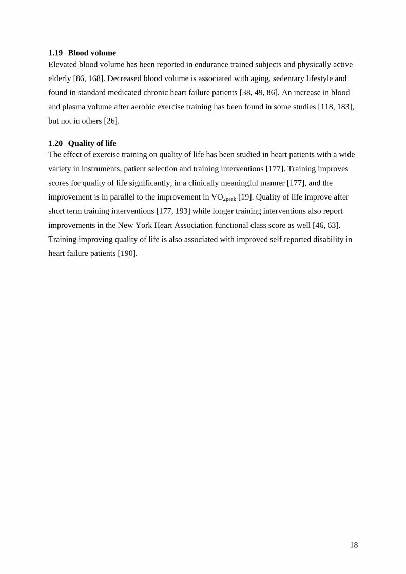
Figure 1. Testing of oxygen uptake
3.2 Cardiac output and stroke volume Cardiac output was measured through study I-IV using the single breath gas technique to the
Sensormedics Vmax Spectra 229 (SensorMedics Corp, California, USA). The test were
initiated with a 10 minute warm up period on the treadmill followed by gradually increasing
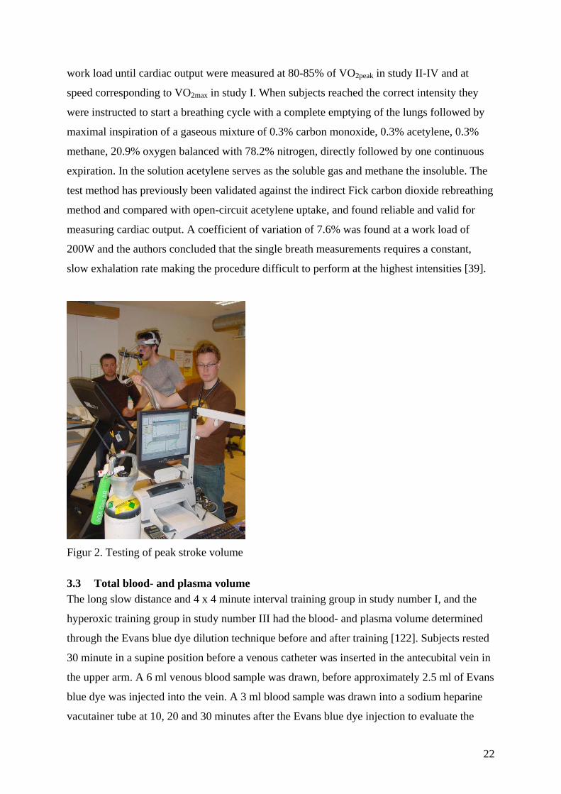
22
work load until cardiac output were measured at 80-85% of VO2peak in study II-IV and at
speed corresponding to VO2max in study I. When subjects reached the correct intensity they
were instructed to start a breathing cycle with a complete emptying of the lungs followed by
maximal inspiration of a gaseous mixture of 0.3% carbon monoxide, 0.3% acetylene, 0.3%
methane, 20.9% oxygen balanced with 78.2% nitrogen, directly followed by one continuous
expiration. In the solution acetylene serves as the soluble gas and methane the insoluble. The
test method has previously been validated against the indirect Fick carbon dioxide rebreathing
method and compared with open-circuit acetylene uptake, and found reliable and valid for
measuring cardiac output. A coefficient of variation of 7.6% was found at a work load of
200W and the authors concluded that the single breath measurements requires a constant,
slow exhalation rate making the procedure difficult to perform at the highest intensities [39].
Figur 2. Testing of peak stroke volume
3.3 Total blood- and plasma volume The long slow distance and 4 x 4 minute interval training group in study number I, and the
hyperoxic training group in study number III had the blood- and plasma volume determined
through the Evans blue dye dilution technique before and after training [122]. Subjects rested
30 minute in a supine position before a venous catheter was inserted in the antecubital vein in
the upper arm. A 6 ml venous blood sample was drawn, before approximately 2.5 ml of Evans
blue dye was injected into the vein. A 3 ml blood sample was drawn into a sodium heparine
vacutainer tube at 10, 20 and 30 minutes after the Evans blue dye injection to evaluate the
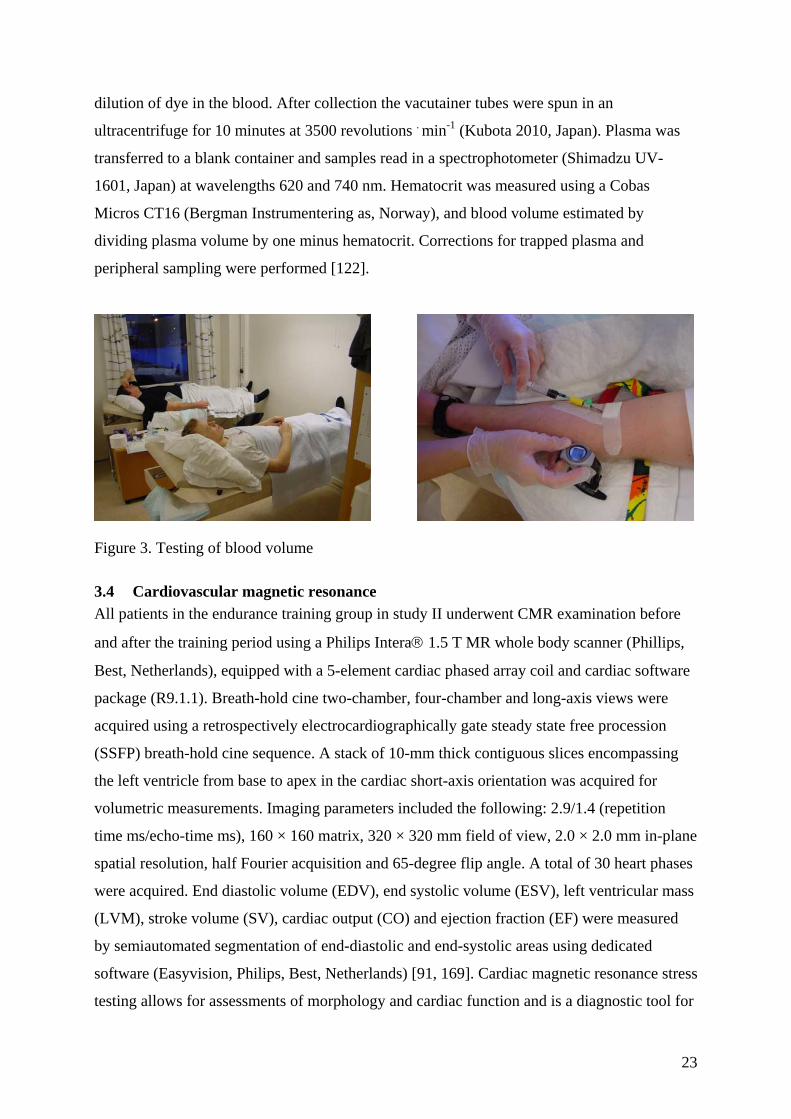
23
dilution of dye in the blood. After collection the vacutainer tubes were spun in an
ultracentrifuge for 10 minutes at 3500 revolutions . min-1 (Kubota 2010, Japan). Plasma was
transferred to a blank container and samples read in a spectrophotometer (Shimadzu UV-
1601, Japan) at wavelengths 620 and 740 nm. Hematocrit was measured using a Cobas
Micros CT16 (Bergman Instrumentering as, Norway), and blood volume estimated by
dividing plasma volume by one minus hematocrit. Corrections for trapped plasma and
peripheral sampling were performed [122].
Figure 3. Testing of blood volume
3.4 Cardiovascular magnetic resonance All patients in the endurance training group in study II underwent CMR examination before
and after the training period using a Philips Intera® 1.5 T MR whole body scanner (Phillips,
Best, Netherlands), equipped with a 5-element cardiac phased array coil and cardiac software
package (R9.1.1). Breath-hold cine two-chamber, four-chamber and long-axis views were
acquired using a retrospectively electrocardiographically gate steady state free procession
(SSFP) breath-hold cine sequence. A stack of 10-mm thick contiguous slices encompassing
the left ventricle from base to apex in the cardiac short-axis orientation was acquired for
volumetric measurements. Imaging parameters included the following: 2.9/1.4 (repetition
time ms/echo-time ms), 160 × 160 matrix, 320 × 320 mm field of view, 2.0 × 2.0 mm in-plane
spatial resolution, half Fourier acquisition and 65-degree flip angle. A total of 30 heart phases
were acquired. End diastolic volume (EDV), end systolic volume (ESV), left ventricular mass
(LVM), stroke volume (SV), cardiac output (CO) and ejection fraction (EF) were measured
by semiautomated segmentation of end-diastolic and end-systolic areas using dedicated
software (Easyvision, Philips, Best, Netherlands) [91, 169]. Cardiac magnetic resonance stress
testing allows for assessments of morphology and cardiac function and is a diagnostic tool for
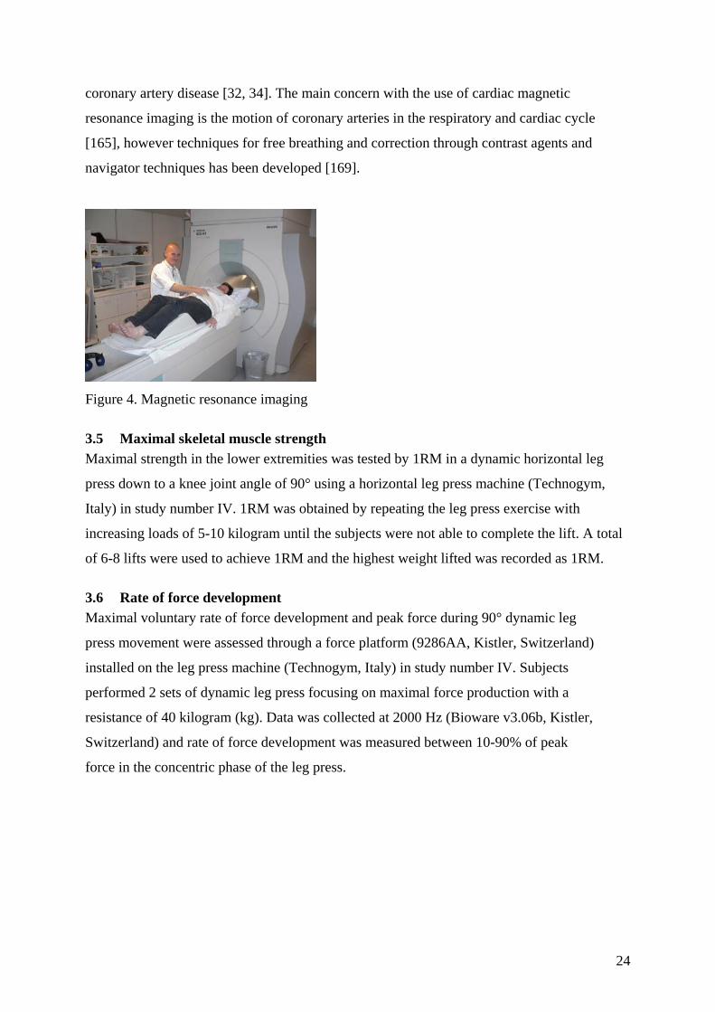
24
coronary artery disease [32, 34]. The main concern with the use of cardiac magnetic
resonance imaging is the motion of coronary arteries in the respiratory and cardiac cycle
[165], however techniques for free breathing and correction through contrast agents and
navigator techniques has been developed [169].
Figure 4. Magnetic resonance imaging
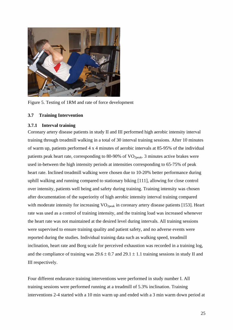
3.5 Maximal skeletal muscle strength Maximal strength in the lower extremities was tested by 1RM in a dynamic horizontal leg
press down to a knee joint angle of 90° using a horizontal leg press machine (Technogym,
Italy) in study number IV. 1RM was obtained by repeating the leg press exercise with
increasing loads of 5-10 kilogram until the subjects were not able to complete the lift. A total
of 6-8 lifts were used to achieve 1RM and the highest weight lifted was recorded as 1RM.
3.6 Rate of force development Maximal voluntary rate of force development and peak force during 90° dynamic leg
press movement were assessed through a force platform (9286AA, Kistler, Switzerland)
installed on the leg press machine (Technogym, Italy) in study number IV. Subjects
performed 2 sets of dynamic leg press focusing on maximal force production with a
resistance of 40 kilogram (kg). Data was collected at 2000 Hz (Bioware v3.06b, Kistler,
Switzerland) and rate of force development was measured between 10-90% of peak
force in the concentric phase of the leg press.
25
Figure 5. Testing of 1RM and rate of force development
3.7 Training Intervention
3.7.1 Interval training Coronary artery disease patients in study II and III performed high aerobic intensity interval
training through treadmill walking in a total of 30 interval training sessions. After 10 minutes
of warm up, patients performed 4 x 4 minutes of aerobic intervals at 85-95% of the individual
patients peak heart rate, corresponding to 80-90% of VO2peak. 3 minutes active brakes were
used in-between the high intensity periods at intensities corresponding to 65-75% of peak
heart rate. Inclined treadmill walking were chosen due to 10-20% better performance during
uphill walking and running compared to stationary biking [111], allowing for close control
over intensity, patients well being and safety during training. Training intensity was chosen
after documentation of the superiority of high aerobic intensity interval training compared
with moderate intensity for increasing VO2peak in coronary artery disease patients [153]. Heart
rate was used as a control of training intensity, and the training load was increased whenever
the heart rate was not maintained at the desired level during intervals. All training sessions
were supervised to ensure training quality and patient safety, and no adverse events were
reported during the studies. Individual training data such as walking speed, treadmill
inclination, heart rate and Borg scale for perceived exhaustion was recorded in a training log,
and the compliance of training was 29.6 ± 0.7 and 29.1 ± 1.1 training sessions in study II and
III respectively.
Four different endurance training interventions were performed in study number I. All
training sessions were performed running at a treadmill of 5.3% inclination. Training
interventions 2-4 started with a 10 min warm up and ended with a 3 min warm down period at
26
70% of maximal heart rate. Subjects carried out three training sessions per week for eight
weeks, a total of 24 sessions. The four training interventions included:
1. Low slow distance running (LSD): The first group performed a continuous run at 70%
of maximal heart rate for 45 min
2. Lactate threshold running (LT): The second group performed a continuous run at
lactate threshold at 85% of maximal heart rate for 24.25 minutes.
3. 15 x 15 seconds interval running (15x15s): The third group performed 47 repetitions
of 15 s intervals at 90 to 95% of maximal heart rate with 15 seconds active resting-
periods at warm up velocity, corresponding to 70% maximal heart rate between each
interval.
4. 4 x 4 minute interval running (4x4 min): A fourth group trained 4 x 4 minutes interval
training at 90-95% of maximal heart rate with 3 minutes active resting-periods at 70%
of maximal heart rate between each interval.
Each training intervention was made equal in terms of energy consumption after an accurate
calculation and a pilot study where active periods and warm up was included. On average the
total oxygen uptake for the training protocols was 130 ± 15 Litre or approximately 650 Kcal
per training session.
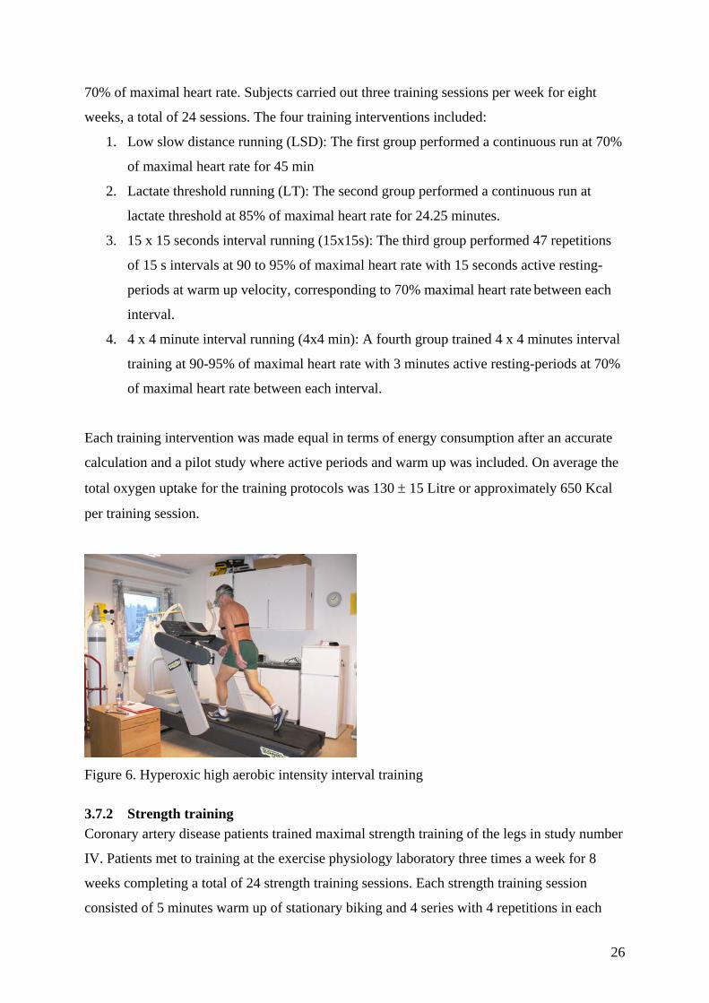
Figure 6. Hyperoxic high aerobic intensity interval training
3.7.2 Strength training Coronary artery disease patients trained maximal strength training of the legs in study number
IV. Patients met to training at the exercise physiology laboratory three times a week for 8
weeks completing a total of 24 strength training sessions. Each strength training session
consisted of 5 minutes warm up of stationary biking and 4 series with 4 repetitions in each
27
series of horizontal dynamic leg press. Exercise was made with emphasis on maximal
mobilization of force in the concentric action and subjects started the concentric movement
when the knee angle corresponded to 90°. Subjects trained with a progressive work load of
85-90% of the individual 1RM. When subjects were able to perform more than four
repetitions in a set, the load was increased by 2.5 kg. A 2 minutes rest period was employed
between each set of exercise. The compliance of training was 23.8 ± 0.4 training sessions.
3.8 Statistical analysis Statistical analyses were performed using the software program SPSS, version 11.0-14.0
(Statistical Package for Social Science, Chicago, USA). Table values are expressed as mean ±
standard deviation (SD), while figure values are expressed as mean percentage change and
data variability as standard error (SE). A two-tailed p < 0.05 was accepted as statistically
significant for all tests. Q-Q plots were used to test data for normal distribution. Due to the
relatively small sample size non parametric statistics tests were chosen in paper II-IV.
Changes within groups were determined by the Wilcoxon signed ranks test, while differences
between groups were calculated by using the Mann-Whitney U-test on delta changes from pre
to post test. Relationships between variables were assessed with correlation analysis. In paper
number I differences within and between groups was calculated through a two-way analysis
of repeated measures ANOVA (least significance difference test) comparing means for
continuous variables.
28
4 Summary of Results Paper I. Aerobic High-Intensity Intervals Improve VO2max More Than Moderate Training
The objectives of this study was to compare the effects of aerobic endurance training at
different intensities- and methods matched for total work and frequency.
1. VO2max increased by 5.5% in the 15/15 and by 7.2 % in the 4x4 group after training,
with no difference in the training response between the 15/15 and 4x4 group.
2. No change in VO2max was detected in the LT and LSD group.
3. Running economy significantly improved in all the training groups by 7.5-11.7%, but
no difference was detected between groups.
4. Lactate threshold did not change in any of the groups when expressed as % VO2max.
Velocity at lactate threshold was however significantly improved by an average of
9.6% in all four groups as a consequence of change in running economy and VO2max.
5. Stroke volume changed from pre to post training in the 15/15 and the 4x4 min group.
No significant difference was observed between the two groups, however the smallest
p value was detected in the 4 x 4 min group.
6. No significant haematological response to training was found.
Paper II. How to Improve Stroke Volume in Heart Patients
The objectives of this study was to investigate to which extent aerobic interval training at 85-
95% of maximal heart rate improves peak stroke volume and myocardial ejection fraction in
coronary artery disease patients.
1. Peak stroke volume significantly improved by 23% after interval training.
2. Resting left ventricle ejection fraction significantly improved by 5% after interval
training.
3. Resting left ventricle end diastolic volume and myocardial weight as well as cardiac
output was unchanged after training. There was a non-significant trend towards
decreased end systolic volume and heart rate, as well as increased stroke volume.
4. VO2peak significantly improved by 17% after high aerobic intensity interval training
29
Paper III. Aerobic interval training improve VO2peak in heart patients; no additional effect
from hyperoxia
The objectives of this study were to investigate if 100% oxygen supplementation during high
aerobic intensity interval training improves training performance and VO2peak in coronary
artery disease patients.
1. VO2peak improved 16% and 17% after 30 training session of high aerobic intensity
interval training at 85-95% of peak heart rate in the hyperoxic- and normoxic training
group respectively.
2. VO2peak improved 0.53 and 0.57% per training session in the two training groups
3. VO2peak was equal in hyperoxia (65% O2) and normoxia (21% O2) in the hyperoxic
training group both before and after interval training.
4. Peak stroke volume increased significantly after high aerobic intensity interval
training in both training groups.
5. Blood volume did not change after 10 weeks of hyperoxic interval training in the
hyperoxic training group.
6. Quality of life improved in both training groups after interval training.
Paper IV. Strength Training Restores Walking in Heart Patients
The objectives of this study were to investigate the effect of maximal strength training of the
legs has upon leg strength, rate of force development and walking mechanical efficiency in
coronary artery disease patients.
1. Maximal strength in leg press measured through one repetition maximum increased by
44% after strength training.
2. This corresponds to an increase in strength by 1.6% per training session.
3. Maximal strength training increased rate of force development by 85%.
4. Maximal strength training increased peak force by 18%.
5. Maximal strength training increased walking efficiency by 35%.
6. Quality of life increased significant in the score for mental health after strength
training.
7. Maximal strength training did not change total serum testosterone.

30
5 Discussion The present thesis reports that high aerobic intensity training at 90-95% of maximal heart rate
improves VO2max and maximal cardiac stroke volume in healthy young men, whereas
isocaloric training protocols at lower intensities does not result in any changes (I). High
aerobic intensity interval training at 85-95% of peak heart rate improves VO2peak and peak
cardiac stroke volume in coronary artery disease patients to the same extent as healthy young
men (II-III). Hyperoxic high aerobic intensity interval training gives no additional
improvements in VO2peak or peak cardiac stroke volume compared with normoxic training in
coronary artery disease patients. Patients were oxygen demand limited in the skeletal muscles
at VO2peak during acute hyperoxic testing both before and after training (III). Maximal
strength training of the legs enhances muscular strength and walking efficiency in coronary
artery disease patients despite no change in VO2peak (IV).
5.1 Improvements in maximal aerobic power The present thesis confirms that the intensity of endurance training is the most important
factor to improve maximal oxygen uptake (VO2max) in healthy young men (I). This is also in
line with previous studies in cardiovascular disease patients [153, 193]. The present thesis
demonstrates and confirms that VO2max, VO2peak and performance increase significantly with
high aerobic intensity interval training at 85-95% of peak heart rate. The increase in aerobic
power is achieved after a few weeks of training, in both healthy men and coronary heart
disease patients (I-III). In healthy young men only training intensities at 90-95% of maximal
heart rate, performed after interval principles improved VO2max (I). This differs from previous
studies of coronary heart disease and heart failure patients, where long slow distance training
at 70% of peak heart rate improved VO2peak as well. The improvement in VO2peak was
however only half of the outcome from high aerobic intensity interval training [153, 193]. The
VO2max was higher in the healthy young men (I) compared with previous studies of
cardiovascular disease patients [153, 193]. The mean VO2max was between 55 and 60 ml . kg-1 .
min-1 in study number I, while mean VO2peak ranged from ~13 ml . kg-1 . min-1 [193] to ~31 ml .
kg-1 . min-1 [153] in previous studies of heart patients. The results may imply that low intensity
endurance training may increase VO2max when VO2max is initially low. When choosing mode
and intensity of endurance training the amount of improvement desired per training session,
and the initial level of aerobic power might be considered before expectations of
improvements are set.
31
High aerobic intensity interval training improves VO2peak in coronary artery disease patients to
a greater extent than VO2max in healthy young men when expressed as percent improvement
(I-III). VO2peak increased by an average of 0.53-0.57% per training session in coronary artery
disease patients. Half the improvement in VO2max per training session was observed in healthy
young men with an average of 0.23-0.30 % increase in VO2max. The relative difference is due
to the lower initial aerobic power observed in coronary artery disease patients. When
comparing the absolute change in aerobic power surprisingly similar results are observed
between studies. On average coronary artery disease patients improved VO2peak by 4 ml . kg-1 .
min-1 and 4.5 ml . kg-1 . min-1 in study II and III, respectively. In study number I, high aerobic
intensity interval training by 15 x 15 seconds and 4 x 4 minutes improved VO2max on average
3.9 ml . kg-1 . min-1 and 4.9 ml . kg-1 . min-1, respectively. Converted to improvement per training
session, 0.13 ml . kg-1 . min-1, 0.15 ml . kg-1 . min-1, 0.16 ml . kg-1 . min-1 and 0.20 ml . kg-1 . min-1
was detected in study II, III and group 15x15 second and 4 x 4 minutes in study I,
respectively. This implies that coronary artery disease patients through supervised training
can perform high aerobic intensity interval training with equal quality as healthy young male
subjects. So, despite documented coronary artery disease and myocardial insufficiency
coronary artery disease patients achieve similar training outcomes as healthy young men
when expressed as absolute values. The similarity in the training outcome between studies
serves as an indication of the potential high intensity interval training possesses as long as it is
performed aerobically. The equality in the training response might serve as an argument for
incorporating high aerobic intensity interval training as part of recommendations for
cardiovascular disease patients.
When considering the importance of high VO2peak in association with mortality in
cardiovascular disease patients [117] it should be noticed that high aerobic intensity interval
training rapidly increases VO2peak in a high mortality risk population. The mean increase in
VO2peak was approximately 4 – 4.5 ml . kg-1 . min-1 after 30 interval training sessions in
coronary artery disease patients (II-III). According to Myers et al. [117] a 1 MET (3.5 ml . kg-1
. min-1) increase in aerobic power corresponds with a 12 % reduction in mortality risk. High
aerobic intensity training thereby serves as a non pharmaceutical treatment, substantially
reducing the risk of mortality in only a few weeks. In addition training might serve as a
protective agent against further development of coronary artery disease [176], and improve
risk factors related to cardiovascular disease [14, 175]. A large improvement in VO2peak
inflicted by high aerobic intensity interval training in a short period of time is both significant

32
and clinically meaningful for the patients. Training might appear motivational since patients
quickly sense the effect of training in their daily life.
Current guidelines regarding training and health recommends 30 minutes of not necessarily
structured, moderate exercise, preferably every day of the week to ensure health benefits
[175]. The recommendation might be questioned in view of the result from the present thesis,
and the importance of high aerobic power for reducing mortality [57, 113, 117]. One might
argue that some elements of high aerobic intensity training are needed to ensure good health
in both healthy subjects and coronary artery disease patients. This might be particular
important in coronary artery disease patients with low initial VO2peak, where significant
improvements in fitness and risk of mortality might be obtained in just a few weeks of
training (II-III). If a relationship exist between cardiovascular fitness and health aspects the
fact that high aerobic intensity training improves VO2max in healthy subjects is important to
consider. According to the data from the present study (I), a young and healthy population
would probably not experience any improvements in VO2max following the recommendations
from the present guidelines and training or activity would merely serve as a means of
maintenance. High aerobic intensity training is reported to be twice as effective as low
intensity training in improving VO2peak in heart patients [153, 193]. In this population
cardiovascular fitness has been proven vital for future development of the disease and life
expectancy [58, 85]. With the important link between cardiovascular fitness and mortality risk
high aerobic intensity interval training has the ability to quickly increase cardiopulmonary
fitness and thereby health prospects as well.
Maximal strength training of the legs did not enhance VO2peak in coronary artery disease
patients (IV), however, as expected the work efficiency was improved. VO2max is minimally
effected by maximal strength training [94], however enhanced muscular strength may
stimulate to increased voluntary endurance activity. With a limited VO2peak in coronary artery
disease patients, improvements in walking efficiency directly translate into similarly
improved walking ability and performance.
5.2 Myocardial changes with training In the present thesis high aerobic intensity interval training significantly improved peak stroke
volume in both coronary artery disease patients and healthy young males (I-III). Peak stroke
volume was initially higher in healthy young men compared with coronary artery disease
33
patients with mean values between ~130 and 155 ml . beat-1 in healthy young men and ~90 and
114 ml . beat-1 in coronary artery disease patients before training. Peak stroke volume
improved relatively the most after training in coronary artery disease patients with ~ 15-17 %
improvement (II-III) while healthy young men improved stroke volume by ~10% (I). The
slightly larger percent increase in stroke volume observed in coronary artery disease patients
after training compared to healthy young men, matches the larger percent improvement in
aerobic power noted in coronary artery disease patients compared with healthy young men.
The absolute delta values are however, as observed with VO2peak and VO2max, surprisingly
equal between studies. Maximal stroke volume improve with ~14-15 ml . beat-1 and peak
stroke volume by ~15-21 ml. beat-1 in healthy young men (I) and coronary artery disease
patients (II-III), respectively. The improvement per interval training session was 0.58-0.63 ml
. beat-1 and 0.51-0.70 ml . beat-1 in the healthy young men (I) and coronary artery disease
patients, respectively (II-III). This implies that despite reduced initial stroke volume, reduced
myocardial efficiency and documented ischemic heart disease, coronary artery disease
patients have the same absolute myocardial response to high aerobic intensity interval training
as healthy young men. High aerobic intensity interval training thereby seems to inflict the
same shear stress on the cardiovascular system improving the oxygen supply to working
skeletal muscles in both young healthy men and coronary artery disease patients. Stroke
volume deterioration documented with aging and disease may thereby primary reflect
increasing levels of inactivity, at least before ischemia and infarction may disrupt peak
myocardial function.
High aerobic intensity interval training improved resting left ventricle ejection fraction in
coronary artery disease patients (II). Improvements in peak stroke volume in coronary artery
disease patients is probably due to increased myocardial contractility as observed with
increased resting left ventricular ejection fraction. Improvements in resting left ventricular
ejection fraction has previously been reported in old heart failure patients together with a
reversal of left ventricle remodelling [193]. High aerobic intensity interval training did not
change the left ventricle dimension, indicating that a larger dose of high aerobic intensity
interval training might be needed to achieve the reported increase in left ventricle dimension
observed in endurance trained female athletes [192]. In the present study (II), there was a
trend toward decreased end systolic volume with a subsequent increase in stroke volume.
Heart rate decreased correspondingly, maintaining an unchanged cardiac output. All those
changes are concordant, indicating that the trend is real. In addition, the result was a

34
significant increase in resting ejection fraction, indicating an enhanced left ventricular systolic
performance. Left ventricle ejection fraction and reversed myocardial remodelling
normalizing myocardial size has been reported to be dependent of training intensity, with only
aerobic interval training demonstrating changes in heart failure patients [193].
Training intensities below 85% of maximal heart rate did not improve maximal stroke volume
or VO2max in healthy young men (I). The present thesis thereby reports a dependency of
training intensity in the effort to improve maximal stroke volume in healthy individuals. The
effect of long slow distance training on peak stroke volume was not investigated in coronary
artery disease patients in the present studies (II-III). Based on the similarities in the response
in VO2peak and peak stroke volume detected in healthy men and coronary artery disease
patients, a logic deduction might be expecting peak stroke volume to follow the improvement
in VO2peak reported in the study by Rognmo et al. [153]. The effect of long slow distance
training in their study was half that of high aerobic intensity training in coronary artery
disease patients. The fact that stroke volume plateaus at different level of maximal heart rate
[54, 196] may explain why high aerobic intensity training is needed to improve VO2max and
maximal stroke volume in healthy young men. When training at 90-95% of maximal heart
rate the healthy young men train at the highest cardiac output they can maintain over 3-8
minutes influencing the cardiovascular system maximally. Training at a lower intensity may
not inflict enough shear stress on the cardiovascular system due to training at a lower cardiac
output. According to the data from Zhou et al. [196] and Gledhill et al. [54] cardiac patients
with low initial VO2peak reach peak cardiac output at a low percentage of maximal heart rate,
thereby low intensity training may also increase peak stroke volume and VO2peak in these
patients as well [153].
The present thesis displays a differentiation in stroke volume response with endurance
training intensity. Maximal or peak stroke volume in moderately trained healthy young men
and coronary artery disease patients improve to a similar extent after high aerobic intensity
interval training. This highlights the importance of training intensity in healthy subjects and
particularly in coronary artery disease patients with already reduced myocardial function. The
improvement in stroke volume seems to follow the changes in aerobic power in both healthy
men and coronary artery disease patients. This indicates that myocardial function and skeletal
muscle metabolism are closely matched and that training responses at the supply and demand
level go hand in hand.
35
Maximal strength training with high load and focus upon maximal voluntary contraction does
not change stroke volume or cardiac output in coronary artery disease patients (IV), therefore
intensity dependent endurance training must be applied to achieve improvements in
myocardial function. Strength training may however serve as an important means for better
circulatory management and blood pressure control in coronary artery disease patients [104].
The present studies together with others [193] suggest that that coronary artery disease
patients following the traditional part of official guidelines will not experience optimal
coronary adaptations from training on a dysfunctional myocardium [193].
5.3 Demand and supply limitations to training High aerobic intensity interval training increases the oxygen delivery to working skeletal
muscles through elevated peak stroke volume in both healthy individuals and coronary artery
disease patients (I-III). Peak stroke volume is elevated to the same extent as VO2max or VO2peak
implying that the oxygen demand is at least on level with the supply of oxygen, but might be
higher.
In coronary artery disease patients no acute effect of hyperoxia was found on VO2peak and
performance, despite significantly increased haemoglobin oxygen saturation. This observation
indicates a demand limitation to oxygen consumption in the working skeletal muscles of the
coronary artery disease patients at the inclusion point in the study (III). Oxygen supply often
limits VO2max in the healthy human skeletal muscles [92, 147-149, 157, 181], however
metabolic limitations in VO2max is suggested in unfit subjects [182]. Depressed oxidative
capacity in the skeletal muscles of heart failure patients has been reported, implying that
VO2peak is limited by the oxidative capacity of mitochondria in the working muscles [41]. This
is in agreement with the findings in the present study of coronary artery disease patients
where 65% inspired oxygen had no effect on VO2peak and performance. Some studies report
increased VO2peak and performance in coronary artery disease patients exposed to hyperoxia,
while others do not observe changes in VO2peak in line with the observations in paper number
III [146, 154]. As discussed in the paper (III) several other factors not controlled for in the
study, as vascular vasoconstriction restricting oxygen supply to the working skeletal muscles,
or the level of myocardial ischemia affecting myocardial contractility may also have
contributed to the lack of improvement in exercise performance. In addition it is important to
note that the coronary artery disease patients did not suffer from exercise induced hypoxemia
during peak normoxic exercise, thereby oxygen delivery was not initially limited in normoxia.
36
Endurance trained healthy men were not investigated during hyperoxic exposure thereby no
conclusions may be drawn in that context.
Hyperoxic high aerobic intensity interval training gave no additional effect on performance
outcome compared with normoxic training. The present study (III) report that 30 hyperoxic
high aerobic intensity interval training sessions increases VO2peak and peak stroke volume to
the same extent as normoxic training. This implies that under both training environments
there is an equal oxygen supplementation and use of oxygen in the energy converting process
in the working skeletal muscles. Thereby an equal training load was performed during both
hyperoxic and normoxic training, and no additional shear stress was initiated to the
cardiovascular system during hyperoxic training. The result is an equal high peak stroke
volume and VO2peak response between the two training groups.
Further investigations may reveal if hyperoxia is beneficial in severely ischemic coronary
artery disease patients with ischemic coronary contractile restrictions. The present study (III)
demonstrates that a VO2peak below 35 ml . kg-1 . min-1 is associated with a metabolic skeletal
muscle demand limitation in coronary artery disease patients. VO2peak above this level might
be needed before the limitations to exercise is switched from oxygen demand towards oxygen
supply limitations. The present data (III) is in line with other studies were hyperoxic
endurance training is reported to have no additional effect on VO2max, even with higher
VO2max values reported in the subjects participating [134, 140]. The practical implication from
the present study (III) is that mildly ischemic coronary artery disease patients does not benefit
from hyperoxic supplementation during training and that thirty high aerobic intensity interval
training sessions did not seem to be enough to switch from oxygen demand to oxygen supply
limitation to exercise, thereby aerobic interval training can safely be performed in normoxia
with equally high performance outcome.
5.4 Maximal strength training Maximal strength training focusing on heavy loads, maximal voluntary contraction and few
repetitions improves muscle strength and muscular contractility in coronary artery disease
patients (IV), in line with other patient groups [75] and healthy subjects [72, 74]. Maximal
strength training was well managed by the coronary artery disease patients and no adverse
events were reported during strength training (IV). Improved muscle strength is important in
many contexts, one being the occurrence of muscle atrophy and muscle weakness in coronary
37
patients [180], another muscular strength as a predictor of survival and the aggravation of
muscle metabolism and skeletal muscle function with progressing New York Heart
Association grading [10, 79, 189]. Thirdly low muscle strength is associated with high levels
of disability in heart disease patients, reporting higher levels of disability than healthy age
matched individuals [138], together with the loss of independence [40]. Low skeletal
muscular strength and exercise impairment is reported to be due to skeletal muscular defects
rather than limitations from the heart disease itself [109]. The advantage of maximal strength
training is a rapid increase in skeletal muscular strength without any gain in body weight.
Increased skeletal muscular strength renders the possibility to reduce disability and move the
threshold of daily activities further away from 1RM. Another positive effect is enhanced level
of daily activity, positively affecting cardiopulmonary fitness. Supervised training has the
potential to reverse the fear of being physical active in this patient group [120]. Increased
muscular strength is especially important in heart patients since increasing 1RM attenuates the
circulatory response giving better circulatory management in day to day activities [104].
Maximal strength training increases the rate of force development in coronary artery disease
patients (III), to the same extent as healthy subjects [72], and COPD patients [75]. The rate of
force development is important in terms of the ability to rapidly develop force in the skeletal
muscles, and contribute to better walking, stair climbing and prevents falls [16, 51, 94, 171].
Increased skeletal muscle strength through neural adaptations reported as increased rate of
force development makes large increases in muscle strength feasible without increasing body
weight, and are together with improved walking performance an indication of the importance
of training both the neurological and the morphological aspects of the muscle [171]. Increased
muscular strength could mean shifting the load of daily activity from heavy, to tolerable and
repeatable [103], and the present study (III) demonstrates that a large increase in muscular
strength is possible through maximal strength training, without any adverse events, and
minimal time spent exercising. The importance of maximal strength training is highlighted
even more through its effect on endurance performance in coronary artery disease patients.
Maximal strength training did not change serum testosterone concentration in coronary artery
disease patients (IV). The coronary artery disease patients already within the laboratory
normal range systemic testosterone levels did not change testosterone levels in response to
training. Low testosterone levels have been reported in connection with cardiovascular
disease [172], and the use of testosterone supplementation has been discussed in this patient
38
group. In the present thesis training did not affect serum testosterone concentration and
thereby cannot at the present time be recommended as an alternative to testosterone
supplementation in cardiovascular disease populations with low testosterone levels.
5.5 Walking efficiency and running economy Maximal strength training affects endurance performance in addition to improving skeletal
muscular strength and contractility. In coronary artery disease patients submaximal walking
efficiency is reduced [78], but returns to the level of healthy age matched adults after 8 weeks
of maximal strength training (IV). Maximal strength training improves walking mechanical
efficiency considerably by 35% after only 24 training sessions. Strength training as part of
rehabilitation of coronary artery diseases has the past years received more focus, after periods
with endurance training primarily receiving attention [22]. This study verifies the importance
of integrating maximal strength training in a rehabilitation setting since coronary artery
disease patients gains a substantial strength and endurance outcome through a minimal
training effort. This might prove very important for coronary artery disease patients whom
due to the fear of the consequences of the disease often are inactive and thereby develops
reduced skeletal muscle function and reduced physical capacity [109]. Improvements in
submaximal endurance performance may stimulate to increased level of voluntary endurance
activity at a higher intensity and increase the total daily energy expenditure [5], an important
aspect in further development or regression of coronary artery disease [64]. Also increased
skeletal muscular strength gives better management of daily living and increase the
independence of elderly coronary artery disease patients [6]. Increasing the daily activity level
has the potential to decrease the progression of the cardiovascular disease and improve the
mortality risk due to increased maximal oxygen uptake [117, 121, 124]. In addition increased
skeletal muscular strength and endurance performance improves physical function [28], and
has the potential to better circulatory management in day to day activities [104]. Coronary
artery disease patients are limited in their ability to walk [78] and report higher levels of
disability that healthy age matched individuals [138]. Maximal strength training may thereby
be necessary to imply to increase skeletal muscle strength and physical function [28] before
patients are able to perform endurance training activities. Strength training has been reported
to lower risk factors associated with coronary artery disease [15, 81] most likely through
increased activity levels. Combining maximal strength training and aerobic interval training
may even produce a larger training outcome of the endurance training due to improved
submaximal and maximal endurance performance.
39
Improvements in work economy was also detected after long slow distance training, lactate
threshold, 15 x 15 second and 4 x 4 minutes interval training in healthy young men (I). The
improvement in work economy was approximately 5% in all groups. Work economy
improved independent of improvements in VO2max, demonstrating that running economy is
not affected by the running speed used during training [68, 69]. No significant change in
lactate threshold was detected in the present study when expressed as percent VO2max however
the lactate threshold running speed was improved on average by 9.6% in all the groups.
Improved running speed is to be expected due to increased running economy in all training
groups.
5.6 Blood volume Blood volume was unchanged after high and low intensity endurance training in healthy
young men (I), or after hyperoxic high aerobic intensity interval training in coronary artery
disease patients (III). In study number I, mean blood volume ranged from 5.8 to 6.1 litres,
while an average of 4.3 litres was measured in coronary artery disease patients in study III.
Elevated blood volume has been observed in endurance trained individuals and physically
active elderly [86, 168]. A low blood volume is associated with aging, a sedentary lifestyle
and is found in medicated chronic heart failure patients [38, 49, 86]. A lower VO2peak, higher
age and the presence of coronary artery disease separated subjects in study III from study
number I. Thereby cardiovascular disease, medication, age and inactivity in the
cardiovascular disease patients compared with healthy young men may explain the initial
difference in blood volume between studies. In addition two women participating in the
cardiovascular disease study (III) might lower blood volume in this group compared with just
male participants in study number I. No change in blood volume was detected as a result of
endurance training in the present thesis (study I & III), neither after long slow distance
training or high aerobic intensity interval training (I). Increased blood and plasma volume has
been found in some studies after aerobic exercise training [118, 183], but not in others [26].
Blood volume may already have been well expanded before training for both the studies (I &
III), reflecting that normal levels of daily activity may be enough to maintain blood volume.
In the present investigations no long term data can explain the difference in blood volume
between the studies, however data might serve as an indication for an age or disease related
reduction in blood volume in cardiovascular disease patients. The amount of blood volume is
important for circulatory distribution of oxygen and a reduced blood volume might affect

40
endurance performance negatively. The present training interventions does not improve blood
volume thereby its effect on circulation and oxygen delivery cannot contribute in the
explanation of the improvements in VO2max and VO2peak as a result of endurance training. At
the present time the described training interventions cannot be recommended as a means for
improving blood volume, although effects from longer periods of training cannot be ruled out.
5.7 Quality of life In the studies of coronary artery disease patients aspects of measured quality of life was
improved both due to high aerobic intensity interval training and maximal strength training.
The improvement in quality of life is essential due to its clinical importance [177], connection
with improved coronary functional class score [46, 63] and self reported disability [190]. Both
improvements in VO2peak (II-III), maximal muscular strength and walking efficiency (IV)
improves quality of life, implying that several training interventions has the potential to affect
physical and mental well being and thereby improve daily functionality and abilities.
5.8 Training is medicine The present thesis demonstrates that supervised high aerobic intensity interval training result
in an equally high training outcome in VO2peak or VO2max in coronary artery disease patients
and healthy young men, and that training improves the blood supply to the exercising skeletal
muscles through a larger cardiac stroke volume. Training as treatment of coronary artery
disease affects further disease development, mortality, reduces the rate of rehospitalisation
and improve overall heath status [184]. Training has the potential to reduce the cost of
pharmaceuticals and correct the reduced physical state and function many coronary artery
disease patients often find themselves in, inspite of modern medical treatment [138, 177, 197].
In addition both endurance and maximal strength training may be performed in coronary
artery disease patients the same way as in healthy individuals without any adverse effects.
41
6 Conclusions High aerobic intensity interval training was more effective than continues training with low to
moderate intensity in improving maximal oxygen uptake in healthy young men. Maximal
stroke volume improved due to high aerobic intensity interval training only.
High aerobic intensity interval training at 85-95% of peak heart rate significantly improved
peak stroke volume and resting left ventricular ejection fraction in coronary artery disease
patients.
Coronary artery disease patients showed peripheral limitations in peak oxygen uptake both
before and after 10 weeks of hyperoxic training, thereby hyperoxic training did not improve
the effect of the training over normoxia.
Maximal leg press exercise focusing on few repetitions with heavy load and maximal
concentric contractions improved maximal strength, rate of force development, walking
mechanical efficiency and thus walking performance capacity in coronary artery disease
patients through a minimal exercise effort.
42
7 References 1. Abinader EG, Sharif DS, and Goldhammer E. Effects of low altitude on exercise performance in patients with congestive heart failure after healing of acute myocardial infarction. Am J Cardiol 1999; 83: 383-387
2. Ades PA. Cardiac rehabilitation and secondary prevention of coronary heart disease. N Engl J Med 2001; 345: 892-902
3. Ades PA, Ballor DL, Ashikaga T, Utton JL, and Nair KS. Weight training improves walking endurance in healthy elderly persons. Ann Intern Med 1996; 124: 568-572
4. Ades PA, Huang D, and Weaver SO. Cardiac rehabilitation participation predicts lower rehospitalization costs. Am Heart J 1992; 123: 916-921
5. Ades PA, Savage PD, Brochu M, Tischler MD, Lee NM, and Poehlman ET. Resistance training increases total daily energy expenditure in disabled older women with coronary heart disease. J Appl Physiol 2005; 98: 1280-1285
6. Ades PA, Savage PD, Cress ME, Brochu M, Lee NM, and Poehlman ET. Resistance training on physical performance in disabled older female cardiac patients. Med Sci Sports Exerc 2003; 35: 1265-1270
7. Almasbakk B and Hoff J. Coordination, the determinant of velocity specificity? J Appl Physiol 1996; 81: 2046-2052
8. American college of sports medicine position stand. Exercise for patients with coronary artery disease. Med Sci Sports Exerc 1994; 26: i-v
9. Andersen P and Saltin B. Maximal perfusion of skeletal muscle in man. J Physiol 1985; 366: 233-249
10. Anker SD, Swan JW, Volterrani M, Chua TP, Clark AL, Poole-Wilson PA, and Coats AJ. The influence of muscle mass, strength, fatigability and blood flow on exercise capacity in cachectic and non-cachectic patients with chronic heart failure. Eur Heart J 1997; 18: 259-269
11. Arbab-Zadeh A, Dijk E, Prasad A, Fu Q, Torres P, Zhang R, Thomas JD, Palmer D, and Levine BD. Effect of aging and physical activity on left ventricular compliance. Circulation 2004; 110: 1799-1805
12. Arena R, Myers J, Williams MA, Gulati M, Kligfield P, Balady GJ, Collins E, and Fletcher G. Assessment of functional capacity in clinical and research settings: A scientific statement from the american heart association committee on exercise, rehabilitation, and prevention of the council on clinical cardiology and the council on cardiovascular nursing. Circulation 2007; 116: 329-343
13. Bader DS, Maguire TE, Spahn CM, O'Malley CJ, and Balady GJ. Clinical profile and outcomes of obese patients in cardiac rehabilitation stratified according to national heart, lung, and blood institute criteria. J Cardiopulm Rehabil 2001; 21: 210-217
43
14. Balady GJ, Williams MA, Ades PA, Bittner V, Comoss P, Foody JA, Franklin B, Sanderson B, and Southard D. Core components of cardiac rehabilitation/secondary prevention programs: 2007 update: A scientific statement from the american heart association exercise, cardiac rehabilitation, and prevention committee, the council on clinical cardiology; the councils on cardiovascular nursing, epidemiology and prevention, and nutrition, physical activity, and metabolism; and the american association of cardiovascular and pulmonary rehabilitation. J Cardiopulm Rehabil Prev 2007; 27: 121-129
15. Banz WJ, Maher MA, Thompson WG, Bassett DR, Moore W, Ashraf M, Keefer DJ, and Zemel MB. Effects of resistance versus aerobic training on coronary artery disease risk factors. Exp Biol Med (Maywood) 2003; 228: 434-440
16. Bassey EJ, Fiatarone MA, O'Neill EF, Kelly M, Evans WJ, and Lipsitz LA. Leg extensor power and functional performance in very old men and women. Clin Sci (Lond) 1992; 82: 321-327
17. Behm DG. Neuromuscular implications and applications of resistance training. Journal of Strength and Conditioning Research 1995; 9: 264-274
18. Behm DG and Sale DG. Intended rather than actual movement velocity determines velocity-specific training response. J Appl Physiol 1993; 74: 359-368
19. Belardinelli R, Georgiou D, Cianci G, and Purcaro A. Randomized, controlled trial of long-term moderate exercise training in chronic heart failure: Effects on functional capacity, quality of life, and clinical outcome. Circulation 1999; 99: 1173-1182
20. Belardinelli R, Georgiou D, Ginzton L, Cianci G, and Purcaro A. Effects of moderate exercise training on thallium uptake and contractile response to low-dose dobutamine of dysfunctional myocardium in patients with ischemic cardiomyopathy. Circulation 1998; 97: 553-561
21. Bemben MG, Massey BH, Bemben DA, Misner JE, and Boileau RA. Isometric muscle force production as a function of age in healthy 20- to 74-yr-old men. Med Sci Sports Exerc 1991; 23: 1302-1310
22. Bjarnason-Wehrens B, Mayer-Berger W, Meister ER, Baum K, Hambrecht R, and Gielen S. Recommendations for resistance exercise in cardiac rehabilitation. Recommendations of the german federation for cardiovascular prevention and rehabilitation. Eur J Cardiovasc Prev Rehabil 2004; 11: 352-361
23. Blair SN, Kohl HW, 3rd, Paffenbarger RS, Jr., Clark DG, Cooper KH, and Gibbons LW. Physical fitness and all-cause mortality. A prospective study of healthy men and women. Jama 1989; 262: 2395-2401
24. Boutcher SH, McLaren PF, Cotton Y, and Boutcher Y. Stroke volume response to incremental submaximal exercise in aerobically trained, active, and sedentary men. Can J Appl Physiol 2003; 28: 12-26
25. Bouvier F, Saltin B, Nejat M, and Jensen-Urstad M. Left ventricular function and perfusion in elderly endurance athletes. Med Sci Sports Exerc 2001; 33: 735-740
44
26. Branch JD, 3rd, Pate RR, Bourque SP, Convertino VA, Durstine JL, and Ward DS. Exercise training and intensity does not alter vascular volume responses in women. Aviat Space Environ Med 1999; 70: 1070-1076
27. Brochu M, Poehlman ET, and Ades PA. Obesity, body fat distribution, and coronary artery disease. J Cardiopulm Rehabil 2000; 20: 96-108
28. Brochu M, Savage P, Lee M, Dee J, Cress ME, Poehlman ET, Tischler M, and Ades PA. Effects of resistance training on physical function in older disabled women with coronary heart disease. J Appl Physiol 2002; 92: 672-678
29. Brooks GA FT, White TP, Baldwin KM. Exercise physiology. Human bioenergetics and its applications Mayfield Publishing company Mountain View 2000
30. Buller NP, Jones D, and Poole-Wilson PA. Direct measurement of skeletal muscle fatigue in patients with chronic heart failure. Br Heart J 1991; 65: 20-24
31. Bunc V and Heller J. Energy cost of running in similarly trained men and women. Eur J Appl Physiol Occup Physiol 1989; 59: 178-183
32. Burgstahler C, Kunze M, Gawaz MP, Rasche V, Wohrle J, Hombach V, and Merkle N. Adenosine stress first pass perfusion for the detection of coronary artery disease in patients with aortic stenosis: A feasibility study. Int J Cardiovasc Imaging 2008; 24: 195-200
33. Byrnes WC, Mihevic PM, Freedson PS, and Horvath SM. Submaximal exercise quantified as percent of normoxic and hyperoxic maximum oxygen uptakes. Med Sci Sports Exerc 1984; 16: 572-577
34. Cheng AS, Pegg TJ, Karamitsos TD, Searle N, Jerosch-Herold M, Choudhury RP, Banning AP, Neubauer S, Robson MD, and Selvanayagam JB. Cardiovascular magnetic resonance perfusion imaging at 3-tesla for the detection of coronary artery disease: A comparison with 1.5-tesla. J Am Coll Cardiol 2007; 49: 2440-2449
35. Church TS, Lavie CJ, Milani RV, and Kirby GS. Improvements in blood rheology after cardiac rehabilitation and exercise training in patients with coronary heart disease. Am Heart J 2002; 143: 349-355
36. Clausen JP and Trap-Jensen J. Heart rate and arterial blood pressure during exercise in patients with angina pectoris. Effects of training and of nitroglycerin. Circulation 1976; 53: 436-442
37. Cohen MV. Myocardial ischemia is not a prerequisite for the stimulation of coronary collateral development. Am Heart J 1993; 126: 847-855
38. Davy KP and Seals DR. Total blood volume in healthy young and older men. J Appl Physiol 1994; 76: 2059-2062
39. Dibski DW, Smith DJ, Jensen R, Norris SR, and Ford GT. Comparison and reliability of two non-invasive acetylene uptake techniques for the measurement of cardiac output. Eur J Appl Physiol 2005; 94: 670-680
40. Doherty TJ. Invited review: Aging and sarcopenia. J Appl Physiol 2003; 95: 1717-1727
45
41. Drexler H, Riede U, Munzel T, Konig H, Funke E, and Just H. Alterations of skeletal muscle in chronic heart failure. Circulation 1992; 85: 1751-1759
42. Edwards WF, BD; Iyrilboz, Y; Dodd, SL. 29 physiological and expense implications of ptca rehabilitation. Medicine & Science in Sports & Exercise 1990; 22: S5
43. Ehsani AA. Mechanisms responsible for enhanced stroke volume after exercise training in coronary heart disease. Eur Heart J 1987; 8 Suppl G: 9-14
44. Ehsani AA, Biello DR, Schultz J, Sobel BE, and Holloszy JO. Improvement of left ventricular contractile function by exercise training in patients with coronary artery disease. Circulation 1986; 74: 350-358
45. Ekblom B, Huot R, Stein EM, and Thorstensson AT. Effect of changes in arterial oxygen content on circulation and physical performance. J Appl Physiol 1975; 39: 71-75
46. Erbs S, Linke A, Gielen S, Fiehn E, Walther C, Yu J, Adams V, Schuler G, and Hambrecht R. Exercise training in patients with severe chronic heart failure: Impact on left ventricular performance and cardiac size. A retrospective analysis of the leipzig heart failure training trial. Eur J Cardiovasc Prev Rehabil 2003; 10: 336-344
47. Erikssen G. Physical fitness and changes in mortality: The survival of the fittest. Sports Med 2001; 31: 571-576
48. Featherstone JF, Holly RG, and Amsterdam EA. Physiologic responses to weight lifting in coronary artery disease. Am J Cardiol 1993; 71: 287-292
49. Feigenbaum MS, Welsch MA, Mitchell M, Vincent K, Braith RW, and Pepine CJ. Contracted plasma and blood volume in chronic heart failure. J Am Coll Cardiol 2000; 35: 51-55
50. Ferguson S, Gledhill N, Jamnik VK, Wiebe C, and Payne N. Cardiac performance in endurance-trained and moderately active young women. Med Sci Sports Exerc 2001; 33: 1114-1119
51. Fleming BE, Wilson DR, and Pendergast DR. A portable, easily performed muscle power test and its association with falls by elderly persoms. Arch Phys Med Rehabil 1991; 72: 886-889
52. Frontera WR, Meredith CN, O'Reilly KP, Knuttgen HG, and Evans WJ. Strength conditioning in older men: Skeletal muscle hypertrophy and improved function. J Appl Physiol 1988; 64: 1038-1044
53. Gielen S, Schuler G, and Hambrecht R. Exercise training in coronary artery disease and coronary vasomotion. Circulation 2001; 103: E1-6
54. Gledhill N, Cox D, and Jamnik R. Endurance athletes' stroke volume does not plateau: Major advantage is diastolic function. Med Sci Sports Exerc 1994; 26: 1116-1121
55. Goldspink G. Cellular and molecular aspects of adaptation in skeletal muscle. in Komi P: Strength and power in sport. London Blackwell Scientific Publications; 1992. 211-229
46
56. Gonzalez-Alonso J, Richardson RS, and Saltin B. Exercising skeletal muscle blood flow in humans responds to reduction in arterial oxyhaemoglobin, but not to altered free oxygen. J Physiol 2001; 530: 331-341
57. Gulati M, Pandey DK, Arnsdorf MF, Lauderdale DS, Thisted RA, Wicklund RH, Al-Hani AJ, and Black HR. Exercise capacity and the risk of death in women: The st james women take heart project. Circulation 2003; 108: 1554-1559
58. Hagberg JM. Physiologic adaptations to prolonged high-intensity exercise training in patients with coronary artery disease. Med Sci Sports Exerc 1991; 23: 661-667
59. Hakkinen K, Alen M, Kallinen M, Newton RU, and Kraemer WJ. Neuromuscular adaptation during prolonged strength training, detraining and re-strength-training in middle-aged and elderly people. Eur J Appl Physiol 2000; 83: 51-62
60. Hakkinen K, Kallinen M, Izquierdo M, Jokelainen K, Lassila H, Malkia E, Kraemer WJ, Newton RU, and Alen M. Changes in agonist-antagonist emg, muscle csa, and force during strength training in middle-aged and older people. J Appl Physiol 1998; 84: 1341-1349
61. Hambrecht R, Adams V, Erbs S, Linke A, Krankel N, Shu Y, Baither Y, Gielen S, Thiele H, Gummert JF, Mohr FW, and Schuler G. Regular physical activity improves endothelial function in patients with coronary artery disease by increasing phosphorylation of endothelial nitric oxide synthase. Circulation 2003; 107: 3152-3158
62. Hambrecht R, Fiehn E, Weigl C, Gielen S, Hamann C, Kaiser R, Yu J, Adams V, Niebauer J, and Schuler G. Regular physical exercise corrects endothelial dysfunction and improves exercise capacity in patients with chronic heart failure. Circulation 1998; 98: 2709-2715
63. Hambrecht R, Gielen S, Linke A, Fiehn E, Yu J, Walther C, Schoene N, and Schuler G. Effects of exercise training on left ventricular function and peripheral resistance in patients with chronic heart failure: A randomized trial. Jama 2000; 283: 3095-3101
64. Hambrecht R, Niebauer J, Marburger C, Grunze M, Kalberer B, Hauer K, Schlierf G, Kubler W, and Schuler G. Various intensities of leisure time physical activity in patients with coronary artery disease: Effects on cardiorespiratory fitness and progression of coronary atherosclerotic lesions. J Am Coll Cardiol 1993; 22: 468-477
65. Hambrecht R, Walther C, Mobius-Winkler S, Gielen S, Linke A, Conradi K, Erbs S, Kluge R, Kendziorra K, Sabri O, Sick P, and Schuler G. Percutaneous coronary angioplasty compared with exercise training in patients with stable coronary artery disease: A randomized trial. Circulation 2004; 109: 1371-1378
66. Hambrecht R, Wolf A, Gielen S, Linke A, Hofer J, Erbs S, Schoene N, and Schuler G. Effect of exercise on coronary endothelial function in patients with coronary artery disease. N Engl J Med 2000; 342: 454-460
67. Haskell WL, Alderman EL, Fair JM, Maron DJ, Mackey SF, Superko HR, Williams PT, Johnstone IM, Champagne MA, Krauss RM, and et al. Effects of intensive multiple risk factor reduction on coronary atherosclerosis and clinical cardiac events in men and women with coronary artery disease. The stanford coronary risk intervention project (scrip). Circulation 1994; 89: 975-990
47
68. Helgerud J. Maximal oxygen uptake, anaerobic threshold and running economy in women and men with similar performances level in marathons. Eur J Appl Physiol Occup Physiol 1994; 68: 155-161
69. Helgerud J, Engen LC, Wisloff U, and Hoff J. Aerobic endurance training improves soccer performance. Med Sci Sports Exerc 2001; 33: 1925-1931
70. Hickson RC, Dvorak BA, Gorostiaga EM, Kurowski TT, and Foster C. Potential for strength and endurance training to amplify endurance performance. J Appl Physiol 1988; 65: 2285-2290
71. Hickson RC, Foster C, Pollock ML, Galassi TM, and Rich S. Reduced training intensities and loss of aerobic power, endurance, and cardiac growth. J Appl Physiol 1985; 58: 492-499
72. Hoff J, Gran A, and Helgerud J. Maximal strength training improves aerobic endurance performance. Scand J Med Sci Sports 2002; 12: 288-295
73. Hoff J and Helgerud J. Endurance and strength training for soccer players: Physiological considerations. Sports Med 2004; 34: 165-180
74. Hoff J, Helgerud J, and Wisloff U. Maximal strength training improves work economy in trained female cross-country skiers. Med Sci Sports Exerc 1999; 31: 870-877
75. Hoff J, Tjonna AE, Steinshamn S, Hoydal M, Richardson RS, and Helgerud J. Maximal strength training of the legs in copd: A therapy for mechanical inefficiency. Med Sci Sports Exerc 2007; 39: 220-226
76. Hoppeler H and Weibel ER. Limits for oxygen and substrate transport in mammals. J Exp Biol 1998; 201: 1051-1064
77. Horvat M, Yoshida S, Prakash R, Marcus HS, Swan HJ, and Ganz W. Effect of oxygen breathing on pacing-induced angina pectoris and other manifestations of coronary insufficiency. Circulation 1972; 45: 837-844
78. Hoydal KL, Helgerud J, Karlsen T, Stoylen A, Steinshamn S, and Hoff J. Patients with coronary artery- or chronic obstructive pulmonary disease walk with mechanical inefficiency. Scand Cardiovasc J 2007: 1-6
79. Hulsmann M, Quittan M, Berger R, Crevenna R, Springer C, Nuhr M, Mortl D, Moser P, and Pacher R. Muscle strength as a predictor of long-term survival in severe congestive heart failure. Eur J Heart Fail 2004; 6: 101-107
80. Hunter GR, McCarthy JP, and Bamman MM. Effects of resistance training on older adults. Sports Med 2004; 34: 329-348
81. Hurley BF, Hagberg JM, Goldberg AP, Seals DR, Ehsani AA, Brennan RE, and Holloszy JO. Resistive training can reduce coronary risk factors without altering vo2max or percent body fat. Med Sci Sports Exerc 1988; 20: 150-154
82. Izquierdo M, Aguado X, Gonzalez R, Lopez JL, and Hakkinen K. Maximal and explosive force production capacity and balance performance in men of different ages. Eur J Appl Physiol Occup Physiol 1999; 79: 260-267
48
83. Izquierdo M, Hakkinen K, Ibanez J, Anton A, Garrues M, Ruesta M, and Gorostiaga EM. Effects of strength training on submaximal and maximal endurance performance capacity in middle-aged and older men. J Strength Cond Res 2003; 17: 129-139
84. Izquierdo M, Ibanez J, Gorostiaga E, Garrues M, Zuniga A, Anton A, Larrion JL, and Hakkinen K. Maximal strength and power characteristics in isometric and dynamic actions of the upper and lower extremities in middle-aged and older men. Acta Physiol Scand 1999; 167: 57-68
85. Jae SY, Carnethon MR, Heffernan KS, Choi YH, Lee MK, and Fernhall B. Association between cardiorespiratory fitness and prevalence of carotid atherosclerosis among men with hypertension. Am Heart J 2007; 153: 1001-1005
86. Jones PP, Davy KP, DeSouza CA, van Pelt RE, and Seals DR. Absence of age-related decline in total blood volume in physically active females. Am J Physiol 1997; 272: H2534-2540
87. Joyner MJ and Coyle EF. Endurance exercise performance: The physiology of champions. J Physiol 2008; 586: 35-44
88. Karlsdottir AE, Foster C, Porcari JP, Palmer-McLean K, White-Kube R, and Backes RC. Hemodynamic responses during aerobic and resistance exercise. J Cardiopulm Rehabil 2002; 22: 170-177
89. Kasch FW, Boyer JL, Van Camp S, Nettl F, Verity LS, and Wallace JP. Cardiovascular changes with age and exercise. A 28-year longitudinal study. Scand J Med Sci Sports 1995; 5: 147-151
90. Kendziorra K, Walther C, Foerster M, Mobius-Winkler S, Conradi K, Schuler G, Sabri O, Hambrecht R, and Kluge R. Changes in myocardial perfusion due to physical exercise in patients with stable coronary artery disease. Eur J Nucl Med Mol Imaging 2005; 32: 813-819
91. Kim WY, Stuber M, Bornert P, Kissinger KV, Manning WJ, and Botnar RM. Three-dimensional black-blood cardiac magnetic resonance coronary vessel wall imaging detects positive arterial remodeling in patients with nonsignificant coronary artery disease. Circulation 2002; 106: 296-299
92. Knight DR, Schaffartzik W, Poole DC, Hogan MC, Bebout DE, and Wagner PD. Effects of hyperoxia on maximal leg o2 supply and utilization in men. J Appl Physiol 1993; 75: 2586-2594
93. Komi PV. Strength and power in sports Blackwell Scientific Publications, Oxford 1992
94. Komi PV. Strength and power in sport. Blackwell Publishing Oxford 2003
95. Larsson L, Sjodin B, and Karlsson J. Histochemical and biochemical changes in human skeletal muscle with age in sedentary males, age 22--65 years. Acta Physiol Scand 1978; 103: 31-39
96. Levine BD. : What do we know, and what do we still need to know? J Physiol 2008; 586: 25-34

49
97. Lexell J. Evidence for nervous system degeneration with advancing age. J Nutr 1997; 127: 1011S-1013S
98. Libby P, Ridker PM, and Maseri A. Inflammation and atherosclerosis. Circulation 2002; 105: 1135-1143
99. Linke A, Schoene N, Gielen S, Hofer J, Erbs S, Schuler G, and Hambrecht R. Endothelial dysfunction in patients with chronic heart failure: Systemic effects of lower-limb exercise training. J Am Coll Cardiol 2001; 37: 392-397
100. Lipkin DP, Jones DA, Round JM, and Poole-Wilson PA. Abnormalities of skeletal muscle in patients with chronic heart failure. Int J Cardiol 1988; 18: 187-195
101. Mark DB and Lauer MS. Exercise capacity: The prognostic variable that doesn't get enough respect. Circulation 2003; 108: 1534-1536
102. McArdle WD KF, Katch VL. Exercise physiology. Energy, nutrition, and human performance Lippincott Williams & Wilkins Philadelphia 2001
103. McCartney N, McKelvie RS, Haslam DR, and Jones NL. Usefulness of weightlifting training in improving strength and maximal power output in coronary artery disease. Am J Cardiol 1991; 67: 939-945
104. McCartney N, McKelvie RS, Martin J, Sale DG, and MacDougall JD. Weight-training-induced attenuation of the circulatory response of older males to weight lifting. J Appl Physiol 1993; 74: 1056-1060
105. McGuire DK, Levine BD, Williamson JW, Snell PG, Blomqvist CG, Saltin B, and Mitchell JH. A 30-year follow-up of the dallas bedrest and training study: I. Effect of age on the cardiovascular response to exercise. Circulation 2001; 104: 1350-1357
106. McKelvie RS. Exercise training in patients with heart failure: Clinical outcomes, safety, and indications. Heart Fail Rev 2008; 13: 3-11
107. McKelvie RS, McCartney N, Tomlinson C, Bauer R, and MacDougall JD. Comparison of hemodynamic responses to cycling and resistance exercise in congestive heart failure secondary to ischemic cardiomyopathy. Am J Cardiol 1995; 76: 977-979
108. McKelvie RS, Teo KK, McCartney N, Humen D, Montague T, and Yusuf S. Effects of exercise training in patients with congestive heart failure: A critical review. J Am Coll Cardiol 1995; 25: 789-796
109. McKelvie RS, Teo KK, Roberts R, McCartney N, Humen D, Montague T, Hendrican K, and Yusuf S. Effects of exercise training in patients with heart failure: The exercise rehabilitation trial (exert). Am Heart J 2002; 144: 23-30
110. Mian OS, Thom JM, Ardigo LP, Morse CI, Narici MV, and Minetti AE. Effect of a 12-month physical conditioning programme on the metabolic cost of walking in healthy older adults. Eur J Appl Physiol 2007; 100: 499-505
111. Miyamura M and Honda Y. Oxygen intake and cardiac output during maximal treadmill and bicycle exercise. J Appl Physiol 1972; 32: 185-188
50
112. Moore DP, Weston AR, Hughes JM, Oakley CM, and Cleland JG. Effects of increased inspired oxygen concentrations on exercise performance in chronic heart failure. Lancet 1992; 339: 850-853
113. Mora S, Redberg RF, Cui Y, Whiteman MK, Flaws JA, Sharrett AR, and Blumenthal RS. Ability of exercise testing to predict cardiovascular and all-cause death in asymptomatic women: A 20-year follow-up of the lipid research clinics prevalence study. Jama 2003; 290: 1600-1607
114. Morris JN, Clayton DG, Everitt MG, Semmence AM, and Burgess EH. Exercise in leisure time: Coronary attack and death rates. Br Heart J 1990; 63: 325-334
115. Myers J. Cardiology patient pages. Exercise and cardiovascular health. Circulation 2003; 107: e2-5
116. Myers J, Kaykha A, George S, Abella J, Zaheer N, Lear S, Yamazaki T, and Froelicher V. Fitness versus physical activity patterns in predicting mortality in men. Am J Med 2004; 117: 912-918
117. Myers J, Prakash M, Froelicher V, Do D, Partington S, and Atwood JE. Exercise capacity and mortality among men referred for exercise testing. N Engl J Med 2002; 346: 793-801
118. Nagashima K, Mack GW, Haskell A, Nishiyasu T, and Nadel ER. Mechanism for the posture-specific plasma volume increase after a single intense exercise protocol. J Appl Physiol 1999; 86: 867-873
119. Narici MV, Maganaris CN, Reeves ND, and Capodaglio P. Effect of aging on human muscle architecture. J Appl Physiol 2003; 95: 2229-2234
120. Neill WA, Branch LG, De Jong G, Smith NE, Hogan CA, Corcoran PJ, Jette AM, Balasco EM, and Osberg S. Cardiac disability. The impact of coronary heart disease on patients' daily activities. Arch Intern Med 1985; 145: 1642-1647
121. Niebauer J, Hambrecht R, Marburger C, Hauer K, Velich T, von Hodenberg E, Schlierf G, Kubler W, and Schuler G. Impact of intensive physical exercise and low-fat diet on collateral vessel formation in stable angina pectoris and angiographically confirmed coronary artery disease. Am J Cardiol 1995; 76: 771-775
122. Nielsen MH and Nielsen NC. Spectrophotometric determination of evans blue dye in plasma with individual correction for blank density by a modified gaeblers method. Scand J Clin Lab Invest 1962; 14: 605-617
123. Oberman A, Fletcher GF, Lee J, Nanda N, Fletcher BJ, Jensen B, and Caldwell ES. Efficacy of high-intensity exercise training on left ventricular ejection fraction in men with coronary artery disease (the training level comparison study). Am J Cardiol 1995; 76: 643-647
124. Ornish D, Scherwitz LW, Billings JH, Brown SE, Gould KL, Merritt TA, Sparler S, Armstrong WT, Ports TA, Kirkeeide RL, Hogeboom C, and Brand RJ. Intensive lifestyle changes for reversal of coronary heart disease. Jama 1998; 280: 2001-2007
51
125. Osteras H, Helgerud J, and Hoff J. Maximal strength-training effects on force-velocity and force-power relationships explain increases in aerobic performance in humans. Eur J Appl Physiol 2002; 88: 255-263
126. Overend TJ, Paterson DH, and Cunningham DA. The effect of interval and continuous training on the aerobic parameters. Can J Sport Sci 1992; 17: 129-134
127. Paffenbarger RS, Jr., Blair SN, and Lee IM. A history of physical activity, cardiovascular health and longevity: The scientific contributions of jeremy n morris, dsc, dph, frcp. Int J Epidemiol 2001; 30: 1184-1192
128. Paffenbarger RS, Jr., Hyde RT, Wing AL, and Hsieh CC. Physical activity, all-cause mortality, and longevity of college alumni. N Engl J Med 1986; 314: 605-613
129. Pate RR and Kriska A. Physiological basis of the sex difference in cardiorespiratory endurance. Sports Med 1984; 1: 87-98
130. Peltonen JE, Rantamaki J, Niittymaki SP, Sweins K, Viitasalo JT, and Rusko HK. Effects of oxygen fraction in inspired air on rowing performance. Med Sci Sports Exerc 1995; 27: 573-579
131. Peltonen JE, Tikkanen HO, Ritola JJ, Ahotupa M, and Rusko HK. Oxygen uptake response during maximal cycling in hyperoxia, normoxia and hypoxia. Aviat Space Environ Med 2001; 72: 904-911
132. Peltonen JE, Tikkanen HO, and Rusko HK. Cardiorespiratory responses to exercise in acute hypoxia, hyperoxia and normoxia. Eur J Appl Physiol 2001; 85: 82-88
133. Perrault H. Efficiency of movement in health and chronic disease. Clin Invest Med 2006; 29: 117-121
134. Perry CG, Reid J, Perry W, and Wilson BA. Effects of hyperoxic training on performance and cardiorespiratory response to exercise. Med Sci Sports Exerc 2005; 37: 1175-1179
135. Piepoli MF, Corra U, Agostoni PG, Belardinelli R, Cohen-Solal A, Hambrecht R, and Vanhees L. Statement on cardiopulmonary exercise testing in chronic heart failure due to left ventricular dysfunction: Recommendations for performance and interpretation part ii: How to perform cardiopulmonary exercise testing in chronic heart failure. Eur J Cardiovasc Prev Rehabil 2006; 13: 300-311
136. Piepoli MF, Corra U, Agostoni PG, Belardinelli R, Cohen-Solal A, Hambrecht R, and Vanhees L. Statement on cardiopulmonary exercise testing in chronic heart failure due to left ventricular dysfunction: Recommendations for performance and interpretation. Part i: Definition of cardiopulmonary exercise testing parameters for appropriate use in chronic heart failure. Eur J Cardiovasc Prev Rehabil 2006; 13: 150-164
137. Piepoli MF, Davos C, Francis DP, and Coats AJ. Exercise training meta-analysis of trials in patients with chronic heart failure (extramatch). Bmj 2004; 328: 189

52
138. Pinsky JL, Jette AM, Branch LG, Kannel WB, and Feinleib M. The framingham disability study: Relationship of various coronary heart disease manifestations to disability in older persons living in the community. Am J Public Health 1990; 80: 1363-1367
139. Plet J, Pedersen PK, Jensen FB, and Hansen JK. Increased working capacity with hyperoxia in humans. Eur J Appl Physiol Occup Physiol 1992; 65: 171-177
140. Ploutz-Snyder LL, Simoneau JA, Gilders RM, Staron RS, and Hagerman FC. Cardiorespiratory and metabolic adaptations to hyperoxic training. Eur J Appl Physiol Occup Physiol 1996; 73: 38-48
141. Pollock ML. Submaximal and maximal working capacity of elite distance runners. Part i: Cardiorespiratory aspects. Ann N Y Acad Sci 1977; 301: 310-322
142. Pons ER, Jr. and Berg JL. The physiologic advantage of oxygen during exercise in patients with coronary artery insufficiency. Dis Chest 1961; 39: 551-556
143. Porter MM, Vandervoort AA, and Lexell J. Aging of human muscle: Structure, function and adaptability. Scand J Med Sci Sports 1995; 5: 129-142
144. Powell KE, Thompson PD, Caspersen CJ, and Kendrick JS. Physical activity and the incidence of coronary heart disease. Annu Rev Public Health 1987; 8: 253-287
145. Powers SK, Lawler J, Dempsey JA, Dodd S, and Landry G. Effects of incomplete pulmonary gas exchange on vo2 max. J Appl Physiol 1989; 66: 2491-2495
146. Restrick LJ, Davies SW, Noone L, and Wedzicha JA. Ambulatory oxygen in chronic heart failure. Lancet 1992; 340: 1192-1193
147. Richardson RS. What governs skeletal muscle vo2max? New evidence. Med Sci Sports Exerc 2000; 32: 100-107
148. Richardson RS, Grassi B, Gavin TP, Haseler LJ, Tagore K, Roca J, and Wagner PD. Evidence of o2 supply-dependent vo2 max in the exercise-trained human quadriceps. J Appl Physiol 1999; 86: 1048-1053
149. Richardson RS, Leigh JS, Wagner PD, and Noyszewski EA. Cellular po2 as a determinant of maximal mitochondrial o(2) consumption in trained human skeletal muscle. J Appl Physiol 1999; 87: 325-331
150. Riseman JEF and Morton GB. The effect of oxygen on the exercise tolerance of patients with angina pectoris American Heart Journal 1939 18: 150-152
151. Robinson S. Experimental studies of physical fintess in relation to age. Arbeitsphysiology 1938; 10: 251-323
152. Rogers MA, Hagberg JM, Martin WH, 3rd, Ehsani AA, and Holloszy JO. Decline in vo2max with aging in master athletes and sedentary men. J Appl Physiol 1990; 68: 2195-2199
53
153. Rognmo O, Hetland E, Helgerud J, Hoff J, and Slordahl SA. High intensity aerobic interval exercise is superior to moderate intensity exercise for increasing aerobic capacity in patients with coronary artery disease. Eur J Cardiovasc Prev Rehabil 2004; 11: 216-222
154. Russell SD, Koshkarian GM, Medinger AE, Carson PE, and Higginbotham MB. Lack of effect of increased inspired oxygen concentrations on maximal exercise capacity or ventilation in stable heart failure. Am J Cardiol 1999; 84: 1412-1416
155. Saltin B. Hemodynamic adaptations to exercise. Am J Cardiol 1985; 55: 42D-47D
156. Saltin B and Astrand PO. Maximal oxygen uptake in athletes. J Appl Physiol 1967; 23: 353-358
157. Saltin B and Calbet JA. Point: In health and in a normoxic environment, vo2 max is limited primarily by cardiac output and locomotor muscle blood flow. J Appl Physiol 2006; 100: 744-745
158. Savage PD, Lee M, Harvey-Berino J, Brochu M, and Ades PA. Weight reduction in the cardiac rehabilitation setting. J Cardiopulm Rehabil 2002; 22: 154-160
159. Scheel KW, Ingram LA, and Wilson JL. Effects of exercise on the coronary and collateral vasculature of beagles with and without coronary occlusion. Circ Res 1981; 48: 523-530
160. Schulman SP, Fleg JL, Goldberg AP, Busby-Whitehead J, Hagberg JM, O'Connor FC, Gerstenblith G, Becker LC, Katzel LI, Lakatta LE, and Lakatta EG. Continuum of cardiovascular performance across a broad range of fitness levels in healthy older men. Circulation 1996; 94: 359-367
161. Selvanayagam JB, Jerosch-Herold M, Porto I, Sheridan D, Cheng AS, Petersen SE, Searle N, Channon KM, Banning AP, and Neubauer S. Resting myocardial blood flow is impaired in hibernating myocardium: A magnetic resonance study of quantitative perfusion assessment. Circulation 2005; 112: 3289-3296
162. Shephard RJ and Balady GJ. Exercise as cardiovascular therapy. Circulation 1999; 99: 963-972
163. Siscovick DS, Weiss NS, Fletcher RH, and Lasky T. The incidence of primary cardiac arrest during vigorous exercise. N Engl J Med 1984; 311: 874-877
164. Smart N, Fang ZY, and Marwick TH. A practical guide to exercise training for heart failure patients. J Card Fail 2003; 9: 49-58
165. Spuentrup E and Botnar RM. Coronary magnetic resonance imaging: Visualization of the vessel lumen and the vessel wall and molecular imaging of arteriothrombosis. Eur Radiol 2006; 16: 1-14
166. Stathokostas L, Jacob-Johnson S, Petrella RJ, and Paterson DH. Longitudinal changes in aerobic power in older men and women. J Appl Physiol 2004; 97: 781-789
54
167. Sternfeld B, Ngo L, Satariano WA, and Tager IB. Associations of body composition with physical performance and self-reported functional limitation in elderly men and women. Am J Epidemiol 2002; 156: 110-121
168. Stevenson ET, Davy KP, and Seals DR. Maximal aerobic capacity and total blood volume in highly trained middle-aged and older female endurance athletes. J Appl Physiol 1994; 77: 1691-1696
169. Stuber M, Botnar RM, Danias PG, McConnell MV, Kissinger KV, Yucel EK, and Manning WJ. Contrast agent-enhanced, free-breathing, three-dimensional coronary magnetic resonance angiography. J Magn Reson Imaging 1999; 10: 790-799
170. Suetta C, Aagaard P, Magnusson SP, Andersen LL, Sipila S, Rosted A, Jakobsen AK, Duus B, and Kjaer M. Muscle size, neuromuscular activation, and rapid force characteristics in elderly men and women: Effects of unilateral long-term disuse due to hip-osteoarthritis. 2007; 102: 942-948
171. Suetta C, Aagaard P, Rosted A, Jakobsen AK, Duus B, Kjaer M, and Magnusson SP. Training-induced changes in muscle csa, muscle strength, emg, and rate of force development in elderly subjects after long-term unilateral disuse. 2004; 97: 1954-1961
172. Svartberg J, von Muhlen D, Mathiesen E, Joakimsen O, Bonaa KH, and Stensland-Bugge E. Low testosterone levels are associated with carotid atherosclerosis in men. J Intern Med 2006; 259: 576-582
173. Tanaka H and Seals DR. Endurance exercise performance in masters athletes: Age-associated changes and underlying physiological mechanisms. J Physiol 2008; 586: 55-63
174. Thomas TR, Adeniran SB, and Etheridge GL. Effects of different running programs on vo2 max, percent fat, and plasma lipids. Can J Appl Sport Sci 1984; 9: 55-62
175. Thompson PD, Buchner D, Pina IL, Balady GJ, Williams MA, Marcus BH, Berra K, Blair SN, Costa F, Franklin B, Fletcher GF, Gordon NF, Pate RR, Rodriguez BL, Yancey AK, and Wenger NK. Exercise and physical activity in the prevention and treatment of atherosclerotic cardiovascular disease: A statement from the council on clinical cardiology (subcommittee on exercise, rehabilitation, and prevention) and the council on nutrition, physical activity, and metabolism (subcommittee on physical activity). Circulation 2003; 107: 3109-3116
176. Thompson PD, Franklin BA, Balady GJ, Blair SN, Corrado D, Estes NA, 3rd, Fulton JE, Gordon NF, Haskell WL, Link MS, Maron BJ, Mittleman MA, Pelliccia A, Wenger NK, Willich SN, and Costa F. Exercise and acute cardiovascular events placing the risks into perspective: A scientific statement from the american heart association council on nutrition, physical activity, and metabolism and the council on clinical cardiology. Circulation 2007; 115: 2358-2368
177. van Tol BA, Huijsmans RJ, Kroon DW, Schothorst M, and Kwakkel G. Effects of exercise training on cardiac performance, exercise capacity and quality of life in patients with heart failure: A meta-analysis. Eur J Heart Fail 2006; 8: 841-850
178. Vandervoort AA. Aging of the human neuromuscular system. Muscle Nerve 2002; 25: 17-25
55
179. Vandervoort AA and McComas AJ. Contractile changes in opposing muscles of the human ankle joint with aging. J Appl Physiol 1986; 61: 361-367
180. Volaklis KA and Tokmakidis SP. Resistance exercise training in patients with heart failure. Sports Med 2005; 35: 1085-1103
181. Wagner PD. Gas exchange and peripheral diffusion limitation. Med Sci Sports Exerc 1992; 24: 54-58
182. Wagner PD. New ideas on limitations to vo2max. Exerc Sport Sci Rev 2000; 28: 10-14
183. Warburton DE, Haykowsky MJ, Quinney HA, Blackmore D, Teo KK, Taylor DA, McGavock J, and Humen DP. Blood volume expansion and cardiorespiratory function: Effects of training modality. Med Sci Sports Exerc 2004; 36: 991-1000
184. Warburton DE, McKenzie DC, Haykowsky MJ, Taylor A, Shoemaker P, Ignaszewski AP, and Chan SY. Effectiveness of high-intensity interval training for the rehabilitation of patients with coronary artery disease. Am J Cardiol 2005; 95: 1080-1084
185. Welch HG. Hyperoxia and human performance: A brief review. Med Sci Sports Exerc 1982; 14: 253-262
186. Wenger HA and Bell GJ. The interactions of intensity, frequency and duration of exercise training in altering cardiorespiratory fitness. Sports Med 1986; 3: 346-356
187. White FC, Bloor CM, McKirnan MD, and Carroll SM. Exercise training in swine promotes growth of arteriolar bed and capillary angiogenesis in heart. J Appl Physiol 1998; 85: 1160-1168
188. Widlansky ME, Gokce N, Keaney JF, Jr., and Vita JA. The clinical implications of endothelial dysfunction. J Am Coll Cardiol 2003; 42: 1149-1160
189. Wielenga RP, Coats AJ, Mosterd WL, and Huisveld IA. The role of exercise training in chronic heart failure. Heart 1997; 78: 431-436
190. Wielenga RP, Erdman RA, Huisveld IA, Bol E, Dunselman PH, Baselier MR, and Mosterd WL. Effect of exercise training on quality of life in patients with chronic heart failure. J Psychosom Res 1998; 45: 459-464
191. Wilson TM and Tanaka H. Meta-analysis of the age-associated decline in maximal aerobic capacity in men: Relation to training status. Am J Physiol Heart Circ Physiol 2000; 278: H829-834
192. Wisloff U, Helgerud J, Stoylen A, and Ellingsen O. Atrioventricular plane displacement in female endurance athletes. Med Sci Sports Exerc 2001; 33: 1503-1510
193. Wisloff U, Stoylen A, Loennechen JP, Bruvold M, Rognmo O, Haram PM, Tjonna AE, Helgerud J, Slordahl SA, Lee SJ, Videm V, Bye A, Smith GL, Najjar SM, Ellingsen O, and Skjaerpe T. Superior cardiovascular effect of aerobic interval training versus moderate continuous training in heart failure patients: A randomized study. Circulation 2007; 115: 3086-3094
56
194. Yoshinaga K, Beanlands RS, Dekemp RA, Lortie M, Morin J, Aung M, McKelvie R, and Davies RF. Effect of exercise training on myocardial blood flow in patients with stable coronary artery disease. Am Heart J 2006; 151: 1324 e1311-1328
195. You Fang Z and Marwick TH. Mechanisms of exercise training in patients with heart failure. Am Heart J 2003; 145: 904-911
196. Zhou B, Conlee RK, Jensen R, Fellingham GW, George JD, and Fisher AG. Stroke volume does not plateau during graded exercise in elite male distance runners. Med Sci Sports Exerc 2001; 33: 1849-1854
197. Zipes DP, Libby P, Bonow RO, and Braunwald E. Braunwald's heart disease. A textbook of cardiovascular medicine Elsevier Saunders, Philadelphia, 2005
198. Aagaard P, Simonsen EB, Andersen JL, Magnusson P, and Dyhre-Poulsen P. Neural adaptation to resistance training: Changes in evoked v-wave and h-reflex responses. J Appl Physiol 2002; 92: 2309-2318
199. Åstrand K, Rodahl PO, Dahl HA, and Strømme SB. Textbook of work physiology. Physiological bases of exercise Human Kinetics Leeds 2003
200. Åstrand PO and Rodahl K. Textbook of work physiology. Physiological bases of exercise McGraw-Hill Book Company New York 1986

PAPER I

Is not included due to copyright

PAPER II

Is not included due to copyright

PAPER III

Aerobic interval training improves VO2peak in coronary artery disease patients; no
additional effect from hyperoxia
Hyperoxic aerobic interval training
Trine Karlsen, MSc a, Jan Hoff, PhD a,b, Asbjørn Støylen, PhD a,c, Mie Cappelen Skovholdt, MS a, Kari Gulbrandsen Aarhus, MS a, Jan Helgerud, PhD a,d
a. Department of Circulation and Medical Imaging, Faculty of Medicine, Norwegian
University of Science and Technology b. Department of Physical Medicine and
Rehabilitation, St. Olav University Hospital c. Department of Cardiology, St.Olav University
Hospital. d. Hokksund Medical Rehabilitation Centre.
1

Abstract
Objectives. To investigate whether hyperoxic aerobic interval training improves training quality
in coronary artery disease patients. Design. Twenty-one stable coronary artery disease patients
were recruited to hyperoxic (n=10) and normoxic (n=11) groups (age: 62.4 ± 6.8 years). Patients
underwent 30 supervised 4x4 minutes interval training sessions using treadmill walking, at 85-
95% of peak heart rate. Results. Arterial saturation was significantly increased by 3% at pretest
from normoxic to hyperoxic testing conditions. Peak oxygen uptake and stroke volume increased
significantly by 16% and 17% (p< 0.05) and by 16 % and 18 % (p< 0.05) in the hyperoxic and
normoxic training groups respectively. No difference was revealed between groups for peak
oxygen uptake and stroke volume. Blood volumes were unchanged from pre to post training.
Peak oxygen uptake measured in normoxia and hyperoxia in the hyperoxia training group
revealed no difference. Conclusion. The present study shows that breathing 100% oxygen
enriched air during aerobic interval training in stable coronary artery disease patients does not
improve peak oxygen uptake above the level attained with normoxic training.
Keywords
100% oxygen; Endurance training; 4x4 minutes intervals; maximal oxygen uptake; Stroke
volume
2
Introduction
Endurance training is an effective means in prevention and rehabilitation of coronary artery
disease and VO2max identified as the single best predictor of mortality [8]. Our research group has
previously shown that in short term interventions high intensity aerobic interval training is
superior to moderate intensity training for increasing aerobic power in coronary artery disease
(CAD) patients [18]. Reduced stroke volume has been reported at rest and maximal exercise in
coronary artery disease patients in whom exercise induced myocardial ischemia may reduce the
myocardial contractile function [20]. Improvements in VO2max after aerobic interval training has
been linked to improved maximal stroke volume [5]. Increased left ventricular ejection fraction
and remodeling of the left ventricle associated with increased VO2max have been noted in heart
failure patients after interval training [24]. A few training studies have investigated the effect of
hyperoxic training on healthy subjects, showing a 8-9 % increase in exercise load at the same
heart rate during hyperoxic training, but none have found significant effect on VO2max after 5-
and 6 weeks of hyperoxic endurance training [11, 13]. These finding are in contrast to the well
documented increase seen in VO2max and performance during acute inspiration of hyperoxic gas
[10, 12, 16]. In CAD patients oxygen breathing increased the angina threshold allowing the heart
to do more work before the development of coronary insufficiency [6], and increased exercise
performance [6, 7, 14, 17]. Some studies recommend use of oxygen during physical activity,
especially to patients with anginal pain and ischemic ST depression after exercise. Aerobic
interval training in hyperoxia may increase the exercise power output from normoxic training
without raising the already high training heart rate, with the potential of increasing the training
outcome [11, 13]. Both duration and intensity of the training intervention in hyperoxia are
crucial. No hyperoxic training study has to our knowledge been performed with CAD patients,
who may have reduced ability for oxygen delivery to the working muscles through reduced
myocardial contractility and stroke volume if ischemic [20]. If hyperoxia increases the training
work load at the normoxic training heart rate, it might increase the training quality, improving
stroke volume and VO2peak to a greater extent than normoxic exercise. The purpose of this study
was to investigate whether breathing 100% oxygen enriched air during aerobic interval training
in cardiovascular disease patients improves the training outcome compared to aerobic interval
training in normoxic conditions.
3
Methods
Twenty-one clinically diagnosed stable coronary artery disease (CAD) patients were recruited
and randomly allocated to a hyperoxic training group (HT) n = 10, and a normoxic training
group (NT) n = 11 from the St. Olav University Hospital of Trondheim. Physical descriptions of
patients are shown in table 1. Inclusion criteria were stable CAD, angina pectoris class I-III in
the Canadian Cardiovascular Society Classification (CCS), ischemia in exercise
electrocardiogram, or angiographically documented cardiovascular disease. Exclusion criteria
were unstable angina pectoris, myocardial infarction during the last month, percutaneous
coronary intervention (PCI) during the last month, left ventricular ejection fraction below 40%,
complex ventricular arrhythmias, and orthopedic or neurological limitations to exercise. The
following number of patients used the listed medication; beta-blockers (13), antiplatelet agents
(15), statins (13), angiotensin-converting-enzyme inhibitors (3), long-acting nitrates (2), and
diuretics (2). No change in medication was reported during the study.
The study protocol was approved by the regional committee for medical research ethics, and was
accomplished according to the declaration of Helsinki. Written consent was obtained from the
subjects. Two patients dropped out of the study while 1 patient was excluded due to repeated
non- cardiac illness affecting training quality.
Exercise testing were performed pre and post the exercise training period. VO2peak were tested in
both normoxia (21% oxygen) and hyperoxia (65% oxygen) in the HT in a random order 2 days
apart while VO2peak were tested in normoxia (21% oxygen) in the NT. Respiratory testing (V-
max Spectra, SensorMedics, USA) were performed during treadmill walking at 3-5 km per hour
(Technogym, Italy). The treadmill inclination was raised (1-3%/min) until subjects reached
exhaustion, and the average of the three continuous highest 10 seconds measurements
determined VO2peak. Criteria for exhaustion were an RQ value above 1.0 and a Borg scale value
above 15. RQ was above 1.0 in all patients and Borg scale values above 15 were observed in 14
patients. In addition, the authors did a subjective evaluation of the level of exhaustion through
observations of ventilation, walking action and facial expressions in patients at the end stage of
the test.
4
Heart rate (HR) was measured by a heart rate monitor (Polar Sport, Finland), while arterial
oxygen saturation (SpO2) was recorded by pulsoximetry in the HT (Critcare Systems INC,
USA). A capillary blood sample taken immediately after the tests, was analyzed for lactate using
an YSI 1500 sport tester (YSI Incorporated, USA). Patients self reported exercise exertion
through the 6-20 Borg scale for ratings of perceived exertion [1].Cardiac output (CO) was
measured during treadmill walking at 80% of VO2peak through acetylene breathing according to
the methods previously described by Helgerud et al [5]. During steady state walking patients did
one complete inspiration and expiration of acetylene gas mixture where inspiration and
expiration values of gases were used to calculate cardiac output. The test method has previously
been validated and a coefficient of variation of 7.6% was found [3]. Blood- and plasma volume
was measured before and after training in the HT using the Evans blue dye dilution technique
[9], according to procedures previously described [5]. The questionnaires SF36 and Macnew
were distributed to the patients for measurements of quality of life before and after the training
periods.
Interval Training
Patients completed 30 interval sessions with treadmill walking in three weekly sessions during
10 weeks after the initial testing. Subjects in the hyperoxic and the normoxic training group had
a compliance of respectively 29.6 ± 0.7 and 29.1 ± 1.1 training sessions. 100% oxygen enriched
air was distributed to the patients in the HT during exercise. Gas was distributed to the patients
from a Douglas bag connected to a gas tank, and patient breathed through a face mask and a
three way valve system. No discomfort was noted due to training with a facial mask, and
consequently all subjects were able to adhere properly to the training intensity. After 5 minutes
warm up, patients continued with 4 times 4 minutes of interval training, with 3 minutes active
breaks in-between each interval. During the 4 minutes intervals patients trained at 85-95% of
HRpeak, while during active breaks intensity was at 60-70% of HRpeak. To compensate for
increased VO2peak capacity, treadmill speed and grade were increased several times during the
study, to make sure the patients trained at 85-95% of their HRpeak at all times. HR and SpO2 were
recorded during exercise and patients reported self reported exercise stamina through the 6-20
Borg scale for ratings of perceived exertion [1].
5
All training sessions were supervised by an exercise physiologist. Two subjects in each training
group experienced angina pain during the start of training sessions early in the study, but were
able to continue training without having to use nitroglycerine. No other cardiac related incidents
were reported during the study.
All values are expressed as mean ± standard deviation (SD). Changes within groups were
determined by the Wilcoxon signed ranks test. Differences between the HT and the NT response
to training were calculated by using the Mann-Whitney U-test. A two-tailed p < 0.05 was
accepted as statistically significant for all tests.
With a power of 0.80 and a two sided α value of 0.05 and an expected difference in training
work load of 8% the calculated number of subjects needed in each group was 7.
Results
VO2peak, performance, peak ventilation, peak cardiac output and stroke volume increased
significantly from pre to post training in both training groups (figure 1; table 2; table 3) together
with scores for physical (10%) and social (9%) quality of life in the HT, and total (9%), physical
(13%) and social score (10%) in the NT measured by the Macnew questionnaire. No difference
was found between groups in the improvement in VO2peak, performance, peak cardiac output and
stroke volume from pre to post the training intervention. Peak oxygen saturation was
significantly increased by 3% (p < 0.05) from normoxic to hyperoxic testing in the HT (table 2;
table 4). VO2peak and performance in the HT was equal in hyperoxic and normoxic testing both
before and after training (table 2; table 4). Peak heart rate and lactate increased significantly from
pre to post training in the HT, and the HT had a significantly greater increase in peak ventilation
than the NT, while the opposite were true for perceived exertion through the Borg scale (table 2).
All other measures, including total blood volume were not significantly different (table 5).
The Borg rate of perceived exertion after each training session was 16.3 ± 0.7 for the HT, and
14.4 ± 1 for the NT. One patient in the HT showed an elevated exercise ST segment after the
training period and was referred for further examination at the cardiac unit at the University
hospital.
6
Discussion
The most important finding in this experiment was that stable coronary artery disease patients
breathing 100% oxygen enriched air during interval training did not show a superior training
effect over patients training in normoxic conditions. Although arterial oxygen saturation was
significantly improved in the hyperoxic pretest compared to the normoxic pretest in the
hyperoxic training group, hyperoxic training showed no additional effect on peak VO2,
performance, cardiac output or stroke volume compared to normoxic training. In addition no
acute effect of hyperoxia was detected on VO2peak in the hyperoxic training group before or after
the training period.
Hyperoxic breathing in cardiovascular disease patients may improve oxygen delivery to the
myocardium and the skeletal muscles. The use of hyperoxic gas has been found to restore
electrocardiographic abnormalities in cardiovascular disease patients [14], and may protect the
myocardium from the hypoxic effect of exercise and increase the angina threshold [6]. The
negative effect of ischemia with reduced myocardial contractility and stroke volume during high
intensity training may thereby be prevented. In the present study, a 3% increase in arterial
oxygen saturation may not be sufficient to overcome a potential ischemic exercise restriction
during interval exercise, or an oxygen desaturation of 95% may not have been low enough to
cause myocardial insufficiencies in normoxia effecting aerobic performance. A significant
increase in SpO2 from 95% to 98% has been reported to be sufficient to increase exercise
performance in chronic heart failure patients [7], so our patients may be at borderline in terms of
getting effect from oxygen supplementation on hemoglobin oxygen saturation and training load.
In addition the present study may not have succeeded in recruiting a severe enough ischemic
patient population to gain effect from increased myocardial oxygen delivery. Due to treatment
options like PCI and coronary bypass surgery fewer severe angina patients seems to be available
as volunteers, and the level of angina might be milder. Hyperoxic supplementation permits the
ischemic heart to carry out more work before coronary insufficiency develop [6] and may
therefore be effective in a severe ischemic patient group with greater limitation in cardiovascular
function during peak exercise.
7
A great number of studies have found VO2max and performance increased in acute hyperoxia,
alongside an increased hemoglobin oxygen saturation and arterial oxygen content [6, 10, 12, 14,
16, 17], however all data do not point in the same direction in terms of heart patients. When
studying the acute effect of hyperoxia in coronary artery disease patients, some investigations did
not detect improvements in VO2peak and performance. In one investigation, haemoglobin oxygen
saturation was not increased in hyperoxia compared to normoxia, however, a trend of reduction
in leg blood flow was detected implying that hyperoxia did not improve muscle oxygen delivery,
thereby explaining the lack of effect on leg oxygen uptake and performance [19]. In another
study of heart failure patients haemoglobin oxygen saturation was increased in hyperoxia
however no improvements in VO2max and performance was noted [15]. The present study
displays similar results with equal VO2peak in hyperoxia and normoxia both before and after the
hyperoxic training intervention. The lack of improvement in VO2peak in hyperoxia may be a result
of decreased leg blood flow. In a study by Russell and colleagues [19], a trend towards reduced
leg blood flow was observed despite of no change in haemoglobin oxygen saturation. In the
present study, in which SpO2 was significantly increased during hyperoxic testing, one might
suggest that leg blood flow may significantly be reduced resulting in no additional oxygen
delivery to the working skeletal muscles serving as an explanation for absence of additional
oxygen consumption in hyperoxia and the lack of effect from hyperoxic training. This could
explain the deficiency of accumulative effect of hyperoxia on VO2peak and stroke volume over
time with aerobic interval training despite increased haemoglobin oxygen saturation and the
theoretical possibility of increased oxygen delivery to the working muscles.
Increased training work load has been reported during hyperoxic training in previous studies [11,
13]. In those training studies in healthy subjects hyperoxic exposure enabled the subjects to
increase the training work load by 8-9% at the normoxic training heart rate. Despite increased
training work load hyperoxic training was not found to be superior in terms of VO2max and
performance enhancement [11, 13]. This is in line with the present study where no difference
was found between the effects of interval training in hyperoxia and normoxia. In the study by
Ploutz-Snyder et al [13] the lack of improvement in VO2max after hyperoxic training may be
explained by the relatively low training intensity (70% of maximal heart rate), while intensity
levels above 85% of peak heart rate was used by Perry et al [11] and in the present study, and
8
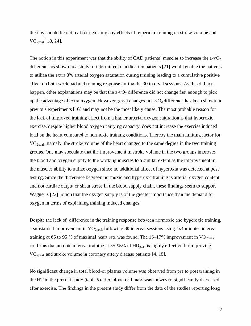
thereby should be optimal for detecting any effects of hyperoxic training on stroke volume and
VO2peak [18, 24].
The notion in this experiment was that the ability of CAD patients` muscles to increase the a-vO2
difference as shown in a study of intermittent claudication patients [21] would enable the patients
to utilize the extra 3% arterial oxygen saturation during training leading to a cumulative positive
effect on both workload and training response during the 30 interval sessions. As this did not
happen, other explanations may be that the a-vO2 difference did not change fast enough to pick
up the advantage of extra oxygen. However, great changes in a-vO2 difference has been shown in
previous experiments [16] and may not be the most likely cause. The most probable reason for
the lack of improved training effect from a higher arterial oxygen saturation is that hyperoxic
exercise, despite higher blood oxygen carrying capacity, does not increase the exercise induced
load on the heart compared to normoxic training conditions. Thereby the main limiting factor for
VO2peak, namely, the stroke volume of the heart changed to the same degree in the two training
groups. One may speculate that the improvement in stroke volume in the two groups improves
the blood and oxygen supply to the working muscles to a similar extent as the improvement in
the muscles ability to utilize oxygen since no additional affect of hyperoxia was detected at post
testing. Since the difference between normoxic and hyperoxic training is arterial oxygen content
and not cardiac output or shear stress in the blood supply chain, these findings seem to support
Wagner’s [22] notion that the oxygen supply is of the greater importance than the demand for
oxygen in terms of explaining training induced changes.
Despite the lack of difference in the training response between normoxic and hyperoxic training,
a substantial improvement in VO2peak following 30 interval sessions using 4x4 minutes interval
training at 85 to 95 % of maximal heart rate was found. The 16–17% improvement in VO2peak
confirms that aerobic interval training at 85-95% of HRpeak is highly effective for improving
VO2peak and stroke volume in coronary artery disease patients [4, 18].
No significant change in total blood-or plasma volume was observed from pre to post training in
the HT in the present study (table 5). Red blood cell mass was, however, significantly decreased
after exercise. The findings in the present study differ from the data of the studies reporting long
9

term changes in plasma volume with aerobic exercise [23], but in line with others having found
no change [2, 5]. A reduced red cell mass could decrease the oxygen carrying capacity of the
blood, and thereby could not explain the improved VO2peak in this experiment.
In the present study the HT group displayed a significant improvement in peak heart rate and
lactate concentration from pre to post training. Both the HT and the NT training groups improved
peak ventilation from pre to post training as expected from improvements in VO2peak. The HT
group did however improve ventilation to a greater extent than the NT group. Improvements in
these variables in the HT group may be a result of a greater motivation to push through to
exhaustion during VO2peak post testing. Part of the increased heart rate may be due to atria
fibrillation during exercise, and not a true difference in exertion during pre and post testing. In
the NT group significant increase in self reported perceived exertion from pre to post training
may be a factor of patients adjusting their perception of exercise strain over the course of the
training intervention. Despite differences between the groups at the pretesting point in the Borg
scale, no significant difference in R values was noted between the groups or in any of the
exercise groups between the pre and the post tests. This implies that the level of strain and
hyperventilation was equal between the tests. Therefore, change in the perceived exertion was
not an effect of greater level of exhaustion in the post test, but a factor of the patients’
interpretation of the scale.
In the present study breathing 100% oxygen enriched air during high intensity aerobic interval
training improves VO2peak to the same extent as ambient air training in stable coronary artery
disease patients with mild to moderate coronary ischemic response to exercise.
10
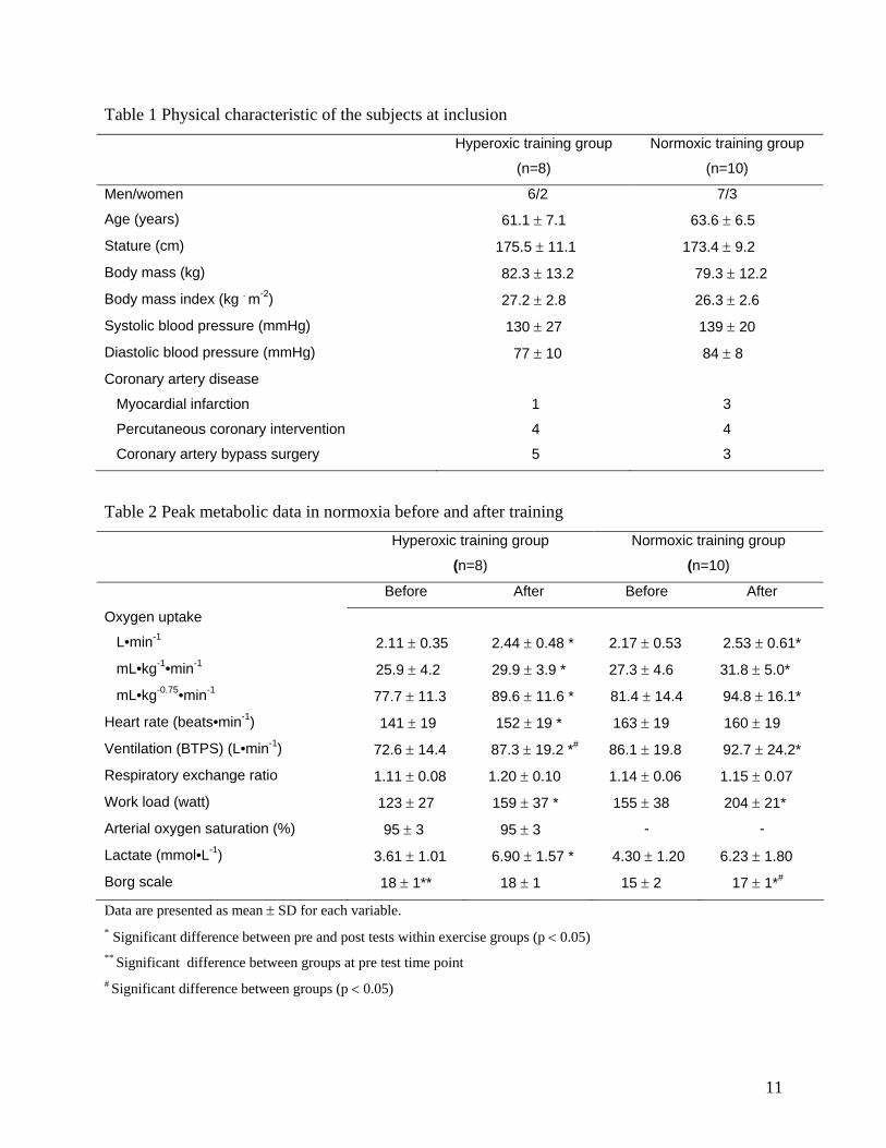
Table 1 Physical characteristic of the subjects at inclusion Hyperoxic training group
(n=8)
Normoxic training group
(n=10)
Men/women 6/2 7/3
Age (years) 61.1 ± 7.1 63.6 ± 6.5
Stature (cm) 175.5 ± 11.1 173.4 ± 9.2
Body mass (kg) 82.3 ± 13.2 79.3 ± 12.2
Body mass index (kg . m-2) 27.2 ± 2.8 26.3 ± 2.6
Systolic blood pressure (mmHg) 130 ± 27 139 ± 20
Diastolic blood pressure (mmHg) 77 ± 10 84 ± 8
Coronary artery disease
Myocardial infarction 1 3
Percutaneous coronary intervention 4 4
Coronary artery bypass surgery 5 3
Table 2 Peak metabolic data in normoxia before and after training Hyperoxic training group
(n=8)
Normoxic training group
(n=10)
Before After Before After
Oxygen uptake
L•min-1 2.11 ± 0.35 2.44 ± 0.48 * 2.17 ± 0.53 2.53 ± 0.61*
mL•kg-1•min-1 25.9 ± 4.2 29.9 ± 3.9 * 27.3 ± 4.6 31.8 ± 5.0*
mL•kg-0.75•min-1 77.7 ± 11.3 89.6 ± 11.6 * 81.4 ± 14.4 94.8 ± 16.1*
Heart rate (beats•min-1) 141 ± 19 152 ± 19 * 163 ± 19 160 ± 19
Ventilation (BTPS) (L•min-1) 72.6 ± 14.4 87.3 ± 19.2 *# 86.1 ± 19.8 92.7 ± 24.2*
Respiratory exchange ratio 1.11 ± 0.08 1.20 ± 0.10 1.14 ± 0.06 1.15 ± 0.07
Work load (watt) 123 ± 27 159 ± 37 * 155 ± 38 204 ± 21*
Arterial oxygen saturation (%) 95 ± 3 95 ± 3 - -
Lactate (mmol•L-1) 3.61 ± 1.01 6.90 ± 1.57 * 4.30 ± 1.20 6.23 ± 1.80
Borg scale 18 ± 1** 18 ± 1 15 ± 2 17 ± 1*#
Data are presented as mean ± SD for each variable. * Significant difference between pre and post tests within exercise groups (p < 0.05) ** Significant difference between groups at pre test time point
# Significant difference between groups (p < 0.05)
11
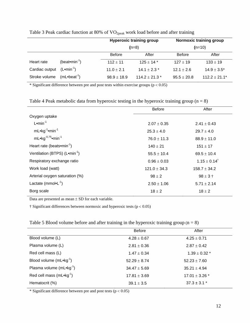
Table 3 Peak cardiac function at 80% of VO2peak work load before and after training Hyperoxic training group
(n=8)
Normoxic training group (n=10)
Before After Before After
Heart rate (beat•min-1) 112 ± 11 125 ± 14 * 127 ± 19 133 ± 19
Cardiac output (L•min-1) 11.0 ± 2.1 14.1 ± 2.3 * 12.1 ± 2.6 14.9 ± 3.5*
Stroke volume (mL•beat-1) 98.9 ± 18.9 114.2 ± 21.3 * 95.5 ± 20.8 112.2 ± 21.1*
* Significant difference between pre and post tests within exercise groups (p < 0.05)
Table 4 Peak metabolic data from hyperoxic testing in the hyperoxic training group (n = 8) Before After
Oxygen uptake
L•min-1 2.07 ± 0.35 2.41 ± 0.43
mL•kg-1•min-1 25.3 ± 4.0 29.7 ± 4.0
mL•kg-0,75•min-1 76.0 ± 11.3 88.9 ± 11.0
Heart rate (beats•min-1) 140 ± 21 151 ± 17
Ventilation (BTPS) (L•min-1) 55.5 ± 10.4 69.5 ± 10.4
Respiratory exchange ratio 0.96 ± 0.03 1.15 ± 0.14†
Work load (watt) 121.0 ± 34.3 158.7 ± 34.2
Arterial oxygen saturation (%) 98 ± 2 98 ± 3 †
Lactate (mmol•L-1) 2.50 ± 1.06 5.71 ± 2.14
Borg scale 18 ± 2 18 ± 2
Data are presented as mean ± SD for each variable.
† Significant differences between normoxic and hyperoxic tests (p < 0.05)
Table 5 Blood volume before and after training in the hyperoxic training group (n = 8) Before After
Blood volume (L) 4.28 ± 0.67 4.25 ± 0.71
Plasma volume (L) 2.81 ± 0.36 2.87 ± 0.42
Red cell mass (L) 1.47 ± 0.34 1.39 ± 0.32 *
Blood volume (mL•kg-1) 52.29 ± 8.74 52.23 ± 7.60
Plasma volume (mL•kg-1) 34.47 ± 5.69 35.21 ± 4.94
Red cell mass (mL•kg-1) 17.81 ± 3.69 17.01 ± 3.26 *
Hematocrit (%) 39.1 ± 3.5 37.3 ± 3.1 *
* Significant difference between pre and post tests (p < 0.05)
12
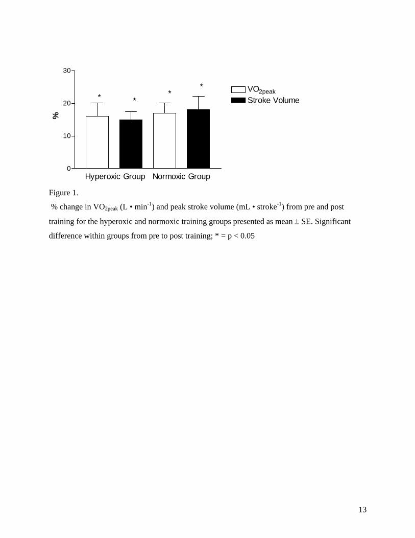
0
10
20
30
VO2peakStroke Volume*
***
Hyperoxic Group Normoxic Group
%
Figure 1.
% change in VO2peak (L • min-1) and peak stroke volume (mL • stroke-1) from pre and post
training for the hyperoxic and normoxic training groups presented as mean ± SE. Significant
difference within groups from pre to post training; * = p < 0.05
13

Acknowledgements
We gratefully acknowledge the assistance of MSc Siri Bjørgen, MSc Vigdis Schnell Husby and
research nurse Aud Hiller. The study was supported by grants from The Norwegian University of
Science and Technology and St. Olav University Hospital.
14
References
1. Borg E and Kaijser L. A comparison between three rating scales for perceived exertion and two different work tests. Scand J Med Sci Sports 2006; 16: 57-69
2. Branch JD, 3rd, Pate RR, Bourque SP, Convertino VA, Durstine JL, and Ward DS. Exercise training and intensity does not alter vascular volume responses in women. Aviat Space Environ Med 1999; 70: 1070-1076
3. Dibski DW, Smith DJ, Jensen R, Norris SR, and Ford GT. Comparison and reliability of two non-invasive acetylene uptake techniques for the measurement of cardiac output. Eur J Appl Physiol 2005; 94: 670-680
4. Erbs S, Linke A, Gielen S, Fiehn E, Walther C, Yu J, Adams V, Schuler G, and Hambrecht R. Exercise training in patients with severe chronic heart failure: Impact on left ventricular performance and cardiac size. A retrospective analysis of the leipzig heart failure training trial. Eur J Cardiovasc Prev Rehabil 2003; 10: 336-344
5. Helgerud J, Hoydal K, Wang E, Karlsen T, Berg P, Bjerkaas M, Simonsen T, Helgesen C, Hjorth N, Bach R, and Hoff J. Aerobic high-intensity intervals improve vo2max more than moderate training. Med Sci Sports Exerc 2007; 39: 665-671
6. Horvat M, Yoshida S, Prakash R, Marcus HS, Swan HJ, and Ganz W. Effect of oxygen breathing on pacing-induced angina pectoris and other manifestations of coronary insufficiency. Circulation 1972; 45: 837-844
7. Moore DP, Weston AR, Hughes JM, Oakley CM, and Cleland JG. Effects of increased inspired oxygen concentrations on exercise performance in chronic heart failure. Lancet 1992; 339: 850-853
8. Myers J, Prakash M, Froelicher V, Do D, Partington S, and Atwood JE. Exercise capacity and mortality among men referred for exercise testing. N Engl J Med 2002; 346: 793-801
9. Nielsen MH and Nielsen NC. Spectrophotometric determination of evans blue dye in plasma with individual correction for blank density by a modified gaeblers method. Scand J Clin Lab Invest 1962; 14: 605-617
10. Peltonen JE, Tikkanen HO, Ritola JJ, Ahotupa M, and Rusko HK. Oxygen uptake response during maximal cycling in hyperoxia, normoxia and hypoxia. Aviat Space Environ Med 2001; 72: 904-911
11. Perry CG, Reid J, Perry W, and Wilson BA. Effects of hyperoxic training on performance and cardiorespiratory response to exercise. Med Sci Sports Exerc 2005; 37: 1175-1179
12. Plet J, Pedersen PK, Jensen FB, and Hansen JK. Increased working capacity with hyperoxia in humans. Eur J Appl Physiol Occup Physiol 1992; 65: 171-177
15

13. Ploutz-Snyder LL, Simoneau JA, Gilders RM, Staron RS, and Hagerman FC. Cardiorespiratory and metabolic adaptations to hyperoxic training. Eur J Appl Physiol Occup Physiol 1996; 73: 38-48
14. Pons ER, Jr. and Berg JL. The physiologic advantage of oxygen during exercise in patients with coronary artery insufficiency. Dis Chest 1961; 39: 551-556
15. Restrick LJ, Davies SW, Noone L, and Wedzicha JA. Ambulatory oxygen in chronic heart failure. Lancet 1992; 340: 1192-1193
16. Richardson RS, Grassi B, Gavin TP, Haseler LJ, Tagore K, Roca J, and Wagner PD. Evidence of o2 supply-dependent vo2 max in the exercise-trained human quadriceps. J Appl Physiol 1999; 86: 1048-1053
17. Riseman JEF and Morton GB. The effect of oxygen on the exercise tolerance of patients with angina pectoris American Heart Journal 1939 18: 150-152
18. Rognmo O, Hetland E, Helgerud J, Hoff J, and Slordahl SA. High intensity aerobic interval exercise is superior to moderate intensity exercise for increasing aerobic capacity in patients with coronary artery disease. Eur J Cardiovasc Prev Rehabil 2004; 11: 216-222
19. Russell SD, Koshkarian GM, Medinger AE, Carson PE, and Higginbotham MB. Lack of effect of increased inspired oxygen concentrations on maximal exercise capacity or ventilation in stable heart failure. Am J Cardiol 1999; 84: 1412-1416
20. Selvanayagam JB, Jerosch-Herold M, Porto I, Sheridan D, Cheng AS, Petersen SE, Searle N, Channon KM, Banning AP, and Neubauer S. Resting myocardial blood flow is impaired in hibernating myocardium: A magnetic resonance study of quantitative perfusion assessment. Circulation 2005; 112: 3289-3296
21. Slordahl SA, Wang E, Hoff J, Kemi OJ, Amundsen BH, and Helgerud J. Effective training for patients with intermittent claudication. Scand Cardiovasc J 2005; 39: 244-249
22. Wagner PD. New ideas on limitations to vo2max. Exerc Sport Sci Rev 2000; 28: 10-14
23. Warburton DE, Haykowsky MJ, Quinney HA, Blackmore D, Teo KK, Taylor DA, McGavock J, and Humen DP. Blood volume expansion and cardiorespiratory function: Effects of training modality. Med Sci Sports Exerc 2004; 36: 991-1000
24. Wisloff U, Stoylen A, Loennechen JP, Bruvold M, Rognmo O, Haram PM, Tjonna AE, Helgerud J, Slordahl SA, Lee SJ, Videm V, Bye A, Smith GL, Najjar SM, Ellingsen O, and Skjaerpe T. Superior cardiovascular effect of aerobic interval training versus moderate continuous training in heart failure patients: A randomized study. Circulation 2007; 115: 3086-3094
16

PAPER IV

Is not included due to copyright
Dissertations at the Faculty of Medicine, NTNU 1977 1. Knut Joachim Berg: EFFECT OF ACETYLSALICYLIC ACID ON RENAL FUNCTION 2. Karl Erik Viken and Arne Ødegaard: STUDIES ON HUMAN MONOCYTES CULTURED IN
VITRO 1978 3. Karel Bjørn Cyvin: CONGENITAL DISLOCATION OF THE HIP JOINT. 4. Alf O. Brubakk: METHODS FOR STUDYING FLOW DYNAMICS IN THE LEFT
VENTRICLE AND THE AORTA IN MAN. 1979 5. Geirmund Unsgaard: CYTOSTATIC AND IMMUNOREGULATORY ABILITIES OF HUMAN
BLOOD MONOCYTES CULTURED IN VITRO 1980 6. Størker Jørstad: URAEMIC TOXINS 7. Arne Olav Jenssen: SOME RHEOLOGICAL, CHEMICAL AND STRUCTURAL PROPERTIES
OF MUCOID SPUTUM FROM PATIENTS WITH CHRONIC OBSTRUCTIVE BRONCHITIS 1981 8. Jens Hammerstrøm: CYTOSTATIC AND CYTOLYTIC ACTIVITY OF HUMAN
MONOCYTES AND EFFUSION MACROPHAGES AGAINST TUMOR CELLS IN VITRO 1983 9. Tore Syversen: EFFECTS OF METHYLMERCURY ON RAT BRAIN PROTEIN. 10. Torbjørn Iversen: SQUAMOUS CELL CARCINOMA OF THE VULVA. 1984 11. Tor-Erik Widerøe: ASPECTS OF CONTINUOUS AMBULATORY PERITONEAL DIALYSIS. 12. Anton Hole: ALTERATIONS OF MONOCYTE AND LYMPHOCYTE FUNCTIONS IN
REALTION TO SURGERY UNDER EPIDURAL OR GENERAL ANAESTHESIA. 13. Terje Terjesen: FRACTURE HEALING AN STRESS-PROTECTION AFTER METAL PLATE
FIXATION AND EXTERNAL FIXATION. 14. Carsten Saunte: CLUSTER HEADACHE SYNDROME. 15. Inggard Lereim: TRAFFIC ACCIDENTS AND THEIR CONSEQUENCES. 16. Bjørn Magne Eggen: STUDIES IN CYTOTOXICITY IN HUMAN ADHERENT
MONONUCLEAR BLOOD CELLS. 17. Trond Haug: FACTORS REGULATING BEHAVIORAL EFFECTS OG DRUGS. 1985 18. Sven Erik Gisvold: RESUSCITATION AFTER COMPLETE GLOBAL BRAIN ISCHEMIA. 19. Terje Espevik: THE CYTOSKELETON OF HUMAN MONOCYTES. 20. Lars Bevanger: STUDIES OF THE Ibc (c) PROTEIN ANTIGENS OF GROUP B
STREPTOCOCCI. 21. Ole-Jan Iversen: RETROVIRUS-LIKE PARTICLES IN THE PATHOGENESIS OF PSORIASIS. 22. Lasse Eriksen: EVALUATION AND TREATMENT OF ALCOHOL DEPENDENT
BEHAVIOUR. 23. Per I. Lundmo: ANDROGEN METABOLISM IN THE PROSTATE. 1986 24. Dagfinn Berntzen: ANALYSIS AND MANAGEMENT OF EXPERIMENTAL AND CLINICAL
PAIN. 25. Odd Arnold Kildahl-Andersen: PRODUCTION AND CHARACTERIZATION OF
MONOCYTE-DERIVED CYTOTOXIN AND ITS ROLE IN MONOCYTE-MEDIATED CYTOTOXICITY.
26. Ola Dale: VOLATILE ANAESTHETICS. 1987 27. Per Martin Kleveland: STUDIES ON GASTRIN. 28. Audun N. Øksendal: THE CALCIUM PARADOX AND THE HEART. 29. Vilhjalmur R. Finsen: HIP FRACTURES 1988 30. Rigmor Austgulen: TUMOR NECROSIS FACTOR: A MONOCYTE-DERIVED REGULATOR
OF CELLULAR GROWTH. 31. Tom-Harald Edna: HEAD INJURIES ADMITTED TO HOSPITAL. 32. Joseph D. Borsi: NEW ASPECTS OF THE CLINICAL PHARMACOKINETICS OF
METHOTREXATE.
33. Olav F. M. Sellevold: GLUCOCORTICOIDS IN MYOCARDIAL PROTECTION. 34. Terje Skjærpe: NONINVASIVE QUANTITATION OF GLOBAL PARAMETERS ON LEFT
VENTRICULAR FUNCTION: THE SYSTOLIC PULMONARY ARTERY PRESSURE AND CARDIAC OUTPUT.
35. Eyvind Rødahl: STUDIES OF IMMUNE COMPLEXES AND RETROVIRUS-LIKE ANTIGENS IN PATIENTS WITH ANKYLOSING SPONDYLITIS.
36. Ketil Thorstensen: STUDIES ON THE MECHANISMS OF CELLULAR UPTAKE OF IRON FROM TRANSFERRIN.
37. Anna Midelfart: STUDIES OF THE MECHANISMS OF ION AND FLUID TRANSPORT IN THE BOVINE CORNEA.
38. Eirik Helseth: GROWTH AND PLASMINOGEN ACTIVATOR ACTIVITY OF HUMAN GLIOMAS AND BRAIN METASTASES - WITH SPECIAL REFERENCE TO TRANSFORMING GROWTH FACTOR BETA AND THE EPIDERMAL GROWTH FACTOR RECEPTOR.
39. Petter C. Borchgrevink: MAGNESIUM AND THE ISCHEMIC HEART. 40. Kjell-Arne Rein: THE EFFECT OF EXTRACORPOREAL CIRCULATION ON
SUBCUTANEOUS TRANSCAPILLARY FLUID BALANCE. 41. Arne Kristian Sandvik: RAT GASTRIC HISTAMINE. 42. Carl Bredo Dahl: ANIMAL MODELS IN PSYCHIATRY. 1989 43. Torbjørn A. Fredriksen: CERVICOGENIC HEADACHE. 44. Rolf A. Walstad: CEFTAZIDIME. 45. Rolf Salvesen: THE PUPIL IN CLUSTER HEADACHE. 46. Nils Petter Jørgensen: DRUG EXPOSURE IN EARLY PREGNANCY. 47. Johan C. Ræder: PREMEDICATION AND GENERAL ANAESTHESIA IN OUTPATIENT
GYNECOLOGICAL SURGERY. 48. M. R. Shalaby: IMMUNOREGULATORY PROPERTIES OF TNF-α AND THE RELATED
CYTOKINES. 49. Anders Waage: THE COMPLEX PATTERN OF CYTOKINES IN SEPTIC SHOCK. 50. Bjarne Christian Eriksen: ELECTROSTIMULATION OF THE PELVIC FLOOR IN FEMALE
URINARY INCONTINENCE. 51. Tore B. Halvorsen: PROGNOSTIC FACTORS IN COLORECTAL CANCER. 1990 52. Asbjørn Nordby: CELLULAR TOXICITY OF ROENTGEN CONTRAST MEDIA. 53. Kåre E. Tvedt: X-RAY MICROANALYSIS OF BIOLOGICAL MATERIAL. 54. Tore C. Stiles: COGNITIVE VULNERABILITY FACTORS IN THE DEVELOPMENT AND
MAINTENANCE OF DEPRESSION. 55. Eva Hofsli: TUMOR NECROSIS FACTOR AND MULTIDRUG RESISTANCE. 56. Helge S. Haarstad: TROPHIC EFFECTS OF CHOLECYSTOKININ AND SECRETIN ON THE
RAT PANCREAS. 57. Lars Engebretsen: TREATMENT OF ACUTE ANTERIOR CRUCIATE LIGAMENT INJURIES. 58. Tarjei Rygnestad: DELIBERATE SELF-POISONING IN TRONDHEIM. 59. Arne Z. Henriksen: STUDIES ON CONSERVED ANTIGENIC DOMAINS ON MAJOR OUTER
MEMBRANE PROTEINS FROM ENTEROBACTERIA. 60. Steinar Westin: UNEMPLOYMENT AND HEALTH: Medical and social consequences of a
factory closure in a ten-year controlled follow-up study. 61. Ylva Sahlin: INJURY REGISTRATION, a tool for accident preventive work. 62. Helge Bjørnstad Pettersen: BIOSYNTHESIS OF COMPLEMENT BY HUMAN ALVEOLAR
MACROPHAGES WITH SPECIAL REFERENCE TO SARCOIDOSIS. 63. Berit Schei: TRAPPED IN PAINFUL LOVE. 64. Lars J. Vatten: PROSPECTIVE STUDIES OF THE RISK OF BREAST CANCER IN A
COHORT OF NORWEGIAN WOMAN. 1991 65. Kåre Bergh: APPLICATIONS OF ANTI-C5a SPECIFIC MONOCLONAL ANTIBODIES FOR
THE ASSESSMENT OF COMPLEMENT ACTIVATION. 66. Svein Svenningsen: THE CLINICAL SIGNIFICANCE OF INCREASED FEMORAL
ANTEVERSION. 67. Olbjørn Klepp: NONSEMINOMATOUS GERM CELL TESTIS CANCER: THERAPEUTIC
OUTCOME AND PROGNOSTIC FACTORS. 68. Trond Sand: THE EFFECTS OF CLICK POLARITY ON BRAINSTEM AUDITORY EVOKED
POTENTIALS AMPLITUDE, DISPERSION, AND LATENCY VARIABLES.
69. Kjetil B. Åsbakk: STUDIES OF A PROTEIN FROM PSORIATIC SCALE, PSO P27, WITH RESPECT TO ITS POTENTIAL ROLE IN IMMUNE REACTIONS IN PSORIASIS.
70. Arnulf Hestnes: STUDIES ON DOWN´S SYNDROME. 71. Randi Nygaard: LONG-TERM SURVIVAL IN CHILDHOOD LEUKEMIA. 72. Bjørn Hagen: THIO-TEPA. 73. Svein Anda: EVALUATION OF THE HIP JOINT BY COMPUTED TOMOGRAMPHY AND
ULTRASONOGRAPHY. 1992 74. Martin Svartberg: AN INVESTIGATION OF PROCESS AND OUTCOME OF SHORT-TERM
PSYCHODYNAMIC PSYCHOTHERAPY. 75. Stig Arild Slørdahl: AORTIC REGURGITATION. 76. Harold C Sexton: STUDIES RELATING TO THE TREATMENT OF SYMPTOMATIC NON-
PSYCHOTIC PATIENTS. 77. Maurice B. Vincent: VASOACTIVE PEPTIDES IN THE OCULAR/FOREHEAD AREA. 78. Terje Johannessen: CONTROLLED TRIALS IN SINGLE SUBJECTS. 79. Turid Nilsen: PYROPHOSPHATE IN HEPATOCYTE IRON METABOLISM. 80. Olav Haraldseth: NMR SPECTROSCOPY OF CEREBRAL ISCHEMIA AND REPERFUSION
IN RAT. 81. Eiliv Brenna: REGULATION OF FUNCTION AND GROWTH OF THE OXYNTIC MUCOSA. 1993 82. Gunnar Bovim: CERVICOGENIC HEADACHE. 83. Jarl Arne Kahn: ASSISTED PROCREATION. 84. Bjørn Naume: IMMUNOREGULATORY EFFECTS OF CYTOKINES ON NK CELLS. 85. Rune Wiseth: AORTIC VALVE REPLACEMENT. 86. Jie Ming Shen: BLOOD FLOW VELOCITY AND RESPIRATORY STUDIES. 87. Piotr Kruszewski: SUNCT SYNDROME WITH SPECIAL REFERENCE TO THE
AUTONOMIC NERVOUS SYSTEM. 88. Mette Haase Moen: ENDOMETRIOSIS. 89. Anne Vik: VASCULAR GAS EMBOLISM DURING AIR INFUSION AND AFTER
DECOMPRESSION IN PIGS. 90. Lars Jacob Stovner: THE CHIARI TYPE I MALFORMATION. 91. Kjell Å. Salvesen: ROUTINE ULTRASONOGRAPHY IN UTERO AND DEVELOPMENT IN
CHILDHOOD. 1994 92. Nina-Beate Liabakk: DEVELOPMENT OF IMMUNOASSAYS FOR TNF AND ITS SOLUBLE
RECEPTORS. 93. Sverre Helge Torp: erbB ONCOGENES IN HUMAN GLIOMAS AND MENINGIOMAS. 94. Olav M. Linaker: MENTAL RETARDATION AND PSYCHIATRY. Past and present. 95. Per Oscar Feet: INCREASED ANTIDEPRESSANT AND ANTIPANIC EFFECT IN
COMBINED TREATMENT WITH DIXYRAZINE AND TRICYCLIC ANTIDEPRESSANTS. 96. Stein Olav Samstad: CROSS SECTIONAL FLOW VELOCITY PROFILES FROM TWO-
DIMENSIONAL DOPPLER ULTRASOUND: Studies on early mitral blood flow. 97. Bjørn Backe: STUDIES IN ANTENATAL CARE. 98. Gerd Inger Ringdal: QUALITY OF LIFE IN CANCER PATIENTS. 99. Torvid Kiserud: THE DUCTUS VENOSUS IN THE HUMAN FETUS. 100. Hans E. Fjøsne: HORMONAL REGULATION OF PROSTATIC METABOLISM. 101. Eylert Brodtkorb: CLINICAL ASPECTS OF EPILEPSY IN THE MENTALLY RETARDED. 102. Roar Juul: PEPTIDERGIC MECHANISMS IN HUMAN SUBARACHNOID HEMORRHAGE. 103. Unni Syversen: CHROMOGRANIN A. Phsysiological and Clinical Role. 1995 104. Odd Gunnar Brakstad: THERMOSTABLE NUCLEASE AND THE nuc GENE IN THE
DIAGNOSIS OF Staphylococcus aureus INFECTIONS. 105. Terje Engan: NUCLEAR MAGNETIC RESONANCE (NMR) SPECTROSCOPY OF PLASMA
IN MALIGNANT DISEASE. 106. Kirsten Rasmussen: VIOLENCE IN THE MENTALLY DISORDERED. 107. Finn Egil Skjeldestad: INDUCED ABORTION: Timetrends and Determinants. 108. Roar Stenseth: THORACIC EPIDURAL ANALGESIA IN AORTOCORONARY BYPASS
SURGERY. 109. Arild Faxvaag: STUDIES OF IMMUNE CELL FUNCTION in mice infected with MURINE
RETROVIRUS. 1996
110. Svend Aakhus: NONINVASIVE COMPUTERIZED ASSESSMENT OF LEFT VENTRICULAR FUNCTION AND SYSTEMIC ARTERIAL PROPERTIES. Methodology and some clinical applications.
111. Klaus-Dieter Bolz: INTRAVASCULAR ULTRASONOGRAPHY. 112. Petter Aadahl: CARDIOVASCULAR EFFECTS OF THORACIC AORTIC CROSS-
CLAMPING. 113. Sigurd Steinshamn: CYTOKINE MEDIATORS DURING GRANULOCYTOPENIC
INFECTIONS. 114. Hans Stifoss-Hanssen: SEEKING MEANING OR HAPPINESS? 115. Anne Kvikstad: LIFE CHANGE EVENTS AND MARITAL STATUS IN RELATION TO RISK
AND PROGNOSIS OF CANSER. 116. Torbjørn Grøntvedt: TREATMENT OF ACUTE AND CHRONIC ANTERIOR CRUCIATE
LIGAMENT INJURIES. A clinical and biomechanical study. 117. Sigrid Hørven Wigers: CLINICAL STUDIES OF FIBROMYALGIA WITH FOCUS ON
ETIOLOGY, TREATMENT AND OUTCOME. 118. Jan Schjøtt: MYOCARDIAL PROTECTION: Functional and Metabolic Characteristics of Two
Endogenous Protective Principles. 119. Marit Martinussen: STUDIES OF INTESTINAL BLOOD FLOW AND ITS RELATION TO
TRANSITIONAL CIRCULATORY ADAPATION IN NEWBORN INFANTS. 120. Tomm B. Müller: MAGNETIC RESONANCE IMAGING IN FOCAL CEREBRAL ISCHEMIA. 121. Rune Haaverstad: OEDEMA FORMATION OF THE LOWER EXTREMITIES. 122. Magne Børset: THE ROLE OF CYTOKINES IN MULTIPLE MYELOMA, WITH SPECIAL
REFERENCE TO HEPATOCYTE GROWTH FACTOR. 123. Geir Smedslund: A THEORETICAL AND EMPIRICAL INVESTIGATION OF SMOKING,
STRESS AND DISEASE: RESULTS FROM A POPULATION SURVEY. 1997 124. Torstein Vik: GROWTH, MORBIDITY, AND PSYCHOMOTOR DEVELOPMENT IN
INFANTS WHO WERE GROWTH RETARDED IN UTERO. 125. Siri Forsmo: ASPECTS AND CONSEQUENCES OF OPPORTUNISTIC SCREENING FOR
CERVICAL CANCER. Results based on data from three Norwegian counties. 126. Jon S. Skranes: CEREBRAL MRI AND NEURODEVELOPMENTAL OUTCOME IN VERY
LOW BIRTH WEIGHT (VLBW) CHILDREN. A follow-up study of a geographically based year cohort of VLBW children at ages one and six years.
127. Knut Bjørnstad: COMPUTERIZED ECHOCARDIOGRAPHY FOR EVALUTION OF CORONARY ARTERY DISEASE.
128. Grethe Elisabeth Borchgrevink: DIAGNOSIS AND TREATMENT OF WHIPLASH/NECK SPRAIN INJURIES CAUSED BY CAR ACCIDENTS.
129. Tor Elsås: NEUROPEPTIDES AND NITRIC OXIDE SYNTHASE IN OCULAR AUTONOMIC AND SENSORY NERVES.
130. Rolf W. Gråwe: EPIDEMIOLOGICAL AND NEUROPSYCHOLOGICAL PERSPECTIVES ON SCHIZOPHRENIA.
131. Tonje Strømholm: CEREBRAL HAEMODYNAMICS DURING THORACIC AORTIC CROSSCLAMPING. An experimental study in pigs.
1998 132. Martinus Bråten: STUDIES ON SOME PROBLEMS REALTED TO INTRAMEDULLARY
NAILING OF FEMORAL FRACTURES. 133. Ståle Nordgård: PROLIFERATIVE ACTIVITY AND DNA CONTENT AS PROGNOSTIC
INDICATORS IN ADENOID CYSTIC CARCINOMA OF THE HEAD AND NECK. 134. Egil Lien: SOLUBLE RECEPTORS FOR TNF AND LPS: RELEASE PATTERN AND
POSSIBLE SIGNIFICANCE IN DISEASE. 135. Marit Bjørgaas: HYPOGLYCAEMIA IN CHILDREN WITH DIABETES MELLITUS 136. Frank Skorpen: GENETIC AND FUNCTIONAL ANALYSES OF DNA REPAIR IN HUMAN
CELLS. 137. Juan A. Pareja: SUNCT SYNDROME. ON THE CLINICAL PICTURE. ITS DISTINCTION
FROM OTHER, SIMILAR HEADACHES. 138. Anders Angelsen: NEUROENDOCRINE CELLS IN HUMAN PROSTATIC CARCINOMAS
AND THE PROSTATIC COMPLEX OF RAT, GUINEA PIG, CAT AND DOG. 139. Fabio Antonaci: CHRONIC PAROXYSMAL HEMICRANIA AND HEMICRANIA
CONTINUA: TWO DIFFERENT ENTITIES? 140. Sven M. Carlsen: ENDOCRINE AND METABOLIC EFFECTS OF METFORMIN WITH
SPECIAL EMPHASIS ON CARDIOVASCULAR RISK FACTORES. 1999
141. Terje A. Murberg: DEPRESSIVE SYMPTOMS AND COPING AMONG PATIENTS WITH CONGESTIVE HEART FAILURE.
142. Harm-Gerd Karl Blaas: THE EMBRYONIC EXAMINATION. Ultrasound studies on the development of the human embryo.
143. Noèmi Becser Andersen:THE CEPHALIC SENSORY NERVES IN UNILATERAL HEADACHES. Anatomical background and neurophysiological evaluation.
144. Eli-Janne Fiskerstrand: LASER TREATMENT OF PORT WINE STAINS. A study of the efficacy and limitations of the pulsed dye laser. Clinical and morfological analyses aimed at improving the therapeutic outcome.
145. Bård Kulseng: A STUDY OF ALGINATE CAPSULE PROPERTIES AND CYTOKINES IN RELATION TO INSULIN DEPENDENT DIABETES MELLITUS.
146. Terje Haug: STRUCTURE AND REGULATION OF THE HUMAN UNG GENE ENCODING URACIL-DNA GLYCOSYLASE.
147. Heidi Brurok: MANGANESE AND THE HEART. A Magic Metal with Diagnostic and Therapeutic Possibilites.
148. Agnes Kathrine Lie: DIAGNOSIS AND PREVALENCE OF HUMAN PAPILLOMAVIRUS INFECTION IN CERVICAL INTRAEPITELIAL NEOPLASIA. Relationship to Cell Cycle Regulatory Proteins and HLA DQBI Genes.
149. Ronald Mårvik: PHARMACOLOGICAL, PHYSIOLOGICAL AND PATHOPHYSIOLOGICAL STUDIES ON ISOLATED STOMACS.
150. Ketil Jarl Holen: THE ROLE OF ULTRASONOGRAPHY IN THE DIAGNOSIS AND TREATMENT OF HIP DYSPLASIA IN NEWBORNS.
151. Irene Hetlevik: THE ROLE OF CLINICAL GUIDELINES IN CARDIOVASCULAR RISK INTERVENTION IN GENERAL PRACTICE.
152. Katarina Tunòn: ULTRASOUND AND PREDICTION OF GESTATIONAL AGE. 153. Johannes Soma: INTERACTION BETWEEN THE LEFT VENTRICLE AND THE SYSTEMIC
ARTERIES. 154. Arild Aamodt: DEVELOPMENT AND PRE-CLINICAL EVALUATION OF A CUSTOM-
MADE FEMORAL STEM. 155. Agnar Tegnander: DIAGNOSIS AND FOLLOW-UP OF CHILDREN WITH SUSPECTED OR
KNOWN HIP DYSPLASIA. 156. Bent Indredavik: STROKE UNIT TREATMENT: SHORT AND LONG-TERM EFFECTS 157. Jolanta Vanagaite Vingen: PHOTOPHOBIA AND PHONOPHOBIA IN PRIMARY
HEADACHES 2000 158. Ola Dalsegg Sæther: PATHOPHYSIOLOGY DURING PROXIMAL AORTIC CROSS-
CLAMPING CLINICAL AND EXPERIMENTAL STUDIES 159. xxxxxxxxx (blind number) 160. Christina Vogt Isaksen: PRENATAL ULTRASOUND AND POSTMORTEM FINDINGS – A
TEN YEAR CORRELATIVE STUDY OF FETUSES AND INFANTS WITH DEVELOPMENTAL ANOMALIES.
161. Holger Seidel: HIGH-DOSE METHOTREXATE THERAPY IN CHILDREN WITH ACUTE LYMPHOCYTIC LEUKEMIA: DOSE, CONCENTRATION, AND EFFECT CONSIDERATIONS.
162. Stein Hallan: IMPLEMENTATION OF MODERN MEDICAL DECISION ANALYSIS INTO CLINICAL DIAGNOSIS AND TREATMENT.
163. Malcolm Sue-Chu: INVASIVE AND NON-INVASIVE STUDIES IN CROSS-COUNTRY SKIERS WITH ASTHMA-LIKE SYMPTOMS.
164. Ole-Lars Brekke: EFFECTS OF ANTIOXIDANTS AND FATTY ACIDS ON TUMOR NECROSIS FACTOR-INDUCED CYTOTOXICITY.
165. Jan Lundbom: AORTOCORONARY BYPASS SURGERY: CLINICAL ASPECTS, COST CONSIDERATIONS AND WORKING ABILITY.
166. John-Anker Zwart: LUMBAR NERVE ROOT COMPRESSION, BIOCHEMICAL AND NEUROPHYSIOLOGICAL ASPECTS.
167. Geir Falck: HYPEROSMOLALITY AND THE HEART. 168. Eirik Skogvoll: CARDIAC ARREST Incidence, Intervention and Outcome. 169. Dalius Bansevicius: SHOULDER-NECK REGION IN CERTAIN HEADACHES AND
CHRONIC PAIN SYNDROMES. 170. Bettina Kinge: REFRACTIVE ERRORS AND BIOMETRIC CHANGES AMONG
UNIVERSITY STUDENTS IN NORWAY. 171. Gunnar Qvigstad: CONSEQUENCES OF HYPERGASTRINEMIA IN MAN
172. Hanne Ellekjær: EPIDEMIOLOGICAL STUDIES OF STROKE IN A NORWEGIAN POPULATION. INCIDENCE, RISK FACTORS AND PROGNOSIS
173. Hilde Grimstad: VIOLENCE AGAINST WOMEN AND PREGNANCY OUTCOME. 174. Astrid Hjelde: SURFACE TENSION AND COMPLEMENT ACTIVATION: Factors influencing
bubble formation and bubble effects after decompression. 175. Kjell A. Kvistad: MR IN BREAST CANCER – A CLINICAL STUDY. 176. Ivar Rossvoll: ELECTIVE ORTHOPAEDIC SURGERY IN A DEFINED POPULATION. Studies
on demand, waiting time for treatment and incapacity for work. 177. Carina Seidel: PROGNOSTIC VALUE AND BIOLOGICAL EFFECTS OF HEPATOCYTE
GROWTH FACTOR AND SYNDECAN-1 IN MULTIPLE MYELOMA. 2001 178. Alexander Wahba: THE INFLUENCE OF CARDIOPULMONARY BYPASS ON PLATELET
FUNCTION AND BLOOD COAGULATION – DETERMINANTS AND CLINICAL CONSEQUENSES
179. Marcus Schmitt-Egenolf: THE RELEVANCE OF THE MAJOR hISTOCOMPATIBILITY COMPLEX FOR THE GENETICS OF PSORIASIS
180. Odrun Arna Gederaas: BIOLOGICAL MECHANISMS INVOLVED IN 5-AMINOLEVULINIC ACID BASED PHOTODYNAMIC THERAPY
181. Pål Richard Romundstad: CANCER INCIDENCE AMONG NORWEGIAN ALUMINIUM WORKERS
182. Henrik Hjorth-Hansen: NOVEL CYTOKINES IN GROWTH CONTROL AND BONE DISEASE OF MULTIPLE MYELOMA
183. Gunnar Morken: SEASONAL VARIATION OF HUMAN MOOD AND BEHAVIOUR 184. Bjørn Olav Haugen: MEASUREMENT OF CARDIAC OUTPUT AND STUDIES OF
VELOCITY PROFILES IN AORTIC AND MITRAL FLOW USING TWO- AND THREE-DIMENSIONAL COLOUR FLOW IMAGING
185. Geir Bråthen: THE CLASSIFICATION AND CLINICAL DIAGNOSIS OF ALCOHOL-RELATED SEIZURES
186. Knut Ivar Aasarød: RENAL INVOLVEMENT IN INFLAMMATORY RHEUMATIC DISEASE. A Study of Renal Disease in Wegener’s Granulomatosis and in Primary Sjögren’s Syndrome
187. Trude Helen Flo: RESEPTORS INVOLVED IN CELL ACTIVATION BY DEFINED URONIC ACID POLYMERS AND BACTERIAL COMPONENTS
188. Bodil Kavli: HUMAN URACIL-DNA GLYCOSYLASES FROM THE UNG GENE: STRUCTRUAL BASIS FOR SUBSTRATE SPECIFICITY AND REPAIR
189. Liv Thommesen: MOLECULAR MECHANISMS INVOLVED IN TNF- AND GASTRIN-MEDIATED GENE REGULATION
190. Turid Lingaas Holmen: SMOKING AND HEALTH IN ADOLESCENCE; THE NORD-TRØNDELAG HEALTH STUDY, 1995-97
191. Øyvind Hjertner: MULTIPLE MYELOMA: INTERACTIONS BETWEEN MALIGNANT PLASMA CELLS AND THE BONE MICROENVIRONMENT
192. Asbjørn Støylen: STRAIN RATE IMAGING OF THE LEFT VENTRICLE BY ULTRASOUND. FEASIBILITY, CLINICAL VALIDATION AND PHYSIOLOGICAL ASPECTS
193. Kristian Midthjell: DIABETES IN ADULTS IN NORD-TRØNDELAG. PUBLIC HEALTH ASPECTS OF DIABETES MELLITUS IN A LARGE, NON-SELECTED NORWEGIAN POPULATION.
194. Guanglin Cui: FUNCTIONAL ASPECTS OF THE ECL CELL IN RODENTS 195. Ulrik Wisløff: CARDIAC EFFECTS OF AEROBIC ENDURANCE TRAINING:
HYPERTROPHY, CONTRACTILITY AND CALCUIM HANDLING IN NORMAL AND FAILING HEART
196. Øyvind Halaas: MECHANISMS OF IMMUNOMODULATION AND CELL-MEDIATED CYTOTOXICITY INDUCED BY BACTERIAL PRODUCTS
197. Tore Amundsen: PERFUSION MR IMAGING IN THE DIAGNOSIS OF PULMONARY EMBOLISM
198. Nanna Kurtze: THE SIGNIFICANCE OF ANXIETY AND DEPRESSION IN FATIQUE AND PATTERNS OF PAIN AMONG INDIVIDUALS DIAGNOSED WITH FIBROMYALGIA: RELATIONS WITH QUALITY OF LIFE, FUNCTIONAL DISABILITY, LIFESTYLE, EMPLOYMENT STATUS, CO-MORBIDITY AND GENDER
199. Tom Ivar Lund Nilsen: PROSPECTIVE STUDIES OF CANCER RISK IN NORD-TRØNDELAG: THE HUNT STUDY. Associations with anthropometric, socioeconomic, and lifestyle risk factors
200. Asta Kristine Håberg: A NEW APPROACH TO THE STUDY OF MIDDLE CEREBRAL ARTERY OCCLUSION IN THE RAT USING MAGNETIC RESONANCE TECHNIQUES
2002 201. Knut Jørgen Arntzen: PREGNANCY AND CYTOKINES 202. Henrik Døllner: INFLAMMATORY MEDIATORS IN PERINATAL INFECTIONS 203. Asta Bye: LOW FAT, LOW LACTOSE DIET USED AS PROPHYLACTIC TREATMENT OF
ACUTE INTESTINAL REACTIONS DURING PELVIC RADIOTHERAPY. A PROSPECTIVE RANDOMISED STUDY.
204. Sylvester Moyo: STUDIES ON STREPTOCOCCUS AGALACTIAE (GROUP B STREPTOCOCCUS) SURFACE-ANCHORED MARKERS WITH EMPHASIS ON STRAINS AND HUMAN SERA FROM ZIMBABWE.
205. Knut Hagen: HEAD-HUNT: THE EPIDEMIOLOGY OF HEADACHE IN NORD-TRØNDELAG 206. Li Lixin: ON THE REGULATION AND ROLE OF UNCOUPLING PROTEIN-2 IN INSULIN
PRODUCING ß-CELLS 207. Anne Hildur Henriksen: SYMPTOMS OF ALLERGY AND ASTHMA VERSUS MARKERS OF
LOWER AIRWAY INFLAMMATION AMONG ADOLESCENTS 208. Egil Andreas Fors: NON-MALIGNANT PAIN IN RELATION TO PSYCHOLOGICAL AND
ENVIRONTENTAL FACTORS. EXPERIENTAL AND CLINICAL STUDES OF PAIN WITH FOCUS ON FIBROMYALGIA
209. Pål Klepstad: MORPHINE FOR CANCER PAIN 210. Ingunn Bakke: MECHANISMS AND CONSEQUENCES OF PEROXISOME PROLIFERATOR-
INDUCED HYPERFUNCTION OF THE RAT GASTRIN PRODUCING CELL 211. Ingrid Susann Gribbestad: MAGNETIC RESONANCE IMAGING AND SPECTROSCOPY OF
BREAST CANCER 212. Rønnaug Astri Ødegård: PREECLAMPSIA – MATERNAL RISK FACTORS AND FETAL
GROWTH 213. Johan Haux: STUDIES ON CYTOTOXICITY INDUCED BY HUMAN NATURAL KILLER
CELLS AND DIGITOXIN 214. Turid Suzanne Berg-Nielsen: PARENTING PRACTICES AND MENTALLY DISORDERED
ADOLESCENTS 215. Astrid Rydning: BLOOD FLOW AS A PROTECTIVE FACTOR FOR THE STOMACH
MUCOSA. AN EXPERIMENTAL STUDY ON THE ROLE OF MAST CELLS AND SENSORY AFFERENT NEURONS
2003 216. Jan Pål Loennechen: HEART FAILURE AFTER MYOCARDIAL INFARCTION. Regional
Differences, Myocyte Function, Gene Expression, and Response to Cariporide, Losartan, and Exercise Training.
217. Elisabeth Qvigstad: EFFECTS OF FATTY ACIDS AND OVER-STIMULATION ON INSULIN SECRETION IN MAN
218. Arne Åsberg: EPIDEMIOLOGICAL STUDIES IN HEREDITARY HEMOCHROMATOSIS: PREVALENCE, MORBIDITY AND BENEFIT OF SCREENING.
219. Johan Fredrik Skomsvoll: REPRODUCTIVE OUTCOME IN WOMEN WITH RHEUMATIC DISEASE. A population registry based study of the effects of inflammatory rheumatic disease and connective tissue disease on reproductive outcome in Norwegian women in 1967-1995.
220. Siv Mørkved: URINARY INCONTINENCE DURING PREGNANCY AND AFTER DELIVERY: EFFECT OF PELVIC FLOOR MUSCLE TRAINING IN PREVENTION AND TREATMENT
221. Marit S. Jordhøy: THE IMPACT OF COMPREHENSIVE PALLIATIVE CARE 222. Tom Christian Martinsen: HYPERGASTRINEMIA AND HYPOACIDITY IN RODENTS –
CAUSES AND CONSEQUENCES 223. Solveig Tingulstad: CENTRALIZATION OF PRIMARY SURGERY FOR OVARAIN CANCER.
FEASIBILITY AND IMPACT ON SURVIVAL 224. Haytham Eloqayli: METABOLIC CHANGES IN THE BRAIN CAUSED BY EPILEPTIC
SEIZURES 225. Torunn Bruland: STUDIES OF EARLY RETROVIRUS-HOST INTERACTIONS – VIRAL
DETERMINANTS FOR PATHOGENESIS AND THE INFLUENCE OF SEX ON THE SUSCEPTIBILITY TO FRIEND MURINE LEUKAEMIA VIRUS INFECTION
226. Torstein Hole: DOPPLER ECHOCARDIOGRAPHIC EVALUATION OF LEFT VENTRICULAR FUNCTION IN PATIENTS WITH ACUTE MYOCARDIAL INFARCTION
227. Vibeke Nossum: THE EFFECT OF VASCULAR BUBBLES ON ENDOTHELIAL FUNCTION 228. Sigurd Fasting: ROUTINE BASED RECORDING OF ADVERSE EVENTS DURING
ANAESTHESIA – APPLICATION IN QUALITY IMPROVEMENT AND SAFETY 229. Solfrid Romundstad: EPIDEMIOLOGICAL STUDIES OF MICROALBUMINURIA. THE
NORD-TRØNDELAG HEALTH STUDY 1995-97 (HUNT 2)
230. Geir Torheim: PROCESSING OF DYNAMIC DATA SETS IN MAGNETIC RESONANCE IMAGING
231. Catrine Ahlén: SKIN INFECTIONS IN OCCUPATIONAL SATURATION DIVERS IN THE NORTH SEA AND THE IMPACT OF THE ENVIRONMENT
232. Arnulf Langhammer: RESPIRATORY SYMPTOMS, LUNG FUNCTION AND BONE MINERAL DENSITY IN A COMPREHENSIVE POPULATION SURVEY. THE NORD-TRØNDELAG HEALTH STUDY 1995-97. THE BRONCHIAL OBSTRUCTION IN NORD-TRØNDELAG STUDY
233. Einar Kjelsås: EATING DISORDERS AND PHYSICAL ACTIVITY IN NON-CLINICAL SAMPLES
234. Arne Wibe: RECTAL CANCER TREATMENT IN NORWAY – STANDARDISATION OF SURGERY AND QUALITY ASSURANCE
2004 235. Eivind Witsø: BONE GRAFT AS AN ANTIBIOTIC CARRIER 236. Anne Mari Sund: DEVELOPMENT OF DEPRESSIVE SYMPTOMS IN EARLY
ADOLESCENCE 237. Hallvard Lærum: EVALUATION OF ELECTRONIC MEDICAL RECORDS – A CLINICAL
TASK PERSPECTIVE 238. Gustav Mikkelsen: ACCESSIBILITY OF INFORMATION IN ELECTRONIC PATIENT
RECORDS; AN EVALUATION OF THE ROLE OF DATA QUALITY 239. Steinar Krokstad: SOCIOECONOMIC INEQUALITIES IN HEALTH AND DISABILITY.
SOCIAL EPIDEMIOLOGY IN THE NORD-TRØNDELAG HEALTH STUDY (HUNT), NORWAY
240. Arne Kristian Myhre: NORMAL VARIATION IN ANOGENITAL ANATOMY AND MICROBIOLOGY IN NON-ABUSED PRESCHOOL CHILDREN
241. Ingunn Dybedal: NEGATIVE REGULATORS OF HEMATOPOIETEC STEM AND PROGENITOR CELLS
242. Beate Sitter: TISSUE CHARACTERIZATION BY HIGH RESOLUTION MAGIC ANGLE SPINNING MR SPECTROSCOPY
243. Per Arne Aas: MACROMOLECULAR MAINTENANCE IN HUMAN CELLS – REPAIR OF URACIL IN DNA AND METHYLATIONS IN DNA AND RNA
244. Anna Bofin: FINE NEEDLE ASPIRATION CYTOLOGY IN THE PRIMARY INVESTIGATION OF BREAST TUMOURS AND IN THE DETERMINATION OF TREATMENT STRATEGIES
245. Jim Aage Nøttestad: DEINSTITUTIONALIZATION AND MENTAL HEALTH CHANGES AMONG PEOPLE WITH MENTAL RETARDATION
246. Reidar Fossmark: GASTRIC CANCER IN JAPANESE COTTON RATS 247. Wibeke Nordhøy: MANGANESE AND THE HEART, INTRACELLULAR MR RELAXATION
AND WATER EXCHANGE ACROSS THE CARDIAC CELL MEMBRANE 2005 248. Sturla Molden: QUANTITATIVE ANALYSES OF SINGLE UNITS RECORDED FROM THE
HIPPOCAMPUS AND ENTORHINAL CORTEX OF BEHAVING RATS 249. Wenche Brenne Drøyvold: EPIDEMIOLOGICAL STUDIES ON WEIGHT CHANGE AND
HEALTH IN A LARGE POPULATION. THE NORD-TRØNDELAG HEALTH STUDY (HUNT)
250. Ragnhild Støen: ENDOTHELIUM-DEPENDENT VASODILATION IN THE FEMORAL ARTERY OF DEVELOPING PIGLETS
251. Aslak Steinsbekk: HOMEOPATHY IN THE PREVENTION OF UPPER RESPIRATORY TRACT INFECTIONS IN CHILDREN
252. Hill-Aina Steffenach: MEMORY IN HIPPOCAMPAL AND CORTICO-HIPPOCAMPAL CIRCUITS
253. Eystein Stordal: ASPECTS OF THE EPIDEMIOLOGY OF DEPRESSIONS BASED ON SELF-RATING IN A LARGE GENERAL HEALTH STUDY (THE HUNT-2 STUDY)
254. Viggo Pettersen: FROM MUSCLES TO SINGING: THE ACTIVITY OF ACCESSORY BREATHING MUSCLES AND THORAX MOVEMENT IN CLASSICAL SINGING
255. Marianne Fyhn: SPATIAL MAPS IN THE HIPPOCAMPUS AND ENTORHINAL CORTEX 256. Robert Valderhaug: OBSESSIVE-COMPULSIVE DISORDER AMONG CHILDREN AND
ADOLESCENTS: CHARACTERISTICS AND PSYCHOLOGICAL MANAGEMENT OF PATIENTS IN OUTPATIENT PSYCHIATRIC CLINICS
257. Erik Skaaheim Haug: INFRARENAL ABDOMINAL AORTIC ANEURYSMS – COMORBIDITY AND RESULTS FOLLOWING OPEN SURGERY
258. Daniel Kondziella: GLIAL-NEURONAL INTERACTIONS IN EXPERIMENTAL BRAIN DISORDERS
259. Vegard Heimly Brun: ROUTES TO SPATIAL MEMORY IN HIPPOCAMPAL PLACE CELLS 260. Kenneth McMillan: PHYSIOLOGICAL ASSESSMENT AND TRAINING OF ENDURANCE
AND STRENGTH IN PROFESSIONAL YOUTH SOCCER PLAYERS 261. Marit Sæbø Indredavik: MENTAL HEALTH AND CEREBRAL MAGNETIC RESONANCE
IMAGING IN ADOLESCENTS WITH LOW BIRTH WEIGHT 262. Ole Johan Kemi: ON THE CELLULAR BASIS OF AEROBIC FITNESS, INTENSITY-
DEPENDENCE AND TIME-COURSE OF CARDIOMYOCYTE AND ENDOTHELIAL ADAPTATIONS TO EXERCISE TRAINING
263. Eszter Vanky: POLYCYSTIC OVARY SYNDROME – METFORMIN TREATMENT IN PREGNANCY
264. Hild Fjærtoft: EXTENDED STROKE UNIT SERVICE AND EARLY SUPPORTED DISCHARGE. SHORT AND LONG-TERM EFFECTS
265. Grete Dyb: POSTTRAUMATIC STRESS REACTIONS IN CHILDREN AND ADOLESCENTS 266. Vidar Fykse: SOMATOSTATIN AND THE STOMACH 267. Kirsti Berg: OXIDATIVE STRESS AND THE ISCHEMIC HEART: A STUDY IN PATIENTS
UNDERGOING CORONARY REVASCULARIZATION 268. Björn Inge Gustafsson: THE SEROTONIN PRODUCING ENTEROCHROMAFFIN CELL,
AND EFFECTS OF HYPERSEROTONINEMIA ON HEART AND BONE 2006 269. Torstein Baade Rø: EFFECTS OF BONE MORPHOGENETIC PROTEINS, HEPATOCYTE
GROWTH FACTOR AND INTERLEUKIN-21 IN MULTIPLE MYELOMA 270. May-Britt Tessem: METABOLIC EFFECTS OF ULTRAVIOLET RADIATION ON THE
ANTERIOR PART OF THE EYE 271. Anne-Sofie Helvik: COPING AND EVERYDAY LIFE IN A POPULATION OF ADULTS
WITH HEARING IMPAIRMENT 272. Therese Standal: MULTIPLE MYELOMA: THE INTERPLAY BETWEEN MALIGNANT
PLASMA CELLS AND THE BONE MARROW MICROENVIRONMENT 273. Ingvild Saltvedt: TREATMENT OF ACUTELY SICK, FRAIL ELDERLY PATIENTS IN A
GERIATRIC EVALUATION AND MANAGEMENT UNIT – RESULTS FROM A PROSPECTIVE RANDOMISED TRIAL
274. Birger Henning Endreseth: STRATEGIES IN RECTAL CANCER TREATMENT – FOCUS ON EARLY RECTAL CANCER AND THE INFLUENCE OF AGE ON PROGNOSIS
275. Anne Mari Aukan Rokstad: ALGINATE CAPSULES AS BIOREACTORS FOR CELL THERAPY
276. Mansour Akbari: HUMAN BASE EXCISION REPAIR FOR PRESERVATION OF GENOMIC STABILITY
277. Stein Sundstrøm: IMPROVING TREATMENT IN PATIENTS WITH LUNG CANCER – RESULTS FROM TWO MULITCENTRE RANDOMISED STUDIES
278. Hilde Pleym: BLEEDING AFTER CORONARY ARTERY BYPASS SURGERY - STUDIES ON HEMOSTATIC MECHANISMS, PROPHYLACTIC DRUG TREATMENT AND EFFECTS OF AUTOTRANSFUSION
279. Line Merethe Oldervoll: PHYSICAL ACTIVITY AND EXERCISE INTERVENTIONS IN CANCER PATIENTS
280. Boye Welde: THE SIGNIFICANCE OF ENDURANCE TRAINING, RESISTANCE TRAINING AND MOTIVATIONAL STYLES IN ATHLETIC PERFORMANCE AMONG ELITE JUNIOR CROSS-COUNTRY SKIERS
281. Per Olav Vandvik: IRRITABLE BOWEL SYNDROME IN NORWAY, STUDIES OF PREVALENCE, DIAGNOSIS AND CHARACTERISTICS IN GENERAL PRACTICE AND IN THE POPULATION
282. Idar Kirkeby-Garstad: CLINICAL PHYSIOLOGY OF EARLY MOBILIZATION AFTER CARDIAC SURGERY
283. Linn Getz: SUSTAINABLE AND RESPONSIBLE PREVENTIVE MEDICINE. CONCEPTUALISING ETHICAL DILEMMAS ARISING FROM CLINICAL IMPLEMENTATION OF ADVANCING MEDICAL TECHNOLOGY
284. Eva Tegnander: DETECTION OF CONGENITAL HEART DEFECTS IN A NON-SELECTED POPULATION OF 42,381 FETUSES
285. Kristin Gabestad Nørsett: GENE EXPRESSION STUDIES IN GASTROINTESTINAL PATHOPHYSIOLOGY AND NEOPLASIA
286. Per Magnus Haram: GENETIC VS. AQUIRED FITNESS: METABOLIC, VASCULAR AND CARDIOMYOCYTE ADAPTATIONS
287. Agneta Johansson: GENERAL RISK FACTORS FOR GAMBLING PROBLEMS AND THE PREVALENCE OG PATHOLOGICAL GAMBLING IN NORWAY
288. Svein Artur Jensen: THE PREVALENCE OF SYMPTOMATIC ARTERIAL DISEASE OF THE LOWER LIMB
289. Charlotte Björk Ingul: QUANITIFICATION OF REGIONAL MYOCARDIAL FUNCTION BY STRAIN RATE AND STRAIN FOR EVALUATION OF CORONARY ARTERY DISEASE. AUTOMATED VERSUS MANUAL ANALYSIS DURING ACUTE MYOCARDIAL INFARCTION AND DOBUTAMINE STRESS ECHOCARDIOGRAPHY
290. Jakob Nakling: RESULTS AND CONSEQUENCES OF ROUTINE ULTRASOUND SCREENING IN PREGNANCY – A GEOGRAPHIC BASED POPULATION STUDY
291. Anne Engum: DEPRESSION AND ANXIETY – THEIR RELATIONS TO THYROID DYSFUNCTION AND DIABETES IN A LARGE EPIDEMIOLOGICAL STUDY
292. Ottar Bjerkeset: ANXIETY AND DEPRESSION IN THE GENERAL POPULATION: RISK FACTORS, INTERVENTION AND OUTCOME – THE NORD-TRØNDELAG HEALTH STUDY (HUNT)
293. Jon Olav Drogset: RESULTS AFTER SURGICAL TREATMENT OF ANTERIOR CRUCIATE LIGAMENT INJURIES – A CLINICAL STUDY
294. Lars Fosse: MECHANICAL BEHAVIOUR OF COMPACTED MORSELLISED BONE – AN EXPERIMENTAL IN VITRO STUDY
295. Gunilla Klensmeden Fosse: MENTAL HEALTH OF PSYCHIATRIC OUTPATIENTS BULLIED IN CHILDHOOD
296. Paul Jarle Mork: MUSCLE ACTIVITY IN WORK AND LEISURE AND ITS ASSOCIATION TO MUSCULOSKELETAL PAIN
297. Björn Stenström: LESSONS FROM RODENTS: I: MECHANISMS OF OBESITY SURGERY – ROLE OF STOMACH. II: CARCINOGENIC EFFECTS OF HELICOBACTER PYLORI AND SNUS IN THE STOMACH
2007 298. Haakon R. Skogseth: INVASIVE PROPERTIES OF CANCER – A TREATMENT TARGET ?
IN VITRO STUDIES IN HUMAN PROSTATE CANCER CELL LINES 299. Janniche Hammer: GLUTAMATE METABOLISM AND CYCLING IN MESIAL TEMPORAL
LOBE EPILEPSY 300. May Britt Drugli: YOUNG CHILDREN TREATED BECAUSE OF ODD/CD: CONDUCT
PROBLEMS AND SOCIAL COMPETENCIES IN DAY-CARE AND SCHOOL SETTINGS 301. Arne Skjold: MAGNETIC RESONANCE KINETICS OF MANGANESE DIPYRIDOXYL
DIPHOSPHATE (MnDPDP) IN HUMAN MYOCARDIUM. STUDIES IN HEALTHY VOLUNTEERS AND IN PATIENTS WITH RECENT MYOCARDIAL INFARCTION
302. Siri Malm: LEFT VENTRICULAR SYSTOLIC FUNCTION AND MYOCARDIAL PERFUSION ASSESSED BY CONTRAST ECHOCARDIOGRAPHY
303. Valentina Maria do Rosario Cabral Iversen: MENTAL HEALTH AND PSYCHOLOGICAL ADAPTATION OF CLINICAL AND NON-CLINICAL MIGRANT GROUPS
304. Lasse Løvstakken: SIGNAL PROCESSING IN DIAGNOSTIC ULTRASOUND: ALGORITHMS FOR REAL-TIME ESTIMATION AND VISUALIZATION OF BLOOD FLOW VELOCITY
305. Elisabeth Olstad: GLUTAMATE AND GABA: MAJOR PLAYERS IN NEURONAL METABOLISM
306. Lilian Leistad: THE ROLE OF CYTOKINES AND PHOSPHOLIPASE A2s IN ARTICULAR CARTILAGE CHONDROCYTES IN RHEUMATOID ARTHRITIS AND OSTEOARTHRITIS
307. Arne Vaaler: EFFECTS OF PSYCHIATRIC INTENSIVE CARE UNIT IN AN ACUTE PSYCIATHRIC WARD
308. Mathias Toft: GENETIC STUDIES OF LRRK2 AND PINK1 IN PARKINSON’S DISEASE 309. Ingrid Løvold Mostad: IMPACT OF DIETARY FAT QUANTITY AND QUALITY IN TYPE 2
DIABETES WITH EMPHASIS ON MARINE N-3 FATTY ACIDS 310. Torill Eidhammer Sjøbakk: MR DETERMINED BRAIN METABOLIC PATTERN IN
PATIENTS WITH BRAIN METASTASES AND ADOLESCENTS WITH LOW BIRTH WEIGHT
311. Vidar Beisvåg: PHYSIOLOGICAL GENOMICS OF HEART FAILURE: FROM TECHNOLOGY TO PHYSIOLOGY
312. Olav Magnus Søndenå Fredheim: HEALTH RELATED QUALITY OF LIFE ASSESSMENT AND ASPECTS OF THE CLINICAL PHARMACOLOGY OF METHADONE IN PATIENTS WITH CHRONIC NON-MALIGNANT PAIN
313. Anne Brantberg: FETAL AND PERINATAL IMPLICATIONS OF ANOMALIES IN THE GASTROINTESTINAL TRACT AND THE ABDOMINAL WALL
314. Erik Solligård: GUT LUMINAL MICRODIALYSIS 315. Elin Tollefsen: RESPIRATORY SYMPTOMS IN A COMPREHENSIVE POPULATION
BASED STUDY AMONG ADOLESCENTS 13-19 YEARS. YOUNG-HUNT 1995-97 AND 2000-01; THE NORD-TRØNDELAG HEALTH STUDIES (HUNT)
316. Anne-Tove Brenne: GROWTH REGULATION OF MYELOMA CELLS 317. Heidi Knobel: FATIGUE IN CANCER TREATMENT – ASSESSMENT, COURSE AND
ETIOLOGY 318. Torbjørn Dahl: CAROTID ARTERY STENOSIS. DIAGNOSTIC AND THERAPEUTIC
ASPECTS 319. Inge-Andre Rasmussen jr.: FUNCTIONAL AND DIFFUSION TENSOR MAGNETIC
RESONANCE IMAGING IN NEUROSURGICAL PATIENTS 320. Grete Helen Bratberg: PUBERTAL TIMING – ANTECEDENT TO RISK OR RESILIENCE ?
EPIDEMIOLOGICAL STUDIES ON GROWTH, MATURATION AND HEALTH RISK BEHAVIOURS; THE YOUNG HUNT STUDY, NORD-TRØNDELAG, NORWAY
321. Sveinung Sørhaug: THE PULMONARY NEUROENDOCRINE SYSTEM. PHYSIOLOGICAL, PATHOLOGICAL AND TUMOURIGENIC ASPECTS
322. Olav Sande Eftedal: ULTRASONIC DETECTION OF DECOMPRESSION INDUCED VASCULAR MICROBUBBLES
323. Rune Bang Leistad: PAIN, AUTONOMIC ACTIVATION AND MUSCULAR ACTIVITY RELATED TO EXPERIMENTALLY-INDUCED COGNITIVE STRESS IN HEADACHE PATIENTS
324. Svein Brekke: TECHNIQUES FOR ENHANCEMENT OF TEMPORAL RESOLUTION IN THREE-DIMENSIONAL ECHOCARDIOGRAPHY
325. Kristian Bernhard Nilsen: AUTONOMIC ACTIVATION AND MUSCLE ACTIVITY IN RELATION TO MUSCULOSKELETAL PAIN
326. Anne Irene Hagen: HEREDITARY BREAST CANCER IN NORWAY. DETECTION AND PROGNOSIS OF BREAST CANCER IN FAMILIES WITH BRCA1GENE MUTATION
327. Ingebjørg S. Juel : INTESTINAL INJURY AND RECOVERY AFTER ISCHEMIA. AN EXPERIMENTAL STUDY ON RESTITUTION OF THE SURFACE EPITHELIUM, INTESTINAL PERMEABILITY, AND RELEASE OF BIOMARKERS FROM THE MUCOSA
328. Runa Heimstad: POST-TERM PREGNANCY 329. Jan Egil Afset: ROLE OF ENTEROPATHOGENIC ESCHERICHIA COLI IN CHILDHOOD
DIARRHOEA IN NORWAY 330. Bent Håvard Hellum: IN VITRO INTERACTIONS BETWEEN MEDICINAL DRUGS AND
HERBS ON CYTOCHROME P-450 METABOLISM AND P-GLYCOPROTEIN TRANSPORT 331. Morten André Høydal: CARDIAC DYSFUNCTION AND MAXIMAL OXYGEN UPTAKE
MYOCARDIAL ADAPTATION TO ENDURANCE TRAINING 2008 332. Andreas Møllerløkken: REDUCTION OF VASCULAR BUBBLES: METHODS TO PREVENT
THE ADVERSE EFFECTS OF DECOMPRESSION 333. Anne Hege Aamodt: COMORBIDITY OF HEADACHE AND MIGRAINE IN THE NORD-
TRØNDELAG HEALTH STUDY 1995-97 334. Brage Høyem Amundsen: MYOCARDIAL FUNCTION QUANTIFIED BY SPECKLE
TRACKING AND TISSUE DOPPLER ECHOCARDIOGRAPHY – VALIDATION AND APPLICATION IN EXERCISE TESTING AND TRAINING
335. Inger Anne Næss: INCIDENCE, MORTALITY AND RISK FACTORS OF FIRST VENOUS THROMBOSIS IN A GENERAL POPULATION. RESULTS FROM THE SECOND NORD-TRØNDELAG HEALTH STUDY (HUNT2)
336. Vegard Bugten: EFFECTS OF POSTOPERATIVE MEASURES AFTER FUNCTIONAL ENDOSCOPIC SINUS SURGERY
337. Morten Bruvold: MANGANESE AND WATER IN CARDIAC MAGNETIC RESONANCE IMAGING
338. Miroslav Fris: THE EFFECT OF SINGLE AND REPEATED ULTRAVIOLET RADIATION ON THE ANTERIOR SEGMENT OF THE RABBIT EYE
339. Svein Arne Aase: METHODS FOR IMPROVING QUALITY AND EFFICIENCY IN QUANTITATIVE ECHOCARDIOGRAPHY – ASPECTS OF USING HIGH FRAME RATE
340. Roger Almvik: ASSESSING THE RISK OF VIOLENCE: DEVELOPMENT AND VALIDATION OF THE BRØSET VIOLENCE CHECKLIST
341. Ottar Sundheim: STRUCTURE-FUNCTION ANALYSIS OF HUMAN ENZYMES INITIATING NUCLEOBASE REPAIR IN DNA AND RNA
342. Anne Mari Undheim: SHORT AND LONG-TERM OUTCOME OF EMOTIONAL AND BEHAVIOURAL PROBLEMS IN YOUNG ADOLESCENTS WITH AND WITHOUT READING DIFFICULTIES
343. Helge Garåsen: THE TRONDHEIM MODEL. IMPROVING THE PROFESSIONAL COMMUNICATION BETWEEN THE VARIOUS LEVELS OF HEALTH CARE SERVICES AND IMPLEMENTATION OF INTERMEDIATE CARE AT A COMMUNITY HOSPITAL COULD PROVIDE BETTER CARE FOR OLDER PATIENTS. SHORT AND LONG TERM EFFECTS
344. Olav A. Foss: “THE ROTATION RATIOS METHOD”. A METHOD TO DESCRIBE ALTERED SPATIAL ORIENTATION IN SEQUENTIAL RADIOGRAPHS FROM ONE PELVIS
345. Bjørn Olav Åsvold: THYROID FUNCTION AND CARDIOVASCULAR HEALTH 346. Torun Margareta Melø: NEURONAL GLIAL INTERACTIONS IN EPILEPSY 347. Irina Poliakova Eide: FETAL GROWTH RESTRICTION AND PRE-ECLAMPSIA: SOME
CHARACTERISTICS OF FETO-MATERNAL INTERACTIONS IN DECIDUA BASALIS 348. Torunn Askim: RECOVERY AFTER STROKE. ASSESSMENT AND TREATMENT; WITH
FOCUS ON MOTOR FUNCTION 349. Ann Elisabeth Åsberg: NEUTROPHIL ACTIVATION IN A ROLLER PUMP MODEL OF
CARDIOPULMONARY BYPASS. INFLUENCE ON BIOMATERIAL, PLATELETS AND COMPLEMENT
350. Lars Hagen: REGULATION OF DNA BASE EXCISION REPAIR BY PROTEIN INTERACTIONS AND POST TRANSLATIONAL MODIFICATIONS
351. Sigrun Beate Kjøtrød: POLYCYSTIC OVARY SYNDROME – METFORMIN TREATMENT IN ASSISTED REPRODUCTION
352. Steven Keita Nishiyama: PERSPECTIVES ON LIMB-VASCULAR HETEROGENEITY: IMPLICATIONS FOR HUMAN AGING, SEX, AND EXERCISE
353. Sven Peter Näsholm: ULTRASOUND BEAMS FOR ENHANCED IMAGE QUALITY 354. Jon Ståle Ritland: PRIMARY OPEN-ANGLE GLAUCOMA & EXFOLIATIVE GLAUCOMA.
SURVIVAL, COMORBIDITY AND GENETICS 355. Sigrid Botne Sando: ALZHEIMER’S DISEASE IN CENTRAL NORWAY. GENETIC AND
EDUCATIONAL ASPECTS 356. Parvinder Kaur: CELLULAR AND MOLECULAR MECHANISMS BEHIND
METHYLMERCURY-INDUCED NEUROTOXICITY 357. Ismail Cüneyt Güzey: DOPAMINE AND SEROTONIN RECEPTOR AND TRANSPORTER
GENE POLYMORPHISMS AND EXTRAPYRAMIDAL SYMPTOMS. STUDIES IN PARKINSON’S DISEASE AND IN PATIENTS TREATED WITH ANTIPSYCHOTIC OR ANTIDEPRESSANT DRUGS
358. Brit Dybdahl: EXTRA-CELLULAR INDUCIBLE HEAT-SHOCK PROTEIN 70 (Hsp70) – A ROLE IN THE INFLAMMATORY RESPONSE ?
359. Kristoffer Haugarvoll: IDENTIFYING GENETIC CAUSES OF PARKINSON’S DISEASE IN NORWAY
360. Nadra Nilsen: TOLL.LIKE RECEPTOR – EXPRESSION, REGULATION AND SIGNALING 361. Johan Håkon Bjørngaard: PATIENT SATISFACTION WITH OUTPATIENT MENTAL
HEALTH SERVICES – THE INFLUENCE OF ORGANIZATIONAL FACTORS. 362. Kjetil Høydal : EFFECTS OF HIGH INTENSITY AEROBIC TRAINING IN HEALTHY
SUBJECTS AND CORONARY ARTERY DISEASE PATIENTS; THE IMPORTANCE OF INTENSITY,, DURATION AND FREQUENCY OF TRAINING.
363. Trine Karlsen: TRAINING IS MEDICINE: ENDURANCE3 AND STRANGTH TRAINING IN CORONARY ARTERY DISESE AND HEALTH
Copyright © 2022 FDOKUMEN