Coronary artery bypass grafting: Part 2--optimizing outcomes and future prospects
Upload
khangminh22Category
view
3download
0
Introduction
Ischemic heart disease (IHD) is diagnosed when apatient presents one or more symptoms, signs or com-plications from an inadequate blood supply to my-ocardium.1 This is generally caused by obstruction ofthe coronary arteries for atherosclerosis and it gener-ally manifests itself with angina.2,3 Other symptoms,called ischemic equivalents, are known and can be ap-preciated as breathlessness, sweating, chest discom-fort, shoulder pain, palpitations, jaw pain. If one of
these symptoms appears predictably and reproduciblyat a certain level of exertion and it is relieved with restor nitroglycerine, stable angina is a more appropriatedefinition. Severity of symptoms is focused on theclassification of the Canadian Cardiovascular Society4
(Table 1).Therefore, different phases of coronary heart dis-
ease (CHD) are included in IHD with the exception ofacute coronary syndrome. Actually, patients with ahistory of obstructive or non-obstructive coronary ar-tery disease, who have become asymptomatic withtreatment and need regular follow-up, belong to thiscategory either.5
Narrowing of 70% or more in at least one maincoronary artery has traditionally represented the patho-physiological mechanism underlying stable IHD, caus-ing exercise and stress-related symptoms. Howeverstable IHD is more complex and physiopathology isbased on different mechanisms.6 Microvascular dys-function should be considered when a patient with sta-ble angina shows an insignificant coronaryangiography. Specific diseases can also provoke mi-crovascular disease, such as hypertrophic cardiomyopa-thy or aortic stenosis. In these cases, it is moreappropriate to define them as secondary coronary mi-crovascular disease. Focal or diffuse spasm of normalor non-obstructive plaque-diseased artery can be appre-ciated in patients with angina unrelated to certain levelof exertion. In this case pain episodes occur usually atnight and in the early morning. Nitrates generally re-
Management of stable coronary artery disease:from evidence to clinical practice
Riccardo Gerloni,1 Luciano Mucci,2 Tiziana Ciarambino,3 Manuel Ventura,4 Valeria Baglio,5 Massimiliano Chiuch,5Francesca Saladini,6 Stefania Dorigoni,7 Davide Tizzani,8 Paola Gnerre9
1Emergency Medicine, Cattinara Hospital, Trieste; 2Internal Medicine, Santa Croce Hospital, Fano (PU); 3Internal Medicine,Marcianise Hospital, Caserta; 4General Medicine, Ospedale degli Infermi, Rivoli (TO); 5Medical Clinic, Cattinara Hospital,Trieste; 6Department of Cardiology, Camposampiero Hospital, Padova; 7Internal Medicine, Santa Chiara Hospital, Trento;8Emergency and Surgery Medicine, Ospedale degli Infermi, Rivoli (TO); 9Internal Medicine, San Paolo Hospital, Savona,Italy
ABSTRACT
Ischemic heart disease, the leading cause of death, is extremely diffuse among patients hospitalized in Internal Medicineward so that Internist should be able to manage correctly this disease. The following review revises the most recent literatureand offers a practical clinical guide to be confident on this topic. After having emphasized that clinical overview remains es-sential, it briefly mentions advantages and limits of different investigations, reminds readers of possible alternative etiopatho-geneses of ischemic heart disease (cardiac syndrome X), reports the most appropriate medical therapy, and gives the opportunityto understand appropriateness of specialist strategies such as coronary artery by-pass grafting and percutaneous coronary inter-vention. Finally, it illustrates a rational and evidence-based follow-up of these patients, considering that only a small part ofthem should be followed by a Cardiologist. The aim of a correct management of ischemic heart disease remains to reduce mor-tality and improve the quality of life.
Correspondence: Paola Gnerre, Internal Medicine, San PaoloHospital, via Genova 30, 17100 Savona, Italy. Tel.: +30.019.8404082. E-mail: [email protected]
Key words: Ischemic heart disease; stable coronary artery dis-ease; angina.
Received for publication: 30 June 2016.Revision received: 10 August 2016.Accepted for publication: 17 August 2016.
This work is licensed under a Creative Commons AttributionNonCommercial 4.0 License (CC BY-NC 4.0).
©Copyright R. Gerloni et al., 2017Licensee PAGEPress, ItalyItalian Journal of Medicine 2017; 11:114-133doi:10.4081/itjm.2017.755
[page 114] [Italian Journal of Medicine 2017; 11:755]
Italian Journal of Medicine 2017; volume 11:114-133
Non-co
mmercial
use o
nly

lieve symptoms within minutes and the electrocardio-gram (ECG) shows ST elevation. Finally left ventricu-lar dysfunction secondary to prior acute myocardialnecrosis is another sort of manifestation of stable IHD.
For all these reasons it is quite difficult to estimatethe incidence of IHD because it depends on setting ofpathology. The Health Survey for England reportedthat around 8% of men and 3% of women aged be-tween 55 and 64 years currently have or have hadangina. This estimate reaches up to 14% and 8% re-spectively if you consider men and women aged be-tween 65 and 74 years.7 For the same reason prognosisdepends on the characteristics of selected patients.Generally, estimates of annual mortality rates rangefrom 1.2 to 2.4% with an annual incidence of cardiacdeath between 0.6 and 1.4%.8-13
Clinical overview and the pre-test risk
A correct clinical overview is based on accuratemedical history. Clinician should distinguish typicalpain from atypical form. According to a classificationof the clinical symptoms we can consider:14 i) typicalangina if the pain answers to the characteristics listedabove: location (retrosternal), quality (oppressive), eti-ology (exercise or emotional stress), duration (severalminutes) and remission (rest or nitrates); ii) atypicalangina, differs from typical angina for the absence ofprecipitating factors. Pain often starts at rest with alow level of intensity. In case it increases slowly, itreaches its peak for a maximum of 15 min and then itdecreases, an underlying vasospasm can be consid-ered. An atypical presentation for location and quality,caused by physical effort, but unresponsive to nitrates,may underlie a microvascular etiology; iii) nonanginalpain, linked to non-cardiac causes.
The Bayesian approach uses clinical estimates ofpre-test disease (test PTP: termed pre-probability)along with the diagnostic tests results to generate in-dividualized probabilities of post-test disease. ForCHD clinical estimates of pre-test disease are influ-enced by the prevalence of disease in the studied pop-ulation, in addition to the clinical characteristics of thepatient (age, gender and nature of the symptoms).15
The sensitivity and specificity of a test are influencedby the characteristics of the population on which thetest is performed, therefore by the PTP.
The new European Society of Cardiology (ESC)Guidelines on chronic CHD6 starts from PTP to drawa correct diagnostic process for patients with stablechest pain. The PTP is calculated by crossing age, gen-der and patients’ symptoms.
Non-invasive tests for CHD have a sensitivity andspecificity of around 85%; so we will get less misdi-agnosis, avoiding testing on patients with PTP <15%(data for healthy people) or PTP >85% (data for sickpeople): in these patients, tests should be performedonly for specific and compelling reasons.
ECG exercise testing has low sensitivity (about50%), for which the number of tests with false resultis unacceptable in the population with PTP>65%.Based on this assumption, ESC guidelines6 suggest aflow chart for the diagnostic and therapeutic manage-ment of chronic CHD (Figure 1). The flow chart can-not be applied to patients: i) not candidates forcoronary angiography (for which there is indication tomedical treatment and to possible further diagnosticstudy); ii) with symptoms compatible with unstableangina (for which it is necessary to refer to the specificguidelines); iii) with typical symptoms and ejectionfraction <50% (for which there is indication for coro-nary angiography).
For patients with PTP <15% we should considerother non-cardiac chest pain causes and should notperform specific non-invasive stress-tests. Vasospasticangina should be considered and treated.
Patients with PTP between 15% and 85% shouldperform cardiac stress tests, in particularly imagingstress tests. The ECG exercise testing can be evaluatedif the PTP is between 15% and 65% or other methodsare not available.
For patients with PTP >85% CHD diagnosisshould be clinical and additional tests do not increasethe accuracy.
Coronary computed tomography angiography(CCTA) could also be an alternative to stress tests inpatients with a low PTP (between 15 and 50%)16 forits high negative predictive value.
[Italian Journal of Medicine 2017; 11:755] [page 115]
Management of stable coronary artery disease
Table 1. Severity of angina according to Canadian Cardiovascular Society.
Class I (no limitation of ordinary activity) Angina reproduced with strenuous exertion
Class II (slight limitation of ordinary activity) Angina reproduced on walking rapidly
Class III (marked limitation of ordinary activity) Angina reproduced on walking 100-200 m
Class IV (inability of activity) Angina reproduced for any activity
Non-co
mmercial
use o
nly

Biochemical tests, non-invasive cardiacand coronary investigations
Biochemical tests are recommended to identify car-diovascular risk factors and to determine prognosis. Ac-tually hyperglycemia, dyslipidemia, thyroid disordersand kidney dysfunction should be assessed in every pa-tient with suspected coronary artery disease.6,17
Troponin seems to have a prognostic value in pa-tients with stable coronary disease. The increasedplasma concentrations could be a predictor of futureevents (myocardial infarction or death), but it has nosufficient independent prognostic value in out-of-hos-pital patients.18-20
Other biomarkers such as natriuretic peptide, C-reactive protein or genetic testing show conflicting re-sults to determine prognosis of stable coronary arterydisease.21,22
Resting electrocardiographyResting ECG can show previous myocardial in-
farction with Q waves, or abnormal ventricular repo-larization pattern. Anyway, a normal resting ECG isnot uncommon even in patients with chronicischemia.17,23
The Report of the American College of Cardiologyidentified a worse prognosis for patients presenting ECGpatterns with Q waves in multiple leads or an R wave inV1 indicating a posterior infarction, persistent ST-T-wave inversions, particularly in leads V1 to V3; left bun-dle-branch block (LBBB), bifascicular block, second-or third-degree atrioventricular block, ventricular tach-yarrhythmia, left ventricular (LV) hypertrophy.17
Resting echocardiographyAccording to 2006 ESC Guidelines, the role of
resting echocardiography is to detect or rule out dis-
[page 116] [Italian Journal of Medicine 2017; 11:755]
Review
Figure 1. A correct clinical overview of chest pain. PTP, termed pre-probability; ECG, electrocardiogram. Adapted fromMontalescot et al., 2013.6
Non-co
mmercial
use o
nly

orders such as valvular dysfunctions or hypertrophiccardiomyopathy and to evaluate left ventricular func-tion. Moreover, the development of tissue Doppler im-aging and strain rate measurements gives thepossibility to evaluate diastolic function.24 The authorsof the most recent ESC guidelines6 confirm the indi-cations mentioned above. Echocardiography shouldbe performed in all patients with symptoms of stablecoronary artery disease, because identification of LVfunction stratifies risk6 and, particularly, long-termsurvival. An ejection fraction (EF) <50% is recognizedto be associated with an annual mortality >3%.6 Theseaspects are underlined also in the Canadian and Brazil-ian Guidelines,4,25 where it is emphasized that kineticalterations can facilitate diagnosis as they can bemarkers of previous myocardial infarct or ongoing is-chemia. Echocardiography can also detect complica-tion of IHD such as ventricle thrombosis in akineticsegments, ventricle remodeling and particularlyaneurysmatic deformation, functional mitral regurgi-tation secondary to myocardial infarction.26 In case ofinsufficient echo quality, microbubble infusion maybe a useful adjunct for a better evaluation of wall mo-tion and perfusion, in order to calculate wall motionscore index.25,27
Coronary computed tomography angiographyand calcium scoring
Patients with a low-intermediate risk of obstructiveCHD (15-50%) can be investigated with CCTA.6,17
They should not be obese, without breath holding ca-pabilities; they should be in sinus rhythm with apreferably heart rate of 65-60 b.p.m. (β-blockers orothers rate-lowering medication can be used beforethe exam) and with a favorable calcium score (Agat-son <400).6,17-20 All these precautions permit goodquality images.
Measurement of calcium scoring is calculated asthe product of the coronary artery calcium area bymaximal plaque density (in Hounsfield units) and cal-cified lesions are quantified using Agatson score. Cal-cium score is useful for risk assessment inasymptomatic individuals, identifying the presence ofcoronary atherosclerosis, but it poorly correlates withnarrowing of the lumen, especially in young individ-uals with acute symptoms (who can have a coronaryartery calcification score of zero) and in patients withAgatson score >400 (stenosis can be overesti-mated).6,17,18
Exercise or pharmacological stressechocardiography
Exercise testing with echocardiography is usefulto identify the presence, location and extension of my-ocardial ischemia, during stress test an appropriate bi-
cycle ergometer is used and images are acquired at restand at each stage of exercise, including peak. Sensi-tivity and specificity for detection of coronary arterydisease range from 53 to 93% and from 70 to 100%respectively.1 The use of injectable agents is describedas valuable for a better identification of myocardialperfusion, while tissue Doppler and strain rate im-prove the capability to detect ischemia. TissueDoppler imaging allows regional quantification of my-ocardial motion (velocity), while strain (difference invelocities between adjacent regions) and strain rate(difference per unit length) allow determination of re-gional deformation.1 In patients unable to perform ex-ercise, pharmacological stress test may be performed.Two different approaches are suggested:24 i) infusionof short-acting sympathomimetic drugs such as dobu-tamine in incremental dose protocol which increasesmyocardial oxygen consumption similarly to physicalexercise; ii) infusion of coronary vasodilator agents(i.e., dipyridamole, adenosine) which provides a con-trast between regions supplied by non-pathologicalcoronary arteries (where perfusion increases) and re-gions supplied by stenotic arteries (where a lower in-crease or a decrease of perfusion is predictable).24
Brazilian Guidelines25 report different sensitivity fordifferent stress agent used, in the presence of one-ves-sel disease: 38% for dipyridamole, 70% for exercisestress test, 61% for dobutamine stress test; the additionof atropine to dobutamine is suggested to improve ac-curacy and decreases the rate of ineffective test, espe-cially in patients taking β-blockers.25 In contrast to2006 guidelines, 2013 ESC Guidelines6 considerdobutamine the pharmacological agent of choice toproduce supply-demand mismatch and suggest the useof contrast media and microbubbles to assess myocar-dial perfusion. Their use is considered as mandatorywhen two or more segments are not well visualized atrest.6 Tissue Doppler imaging and strain rate imagingare valuable to improve the diagnostic performance ofstress echocardiography to detect ischemia.6 Stress im-aging compared to exercise ECG test has a superiordiagnostic performance (for stress echo sensitivity 80-85%, specificity 80-88%, vs for exercise ECG 40-45%and 85-90% respectively), ability to quantify and lo-calize areas of ischemia and to provide diagnostic andprognostic information in patients with ECG abnor-malities, particularly in those with previous coronaryartery disease.26-28
As it is mentioned in 2013 ESC guidelines, phys-ical stress test is preferable to pharmacological one,because the former gives information such as exerciseand work load, changes in heart rate and blood pres-sure.6 Regional wall motion abnormalities at rest andinability to perform exercise adequately are the onlytwo situations to prefer pharmacological stress test.
In the American Heart Association (AHA) 2014
[Italian Journal of Medicine 2017; 11:755] [page 117]
Management of stable coronary artery disease
Non-co
mmercial
use o
nly

Guideline for the diagnosis and management of patientswith stable IHD there is no mention of non-invasive ex-aminations as resting or stress echocardiography.29
Cardiac magnetic resonance at restand stress testing
Cardiac magnetic resonance (CMR) can be per-formed at rest or as stress test, using dobutamine infu-sion (to detect wall motion abnormalities induced byischemia) or vasodilator agents like adenosine. More-over, CMR can be useful to assess coronary anatomy(magnetic resonance coronary angiography).6
CMR can be useful in the detection of CHD and,particularly, in assessing myocardial viability, detectionof small subendocardial infarctions and ventricular func-tion before revascularization, risk stratification in sus-pected CHD and evaluation of myocardial ischemia.30
Main and appropriate recommendations on the useof CMR are:31 i) detection of CHD in symptomatic pa-tients: evaluation of chest pain syndrome using va-sodilator perfusion CMR or dobutamine stressfunction CMR and MR coronary angiography; ii) riskassessment of CHD in previous coronary angiography(catheterization or CT) with stenosis of unclear sig-nificance, using vasodilator perfusion CMR or dobu-tamine stress function CMR; iii) detection ofmyocardial scar and viability, using late Gadoliniumenhancement.
Electrocardiogram exercise testing
Stress ECG is a widespread test, used both for IHDdiagnosis and event risk stratification (Duke Treadmillscore).32,33 It is performed by treadmill or bicycle ex-ercise, recording a 12-lead ECG.
The exam is diagnostic when recording ECGdemonstrates a depression ≥0.1 mV, persisting for atleast 0.06-0.08 s after the J-point, in one or more ECGleads at peak exercise or during recovering. High riskof mortality is present in patients whose ST-segmentdepression appears at a reduced workload or persistsinto recover. Important additional non-ECG factors areexercise duration, chronotropic incompetence, angina,ventricular arrhythmias, heart rate recovery, and hemo-dynamic response to exercise (i.e., drop in systolicblood pressure), or when combination scores such asthe Duke Treadmill or Lauer scores are applied.17
The exercise stress test should be stopped for thefollowing reasons: symptoms limitation, combinationof symptoms such as pain with significant ST-changes; marked ST-depression, ST-elevation >1 mm,significant arrhythmia, sustained fall in systolic bloodpressure >10 mmHg, marked hypertension (>250mmHg systolic or >115 mmHg diastolic); achieve-ment of maximum predicted heart rate. Limitations tothe correct interpretation of the test are the presence
of LBBB, paced rhythm and Wolff Parkinson White(WPW) syndrome, because in these cases ECGchanges cannot be evaluated. Additionally, false pos-itive results are more frequent in patients with abnor-mal resting ECG for left ventricular hypertrophy(LVH), electrolyte imbalance, intraventricular conduc-tion abnormalities and use of digitalis. Antianginaltherapy should be stopped for 24-48 h before test. Itis not diagnostic if at least 85% of maximum heart rateis not achieved.24
Exercise stress testing can also be useful to evaluatethe efficacy of treatment, to assist prescription of exer-cise after control of symptoms, to calculate the patient’sevent risk (using Duke Treadmill score that combinesexercise time, ST-deviation and angina).34-37
Single-photon emission computed tomographymyocardial perfusion imaging, stress perfusionscintigraphy and positron emission tomography
Myocardial perfusion single-photon emissioncomputed tomography myocardial perfusion imaging(SPECT) is generally performed using Tc-99m or Tl-201 and it is conducted by using vasodilator agents(adenosine or regadenoson, dipyridamole) or dobuta-mine. The major limit is high radiation exposure. My-ocardial hypoperfusion is characterized by reducedtracer uptake at rest or after stress. Stress perfusionscintigraphy is usually ECG-gated SPECT that addsadditional information about ejection fraction and leftventricular motility. Another important rule of SPECTis to assess myocardial viability, evaluating radionu-clides distribution after nitrate administration.
Brindis et al.38 defined the following main appro-priate criteria of SPECT myocardial perfusion imag-ing: i) detection of CHD in asymptomatic andsymptomatic patients (evaluation of chest pain syn-drome) when PTP is intermediate; ii) risk assessmentwith prior test results: worsening of the symptoms, orfollow-up in stable disease; iii) risk assessment withprior test results in unstable angina/non ST-elevationmyocardial infarction (NSTEMI), STEMI, pre-oper-ative or post-revascularization [percutaneous coronaryintervention (PCI) or coronary artery bypass grafting(CABG)]; iv) risk assessment with prior test results:prior coronary calcium Agatston Score greater than orequal to 400; v) assessment of viability/ischemia: is-chemic cardiomyopathy (including SPECT imagingfor wall motion and ventricular function) in patientseligible for revascularization.
In comparison with SPECT, positron emission to-mography (PET) is less available and more expensive.However, it offers a high sensitivity technique in thedetection of CHD (in particular microvascular dis-ease), quantifying myocardial blood flow usingpositron-emitting radiotracers (like 82Rubidium or 13N-ammonia).39,40
[page 118] [Italian Journal of Medicine 2017; 11:755]
Review
Non-co
mmercial
use o
nly

Management of stable ischemic heart disease
Symptoms control, improved survival and preven-tion of cardiovascular events are the main aims of ther-apy in patients affected by stable IHD.41,42 Optimalmanagement requires risk factors control (such as treat-ment of hypertension, dyslipidemia and diabetes mel-litus, cessation of smoking, weight reduction, stressreduction), regular physical activity and therapies as-sumption, such as antianginal and antiplatelet drugs.43,44
β-blockers, used as first line therapy, improve ex-ercise tolerance and reduce anginal episodes by limit-ing oxygen consumption for their inotropic andchronotropic action, and increasing perfusion of is-chemic areas for diastole prolongation. Further, theyimprove survival in patients with stable coronary dis-ease with systolic heart failure or previous myocardialinfarction. In contrast, there is not enough evidencethat β-blockers reduce the risk of death in patients withstable coronary heart disease in the absence of recentmyocardial infarction or heart failure.45,46 β-blockersmay be used in combination with dihydropyridines(DHP) calcium channel blockers, but they should beavoided in combination with non-DHPs, due to therisk of bradycardia or atrioventricular block.6 Further,they should not be used in patients with vasospasticor variant (Prinzmetal’s) angina, because they may in-duce coronary vasospasm.
Calcium channel blockers are generally used assecond line therapy when β-blockers cause side effectsor are contraindicated. Calcium channel blockers de-crease contractility and induce coronary and periph-eral vasodilatation, reducing peripheral vascularresistance.47 Non-DHPs reduce also the heart rate in-hibiting sinus node. Verapamil is used in all varietiesof angina - effort, vasospastic, unstable - supraventric-ular tachycardia and hypertension. DHPs may be usedin association with β-blockers due to greater antiang-inal effect, even if this combination is oftenunderused.48
Nitrates induce coronary arteriolar and venous va-sodilatation by means of their active component nitricoxide, so reducing the preload. Nitrates may be short-acting or long acting. Long-acting nitrates are used forangina prophylaxis, but they are not continuously ef-fective if taken over a prolonged period, because theyinduce tolerance that should be prevented by changesin dosing and timing of administration or using slow-release preparations.49-51
Ranolazine, a late sodium channel blocker, is an-other antianginal agent without influences on bloodpressure and heart rate, used alone or in combinationwith β blockers.52 It is used in patients intolerant ornon-responders to first-line agents as β-blockers orcalcium antagonists. QTc should be monitored be-cause ranolazine prolongs QT duration. In patients
with stable chronic angina, ranolazine reduces recur-rent ischemia53 and improves HbA1C in diabetics fora better insulin secretion.54,55 Improvement of patientswith chronic angina is demonstrated in a series of tri-als which consider different outcomes such as exercisestress test performance (duration, time to angina, timeto ST-segment depression),56-58 quality of life,59 andfrequency of angina with need for nitroglyercin.60-62
Ranolazine is well tolerated and its most frequent ad-verse events reported were dizziness (11.8%), consti-pation (10.9%), and peripheral edema (8.3%).63
Ranolazine is contraindicated with potent inhibitorsof the CYP3A4 pathway including certain antifungals(ketoconazole and other azole class), antibiotics(macrolides, clarithromycin), HIV protease inhibitors,diltiazem, and grapefruit products.64
Ivabradine reduces heart rate, inhibiting the sinusnode pacemaking current. So, it decreases myocardialoxygen demand without effects on blood pressure andcontractility. Its use is approved for therapy of chronicstable angina in patients intolerant to β-blockers or un-controlled by the latter, because it reduces heart rateand the number of angina attacks.65 It decreases symp-toms in patients with stable angina pectoris and im-proves outcomes in patients with systolic heartfailure.66-68 Ivabradine is shown to reduce disability,by limiting angina episodes and disease perception.As recently demonstrated in a substudy,69 this positivedrug effect is maintained over 3 years of follow-upand the best improvement is evident for patients in thelowest tertile for angina frequency score at baseline.Statistical analyses also reveal an improvement inphysical limitation at 6 months, despite this datumloses its statistical significance on time. However, inthis substudy a positive trend on angina-related qualityof life emerges for patients treated with ivabradine andthis aspect should be kept in mind by Internists whogenerally deal with very old patients.
Nicorandil is a nitrate derivative of nicotinamidethat can be used for the prevention and long-termtreatment of angina. This drug dilates epicardial coro-nary arteries and stimulating ATP-sensitive potassiumchannels in smooth muscle cells of blood vessels. Theprospective study IONA (impact of nicorandil inangina) reported a reduction of cardiovascular events(such as coronary heart disease death, non-fatal my-ocardial infarction, or unplanned hospitalization forcardiac chest pain) by 14% in 2565 patients takingNicorandil (against 2561 patients assessed to placebo)in follow up for an average of 1.6 years (range 1-3years).70,71
Other antianginal drugs, such as allopurinol, mol-sidomine and trimetazidine, may be used on few evi-dence of their efficacy. Finally, antiplatelet agents areused in the prevention of vascular events. If there areno contraindications, all patients with stable angina
[Italian Journal of Medicine 2017; 11:755] [page 119]
Management of stable coronary artery disease
Non-co
mmercial
use o
nly
pectoris are treated with aspirin. Patients allergic toaspirin can be treated with clopidogrel - antagonist ofthe platelet ADP receptor P2Y12.72 Prasugrel and tica-grelor have not been evaluated in patients with stableangina. Finally, combined antiplatelet therapy is ben-eficial in patients with stable angina who have under-gone elective PCI. Patients with documented CHDhave a very high risk of cardiovascular events and re-ceive benefits from statins treatment (LDL-C target of1.8 mmol/L or 70 mg/dL).73 Statins can reduce mor-tality and the incidence of acute coronary syndromes;in addition, there is increasing evidence that they re-duce the incidence of anginal episodes in patients withstable IHD. In the AVERT trial, patients treated withatorvastatin had a reduction in the rate of hospitaliza-tion for worsening angina compared to patients treatedwith angioplasty and without statins.74 In the DUAALtrial, patients with stable IHD were randomly assignedto one of three treatments: amlodipine, atorvastatin oramlodipine plus atorvastatin. During 48 hours, pa-tients treated with atorvastatin had a reduction of is-chemic episodes and more than 50 percent of patientswere asymptomatic at 26 weeks.75 The use of ACE in-hibitors in stable IHD is controversial; actually clinicaltrials have not evenly demonstrated that the ACE in-hibitors reduce cardiovascular mortality, non-fatalheart attack, stroke and heart failure in patients withatherosclerosis and preserved left ventricularfunction.76 Instead, it is appropriate to use ACE in-hibitors in patients with stable angina if hypertension,chronic kidney disease, diabetes or ventricular dys-function (LVEF ≤40%) coexist.77
Invasive treatment of cardiac ischemia:coronary artery bypass grafting orpercutaneous coronary intervention?
In patients with refractory symptoms to medicaltreatment the choice is revascularization strategy, per-cutaneous or surgical revascularization.6 ThereforeCABG or PCI, may be indicated in flow-limiting coro-nary stenosis to reduce myocardial ischemia and its
adverse clinical manifestations.78-80 Subsisting similargoals, revascularization and medical therapy must beconsidered as complementary.
CABG was performed for the first time in 1964.81
The first PCI was introduced thirteen years later.82
Both revascularization techniques have improved inthe following years: CABG with the use of arterialconduits, PCI with the advent of stent.83
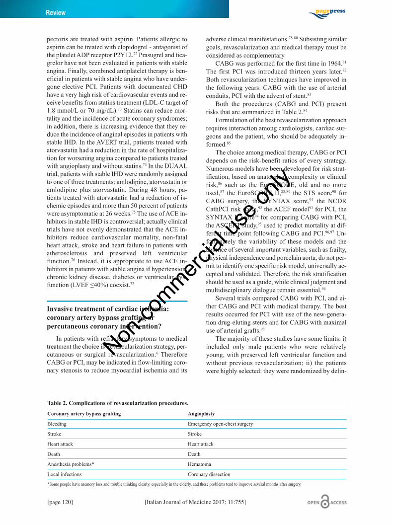
Both the procedures (CABG and PCI) presentrisks that are summarized in Table 2.84
Formulation of the best revascularization approachrequires interaction among cardiologists, cardiac sur-geons and the patient, who should be adequately in-formed.85
The choice among medical therapy, CABG or PCIdepends on the risk-benefit ratios of every strategy.Numerous models have been developed for risk strat-ification, based on anatomical complexity or clinicalrisk,86 such as the EuroSCORE, old and no moreused,87 the EuroSCORE II,88,89 the STS score90 forCABG surgery, the SYNTAX score,91 the NCDRCathPCI risk score,92 the ACEF model93 for PCI, theSYNTAX II score94 for comparing CABG with PCI,the ASCERT study,95 used to predict mortality at dif-ferent time point following CABG and PCI.96,97 Un-fortunately the variability of these models and theabsence of several important variables, such as frailty,physical independence and porcelain aorta, do not per-mit to identify one specific risk model, universally ac-cepted and validated. Therefore, the risk stratificationshould be used as a guide, while clinical judgment andmultidisciplinary dialogue remain essential.94
Several trials compared CABG with PCI, and ei-ther CABG and PCI with medical therapy. The bestresults occurred for PCI with use of the new-genera-tion drug-eluting stents and for CABG with maximaluse of arterial grafts.98
The majority of these studies have some limits: i)included only male patients who were relativelyyoung, with preserved left ventricular function andwithout previous revascularization; ii) the patientswere highly selected: they were randomized by delin-
[page 120] [Italian Journal of Medicine 2017; 11:755]
Review
Table 2. Complications of revascularization procedures.
Coronary artery bypass grafting Angioplasty
Bleeding Emergency open-chest surgery
Stroke Stroke
Heart attack Heart attack
Death Death
Anesthesia problems* Hematoma
Local infections Coronary dissection
*Some people have memory loss and trouble thinking clearly, especially in the elderly, and these problems tend to improve several months after surgery.
Non-co
mmercial
use o
nly

eation of coronary anatomy by angiography, withoutroutine assessment of ischemia; iii) the revasculariza-tion was performed when medical therapy failed; iv)the proportion of patients who did not undergo revas-cularization progressively declined during follow-up,camouflaging differences between the two strategies(CABG and PCI); v) time of follow-up was limited,usually less than five years: so reducing the possibleevaluations about the advantages of CABG related toarterial grafts.98
Medicines are the primary options for stable, low-risk CHD, and should be given to all CHD patients.Medically refractory is a useful high-risk marker ofpotential benefit from revascularization.
The COURAGE trial reported no difference indeath, myocardial infarction and stroke with PCI com-pared with medical therapy for stable angina. On theother hand, patients without angina were significantlymore numerous in the PCI group at one and three years.The difference was minor at five years, when patientsin both groups underwent additional revascularization(21% in the PCI group and 33% in the medical therapygroup respectively).99 Most of other meta-analyses donot describe differences between PCI and medical treat-ment in terms of death, myocardial infarction, un-planned revascularization or angina.100-103
The results in terms of rates of death, cardiac deathand non-fatal myocardial infarction are similar if com-paring early generation drug-eluting stents versus bare-metal stents, while a relative risk reduction in the needfor subsequent or repeat target vessel revascularizationwas registered with the use of drug-eluting stents.103,104
Compared with bare-metal stents and early gen-eration drug-eluting stents, new-generation drug-eluting stents have also improved safety outcomesincluding death, myocardial infarction and stentthrombosis.103,105-108
The superiority of CABG with internal mammaryartery to medical therapy for specific subset of CHD re-sulted into two meta-analyses that demonstrated sur-vival benefit in patients with left main or three-vesselCHD, particularly when the proximal left anterior de-scending coronary artery was involved. Benefits weregreater in patients with severe symptoms, early positiveexercise tests and impaired left ventricular func-tion.109,110
It is very difficult to choose between the two revas-cularization strategies (CABG or PCI), because neitherPCI nor CABG alone can provide a solution for the en-tire spectrum of CHD patients who need revasculariza-tion. CABG permits more complete revascularizationthan PCI, especially with chronic proximal occlusions.88
The choice of the most appropriate strategy of my-ocardial revascularization remains debatable in manypatients. Randomized trials comparing surgery(CABG) to angioplasty (PTCA) have shown that both
modalities are equivalent in terms of survival or in-farct free survival. In patients with type 2 diabetesmellitus in the BARI 2D Trial, a strategy of revascu-larization with CABG or PCI resulted in no differencein mortality compared with optimal medical therapy,111
but patients treated with PTCA required many moreadmissions for additional revascularization proceduresduring follow up, with increasing of the time awayfrom a normal active life, and increasing costs in thetreatment of proximal left anterior descending coro-nary artery disease112,113 and left main coronary arterydisease (in this case some trials demonstrated morerisk of stroke in the group treated with CABG).114,115
In case of three-vessel coronary artery disease, thestudies are discordant: a meta-analysis including pa-tients treated before the use of drug-eluting stentsdemonstrated no differences between PCI and CABGin terms of mortality in all patients, but with reducedmortality in diabetics and in aged 65 years or more pa-tients if treated with CABG.116 A following meta-analysis with patients treated with drug-eluting stentsreported a significant reduction in mortality, myocar-dial infarction and repeat revascularization in thosegroups performed with CABG, but with increasingrisk of stroke.117 There is notable consistency in thefindings on the survival advantage of CABG over PCIfor more severe three-vessel CHD.98
The decision to treat patients with CABG has beenlargely based on CHD extension and left ventricularfunction. Considering patients with stable and unsta-ble angina (excluding recent myocardial infarction),those with left main narrowing >50% or three-vesselstenoses >70%, or even two-vessel stenoses >70%,where one of the vessels is the proximal left anteriordescending, presented minor mortality if treated withCABG.118
Specific advantages of CABG include: revascular-ization of chronically occluded vessels with collateralssupplying viable myocardium, protection of territoriesrather than simply treating lesions, greater durabilityof conduits compared to bare-metal stents, while inthis case the difference with drug-eluting stents ispoor. Based on these principles, physiologic ratherthan anatomic considerations are most useful in deter-mining whether and how urgently to revascularize:STEMI is an emergent indication and high-risk non-STEMI an urgent indication. Coronary anatomy, in-cluding both number of vessels and lesioncharacteristics, continues to influence decision onreperfusion strategy and patient specific strategies.119
The majority of the PCI versus CABG trials en-rolled populations that were at relatively low risk forischemic clinical events. Trials demonstrated few hardoutcome (survival, MI, or stroke) differences betweenCABG and PCI. CABG continues to be the most suit-able revascularization option for patients with multi-
[Italian Journal of Medicine 2017; 11:755] [page 121]
Management of stable coronary artery disease
Non-co
mmercial
use o
nly

vessel, multi-lesion CHD, PCI is the acute stabiliza-tion method of choice for patients with on-going is-chemia and acute MI, especially among patients withhemodynamic compromise, and/or major comorbidity.Bypass surgery remains the treatment of choice in di-abetics only in case of complex coronary anatomysuch as multi-vessel CHD, left main CHD and in pa-tients with multi-vessel disease and impaired ventri-cles.119
Cardiac syndrome X: angina pectoriswith normal coronary arteries
Cardiac syndrome X is a clinical syndrome thatrefers to patients with angina and normal coronary ar-tery, who can show evidence of coronary microvascu-lar dysfunction or abnormalities on stress testing.Microvascular dysfunction causing ischemia is due toendothelial cell dysfunction, abnormal cardiac adren-ergic tone and occult small vessel coronary artery dis-ease. The cardiac microvasculature may have areduced vasodilator, or even a paradoxical vasocon-strictor response to several pharmacologic agents andexercise.120 Because of these abnormalities, cardiacsyndrome X has also been called microvascularangina. Increased cardiac sympathetic tone121 and in-creased response to β-adrenergic stimulation122 cancontribute to autonomic system abnormalities. Actu-ally, enhanced sensitivity to intracardiac pain or theso-called sensitive heart syndrome may result fromsympathovagal imbalance with sympathetic predom-inance123 or reduced activity of the endogenous opioidsystem.124
Patients with cardiac syndrome X are younger thanthose with atherosclerotic cardiovascular disease andpredominantly female.125 In fact, myocardial ischemiaand/or coronary microvascular dysfunction is presentin 20 to 50 percent of women with chest pain and nor-mal coronary arteries.126 Patient with cardiac syn-drome X may have an acute coronary syndrome dueto a ruptured atheromatous plaque and no residual le-sions greater than 50 percent.127 Normal coronaryanatomy or vessels without ≥50 percent stenosis havebeen reported in 9-14% of patients with a non-ST el-evation ACS.128
Management
Considering patients with cardiac syndrome X, thechest pain is similar to classic angina pectoris in aboutone-half of them. It may be precipitated by effort, butalso occurs at rest.129 However, compared to no inter-vention, exercise training improved exercise capacityby 34 percent and delayed the onset of pain during ex-ercise by 100 percent, although the maximum painwas unchanged.130
The episodes are predominantly exertional, fre-quently occur from midnight to early morning andeach episode generally lasts 5 to 15 min. Many undi-agnosed patients with anginal type chest pain relievesymptoms with sublingual nitroglycerin.131 Tachycar-dia, hypertension, diaphoresis, and a gallop rhythmcan be noticed when symptoms are present.
A 12-lead ECG should be performed in all patientswith a history of chest pain. The ECG is usually nor-mal between episodes. Transient ECG changes, withST segment depression are common.132 The absenceof ECG changes does not exclude a cardiac etiology.ST-segment elevation, that is the hallmark of variant(Prinzmetal’s) angina, is not a feature of cardiac syn-drome X.133 Ambulatory ECG monitoring for 24 hmay be helpful for documenting ST segment changes.
The typical finding on the exercise ECG is hori-zontal or down-sloping ST segment depression. Exer-cise thallium-201 myocardial scintigraphy maydemonstrate regional myocardial perfusion defectsduring exercise.134 Studies have demonstrated neitherperfusion defects nor regional wall motion abnormal-ities after dobutamine or transesophageal atrialpacing,135 because the ischemia is limited to the suben-docardium.136
A coronary angiogram showing normal epicardialcoronary arteries (<30 percent diameter reduction) isnecessary to diagnose cardiac syndrome X. Lesionsbetween 30 and 50 percent may be evaluated with ei-ther fractional flow reserve or intravascular ultra-sound.137
Measurement of coronary flow reserve (CFR) canshow microvascular disease.138 CFR is the ratio of max-imal hyperemic coronary blood flow, measured after in-fusion of a coronary vasodilator such as adenosine ordipyridamole, to resting or basal coronary blood flow.Normal CFR ranges from 2.5 to 5. Occasionally it isgreater than 5. Maximal coronary blood flow should beat least 2.5 times the resting blood flow. CMR can beuseful for studying patients with a possible diagnosis ofcardiac syndrome X and it is able to detect regional dif-ferences in myocardial blood flow.139
LVH, right ventricular hypertrophy, stress inducedcardiomyopathy, systemic amyloidosis140 should beconsidered and exclude before diagnosing syndrome X.
Patients with cardiac syndrome X and stableangina generally have an excellent prognosis, whilethose with acute coronary syndromes have an appre-ciable acute mortality.141
All patients with cardiac syndrome X should betreated with aggressive risk factor reduction and sub-lingual nitroglycerin.
The 2013 ESC guidelines on the management ofstable coronary heart disease recommend that all pa-tients receive secondary prevention medications in-cluding aspirin and statins and that β blockers should
[page 122] [Italian Journal of Medicine 2017; 11:755]
Review
Non-co
mmercial
use o
nly

be used as first line therapy.6 Calcium channel block-ers are used if β-blockers are not effective. Statinshave been shown to improve coronary artery endothe-lial function142 and β-blockers are most effective in re-ducing the frequency and severity of angina and inimproving exercise tolerance.143 Atenolol significantlyreduces anginal episodes and improves quality-of-lifemeasures.
Ranolazine is a novel anti-anginal agent and it re-sulted in significantly improved myocardial perfu-sion.144
Hormone replacement therapy may be beneficialin some postmenopausal women with chest pain andestrogen deficiency. Estrogen may act by improvingendothelium-dependent coronary vasomotion.145
Ace-inhibitors, imipramine and L-arginine andspinal cord stimulation have a secondary role in pa-tients who persist symptomatic.146
Follow up of patient with stable ischemic heartdisease
The management of stable IHD aims to relievesymptoms, to improve the quality of life and to reducethe risk of acute coronary syndrome, heart failure anddeath.
The literature has shown that a periodic follow-upof these patients reduces the individual cardiovascularrisk and increases the quality of life.147 However, norandomized trials assess the prognostic impact of dif-ferent follow-up strategies,6 therefore most of theguidance provided by the international literature andthe scientific community are based on expert opinionor on small retrospective studies or registries.
Guidelines recommend clinical evaluations everyfour to six months in the first year after the ischemicevent. After the first year, in patients with stable con-ditions, even in patients with important systolic dys-function, the follow-up can be performed every 6 to12 months, if the patient is enough reliable to call foran appointment in case symptoms or functional capac-ity become worse. This follow-up should be per-formed by a general practitioner, with possiblereference to a cardiologist in cases of doubts and un-certainties.6,17
Additional prospective studies are required to es-tablish appropriate follow up strategies and efficienttime intervals for evaluation. There is not yet a prog-nostic score for detection of patients with stable IHDwho are at high risk of major adverse cardiac events.Surely adverse outcomes are associated with time toischemia during exercise, frequency and severity ofangina, onset of recurrent symptoms <6 months, priorinfarction or CABG, resting ECG abnormalities, LVHor enlargement or abnormal function, coronary an-giography stenosis, diabetes mellitus, comorbidities,age, smoking and sex.17,148-150
The guidelines do not define precisely the ele-ments needed in follow-up visits. In general terms, theliterature suggests careful interval medical historyevaluation, limitations in physical activity, assessmentof symptoms and clinical and functional status, phys-ical examination (including measurement of weightand waist circumference and BMI, as well as bloodpressure and heart rate), and execution of first levellaboratory exams according to patient’s clinical eval-uation.6,17,147,151
Medical examination should consider adherenceto drug and behavioral therapy, onset of adverse ef-fects, monitoring of cardiovascular risk factors and co-morbidities, recurrence of ischemia, onset ofsymptoms and signs suggestive of heart failure, ar-rhythmias, heart valve disease and peripheral vasculardisease. The American guidelines suggest SAQ (Seat-tle angina questionnaire, a 19-item, self-reportedquestionnaire validated to quantify the symptoms,functional limitations and quality of life of patientswith stable IHD) as a tool for monitoring the patient’sclinical status.17
Glucose, creatinine, lipid profile and glycated he-moglobin in diabetic patients should be periodicallyevaluated. Blood count, serum electrolytes and thyroidfunction should be monitored annually.17,152
The serial measurement of natriuretic peptides (tooptimize the therapy in the case of a concurrent situa-tion of chronic heart failure) showed contrasting re-sults. However, it is demonstrated that high levels ofnatriuretic peptides are associated with worse progno-sis, instead their reduction is associated with a betterprognosis.6,153
Resting electrocardiogram should be performedannually, even in stable patients. A supplementaryelectrocardiogram should be performed in case ofanginal symptoms modification, onset of syncope orpre-syncope or symptoms compatible with arrhyth-mias, assumption of new drugs with possible second-ary abnormalities of electrical conduction.6
Echocardiography should be considered only incase of clinical suspicion: onset or worsening of heartfailure or valve disease, evidence of new ischemia.17,152
The American Guidelines do not suggest a periodicechocardiographic evaluation in case of stable diseaseor in low risk patients.17
Stress testing is appropriate in the presence of newor worsening symptoms, once unstable angina or acutecoronary syndrome have been excluded. The recom-mendations are similar to the ones used for the diag-nosis of suspected IHD. Therefore, a candidate totreadmill stress testing should have an interpretableresting ECG. If resting ECG is uninterpretable orshows LBBB, a stress echocardiography, or a myocar-dial scintigraphy or, as a second choice, a cardiac MRIshould be performed. These same exams should be
[Italian Journal of Medicine 2017; 11:755] [page 123]
Management of stable coronary artery disease
Non-co
mmercial
use o
nly

considered as first choice in the event of high risk pa-tient or known multi-vessel diseases.17 CCTA could beconsidered in patients with previous CABG or previ-ous placement of stents >3 mm in order to assess theirpatency, in the context of absence of severe calcifica-tions.17
The international guidelines highlight the utility toperform the same test in the same patients to avoidtest-dependant discrepancies.
The appropriateness of performing non-invasivetesting in patients who are asymptomatic or have sta-ble symptoms depends on factors related to the likeli-hood of significant findings. There is no current datademonstrating that a follow-up strategy based on theuse of stress tests in patients persistently asympto-matic, would improve their outcomes.154,155
The management of patient with stableischemic heart diseaseRationale and objective
IHD is a frequent and disabling disease, associatedwith an appreciable incidence of acute coronary eventsand increased mortality. For this reason, Internistshould know how to treat these patients at best, refer-ring only a small part of them to Cardiologists. It isnecessary a great effort because therapy and invasivetreatments are continuously progressing and Clini-cians should be constantly updated. Therefore, thegoal of this work is to raise awareness of the clinicalmanagement of stable IHD through a better knowl-edge of the most recent revisions and guidelines.Some particular aspects will be omitted because theyare considered too specialistic and are already outlinedin the previous part of this monography.
Methodology
In order to provide evidence-based recommenda-tions for the management of patients with stable IHD,we first verified the existence of guidelines on thematter. Therefore, we conducted a search using thefollowing database-guidelines: i) Scottish Intercolle-giate Guidelines Network (SIGN); ii) Institute forClinical Systematic Improvement (ICSI); iii) NationalInstitute for Health and Clinical Excellence (NICE) -National Health System (NHS) evidence; iv) NationalGuideline Clearinghouse (NGC); v) Canadian Med-ical Association, CMA Infobase; vi) New ZealandGuidelines Group; vii) Italian National Health SystemGuidelines; viii) Clinical Practice Guidelines Portal;ix) eGuidelines.
The research was carried out by seven authors in-dependently, using the terms stable, ischemic, heart,disease as key words, when the site included thesearch function, and in other cases we listed the last
manually guidelines stored in the database. The re-sults obtained separately were then compared anddiscussed together. The guidelines thus obtainedwere evaluated using the AGREE instrument156 (Ap-praisal of Guidelines, Research and Evaluation II,22) by 4 authors independently. AGREE II assessescompliance with 23 requirements, meeting 6 do-mains as the explanation of the purpose, the clarity,the involvement of all stakeholders, the rigor of de-velopment, applicability and editorial independenceof the same. Each author assessed the compliance ofindividual requirements with a score from 1 (dis-agree completely) to 7 (complete agreement). Thescores assigned by each author were added within in-dividual domains and reported with the highest andthe lowest score possible within the domain based onthe number of requirements included and the numberof evaluators.
Results
Through the databases listed above, we identified4 guidelines which we evaluated with AGREE method(Table 3). Other references were excluded because toospecific and non-functional for our work. By usingAGREE criteria we judge the NICE guidelines157 onstable IHD to be the best.
Actually, it contains excellent description of targetpopulation, objectives and purpose, it does not forgetto consider target-population preferences. It empha-sizes the role of informed decisions on risks and ben-efits of different treatments. Further it underlinescultural lacks, unresolved questions of managementand it always suggests adequate trials to resolve theuncertainties. It is a complete guideline elaborated in468 pages; however, there is a shorter version, suitablefor clinicians.
ESC6 and AHA17 guidelines are both good guide-lines. ESC guidelines contain clear messages, elabo-rated with many tables and easy to access. However,there is not a precise description of the target popula-tion, there are not any considerations about possiblebarriers to guidelines implementation. Further, the
[page 124] [Italian Journal of Medicine 2017; 11:755]
Review
Table 3. Evaluation of the guidelines on sepsis using theAppraisal of Guidelines, Research and Evaluation II(AGREE) method.
Guideline AGREE evaluation
National Institute for Health and Clinical Excellence (NICE) 81%
European Society of Cardiology (ESC) 57%
American Heart Association (AHA) 57%
Canadian Cardiovascular Society (CCS) 43%
Non-co
mmercial
use o
nly

economic aspects are marginally described. AHA isschematic and easy to assess, but it lacks some impor-tant aspects: the process used to produce recommen-dation is not described, time and methods of guidelineupdate are ignored, the barriers to guideline imple-mentation are not considered.
Canadian Cardiovascular Society (CCS) Guide-lines4 are really synthetic and clear, but there are mod-est indications on the most recent non-invasive testsuch as MR and PET. They discuss marginally differ-ent revascularization approaches without consideringcost, risk and benefits. Barriers to guidelines imple-mentation are not mentioned; a precise description ofthe target population is absent.
The importance of a correct clinical overview
Management of stable angina needs great compe-tence and optimal clinical ability since the beginning.Actually, the most important approach is based on acorrect clinical overview, starting from symptoms re-ported analyses. Capacity of distinguishing typicalfrom atypical syndrome is the first necessary step re-quired.4 Indeed, risk factors (particularly age and gen-der) and symptoms permit to stratify pre-testprobability of disease.4,6 According to this result cli-nician can suggest to perform nothing or angiographyor a non-invasive test to demonstrate clinical suspectof angina. ECG exercise has not a great sensibility andis not advisable when pre-test probability is over 65%.Further, it should not be used in case of LVEF <50%,exercise limited by orthopedic or other non-cardiacproblems or equivocal ECG, such as LBBB, WPW orpaced rhythm.6,24 Obviously biochemical examinationand echocardiography are two precious tests whichcan facilitate the clinician’s approach. Actually, in caseof systolic dysfunction it is mandatory to know coro-nary anatomy and coronary angiography is preferredto CCTA when patient suffers from a typical chestpain.6 Troponin, if correctly used, can be helpful to cli-nician to distinguish acute coronary syndrome. In thiscase patient management and treatment are based onNSTEMI guidelines6 (Figure 2).
Main tests at first medical examination and duringfollow-up
Every patient with a diagnosis of stable anginashould assess thyroid and kidney function, lipid pro-file, glycemia and blood count to limit disease pro-gression (controlling risk factors) and ischemiatriggers (anemia, hyperthyroidism).6-17 These examsshould be repeated at least every year during follow-up4,17,152 (Table 4).
ECG should be repeated every year and in case ofanginal symptoms modification, onset of syncope orpre-syncope or symptoms compatible with arrhyth-mias, assumption of new drugs with possible second-ary abnormalities of electrical conduction.6 If nothingemerges from basal ECG, stress test should be per-formed to investigate potential progression of coro-nary artery disease or possible stent or graft stenosis.
During follow-up echocardiography should beconsidered only in case of clinical suspicion: onset orworsening of heart failure or valve disease, evidenceof new ischemia.17,152
Stress test are useful to demonstrate a suspectedclinical diagnosis of angina and to stratify severity.6,158
Stress ECG and echocardiography are the most usedfor their simplicity, low cost and wide-spread avail-ability, whereas stress MR and scintigraphy are not soavailable outside of academic practice settings.158
However MR is recommended in patients in whom,despite the use of echo contrast agents, transthoracicechocardiography cannot be reliable for restrictedacoustic window.6 When choosing the most appropri-ate test for a specific patient, clinician should considerpatient characteristics, potential contraindications totesting, limitations of each modality, local availabilityand local expertise.4
Angiography is useful in case of symptomatic pa-tients in optimal medical therapy to evaluate the pos-sibility of a revascularization therapy. It can beperformed in asymptomatic patients with suspectedthree vessels or left main coronary anatomy,157 be-cause medical treatment is inferior to invasive revas-cularization such as CABG in this setting. Anotherdebatable indication is represented by high-risk pa-
[Italian Journal of Medicine 2017; 11:755] [page 125]
Management of stable coronary artery disease
Table 4. Main tests in stable ischemic heart disease.
Test Utility Indication
Blood exams Control disease progression and ischemia triggers Every patient, every year
ECG Discover ischemic signs and prognosis Every patient, every year
Echocardiography Exclude other cardiopathy and evaluate ejection fraction Every patient
Stress test Diagnosis and stratification When PTP is <85% or follow-up of high risk patients
Angiography Revascularization or anatomy investigation Uncontrolled symptoms or possible complex lesions or high risk patients
ECG, electrocardiogram; PTP, termed pre-probability.
Non-co
mmercial
use o
nly

Review
Table 5. Stratification of risk outcome.
Non-invasive test High-risk outcome
Exercise Treadmill >2 mm of ST depression at low workload Exercise-induced ST elevation Exercise-induced ventricular tachycardia/fibrillation Failure to increase blood pressure >120 mmHg or sustained decrease >10 mmHg during exercise
Myocardial perfusion imaging Resting perfusion abnormalities >10% of the myocardium Stress-induced perfusion abnormalities >10% of the myocardium or indicating multiple coronary obstruction Severe stress-induced left ventricular dysfunction
Stress echocardiography Inducible kinetic abnormalities involving >2 coronary beds Kinetic abnormalities developing at low dose of dobutamine
Coronary computed tomographic angiography Multi-vessel or left main stenosis
Figure 2. Correct management of chest pain. ECG, electrocardiogram; EF, ejection fraction; CCTA, coronary computedtomography angiography; PTP, termed pre-probability.Non
-commerc
ial us
e only

tients and this common practice is underinvestigation.158 LVEF, ischemic and anatomic burdenare the fundamental triad which offers prognosticstratification of patient with stable IHD4 (Table 5).
Optimal medical therapy
Lifestyle modifications and risk factors control areessential aspects to emphasize.6 Further therapy isbased on anti-anginal drugs and drugs for secondaryprevention of cardiovascular disease.43,44 The formertreatment is based on decreasing oxygen consumption:β-blockers and calcium channel blockers represent themain stems.6,157 Clinicians should choose treatment ac-cording to contraindication or patient preference. β-blockers are preferable in case myocardial infarcthistory, because these drugs demonstrated a favorableeffect on mortality.159
Long acting nitrates, ranolazine, ivabradine, nico-randil are possible second line alternative treatmentswhenever first line treatments are contraindicated orare not tolerated.6 Comorbidities, contraindications,person’s preference and drug costs are the elements to
evaluate for appropriate drug prescription. A tripleanti-anginal therapy should be considered only in pa-tients complaining persisting symptoms and present-ing non-revascularizable CHD.157
Secondary cardiovascular prevention aims to re-duce long-term risk of cardiovascular events. Statinsand low dose of aspirin should be prescribed to everypatient with stable coronary artery disease, after hav-ing considered allergies, intolerance and risk of bleed-ing.6,63-65 Aspirin reduces myocardial infarct incidencebut not fatal events. At the same time its use is associ-ated with an increased risk of bleeding.160,161 For thesereasons this treatment should be introduced after hav-ing considered bleeding risk and comorbidity. ACE-inhibitors are not routinary prescribed in patients withstable coronary artery disease67 because their advan-tages are limited to other settings such as heart failure,hypertension and myocardial infarction.
Medical treatment or invasive approach?
A clear and undebatable indication for an invasiveapproach is persisting symptoms in optimal ther-
[Italian Journal of Medicine 2017; 11:755] [page 127]
Management of stable coronary artery disease
Figure 3. Angiography indications. EF, ejection fraction; PCI, percutaneous coronary intervention; CABG, coronaryartery by-pass grafting.
Non-co
mmercial
use o
nly

apy4,6,17,157 (Figure 3). Actually coronary artery revascu-larization with either CABG or PCI should be per-formed in symptomatic patients, uncontrolled withoptimal medical therapy or in case revascularizationcould improve survival (left main coronary artery dis-ease; three vessel disease, coronary artery disease, par-ticularly with a reduced left ventricular ejection fraction(usually <40 percent); or two vessel disease with morethan a 75 percent stenosis in the left anterior descendingartery proximal to the first major septal artery).102-108
However this last indication is based on randomized tri-als of CABG versusmedical treatment that were carriedout over 30 years ago, when statins and renin-an-giotensin system inhibitors were not available.
If revascularization is desirable, clinician shouldconsider risks and benefits of CABG and PCI, sever-ity and complexity of the person’s coronary diseaseand other relevant clinical factors and comorbidi-ties.157 A multidisciplinary team should explain var-ious options to the patient and take account of hispreference.
If the patient has anatomically less complex coro-nary disease and does not express a preference, clini-cians should take account of the evidence that PCImay be a more cost-effective procedure. For patientswith anatomically complex three-vessel disease, withor without involvement of the left main stem, and forpeople with multi-vessel disease who have diabetes orare aged >65 years, clinicians should take account ofthe potential survival advantage of CABG over PCIwhen advising patients about the appropriate revascu-larization strategy.162
References1. Kones R, Rumana U. Stable ischemic heart disease. Car-
diol Clin 2014;32:333-51.2. Valgimigli M, Biscaglia S. Stable angina pectoris. Curr
Atheroscler Rep 2014;16:422.3. Crea F, Pupita G, Galassi AR, et al. Role of adenosine
in pathogenesis of anginal pain. Circulation1990;81:164-72.
4. Mancini GB, Gosselin G, Chow B, et al. Canadian Car-diovascular Society guidelines for the diagnosis andmanagement of stable ischemic heart disease. Can J Car-diol 2014;30:837-49.
5. Wilson JF. In the clinic, stable ischemic heart disease.Ann Intern Med 2014;160:1-16.
6. Montalescot G, Sechtem U, Achenbach S. ESC guide-lines on the management of stable coronary artery dis-ease. Eur Heart J 2013;34:2949-3003.
7. Information Centre for Health and Social Care. HealthSurvey for England. Cardiovascular Disease and RiskFactors in Adults. Vol. 1. London: HMSO; 2008.
8. Boden WE, O’Rourke RA, Teo KK, et al. Optimal med-ical therapy with or without PCI for stablecoronary dis-ease. N Eng J Med 2007;356:1503-16.
9. Chung SC, Hlatky MA, Faxon D, et al. The effect of age
on clinical outcomes and health status BARI 2D (BypassAngioplasty Revascularization Investigation in Type 2Diabetes). J Am Coll Cardiol 2011;58:810-9.
10. Frye RL, August P, Brooks MM, et al. A randomizedtrial of therapies for type 2 diabetes and coronary arterydisease. N Eng J Med 2009;360:2503-15.
11. Henderson RA, Pocock SJ, Clayton TC, et al. Seven-year outcome in the RITA-2 trial: coronary angioplastyversus medical therapy. J Am Coll Cardiol 2003;42:1161-70.
12. Poole-Wilson PA, Lubsen J, Kirwan BA, et al. Effect oflong-acting nifedipine on mortality and cardiovascularmorbidity in patients with stable angina requiring treat-ment (ACTION trial): randomised controlled trial.Lancet 2004;364:849-57.
13. Steg PG, Greenlaw N, Tardif JC, et al. Women and menwith stable coronary artery disease have similar clinicaloutcomes: insights from the international prospectiveCLARIFY registry. Eur Heart J 2012;33:2831-840.
14. Diamond GA. A clinically relevant classification ofchest discomfort. J Am Coll Cardiol 1983;1:574-5.
15. Diamond GA, Forrester JS. Analysis of probability asan aid in the clinical diagnosis of coronary-artery dis-ease. N Eng J Med 1979;300:1350-8.
16. Genders TS, Meijboom WB, Meijs MF. CT coronary an-giografy in patients suspected of having coronary arterydisease: decision making from various perspective in theface of uncertainty. Radiology 2009;253:734-44.
17. Fihn SD, Gardin JM, Abrams J, et al. ACCF/AHA/ACP/AATS/PCNA/SCAI/STS Guideline for the diag-nosis and management of patients with stable ischemicheart disease. J Am Coll Cardiol 2012;60:e44-e164.
18. Zethelius B, Johnston N, Venge P. Troponin I as a pre-dictor of coronary heart disease and mortality in 70-year-old men: a community-based cohort study.Circulation 2006;113:1071-8.
19. Omland T, De Lemos JA, Sabatine MS, et al. A sensitivecardiac troponin T assay in stable coronary artery dis-ease. N Eng J Med 2009;361:2538-47.
20. Ndrepepa G, Braun S, Mehilli J, et al. Prognostic valueof sensitive troponin T in patients with stable and unsta-ble angina and undetectable conventional troponin. AmHeart J 2011;161:68-75.
21. Oremus M, Raina PS, Santaguida P, et al. A systematicreview of BNP as a predictor of prognosis in personswith coronary artery disease. Clin Biochem 2008;41:260-5.
22. Buckley DI, Fu R, Freeman M, et al. C-reactive proteinas a risk factor for coronary heart disease: a systematicreview and meta-analyses for the U.S. Preventive Serv-ices Task Force. Ann Intern Med 2009;151:483-95.
23. Lee TH, Cook EF, Weisberg MC, et al. Impact of theavailability of a prior electrocardiogram on the triage ofthe patient with acute chest pain. J Gen Intern Med1990;5:381-8.
24. Fox K, Garcia MA, Ardissino D, et al. Guidelines on themanagement of stable angina pectoris: executive sum-mary The Task Force on the Management of StableAngina Pectoris of the European Society of Cardiology.Eur Heart J 2006;27:1341-81.
25. Cesar LA, Ferreira JF, Armaganijan D, et al. Guidelinefor stable coronary artery disease. Arq Bras Cardiol2014;103:1-56.
[page 128] [Italian Journal of Medicine 2017; 11:755]
Review
Non-co
mmercial
use o
nly

26. Pino GP, Baldini U, Borrello F, Terranova A. Cardiopa-tia ischemica cronica, cap. 28. Linee Guida SIEC; 2011.Available from: https://www.siec.it/linee-guida/
27. Skinner JS, Smeeth L, Kendall JM, et al. NICE guid-ance. Chest pain of recent onset: assessment and diag-nosis of recent onset chest pain or discomfort ofsuspected cardiac origin. Heart 2010;96:974-8.
28. Montalescot G, Sechtem U, Achenbach S, et al. 2013ESC guidelines on the management of stable coronaryartery disease - addenda. ESC Guidelines - addenda;available from: www.escardio.org/guidelines
29. Fihn SD, Blankenship JC, Alexander KP, et al. 2014ACC/AHA/AATS/PCNA/SCAI/STS focused update ofthe guideline for the diagnosis and management of pa-tients with stable ischemic heart disease: a report of theAmerican College of Cardiology/American Heart Asso-ciation Task Force on Practice Guidelines, and theAmerican Association for Thoracic Surgery, PreventiveCardiovascular Nurses Association, Society for Cardio-vascular Angiography and Interventions, and Society ofThoracic Surgeons. Circulation 2014;130:1749-67.
30. Bruder O, Wagner A, Lombardi M, et al. European car-diovascular magnetic resonance (EuroCMR) registry -multi national results from 57 centers in 15 countries. JCardiovasc Magnet Reson 2013;15:9.
31. Hendel RC, Patel MR, Kramer CM, et al. ACCF/ACR/SCCT/SCMR/ASNC/NASCI/SCAI/SIR 2006 appropri-ateness criteria for cardiac computed tomography andcardiac magnetic resonance imaging: a report of theAmerican College of Cardiology Foundation QualityStrategic Directions Committee Appropriateness Crite-riaWorking Group, American College of Radiology, So-ciety of Cardiovascular Computed Tomography, Societyfor Cardiovascular Magnetic Resonance, American So-ciety of Nuclear Cardiology, North American Societyfor Cardiac Imaging, Society for Cardiovascular An-giography and Interventions, and Society of Interven-tional Radiology. J Am Coll Cardiol 2006;48:1475-97.
32. Mark DB, Shaw L, Harrell FE Jr, et al. Prognostic valueof a treadmill exercise score in outpatients with sus-pected coronary artery disease. N Engl J Med 1991;325:849-53.
33. Mark DB, Hlatky MA, Harrell FE Jr, et al. Exercisetreadmill score for predicting prognosis in coronary ar-tery disease. Ann Intern Med 1987;106:793-800.
34. Gianrossi R, Detrano R, Mulvihill D, et al. Exercise-in-duced ST depression in the diagnosis of coronary arterydisease. A meta-analysis. Circulation 1989;80:87-98.
35. Kwok Y, Kim C, Grady D, et al. Meta-analysis of exer-cise testing to detect coronary artery disease in women.Am J Cardiol 1999;83:660-6.
36. Gibson SR. The diagnostic and prognostic value of ex-ercise electrocardiography in asymptomatic subjects andstable symptomatic patients. Curr Opin Cardiol 1991;6:536-46.
37. Ashley EA, Myers J, Froelicher V. Exercise testing inclinical medicine. Lancet 2000;356:1592-7.
38. Brindis RG, Douglas PS, Hendel RC, et al. ACCF/ASNC appropriateness criteria for single-photon emis-sion computed tomography myocardial perfusion imag-ing (SPECT MPI): a report of the American College ofCardiology Foundation Strategic Directions CommitteeAppropriateness Criteria Working Group and the Amer-
ican Society of Nuclear Cardiology. J Am Coll Cardiol2005;46:1587-605.
39. Schwitter J, Wacker CM, van Rossum AC, et al. MR-IMPACT: comparison of perfusion-cardiac magneticresonance with single photon emission computed to-mography for the detection of coronary artery disease ina multicentre, multivendor, randomized trial. Eur HeartJ 2008;29:480-9.
40. Nandalur KR, Dwamena BA, Choudhri AF, et al. Diag-nostic performance of positron emission tomography inthe detection of coronary artery disease: a meta-analysis.Acad Radiol 2008;15:444-51.
41. Gibbons RJ, Abrams J, Chatterjee K, et al. ACC/AHA2002 Guideline update for the management of patientswith chronic stable angina. Circulation 2003;107:149-58.
42. Abrams J, Thadani U. Therapy of stable angina pectoris:the uncomplicated patient. Circulation 2005;112:e255-9.
43. Fihn SD, Gardin JM, Abrams J, et al. 2012 ACCF/AHA/ACP/AATS/PCNA/SCAI/STS guideline for the diagno-sis and management of patients with stable ischemicheart disease: a report of the American College of Car-diology Foundation/American Heart Association taskforce on practice guidelines, and the American Collegeof Physicians, American Association for Thoracic Sur-gery, Preventive Cardiovascular Nurses Association, So-ciety for Cardiovascular Angiography and Interventions,and Society of Thoracic Surgeons. Circulation 2012;126:e354-471.
44. Winniford MD, Wheelan KR, Kremers MS, et al. Smok-ing-induced coronary vasoconstriction in patients withatherosclerotic coronary artery disease: evidence foradrenergically mediated alterations in coronary arterytone. Circulation 1986;73:662-67.
45. Bangalore S, Steg G, Deedwania P, et al. β-Blocker useand clinical outcomes in stable outpatients with and with-out coronary artery disease. JAMA 2012;308:1340-9.
46. Andersson C, Shilane D, Go AS, et al. β-blocker ther-apy and cardiac events among patients with newly di-agnosed coronary heart disease. J Am Coll Cardiol2014;64:247-52.
47. Braunwald E. Mechanism of action of calcium-channel-blocking agents. N Engl J Med 1982;307:1618-27.
48. Emanuelsson H, Egstrup K, Nikus K, et al. Antianginalefficacy of the combination of felodipine-metoprolol10/100 mg compared with each drug alone in patientswith stable effort-induced angina pectoris: a multicenterparallel group study. The TRAFFIC Study Group. AmHeart J 1999;137:854-62.
49. Thadani U, Fung HL, Darke AC, et al. Oral isosorbidedinitrate in angina pectoris: comparison of duration ofaction an dose-response relation during acute and sus-tained therapy. Am J Cardiol 1982;49:411-9.
50. Henderson RA, O’Flynn N. Management of stableangina: summary of NICE guidance. Heart 2012;98:500-7.
51. Parker JO. Eccentric dosing with isosorbide-5-mononi-trate in angina pectoris. Am J Cardiol 1993;72:871-6.
52. Chaitman BR. Ranolazine for the treatment of chronicangina and potential use in other cardiovascular condi-tions. Circulation 2006;113:2462-72.
53. Morrow DA, Scirica BM, Karwatowska-Prokopczuk E,et al. Effects of ranolazine on recurrent cardiovascular
[Italian Journal of Medicine 2017; 11:755] [page 129]
Management of stable coronary artery disease
Non-co
mmercial
use o
nly

events in patients with non-ST-elevation acute coronarysyndromes: the MERLIN-TIMI 36 randomized trial.JAMA 2007;297:1775-83.
54. Morrow DA, Scirica BM, Chaitman BR, et al. Evalua-tion of the glycometabolic effects of ranolazine in pa-tients with and without diabetes mellitus in theMERLIN-TIMI 36 randomized controlled trial. Circu-lation 2009;119:2032-9.
55. Kosiborod M, Arnold SV, Spertus JA, et al. Evaluationof Ranolazine in Patients with Type 2 Diabetes Mellitusand Chronic Stable Angina. Results from the TERISArandomized clinical trial. J Am Coll Cardiol2013;61:2038-45.
56. Chaitman BR, Skettino SL, Parker JO, et al. Antiis-chemic effects and long-term survival during ranolazinemonotherapy in patients with chronic severe angina. JAm Coll Cardiol 2004;43:1375-82.
57. Chaitman BR, Pepine CJ, Parker JO, et al. Effects of ra-nolazine with atenolol, amlodipine, or diltiazem on ex-ercise tolerance and angina frequency in patients withsevere chronic angina: a randomized controlled trial.JAMA 2004;291:309-16.
58. Rousseau MF, Pouleur H, Cocco G, et al. Comparativeefficacy of ranolazine versus atenolol for chronic anginapectoris. Am J Cardiol 2005;95:311-6.
59. Stone PH, Gratsiansky NA, Blokhin A, et al; ERICA In-vestigators. Antianginal efficacy of ranolazine whenadded to treatment with amlodipine: the ERICA (Effi-cacy of Ranolazine in Chronic Angina) trial. J Am CollCardiol 2006;48:566-75.
60. Henry TD, Satran D, Hodges JS, et al. Traverse JH.Long-term survival in patients with refractory angina.Eur Heart J 2013;34:2683-8.
61. Thadani U, Ezekowitz M, Fenney L, et al. Double-blindefficacy and safety study of a novel anti-ischemic agent,ranolazine, versus placebo in patients with chronic sta-ble angina pectoris. Circulation 1994;90:726-34.
62. Kosiborod M, Arnold SV, Spertus JA, et al. Evaluationof ranolazine in patients with type 2 diabetes mellitusand chronic stable angina: results from the TERISA ran-domized clinical trial (type 2 diabetes evaluation of ra-nolazine in subjects with chronic stable angina). J AmColl Cardiol 2013;61:2038-45.
63. Koren MJ, Crager MR, Sweeney M. Long-term safetyof a novel antianginal agent in patients with severechronic stable angina: the ranolazine open label experi-ence (ROLE). J Am Coll Cardiol 2007;49:1027-34.
64. Fihn SD, Gardin JM, Abrams J, et al. 2012 ACCF/AHA/ACP/AATS/PCNA/SCAI/STS guideline for thediagnosis and management of patients with stable is-chemic heart disease: a report of the American Collegeof Cardiology Foundation/American Heart Associationtask force on practice guidelines, and the American Col-lege of Physicians, American Association for ThoracicSurgery, Preventive Cardiovascular Nurses Association,Society for Cardiovascular Angiography and Interven-tions, and Society of Thoracic Surgeons. Circulation2012;126:354-471.
65. Lopatin YM. Russian Multicenter Observational Pro-gram NACHALO. Evaluation of the antianginal effi-cacy of ivabradine in patients with ischemic heartdisease complicated by heart failure. Kardiologiia2015;55:5-11.
66. Tardif JC, Ford I, Tendera M, et al. Efficacy of ivabra-dine, a new selective I(f) inhibitor, compared withatenolol in patients with chronic stable angina. Eur HeartJ 2005;26:2529-36.
67. Fox K, Ford I, Steg PG, et al. Ivabradine for patientswith stable coronary artery disease and left-ventricularsystolic dysfunction (BEAUTIFUL): a randomised,double-blind, placebo-controlled trial. Lancet 2008;372:807-16.
68. Fox K, Ford I, Steg PG, et al. Ivabradine in stable coro-nary artery disease without clinical heart failure. N EnglJ Med 2014;371:1091-9.
69. Tendera M, Chassany O, Ferrari R, et al. Quality of lifewith ivabradine in patients with angina pectoris thestudy assessing the morbidity-mortality benefits of theif inhibitor ivabradine in patients with coronary arterydisease quality of life substudy. Circ Cardiovasc QualOutcomes 2016;9:31-8.
70. Ford I (The IONA Study Group). Effect of nicorandil oncoronary events in patients with stable angina: the im-pact of nicorandil in angina (IONA) randomised trial.Lancet 2002;359:1269-75.
71. Ford I (The IONA Study Group). Impact of nicorandilin angina: subgroup analyses IONA Study Group. Heart2004;90:1427-30.
72. Carolei A. CAPRIE Steering Committee: A randomised,blinded, trial of clopidogrel versus aspirin in patients atrisk of ischaemic events (CAPRIE). Lancet 1996;348:1329-39.
73. Reiner Z, Catapano AL, De Backer G, et al. ESC/EASGuidelines for the management of dyslipidaemias: theTask Force for the management of dyslipidaemias of theEuropean Society of Cardiology (ESC) and the Euro-pean Atherosclerosis Society (EAS). Eur Heart J2011;32:1769-818.
74. Pitt B, Waters D, Brown WV, et al. Aggressive lipid-lowering therapy compared with angioplasty in stablecoronary artery disease. Atorvastatin versus Revascular-ization Treatment Investigators. N Engl J Med 1999;341:70-6.
75. Deanfield JE, Sellier P, Thaulow E, et al. Potent anti-is-chaemic effects of statins in chronic stable angina: in-cremental benefit beyond lipid lowering? Eur Heart J2010;31:2650-9.
76. Dagenais GR, Pogue J, Fox K, et al. Angiotensin-con-verting-enzyme inhibitors in stable vascular diseasewithout left ventricular systolic dysfunction or heart fail-ure: a combined analysis of three trials. Lancet 2006;368:581-8.
77. Braunwald E, Domanski MJ, Fowler SE, et al. An-giotensin-converting-enzyme inhibition in stable coro-nary artery disease. N Eng J Med 2004;351:2058-68.
78. Kolh P, Wijns W, Danchin N, et al. Guidelines on my-ocardial revascularization. Eur J Cardiothorac Surg2010;38:S1-52.
79. Wijns W, Kolh P, Danchin N, et al. Guidelines on my-ocardial revascularization: The Task Force on Myocar-dial Revascularization of the European Society ofCardiology (ESC) and the European Association forCardio-Thoracic Surgery (EACTS). Eur Heart J2010;31:2501-55.
80. Deb S, Wijeysundera HC, Ko DT, et al. Coronary arterybypass graft surgery vs.percutaneous interventions in
[page 130] [Italian Journal of Medicine 2017; 11:755]
Review
Non-co
mmercial
use o
nly

coronary revascularization: a systematic review. JAMA2013;310:2086-95.
81. Head SJ, Kieser TM, Falk V, et al. Coronary artery by-pass grafting: Part 1: the evolution over the first 50years. Eur Heart J 2013;34:2862-72.
82. Gruntzig A. Transluminal dilatation of coronary-arterystenosis. Lancet 1978;1:263.
83. Stefanini GG, Holmes DR Jr. Drug-eluting coronary-artery stents. N Engl J Med 2013;368:254-65.
84. Healthwise Staff. Anticoagulants for heart attack and un-stable angina. Last Revised: February 13, 2013. Avail-able from: http://www.wakemed.org/body.cfm?id=4616&action=detail&AEProductID=HW_Knowledge-base&AEArticleID=tx4242
85. Head SJ, Kaul S, Mack MJ, et al. The rationale for HeartTeam decision-making for patients with stable, complexcoronary artery disease. Eur Heart J 2013;34:2510-8.
86. Head SJ, Holmes DR Jr., Mack MJ, et al. InvestigatorsS. Risk profile and 3-year outcomes from the SYNTAXpercutaneous coronary intervention and coronary arterybypass grafting nested registries. JACC Cardiovasc In-terv 2012;5:618-25.
87. Nashef SA, Roques F, Michel P, et al. European systemfor cardiac operative risk evaluation (EuroSCORE). EurJ Cardiothorac Surg 1999;16:9-13.
88. Nashef SA, Roques F, Sharples LD, et al. EuroSCOREII. Eur J Cardiothorac Surg 2012;41:734-44; discussion744-5.
89. Biancari F, Vasques F, Mikkola R, et al. Validation ofEuro SCORE II in patients undergoing coronary arterybypass surgery. Ann Thorac Surg 2012;93:1930-5.
90. Shahian DM, O’Brien SM, Filardo G, et al. Society ofThoracic Surgeons Quality Measurement Task F. TheSociety of Thoracic Surgeons 2008 cardiac surgery riskmodels: part 3: valve plus coronary artery bypass graft-ing surgery. Ann Thorac Surg 2009;88:S43-62.
91. Mohr FW, Morice MC, Kappetein AP, et al. Coronary ar-tery bypass graft surgery vs. percutaneous coronary inter-vention in patients with three-vessel disease and left maincoronary disease: 5-year follow-up of the randomised,clinical SYNTAX trial. Lancet 2013;381:629-38.
92. Peterson ED, Dai D, DeLong ER, et al. Contemporarymortality risk prediction for percutaneous coronary in-tervention: results from 588,398 procedures in the Na-tional Cardiovascular Data Registry. J Am Coll Cardiol2010;55:1923-32.
93. Wykrzykowska JJ, Garg S, Onuma Y, et al. Value of age,creatinine, and ejection fraction (ACEF score) in assess-ing risk in patients undergoing percutaneous coronaryinterventions in the ’All-Comers’ LEADERS trial. CircCardiovasc Interv 2011;4:47-56.
94. Farooq V, van Klaveren D, Steyerberg EW, et al.Anatomical and clinical characteristics to guide decisionmaking between coronary artery bypass surgery and per-cutaneous coronary intervention for individual patients:development and validation of SYNTAX score II.Lancet 2013;381:639-50.
95. Weintraub WS, Grau-Sepulveda MV, Weiss JM, et al.Comparative effectiveness of revascularization strate-gies. N Engl J Med 2012;366:1467-76.
96. Shahian DM, O’Brien SM, Sheng S, et al. Predictors oflong-term survival after coronary artery bypass graftingsurgery: results from the Society of Thoracic Surgeons
Adult Cardiac Surgery Database (the ASCERT study).Circulation 2012;125:1491-500.
97. Weintraub WS, Grau-Sepulveda MV, Weiss JM, et al.Prediction of long-term mortality after percutaneouscoronary intervention in older adults: results from theNational Cardiovascular Data Registry. Circulation2012;125:1501-10.
98. Windecker S, Kolh P, Alfonso F, et al. 2014 ESC/EACTSGuidelines on myocardial Revascularization. The TaskForce on Myocardial Revascularization of the EuropeanSociety of Cardiology (ESC) and the European Associa-tion for Cardio-Thoracic Surgery (EACTS). EuropeanHeart Journal web addenda; available from:https://www.escardio.org/static_file/Escardio/Guide-lines/publications/PCIMR-Web%20addenda.pdf
99. Boden WE, O’Rourke RA, Teo KK et al. Optimal med-ical therapy with or without PCI for stable coronary dis-ease. N Engl J Med 2007;356:1503-16.
100. Bucher HC, Hengstler P, Schindler C, et al. Percuta-neous transluminal coronary angioplasty vs. medicaltreatment for non-acute coronary heart disease: meta-analysis of randomised controlled trials. BMJ 2000;32:73-7.
101. Katritsis DG, Ioannidis JP. Percutaneous coronary in-tervention vs. conservative therapy in nonacute coro-nary artery disease: a meta-analysis. Circulation2005;111:2906-12.
102. Stergiopoulos K, Brown DL. Initial coronary stent im-plantation with medical therapy vs.medical therapyalone for stable coronary artery disease: meta-analysisof randomized controlled trials. Arch Intern Med2012;172:312-9.
103. Bangalore S, Kumar S, Fusaro M et al. Short- and long-term outcomes with drug-eluting and bare-metal coro-nary stents: a mixed-treatment comparison analysis of117,762 patient-years of follow-up from randomizedtrials. Circulation 2012;125:2873-91.
104. Stettler C, Wandel S, Allemann S, et al. Outcomes as-sociated with drug-eluting and bare-metal stents: a col-laborative network meta-analysis. Lancet 2007;370:937-48.
105. Stefanini GG, Byrne RA, Serruys PW, et al. Biodegrad-able polymer drug-eluting stents reduce the risk of stentthrombosis at 4 years in patients undergoing percuta-neous coronary intervention: a pooled analysis of indi-vidual patient data from the ISAR-TEST 3,ISAR-TEST 4, and LEADERS randomized trials. EurHeart J 2012;33:1214-22.
106. Stefanini GG, Baber U, Windecker S, et al. Safety andefficacy of drug-eluting stents in women: a patient-levelpooled analysis of randomized trials. Lancet2013;382:1879-88.
107. Stefanini GG, Kalesan B, Serruys PW, et al. Long-termclinical outcomes of biodegradable polymer biolimus-eluting stents vs. durable polymer sirolimus-elutingstents in patients with coronary artery disease (LEAD-ERS): 4 year follow-up of a randomised non-inferioritytrial. Lancet 2011;378:1940-8.
108. Palmerini T, Biondi-Zoccai G, Della Riva D, et al. Stentthrombosis with drug-eluting and bare-metal stents: ev-idence from a comprehensive network meta-analysis.Lancet 2012;379:1393-402.
109. Yusuf S, Zucker D, Peduzzi P, et al. Effect of coronary
[Italian Journal of Medicine 2017; 11:755] [page 131]
Management of stable coronary artery disease
Non-co
mmercial
use o
nly
artery bypass graft surgery on survival: overview of 10-year results from randomised trials by the Coronary Ar-tery Bypass Graft Surgery Trialists Collaboration.Lancet 1994;344:563-70.
110. Jeremias A, Kaul S, Rosengart TK, et al. The impact ofrevascularization on mortality in patients with nonacutecoronary artery disease. Am J Med 2009;122:152-61.
111. Detre KM, Frye RL, Genuth S. A symposium: treat-ment of coronary artery disease and type 2 diabetes: therationale for the bypass angioplasty revascularizationinvestigation 2 diabetes (BARI 2D) trial. Am J Cardiol2006;97:1G-65G.
112. Kapoor JR, Gienger AL, Ardehali R, et al. Isolated dis-ease of the proximal left anterior descending arterycomparing the effectiveness of percutaneous coronaryinterventions and coronary artery bypass surgery. JACCCardiovasc Interv 2008;1:483-91.
113. Aziz O, Rao C, Panesar SS, et al. Meta-analysis of min-imally invasive internal thoracic artery bypass vs. per-cutaneous revascularization for isolated lesions of theleft anterior descending artery. BMJ 2007;334:617.
114. Morice MC, Serruys PW, Kappetein AP, et al. Out-comes in patients with de novo left main diseasetreated with either percutaneous coronary interven-tion using paclitaxel-eluting stents or coronary arterybypass graft treatment in the synergy between percu-taneous coronary intervention with TAXUS and car-diac surgery (SYNTAX) trial. Circulation 2010;121:2645-53.
115. Capodanno D, Stone GW, Morice MC, et al. Percuta-neous coronary intervention vs. coronary artery bypassgraft surgery in left main coronary artery disease: ameta-analysis of randomized clinical data. J Am CollCardiol 2011;58:1426-32.
116. Hlatky MA, Boothroyd DB, Bravata DM, et al. Coro-nary artery bypass surgery compared with percutaneouscoronary interventions for multivessel disease: a col-laborative analysis of individual patient data from tenrandomised trials. Lancet 2009;373:1190-7.
117. Sipahi I, Akay MH, Dagdelen S, et al. Coronary arterybypass grafting vs percutaneous coronary interventionand long-term mortality and morbidity in multivesseldisease: meta-analysis of randomized clinical trials ofthe arterial grafting and stenting era. JAMA Intern Med2014;174:223-30.
118. Mehta RH, Lopes RD, Ballotta A, et al. Percutaneouscoronary intervention or coronary artery bypass surgeryfor cardiogenic shock and multivessel coronary arterydisease? Am Heart J 2010;159:141-7.
119. Brooks MM, Frye RL, Genuth S, et al. Hypotheses, de-sign and methods for the Bypass Angioplasty Revascu-larization Investigation 2 (BARI 2D) trial. Am J Cardiol2006;97:9G-19G.
120. Reis SE, Holubkov R, Conrad Smith AJ, et al. Coronarymicrovascular dysfunction is highly prevalent inwomen with chest pain in the absence of coronary ar-tery disease: results from the NHLBI WISE study. AmHeart J 2001;141:735.
121. Lanza GA, Giordano A, Pristipino C, et al. Abnormalcardiac adrenergic nerve function in patients with syn-drome X detected by [123I] metaiodobenzylguanidinemyocardial scintigraphy. Circulation 1997;96:821.
122. Madaric J, Bartunek J, Verhamme K, et al. Hyperdy-
namic myocardial response to beta-adrenergic stimula-tion in patients with chest pain and normal coronary ar-teries. J Am Coll Cardiol 2005;46:1270.
123. Cannon RO. The sensitive heart. A syndrome of abnor-mal cardiac pain perception. JAMA 1995;273:883-7.
124. Fedele F, Agati L, Pugliese M et al. Role of central en-dogenous opiate system in patients with syndrome X.Am Heart J 1998;136:1003-9.
125. Kaski JC, Rosano GM, Collins P, et al. Cardiac syn-drome X: clinical characteristics and left ventricularfunction. Long-term follow-up study. J Am Coll Car-diol 1995;25:807.
126. Johnson BD, Shaw LJ, Buchthal SD, et al. Prognosisin women with myocardial ischemia in the absence ofobstructive coronary disease: results from the NationalInstitutes of Health-National Heart, Lung, and BloodInstitute-Sponsored Women’s Ischemia SyndromeEvaluation (WISE). Circulation 2004;109:2993.
127. Germing A, Lindstaedt M, Ulrich S, et al. Normal an-giogram in acute coronary syndrome-preangiographicrisk stratification, angiographic findings and follow-up.Int J Cardiol 2005;99:19.
128. Bugiardini R, Manfrini O, De Ferrari GM. Unansweredquestions for management of acute coronary syndrome:risk stratification of patients with minimal disease ornormal findings on coronary angiography. Arch InternMed 2006;166:1391.
129. Rosen SD, Uren NG, Kaski JC, et al. Coronary va-sodilator reserve, pain perception, and sex in patientswith syndrome X. Circulation 1994;90:50-60.
130. Eriksson BE, Tyni-LennèR, Svedenhag J, et al. Physicaltraining in syndrome X: physical training counteractsdeconditioning and pain in Syndrome X. J Am CollCardiol 2000;36:1619.
131. Kanatsuka H, Eastham CL, Marcus ML, et al. Effectsof nitroglycerin on the coronary microcirculation innormal and ischemic myocardium. J Cardiovasc Phar-macol 1992;19:755.
132. Lanza GA, Manzoli A, Pasceri V, et al. Ischemic-likeST-segment changes during Holter monitoring in pa-tients with angina pectoris and normal coronary arteriesbut negative exercise testing. Am J Cardiol 1997;79:1.
133. Maseri A. Ischemic heart disease. London: ChurchillLivingstone; 1995.
134. Fragasso G, Rossetti E, Dosio F, et al. High prevalenceof the thallium-201 reverse redistribution phenomenonin patients with syndrome X. Eur Heart J 1996;17:1482.
135. Anselmi M, Golia G, Marino P, et al. Comparison ofleft ventricular function and volumes during trans-esophageal atrial pacing combined with two-dimen-sional echocardiography in patients with syndrome X,atherosclerotic coronary artery disease, and normal sub-jects. Am J Cardiol 1997; 80:1261.
136. Panting JR, Gatehouse PD, Yang GZ, et al. Abnormalsubendocardial perfusion in cardiac syndrome X de-tected by cardiovascular magnetic resonance imaging.N Engl J Med 2002;346:1948.
137. Atmaca Y, Ozdemir AO, Ozdol C, et al. Angiographicevaluation of myocardial perfusion in patients with syn-drome X. Am J Cardiol 2005;96:803.
138. Reis SE, Holubkov R, Conrad Smith AJ, et al. Coronarymicrovascular dysfunction in highly prevalent inwomen with chest pain in the absence of coronary ar-
[page 132] [Italian Journal of Medicine 2017; 11:755]
Review
Non-co
mmercial
use o
nly

tery disease: results from the NHLBI WISE study. AmHeart J 2001;141:735-41.
139. Lee DC, Simonetti OP, Harris KR, et al. Magnetic res-onance versus radionuclide pharmacological stress per-fusion imaging for flow-limiting stenoses of varyingseverity. Circulation 2004;110:58-65.
140. Mueller PS, Edwards WD, Gertz MA. Symptomatic is-chemic heart disease resulting from obstructive intramu-ral coronary amyloidosis. Am J Med 2000;109:181-8.
141. Sicari R, Palinkas A, Pasanisi EG, et al. Long-term sur-vival of patients with chest pain syndrome and angio-graphically normal or near-normal coronary arteries:the additional prognostic value of dipyridamoleechocardiography test (DET). Eur Heart J2005;26:2136.
142. Treasure CB, Klein JL, Weintraub WS, et al. Beneficialeffects of cholesterol-lowering therapy on the coronaryendothelium in patients with coronary artery disease. NEngl J Med 1995;332:481.
143. Fragasso G, Chierchia SL, Pizzetti G, et al. Impairedleft ventricular filling dynamics in patients with anginaand angiographically normal coronary arteries: effectof beta adrenergic blockade. Heart 1997;77:32.
144. Mehta PK, Goykhman P, Thomson LE, et al. Ra-nolazine improves angina in women with evidence ofmyocardial ischemia but no obstructive coronary arterydisease. JACC Cardiovasc Imaging 2011;4:514.
145. Roqué M, Heras M, Roig E, et al. Short-term effects oftransdermal estrogen replacement therapy on coronaryvascular reactivity in postmenopausal women withangina pectoris and normal results on coronary an-giograms. J Am Coll Cardiol 1998;31:139-43.
146. Lanza GA, Sestito A, Sgueglia GA, et al. Effect ofspinal cord stimulation on spontaneous and stress-in-duced angina and 'ischemia-like' ST-segment depres-sion in patients with cardiac syndrome X. Eur Heart J2005;26:983.
147. Mayer-Berger W, Simic D, Mahmoodzad J, et al. Effi-cacy of a long-term secondary prevention programmefollowing inpatient cardiovascular rehabilitation on riskand health-related quality of life in a low-education co-hort: a randomized controlled study. Eur J Prev Cardiol2014;21:145-52.
148. Daly CA, De Stavola B, Sendon JLL, et al. Predictingprognosis in stable angina - results from the Euro heartsurvey of stable angina: prospective observationalstudy. BMJ 2006;332:262-7.
149. Daly C, Norrie J, Murdoch DL, et al. The value of rou-tine non-invasive tests to predict clinical outcome instable angina. Eur Heart J 2003;24:532-40.
150. Clayton TC, Lubsen J, Pocock SJ, et al. Risk score forpredicting death, myocardial infarction, and stroke inpatients with stable angina, based on a large ran-domised trial cohort of patients. BMJ 2005;331:869.
151. Specchia G, Boccanelli A, Cella C, et al. Linee guidasulla cardiopatia ischemica cronica. Documento in col-laborazione con associazione cardiologi extraospedalieri.Available from: http://docplayer.it/4257089-Linee-guida-sulla-cardiopatia-ischemica-cronica.html
152. Lettino M, Mazzuero A, Tavazzi L. Cardiopatia is-chemica cronica: dalla evidenza scientifica alla praticaclinica. Roma: Verduci Editore; 2008.
153. Porapakkham P, Zimmet H, Billah B, et al. B-type na-triuretic peptide-guided heart failure therapy: a meta-analysis. Arch Intern Med 2010;170:507-14.
154. Harb SC, Cook T, Jaber WA, et al. Exercise testing inasymptomatic patients after revascularization: are out-comes altered? Arch Intern Med 2012;172:854-61.
155. Hachamovitch R, Hayes S, Friedman JD, et al. Deter-minants of risk and its temporal variation in patientswith normal stress myocardial perfusion scans: what isthe warranty period of a normal scan? J Am Coll Car-diol 2003;41:1329-40.
156. AGREE Next Steps Consortium. The AGREE II In-strument [Electronic version]; 2009. Available from:http://www.agreetrust.org
157. National Institute for Health Care and Excellence(NICE). Stable angina: management. Clinical guideline[CG126] - Published date: July 2011. Available from:https://www.nice.org.uk/guidance/cg126
158. ClinicalTrials.gov. International Study of ComparativeHealth Effectiveness with Medical and Invasive Ap-proaches (ISCHEMIA). Available from: http://clinical-trials.gov/ct2/show/NCT01471522?term=ISCHEMIA&rank=1
159. Yusuf S, Wittes J, Friedman L. Overview of results of ran-domized clinical trials in heart disease. I. Treatments fol-lowing myocardial infarction. JAMA 1988;260:2088-93.
160. Gori T, Parker JD. Nitrate-induced toxicity and precon-ditioning: a rationale for reconsidering the use of thesedrugs. J Am Coll Cardiol 2008;52:251-4.
161. Serebruany VL, Steinhubl SR, Berger PB, et al. Analysisof risk of bleeding complications after different doses ofaspirin in 192036 patients enrolled in 31 randomizedcontrolled trials. Am J Cardiol 2005;95:1218-22.
162. Windecker S, Stortecky S, Stefanini GG, et al. Revas-cularisation versus medical treatment in patients withstable coronary artery disease: network meta-analysis.BMJ 2014;348:g3859.
[Italian Journal of Medicine 2017; 11:755] [page 133]
Management of stable coronary artery disease
Non-co
mmercial
use o
nly
Copyright © 2022 FDOKUMEN