Potent and reversible interaction of silver with pure Na,K-ATPase and Na,K-ATPase-liposomes
Upload
independentCategory
view
2download
0
An Arabidopsis ATPase gene involved in nematode-inducedsyncytium development and abiotic stress responses
Muhammad Amjad Ali1,†,‡, Stephan Plattner1,†, Zoran Radakovic2, Krzysztof Wieczorek1, Abdelnaser Elashry1,2,
Florian M.W. Grundler2, Moritz Ammelburg3, Shahid Siddique1,2 and Holger Bohlmann1,*1Division of Plant Protection, Department of Crop Sciences, University of Natural Resources and Life Sciences Vienna,
Universit€ats- und Forschungszentrum Tulln, Konrad Lorenz Straße 24, Tulln 3430, Austria,2Department of Molecular Phytomedicine, Institut fur Nutzpflanzenwissenschaften und Ressourcenschutz, University of
Bonn, Bonn 53115, Germany, and3Department 1, Protein Evolution, Max Planck Institute for Developmental Biology, Spemannstraße 35, Tubingen 72076,
Germany
Received 8 November 2012; revised 8 February 2013; accepted 4 March 2013; published online 8 March 2013.
*For correspondence (e-mail [email protected]).†These authors contributed equally to this work.‡Present address: Department of Bioinformatics & Biotechnology, GC University, 38000 Faisalabad, Pakistan.
SUMMARY
The beet cyst nematode Heterodera schachtii induces syncytia in the roots of Arabidopsis thaliana, which
are its only nutrient source. One gene, At1g64110, that is strongly up-regulated in syncytia as shown by
RT-PCR, quantitative RT-PCR, in situ RT-PCR and promoter::GUS lines, encodes an AAA+-type ATPase.
Expression of two related genes in syncytia, At4g28000 and At5g52882, was not detected or not different
from control root segments. Using amiRNA lines and T-DNA mutants, we show that At1g64110 is important
for syncytium and nematode development. At1g64110 was also inducible by wounding, jasmonic acid,
salicylic acid, heat and cold, as well as drought, sodium chloride, abscisic acid and mannitol, indicating
involvement of this gene in abiotic stress responses. We confirmed this using two T-DNA mutants that were
more sensitive to abscisic acid and sodium chloride during seed germination and root growth. These
mutants also developed significantly smaller roots in response to abscisic acid and sodium chloride. An
in silico analysis showed that ATPase At1g64110 (and also At4g28000 and At5g52882) belong to the ‘meiotic
clade’ of AAA proteins that includes proteins such as Vps4, katanin, spastin and MSP1.
Keywords: Arabidopsis thaliana, Heterodera schachtii, syncytium, AAA+ ATPase, GUS, amiRNA, abiotic
stress, DAA1.
INTRODUCTION
Nematodes are a large group of animals that include free-
living and parasitic species of animals, humans and plants.
Plant pathogenic nematodes parasitize a large variety of
plant species, especially the roots, and cause serious
damage to crop plants. Among the economically important
pathogens are two main groups with a sedentary lifestyle.
They induce a feeding site within the plant root that con-
sists of several giant cells in case of root-knot nematodes
(genus Meloidogyne) or a syncytium in case of cyst nema-
todes (genera Heterodera and Globodera). These feeding
sites are the sole source of nutrients throughout the whole
life of these sedentary nematodes (Gheysen and Mitchum,
2011).
Cyst nematodes enter the plant roots as second-stage
juveniles (J2). They select a single root cell (initial syncytial
cell) within the central cylinder, and induce a syncytium
that expands by incorporating up to a few hundred neigh-
bouring cells by partial cell-wall dissolution. Adult male
cyst nematodes only feed from the syncytium until their
third moult, and then leave their feeding site to mate with
females. Female cyst nematodes never leave their feeding
site and continue to feed after fertilization. They produce
several hundred eggs that remain within their enlarged
bodies, which subsequently harden to form cysts, which
protect the eggs until infective J2 larvae hatch again under
favourable conditions (Sobczak and Golinowski, 2011).
Development of the syncytium from the initial syncytial
cell requires partial cell-wall dissolutions to neighbouring
cells, and is probably initiated by nematode secretions that
are delivered into the plant through the nematode stylet.
© 2013 The AuthorsThe Plant Journal © 2013 John Wiley & Sons Ltd
852
The Plant Journal (2013) 74, 852–866 doi: 10.1111/tpj.12170

The development of syncytia requires coordinated expres-
sion of a variety of plant genes, including expansins, cellu-
lases and pectinases that are important for the degradation
of cell walls, leading to incorporation of new cells into the
growing syncytium (Goellner et al., 2001; Wieczorek et al.,
2006, 2008). Syncytial cell walls are not only partially
degraded but also undergo modifications that require the
synthesis of new cell-wall polysaccharides. Cell-wall
ingrowths that are thought to be important for the trans-
port of water and solutes are produced at the interface
between syncytia and xylem vessels in syncytia associated
with female nematodes (Jones and Northcote, 1972; Siddique
et al., 2012), and the outer cell walls of the syncytium are
strengthened. The cells of the syncytium undergo drastic
changes in ultrastructure and activity. The large central
vacuole is fragmented into many small vacuoles, and the
nuclei and nucleoli are enlarged due to endo-reduplication
of DNA (Niebel et al., 1996). Syncytia show high metabolic
activity, evident from proliferation of the endoplasmic
reticulum and accumulation of ribosomes and mitochon-
dria in a dense granular cytoplasm (Golinowski et al.,
1996; Sobczak et al., 1997). At the transcriptome level, pref-
erential up-regulation of GO categories related to meta-
bolic activity was found (Szakasits et al., 2009). The
nematodes withdraw solutes and nutrients from the syncy-
tium through a feeding tube formed at the tip of their
stylet, thus creating a strong sink within the plant. The
feeding tube also acts as a molecular sieve, such that only
molecules up to 20–30 kDa are taken up (B€ockenhoff and
Grundler, 1994; Urwin et al., 1998).
The sugar beet cyst nematode Heterodera schachtii com-
pletes its lifecycle on Arabidopsis thaliana roots in vitro
within 6 weeks (Sijmons et al., 1991). This interaction has
been established as a model system due to the fact that
the translucent Arabidopsis roots facilitate microscopic
study of the development of this and other nematode spe-
cies inside living roots (Wyss and Grundler, 1992). We
have recently performed a transcriptome analysis of syncy-
tia induced by H. schachtii in Arabidopsis roots that
revealed that, of 21 138 genes represented on the Affyme-
trix GeneChip, 18.4% had a higher expression level and
15.8% had a lower expression level in syncytia compared
with control roots (Szakasits et al., 2009).
One of the most strongly up-regulated genes (At1g64110,
Table S1) was found to encode an AAA+-type ATPase. This
gene was also found to be regulated by the R2R3 MYB
transcription factor DUO1 and induced in sperm cells (Borg
et al., 2011). It was therefore named DAA1 (DUO1-activated
ATPase1). AAA+-type ATPases form a large superfamily of
proteins that contain a P-loop NTPase domain. They have a
diverse range of functions, for example as subunits of
proteases or as molecular chaperones involved in the unfold-
ing and disaggregation of macromolecules (Iyer et al., 2004;
Ammelburg et al., 2006). Considering the rearrangements
that take place in the cells that are incorporated into the syn-
cytium, it is clear that such activities are needed for the
development of syncytia. We therefore studied expression
of the At1g64110/DAA1 gene in detail, and show its impor-
tance for the biotic interaction with the beet cyst nematode
H. schachtii and also for abiotic stress responses.
RESULTS
Expression of ORTHO000440 genes in Arabidopsis
The At1g64110/DAA1 gene belongs to a small sub-family
of three very similar genes in A. thaliana and Arabidopsis
lyrata, designated ORTHO000440 by PLAZA (http://bioinfor
matics.psb.ugent.be/plaza/, Proost et al., 2009) within the
gene family HOM000025. The two other genes in this
sub-family are At4g28000, which is expressed at very low
levels in roots and syncytia (Szakasits et al., 2009), and
At5g52882, which is not represented on the Affymetrix
Arabidopsis GeneChip (Table S1). We performed a semi-
quantitative RT-PCR using RNA isolated from syncytia cut
from infected roots at 5 and 15 dpi (days post-inoculation).
Corresponding root segments without nematode infec-
tion were used as a control (Figure S1). Expression of
At1g64110 and At5g52882 was clearly detected. Expression
of At1g64110 was stronger in syncytia than in control root
segments, but there was not much difference between syn-
cytia and control root segments in terms of expression of
At5g52882, confirming transcriptome data (Szakasits et al.,
2009). We then used quantitative RT-PCR (Table 1) to com-
pare expression in 15 dpi syncytia cut from infected roots
with that in control root segments without root tips (as
used by Szakasits et al., 2009). Expression of At1g64110
was strongly up-regulated in syncytia, with a fold change
of 97.2, but expression of At5g52882 was not different in
syncytia compared to control root segments (fold change
of 0.95). In order to localize expression of At1g64110 in
and around the syncytium, we used in situ RT-PCR on sec-
tions from 15 dpi syncytia (Figure 1). A high level of
At1g64110 transcripts was clearly detected in syncytia. In
uninfected root sections, expression was detected in peri-
cycle cells but not in cells of the stele. No products were
observed in control root sections without Taq polymerase.
Table 1 ORTHO000440 gene expression in syncytia (quantitativeRT-PCR)
DDCt (log2)Foldchange
At1g64110 7.255 97.2At5g52882 0.08 0.95
Expression in 15 dpi syncytia (cut from the roots) compared tocontrol root segments comprising the elongation zone withoutroot tips. Values are the means of three technical and threebiological replicates.
© 2013 The AuthorsThe Plant Journal © 2013 John Wiley & Sons Ltd, The Plant Journal, (2013), 74, 852–866
ATPase gene in syncytia 853

We performed RT-PCR for all three genes using RNA
from various organs and growth stages (Figure 2). Expres-
sion of At1g64110 and At5g52882 was detected in most
organs. At1g64110 and At5g52882 were expressed in the
roots and shoots of 5- and 14-day-old seedlings.
At1g64110 was expressed in stems and flowers but not in
siliques, whereas At5g52882 was expressed in stems and
flowers and also in siliques. Expression of At4g28000 was
only detected in siliques and weakly in flowers. The results
for At1g64110 and At4g28000 are in line with GeneChip
data available at Genevestigator (https://www.genevestiga
tor.com/, Zimmermann et al., 2004) (Figure S2).
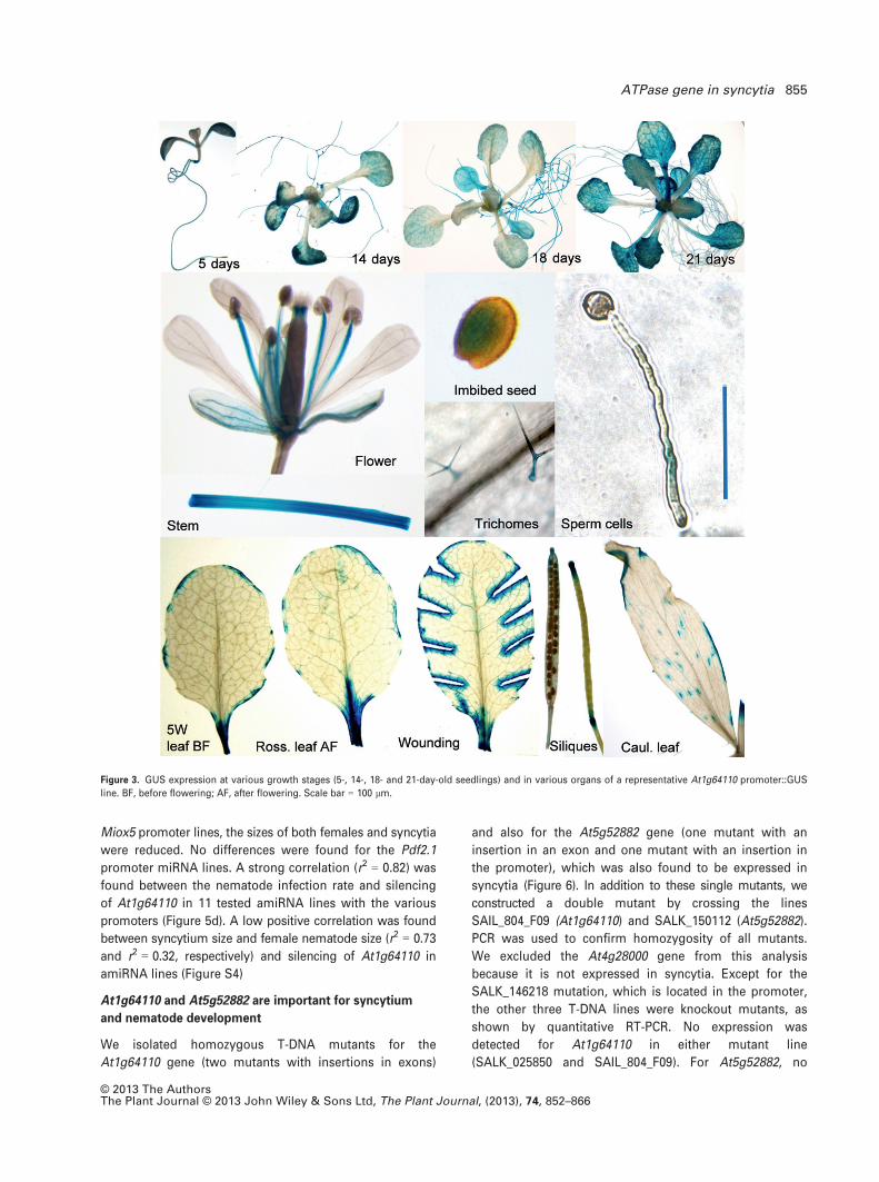
GUS expression analysis of At1g64110
Of the three genes of this sub-family, only At1g64110
expression was strongly induced in syncytia. We therefore
created promoter::GUS lines to further study its expres-
sion. A representative line was selected, and the general
GUS expression pattern was assessed (Figure 3). Seed-
lings showed staining in the roots and the shoots. Older
rosette leaves were not stained except for trichomes, with
some staining at the edge of the leaves. We also noted
staining at the petiole where the leaves were cut from the
plant, suggesting induction of the gene by wounding. We
confirmed this by cutting the leaves, which led to staining
at the cut sites. Cauline leaves also showed some staining
at the edges, and also strong staining in the trichomes.
Stems were also stained, and we observed staining of the
filaments of the anthers and the veins of the sepals in
flowers. Some staining was also observed at the base of
the stigma, and also in young siliques but not old siliques.
Young siliques were also stained at the abscission zone,
and seeds were only stained after imbibition. Lastly, GUS
expression was observed in sperm cells after artificial ger-
mination of pollen grains.
The promoter::GUS line was used to determine the
expression of At1g64110 during syncytium development
(Figure 4). Even at 12 hpi (hours post-inoculation), faint
GUS staining was visible that appeared to be associated
with cells that are then incorporated into the developing
syncytium. Clear GUS staining of the syncytium was
detected at 24 hpi, and at all time points thereafter (48 hpi,
3 dpi, 4 dpi, 5 dpi, 9 dpi and 12 dpi), up to 15 dpi. At
20 dpi, the GUS staining was much weaker. Uninfected
seedlings showed GUS staining in roots and shoots at all
time points (Figure S3) that decreased in older seedlings,
and there was usually no staining in roots by 22 days.
Down-regulation of At1g64110 enhances resistance
against H. schachtii
The strong up-regulation of At1g64110 in syncytia
indicated an important function of this gene for syncytium
development. We tested this by producing artificial micro
RNA (amiRNA) lines to down-regulate the gene using three
promoters: the CaMV 35S promoter, the Pdf2.1 promoter
(Siddique et al., 2011), and the Miox5 promoter (Siddique
et al., 2009). The 35S promoter has been reported to be
only active in younger syncytia and down-regulated in old
syncytia (Urwin et al., 1997; Goverse et al., 1998), whereas
both Pdf2.1 and Miox5 have been shown to be strongly
up-regulated in syncytia. For each promoter, we selected
two independent homozygous lines with at least 50%
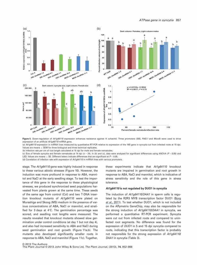
down-regulation of the At1g64110 gene (Figure 5a). We
performed infection assays for all lines, and found that the
number of female nematodes was significantly reduced in
each line but the number of males was not significantly
different (Figure 5b). Contrasting results were found for
the size of syncytia and females in these lines (Figure 5c).
In the 35S promoter lines, the size of the syncytia was
significantly reduced but not the size of females. In the
(a) (b) (c)
Figure 1. In situ RT-PCR of At1g64110 expression in 15 dpi syncytia and uninfected roots.
(a) Strong signal for At1g64110 mRNA in the syncytium. In addition, staining is visible in pericycle cells.
(b) Control reaction performed without Taq polymerase on a section of 15 dpi syncytium. Staining is not visible in either the feeding site or surrounding cells.
(c) Control reaction on a root section above the syncytium showing signals for At1g64110 in pericycle cells. S, syncytium; Ep, epidermis; En, endodermis;
P, pericycle. Scale bars = 50 lm.
Figure 2. RT-PCR using RNA isolated from seedlings grown on MS medium
(5S, 5-day-old shoots; 14S, 14-dayold shoots; 5R, 5-day-old roots; 14R,
14-day-old roots) or on soil (5WL, 5-week-old leaves; CL, cauline leaves, ST,
stems; FL, flowers; SIL, siliques). Primers for the 18S gene were used for
control reactions.
© 2013 The AuthorsThe Plant Journal © 2013 John Wiley & Sons Ltd, The Plant Journal, (2013), 74, 852–866
854 Muhammad Amjad Ali et al.
Miox5 promoter lines, the sizes of both females and syncytia
were reduced. No differences were found for the Pdf2.1
promoter miRNA lines. A strong correlation (r2 = 0.82) was
found between the nematode infection rate and silencing
of At1g64110 in 11 tested amiRNA lines with the various
promoters (Figure 5d). A low positive correlation was found
between syncytium size and female nematode size (r2 = 0.73
and r2 = 0.32, respectively) and silencing of At1g64110 in
amiRNA lines (Figure S4)
At1g64110 and At5g52882 are important for syncytium
and nematode development
We isolated homozygous T-DNA mutants for the
At1g64110 gene (two mutants with insertions in exons)
and also for the At5g52882 gene (one mutant with an
insertion in an exon and one mutant with an insertion in
the promoter), which was also found to be expressed in
syncytia (Figure 6). In addition to these single mutants, we
constructed a double mutant by crossing the lines
SAIL_804_F09 (At1g64110) and SALK_150112 (At5g52882).
PCR was used to confirm homozygosity of all mutants.
We excluded the At4g28000 gene from this analysis
because it is not expressed in syncytia. Except for the
SALK_146218 mutation, which is located in the promoter,
the other three T-DNA lines were knockout mutants, as
shown by quantitative RT-PCR. No expression was
detected for At1g64110 in either mutant line
(SALK_025850 and SAIL_804_F09). For At5g52882, no
Figure 3. GUS expression at various growth stages (5-, 14-, 18- and 21-day-old seedlings) and in various organs of a representative At1g64110 promoter::GUS
line. BF, before flowering; AF, after flowering. Scale bar = 100 lm.
© 2013 The AuthorsThe Plant Journal © 2013 John Wiley & Sons Ltd, The Plant Journal, (2013), 74, 852–866
ATPase gene in syncytia 855

expression was detected in SALK_150112; however, nor-
mal expression was detected in SALK_146218 (Table 2).
We analysed the seedling root phenotype of these
mutants. There was a significant decrease in root area
and length for SALK_146218 (At5g52882) compared to
wild-type seedlings. For all other mutants, including the
double mutant, no differences were found (Figure S5).
We tested the susceptibility of the mutants (single and
double) and compared them with Col wild-type plants
(Figure 7). Single mutants of gene At5g52882 had the
least effect. The number of female and male nematodes
was not significantly different from wild-type, and also
the size of syncytia and of female nematodes was the
same. However, both single mutants of gene At1g64110
were clearly more resistant than wild-type. They sup-
ported fewer females and males, and also syncytia and
females were smaller than those from wild-type plants.
The double mutant was not significantly different from
the At1g64110 single mutants. We also analysed expres-
sion of the defence marker genes Pdf1.2 (Thomma et al.,
1998) and PR1 (Uknes et al., 1992) in the mutants (Fig-
ure 8). We found no difference in expression in seedlings
of any of the mutants compared to wild-type seedlings.
At1g64110 is involved in the response to abiotic stress
The finding that At1g64110 was induced by wounding
prompted us to test its induction by jasmonic acid (JA).
We also included salicylic acid (SA) as it is commonly
involved in resistance responses to biotrophic pathogens.
Treatment with methyl jasmonic acid (MeJA) and SA was
performed by infiltrating seedlings growing on MS
medium. As shown in Figure 9, At1g64110 expression was
induced by JA, especially after 3 and 12 h. The strongest
response to SA was found at 3 h, after which it declined,
but increased again at 48 h.
We further performed quantitative RT-PCR to study
expression of the ATPase gene At1g64110 in response to
various abiotic stress conditions, including wounding,
abscisic acid (ABA), cold, heat, drought, salt and osmotic
stresses at the 14-day seedling stage and 5-week rosette
Figure 4. GUS expression in syncytia of a rep-
resentative At1g64110 promoter::GUS line at
various times after inoculation. There was
strong expression of GUS throughout the
development of nematode-induced syncytia,
starting from cell differentiation for syncytia for-
mation (12 hpi) up to mature syncytia (15 dpi),
and decreasing at 20 dpi. Asterisks indicate
cells differentiating into syncytia; N, nematode;
S, syncytium. Scale bar = 100 lm. The last
image shows the root elongation zone and root
tip from a non-infected plant.
© 2013 The AuthorsThe Plant Journal © 2013 John Wiley & Sons Ltd, The Plant Journal, (2013), 74, 852–866
856 Muhammad Amjad Ali et al.
stage. The At1g64110 gene was highly induced in response
to these various abiotic stresses (Figure 10). However, the
induction was more profound in response to ABA, manni-
tol and NaCl at the early seedling stage. To test the impor-
tance of this gene in the response to these physiological
stresses, we produced synchronized seed populations har-
vested from plants grown at the same time. These seeds
of the same age from control (Col) and two T-DNA inser-
tion knockout mutants of At1g64110 were plated on
Murashige and Skoog (MS) medium in the presence of var-
ious concentrations of ABA, NaCl or mannitol, and strati-
fied for 3 days at 4°C. The germination percentage was
scored, and seedling root lengths were measured. The
results revealed that knockout mutants showed slow ger-
mination under control conditions (at day 1 but not day 4),
and also had increased sensitivity to ABA and NaCl during
seed germination and root growth (Figure 11a,b). The
mutants also developed significantly smaller roots in
response to ABA, NaCl and mannitol (Figure 11c). Together,
these experiments indicate that At1g64110 knockout
mutants are impaired in germination and root growth in
response to ABA, NaCl and mannitol, which is indicative of
stress sensitivity and the role of this gene in stress
tolerance.
At1g64110 is not regulated by DUO1 in syncytia
The induction of At1g64110/DAA1 in sperm cells is regu-
lated by the R2R3 MYB transcription factor DUO1 (Borg
et al., 2011). To test whether DUO1, which is not included
on the Affymetrix GeneChip, may also be responsible for
the strong induction of At1g64110/DAA1 in syncytia, we
performed a quantitative RT-PCR experiment. Syncytia
were cut out from infected roots and compared to unin-
fected root segments. No difference was found for the
expression of DUO1 in 5 and 15 dpi syncytia compared to
roots, indicating that this transcription factor is probably
not responsible for the strong expression of At1g64110/
DAA1 in syncytia (Table 3).
(a) (b)
(c) (d)
Figure 5. Down-regulation of At1g46110 expression enhances resistance against H. schachtii. Three promoters (35S, Pdf2.1 and Miox5) were used to drive
expression of an artificial At1g46110 miRNA gene.
(a) At1g46110 expression in miRNA lines measured by quantitative RT-PCR relative to expression of the 18S gene in syncytia cut from infected roots at 15 dpi.
Values are means � SEM for three biological and three technical replicates.
(b) Infection rate per cm of root length calculated at 15 dpi for male and female nematodes.
(c) Size of female syncytia and female nematodes at 14 dpi (n = 24). In (b) and (c), data were analysed for significant differences using ANOVA (P < 0.05) and
LSD. Values are means � SE. Different letters indicate differences that are significant at P < 0.05.
(d) Correlation of infection rate with expression of At1g64110 in miRNA lines with various promoters.
© 2013 The AuthorsThe Plant Journal © 2013 John Wiley & Sons Ltd, The Plant Journal, (2013), 74, 852–866
ATPase gene in syncytia 857

Sequence analysis of ORTHO000440 proteins
The AAA+ ATPase proteins studied here are very similar
at the protein level as shown by a sequence alignment
(Figure S6). They do not contain a signal sequence (Petersen
et al., 2011) but do contain a transmembrane helix at the
N-terminus (Figure S7). They contain two AAA domains,
but only the second one appears to be functional as it
comprises the catalytical Walker A and B sequences
(Walker et al., 1982), which are missing from the first
domain. Furthermore, a C-terminal extension, similar to
the one found in Vps4 (Babst et al., 1998) and presumably
important for oligomerization, was detected. Sequence
comparison with other ATPases positioned the
ORTHO000440 proteins within the central cluster of the
AAA family, within the AAA+ superfamily (Ammelburg
et al., 2006). Further sub-typing allowed assignment of
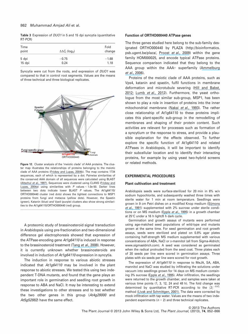
At1g64110 to the ‘meiotic clade’ of AAA proteins (Frickey
and Lupas, 2004b), containing AAA proteins such as Vps4,
katanin, spastin and MSP1 (Figure 12). The most pro-
nounced similarity detected is to the membrane-bound
MSP1 (mitochondrial sorting of proteins 1) proteins of
fungi and metazoa, with BLAST P values as low as 1.0e-70.
DISCUSSION
Expression of ATPase genes in response to nematode
infection
The ATPase gene At1g64110 was among the most strongly
up-regulated genes found in a transcriptome analysis of
syncytia induced by the beet cyst nematode H. schachtii in
Arabidopsis roots (Szakasits et al., 2009). Here we con-
firmed the expression in syncytia using RT-PCR, in situ RT-
PCR and GUS analysis. Arabidopsis contains two other
genes that are related to At1g64110: At4g28000 and
At5g52882. At4g28000 was expressed at a very low level in
syncytia and control root segments (Szakasits et al., 2009),
and was only detected in the present study in flowers and
siliques using RT-PCR; thus we have excluded this gene
from further experiments. At5g52882 is not represented on
the Affymetrix Arabidopsis GeneChip, and therefore no
information was available about its expression. We there-
fore tested the expression of this gene in syncytia using
RT-PCR and quantitative RT-PCR. Expression was clearly
detected in syncytia and roots, but was not up-regulated in
syncytia, in contrast to At1g64110 expression.
At1g64110 is expressed in sperm cells
It has been shown that At1g64110 is expressed in sperm
cells, and that this expression is dependent on the R2R3
MYB transcription factor DUO1 (Borg et al., 2011). We con-
Figure 6. T-DNA mutants for At1g46110 and
At5g52882. The intron/exon structure and the
site of the T-DNA insertion are shown for both
genes. PCR using gene-specific primers and the
T-DNA primer confirmed homozygous mutants.
Table 2 Expression of At1g64110 and At5g52882 in T-DNAmutants
Locus T-DNA mutant lines Expression
At1g64110 SAIL_804_F09 NDSALK_025850 ND
At5g52882 SALK_150112 NDSALK_146218 +
ND, not detected; +, normal expression.RNA was extracted from 7-day-old seedlings to detect expressionof At1g64110 and At5g52882 in corresponding T-DNA mutant linesby quantitative RT-PCR.
© 2013 The AuthorsThe Plant Journal © 2013 John Wiley & Sons Ltd, The Plant Journal, (2013), 74, 852–866
858 Muhammad Amjad Ali et al.

firmed the expression of At1g64110 in sperm cells using
our promoter::GUS line. Unfortunately, DUO1 (At3g60460)
is not represented on the Arabidopsis GeneChip, and its
expression in syncytia was therefore not known. We there-
fore used quantitative RT-PCR to show that expression of
DUO1 was not induced in syncytia compared to uninfected
control root segments. Furthermore, examination of the
expression level in syncytia (Szakasits et al., 2009) of
DUO1 target genes in sperm cells (Borg et al., 2011)
revealed that, other than At1g64110, only one gene (DAN1)
was up-regulated in syncytia compared to control root seg-
ments and DAW1 was down-regulated. All other DUO1 tar-
get genes were expressed at a very low level in roots and
syncytia (Table S3). Thus, it is unlikely that DUO1 is
responsible for the strong up-regulation of At1g64110 in
syncytia.
ATPase genes are important for syncytium and nematode
development
The strong up-regulation of At1g64110 in syncytia
suggested an important function of this gene for syncy-
tium and nematode development. We confirmed this
assumption using amiRNA lines and T-DNA knockout
mutants. Expression of the amiRNA gene was driven by
one of three promoters, and all lines achieved at least
50% reduction of the transcript level in syncytia cut from
infected Arabidopsis roots. All lines showed an effect, and
especially the number of females developing on these roots
was reduced. The reduction in syncytium size in some ami-
RNA lines and in the At1g64110 T-DNA mutants showed
(a)
(b)
Figure 7. Infection assays for knockout mutants
of At1g64110 (SAIL_804_F09 and SALK_025850),
At5g52882 (SALK_150112 and SALK_146218)
and a double mutant (SALK_150112/
SAIL_804_F09) compared with Col wild-type
plants.
(a) Infection rate per cm root length calculated
at 15 dpi, with wild-type set as 100%, for female
and male nematodes.
(b) Size of female syncytia and female nema-
todes at 14 dpi (n = 30). Different letters indi-
cate differences that are significant at P < 0.05
(ANOVA and LSD). Values are means � SE.
Figure 8. Expression of defence marker genes in At1g46110 and At5g52882
T-DNA mutants. Relative expression of Pdf1.2 and PR1 measured by quanti-
tative RT-PCR in seedlings of knockout mutants compared with Col wild-
type plants. There was no change in expression of Pdf1.2 and PR1 in any of
the mutants. Values are means � SEM for three technical and three biologi-
cal replicates.
© 2013 The AuthorsThe Plant Journal © 2013 John Wiley & Sons Ltd, The Plant Journal, (2013), 74, 852–866
ATPase gene in syncytia 859

that At1g64110 is important for syncytium development.
The smaller syncytia provide fewer nutrients for the nema-
todes, and thus influence the development of nematodes.
The observed effects were stronger with the two T-DNA
mutants that were used, probably due to the fact that the
miRNA lines still had a significant level of At1g64110 tran-
scripts while both At1g64110 T-DNA mutants were knock-
out mutants. The effect of the At5g52882 mutants was less
pronounced. An At1g64110 and At5g52882 double mutant
showed an additive effect, especially on the number of
female nematodes developing on the roots. These data
indicate that both proteins (and perhaps also At4g28000)
have the same function, and that a high level of these pro-
teins is needed for fully functional syncytia.
The ATPase genes discussed here may also be important
for the development of clubroot disease. The pathogen
Plasmodiophora brassiceae induces formation of galls in
the roots of Brassicaceae, including Arabidopsis, which are
a severe nutrient sink for the plant, similar to the syncytia
induced by cyst nematodes. In a transcriptome analysis of
clubroots of Arabidopsis roots, it was found that expression
of At1g64110 was almost 54-fold up-regulated (Siemens
et al., 2006). It may be interesting to determine whether
down-regulation of this gene led to enhanced resistance
against Plasmodiophora brassiceae. Furthermore, induction
of the gene by SA indicates that it may also be involved in
resistance responses to other pathogens.
The ATPase gene At1g64110 is involved in abiotic stress
responses
We found that At1g64110 was inducible by MeJA and
wounding, heat, cold and drought, as well as NaCl, ABA
and mannitol. Involvement of the ATPase gene At1g64110
in abiotic stress has been reported previously. Analysis of
drought responses of Boechera holboelli, a relative of Ara-
bidopsis, found that the B. holboelli homologue of
At1g64110 was up-regulated during drought responses
(Knight et al., 2006). Drought responses in Arabidopsis are
regulated by the plasma membrane histidine kinase ATHK1,
and At1g64110 is one of the genes that was induced in
ATHK1 over-expression lines and down-regulated in an
athk1 mutant (Wohlbach et al., 2008). However, expression
of ATHK1 in syncytia was rather low, and was not different
from that in control root segments (Szakasits et al.,
2009), which indicates that the expression of At1g64110 in
syncytia may not be regulated by ATHK1. Furthermore,
among the genes reported by Wohlbach et al. (2008) as
up-regulated by ATHK1, only 11 were up-regulated in syncytia
compared to control root segments (Szakasits et al., 2009),
while three were down-regulated and the majority (24)
showed the same expression in syncytia and roots (Table S4;
data from Szakasits et al., 2009). Another transcription factor
Figure 9. Relative expression of the At1g64110 gene in response to MeJA
(100 lM) and SA (1 mM). The graph shows the results for one of two experi-
ments with similar results.
Figure 10. Relative expression of At1g64110 in
wild-type seedlings subjected to various stres-
ses at early (14-day-old seedlings on MS med-
ium) and late (5-week-old rosette leaves on soil)
developmental stages compared to control
plants as measured by quantitative RT-PCR.
The experiment was repeated twice with three
technical replicates, and the data are shown as
DDC t : The values are means � SE.
© 2013 The AuthorsThe Plant Journal © 2013 John Wiley & Sons Ltd, The Plant Journal, (2013), 74, 852–866
860 Muhammad Amjad Ali et al.

that was shown to regulate the expression of At1g64110
was AREB1, a leucine zipper protein encoded by At1g45249
that binds to the ABRE motif in the promoter region of ABA-
inducible genes (Fujita et al., 2005). Again, most of the
genes that were found to be up-regulated in transgenic lines
over-expressing a constitutive active form of AREB1
(AREB1DQT) were not up-regulated in syncytia (Szakasits
et al., 2009), indicating that AREB1 is also probably not
involved in the strong up-regulation of At1g64110 in syncy-
tia (Table S5).
(a)
(b)
(c)
Figure 11. Physiological responses of
At1g64110 mutants (SAIL_804_F09 and
SALK_025850).
(a) Germination of wild-type and two mutants
grown for 10 days on MS agar plates contain-
ing various ABA concentrations.
(b) Effects of ABA and NaCl on germination at
day 1 and day 4.
(c) Effects of NaCl and mannitol on the root
length of seedlings grown on MS agar plates.
Data are means � SE of three experiments with
n = 20 (germination) and n = 6 (root length).
© 2013 The AuthorsThe Plant Journal © 2013 John Wiley & Sons Ltd, The Plant Journal, (2013), 74, 852–866
ATPase gene in syncytia 861
A proteomic study of brassinosteroid signal transduction
in Arabidopsis using pre-fractionation and two-dimensional
difference gel electrophoresis showed that expression of
the ATPase-encoding gene At1g64110 is induced in response
to the brassinosteroid treatment (Tang et al., 2008). However,
it is currently unknown whether brassinosteroids are
involved in induction of At1g64110 expression in syncytia.
The induction in response to various abiotic stresses
indicated that At1g64110 may be involved in the plant
response to abiotic stresses. We tested this using two inde-
pendent T-DNA mutants, and found that the gene plays an
important role in germination and seedling root growth in
response to ABA and NaCl. It may be interesting to extend
these investigations to other stresses and to test whether
the two other genes in this group (At4g28000 and
At5g52882) have the same effect.
Function of ORTHO000440 ATPase genes
The three genes studied here belong to the sub-family des-
ignated ORTHO000440 by PLAZA (http://bioinformatics.
psb.ugent.be/plaza/, Proost et al., 2009) within the gene
family HOM000025, and encode typical ATPase proteins.
Sequence comparison indicated that they belong to the
AAA group within the AAA+ superfamily (Ammelburg
et al., 2006).
Proteins of the meiotic clade of AAA proteins, such as
Vps4, katanin and spastin, fulfil functions in membrane
deformation and microtubule severing (Hill and Babst,
2012; Lumb et al., 2012). Furthermore, the yeast ortho-
logue from the most similar sub-group, MSP1, has been
shown to play a role in insertion of proteins into the inner
mitochondrial membrane (Nakai et al., 1993). The rather
close relationship of At1g64110 to these proteins impli-
cates this plant-specific sub-group in the remodelling of
membranes and shaping of their protein content. Such
activities are relevant for processes such as formation of
a syncytium or the response to stress, and provide a plau-
sible explanation for the effects observed. To further
explore the specific function of At1g64110 and related
ATPases in Arabidopsis, it will be important to identify
their subcellular location and to identify their interacting
proteins, for example by using yeast two-hybrid screens
or related methods.
EXPERIMENTAL PROCEDURES
Plant cultivation and treatment
Arabidopsis seeds were surface-sterilized for 20 min in 6% w/vsodium hypochlorite, and subsequently washed three times withsterile water for 1 min at room temperature. Seedlings weregrown in 9 cm Petri dishes on a modified Knop medium (Sijmonset al., 1991) supplemented with 2% sucrose under sterile condi-tions or on MS medium (Epple et al., 1995) in a growth chamberat 25°C under a 16 h light/8 h dark cycle.
Germination and growth assays of mutants were performedusing age-matched seed populations of wild-type and mutantsgrown at the same time. For seed germination and root growthassays, seeds were sterilized and plated on 0.8% agar platescontaining half-strength MS medium supplemented with variousconcentrations of ABA, NaCl or D-mannitol (all from Sigma-Aldrich;www.sigmaaldrich.com). A seed was considered as germinatedwhen the radical protruded from the seed coat. Three plates eachof 20 seeds per line were scored in germination assays. Threeplates with six seeds per line were scored for root growth.
The expression of At1g64110 in response to MeJA, SA, ABA,mannitol and NaCl was studied by infiltrating the solutions undervacuum into seedlings grown for 14 days on MS medium contain-ing 3% sucrose (Epple et al., 1995). After infiltration, the seedlingswere returned to the growth chamber, and samples were taken atvarious time points (1, 3, 12, 24 and 48 h). The fold change wasdetermined by quantitative RT-PCR according to the ð2Þ�DDCt
method (Livak and Schmittgen, 2001). The data were corrected bymock infiltration with tap water. Values are the means of two inde-pendent experiments (n = 2) and three technical replicates.
Table 3 Expression of DUO1 in 5 and 15 dpi syncytia (quantitativeRT-PCR)
Timepoint DDCt (log2)
Foldchange
5 dpi �0.75 �1.6815 dpi 0.24 1.18
Syncytia were cut from the roots, and expression of DUO1 wascompared to that in control root segments. Values are the meansof three technical and three biological replicates.
Figure 12. Cluster analysis of the ‘meiotic clade’ of AAA proteins. The clus-
ter map illustrates the relationships of proteins belonging to the meiotic
clade of AAA proteins (Frickey and Lupas, 2004b). The map contains 1734
sequences, each of which is represented by a dot. Pairwise similarities of
the conserved AAA domain of all sequences were calculated using BLAST
(Altschul et al., 1997). Sequences were clustered using CLANS (Frickey and
Lupas, 2004a) using similarities with P values < 1.0e-50. Darker lines
between two dots indicate lower BLAST P values. The At1g64110/
ORTHO000440 cluster (red dots) shows the tightest connections to MSP1
proteins from fungi and metazoa (yellow dots). However, the Spastin
(green), Katanin (blue) and Vps4 (purple) clusters also show strong similari-
ties to the At1g64110/ORTHO000440 (red) group.
© 2013 The AuthorsThe Plant Journal © 2013 John Wiley & Sons Ltd, The Plant Journal, (2013), 74, 852–866
862 Muhammad Amjad Ali et al.
For treatment of older plants, we used 5-week-old rosette leavesfrom plants grown on soil. Wounding was performed using for-ceps, heat treatment was at 37°C, and cold treatment was at 4°C.For drought treatment, soil-grown plants were not watered for7 days, while seedlings growing in Petri dishes on MS mediumwere treated by opening the plates and incubating them on a ster-ile bench. Untreated plants were used as a control for thesetreatments.
Cloning of an amiRNA construct for At1g64110
Engineering of the precursor containing the artificial microRNA(amiRNA) was performed as described by Schwab et al. (2006).The primer sequences (Table S2) used to create the precursor ofthe artificial miRNA (amiRNA sequence: 5′-TAAACGTTTATGAAAC-TCGCC-3′) were generated using the web-based tool ‘Web micro-RNA Designer’ at http://wmd.weigelworld.org/cgi-bin/mirnatools.pl (Schwab et al., 2006).
The amiRNA-containing precursor was generated by overlap-ping PCR as described by Schwab et al. (2006). The first roundamplified fragments (a) to (c). These were subsequently fused byPCR (d). Oligonucleotide primers I–IV were used to replace themiRNA319a region with the artificial sequence in plasmidpUCmi319a (see Methods S1). Primers A and B were based on thetemplate plasmid sequence. Regeneration of functional miRNAprecursors was achieved by combining PCR products A-IV, II-IIIand I-B in a single reaction with primers A and B.
Arabidopsis transformation
Binary vectors were introduced into Agrobacterium tumefaciensGV3101 for transformation of Arabidopsis Col-0 plants by thefloral-dip method as modified by Logemann et al. (2006). Transgenicplants were selected on MS medium containing 50 lg ml�1 kana-mycin and 250 lg ml�1 timentin. Dry seeds were sprinkled ontothe selection plates at approximately 25–35 seeds per cm2. Theplates were incubated at 22°C with a photoperiod of 16 h light/8 hdark for the selection of kanamycin-resistant plants. Arabidopsisplants resistant to kanamycin were transferred to soil in a growthchamber to produce seeds.
RNA isolation
Root segments containing syncytia were excised at 15 dpi andimmediately frozen in liquid nitrogen. Control root segments werecollected and frozen as described previously (Szakasits et al.,2009). Total RNA was isolated using an RNeasy plant mini kit(Qiagen; www.qiagen.com) according to the manufacturer’sinstructions, including DNase I digestion. The quality and quantityof the RNA was assessed using an Agilent 2100 bioanalyzer(Agilent Technologies; www.home.agilent.com). Reverse tran-scription was performed using SuperScript III reverse transcrip-tase (Invitrogen; www.invitrogen.com) and random primers [oligo(dN6)] according to the manufacturer’s instructions.
Semi-quantitative RT-PCR
RT-PCR was performed using RT-PCR Master Mix (USB; www.affymetrix.com/estore/browse/brand/usb/usb.jsp) according to themanufacturer’s instructions. The primers used are shown in TableS2.
Quantitative RT-PCR
Real-time RT-PCR was performed using an ABI PRISM 7300sequence detector (Applied Biosystems; www.invitrogen.com/site/
us/en/home/brands/Applied-Biosystems.html?CID=fl-AppliedBiosystems) as described previously (Siddique et al., 2009). Eachquantitative PCR sample contained 12.5 ll Platinum SYBR GreenqPCR SuperMix with UDG (uracil DNA glycosylase) and ROX (5-carboxy-X-rhodamine) reference dye (Invitrogen), 2 mM MgCl2,0.5 ll forward and reverse primer (10 lM), 2 ll cDNA and sufficientwater to obtain a 25 ll total reaction volume. The primers usedare shown in Table S2. All samples were diluted 1:3, and wereanalysed in technical triplicates.
Nematode infection assays
H. schachtii cysts were harvested from in vitro stock cultures onroots of mustard plants (Sinapis alba cv. Albatros) growing on0.29 Knop medium (Sijmons et al., 1991) supplemented with 2%sucrose. Hatching of J2 from the cysts was stimulated by soakingin 3 mM ZnCl2. Larvae were then washed three times in sterilewater, and the J2 were resuspended in 0.5% w/v gelrite (Duchefa;www.duchefa-biochemie.nl/). Roots of 12-day-old Arabidopsisseedlings growing on Knop medium were inoculated under sterileconditions with approximately 50-60 J2 per plant. Three indepen-dent experiments were performed, each comprising five Petridishes with a total of approximately ten seedlings. At 14 dpi, thesize of syncytia (associated with females) and of females wasmeasured as described by Siddique et al. (2009). Briefly, syncytiaassociated with females were randomly selected and photo-graphed (longitudinal optical sections) using a Zeiss Axiovert200M inverted microscope equipped with a Zeiss Axiocam digitalcamera (Zeiss, http://www.zeiss.com/). The syncytia and femaleswere outlined using the Axiovision Kontour tool, and the area wasdetermined. The mean size of syncytia and females was calcu-lated, and data were further statistically analysed using single-factor ANOVA (P < 0.05) and fisher’s least significant difference test.One day later (15 dpi), the number of males and females wascounted. The data were expressed as nematodes per cm of rootlength, and were analysed using single-factor ANOVA (P < 0.05) andDuncan’s multiple range test.
GUS reporter analysis
The promoter region of At1g64110 was amplified by PCR using 50ng Arabidopsis genomic DNA as template. Primers (Table S2)included restriction sites for NcoI and EcoRI (underlined) for sub-sequent cloning into binary vector pPZP3425 (Szakasits et al.,2007). This plasmid contains a double enhanced 35S promoterand a TMV (tobacco mosaic virus) omega element driving theGUS gene. During the cloning procedure, the double enhanced35S promoter and TMV omega element were replaced by the pro-moter fragment. The promoter sequence was confirmed bysequencing. We produced 17 GUS lines and tested seven in detailfor GUS expression in seedlings and syncytia. From these, weselected the one line that was used here. Histochemical detectionof GUS activity was performed by staining using X-Gluc (Biomol;www.biomol.de) in 0.1 M sodium phosphate buffer pH 7.0, 0.1%Triton X-100, 0.5 mM K3[Fe(CN)6], 0.5 mM K4[Fe(CN)6] and 10 mMNa2EDTA. After staining, chlorophyll was removed from photo-synthetic tissues using 70% v/v ethanol. Various plant parts werestained after flowering of the plants. For seed imbibition, seedswere imbibed at 4°C on filter papers soaked in sterile water undercontinuous white light for 4 days before GUS staining for 24 h.For analysis of syncytia, infected roots containing syncytia werestained at various times after inoculation by overnight incubationwith X-Gluc. The stained syncytia and uninfected roots were pho-tographed under the Axiovert 200M inverted microscope with anintegrated camera (AxioCam MRc5).
© 2013 The AuthorsThe Plant Journal © 2013 John Wiley & Sons Ltd, The Plant Journal, (2013), 74, 852–866
ATPase gene in syncytia 863
In vitro pollen germination and GUS staining
For sperm cell staining, fresh anthers were used for in vitro pollengermination. The medium for in vitro pollen germination contained18% sucrose, 0.01% boric acid, 1 mM CaCl2, 1 mM Ca(NO3)2, 1 mM
MgSo4 and 0.5% agar (Difco; www.bd.com) at pH 7.0. Sucrosewas dissolved first, and then other ingredients were added.Finally, agar was added and the medium was boiled for 3–4 minto dissolve agar (Li et al., 1999). The medium was poured intosmall Petri dishes (35 9 10 mm). Pollen was dusted onto the med-ium and allowed to germinate for 3–5 h or sometimes overnightat room temperature. After pollen germination, histochemicaldetection of GUS activity was performed by staining using theabove-mentioned X-Gluc solution and incubation overnight at37°C. The pollen tubes containing stained sperm cells were photo-graphed as described above.
In situ RT-PCR
In situ RT-PCR was performed as described by Koltai and Bird(2000) and Urbanczyk-Wochniak et al. (2002). Syncytia at 15 dpiwere dissected from roots and immediately placed into coldfixation solution (63% v/v ethanol, 2% v/v formalin). After 24 h,syncytia were embedded in 4% low-melting-point agarose, and25 lm thick sections were prepared using a vibratome (VT100;Leica, http://www.leica.com/). RT-PCR was then performed usingdigoxigenin-labelled dUTP and primers (Table S2). After a stainingreaction with nitroblue tetrazolium/5-bromo-4-chloro-3-indolylphosphate, cross-sections were photographed under an Axiovert200M inverted microscope with an integrated camera (AxioCamMRc5). Full details of the method are provided in Wieczorek et al.(2006).
Mutant screening
Seeds of SAIL_804_F09, SALK_023626 (At1g64110), SALK_150112and SALK_146218 (At5g52882) were obtained from the Notting-ham Arabidopsis Stock Centre (http://arabidopsis.info/) (Sessionset al., 2002; Alonso et al., 2003). Genomic DNA from segregat-ing plants was screened by PCR using the primers listed inTable S2. Primer sequences were obtained using the tool sup-plied at the SIGnAL website (http://signal.salk.edu/tdnaprimers.2.html).
Root length and area measurement
Seeds of Arabidopsis plants (Col-0 and T-DNA mutants) weregrown on Knop medium as described above. Twelve-day-oldplants (corresponding to the infection time point) were photo-graphed using a DM4000 microscope (Leica; http://www.leica-microsystems.com) and an Olympus digital camera (http://www.olympus-europa.com/). The length and diameter of mainand all lateral roots was calculated using the measurement tool ofLAS software version 4.1 (http://www.leica-microsystems.com/products/microscope-software/materials-sciences/materials-imaging/details/product/leica-las-interactive-measurement/). The total rootarea of a plant was calculated by adding the areas of all individualroots. Five to ten individual measurements were used to calculatethe mean length and area per plant.
Bioinformatics
Sequence similarity searches indicated the presence of aconserved AAA+ domain in the C-terminal part of At1g64110.Homologues of this domain were identified by searching theNational Center for Biotechnology Information non-redundant
database using BLAST (Altschul et al., 1997). The 2000 most simi-lar high-scoring segment pairs were retrieved and clustered usingCLANS (Frickey and Lupas, 2004a), with BLAST as a comparisontool. Clustering was performed with default parameters at aP value cut-off of 1.0e-50. Searches using HHpred (Soding et al.,2005), a remote homology detection method based on compari-son of profile hidden Markov models, revealed the presence ofanother but degenerate AAA+ domain in the N-terminal portion ofAt1g64110. The N-terminal transmembrane helix was assignedusing TMHMM (Krogh et al., 2001).
ACKNOWLEDGEMENTS
We appreciate the excellent technical assistance of SabineDaxb€ock-Horvath and Martina Niese. This research was supportedby grants P16296-B06, P21067-B12 and P20471-B11 from the Aus-trian Fonds zur F€orderung der Wissenschaftlichen Forschung.M.A.A. and S.M.S. were supported by the Higher Education Com-mission of Pakistan. M.A. is grateful to Andrei Lupas (Department1, Protein Evolution, Max Planck Institute for Developmental Biol-ogy, Tubingen, Germany) for continuous support.
SUPPORTING INFORMATION
Additional Supporting Information may be found in the online ver-sion of this article.Table S1. Gene expression of ATPase genes in syncytia (fromSzakasits et al., 2009) .
Table S2. Primers used in this work.
Table S3. Expression of validated DUO1 target genes (Borg et al.,2011) in syncytia induced by H. schachtii in Arabidopsis roots.
Table S4. Expression of genes identified as ‘significantly differen-tially regulated according to both ATHK1 transcript level and sor-bitol stress condition’ (Wohlbach et al., 2008) in syncytia inducedby H. schachtii in Arabidopsis roots.
Table S5. Expression of genes up-regulated in plants over-expressing AREB1DQT (Fujita et al., 2005) in syncytia induced byH. schachtii in Arabidopsis roots.
Figure S1. Expression of At1g64110 and At1g52882 in syncytia asshown by RT-PCR.
Figure S2. Gene expression of At1g64110 and At4g28000 accord-ing to Genevestigator.
Figure S3. GUS expression in uninfected seedlings.
Figure S4. Correlation of syncytia size and female nematode sizewith the expression of At1g64110 in miRNA lines with various pro-moters.
Figure S5. Phenotypic analysis of T-DNA mutants.
Figure S6. Sequence alignment of ORTHO000440 ATPases.
Figure S7. Domain composition of At1g64110.
Methods S1. Cloning of pUCmi319a.
REFERENCES
Alonso, J.M., Stepanova, A.N., Leisse, T.J. et al. (2003) Genome-wide inser-
tional mutagenesis of Arabidopsis thaliana. Science, 301, 653–657.Altschul, S.F., Madden, T., Schaffer, A., Zhang, J., Zhang, Z., Miller, W.
and Lipman, D.J. (1997) Gapped BLAST and PSI-BLAST: a new genera-
tion of protein database search programs. Nucleic Acids Res. 25, 3389–3402.
Ammelburg, M., Frickey, T. and Lupas, A.N. (2006) Classification of AAA+proteins. J. Struct. Biol. 156, 2–11.
Babst, M., Wendland, B., Estepa, E.J. and Emr, S.D. (1998) The Vps4p AAA
ATPase regulates membrane association of a Vps protein complex
required for normal endosome function. EMBO J. 17, 2982–2993.B€ockenhoff, A. and Grundler, F.M.W. (1994) Studies on the nutrient uptake
by the beet cyst nematode Heterodera schachtii by in situ microinjection
© 2013 The AuthorsThe Plant Journal © 2013 John Wiley & Sons Ltd, The Plant Journal, (2013), 74, 852–866
864 Muhammad Amjad Ali et al.
of fluorescent probes into the feeding structures in Arabidopsis thaliana.
Parasitology, 109, 249–255.Borg, M., Brownfield, L., Khatab, H., Sidorova, A., Lingaya, M. and Twell, D.
(2011) The R2R3 MYB transcription factor DUO1 activates a male germ-
line-specific regulon essential for sperm cell differentiation in Arabidop-
sis. Plant Cell, 23, 534–549.Epple, P., Apel, K. and Bohlmann, H. (1995) An Arabidopsis thaliana thionin
gene is inducible via a signal-transduction pathway different from that
for pathogenesis-related proteins. Plant Physiol. 109, 813–820.Frickey, T. and Lupas, A.N. (2004a) CLANS: a Java application for visualizing
protein families based on pairwise similarity. Bioinformatics, 20, 3702–3704.
Frickey, T. and Lupas, A.N. (2004b) Phylogenetic analysis of AAA proteins.
J. Struct. Biol. 146, 2–10.Fujita, Y., Fujita, M., Satoh, R., Maruyama, K., Parvez, M.M., Seki, M., Hira-
tsu, K., Ohme-Takagi, M., Shinozaki, K. and Yamaguchi-Shinozaki, K.
(2005) AREB1 is a transcription activator of novel ABRE-dependent ABA
signaling that enhances drought stress tolerance in Arabidopsis. Plant
Cell, 17, 3470–3488.Gheysen, G. and Mitchum, M.G. (2011) How nematodes manipulate plant
development pathways for infection. Curr. Opin. Plant Biol. 14, 415–421.
Goellner, M., Wang, X.H. and Davis, E.L. (2001) Endo-b-1,4-glucanaseexpression in compatible plant–nematode interactions. Plant Cell, 13,
2241–2255.Golinowski, W., Grundler, F.M.W. and Sobczak, M. (1996) Changes in
the structure of Arabidopsis thaliana during female development of
the plant-parasitic nematode Heterodera schachtii. Protoplasma, 194,
103–116.Goverse, A., Biesheuvel, J., Wijers, G.J., Gommers, F.J., Bakker, J., Schots,
A. and Helder, J. (1998) In planta monitoring of the activity of two con-
stitutive promoters, CaMV 35S and TR2′, in developing feeding cells
induced by Globodera rostochiensis using green fluorescent protein in
combination with confocal laser scanning microscopy. Physiol. Mol.
Plant Pathol. 52, 275–284.Hill, C.P. and Babst, M. (2012) Structure and function of the membrane
deformation AAA ATPase Vps4. Biochim. Biophys. Acta, 1823, 172–181.Iyer, L.M., Leipe, D.D., Koonin, E.V. and Aravind, L. (2004) Evolutionary his-
tory and higher order classification of AAA plus ATPases. J. Struct. Biol.
146, 11–31.Jones, M.G. and Northcote, D.H. (1972) Nematode-induced syncytium – a
multinucleate transfer cell. J. Cell Sci. 10, 789–809.Knight, C.A., Vogel, H., Kroymann, J., Shumate, A., Witsenboer, H. and
Mitchell-Olds, T. (2006) Expression profiling and local adaptation of
Boechera holboellii populations for water use efficiency across a
naturally occurring water stress gradient. Mol. Ecol. 15, 1229–1237.Koltai, H. and Bird, D.M. (2000) High throughput cellular localization of
specific plant mRNAs by liquid-phase in situ reverse transcription-poly-
merase chain reaction of tissue sections. Plant Physiol. 123, 1203–1212.
Krogh, A., Larsson, B., von Heijne, G. and Sonnhammer, E.L. (2001) Predict-
ing transmembrane protein topology with a hidden Markov model:
application to complete genomes. J. Mol. Biol. 305, 567–580.Li, H., Lin, Y., Heath, R.M., Zhu, M.X. and Yang, Z. (1999) Control of pollen
tube tip growth by a Rop GTPase-dependent pathway that leads to
tip-localized calcium influx. Plant Cell, 11, 1731–1742.Livak, K.J. and Schmittgen, T.D. (2001) Analysis of relative gene expression
data using real-time quantitative PCR and the 2� DDCt method. Methods,
25, 402–408.Logemann, E., Birkenbihl, R.P., Ulker, B. and Somssich, I.E. (2006) An
improved method for preparing Agrobacterium cells that simplifies the
Arabidopsis transformation protocol. Plant Methods, 2, 16.
Lumb, J.H., Connell, J.W., Allison, R. and Reid, E. (2012) The AAA ATPase
spastin links microtubule severing to membrane modelling. Biochim.
Biophys. Acta, 1823, 192–197.Nakai, M., Endo, T., Hase, T. and Matsubara, H. (1993) Intramitochondrial
protein sorting. Isolation and characterization of the yeast MSP1 gene
which belongs to a novel family of putative ATPases. J. Biol. Chem. 268,
24262–24269.Niebel, A., Engler, J.D., Hemerly, A., Ferreira, P., Inze, D., VanMontagu, M.
and Gheysen, G. (1996) Induction of cdc2a and cyc1At expression in
Arabidopsis thaliana during early phases of nematode-induced feeding
cell formation. Plant J. 10, 1037–1043.Petersen, T.N., Brunak, S., von Heijne, G. and Nielsen, H. (2011) SignalP 4.0:
discriminating signal peptides from transmembrane regions. Nat. Meth-
ods, 8, 785–786.Proost, S., Van Bel, M., Sterck, L., Billiau, K., Van Parys, T., Van de Peer, Y.
and Vandepoele, K. (2009) PLAZA: a comparative genomics resource to
study gene and genome evolution in plants. Plant Cell, 21, 3718–3731.Schwab, R., Ossowski, S., Riester, M., Warthmann, N. and Weigel, D. (2006)
Highly specific gene silencing by artificial microRNAs in Arabidopsis.
Plant Cell, 18, 1121–1133.Sessions, A., Burke, E., Presting, G. et al. (2002) A high-throughput Arabid-
opsis reverse genetics system. Plant Cell, 14, 2985–2994.Siddique, S., Endres, S., Atkins, J.M. et al. (2009) Myo-inositol oxygenase
genes are involved in the development of syncytia induced by Heteroder-
a schachtii in Arabidopsis roots. New Phytol. 184, 457–472.Siddique, S., Wieczorek, K., Szakasits, D., Kreil, D.P. and Bohlmann, H.
(2011) The promoter of a plant defensin gene directs specific expression
in nematode-induced syncytia in Arabidopsis roots. Plant Physiol. Bio-
chem. 49, 1100–1107.Siddique, S., Sobczak, M., Tenhaken, R., Grundler, F.M. and Bohlmann, H.
(2012) Cell wall ingrowths in nematode induced syncytia require UGD2
and UGD3. PLoS ONE, 7, e41515.
Siemens, J., Keller, I., Sarx, J., Kunz, S., Schuller, A., Nagel, W., Schmulling,
T., Parniske, M. and Ludwig-Muller, J. (2006) Transcriptome analysis of
Arabidopsis clubroots indicate a key role for cytokinins in disease devel-
opment. Mol. Plant–Microbe Interact. 19, 480–494.Sijmons, P.C., Grundler, F.M.W., Vonmende, N., Burrows, P.R. and Wyss, U.
(1991) Arabidopsis thaliana as a new model host for plant-parasitic
nematodes. Plant J. 1, 245–254.Sobczak, M. and Golinowski, W. (2011) Cyst nematodes and syncytia. In Ge-
nomics and Molecular Genetics of Plant–Nematode Interactions (Jones,
J., Gheysen, G. and Fenoll, C., eds). New York: Springer, pp. 61–82.Sobczak, M., Golinowski, W. and Grundler, F.M.W. (1997) Changes in the
structure of Arabidopsis thaliana roots induced during development of
males of the plant parasitic nematode Heterodera schachtii. Eur. J. Plant
Pathol. 103, 113–124.Soding, J., Biegert, A. and Lupas, A.N. (2005) The HHpred interactive server
for protein homology detection and structure prediction. Nucleic Acids
Res. 33, W244–W248.
Szakasits, D., Siddique, S. and Bohlmann, H. (2007) An improved pPZP vec-
tor for Agrobacterium-mediated plant transformation. Plant Mol. Biol.
Rep. 25, 115–120.Szakasits, D., Heinen, P., Wieczorek, K., Hofmann, J., Wagner, F., Kreil, D.P.,
Sykacek, P., Grundler, F.M. and Bohlmann, H. (2009) The transcriptome
of syncytia induced by the cyst nematode Heterodera schachtii in Arabid-
opsis roots. Plant J. 57, 771–784.Tang, W.Q., Deng, Z.P., Oses-Prieto, J.A., Suzuki, N., Zhu, S.W., Zhang, X.,
Burlingame, A.L. and Wang, Z.Y. (2008) Proteomics studies of brassinos-
teroid signal transduction using prefractionation and two-dimensional
DIGE. Mol. Cell. Proteomics, 7, 728–738.Thomma, B.P., Eggermont, K., Penninckx, I.A., Mauch-Mani, B., Vogelsang,
R., Cammue, B.P. and Broekaert, W.F. (1998) Separate jasmonate-depen-
dent and salicylate-dependent defense-response pathways in Arabidop-
sis are essential for resistance to distinct microbial pathogens. Proc. Natl
Acad. Sci. USA, 95, 15107–15111.Uknes, S., Mauch-Mani, B., Moyer, M., Potter, S., Williams, S., Dincher, S.,
Chandler, D., Slusarenko, A., Ward, E. and Ryals, J. (1992) Acquired
resistence in Arabidopsis. Plant Cell, 4, 645–656.Urbanczyk-Wochniak, E., Filipecki, M. and Przybecki, Z. (2002) A useful
protocol for in situ RT-PCR on plant tissues. Cell. Mol. Biol. Lett. 7,
7–18.Urwin, P.E., Moller, S.G., Lilley, C.J., McPherson, M.J. and Atkinson, H.J.
(1997) Continual green-fluorescent protein monitoring of cauliflower
mosaic virus 35S promoter activity in nematode-induced feeding cells in
Arabidopsis thaliana. Mol. Plant–Microbe Interact. 10, 394–400.Urwin, P.E., McPherson, M.J. and Atkinson, H.J. (1998) Enhanced trans-
genic plant resistance to nematodes by dual proteinase inhibitor
constructs. Planta, 204, 472–479.Walker, J.E., Saraste, M., Runswick, M.J. and Gay, N.J. (1982) Distantly
related sequences in the a- and b-subunits of ATP synthase, myosin,
© 2013 The AuthorsThe Plant Journal © 2013 John Wiley & Sons Ltd, The Plant Journal, (2013), 74, 852–866
ATPase gene in syncytia 865

kinases and other ATP-requiring enzymes and a common nucleotide
binding fold. EMBO J. 1, 945–951.Wieczorek, K., Golecki, B., Gerdes, L. et al. (2006) Expansins are involved in
the formation of nematode-induced syncytia in roots of Arabidopsis
thaliana. Plant J. 48, 98–112.Wieczorek, K., Hofmann, J., Blochl, A., Szakasits, D., Bohlmann, H. and
Grundler, F.M.W. (2008) Arabidopsis endo-1,4-b-glucanases are involved
in the formation of root syncytia induced by Heterodera schachtii. Plant
J. 53, 336–351.
Wohlbach, D.J., Quirino, B.F. and Sussman, M.R. (2008) Analysis of the
Arabidopsis histidine kinase ATHK1 reveals a connection between
vegetative osmotic stress sensing and seed maturation. Plant Cell, 20,
1101–1117.Wyss, U. and Grundler, F.M.W. (1992) Heterodera schachtii and Arabidopsis
thaliana, a model host–parasite interaction. Nematologica, 38, 488–493.Zimmermann, P., Hirsch-Hoffmann, M., Hennig, L. and Gruissem, W. (2004)
GENEVESTIGATOR. Arabidopsis microarray database and analysis tool-
box. Plant Physiol. 136, 2621-2632.
© 2013 The AuthorsThe Plant Journal © 2013 John Wiley & Sons Ltd, The Plant Journal, (2013), 74, 852–866
866 Muhammad Amjad Ali et al.
Copyright © 2022 FDOKUMEN