Endoscopic features of gastrointestinal tuberculosis and ...

CURRENT STATUSREV IEW
Human gastrointestinal nematode infections: are new controlmethods required?
Gillian Stepek*1, David J. Buttle�, Ian R. Duce* and Jerzy M. Behnke*
*School of Biology, University of Nottingham, Nottingham, UK; and�Division of Genomic Medicine, University of Sheffield,
Sheffield, UK1Current address: Department of Veterinary Parasitology, Faculty of Veterinary Medicine, University of Glasgow, Glasgow, UK
Parasitic infections are a major medical problem throughout
the world, especially in developing countries where they
cause more morbidity and mortality than other infectious
diseases and are the primary cause of death. There are two
main groups of parasites: (a) the protozoa, which are unicel-
lular organisms and include the malaria parasite, Plasmo-
dium; and (b) the helminths, which are metazoan organisms
and include the cestodes, trematodes and nematodes. The
protozoa are responsible for the majority of the mortality
associated with parasitic infections, while the helminths gen-
erally produce long-term (or chronic), debilitating diseases:
one of the reasons why there is more public awareness of
parasitic protozoan infections than of infections with helm-
inths. In this paper, we review the major human gastrointes-
tinal nematodes, the problems associated with the control of
these parasites and a potential new therapeutic approach.
INTERNATIONAL
JOURNAL OF
EXPERIMENTAL
PATHOLOGY
Received for publication:
9 February 2006
Accepted for publication:
22 June 2006
Correspondence:
Jerzy Behnke
School of Biology
University of Nottingham
University Park
Nottingham NG7 2RD, UK
Tel.: 44 115 951 3208;
Fax: 44 115 951 3251;
E-mail:
Summary
Gastrointestinal (GI) nematode infections affect 50% of the human population
worldwide, and cause great morbidity as well as hundreds of thousands of deaths.
Despite modern medical practices, the proportion of the population infected with GI
nematodes is not falling. This is due to a number of factors, the most important
being the lack of good healthcare, sanitation and health education in many develop-
ing countries. A relatively new problem is the development of resistance to the small
number of drugs available to treat GI nematode infections. Here we review the most
important parasitic GI nematodes and the methods available to control them. In
addition, we discuss the current status of new anthelmintic treatments, particularly
the plant cysteine proteinases from various sources of latex-bearing plants and fruits.
Keywords
anthelmintic, control, gastrointestinal nematodes, human, plant cysteine proteinases,
resistance
Int. J. Exp. Path. (2006), 87, 325–341
doi: 10.1111/j.1365-2613.2006.00495.x
� 2006 Blackwell Publishing Ltd 325

Gastrointestinal nematode infections ofhumans
Gastrointestinal (GI) nematode (soil-transmitted helminth)
infections are amongst the most prevalent worldwide,
although this is largely acknowledged only by those working
in this field. It is estimated that there are 3.5 billion cases
worldwide, of which 450 million are individuals who are
seriously ill as a result, the majority of who are children,
and of which 44 million are pregnant women infected with
hookworms. Approximately 125 000 deaths occur per year,
and these are mainly due to infections with the hookworms,
Ancylostoma duodenale and Necator americanus, or the
roundworm, Ascaris lumbricoides. There are 300–500 mil-
lion cases of malaria per year and, although this number is
much less than that for GI nematode infections, the number
of deaths attributable to malaria is far greater, reaching
3 million per year (http://www.who.int/wormcontrol/en/;
http://www.library.csi.cuny.edu/�davis/faculty_page/Parasit_
links/parasitology_links.html). According to Chan (1997),
the prevalence of GI nematode infections has remained
unchanged in over 50 years, with 39 million disability-adjus-
ted life years (DALYs) lost due to these parasites when com-
pared with 35.7 million lost to malaria or 34.1 million lost
to measles. How can this be the case with modern medical
practices? The majority of infections with GI nematodes
remain asymptomatic, and those cases which do cause mor-
bidity are not directly fatal, in contrast to malaria. This may
be one reason why GI nematode infections have been neglec-
ted diseases in terms of public recognition and research
funding.
Of the 342 helminth species that infect humans (Cromp-
ton 1999), the species of greatest medical importance are
Ascaris lumbricoides (roundworm), Ancylostoma duodenale
and Necator americanus (hookworms), Trichuris trichiura
(whipworm), Enterobius vermicularis (pinworm) and Strong-
yloides stercoralis (threadworm) (Table 1). For more than
50 years, the number of cases of GI nematode infections has
increased with the global population, such that over 50% of
the world’s population are affected by the six major GI
nematode species (Chan 1997; Horton 2003). The majority
of these people live in the developing countries, with those
living in rural and urban slums most at risk. This is probably
due to the poor housing, overcrowded living conditions, lack
of adequate sanitation and hygiene and poor education and
health care in these areas, all of which are associated with
poverty. In the case of S. stercoralis in urban slums, one risk
factor for transmission of infection is believed to be close
contact between individuals, linked with poor personal
hygiene (Conway et al. 1995). In addition, warm and humid
climatic conditions appear to favour the survival of free-liv-
ing larval stages of species such as N. americanus, A. duode-
nale and S. stercoralis (Kappus et al. 1994), with the latter
parasite endemic in tropical and sub-tropical countries,
including South East America, the Far East, West Africa,
Italy and Australia. Although GI nematodes are largely
restricted to the tropics and sub-tropics, infections can occur
in the Northern hemisphere when ambient conditions are
favourable. For example, hookworms caused anaemia in tin
miners in Cornwall at the start of the 20th century and also
caused problems among engineers in the Alps (Boycott &
Haldane 1903; Boycott 1911; Foster 1965). In both cases,
the poor hygiene standards of the workers, in warm, moist
conditions, favoured transmission. In general, however,
infections with GI nematodes, with the exception of E. ver-
micularis, are less frequent in temperate climates and techno-
logically developed societies, such as the USA and Western
Europe; nevertheless, a proportion of these populations still
become infected, despite the higher standards of hygiene and
sanitation (Kappus et al. 1994). In particular, E. vermicularis
infections, whilst being present throughout the tropics, are
also widespread in the Northern hemisphere, such as in the
UK, unlike infections with the other major GI nematodes;
this is possibly due to the low temperatures and high
Table 1 The major gastrointestinal (GI) nematode parasites of humans
GI nematode
(species name)
GI nematode
(group name)
Number
infected Distribution Transmission
Ancylostoma duodenale/
Necator americanus
Hookworm 1.3 billion Worldwide, especially tropical regions Skin contact with contaminated soil
Ascaris lumbricoides Roundworm 1.3 billion Worldwide, especially tropical regions Ingestion of eggs
Trichuris trichiura Whipworm 1.05 billion Worldwide, especially tropical regions Ingestion of eggs
Enterobius vermicularis Pinworm 209 million Worldwide Ingestion of eggs; occasionally inhaled
Strongyloides stercoralis Threadworm 30 million Worldwide, especially tropical regions Skin contact with contaminated soil;
autoinfection
326 G. Stepek et al.
� 2006 Blackwell Publishing Ltd, International Journal of Experimental Pathology, 87, 325–341

humidities which favour egg development. Infections with
this nematode are the least harmful, and are considered more
of a nuisance than a serious disease. Nevertheless, they are
most common in crowded residences, particularly in school
children 5–10 years old, and are spread easily between all
family members, with frequent reinfection (Gonzalez &
Javier de la Cabada 1987; Cook 1994; Kucik et al. 2004).
Transmission and life-cycle of human GI nematodes
The six GI nematode species of major importance, A. duode-
nale, N. americanus, A. lumbricoides, T. trichiura, E. vermi-
cularis and S. stercoralis, have direct life-cycles, i.e. only one
host is involved. These six nematode species are all highly
specific to humans, with no animal reservoirs of infection
for any species. Although some animal species, such as pigs,
can become infected with the human GI nematodes, the life-
cycle cannot reach completion in these foreign hosts. The
eggs or larvae of all the major nematodes, with the excep-
tion of E. vermicularis, require a period of development in
the soil to become infective before transmission to the
human host. This requirement, combined with a similar geo-
graphical distribution, generates a high frequency of concur-
rent multiple species infections (usually A. lumbricoides and
T. trichiura; Booth & Bundy 1992), especially in areas
where several species are sympatric. In these endemic
regions, multiple worm infections are more common than
infections with a single species, but are still largely asympto-
matic when the worm burden is low. However, multiple
infections can exacerbate the pathology (Booth et al. 1998).
Infections with A. lumbricoides and T. trichiura are more
likely to be transmitted within the domestic situation where
eggs may persist in household dust, whereas hookworm
infections are more often transmitted in the field, where
shoes are worn infrequently. Whereas Ascaris and Trichuris
can only infect via oral ingestion, hookworms can also infect
the host by skin penetration (see below). This is why the
wearing of shoes is a major factor in the prevention of
hookworm transmission (Killewo et al. 1991).
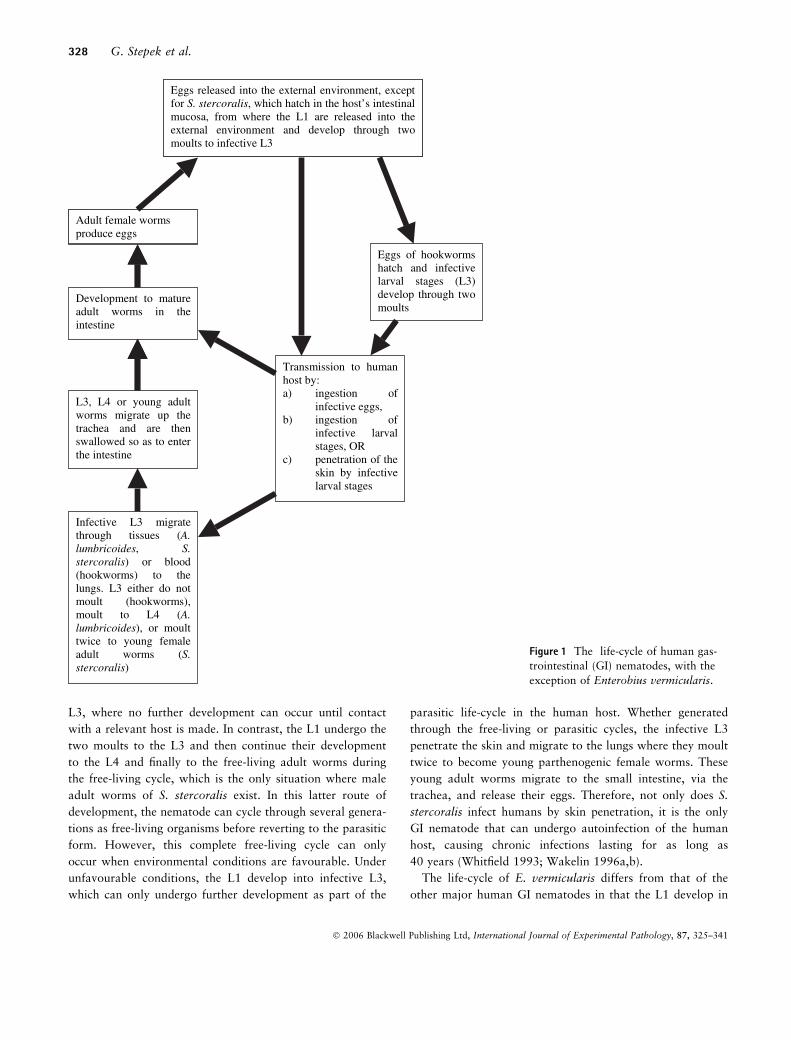
The life-cycles of the major GI nematodes of humans are
essentially similar (Figure 1), but they do have specific differ-
ences. In all, the adult worms reproduce sexually and the
mature female worms produce and release eggs into their
immediate environment of the human intestine. In most spe-
cies, these eggs pass into the external environment via host
faeces and then the L1 (first larval stage) develop within the
eggs. The nematode species differ with respect to subsequent
development, although in all cases, the L1 develop through
four further stages (L2, L3, L4 and pre-adults), with each
stage preceded by a cuticular moult, and each moult causing
an increase in parasite size. However, there are important
differences between species as to where and when these
moults occur. Finally, the pre-adult worms become mature
adult worms, but both the site within the host and the time
for maturation of the pre-adult stage to fully fecund (egg-
laying) females differ between nematode species.
The eggs of both A. lumbricoides and T. trichiura require
a period of external development to become infective even
though the eggs do not hatch in the external environment.
The L1 of T. trichiura develop in the eggs and it is this stage
which is infective to humans. For further development to
occur, the L1 have to be ingested, and this usually occurs
via contaminated food or water. On ingestion, the eggs (con-
taining the infective L1) hatch in the small intestine, from
where the L1 migrate to the colon of the large intestine and
develop, through four moults, to mature adult worms. For
A. lumbricoides, the L1 develop and undergo two moults to
the infective L3 within the eggs, whilst in the external envi-
ronment. As for T. trichiura, the eggs containing the infec-
tive L3 enter the human host by ingestion, where they hatch
and release the L3 into the duodenum. This is followed by
tissue migration to the lungs, via the liver, where the third
moult to L4 occurs. The L4 enter the trachea and are then
swallowed and enter the intestine, where final maturation to
the adult worms occurs (Whitfield 1993; Wakelin 1996a,b).
In contrast, the eggs of hookworms (A. duodenale and
N. americanus) hatch externally and release the L1, which
develop as free-living stages through two moults to the infec-
tive L3. Infection of humans by these L3 is largely by active
skin penetration. The L3 migrate to the lungs and the tra-
chea, from where they are swallowed, and enter the small
intestine to complete their development through the two
remaining moults and final maturation to adult worms.
Whereas N. americanus can only infect humans via skin
penetration, A. duodenale can infect both by skin penetra-
tion and by ingestion of the infective L3. Following inges-
tion, these L3 migrate directly to the small intestine where
they develop, through two moults, to mature adult worms
(Whitfield 1993; Wakelin 1996a,b).
Strongyloides stercoralis is the exception in that the para-
sitic stages are parthenogenic female worms, which release
eggs that hatch internally in the host’s intestine, releasing the
L1. The L1 usually pass out of the host via faeces, but in
some cases, their development can be accelerated whilst still
in the host. In such cases, the L1 moult twice to the infective
L3, which penetrate the gut or the perianal skin surface and
migrate to the lungs (autoinfection). More commonly, the
L1 are released in the host faeces and then follow either the
free-living or parasitic developmental cycle. In the parasitic
cycle, the L1 develop through two moults to the infective
Control of human gastrointestinal nematodes 327
� 2006 Blackwell Publishing Ltd, International Journal of Experimental Pathology, 87, 325–341
L3, where no further development can occur until contact
with a relevant host is made. In contrast, the L1 undergo the
two moults to the L3 and then continue their development
to the L4 and finally to the free-living adult worms during
the free-living cycle, which is the only situation where male
adult worms of S. stercoralis exist. In this latter route of
development, the nematode can cycle through several genera-
tions as free-living organisms before reverting to the parasitic
form. However, this complete free-living cycle can only
occur when environmental conditions are favourable. Under
unfavourable conditions, the L1 develop into infective L3,
which can only undergo further development as part of the
parasitic life-cycle in the human host. Whether generated
through the free-living or parasitic cycles, the infective L3
penetrate the skin and migrate to the lungs where they moult
twice to become young parthenogenic female worms. These
young adult worms migrate to the small intestine, via the
trachea, and release their eggs. Therefore, not only does S.
stercoralis infect humans by skin penetration, it is the only
GI nematode that can undergo autoinfection of the human
host, causing chronic infections lasting for as long as
40 years (Whitfield 1993; Wakelin 1996a,b).
The life-cycle of E. vermicularis differs from that of the
other major human GI nematodes in that the L1 develop in
Eggs released into the external environment, exceptfor S.stercoralis, which hatch in the host’s intestinalmucosa, from where the L1 are released into theexternal environment and develop through twomoults to infective L3
Adult female worms produce eggs
Eggs of hookwormshatch and infectivelarval stages (L3)develop through twomoults
Transmission to humanhost by: a) ingestion of
infective eggs, b) ingestion of
infective larvalstages, OR
c) penetration of theskin by infectivelarval stages
Development to matureadult worms in theintestine
Infective L3 migratethrough tissues (A.lumbricoides, S.stercoralis) or blood(hookworms) to thelungs. L3 either do notmoult (hookworms),moult to L4 (A.lumbricoides), or moulttwice to young femaleadult worms (S.stercoralis)
L3, L4 or young adultworms migrate up thetrachea and are thenswallowed so as to enterthe intestine
Eggs released into the external environment, exceptfor S. stercoralis, which hatch in the host’s intestinalmucosa, from where the L1 are released into theexternal environment and develop through twomoults to infective L3
Adult female worms produce eggs
Figure 1 The life-cycle of human gas-
trointestinal (GI) nematodes, with the
exception of Enterobius vermicularis.
328 G. Stepek et al.
� 2006 Blackwell Publishing Ltd, International Journal of Experimental Pathology, 87, 325–341
the eggs on the perianal skin or under the fingernails. The
majority of the infective eggs are ingested, or inhaled and
swallowed after being coughed up, and hatch in the small
intestine, where they release the L1. These L1 undergo four
moults to the adult stage, usually in the large intestine and
the appendix. Gravid females then migrate to the anus and
deposit eggs on the perianal skin (Whitfield 1993; Wakelin
1996a,b).
The survival of nematodes in the GI tract is favoured by
the high availability of food, and an easy exit to the external
environment ensures continuation of the life-cycle. Whereas
most of the major GI nematodes obtain their nutrients by
attaching and feeding on the mucosa of the intestinal tract,
Ascaris also feeds on the contents of the intestinal lumen.
Hookworms attach to the villi of the gut, abrading the
mucosal surface and feeding on the mucosal tissues of the
intestine, internalizing boluses of tissue and sucking in blood
from the underlying capillaries, making anaemia a promin-
ent symptom of the disease caused by these parasites
(Wakelin 1996b).
Zoonoses
Zoonotic diseases are of worldwide importance, and involve
a large range of animals that are consumed by humans or
associated with them. However, it is not possible to cover
them all in this review, and so we have concentrated on the
nematodes that are acquired from household pets, predomin-
antly dogs. Zoonoses are particularly prevalent in rural areas
of developing countries, such as India. Here, animals are
often found living alongside humans in conditions of over-
crowding, poor socio-economy and poor sanitation and
hygiene, and there is frequently insufficient medical care and
veterinary services and an unawareness of zoonotic diseases.
One of the most important zoonotic infections caused by
GI nematodes is toxocariasis, caused by the dog parasite,
Toxocara canis, which is closely related to A. lumbricoides
and has a similar life-cycle. This nematode has a worldwide
distribution, and is one of the most common zoonotic GI
nematode infections in children. Due to environmental con-
tamination by Toxocara eggs in areas frequented by chil-
dren, such as sand-pits, frequent transmission of the
nematode occurs from soil contaminated with dog faeces to
humans (Schantz 1991; Traub et al. 2005). The majority of
people infected with Toxocara remain asymptomatic, whilst
a minority develop the severe or fatal diseases of visceral
larva migrans, ocular larva migrans and covert toxocariasis.
In these diseases, the eggs hatch in the small intestine and
the larvae migrate to the liver, lungs, eyes and other body
organs, where they cause tissue necrosis, chronic liver
disease, oedema, haemorrhage and eosinophilia. Most cases
are preventable by improvements to hygiene, elimination of
the parasite from dogs (especially puppies, due to transpla-
cental transmission), and preventing children from playing
in areas frequented by infected animals (Hartleb & Janus-
zewski 2001; Baboolal & Rawlins 2002; Traub et al. 2005).
Infection of humans with the canine hookworm, Ancylo-
stoma ceylanicum, occurs in areas of low socio-economic
conditions, such as the Philippines and South America,
where proper footwear is unaffordable and infected dogs
reside in the same environment as humans (Velasquez &
Cabrera 1968). However, it is not just the poorest regions
of the world where canine hookworms can infect humans:
Ancylostoma caninum frequently infects humans in tropical
and temperate countries, including Australia and the USA.
This is mainly due to the large number of dogs owned as
pets and to the behaviour of both humans and dogs, e.g.
people tend to walk barefoot on damp grass or on the
beach, where they may be exposed to the infective skin-pen-
etrating larvae as these areas are also frequented by infected
dogs. Infections with A. caninum can result in eosinophilic
enteritis, which is mainly restricted to the small intestine.
Although eosinophilic enteritis is treatable, it can be recur-
rent, possibly following a seasonal pattern as in dogs, and
this is believed to be due to the reactivation of dormant L3.
The presence of the infective L3 of canine hookworms in
humans can also lead to the development of cutaneous larva
migrans in these abnormal hosts (Prociv & Croese 1996;
Traub et al. 2005).
Consequences of infections with GI nematodes
In the case of heavy infections with GI nematodes, the most
common complaints are intestinal, such as diarrhoea,
abdominal pain and, in the case of A. lumbricoides, obstruc-
tion of the gut (Gilles 1968; Gonzalez & Javier de la
Cabada 1987; Clinch & Stephens 2000; Kucik et al. 2004).
However, these parasitic infections have more serious conse-
quences, as outlined below.
Anaemia
Anaemia is a major consequence of infection with GI nema-
todes, in particular the hookworms, but heavy T. trichiura
infections have also been found to cause iron-deficiency
anaemia (Gilgen et al. 2001). The majority of cases of hook-
worm-induced anaemia occur in people who live rurally in
developing countries and rely on agricultural labour for the
main family income. Anaemia reduces the physical ability to
carry out the work associated with this way of life, leading
Control of human gastrointestinal nematodes 329
� 2006 Blackwell Publishing Ltd, International Journal of Experimental Pathology, 87, 325–341
to poor nutrition and increased hookworm infection, thus
setting up a vicious circle (Crompton 1986). The extent of
anaemia is dependent on the intensity of infection, the
infecting nematode species (A. duodenale causes much
greater blood loss than N. americanus), and the level of iron
intake and host nutritional status (Albonico et al. 1998).
The severity of iron-deficiency anaemia is greater in preg-
nant women infected with hookworms, compared with
non-pregnant infected women, due to the natural increased
iron requirement during pregnancy. Increasing severity of
anaemia can lead to the death of the woman and increased
risks to the unborn foetus, such as premature delivery. Thus,
hookworms are a major contributing factor to the world-
wide incidence of anaemia, particularly in malnourished
pregnant women in the developing countries (Bundy et al.
1995; Dreyfuss et al. 2000).
Malnutrition
Livestock infected with GI nematodes vary in their ability
to cope with these infections, and this ability, together
with the severity of infection, is dependent on the nutri-
tional status of the host (Coop & Kyriazakis 2001). It is
believed that the same is true for GI nematode infections
of humans. Malnutrition occurs in people living in condi-
tions where GI nematode infections are endemic, i.e. high
levels of poverty, poor sanitation and a lack of adequate
hygiene standards, and results in iron deficiency, resulting
in anaemia (see above). Infections with GI nematodes are
more problematic in children suffering from malnutrition
(Stephenson et al. 2000). In the young, not only do intes-
tinal nematodes have a detrimental effect on the host’s
nutritional status, but they also cause a loss of appetite
and affect the host’s physical, cognitive and social devel-
opment (Crompton 1986; Dossa et al. 2001). These latter
effects are largely due to the malnourished state of the
helminth-infected children (Levav et al. 1995). Malnutri-
tion is exacerbated after infection with GI nematodes
because the nematodes damage the intestinal mucosal
epithelial cells whilst feeding, resulting in the prevention
of nutrient absorption by the host and leading to stunted
growth (Stephensen 1999). This, in turn, has consequences
for the work productivity of adults in developing countries
(Guyatt 2000). A single dose of albendazole to helminth-
infected children in Kenya greatly improved their growth
rate, weight, physical fitness and activity and appetite
(Stephenson et al. 1993). Thus, by treating GI nematode
infections, normal growth rate and appetite are restored,
and productivity is also increased, especially in developing
countries where the work is physically demanding.
School attendance and cognitive function
Moderate to heavy burdens of T. trichiura or hookworms in
school children result in poor school attendance and low
cognitive function when compared with the performance of
uninfected children. The effect on cognitive function stems
from malnutrition (see above), with or without anaemia,
and, in undernourished children, is related to the intensity of
GI nematode infection. However, the poor cognitive per-
formance is reversible following treatment with anthelmintic
drugs. Therefore, improvements in nutrition and lowering
the incidence of infection are likely to lead to improvements
in school performance (Nokes et al. 1992a,b; Simeon et al.
1995).
Secondary infections
There are two forms of severe infection with S. stercoralis:
hyperinfective strongyloidiasis and disseminated strongyloid-
iasis, both of which can be life-threatening in the more sus-
ceptible immunosuppressed patients. Both severe forms
involve the presence of a massive worm burden. These
worms are concentrated in the intestinal and respiratory
tracts in the hyperinfective form, but are spread throughout
many organs, including the central nervous system, during
disseminated strongyloidiasis. Both severe forms cause gas-
trointestinal, cutaneous and respiratory problems and, in the
case of the disseminated disease, these are often fatal due to
the increased risk of meningitis and other secondary bacter-
ial infections resulting from infection with this nematode
(Gilles 1968; Bwibo 1971; Milder et al. 1981; Gonzalez &
Javier de la Cabada 1987; Sturchler 1987; Fisher et al.
1993; Jain et al. 1994; Schneider & Rogers 1997; Tsai et al.
2002; Keiser & Nutman 2004).
Infections with the major human GI nematodes impair the
immune response to other serious infections, such as tuber-
culosis (TB) and human immunodeficiency virus (HIV),
which are each controlled by a Th1 immune response. This
type of immune response is downregulated by helminth
infections; hence, the potential contribution to the rising
numbers of TB and HIV infections in the developing coun-
tries. Helminth infections also induce the production of the
T regulatory cytokines, interleukin (IL)-10 and transforming
growth factor (TGF)-b, which are immunosuppressive and
are believed to inhibit immune responses that protect against
TB. This downregulation of the host’s immune response is
believed to be responsible for the more rapid progression of
HIV infection to AIDS in the developing countries (Bentwich
et al. 1999; Othieno et al. 1999; Elliott et al. 2003). Individ-
uals with helminth infections show marked Th2 immune
330 G. Stepek et al.
� 2006 Blackwell Publishing Ltd, International Journal of Experimental Pathology, 87, 325–341
responses and are chronically immune-activated, which
increases their susceptibility to HIV and TB. The vaccines
under current development against HIV and TB may have
no effect in areas where helminth infections are endemic, i.e.
the areas of highest HIV and TB incidence. This is due to
the pre-existing Th2 immune responses of the infected indi-
viduals who may not be able to develop a protective Th1
response upon vaccination (Bentwich et al. 1995; Borkow &
Bentwich 2000). However, one potential vaccine candidate
against HIV involves the use of an adjuvant which changes
the immune response of affected individuals from Th2 to
Th1. Adjuvants that do not cause a change to a Th1
response have no effect on the protection against these infec-
tions (Ayash-Rashkovsky et al. 2002). Thus, the existing
presence of helminth infections increases the susceptibility of
individuals to secondary infections requiring a Th1 immune
response for protection.
Control of GI nematodes of humans
Anthelmintics
Now, the major means of controlling human GI nematode
infections is by the administration of one of the four che-
motherapeutic anthelmintic drugs recommended by the
WHO for the treatment of these infections. These drugs are
albendazole, mebendazole, levamisole and pyrantel, and
there is at least one anthelmintic drug which can be used to
treat each of the major GI nematodes of humans (Table 2).
These drugs belong to two distinct classes: group 1, the
benzimidazoles (albendazole and mebendazole), and group
2, the imidazothiazoles/tetrahydropyrimidines (levamisole
and pyrantel). There is a third class of anthelmintics, the
macrocyclic lactones (group 3; e.g. ivermectin), which are
used in the treatment of GI nematode infections of live-
stock, but which have only recently been registered for use
in humans against strongyloidiasis in France, Australia and
the USA, although ivermectin has been available for use
against filarial nematodes for several years (Albonico et al.
1999).
The benzimidazoles are broad spectrum drugs that bind to
free b-tubulin, inhibiting its polymerization and so interfer-
ing with microtubule-dependent glucose uptake by the para-
site. The imidazothiazoles/tetrahydropyrimidines stimulate
the nicotinic acetylcholine receptors, resulting in over-stimu-
lation, blockade of the neuromuscular junctions, and rigid
paralysis of the worms. The parasites are then unable to
move in the intestinal tract and are swept out by the peristal-
tic action in the intestine. The macrocyclic lactones act by
opening glutamate-gated chloride channels, increasing chlor-
ide ion conductance, leading to defects in neurotransmission
and flaccid paralysis. There is a fourth class of anthelmintics,
the heterocyclic ethyleneamines, of which the best known
member, piperazine, is only used against A. lumbricoides
and E. vermicularis. This drug acts by reversibly inhibiting
neuromuscular transmission by stimulating gamma-aminobu-
tyric acid (GABA) receptors in nematode muscle, causing
flaccid paralysis of the worms, which are then removed by
normal intestinal peristalsis (Rang et al. 2003).
These anthelmintics are relatively safe, having a very few
minor gastrointestinal side-effects. However, none are recom-
mended for use by pregnant women, particularly those in the
first trimester. Although they can be administered in single
doses, these drugs are more effective during treatment with
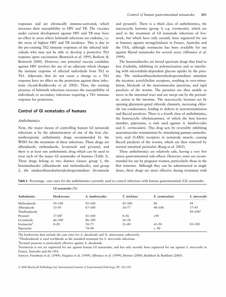
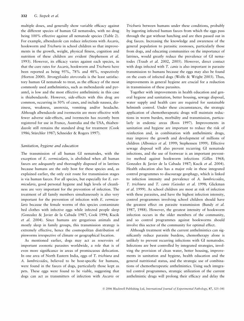
Table 2 Percentage cure rates for the anthelmintics currently used to control infections with human gastrointestinal (GI) nematodes
Anthelmintic
GI nematodes (%)
Hookworms A. lumbricoides T. trichiura E. vermicularis S. stercoralis
Mebendazole 95–100 95–100 45–100 96 44
Albendazole 33–95 67–100 10–77 40–100 17–95
Thiabendazole 89–100*
Pyrantel 37–88� 81–100 0–56 >90
Levamisole 66–100 86–100 16–18
Ivermectin� 0–20 50–75 11–80 61–94 83–100
Piperazine 74–94 c. 90
The hookworm data include the cure rates for A. duodenale and N. americanus collectively.
*Thiabendazole is used worldwide as the standard treatment for S. stercoralis infections.�Pyrantel pamoate is particularly effective against A. duodenale.�Ivermectin is not yet registered for use against human GI nematodes, and has only recently been registered for use against S. stercoralis in
France, Australia and the USA.
Sources: Freedman et al. (1989); Naquira et al. (1989); Albonico et al. (1999); Horton (2000); Burkhart & Burkhart (2005).
Control of human gastrointestinal nematodes 331
� 2006 Blackwell Publishing Ltd, International Journal of Experimental Pathology, 87, 325–341
multiple doses, and generally show variable efficacy against
the different species of human GI nematodes, with no drug
being 100% effective against all nematode species (Table 2).
For example, albendazole can reduce infections with Ascaris,
hookworm and Trichuris in school children so that improve-
ments in the growth, weight, physical fitness, cognition and
nutrition of these children are evident (Stephenson et al.
1993). However, its efficacy varies against each species, in
that the cure rates for Ascaris, hookworm and Trichuris have
been reported as being 95%, 78% and 48%, respectively
(Horton 2000). Strongyloides stercoralis is the least satisfac-
tory human GI nematode to treat, as the efficacy of the most
commonly used anthelmintics, such as mebendazole and pyr-
antel, is low and the most effective anthelmintic in this case
is thiabendazole. However, side-effects with this drug are
common, occurring in 50% of cases, and include nausea, diz-
ziness, weakness, anorexia, vomiting and/or headache.
Although albendazole and ivermectin are more effective with
fewer adverse side-effects, and ivermectin has recently been
registered for use in France, Australia and the USA, thiaben-
dazole still remains the standard drug for treatment (Cook
1986; Sturchler 1987; Schneider & Rogers 1997).
Sanitation, hygiene and education
The transmission of all human GI nematodes, with the
exception of E. vermicularis, is abolished when all human
faeces are adequately and thoroughly disposed of in latrines
because humans are the only host for these species and, as
explained earlier, the only exit route for transmission stages
is via human faeces. For all species, but especially for E. ver-
micularis, good personal hygiene and high levels of cleanli-
ness are very important for the prevention of infection. The
treatment of all family members simultaneously is also very
important for the prevention of infection with E. vermicu-
laris because the female worms of this species contaminate
bed clothes with infective eggs while infected people sleep
(Gonzalez & Javier de la Cabada 1987; Cook 1994; Kucik
et al. 2004). Since humans are gregarious animals and
mostly sleep in family groups, this transmission strategy is
extremely effective, hence the cosmopolitan distribution of
pinworms irrespective of climate or geographical location.
As mentioned earlier, dogs may act as reservoirs of
important zoonotic parasites worldwide, a role that is of
even more significance in areas of promiscuous defecation.
In one area of North Eastern India, eggs of T. trichiura and
A. lumbricoides, believed to be host-specific for humans,
were found in the faeces of dogs, particularly those kept as
pets. These eggs were found to be viable, suggesting that
dogs can act as transmitters of infection with Ascaris or
Trichuris between humans under these conditions, probably
by ingesting infected human faeces from which the eggs pass
through the gut without hatching and are then passed out in
dog faeces. Increasing the knowledge and awareness of the
general population to parasitic zoonoses, particularly those
from dogs, and educating communities on the importance of
latrines, would greatly reduce the prevalence of GI nema-
todes (Traub et al. 2002, 2005). However, direct contact
with dogs infected with T. canis is also important in parasite
transmission to humans because the eggs may also be found
on the coats of infected dogs (Wolfe & Wright 2003). Thus,
improvements in general hygiene are crucial for a reduction
in transmission of these parasites.
Together with improvements in health education and gen-
eral hygiene and sanitation, better housing, sewage disposal,
water supply and health care are required for sustainable
helminth control. Under these circumstances, the strategic
application of chemotherapeutic drugs will maintain reduc-
tions in worm burden, morbidity and transmission, particu-
larly in endemic areas (Roos 1997). Improvements in
sanitation and hygiene are important to reduce the risk of
reinfection and, in combination with anthelmintic drugs,
may improve the growth and development of millions of
children (Albonico et al. 1999; Stephensen 1999). Effective
sewage disposal will also prevent recurring GI nematode
infections, and the use of footwear is an important prevent-
ive method against hookworm infections (Gilles 1968;
Gonzalez & Javier de la Cabada 1987; Kucik et al. 2004).
Health education also has a major role in these integrated
control programmes to discourage geophagy, which is linked
to infection intensity and prevalence of A. lumbricoides,
T. trichiura and T. canis (Geissler et al. 1998; Glickman
et al. 1999). As school children are most at risk of infection
with these parasites, and have the highest infection intensity,
control programmes involving school children should have
the greatest effect on parasite transmission (Bundy et al.
1987, 1988). However, the greatest intensity of hookworm
infection occurs in the older members of the community,
and so control programmes against hookworms should
involve this sector of the community for optimal effect.
Although treatment with the current anthelmintics can sig-
nificantly reduce parasite burdens, chemotherapy alone is
unlikely to prevent recurring infections with GI nematodes.
Infections are best controlled by integrated strategies, invol-
ving the provision of clean water, better housing, improve-
ments in sanitation and hygiene, health education and the
general nutritional status, and the strategic use of combina-
tions of chemotherapeutic anthelmintics. Using such integra-
ted control programmes, strategic utilization of the current
anthelmintic drugs will prolong their efficacy and delay the
332 G. Stepek et al.
� 2006 Blackwell Publishing Ltd, International Journal of Experimental Pathology, 87, 325–341
onset of resistance (Schantz 1991; Coles 1995; Reynoldson
et al. 1998; Albonico et al. 1999; Dossa et al. 2001).
Problems with current strategies to control GInematode infections of humans
The control of parasitic nematode infections is made difficult
by environmental, social and economic factors. Complete
eradication of helminths is impossible without major chan-
ges to socio-economic conditions throughout the world, and
without the associated financial investment required to pro-
vide these (Albonico et al. 1999). Therefore, an alternative
strategy is to accept that worm infections cannot be eradi-
cated and then to try to keep infections as low as possible
through reduction of transmission of the parasite, thereby
limiting both the number of people at risk of infection and
the associated morbidity (Crompton 1999).
A major problem in the control of GI nematode infec-
tions is the development of resistance to the currently avail-
able anthelmintics. Resistance to the drugs in all three of
the major anthelmintic classes used in livestock is wide-
spread, particularly in Africa, Australia, New Zealand, Asia
and South America (Waller 1997), and there is now a
potential danger of the occurrence of resistance in helm-
inths of humans. However, the treatment of humans with
anthelmintics is less frequent than that of livestock and
occurs as part of better control programmes, which should
slow the appearance of resistance in human helminths
(Geerts et al. 1997; Roos 1997; Geerts & Gryseels 2001;
Horton 2003). Unfortunately, it is a case of when, not if,
resistance develops. Currently, the benzimidazoles are the
drugs which are the most frequently used to treat human
GI nematode infections, but it is to these drugs that live-
stock nematodes have developed the most resistance (Coles
1995). Thus, the spread of resistance among GI nematodes
of livestock to the available anthelmintics provides a crucial
warning about the widespread use of anthelmintics to con-
trol human GI nematodes (Coles 1995; Geerts & Gryseels
2000). Indeed, there have been recent reports indicating
reductions in efficacy and the possible development of resis-
tance to anthelmintics in the human population. For
instance, in southeast Mali, mebendazole and pyrantel have
recently shown worryingly low efficacy against human
infections with N. americanus (de Clercq et al. 1997; Sacko
et al. 1999). Similarly, in the Kimberly region of North
Western Australia, pyrantel lacks efficacy against A. duode-
nale, possibly through development of resistance as a result
of frequent use of this drug (Reynoldson et al. 1997). On
Pemba Island, Zanzibar, the efficacy of mebendazole
against hookworms in school children appeared to have
fallen over a period of 5 years, during which time the chil-
dren were regularly treated with mebendazole (egg reduc-
tion rate fell from 82.4% to 52.1%). This suggests the
possibility of emergence of mebendazole-resistant hook-
worms on Pemba Island, and provides a crucial warning
about the consistent use of anthelmintics as the sole means
to control human GI nematode infections (Albonico et al.
1994, 2003).
The problem of the development of resistance is partic-
ularly worrying because there are few new drugs at the clin-
ical trial stage of development. However, recent studies have
identified some potential candidates. In Peru and Mexico,
nitazoxanide, currently only available to treat protozoan
infections, has been successfully used against ascariasis,
trichuriasis and strongyloidiasis, with few adverse side-
effects (Davila-Gutierrez et al. 2002; Ortiz et al. 2002).
Unfortunately, there are also no vaccines available for the
treatment of human GI nematode infections and there are
unlikely to be any in the foreseeable future.
Strategies for delaying the onset of resistance, and making
the most use of the available anthelmintics, include selective
treatment of populations. By treating only a proportion of
the affected community, usually those who are most likely
to harbour heavy infections, or by treating at a time of the
year when significant proportions of worms are in the host’s
external environment, the selection pressure on worms with
resistant alleles is not as intense as it might otherwise have
been. The worms that avoid treatment (either in non-treated
individuals or in the environment as eggs or larvae) repre-
sent ‘worms in refugia’ with non-resistant genotypes. Thus,
at the parasite population level, non-resistant worms survive
episodes of treatment and, in this way, help to dilute the
resistant alleles in the population and slow down the
increase of resistance that would otherwise occur if all
worms were subjected to the anthelmintic in question (Smith
1990; Smith et al. 1999; Coles 2002, 2005).
Other strategies for slowing the onset of resistance include
the application of combinations of different anthelmintic
drugs. For instance, pyrantel on its own has very poor
efficacy against T. trichiura, but in the combination pyran-
tel–oxantel (oxantel being another group 2 drug), the egg
reduction rate was greater than 80% (Albonico et al. 2002).
Administering levamisole and mebendazole together was
found to significantly increase the efficacy against A. lumb-
ricoides, T. trichiura and hookworms compared with either
drug alone (Albonico et al. 2003). It is likely that increased
combination therapy will slow the development of resist-
ance, particularly when combinations involving different
drugs with distinct modes of action are used (Smith
1990; Coles 2006). Combination therapy is, therefore,
Control of human gastrointestinal nematodes 333
� 2006 Blackwell Publishing Ltd, International Journal of Experimental Pathology, 87, 325–341
advantageous and should be used as the treatment of choice
against soil-transmitted helminths.
Another form of combination therapy which could be
used to control intestinal nematode infections is to combine
treatment programmes for different diseases, such as mal-
aria, schistosomiasis, intestinal helminthiasis and filariasis
(Molyneux & Nantulya 2004), where drug combinations
such as ivermectin and albendazole, or praziquantel and
albendazole, are used to increase the spectrum of efficacy.
Two early studies examined the effect of the combination of
ivermectin and albendazole against the filarial nematode,
Wuchereria bancrofti, and the intestinal helminths, A. lum-
bricoides, T. trichiura and hookworms. Both studies demon-
strated that the combined treatment resulted in a
significantly greater reduction in the prevalence and intensity
of infection of all three intestinal helminths and W. ban-
crofti. Therefore, integrated control programmes for differ-
ent diseases would probably substantially improve the health
of infected populations, especially children (Addiss et al.
1997; Beach et al. 1999). However, these integrated control
programmes are only in their early stages.
In the developing countries, one of the major problems in
the control of GI nematode infections is the expense of ant-
helmintics in relation to family budgets even though, in
some parts of the world, prices have fallen in recent years,
and anthelmintics may cost only a fraction of the price
charged in developed countries. Even so, cheaper, less effi-
cient drugs, such as tetrachlorethylene, may be used or treat-
ments may be shared between family members, so that each
member receives less than the recommended dose (Cook
1986). The use of sub-optimal doses encourages the
spread of parasite genotypes that have resistant alleles to
anthelmintics (Smith 1990). A much greater expense in the
control of human GI nematode infections, especially in the
developing countries, is the provision of clean water sup-
plies, better sanitation and good primary health care (Guyatt
& Evans 1992).
Thus, it is clear that there are numerous problems asso-
ciated with the control of GI nematode infections, and that
novel methods are required.
The search for new anthelmintics
One alternative strategy may be the use of natural products
that have anthelmintic properties. There has been a recent
resurgence in the investigation of naturally occurring sub-
stances for their anthelmintic properties (for examples see
Guarrera 1999; Waller et al. 2001; Githiori et al. 2003;
Hordegen et al. 2003; Hounzangbe-Adote et al. 2005a,b),
and this has been largely due to the presence of resistance
to the current chemotherapeutic anthelmintics. The material
under study is often plant-derived and this line of investiga-
tion has been encouraged by the fact that such plant
extracts have been used traditionally by indigenous people,
mainly in the tropics, against GI nematodes of both
humans and livestock. To date, very few of these putative
natural anthelmintics have been comprehensively assessed
scientifically (e.g. Raj 1974; Desta 1995; Tandon et al.
1997; McGaw et al. 2000; Giday et al. 2003; Beloin et al.
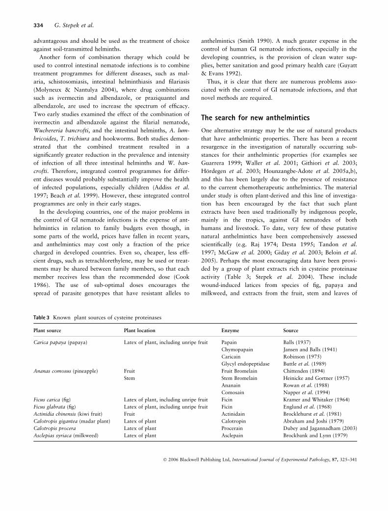
2005). Perhaps the most encouraging data have been provi-
ded by a group of plant extracts rich in cysteine proteinase
activity (Table 3; Stepek et al. 2004). These include
wound-induced latices from species of fig, papaya and
milkweed, and extracts from the fruit, stem and leaves of
Table 3 Known plant sources of cysteine proteinases
Plant source Plant location Enzyme Source
Carica papaya (papaya) Latex of plant, including unripe fruit Papain
Chymopapain
Caricain
Glycyl endopeptidase
Balls (1937)
Jansen and Balls (1941)
Robinson (1975)
Buttle et al. (1989)
Ananas comosus (pineapple) Fruit
Stem
Fruit Bromelain
Stem Bromelain
Ananain
Comosain
Chittenden (1894)
Heinicke and Gortner (1957)
Rowan et al. (1988)
Napper et al. (1994)
Ficus carica (fig) Latex of plant, including unripe fruit Ficin Kramer and Whitaker (1964)
Ficus glabrata (fig) Latex of plant, including unripe fruit Ficin Englund et al. (1968)
Actinidia chinensis (kiwi fruit) Fruit Actinidain Brocklehurst et al. (1981)
Calotropis gigantea (madar plant) Latex of plant Calotropin Abraham and Joshi (1979)
Calotropis procera Latex of plant Procerain Dubey and Jagannadham (2003)
Asclepias syriaca (milkweed) Latex of plant Asclepain Brockbank and Lynn (1979)
334 G. Stepek et al.
� 2006 Blackwell Publishing Ltd, International Journal of Experimental Pathology, 87, 325–341

pineapple. For instance, Ficus glabrata latex is a well-
known anthelmintic in Central and South America. Since
the start of the last century, several other species of fig,
such as Ficus laurifolia, have also been used against
Trichuris, Taenia, Ascaris and Enterobius. In addition,
papaya (Carica papaya) and pineapple (Ananas comosus)
have been used to treat chickens, dogs, pigs and humans
infected with intestinal parasites. However, use of these
traditional treatments only occurred in areas where the
plants were found, and their use quickly declined when the
modern synthetic drugs were introduced (Gaughran 1976).
With the onset of resistance to the synthetic anthelmintics,
interest in this area of research has now been rekindled,
although satisfactory trials in humans that conform to con-
temporary standards are still largely lacking. In one rare
example, in the Amazon basin, the crude latex of F. glabrata
was administered orally for three consecutive days at 1 ml/kg.
After treatment, there were reductions in egg output of
Ascaris (85%), Strongyloides (72%), Ancylostoma/Necator
(55%) and Trichuris (67%), almost matching the efficacy of
the modern, synthetic anthelmintics used as positive controls.
No adverse side-effects associated with the use of fig latex
were noted (Hansson et al. 1986). Studies in livestock are
almost equally as rare, although anecdotal stories suggest the
successful use, as early as the 19th century, of the crude latex
of C. papaya against ascarids, tapeworms, whipworms and
hookworms (Berger & Asenjo 1940). In two well-documented
trials, a single dose of the crude latex of C. papaya provided
comparable anthelmintic efficacy to the currently available
synthetic anthelmintics, with respect to reductions in the egg
output and worm burden, in pigs infected with the round-
worm, Ascaris suum (Satrija et al. 1994) and mice infected
with Heligmosomoides polygyrus, one of the most common
laboratory models for routine screening of potential drug can-
didates (Satrija et al. 1995).
Although the above studies describe anthelmintic efficacy
of the extracts of papaya and fig, the mechanism of action by
which these extracts produced their effects was not assessed.
This is important because current drug legislation requires an
understanding of the mode of action of candidate drugs
before registration for use as alternative treatments against
GI nematode infections in humans. In vitro, F. laurifolia
latex (Robbins 1930) and F. glabrata latex (Hansson et al.
1986) acted rapidly against A. suum, causing the nematode
cuticle to become wrinkled and flaccid, before developing
small blisters, which later perforated releasing the internal
structures, and causing the death of the nematode (Robbins
1930). Purified papain from papaya latex (Berger & Asenjo
1940) and bromelain from fresh pineapple juice (Berger &
Asenjo 1939) also caused rapid ulceration of A. suum,
followed by digestion of the worm in vitro. These studies,
taken together, indicated that the anthelmintic properties of
pineapple juice and of the latex of Ficus species and of
C. papaya had a similar mechanism of action, which differed
to that of the currently available anthelmintics. Most of
these plants are widely available and inexpensive in tropical
and sub-tropical countries, the areas with highest infection
intensity.
Proof that the active principles in these plant extracts
were cysteine proteinases was provided by Stepek et al.
(2005), using H. polygyrus in vitro. The plant extracts, or
cysteine proteinases purified from them, only possessed ant-
helmintic activity if a reducing agent (cysteine) was present.
These enzymes are well known to be active only in a redu-
cing environment. In addition, the specific inactivator of the
papain family of cysteine proteinases, l-trans-epoxysuccinyl-
leucylamido-4-guanidino-butane (E-64), completely inhibited
the anthelmintic activity. It was also demonstrated that this
mechanism was specific to cysteine proteinases, in that it did
not occur with aspartic or serine proteinases found in the
mammalian alimentary canal. Digestion of the cuticle also
occurred when the same enzymes were incubated in vitro
with the rodent GI nematodes, Trichuris muris (Stepek et al.
2006), Protospirura muricola (G. Stepek et al., personal
communication) and A. ceylanicum (G. Stepek et al., perso-
nal communication). The same mechanism of action was
also observed in vivo against H. polygyrus, resulting in the
expulsion of only the affected worms from the murine gas-
trointestinal tract (G. Stepek et al., personal communica-
tion). Moreover, fresh latex from the fig plants, Ficus carica
and Ficus insipida, also partially digested the cestode,
Rodentolepis (Vampirolepis) nana, and the oxyurids, Sypha-
cia obvelata and Aspiculuris tetraptera (de Amorin et al.
1999). The mechanism of action is, therefore, not restricted
to one nematode species, but has the same detrimental effect
on a range of GI nematodes and cestodes.
Potential problems of plant cysteineproteinases as anthelmintics
The cysteine proteinases from plants may seem to be a
viable alternative to current anthelmintics, but there are
some potential problems associated with these enzymes. It is
well known that papain and its homologues are inactive at
low pH. This is for two reasons, the first being due to the
pKa of the essential thiolate anion, which is normally
around pH 4. This means that at pH values below 4 (such
as in the stomach) the enzymes will be largely inactive. The
second reason is that these enzymes suffer irreversible dena-
turation at low pH such that, following passage through the
Control of human gastrointestinal nematodes 335
� 2006 Blackwell Publishing Ltd, International Journal of Experimental Pathology, 87, 325–341
stomach, the enzymes may be irreversibly inactivated even
though the pH then rises to close to their pH optimum of
7–8. It has recently been suggested that these properties, as
well as digestion by the stomach proteinase pepsin, may
mean that the anthelmintic proteinases need to be protected
against low pH to be effective as anthelmintics (Huet et al.
2006). All the successful in vivo trials published to date have
utilized impure enzyme preparations, such as crude papaya
latex. Our recent data demonstrated that, when delivered
orally in the form of crude latex, sufficient enzyme activity
survived passage through the entire gastrointestinal tract of
mice to mediate a potent anthelmintic effect against a nema-
tode species residing in the lower alimentary tract (Stepek
et al. 2006). Possibly the presence of the proteinaceous latex
material is sufficient to locally buffer the stomach contents
against acidity (which would also inactivate pepsin), thus
allowing the enzymes to traverse this otherwise hostile envi-
ronment. We have found that administration of pure papain
to mice infected with H. polygyrus resulted in much lower
efficacy than administration of the same amount of cysteine
proteinase in the form of crude latex (G. Stepek et al.,
unpublished data). It is to be expected that improved formu-
lations and methods of delivery will increase efficacy of the
pure enzymes in the future, especially as Hale (2004) has
recently shown that when bromelain (from the pineapple,
Ananas comosus) was administered with antacid, it retained
its activity throughout the intestinal tract of mice.
Although the toxicities of the purified enzymes appear to
be quite low (chymopapain is already a licensed drug for the
treatment of intervertebral disc disease, and many of the
plant cysteine proteinases have been used as meat tenderisers
and in other aspects of food preparation), problems associ-
ated with allergy may occur. However, these usually arise
following intake through the airways, as is commonly the
case with immunoglobulin E (IgE)-mediated allergy
(Soto-Mera et al. 2000; Nelde et al. 2001), and serious aller-
gic reactions following oral ingestion would be expected to
be extremely rare. However, Hale (2004) found anti-brome-
lain antibodies in the serum of mice following prolonged
(18 weeks) daily oral administration of the enzyme. Brome-
lain is known to erode some of the cell surface markers
essential for inflammatory responses, affect the production
of cytokine secretion, and generally show anti-inflammatory
activity when administered in vivo (Hale & Haynes 1992;
Hale et al. 2002). However, the intestinal mucosa has a
rapid turnover (2–3 days) in rodents, and normal function
would be rapidly restored after either a single oral treatment
or a week of daily treatments. Other causes of toxicity may
be associated with oral administration of some crude latex
preparations. For instance, doses of 10 ml/kg/day for 3 days
of F. carica or F. insipida latex produced fatally high toxic-
ity levels in mice (de Amorin et al. 1999).
As with other control strategies, reinfection is certain to
occur if improvements in hygiene and sanitation standards
are not made, and so a combination of anthelmintic treat-
ment and better hygiene is required. Thus, if improvements
in sanitation and hygiene are not made and sustained, the
use of plant cysteine proteinases as anthelmintics may not
provide much benefit. On the other hand, plant cysteine pro-
teinases, as novel anthelmintics, will only add to the range
of tools and associated strategies that aim to maintain low
prevalence of infection and low morbidity rather than eradi-
cation. In this context, since these enzymes occur naturally
in tropical plants, they offer scope for local production and
may spawn novel agricultural enterprises for developing
countries. This, in turn, may be facilitated by the develop-
ment of new varieties of the source plants that contain more
active enzymes at greater concentrations, through classic
selection programmes or transgenesis once the genes respon-
sible have been identified. At the individual level, they offer
cheap and regular supplies of medicines for the treatment of
nematode infections of impoverished people in the develop-
ing countries.
Conclusions
At the present time, control of GI nematode infections of
humans is not totally adequate, despite the existence of
chemical anthelmintics of varying effectiveness. The inevit-
ability of the development of resistance to the current ant-
helmintics will complicate the future control of GI nematode
infections in humans unless new drugs are discovered, or the
existing anthelmintics are used strategically to avoid the
occurrence of resistance. New anthelmintic drugs, with
mechanisms of action that differ from those that are cur-
rently available, are desperately required for the control of
GI nematodes. In the absence of any new candidates in
chemical drug development, the cysteine proteinases found
naturally in plants, such as papaya, pineapple and fig, repre-
sent a viable option. These treatments would be relatively
inexpensive, are likely to be without serious adverse reac-
tions and have been used by indigenous people of tropical
countries for centuries. Improved methods of delivery will
be expected to bring further advances in terms of efficacy
and safety, and it is imperative that the plant cysteine pro-
teinases are tested to modern pharmaceutical standards and
that their potential benefits are assessed in controlled studies.
Such work should be undertaken with all possible haste, as
the protection given by the current chemical anthelmintics
may soon be limited.
336 G. Stepek et al.
� 2006 Blackwell Publishing Ltd, International Journal of Experimental Pathology, 87, 325–341
Acknowledgements
Our work on the anthelmintic action of the plant cysteine
proteinases was funded by the Leverhulme Trust, with a
grant to DJB, IRD and JMB.
References
Abraham K.I. & Joshi P.N. (1979) Studies on proteinases from
Calotropis gigantea latex. I: Purification and some properties
of two proteinases containing carbohydrate. Biochim. Bio-
phys. Acta 568, 111–119.
Addiss D.G., Beach M.J., Streit T.G. et al. (1997) Randomised
placebo-controlled comparison of ivermectin and albendazole
alone and in combination for Wuchereria bancrofti microfi-
laraemia in Haitian children. Lancet 350, 480–484.
Albonico M., Smith P.G., Hall A., Chwaya H.M., Alawi K.S.,
Savioli L. (1994) A randomised controlled trial comparing
mebendazole and albendazole against Ascaris, Trichuris and
hookworm infections. Trans. R. Soc. Trop. Med. Hyg. 88,
585–589.
Albonico M., Stoltzfus R.J., Savioli L. et al. (1998) Epidemio-
logical evidence for a differential effect of hookworm species,
Ancylostoma duodenale or Necator americanus, on iron sta-
tus of children. Int. J. Epidem. 27, 530–537.
Albonico M., Crompton D.W.T., Savioli L. (1999) Control
strategies for human intestinal nematode infections. Adv.
Parasitol. 42, 277–341.
Albonico M., Bickle Q., Haji H.J. et al. (2002) Evaluation of
the efficacy of pyrantel-oxantel for the treatment of soil-trans-
mitted nematode infections. Trans. R. Soc. Trop. Med. Hyg.
96, 685–690.
Albonico M., Bickle Q., Ramsan M., Montresor A., Savioli L.,
Taylor M. (2003) Efficacy of mebendazole and levamisole
alone or in combination against intestinal nematode infections
after repeated targeted mebendazole treatment in Zanzibar.
Bull. WHO 81, 343–352.
de Amorin A., Borba H.R., Carauta J.P.P., Lopes D., Kaplan
M.A.C. (1999) Anthelmintic activity of the latex of Ficus spe-
cies. J. Ethnopharm. 64, 255–258.
Ayash-Rashkovsky M., Weisman Z., Diveley J., Moss R.B.,
Bentwich Z., Borkow G. (2002) Generation of Th1 immune
responses to inactivated, gp120-depleted HIV-1 in mice with
a dominant Th2 biased immune profile via immunostimulato-
ry oligonucleotides – relevance to AIDS vaccines in develop-
ing countries. Vaccine 20, 2684–2692.
Baboolal S. & Rawlins S.C. (2002) Seroprevalence of toxocaria-
sis in schoolchildren in Trinidad. Trans. R. Soc. Trop. Med.
Hyg. 96, 139–143.
Balls A.K. (1937) Crystalline papain. Science 86, 379.
Beach M.J., Streit T.G., Addiss D.G., Prospere R., Roberts
J.M., Lammie P.J. (1999) Assessment of combined ivermectin
and albendazole for treatment of intestinal helminth and
Wuchereria bancrofti infections in Haitian schoolchildren.
Am. J. Trop. Med. Hyg. 60, 479–486.
Beloin N., Gbeassor M., Akpagana K. et al. (2005) Ethnomedic-
inal uses of Momordica charantia (Cucurbitaceae) in Togo
and relation to its phytochemistry and biological activity.
J. Ethnopharm. 96, 49–55.
Bentwich Z., Kalinkovich A., Weisman Z. (1995) Immune acti-
vation is a dominant factor in the pathogenesis of African
Aids. Immunol. Today 16, 187–191.
Bentwich Z., Kalinkovich A., Weisman Z., Borkow G., Beyers
N., Beyers A.D. (1999) Can eradication of helminthic infec-
tions change the face of Aids and tuberculosis? Immunol.
Today 20, 485–487.
Berger J. & Asenjo C.F. (1939) Anthelmintic activity of fresh
pineapple juice. Science 90, 299–300.
Berger J. & Asenjo C.F. (1940) Anthelmintic activity of crystal-
line papain. Science 91, 387–388.
Booth M. & Bundy D.A.P. (1992) Comparative prevalences of
Ascaris lumbricoides, Trichuris trichiura and hookworm
infections and the prospects for combined control. Parasitol-
ogy 105, 151–157.
Booth M., Bundy D.A.P., Albonico M., Chwaya H.M., Alawi
K.S., Savioli L. (1998) Associations among multiple geohelm-
inth species infections in schoolchildren from Pemba Island.
Parasitology 116, 85–93.
Borkow G. & Bentwich Z. (2000) Eradication of helminthic
infections may be essential for successful vaccination against
HIV and tuberculosis. Bull. WHO 78, 1368–1369.
Boycott A.E. (1911) Ankylostoma infection. Lancet 1, 717–721.
Boycott A.E. & Haldane J.S. (1903) An outbreak of ankylosto-
miasis in England. J. Hyg. 3, 95–136.
Brockbank W.J. & Lynn K.R. (1979) Purification and preli-
minary characterisation of two asclepains from the latex of
Asclepias syriaca L. (milkweed). Biochim. Biophys Acta 578,
13–22.
Brocklehurst K., Baines B.S., Malthouse J.P.G. (1981) Differ-
ences in the interactions of the catalytic groups of the active
centres of actinidin and papain. Rapid purification of fully
active actinidin by covalent chromatography and characteriza-
tion of its active centre by use of two-protonic-state reactivity
probes. Biochem. J. 197, 739–746.
Bundy D.A.P., Cooper E.S., Thompson D.E., Didier J.M., Sim-
mons I. (1987) Epidemiology and population dynamics of
Ascaris lumbricoides and Trichuris trichiura infection in the
same community. Trans. R. Soc. Trop. Med. Hyg. 81, 987–
993.
Bundy D.A.P., Kan S.P., Rose R. (1988) Age-related preval-
ence, intensity and frequency distribution of gastrointestinal
helminth infection in urban slum children from Kuala
Lumpur, Malaysia. Trans. R. Soc. Trop. Med. Hyg. 82,
289–294.
Control of human gastrointestinal nematodes 337
� 2006 Blackwell Publishing Ltd, International Journal of Experimental Pathology, 87, 325–341
Bundy D.A.P., Chan M.S., Savioli L. (1995) Hookworm infec-
tion in pregnancy. Trans. R. Soc. Trop. Med. Hyg. 89, 521–
522.
Burkhart C.N. & Burkhart C.G. (2005) Assessment of fre-
quency, transmission, and genitourinary complications of
enterobiasis (pinworms). Int. J. Dermatol. 44, 837–840.
Buttle D.J., Kembhavi A.A., Sharp S.L., Shute R.E., Rich D.H.,
Barrett A.J. (1989) Affinity purification of the novel cysteine
proteinase papaya proteinase IV and papain from papaya
latex. Biochem. J. 261, 469–476.
Bwibo N.O. (1971) Clinical significance of strongyloides in
African children. J. Trop. Med. Hyg. 74, 79–81.
Chan M.-S. (1997) The global burden of intestinal nematode
infections – fifty years on. Parasitol. Today 13, 438–443.
Chittenden R.H. (1894) On the proteolytic action of bromelin,
the ferment of pineapple juice. J. Physiol. 15, 249–310.
de Clercq D., Sacko M., Behnke J., Gilbert F., Dorny P., Ver-
cruysse J. (1997) Failure of mebendazole in treatment of
human hookworm infections in the southern region of Mali.
Am. J. Trop. Med. Hyg. 57, 25–30.
Clinch C.R. & Stephens M.B. (2000) Case description of ascar-
iasis. Arch. Fam. Med. 9, 1193–1194.
Coles G.C. (1995) Chemotherapy of human nematodes: learning
from the problems in sheep. J. Roy. Soc. Med. 88, 649P–
651P.
Coles G.C. (2002) Cattle nematodes resistant to anthelmintics:
why so few cases? Vet. Res. 33, 481–489.
Coles G.C. (2005) Anthelmintic resistance – looking to the
future: a UK perspective. Res. Vet. Sci. 78, 99–108.
Coles G.C. (2006) Anthelmintic resistance and anthelmintic
combinations. Vet. Rec. (in press).
Conway D.J., Hall A., Anwar K.S., Rahman M.L., Bundy
D.A.P. (1995) Household aggregation of Strongyloides sterco-
ralis infection in Bangladesh. Trans. R. Soc. Trop. Med. Hyg.
89, 258–261.
Cook G.C. (1986) The clinical significance of gastrointestinal
helminths – a review. Trans. R. Soc. Trop. Med. Hyg. 80,
675–685.
Cook G.C. (1994) Enterobius vermicularis infections. Gut 35,
1159–1162.
Coop R.L. & Kyriazakis I. (2001) Influence of host nutrition on
the development and consequences of nematode parasitism in
ruminants. Trends. Parasitol. 17, 325–330.
Crompton D.W.T. (1986) Nutritional aspects of infection.
Trans. R. Soc. Trop. Med. Hyg. 80, 697–705.
Crompton D.W.T. (1999) How much human helminthiasis is
there in the world? J. Parasitol. 85, 397–403.
Davila-Gutierrez C.E., Vasquez C., Trujillo-Hernandez B., Hu-
erta M. (2002) Nitazoxanide compared with quinfamide and
mebendazole in the treatment of helminthic infections and
intestinal protozoa in children. Am. J. Trop. Med. Hyg. 66,
251–254.
Desta B. (1995) Ethiopian traditional herbal drugs. Part I: stud-
ies on the toxicity and therapeutic activity of local taenicidal
medications. J. Ethnopharm. 45, 27–33.
Dossa R.A.M., Ategbo E.A.D., de Koning F.L.H.A., Van
Raaij J.M.A., Hautvast J.G.A.J. (2001) Impact of iron sup-
plementation and deworming on growth performance in
preschool Beninese children. Eur. J. Clin. Nutr. 55, 223–
228.
Dreyfuss M.L., Stoltzfus R.J., Shrestha J.B. et al. (2000) Hook-
worms, malaria and vitamin A deficiency contribute to anae-
mia and iron deficiency among pregnant women in the plains
of Nepal. J. Nutr. 130, 2527–2536.
Dubey V.K., Jagannadham M.V. (2003) Procerain, a stable cys-
teine protease from the latex of Calotropis procera. Phyto-
chemistry 62, 1057–1071.
Elliott A.M., Kyosiimire J., Quigley M.A. et al. (2003)
Eosinophilia and progression to active tuberculosis in HIV-
1-infected Ugandans. Trans. R. Soc. Trop. Med. Hyg. 97,
477–480.
Englund P.T., King T.P., Craig L.C., Walti A. (1968) Studies on
ficin I: its isolation and characterisation. Biochemistry 7,
163–175.
Fisher D., McCarry F., Currie B. (1993) Strongyloidiasis in the
northern territory: under-recognised and under-treated?. Med.
J. Aust. 159, 88–90.
Foster W.D. (1965) A History of Parasitology. E. & S. Living-
stone Ltd, London.
Freedman D.O., Zierdt W.S., Lujan A., Nutman T.B. (1989)
The efficacy of ivermectin in the chemotherapy of gastroin-
testinal helminthiasis in humans. J. Infect. Dis. 159, 1151–
1153.
Gaughran E.R.L. (1976) Ficin: history and present status.
Quart. J. Crude Drug Res. 14, 1–21.
Geerts S. & Gryseels B. (2000) Drug resistance in human hel-
minths: current situation and lessons from livestock. Clin.
Microbiol. Rev. 13, 207–222.
Geerts S. & Gryseels B. (2001) Anthelmintic resistance in human
helminths: a review. Trop. Med. Int. Health 6, 915–921.
Geerts S. Coles G.C., Gryseels B. (1997) Anthelmintic resistance
in human helminths: learning from the problems with worm
control in livestock. Parasitol. Today 13, 149–151.
Geissler P.W., Mwaniki D., Thiong’o F., Friis H. (1998) Geop-
hagy as a risk factor for geohelminth infections: a longitud-
inal study of Kenyan primary schoolchildren. Trans. R. Soc.
Trop. Med. Hyg. 92, 7–11.
Giday M., Asfaw Z., Elmqvist T., Woldu Z. (2003) An ethno-
botanical study of medicinal plants used by the Zay people in
Ethiopia. J. Ethnopharm. 85, 43–52.
Gilgen D., Mascie-Taylor C.G.N., Rosetta L. (2001) Intestinal
helminth infections, anaemia and labour productivity of
female tea pluckers in Bangladesh. Trop. Med. Int. Health 6,
449–457.
338 G. Stepek et al.
� 2006 Blackwell Publishing Ltd, International Journal of Experimental Pathology, 87, 325–341

Gilles H.M. (1968) Gastrointestinal helminthiasis. BMJ 2, 475–
477.
Githiori J.B., Hoglund J., Waller P.J., Baker R.L. (2003)
Evaluation of anthelmintic properties of extracts from some
plants used as livestock dewormers by pastoralist and small-
holder farmers in Kenya against Heligmosomoides polygyrus
infections in mice. Vet. Parasitol. 118, 215–226.
Glickman L.T., Camara A.O., Glickman N.W., McCabe G.P.
(1999) Nematode intestinal parasites of children in rural Gui-
nea, Africa: prevalence and relationship to geophagia. Int. J.
Epidem. 28, 169–174.
Gonzalez S. & Javier de la Cabada F. (1987) Parasitic infections
of the colon and rectum. Bailliere’s Clin. Gastroenterol. 1,
447–467.
Guarrera P.M. (1999) Traditional antihelmintic, antiparasitic
and repellent uses of plants in Central Italy. J. Ethnopharma-
col. 68, 183–192.
Guyatt H. (2000) Do intestinal nematodes affect productivity in
adulthood? Parasitol. Today 16, 153–158.
Guyatt H.L. & Evans D. (1992) Economic considerations for
helminth control. Parasitol. Today 8, 397–402.
Hale L.P. (2004) Proteolytic activity and immunogenicity of
oral bromelain within the gastrointestinal tract of mice. Int.
Immunopharmacol. 4, 255–264.
Hale L.P. & Haynes B.F. (1992) Bromelain treatment of human T
cells removes CD44, CD45RA, E2/MIC2, CD6, CD7, CD8
and Lue 8/LAM1 surface molecules and markedly enhances
CD2-mediated T cell activation. J. Immunol. 149, 3809–3816.
Hale L.P., Greer P.K., Sempowski G.D. (2002) Bromelain treat-
ment alters leukocyte expression of cell surface molecules
involved in cellular adhesion and activation. Clin. Immunol.
104, 183–190.
Hansson A., Veliz G., Naquira C., Amren M., Arroyo M., Arev-
alo G. (1986) Preclinical and clinical studies with latex from
Ficus glabrata HBK, a traditional intestinal anthelmintic in
the Amazonian region. J. Ethnopharm. 17, 105–138.
Hartleb M. & Januszewski K. (2001) Severe hepatic involve-
ment in visceral larva migrans. Eur. J. Gastroenterol. Hepa-
tol. 13, 1245–1249.
Heinicke R.M. & Gortner W.A. (1957) Stem bromelain – a
new protease preparation from pineapple plants. Econ. Bot.
11, 225–234.
Hordegen P., Hertzberg H., Heilmann J., Langhans W., Maurer
V. (2003) The anthelmintic efficacy of five plant products
against gastrointestinal trichostrongylids in artificially infected
lambs. Vet. Parasitol. 117, 51–60.
Horton J. (2000) Albendazole: a review of anthelmintic efficacy
and safety in humans. Parasitology 121, S113–S132.
Horton J. (2003) Human gastrointestinal helminth infections: are
they now neglected diseases? Trends Parasitol. 19, 527–531.
Hounzangbe-Adote S., Fouraste I., Moutairou K., Hoste H.
(2005a) In vitro effects of four tropical plants on the activity
and development of the parasitic nematode, Trichostrongylus
colubriformis. J. Helminthol. 79, 29–33.
Hounzangbe-Adote M.S., Paolini V., Fouraste I., Moutairou K.,
Hoste H. (2005b) In vitro effects of four tropical plants on
three life-cycle stages of the parasitic nematode, Haemonchus
contortus. Res. Vet. Sci. 78, 155–160.
Huet J., Looze Y., Bartik K., Raussens V., Wintjens R., Bous-
sard P. (2006) Structural characterization of the papaya cys-
teine proteinases at low pH. Biochem. Biophys. Res.
Commun. 341, 620–626.
Jain A.K., Agarwal S.K., EL-Sadr W. (1994) Streptococcus bovis
bacteremia and meningitis associated with Strongyloides
stercoralis colitis in a patient infected with Human Immuno-
deficiency Virus. Clin. Infect. Dis. 18, 253–254.
Jansen E.F. & Balls A.K. (1941) Chymopapain: a new crystalline
proteinase from papaya latex. J. Biol. Chem. 137, 459–460.
Kappus K.D., Lundgren R.G., Juranek D.D., Roberts J.M.,
Spencer H.C. (1994) Intestinal parasitism in the United States:
update on a continuing problem. Am. J. Trop. Med. Hyg. 50,
705–713.
Keiser P.B. & Nutman T.B. (2004) Strongyloides stercoralis in
the immunocompromised population. Clin. Microbiol. Rev.
17, 208–217.
Killewo J.Z.J., Cairncross S., Smet J.E.M., Maikwano L.F., Van
Asten H. (1991) Patterns of hookworm and Ascaris infection
in Dar es Salaam. Acta Trop. 48, 247–249.
Kramer D.E. & Whitaker J.R. (1964) Ficus enzymes II: proper-
ties of the proteolytic enzymes from the latex of Ficus carica
variety Kadota. J. Biol. Chem. 239, 2178–2183.
Kucik C.J., Martin G.L., Sortor B.V. (2004) Common intestinal
parasites. Am. Fam. Phys. 69, 1161–1168.
Levav M., Mirsky A.F., Schantz P.M., Castro S., Cruz M.E.
(1995) Parasitic infection in malnourished school children:
effects on behaviour and EEG. Parasitology 110, 103–111.
McGaw L.J., Jager A.K., Van Staden J. (2000) Antibacterial,
anthelmintic and anti-amoebic activity in South African medi-
cinal plants. J. Ethnopharm. 72, 247–263.
Milder J.E., Walzer P.D., Kilgore G., Rutherford I., Klein M.
(1981) Clinical features of Strongyloides stercoralis infection
in an endemic area of the United States. Gastroenterology 80,
1481–1488.
Molyneux D.H. & Nantulya V.M. (2004) Linking disease con-
trol programmes in rural Africa: a pro-poor strategy to reach
Abuja targets and millennium development goals. BMJ 328,
1129–1132.
Napper A.D., Bennett S.P., Borowski M. et al. (1994) Purifica-
tion and characterization of multiple forms of the pineapple-
stem-derived cysteine proteinases ananain and comosain. Bio-
chem. J. 301, 727–735.
Naquira C., Jimenez G., Guerra J.G. et al. (1989) Ivermectin
for human strongyloidiasis and other intestinal helminths.
Am. J. Trop. Med. Hyg. 40, 304–309.
Control of human gastrointestinal nematodes 339
� 2006 Blackwell Publishing Ltd, International Journal of Experimental Pathology, 87, 325–341

Nelde A., Teufel M., Hahn C. et al. (2001) The impact of the
route and frequency of antigen exposure on the IgE response
in allergy. Int. Arch. Allergy Immunol. 124, 461–469.
Nokes C., Grantham-McGregor S.M., Sawyer A.W., Cooper
E.S., Robinson B.A., Bundy D.A.P. (1992a) Moderate to
heavy infections of Trichuris trichiura affect cognitive func-
tion in Jamaican school children. Parasitology 104, 539–547.
Nokes C., Grantham-McGregor S.M., Sawyer A.W., Cooper
E.S., Bundy D.A.P. (1992b) Parasitic helminth infection and
cognitive function in school children. Proc. R. Soc. Lond. B
247, 77–81.
Ortiz J.J., Chegne N.L., Gargala G., Favennec L. (2002) Com-
parative clinical studies of nitazoxanide, albendazole and pra-
ziquantel in the treatment of ascariasis, trichuriasis and
hymenolepiasis in children from Peru. Trans. R. Soc. Trop.
Med. Hyg. 96, 193–196.
Othieno C., Hirsch C.S., Hamilton B.D., Wilkinson K., Ellner
J.J., Toossi Z. (1999) Interaction of Mycobacterium tubercu-
losis-induced transforming growth factor b1 and interleukin-
10. Infect. Immun. 67, 5730–5735.
Prociv P. & Croese J. (1996) Human enteric infection with
Ancylostoma caninum: hookworms reappraised in the light of
a ‘new’ zoonosis. Acta Trop. 62, 23–44.
Raj R.K. (1974) Screening of some indigenous plants for anthel-
mintic action against human Ascaris lumbricoides. Indian
J. Phys. Pharmacol. 18, 129–131.
Rang H.P., Dale M.M., Ritter J.M., Moore P.K. (2003) Chapter
49: Anthelmintic drugs. In: Pharmacology, 5th edn. pp. 687–
692 (eds. H.P. Rang, M.M. Dale, J.M. Ritter, P.K. Moore).
London: Churchill Livingston.
Reynoldson J.A., Behnke J.M., Pallant L.J. et al. (1997) Failure
of pyrantel in treatment of human hookworm infections
(Ancylostoma duodenale) in the Kimberly region of North
West Australia. Acta Trop. 68, 301–312.
Reynoldson J.A., Behnke J.M., Gracey M. et al. (1998) Efficacy
of albendazole against Giardia and hookworm in a remote
Aboriginal community in the north of Western Australia.
Acta Trop. 71, 27–44.
Robbins B.H. (1930) A proteolytic enzyme in ficin, the anthelmin-
tic principle of leche de higueron. J. Biol. Chem. 87, 251–257.
Robinson G.W. (1975) Isolation and characterization of papaya
peptidase A from commercial chymopapain. Biochemistry 14,
3695–3700.
Roos M.H. (1997) The role of drugs in the control of parasitic
nematode infections: must we do without? Parasitology 114,
S137–S144.
Rowan A.D., Buttle D.J., Barrett A.J. (1988) Ananain: a novel
cysteine proteinase found in pineapple stem. Arch. Biochem.
Biophys. 267, 262–270.
Sacko M., de Clercq D., Behnke J.M., Gilbert F.S., Dorny P.,
Vercruysse J. (1999) Comparison of the efficacy of mebendaz-
ole, albendazole and pyrantel in treatment of human
hookworm infections in the southern region of Mali, West
Africa. Trans. R. Soc. Trop. Med. Hyg. 93, 195–203.
Satrija F., Nansen P., Bjørn H., Murtini S., He S. (1994) Effect
of papaya latex against Ascaris suum in naturally infected
pigs. J. Helminthol. 68, 343–346.
Satrija F., Nansen P., Murtini S., He S. (1995) Anthelmintic
activity of papaya latex against patent Heligmosomoides po-
lygyrus infections in mice. J. Ethnopharm. 48, 161–164.
Schantz P.M. (1991) Parasitic zoonoses in perspective. Int. J.
Parasitol. 21, 161–170.
Schneider J.H. & Rogers A.I. (1997) Strongyloidiasis: the pro-
tean parasitic infection. Postgrad. Med. 102, 177–184.
Simeon D.T., Grantham-McGregor S.M., Callender J.E., Wong
M.S. (1995) Treatment of Trichuris trichiura infections
improves growth, spelling scores and school attendance in
some children. J. Nutr. 125, 1875–1883.
Smith G. (1990) A mathematical model for the evolution of ant-
helmintic resistance in a direct life-cycle nematode parasite.
Int. J. Parasitol. 20, 913–921.
Smith G., Grenfell B.T., Isham V., Cornell S. (1999) Anthelmin-
tic resistance revisited: under-dosing, chemoprophylactic strat-
egies, and mating probabilities. Int. J. Parasitol. 29, 77–91.
Soto-Mera M.T., Lopez-Rico M.R., Filgueira J.F., Villamil E.,
Cidras R. (2000) Occupational allergy to papain. Allergy 55,
983–984.
Stepek G., Behnke J.M., Buttle D.J., Duce I.R. (2004) Natural
plant cysteine proteinases as anthelmintics? Trends Parasitol.
20, 322–327.
Stepek G., Buttle D.J., Duce I.R., Lowe A., Behnke J.M. (2005)
Assessment of the anthelmintic effect of natural plant cysteine
proteinases against the gastrointestinal nematode, Heligmo-
somoides polygyrus, in vitro. Parasitology 130, 203–211.
Stepek G., Lowe A.E., Buttle D.J., Duce I.R., Behnke J.M.
(2006) In vitro and in vivo anthelmintic efficacy of plant cys-
teine proteinases against the rodent gastrointestinal nematode,
Trichuris muris. Parasitology 132, 681–689.
Stephensen C.B. (1999) Burden of infection on growth failure.
J. Nutr. 129, 534S–538S.
Stephenson L.S., Latham M.C., Adams E.J., Kinoti S.N., Pertet
A. (1993) Physical fitness, growth and appetite of Kenyan
school boys with hookworm, Trichuris trichiura and Ascaris
lumbricoides infections are improved four months after a sin-
gle dose of albendazole. J. Nutr. 123, 1036–1046.
Stephenson L.S., Latham M.C., Ottesen E.A. (2000) Malnutri-
tion and parasitic helminth infections. Parasitology 121, S23–
S38.
Sturchler D. (1987) Parasitic diseases of the small intestinal
tract. Bailliere’s Clin. Gastroenterol. 1, 397–424.
Tandon V., Pal P., Roy B., Rao H.S.P., Reddy K.S. (1997)
In vitro anthelmintic activity of root-tuber extract of Flemin-
gia vestita, an indigenous plant in Shillong, India. Parasitol.
Res. 83, 492–498.
340 G. Stepek et al.
� 2006 Blackwell Publishing Ltd, International Journal of Experimental Pathology, 87, 325–341
Traub R.J., Robertson I.D., Irwin P., Mencke N., Thompson
R.C.A. (2002) The role of dogs in transmission of gastrointes-
tinal parasites in a remote tea-growing community in north-
eastern India. Am. J. Trop. Med. Hyg. 67, 539–545.
Traub R.J., Robertson I.D., Irwin P.J., Mencke N., Thompson
R.C.A.A. (2005) Canine gastrointestinal parasitic zoonoses in
India. Trends Parasitol. 21, 42–48.
Tsai H.-C., Lee S.S.-J., Liu Y.-C. et al. (2002) Clinical manifes-
tations of strongyloidiasis in southern Taiwan. J. Microbiol.
Immunol. Infect. 35, 29–36.
Velasquez C.C. & Cabrera B.C. (1968) Ancylostoma ceylani-
cum (Looss, 1911) in a Filipino woman. J. Parasitol. 54,
430–431.
Wakelin D. (1996a) Chapter 1: Parasites and parasitism. In:
Immunity to Parasites: How Parasitic Infections are Con-
trolled. 2nd edn. pp. 4–8 (ed. D. Wakelin). Cambridge: Cam-
bridge University Press.
Wakelin D. (1996b) Chapter 7: Gastrointestinal Nematodes. In:
In Immunity to Parasites: How Parasitic Infections are Con-
trolled, 2nd edn. pp. 122–126 (ed. D. Wakelin). Cambridge:
Cambridge University Press.
Waller P.J. (1997) Anthelmintic resistance. Vet. Parasitol. 72,
391–412.
Waller P.J., Bernes G., Thamsborg S.M. et al. (2001) Plants as
de-worming agents of livestock in the Nordic countries: his-
torical perspective, popular beliefs and prospects for the
future. Acta Vet. Scand. 42, 31–44.
Whitfield P.J. (1993) Chapter 2: Parasitic Helminths. In: Mod-
ern Parasitology, 2nd edn. pp. 24–52 (ed. F.E.G. Cox).
Oxford: Blackwell Scientific Publications.
Wolfe A. & Wright I.P. (2003) Human toxocariasis and direct
contact with dogs. Vet. Rec. 152, 419–422.
Control of human gastrointestinal nematodes 341
� 2006 Blackwell Publishing Ltd, International Journal of Experimental Pathology, 87, 325–341
Copyright © 2022 FDOKUMEN