Nematode biodiversity in cereal growing areas of Bolu, Turkey
Upload
independentCategory
view
6download
0
Microreview
Genetic and molecular analysis of nematode–microbeinteractions
Man-Wah Tan1*# and Michael Shapira2,3**1Departments of Genetics and Microbiology andImmunology, Stanford University School of Medicine,Stanford, CA 94305, USA.2Graduate Group in Microbiology and 3Department ofIntegrative Biology, University of California at Berkeley,CA 94720-3102, USA.
Summary
Symbiosis, the living together of unlike organisms,such as between microbes and their multicellulareukaryotic hosts, can be categorized as parasitic,commensal or mutualistic. The establishment ofsymbiosis and the outcome of microbe–host inter-actions are dictated largely by both microbe- andhost-derived factors. Over the last decade, thenematode Caenorhabditis elegans has provided afacile experimental system to study such interac-tions, with parasitic interactions being the primaryfocus. The myriad of genetic and molecular toolsavailable has made C. elegans a powerful modelsystem to interrogate the interactions between ahost and its pathogens, and has provided a greaterunderstanding of the molecular underpinnings ofthese interactions, many of which were found to beconserved across other taxa. Commensal andmutualistic interactions between worms and theirmicrobes, although less studied, have the potentialto enhance our understanding of genetic andmolecular features underlying host–microbe inter-actions. Here, we highlight new insights obtainedin delineating the signalling pathways that func-tion within and between host cells in combat-ing assaults from extracellular and intracellular
pathogens. We also discuss potential new insightsthat could be gained from further studies into com-mensal and mutualistic relationships betweennematodes and microbes.
Introduction
Caenorhabditis elegans is a popular genetically tractablemodel for studying fundamental processes shared by allanimals, and over the last decade has contributed to thefield of host–pathogen interactions. Choice of pathogenswas in most cases guided by the importance of bacterialand fungal pathogens as etiological agents of humandiseases. More than 40 different microbes, including bac-teria, fungi, viruses and microsporidia, have been shownto be pathogenic to C. elegans. The majority of thesepathogens infect primarily the intestine. Accordingly, muchinsight into microbial virulence factors as well as hostimmune modules of recognition and protection againstintestinal infections has been obtained (see Irazoquiet al., 2010 for a recent review). Interactions with otherhost tissues are also beginning to emerge from studiesusing pathogens that infect the C. elegans rectum [Micro-bacterium nematophilum (Hodgkin et al., 2000)], epider-mis (Drechmeria coniospora) or uterus [Leucobacterchromireducens (Muir and Tan, 2008)]. The first naturalintracellular pathogen, Nematocida parisii, a microspo-ridia that survives within intestinal cells, has also beendescribed (Troemel et al., 2008). Although natural viralpathogen for C. elegans has not been identified, severalbroad host range viruses can infect, replicate andassemble within C. elegans cells (Lu et al., 2005; Schottet al., 2005; Wilkins et al., 2005). Pathogens also producea variety of molecules that are toxic to C. elegans; theseinclude pyocyanin, cyanide and pore-forming toxins.Together, they form a large spectrum of pathogenic inter-actions that can be investigated using C. elegans.
While such studies resulted in important insightsregarding pathogenicity, they also demonstrated how con-fined is the scope of interactions exposed this way. Innature C. elegans interacts with rich soil microbial com-munities, of which some are pathogens, maybe even very
Received 16 November, 2010; revised 28 December, 2010; accepted30 December, 2010. For correspondence. *E-mail [email protected]; Tel. (+1) 650 467 6746; Fax (+1) 650 225 6103; **[email protected]; Tel. (+1) 510 643 2579; Fax (+1) 510 6436264.#Current address: Department of Microbial Pathogenesis, Genentech,Inc., South San Francisco, CA 94080, USA; E-mail [email protected]
Cellular Microbiology (2011) 13(4), 497–507 doi:10.1111/j.1462-5822.2011.01570.xFirst published online 30 January 2011
© 2011 Blackwell Publishing Ltd
cellular microbiology

similar to the initially examined human pathogens (e.g.Pseudomonas aeruginosa), but most are not, includingprobably many commensals and even some mutualists. Itis likely that studying the interactions between C. elegansand natural partners it has co-evolved with will expand therange of known interactions, ultimately leading to a betterunderstanding of the clinically relevant pathogenic ones.
This review addresses recent advances in our under-standing of host–pathogen interactions in C. elegans, inparticular the cell-autonomous and non-cell-autonomousevents leading from pathogen recognition to immuneresponses. From this main line of research, we brieflysummarize knowledge gained from studies addressingother aspects of nematode–microbe interactions, i.e.commensalism and mutualism, and their potential for pro-viding additional guiding principles for elucidating themolecular nature of symbiosis.
Host immunity
The innate immune system is the first line of defenceagainst infections and is crucial for survival. Innate immu-nity involves numerous molecular factors that act cell-autonomously within infected tissues, and those producedby neighbouring tissues to exert their effects non-cell-autonomously. Together, they orchestrate changes withininfected animals to ward off infectious agents and torestore homeostasis. Thus, to gain insights into the inter-play between cell-autonomous and non-cell-autonomouseffects on immune regulation, it is important to studyhost–pathogen interactions in the context of an intactorganism. C. elegans is well suited to elucidate bothautonomous and non-autonomous aspects of immuneresponses. Techniques for gene disruption, epistasisanalysis, along with targeted, cell-specific messengerRNA and protein expression, as well as quantitativeanalysis of immune response are well established, thusfacilitating incisive studies on intact animals.
Despite lacking the Toll-NF-kB axis (Pujol et al., 2001),C. elegans can effectively protect itself from diverse patho-gens by integrating several conserved signalling path-ways, which include the insulin/IGF-1 signalling (IIS)(Garsin et al., 2003) and the p38 mitogen-activated proteinkinase (MAPK) (Kim et al., 2002) pathways. The function ofthese signalling modules within cells has been recentlyreviewed (Irazoqui et al., 2010). In this section we expandon their cell- and tissue-specific functions by consideringtheir non-cell-autonomous involvement in orchestratingimmune responses, focusing on epithelial immunity.
Epithelial immunity against extracellular pathogens
Epithelial cells form the first line of defence against micro-bial pathogens. In mammals, the epithelium is responsible
for various antimicrobial defence mechanisms, whichinclude the formation of tight junctions and the secretionof antimicrobial effectors or inflammatory mediators(Mayer and Dalpke, 2007). Despite lacking specializedimmune cells typical of inflammatory responses in verte-brates, investigations into the immune responses in twoC. elegans epithelial tissues – the epidermis (also knownas hypodermis) and the intestine – have provided impor-tant insights into signal transduction pathways and effec-tor functions in epithelial immunity.
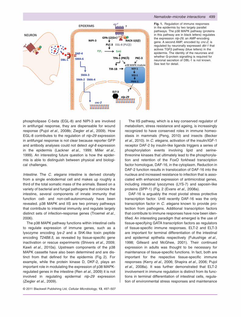
Epidermis. The C. elegans epidermis surrounds the entireanimal and secretes an external collagenous cuticle. A soilfungus, D. coniospora infects C. elegans by germinatingupon contact with the cuticle and produces hyphae thatpierce the cuticle and underlying epidermis. Followinginfection, many genes potentially encoding antimicrobialpeptides, including nlp-29 and cnc-2, are induced in theepidermis. Using nlp-29 and cnc-2 expression as report-ers, the Ewbank group showed that at least two distinctsignalling pathways function in parallel during induction ofantimicrobial responses to D. coniospora (Fig. 1).
Induction of cnc-2 expression is mediated by neuronallyexpressed dbl-1 that activates the TGFb pathway withinthe epidermis. DBL-1 activity is controlled by post-translational modification at the neurones (Zugasti andEwbank, 2009) but the identities of these neurones andthe molecular components that regulate DBL-1 process-ing and secretion have yet to be determined. Transcriptioncofactors that act together with receptor-regulated Smadprotein SMA-3 and the common-mediator Smad proteins(SMA-2 and SMA-4) are also not known.
As summarized in Fig. 1, the conserved p38 MAPKpathway induces nlp-29 expression independently of neu-ronal function (Pujol et al., 2008a; Ziegler et al., 2009).Components of the p38 MAPK pathway include theTribbles-like Kinase NIPI-3 that acts in parallel to GPA-12and RACK-1, the a- and b-subunits of heterotrimericG-proteins, respectively, and upstream of two non-redundant protein kinase C (PKC-3 and TPA-1). The pre-sumptive G-protein-coupled receptor(s) that mediateG-protein signalling has yet to be identified. TPA-1 directlytargets TIR-1B, one of five isoforms of TIR-1, which isrelated to the mammalian adaptor TIRAP, a mediator ofToll-like receptor signalling. Whether protein kinase C acti-vates a member of the p38 MAPK module directlyremains to be determined. Induction of nlp-29 expressionalso requires the epidermis-specific ELT-3 GATA tran-scription factor (Pujol et al., 2008a), but the connectionbetween ELT-3 and p38 MAPK pathway has yet to beestablished.
Interestingly, distinct components of the p38 MAPKsignalling mediate innate immune responses to fungalinfection versus laser or puncture wounding. Whereas
498 M.-W. Tan and M. Shapira
© 2011 Blackwell Publishing Ltd, Cellular Microbiology, 13, 497–507
phospholipase C-beta (EGL-8) and NIPI-3 are involvedin antifungal response, they are dispensable for woundresponse (Pujol et al., 2008b; Ziegler et al., 2009). HowEGL-8 contributes to the regulation of nlp-29 expressionin antifungal response is not clear because reporter GFPand antibody analyses could not detect egl-8 expressionin the epidermis (Lackner et al., 1999; Miller et al.,1999). An interesting future question is how the epider-mis is able to distinguish between physical and biologi-cal challenges.
Intestine. The C. elegans intestine is derived clonallyfrom a single endodermal cell and makes up roughly athird of the total somatic mass of the animals. Based on avariety of bacterial and fungal pathogens that colonize theintestine, several components of innate immunity thatfunction cell- and non-cell-autonomously have beenrevealed. p38 MAPK and IIS are two primary pathwaysthat contribute to intestinal immunity and regulate largelydistinct sets of infection-response genes (Troemel et al.,2006).
The p38 MAPK pathway functions within intestinal cellsto regulate expression of immune genes, such as alysozyme encoding lys-2 and a ShK-like toxin peptideencoding T24B8.5, as revealed by tissue-specific geneinactivation or rescue experiments (Shivers et al., 2009;Kawli et al., 2010a). Upstream components of the p38MAPK cassette have also been determined and are dis-tinct from that defined for the epidermis (Fig. 2). Forexample, while the protein kinase D, DKF-2, plays animportant role in modulating the expression of p38 MAPK-regulated genes in the intestine (Ren et al., 2009) it is notinvolved in regulating epidermal nlp-29 expression(Ziegler et al., 2009).
The IIS pathway, which is a key conserved regulator ofmetabolism, stress resistance and ageing, is increasinglyrecognized to have conserved roles in immune homeo-stasis in mammals (Peng, 2010) and insects (Beckeret al., 2010). In C. elegans, activation of the insulin/IGF-1receptor DAF-2 by insulin-like ligands triggers a series ofphosphorylation events involving lipid and serine-threonine kinases that ultimately lead to the phosphoryla-tion and retention of the FoxO forkhead transcriptionfactor homologue, DAF-16, in the cytoplasm. Reduction inDAF-2 function results in translocation of DAF-16 into thenucleus and increased resistance to infection that is asso-ciated with enhanced expression of antimicrobial genes,including intestinal lysozymes (LYS-7) and saposin-likeproteins (SPP-1) (Fig. 2 (Evans et al., 2008a).
DAF-16 is arguably the most pivotal stress protectivetranscription factor. Until recently DAF-16 was the onlytranscription factor in C. elegans known to provide pro-tection from pathogens. Additional transcription factorsthat contribute to immune responses have now been iden-tified. An interesting paradigm that emerged is the use oftissue-specifying GATA transcription factors as regulatorsof tissue-specific immune responses. ELT-2 and ELT-3are important for terminal differentiation of the intestinaland epidermal epithelia respectively (Fukushige et al.,1998; Gilleard and McGhee, 2001). Their continuedexpression in adults was thought to be necessary formaintenance of tissue-specific functions. In fact, both areimportant for the respective tissue-specific immuneresponses (Kerry et al., 2006; Shapira et al., 2006; Pujolet al., 2008a). It was further demonstrated that ELT-2involvement in immune regulation is distinct from its func-tions in terminal differentiation of intestinal cells, regula-tion of environmental stress responses and maintenance
Fig. 1. Regulation of immune responsesin the epidermis by two largely parallelpathways. The p38 MAPK pathway (proteinsin this pathway are in black letters) regulatesthe expression nlp-29, an AMP-encodinggene. A second AMP, encoded by cnc-2, isregulated by neuronally expressed dbl-1 thatactives TGFb pathway (blue letters) in theepidermis. The identity of the neurones andwhether G-protein signalling is required forneuronal secretion of DBL-1 is not known.See text for detail.
Nematode–microbe interactions 499
© 2011 Blackwell Publishing Ltd, Cellular Microbiology, 13, 497–507

of tissue function and structure (Shapira et al., 2006).Overall, GATA transcription factor appear as tissue-specific integrators of external signals, enabling properstress-specific and tissue-specific responses. Suchhypotheses become more interesting given that the rolesof GATA transcription factors in tissue differentiation areconserved between worms and humans. Both ELT-2 andELT-3 are homologous to GATA4, 5 and 6 in humans,whose expressions are critical for endodermal differentia-tion. GATA6 continues to be expressed in the adult lungand intestine, is induced and is important for protectinglung epithelial cells from P. aeruginosa infection (Shapiraet al., 2006).
ELT-2 is constitutively nuclear (Fukushige et al., 1999),suggesting that for gene induction in response to infectionit must receive relevant signal(s) transduced by otherproteins (Fig. 2). It is yet unknown how this signal isinitiated and by which protein. The co-occurrence ofDNA-binding motifs for DAF-16 and ELT-2 in the5′-untranslated region of a large number of DAF-16-regulated genes (Murphy, 2006) raises the intriguing pos-sibility that FoxO and GATA transcription factors maycooperate to regulate immune homeostasis. Indeed,DAF-16 often functions cooperatively with many transcrip-tion factors, including HSF-1, a heat shock factor (Hsuet al., 2003). Parenthetically, DAF-16, HSF-1 and the heatshock chaperones they regulate are required to protect C.
elegans from bacterial infections (Singh and Aballay,2006), possibly by limiting protein damage due to reactiveoxygen species produced by the host and pathogensduring infection (Mohri-Shiomi and Garsin, 2008). Thus,maintaining protein homeostasis (proteostasis) may be acrucial component of immune response. Further supportfor the proteostasis hypothesis comes for a recent findingthat transcriptional response to infection in C. elegans isconnected to the unfolded proteins response (Richardsonet al., 2010).
What other transcription factors cooperate with or func-tion in parallel to DAF-16 to regulate immunity? Similar toDAF-16, SKN-1 – a distantly related basic leucine-zipper(bZIP) protein that orchestrates the major oxidative stressresponse in vertebrates and yeast – is phosphorylated bythe IIS serine-threonine kinases AKT-1, -2 and SGK-1 andreduced IIS leads to localization of SKN-1 in intestinalnuclei and activation of SKN-1 target genes. SKN-1 func-tions in parallel to DAF-16 to promote longevity (Tulletet al., 2008), but is dispensable for pathogen resistance(Kawli et al., 2010b). Similarly, mutant analyses revealedthat although IIS regulates both ageing and immunity in C.elegans these processes are likely to be mediatedthrough distinct mechanisms involving different serine-threonine kinases (Evans et al., 2008b). ETS-4, an ortho-logue of vertebrate SPDEF transcription factor, is a recentaddition to the list of DAF-16-dependent longevity deter-
Fig. 2. Cell- and non-cell-autonomous regulation of immune responses in the intestine. The IIS pathway (blue letters and arrows) is regulatedprimarily by the insulin peptide released from the neurones. In the intestine, activity of IIS determines the subcellular localization of DAF-16.Whether ELT-2 and ETS-4 cooperate with DAF-16 to regulate gene transcription remains to be determined. A separate G-protein signallingpathway modulates the activity of the p38 MAPK (black letters and arrows) module through a series of enzymes that include phospholipases,which determines the level of diacylglycerol (DAG), and protein kinase C (TPA-1) and protein kinase D (DFK-2). ATF-7 is the transcriptionfactor that mediates p38 MAPK signalling but how it is co-ordinated with other transcription to orchestrated immune gene expression remainsunclear. ZIP-2 appears to regulate immunity independently of p38 MAPK signalling. With the exception of FSHR-1, which functions in parallelto p38 MAPK signalling, the G-protein-coupled receptors that engage Goa and Gqa signalling to affect immune function are currentlyunknown. See text for detail.
500 M.-W. Tan and M. Shapira
© 2011 Blackwell Publishing Ltd, Cellular Microbiology, 13, 497–507

minants. ETS-4-regulated genes are enriched for longev-ity and innate immune effectors, sharing transcriptionaltargets with GATA and FoxO factors (Thyagarajan et al.,2010). These observations provide an opportunity toinvestigate how DAF-16, ETS-4 and ELT-2 may beengaged in the intestine to regulate longevity andimmunity.
Two additional transcription factors belonging to thebZIP family were identified in screens for genetic lesionsthat altered the expression of infection-response gene.ATF-7, an orthologue of the mammalian ATF2/ATF7/CREB5 family, functions as a mediator of p38 MAPK-dependent immune gene expression (Shivers et al.,2010). Loss of atf-7 results in animals that are moresusceptible to a broad panel of pathogens. Another bZIPmember, ZIP-2, appears to have a more specializedimmune function; it is required to regulate expression ofimmunity gene that are independent of p38 MAPK signal-ling and is required for defence against P. aeruginosa butnot another pathogen, Staphylococcus aureus (Esteset al., 2010). Much work remains to clarify how thesetranscription factors integrate signals within the intestineto modulate immune gene expression.
ZIP-2’s contribution to pathogen-specific immuneresponses highlights another feature of C. elegans immu-nity that is of great interest, its ability to differentiatebetween pathogenic and non-pathogenic microbes andinduce a more targeted response. This finding is consis-tent with genome-wide transcriptional profiling studiesshowing that different intestinal bacterial pathogens of C.elegans elicit distinct transcriptional responses from thehost, all of which were distinct from those induced by anepidermal fungal pathogen (Wong et al., 2007). Thus, C.elegans, like mammals and insects, is able to discriminatenot only between pathogenic and non-pathogenicmicrobes but between different classes of microbes aswell. While a clearer picture is beginning to emerge forintracellular and intercellular signalling, the host receptorsthat mediate these responses and the virulence factorsthat activate an immune response have remainedsketchy. Recently, a G-protein-coupled receptor FSHR-1was shown to function in the intestine and in parallel to thep38 MAPK pathway to activate a common set of genes(Fig. 2; Powell et al., 2009), but its ligand and the tran-scription factor that mediate the FSHR-1 responseremains elusive.
Neuronal regulation of epithelial immunity. Recently, sig-nificant strides in elucidating the involvement of neuronalinputs in regulating epidermal and intestinal immunitywere made using the C. elegans system. These are dis-cussed in recent reviews (Zhang and Zhang, 2009; Kawliet al., 2010a). As noted above, neuronal expression ofDBL-1 results in activation of a TGFb-like pathway and
induction of cnc-2 in the epidermis (Fig. 1; Zugasti andEwbank, 2009). Because all receptors and transducersTGFb signalling that mediate DBL-1 effects are stronglyexpressed in the intestine, it is plausible that neuronallysecreted DBL-1 could also modulate intestinal immunity.
In a separate study, it was shown that sustainedreduced secretion from neuronal dense core vesiclesresulted in increased basal and inducible innate immu-nity. This neuronal effect was due to diminished releaseof INS-7, an insulin/IGF-1-like agonist of DAF-2 and theconcomitant activation of DAF-16-dependent immunitygenes in the intestine. Conversely, chronic hypersecre-tion of INS-7 due to abrogation of Goa signalling in thegoa-1 and dgk-1 mutants led to increased signallingthrough DAF-2, suppression of DAF-16 activity in theintestine and increased pathogen susceptibility (Fig. 2;Kawli and Tan, 2008). An added layer of complexity inthis mode of regulation is provided by the tissue-specificroles Gqa signalling. Gqa signalling negatively regulatesGoa signalling to modulate presynaptic levels of diacylg-lycerol (DAG) and neuropeptide release. Unlike Goa,expression of which is restricted to neurones, compo-nents of Gqa signalling, which include EGL-30 (Gqa)and EGL-8 (phospholipase Cb, PLCb), are expressed inboth neurones and intestine. In the neurones, Gqa sig-nalling antagonizes Goa signalling. Thus, reduction ofneuronal Gqa signalling results in decreased INS-7secretion, leading to decreased intestinal IIS and result-ing in increased longevity and pathogen resistance. Inthe intestine Gqa signalling is required for immunitythrough its effects on the activity of p38 MAPK signalling(Fig. 2), and loss of Gqa signalling phenocopies a p38MAPK mutant. Thus, at the organismal level, pathogenresistance is an aggregate of the systemic and cellintrinsic effects Gqa signalling on the activity of IIS andp38 MAPK pathways respectively (Kawli et al., 2010b).
The importance of neuroregulation of immunity isunderscored by the discovery that P. aeruginosa subvertsthis pathway to suppress the expression of DAF-16-dependent immune effectors. Specifically, P. aeruginosainduces the expression of INS-7, which activates intesti-nal IIS and causes DAF-16 to translocate from thenucleus (Evans et al., 2008a). Interestingly, this mecha-nism of immune suppression appears to be conserved asinfection with the mouse enteric pathogen Citrobacterrodentium also resulted in relocalization of FoxO3a to thecytoplasm of intestinal epithelial cells in vivo (Snoekset al., 2008).
Thus, the C. elegans system is well suited for futureinvestigations into whether additional neuropeptides orneurotransmitters also play a role in regulation of immunefunction. An obvious question is whether the neurotrans-mitter serotonin, which is known to modulate the activity ofISS in the intestine (Liang et al., 2006), also affects
Nematode–microbe interactions 501
© 2011 Blackwell Publishing Ltd, Cellular Microbiology, 13, 497–507

immune function. With the increasing appreciation thatactivity of the nervous system could significantly impactimmunological states (Sternberg, 2006), this nascent areaof research in C. elegans promises to produce rewardingfindings.
Immunity against intracellular pathogens
The identification and characterization of intracellularviral, bacterial and eukaryotic pathogens in recent yearshave opened up new and interesting avenues for explo-rations into host immune responses, some of which arelikely to be distinct from those employed against extracel-lular pathogens.
Viruses. A natural virus for C. elegans has not beendescribed and a recent approach to discover viruses inthis nematode suggests that, at least for the commonlaboratory strain N2, C. elegans is not persistentlyinfected with any RNA virus (Wu et al., 2010). Severalbroad-host-range viruses could, however, infect, repli-cate and assemble within C. elegans cells. The vesicularstomatitis virus (VSV), a negative-strand RNA virus thatinfects both insects and mammals, can enter C. eleganscells, express viral proteins and produce new infectiousviral particles (Schott et al., 2005; Wilkins et al., 2005).The positive-strand RNA Flock house virus (FHV) canalso replicate in C. elegans cells (Lu et al., 2005). Thesefindings paved the way for using the C. elegans systemto elucidate the molecular nature of virus–host interac-tions and revealed that key mammalian antiviral path-ways are also conserved in C. elegans (Lu et al., 2005;Schott et al., 2005; Wilkins et al., 2005). These studiesconfirmed that the RNAi machinery is important for pro-tection from virus infection (Lu et al., 2005; Schott et al.,2005; Wilkins et al., 2005), a defence mechanismthat is conserved in both plants and invertebrates(Aliyari and Ding, 2009). Interestingly, C. elegansmutants deficient in Dicer, RDE-1 and RDE-4 showincreased expression of genes predicted to be importantfor immunity against bacterial infections (Welker et al.,2007). It will be interesting to determine whether theRNAi mechanism also affects immunity against otherclasses of pathogens.
Infection of C. elegans by vaccinia DNA virus revealedthe importance of the programmed cell death (PCD)pathway for defence against such infection (Liu et al.,2006). Deficiencies in similar components of PCD alsoresulted in hypersensitivity to Salmonella typhimuriuminfection (Aballay and Ausubel, 2001). S. typhimurium iscurrently the only known bacterium that survives intracel-lularly in C. elegans, suggesting that PCD has generalimportance for protecting worms from intracellularpathogens.
Bacteria. S. typhimurium is found mainly in the lumenand rarely within intestinal epithelial cells of the digestivetract of infected C. elegans. The rare presence of intrac-ellular bacteria was attributed to a robust autophagy effec-tor function in the intestinal cells (Jia et al., 2009).Specifically, Beth Levine and colleagues showed thatknock-down of an autophagy gene bec-1 by RNAiresulted in increased intact bacteria within intestinal cells,which is associated with increased intracellular growth.These observations open up the possibility for furtherrewarding investigations into the mechanisms of autoph-agy in limiting intracellular bacterial growth within thecontext of a whole animal.
Microsporidia. N. parisii is a eukaryotic obligate intracel-lular parasite and the first intracellular pathogen describedfrom wild isolates of Caenorhabiditis from several geo-graphical locations (Troemel et al., 2008). N. parisii is ableto form colonies and multiply within the intestinal cells ofC. elegans. Importantly, genetic alterations of the mainpathways that contribute to intestinal infections by extra-cellular bacteria, the p38 MAPK and IIS pathways, do notappear to affect resistance against this microsporidia(Troemel et al., 2008). These observations suggest thatthese pathways do not contribute to immunity againstmicrosporidia or that this pathogen is able to suppressthese immune pathways. Further investigations into hostdefence responses to N. parisii are likely to reveal newinsights into epithelial immunity.
Detection/recognition of bacteria by the host andthe effects on behaviour
The ability to detect and avoid noxious or dangerousmicrobes is another important defence mechanism for C.elegans. Because C. elegans feeds on soil microorgan-isms it must be able to distinguish between nutritious anddangerous ones. To achieve this C. elegans has evolveda sophisticated chemosensory system consisting of 32sensory neurones, located at both the anterior and theposterior ends of the nematode, to sense and respond toa wide array of chemicals, including microbial products.This olfactory chemotaxis is mediated by G-protein-coupled chemoreceptors, and the specific moleculessensed by these receptors could elicit either an attractionor avoidance behaviour. For example, the acylatedhomoserine lactone (aHSL) autoinducers that Gram-negative bacteria use as communication signals areattractive to C. elegans (Beale et al., 2006), whereas thecyclic lipodepsipentapeptide serrawettin W2, a surfactantsecreted by some Serratia marcescens strains, is a C.elegans repellent (Pradel et al., 2007). It was proposedthat avoidance behaviour triggered by serrawettin detec-tion, which requires primarily in the chemosensory neu-
502 M.-W. Tan and M. Shapira
© 2011 Blackwell Publishing Ltd, Cellular Microbiology, 13, 497–507

rones AWB and TOL-1, the single representative of Toll-like receptor in C. elegans, allows the nematode to avoidharmful microbes. With regards to aHSL, which is pro-duced by both non-pathogens and pathogens, C. elegansis able to associate the molecule with the experience ithas had, a phenomenon known as associative learning(Zhang et al., 2005). For example, naïve C. elegans areattracted to aHSL-producing P. aeruginosa but would berepelled by this same molecule upon subsequent encoun-ters. However, attraction to aHSL-producing bacteria thatis suitable for food would trigger attractive behaviour uponsubsequent encounters. Associative learning is mediatedby the serotonergic ADF neurones and the serotonin-gated chloride channel MOD-1 (Zhang et al., 2005; Bealeet al., 2006). An additional study indicates that signallingthrough the p38 MAPK pathway in the nervous systemregulates the expression of the serotonin biosyntheticenzyme TPH-1 in ADF neurones and is required for patho-gen avoidance (Shivers et al., 2009). Thus, p38 MAPKsignalling functions to regulate both aspects of C. elegansdefence, expression of antimicrobials and microbialavoidance.
While the olfactory chemotaxis system and behaviouraldefence mechanisms it governs may allow worms to effi-ciently defend themselves from pathogenic microbes intheir natural habitat, this system is also being exploited bypathogens to their benefits. For example, Bacillus nema-tocida appears to produce several food-like volatileorganic compounds (VOCs) as chemoattractants toactively lure in C. elegans, and once ingested this patho-gen produces proteases in the intestine to kill, and thusturning the table on, the worms (Niu et al., 2010). It will beinteresting to determine if worms have evolved associa-tive learning mechanism(s) to avoid VOCs upon subse-quent encounters.
Soil bacteria, food quality and effects on C. elegansfeeding behaviour and physiology
Detection and recognition of microbes is necessary notonly for avoiding pathogens, but also for identifying foodsources. Nuanced differentiation between bacteria withdifferent nutritional qualities may provide significantadvantages in the complex soil environment. The regula-tory mechanisms enabling such nuanced differentiationare receiving increasingly more attention as they arefound to control not only behaviour but also physiologyand help maintain homeostasis. Studies focusing on abattery of phylogenetically diverse bacteria isolated fromsoil exposed a range of nutritional qualities of thesemicrobes, as manifested in different rates of worm devel-opment (Avery and Shtonda, 2003). Feeding on low nutri-tional value microbes triggers a starvation responsecharacterized by increased pharyngeal pumping and pro-
nounced roaming behaviour – straight rapid movementseeking new and better food sources (Shtonda and Avery,2006). Low-quality food is also less likely to lead tosatiation-related quiescent behaviour (You et al., 2008).Similar to starvation, low-quality food increases autoph-agy in pharyngeal muscle cells, which is presumed to helpmaintain energy homeostasis in the animal (Kang andAvery, 2009). These studies suggest that C. elegans pro-cesses information relating to the quality of food in itsenvironment and modifies its behaviour accordingly tomaximize energy acquisition. When energy demands arenot met, physiological pathways are activated to maintainenergy homeostasis by recycling. The possibility thatcertain microbes can induce autophagy may be relevantto host immunity. As mentioned above, autophagy is nec-essary for full protection against S. typhimurium. Accord-ingly, it will be interesting to test if the host could increasepathogen resistance by consuming microbes of low nutri-tional value.
The mechanisms underlying feeding behaviours andautophagy, as identified to date, share similarities.Increased pharyngeal pumping during starvation (Youet al., 2006), and activation of autophagy in pharyngealcells (Kang et al., 2007) both require cholinergic muscar-inic signalling. Autophagy was suppressed by addition ofspecific amino acids, activating metabotropic glutamater-gic receptors in the AIY and AIB interneurones, and affect-ing the pharynx, presumably through the release of yetunidentified neuropeptides (Kang and Avery, 2009). AIBneurones were also shown to regulate roaming and qui-escence behaviours in response to food (Bendena et al.,2008), and both AIB and AIY were shown to regulatefeeding-related behaviours downstream to signals fromthe AWC chemosensory neurones, which are the mainregulators of food-seeking behaviour (Bargmann, 2006).Similar to the mechanisms underlying the regulation ofautophagy, the contribution of AIB and AIY to feedingbehaviour was regulated by glutamine (released fromamphid AWC neurones); however, differing from the regu-lation of autophagy, food-seeking behaviours controlledby AIB and AIY were mediated by an AMPA receptor anda glutamate-gated chloride channel respectively (Chala-sani et al., 2007).
The apparent overlap in the contribution of the AIYand/or AIB neurones to the control of both feeding behav-iour and autophagy suggests master regulation of severalprocesses acting in parallel and sharing the common goalof dealing with low food quality. What is specifically rec-ognized in bacteria with low nutritional value is yetunknown. It could be the actual lack of nutrients thatinduces behavioural and physiological processes;however, the involvement of sensory neuronal circuitsassociated with amphid neurones, which typically sensethe outside world, suggests that worms can recognize
Nematode–microbe interactions 503
© 2011 Blackwell Publishing Ltd, Cellular Microbiology, 13, 497–507

molecules released by bacteria that reveal their nutritionalvalue. That worms can recognize microbe-secreted mol-ecules with their amphid neurones is demonstrated by theability of C. elegans to be repelled by S. marcescensserrawettin W2 (Pradel et al., 2007). Interestingly, thisavoidance is controlled by AWB chemosensory neurones,which are similar in their position and in the neurotrans-mitter they release to the previously mentioned AWCneurones.
Expanding knowledge of worm–microbe interactionsby studying mutualism in nematodes
The above studies expand our understanding of themechanisms responsible for microbe recognition by C.elegans and may lead to important insights into howpathogens are recognized. Other interactions with patho-gens, however, exist only in the context of symbiosis (e.g.cell adherence and colonization). Similar to the way rec-ognition of food microbes may teach us about pathogenrecognition, studying interactions of C. elegans with com-mensals and mutualists may expand our knowledge ofsimilar interactions with pathogens. The interactions of C.elegans with its natural soil co-inhabitants are now receiv-ing increased attention by several groups, but this is justthe beginning. An example of what may lie ahead could beobtained by considering studies focusing on mutualisticinteractions between microbes and nematodes other thanC. elegans.
Studies of Heterorhabditis–Photorhabdus interactionsprovide the paradigm for understanding the mechanismsinvolved in establishing symbiosis. Heterorhabditis bacte-riophora is a close relative of C. elegans that has acomplex mutualistic relationship with an enteric ento-mopathogenic bacterium Photorhabdus luminescens(Ciche, 2007; Ffrench-Constant et al., 2007). The multi-phasic complexity of the interactions between the twoprovides the potential for a better understanding ofmechanisms underlying not only bacterial recognition bythe host but also colonization of the worm, invasion andinteractions with worm immunity. The life cycle of P. lumi-nescens begins inside non-feeding infective juvenile (IJ)worms, a development arrest phase similar to dauer(Ciche, 2007). IJ worms seek insect larvae in the soil andpenetrate the larvae upon encounter. Inside the larvae,worms regurgitate 20–30 bacteria, which are sufficient tokill the larvae within a few days. In the cadaver, bacteriafeed on nutrients from the insect larvae, while the wormnow begins to feed on its symbionts. Some of the ingestedbacteria adhere to the intestinal epithelium and generatea biofilm (Ciche et al., 2008; Waterfield et al., 2009). Fromthe intestine, adherent bacteria invade rectal cells wherethey replicate inside vacuoles, while the worm, now wellfed, produces progeny that hatch and develop in their
mother’s body cavities. Worms could develop throughseveral generations in the cadaver. Once resources areexhausted, mothers die, and immediately after, rectalcells lyse, releasing bacteria into the still intact intestine.Bacteria then colonize the pharynx of IJ larvae, and laterinvade the intestinal lumen. When lysis of the mother iscomplete thousands of colonized IJ larvae are released tothe environment seeking new prey. To complete thiscomplex life cycle, bacteria must be capable of adheringto different cells, invade, and grow both extracellularly (asa biofilm) and intracellularly. Moreover, expression offactors important for the different stages must be regu-lated and synchronized with worm development.
Studies aimed at identifying the mechanisms underlyingthe different phases in the symbiosis of Photorhabdus inthe worm revealed several contributing genes. As wouldbe expected, bacteria living in the worm gut should be ableto tolerate the conditions inside. Thus, disruption of thepbgPE operon, involved in synthesis of the bacteriallipopolysaccharide O-antigen, an essential component ofthe bacterial cell wall, resulted in bacterial sensitivity to lowpH and antimicrobial peptides, and a failure to colonize IJlarvae (Bennett and Clarke, 2005). Other genes exemplifythe bacteria contribution to the mutualism. The product ofthe Photorhabdus ngrA gene, a 4′-phosphopantetheinyltransferase, is thought to be involved in biosynthesis offatty acids, polyketides and non-ribosomally synthesizedpeptides, and may contribute to secondary metabolism.Bacteria defective in this gene were unable to competewith wild-type bacteria in colonizing IJs and worms grownon monoxenic culture of ngrA mutants failed to grow anddevelop (Ciche et al., 2001). Similar effects were observedin mutants for the cip genes, which encode proteins con-sisting about 40% of all proteins expressed by Photorhab-dus during symbiosis, proteins that are rich in essentialamino acids (Bintrim and Ensign, 1998).
Factors representing more specific aspects of the inter-actions between Heterorhabditis and Photorhabdus werealso identified. For example, the mad locus, whichincludes genes specifying the synthesis of an usher/chaperone-assembled fimbria, is essential for adherenceto the mother’s posterior intestine and for vertical trans-mission (Somvanshi et al., 2010). Interestingly, theexpression of mad genes was found to depend on anON/OFF invertible promoter switch, which was at the ONposition only in adherent cells.
Studies of Heterorhabditis–Photorhabdus symbiosisare beginning to reveal detailed information on themolecular underpinning of microbe–nematode interac-tions that could have counterparts in C. elegans interac-tions with microbes. The degree of specificity of suchmechanisms depends on how intertwined are the evolu-tionary paths of two organisms. Identifying the natural‘partners’ of C. elegans has the potential to significantly
504 M.-W. Tan and M. Shapira
© 2011 Blackwell Publishing Ltd, Cellular Microbiology, 13, 497–507

increase the range of known host–microbe interactions,similar to what was obtained in studying Heterorhabditis–Photorhabdus interactions, and may thus improve ourunderstanding of host–microbe interactions in general.
Conclusions and future prospects
Studies of host–microbes interactions in C. elegans, whilelargely confined to interactions with pathogens, havebeen beneficial in filling the gaps in our understanding ofpathogen virulence factors and signalling pathways thatregulate immune response. The knowledge gained hasthe potential to shed new light on host–pathogen interac-tions in humans. These advancements include theidentification of novel transcription factors important forimmune responses and a better understanding of the roleof neuronal inputs in regulating innate immune responses.Recent studies of intracellular pathogens have expandedthe utility of the C. elegans to further elucidate the mecha-nisms by which RNA interference, autophagy and PCDfunction to protect the host from infections. Despite muchprogress in elucidating the signalling repertoire in immuneresponses to microbes, none of the canonical patternrecognition receptors has been identified and muchremains to be learned on how pathogens are recognizedby C. elegans. Thus, future investigations will be requiredto determine the precise role of the nervous system inimmunity and how recognition of infection or microbialproducts is integrated with signals of direct recognition ofmicrobes as generated by the more typical pattern recog-nition receptors. Similarly, which proteins take part in theinteractions between microbes and the epithelial tissuesthey colonize is yet unknown. In addition, it is likely that byadapting some of the tools used for studying C. elegans–microbes interactions to other microbe–worm interactionsin the natural context will provide news insights, especiallywith regards to interactions with commensals andmutualists.
References
Aballay, A., and Ausubel, F.M. (2001) Programmed cell deathmediated by ced-3 and ced-4 protects Caenorhabditiselegans from Salmonella typhimurium-mediated killing.Proc Natl Acad Sci USA 98: 2735–2739.
Aliyari, R., and Ding, S.W. (2009) RNA-based viral immunityinitiated by the Dicer family of host immune receptors.Immunol Rev 227: 176–188.
Avery, L., and Shtonda, B.B. (2003) Food transport in the C.elegans pharynx. J Exp Biol 206: 2441–2457.
Bargmann, C.I. (2006) Chemosensation in C. elegans.WormBook 1–29.
Beale, E., Li, G., Tan, M.W., and Rumbaugh, K.P. (2006)Caenorhabditis elegans senses bacterial autoinducers.Appl Environ Microbiol 72: 5135–5137.
Becker, T., Loch, G., Beyer, M., Zinke, I., Aschenbrenner,A.C., Carrera, P., et al. (2010) FOXO-dependent regulationof innate immune homeostasis. Nature 463: 369–373.
Bendena, W.G., Boudreau, J.R., Papanicolaou, T., Maltby,M., Tobe, S.S., and Chin-Sang, I.D. (2008) A Caenorhab-ditis elegans allatostatin/galanin-like receptor NPR-9 inhib-its local search behavior in response to feeding cues. ProcNatl Acad Sci USA 105: 1339–1342.
Bennett, H.P., and Clarke, D.J. (2005) The pbgPE operon inPhotorhabdus luminescens is required for pathogenicityand symbiosis. J Bacteriol 187: 77–84.
Bintrim, S.B., and Ensign, J.C. (1998) Insertional inactivationof genes encoding the crystalline inclusion proteins of Pho-torhabdus luminescens results in mutants with pleiotropicphenotypes. J Bacteriol 180: 1261–1269.
Chalasani, S.H., Chronis, N., Tsunozaki, M., Gray, J.M.,Ramot, D., Goodman, M.B., and Bargmann, C.I. (2007)Dissecting a circuit for olfactory behaviour in Caenorhab-ditis elegans. Nature 450: 63–70.
Ciche, T. (2007) The biology and genome of Heterorhabditisbacteriophora. WormBook 1–9.
Ciche, T.A., Bintrim, S.B., Horswill, A.R., and Ensign, J.C.(2001) A Phosphopantetheinyl transferase homolog isessential for Photorhabdus luminescens to support growthand reproduction of the entomopathogenic nematode Het-erorhabditis bacteriophora. J Bacteriol 183: 3117–3126.
Ciche, T.A., Kim, K.S., Kaufmann-Daszczuk, B., Nguyen,K.C., and Hall, D.H. (2008) Cell invasion and matricideduring Photorhabdus luminescens transmission by Heter-orhabditis bacteriophora nematodes. Appl Environ Micro-biol 74: 2275–2287.
Estes, K.A., Dunbar, T.L., Powell, J.R., Ausubel, F.M., andTroemel, E.R. (2010) bZIP transcription factor zip-2 medi-ates an early response to Pseudomonas aeruginosa infec-tion in Caenorhabditis elegans. Proc Natl Acad Sci USA107: 2153–2158.
Evans, E.A., Kawli, T., and Tan, M.-W. (2008a) Pseudomonasaeruginosa suppresses host immunity by activating theDAF-2 insulin signaling pathway in C. elegans. PLoSPathog 4: e1000175.
Evans, E.A., Chen, W., and Tan, M.-W. (2008b) The daf-2insulin signaling pathway independently regulates agingand immunity in C. elegans through distinct kinases. AgingCell 7: 879–893.
Ffrench-Constant, R.H., Eleftherianos, I., and Reynolds, S.E.(2007) A nematode symbiont sheds light on invertebrateimmunity. Trends Parasitol 23: 514–517.
Fukushige, T., Hawkins, M.G., and McGhee, J.D. (1998) TheGATA-factor elt-2 is essential for formation of the Cae-norhabditis elegans intestine. Dev Biol 198: 286–302.
Fukushige, T., Hendzel, M.J., Bazett-Jones, D.P., andMcGhee, J.D. (1999) Direct visualization of the elt-2 gut-specific GATA factor binding to a target promoter inside theliving Caenorhabditis elegans embryo. Proc Natl Acad SciUSA 96: 11883–11888.
Garsin, D.A., Villanueva, J.M., Begun, J., Kim, D.H., Sifri,C.D., Calderwood, S.B., et al. (2003) Long-lived C. elegansdaf-2 mutants are resistant to bacterial pathogens. Science300: 1921.
Gilleard, J.S., and McGhee, J.D. (2001) Activation of hypo-dermal differentiation in the Caenorhabditis elegans
Nematode–microbe interactions 505
© 2011 Blackwell Publishing Ltd, Cellular Microbiology, 13, 497–507

embryo by GATA transcription factors ELT-1 and ELT-3.Mol Cell Biol 21: 2533–2544.
Hodgkin, J., Kuwabara, P.E., and Corneliussen, B. (2000) Anovel bacterial pathogen, Microbacterium nematophilum,induces morphological change in the nematode C.elegans. Curr Biol 10: 1615–1618.
Hsu, A.L., Murphy, C.T., and Kenyon, C. (2003) Regulation ofaging and age-related disease by DAF-16 and heat-shockfactor. Science 300: 1142–1145.
Irazoqui, J.E., Urbach, J.M., and Ausubel, F.M. (2010) Evo-lution of host innate defence: insights from Caenorhabditiselegans and primitive invertebrates. Nat Rev Immunol 10:47–58.
Jia, K., Thomas, C., Akbar, M., Sun, Q., Adams-Huet, B.,Gilpin, C., and Levine, B. (2009) Autophagy genes protectagainst Salmonella typhimurium infection and mediateinsulin signaling-regulated pathogen resistance. Proc NatlAcad Sci USA 106: 14564–14569.
Kang, C., and Avery, L. (2009) Systemic regulation of star-vation response in Caenorhabditis elegans. Genes Dev 23:12–17.
Kang, C., You, Y.J., and Avery, L. (2007) Dual roles of autoph-agy in the survival of Caenorhabditis elegans during star-vation. Genes Dev 21: 2161–2171.
Kawli, T., and Tan, M.-W. (2008) Neuroendocrine signalsmodulate the innate immunity of Caenorhabditiselegans through insulin signaling. Nat Immunol 9: 1415–1424.
Kawli, T., He, F., and Tan, M.W. (2010a) It takes nerves tofight infections: insights on neuro-immune interactions fromC. elegans. Dis Model Mech 3: 721–731.
Kawli, T., Wu, C., and Tan, M.W. (2010b) Systemic and cellintrinsic roles of Gqalpha signaling in the regulation ofinnate immunity, oxidative stress, and longevity in Cae-norhabditis elegans. Proc Natl Acad Sci USA 107: 13788–13793.
Kerry, S., TeKippe, M., Gaddis, N.C., and Aballay, A. (2006)GATA transcription factor required for immunity to bacterialand fungal pathogens. PLoS ONE 1: e77.
Kim, D.H., Feinbaum, R., Alloing, G., Emerson, F.E., Garsin,D.A., Inoue, H., et al. (2002) A conserved p38 MAP kinasepathway in Caenorhabditis elegans innate immunity.Science 297: 623–626.
Lackner, M.R., Nurrish, S.J., and Kaplan, J.M. (1999)Facilitation of synaptic transmission by EGL-30 Gqalphaand EGL-8 PLCbeta: DAG binding to UNC-13 is requiredto stimulate acetylcholine release. Neuron 24: 335–346.
Liang, B., Moussaif, M., Kuan, C.J., Gargus, J.J., and Sze,J.Y. (2006) Serotonin targets the DAF-16/FOXO signalingpathway to modulate stress responses. Cell Metab 4: 429–440.
Liu, W.H., Lin, Y.L., Wang, J.P., Liou, W., Hou, R.F., Wu, Y.C.,and Liao, C.L. (2006) Restriction of vaccinia virusreplication by a ced-3 and ced-4-dependent pathway inCaenorhabditis elegans. Proc Natl Acad Sci USA 103:4174–4179.
Lu, R., Maduro, M., Li, F., Li, H.W., Broitman-Maduro, G., Li,W.X., and Ding, S.W. (2005) Animal virus replication andRNAi-mediated antiviral silencing in Caenorhabditiselegans. Nature 436: 1040–1043.
Mayer, A.K., and Dalpke, A.H. (2007) Regulation of localimmunity by airway epithelial cells. Arch Immunol Ther Exp(Warsz) 55: 353–362.
Miller, K.G., Emerson, M.D., and Rand, J.B. (1999) Goalphaand diacylglycerol kinase negatively regulate the Gqalphapathway in C. elegans. Neuron 24: 323–333.
Mohri-Shiomi, A., and Garsin, D.A. (2008) Insulin signalingand the heat shock response modulate protein homeosta-sis in the Caenorhabditis elegans intestine during infection.J Biol Chem 283: 194–201.
Muir, R.E., and Tan, M.W. (2008) Virulence of Leucobacterchromiireducens subsp. solipictus to Caenorhabditiselegans: characterization of a novel host–pathogen inter-action. Appl Environ Microbiol 74: 4185–4198.
Murphy, C.T. (2006) The search for DAF-16/FOXO transcrip-tional targets: approaches and discoveries. Exp Gerontol41: 910–921.
Niu, Q., Huang, X., Zhang, L., Xu, J., Yang, D., Wei, K., et al.(2010) A Trojan horse mechanism of bacterial pathogen-esis against nematodes. Proc Natl Acad Sci USA 107:16631–16636.
Peng, S.L. (2010) Forkhead transcription factors in chronicinflammation. Int J Biochem Cell Biol 42: 482–485.
Powell, J.R., Kim, D.H., and Ausubel, F.M. (2009) The Gprotein-coupled receptor FSHR-1 is required for the Cae-norhabditis elegans innate immune response. Proc NatlAcad Sci USA 106: 2782–2787.
Pradel, E., Zhang, Y., Pujol, N., Matsuyama, T., Bargmann,C.I., and Ewbank, J.J. (2007) Detection and avoidance of anatural product from the pathogenic bacterium Serratiamarcescens by Caenorhabditis elegans. Proc Natl AcadSci USA 104: 2295–2300.
Pujol, N., Link, E.M., Liu, L.X., Kurz, C.L., Alloing, G., Tan,M.-W., et al. (2001) A reverse genetic analysis of compo-nents of the Toll signaling pathway in Caenorhabditiselegans. Curr Biol 11: 809–821.
Pujol, N., Zugasti, O., Wong, D., Couillault, C., Kurz, C.L.,Schulenburg, H., and Ewbank, J.J. (2008a) Anti-fungalinnate immunity in C. elegans is enhanced by evolutionarydiversification of antimicrobial peptides. PLoS Pathog 4:e1000105.
Pujol, N., Cypowyj, S., Ziegler, K., Millet, A., Astrain, A.,Goncharov, A., et al. (2008b) Distinct innate immuneresponses to infection and wounding in the C. elegansepidermis. Curr Biol 18: 481–489.
Ren, M., Feng, H., Fu, Y., Land, M., and Rubin, C.S. (2009)Protein kinase D is an essential regulator of C. elegansinnate immunity. Immunity 30: 521–532.
Richardson, C.E., Kooistra, T., and Kim, D.H. (2010) Anessential role for XBP-1 in host protection against immuneactivation in C. elegans. Nature 463: 1092–1095.
Schott, D.H., Cureton, D.K., Whelan, S.P., and Hunter, C.P.(2005) An antiviral role for the RNA interference machineryin Caenorhabditis elegans. Proc Natl Acad Sci USA 102:18420–18424.
Shapira, M., Hamlin, B.J., Rong, J., Chen, K., Ronen, M., andTan, M.-W. (2006) A conserved role for a GATA transcrip-tion factor in regulating epithelial innate immuneresponses. Proc Natl Acad Sci USA 103: 14086–14091.
Shivers, R.P., Kooistra, T., Chu, S.W., Pagano, D.J., and Kim,D.H. (2009) Tissue-specific activities of an immune signal-
506 M.-W. Tan and M. Shapira
© 2011 Blackwell Publishing Ltd, Cellular Microbiology, 13, 497–507

ing module regulate physiological responses to pathogenicand nutritional bacteria in C. elegans. Cell Host Microbe 6:321–330.
Shivers, R.P., Pagano, D.J., Kooistra, T., Richardson, C.E.,Reddy, K.C., Whitney, J.K., et al. (2010) Phosphorylationof the conserved transcription factor ATF-7 by PMK-1 p38MAPK regulates innate immunity in Caenorhabditiselegans. PLoS Genet 6: e1000892.
Shtonda, B.B., and Avery, L. (2006) Dietary choice behaviorin Caenorhabditis elegans. J Exp Biol 209: 89–102.
Singh, V., and Aballay, A. (2006) Heat-shock transcriptionfactor (HSF)-1 pathway required for Caenorhabditiselegans immunity. Proc Natl Acad Sci USA 103: 13092–13097.
Snoeks, L., Weber, C.R., Turner, J.R., Bhattacharyya, M.,Wasland, K., and Savkovic, S.D. (2008) Tumor suppressorFoxo3a is involved in the regulation of lipopolysaccharide-induced interleukin-8 in intestinal HT-29 cells. Infect Immun76: 4677–4685.
Somvanshi, V.S., Viswanathan, P., Jacobs, J.L., Mulks, M.H.,Sundin, G.W., and Ciche, T.A. (2010) The type 2 secretionPseudopilin, gspJ, is required for multihost pathogenicity ofBurkholderia cenocepacia AU1054. Infect Immun 78:4110–4121.
Sternberg, E.M. (2006) Neural regulation of innate immunity:a coordinated nonspecific host response to pathogens. NatRev Immunol 6: 318–328.
Thyagarajan, B., Blaszczak, A.G., Chandler, K.J., Watts, J.L.,Johnson, W.E., and Graves, B.J. (2010) ETS-4 is a tran-scriptional regulator of life span in Caenorhabditis elegans.PLoS Genet 6: e1001125.
Troemel, E.R., Chu, S.W., Reinke, V., Lee, S.S., Ausubel,F.M., and Kim, D.H. (2006) p38 MAPK regulates expres-sion of immune response genes and contributes to longev-ity in C. elegans. PLoS Genet 2: e183.
Troemel, E.R., Felix, M.A., Whiteman, N.K., Barriere, A., andAusubel, F.M. (2008) Microsporidia are natural intracellularparasites of the nematode Caenorhabditis elegans. PLoSBiol 6: 2736–2752.
Tullet, J.M., Hertweck, M., An, J.H., Baker, J., Hwang, J.Y.,Liu, S., et al. (2008) Direct inhibition of the longevity-promoting factor SKN-1 by insulin-like signaling in C.elegans. Cell 132: 1025–1038.
Waterfield, N.R., Ciche, T., and Clarke, D. (2009) Photorhab-dus and a host of hosts. Annu Rev Microbiol 63: 557–574.
Welker, N.C., Habig, J.W., and Bass, B.L. (2007) Genesmisregulated in C. elegans deficient in Dicer, RDE-4, orRDE-1 are enriched for innate immunity genes. RNA 13:1090–1102.
Wilkins, C., Dishongh, R., Moore, S.C., Whitt, M.A., Chow,M., and Machaca, K. (2005) RNA interference is an antivi-ral defence mechanism in Caenorhabditis elegans. Nature436: 1044–1047.
Wong, D., Bazopoulou, D., Pujol, N., Tavernarakis, N., andEwbank, J.J. (2007) Genome-wide investigation revealspathogen-specific and shared signatures in the responseof Caenorhabditis elegans to infection. Genome Biol 8:R194.
Wu, Q., Luo, Y., Lu, R., Lau, N., Lai, E.C., Li, W.X., and Ding,S.W. (2010) Virus discovery by deep sequencing andassembly of virus-derived small silencing RNAs. Proc NatlAcad Sci USA 107: 1606–1611.
You, Y.J., Kim, J., Cobb, M., and Avery, L. (2006) Starvationactivates MAP kinase through the muscarinic acetylcholinepathway in Caenorhabditis elegans pharynx. Cell Metab 3:237–245.
You, Y.J., Kim, J., Raizen, D.M., and Avery, L. (2008) Insulin,cGMP, and TGF-beta signals regulate food intake and qui-escence in C. elegans: a model for satiety. Cell Metab 7:249–257.
Zhang, X., and Zhang, Y. (2009) Neural-immune communi-cation in Caenorhabditis elegans. Cell Host Microbe 5:425–429.
Zhang, Y., Lu, H., and Bargmann, C.I. (2005) Pathogenicbacteria induce aversive olfactory learning in Caenorhab-ditis elegans. Nature 438: 179–184.
Ziegler, K., Kurz, C.L., Cypowyj, S., Couillault, C., Pophillat,M., Pujol, N., and Ewbank, J.J. (2009) Antifungal innateimmunity in C. elegans: PKCdelta links G protein signalingand a conserved p38 MAPK cascade. Cell Host Microbe 5:341–352.
Zugasti, O., and Ewbank, J.J. (2009) Neuroimmune regula-tion of antimicrobial peptide expression by a noncanonicalTGF-beta signaling pathway in Caenorhabditis elegansepidermis. Nat Immunol 10: 249–256.
Nematode–microbe interactions 507
© 2011 Blackwell Publishing Ltd, Cellular Microbiology, 13, 497–507
Copyright © 2022 FDOKUMEN