An approach to evaluate two-electron reduction of 9,10-phenanthraquinone and redox activity of the...
11
Original Contribution An approach to evaluate two-electron reduction of 9,10-phenanthraquinone and redox activity of the hydroquinone associated with oxidative stress Keiko Taguchi a , Sayako Fujii b , Shigeru Yamano c , Arthur K. Cho d , Shinji Kamisuki e , Yumi Nakai f , Fumio Sugawara e , John R. Froines d , Yoshito Kumagai a,d, ⁎ a Doctoral Programs in Medical Sciences, Graduate School of Comprehensive Human Sciences, University of Tsukuba, 1-1-1 Tennodai, Tsukuba, Ibaraki 305-8575, Japan b Master’s Program in Environmental Sciences, University of Tsukuba, 1-1-1 Tennodai, Tsukuba, Ibaraki 305-8575, Japan c Faculty of Pharmaceutical Sciences, Fukuoka University, 8-19-1 Nanakuma, Jonan-ku, Fukuoka 814-0180, Japan d Southern California Particle Center, Institute of the Environment, University of California, Los Angeles, Los Angeles, CA 90095, USA e Department of Applied Biological Science, Faculty of Science and Technology, Tokyo University of Science, Noda, Chiba 278-8510, Japan f Analytical Instruments Division, JEOL Ltd., 3-1-2 Musashino, Akishima, Tokyo 196-8558, Japan Received 19 February 2007; revised 11 May 2007; accepted 11 May 2007 Available online 24 May 2007 Abstract Quinones are widely used as medicines or redox agents. The chemical properties are based on the reactions against an electron donor. 9,10- Phenanthraquinone (PQ), which is a quinone contaminated in airborne particulate matters, forms redox cycling, not Michael addition, with electron donors. Redox cycling of PQ contributes to its toxicity, following generation of reactive oxygen species (ROS). Detoxification of quinones is generally thought to be two-electron reduction forming hydroquinones. However, a hydroquinone of PQ, 9,10-dihydroxyphenanthrene (PQH 2 ), has been never detected itself, because it is quite unstable. In this paper, we succeeded in detecting PQH 2 as its stable derivative, 9,10- diacetoxyphenanthrene (DAP). However, higher concentrations of PQ (N 4 μM) form disproportionately with PQH 2 , producing the 9,10- phenanthraquinone radical (PQ U - ) which is a one-electron reducing product of PQ. In cellular experiments using DAP as a precursor of PQH 2 , it was shown that PQH 2 plays a critical role in the oxidative protein damage and cellular toxicity of PQ, showing that two-electron reduction of PQ can also initiate redox cycling to cause oxidative stress-dependent cytotoxicity. © 2007 Elsevier Inc. All rights reserved. Keywords: Redox cycling; Two-electron reduction; 9,10-Phenanthraquinone; Disproportionation Introduction Ambient particulate matter (PM 2.5 ) and diesel exhaust par- ticles (DEP) are air pollutants that induce a variety of adverse health effects including cardiovascular diseases [1], allergies [2], and cancers [3]. Recently, oxidative stress has been suggested to be a key biological event in the toxicity of the particulates [4,5]. Organic extracts from DEP, which contain high levels of polycyclic aromatic hydrocarbons (PAHs) and their derivatives, are capable of producing reactive oxygen species (ROS) [6–8]. We have found that 9,10-phenanthraqui- none (PQ), a PAH-quinone found in DEP [9] produces superoxide [8,10]. This quinone is toxic to human pulmonary epithelial A549 cells at micromolar concentrations [11] and exhibits both aerobic and anaerobic toxicity in yeast [12]. The Free Radical Biology & Medicine 43 (2007) 789 – 799 www.elsevier.com/locate/freeradbiomed Abbreviations: AKR, aldo-keto reductase; AZQ, 2,5-ethyl(carboethoxya- mino)3,6-diaziridinyl-1,4-benzoquinone; DAP, 9,10-diacetoxyphenanthrene; DEP, diesel exhaust particles; DMPO, 5,5-dimethyl-1-pyrroline N-oxide; DMSO, dimethyl sulfoxide; DNPH, 2,4-dinitrophenylhydrazine; ESI-MS, electrospray ionization-mass spectrometry; ESR, electron spin resonance; Hepes, N-2-hydroxy-ethylpiperazine-N′-2-ethanesulfonic acid; H 2 O 2 , hydrogen peroxide; HPLC, high-performance liquid chromatography; MTT, 3-(4,5- dimethylthiazol-2-yl)-2,5-diphenyltetrazolium bromide; NQO, NAD(P)H:qui- none oxidoreductase; PAGE, polyacrylamide gel electrophoresis; PAH, polycyclic aromatic hydrocarbon; P450R, NADPH-cytochrome P450 reductase; PM 2.5 , ambient particulate matter; PQ, 9,10-phenanthraquinone; PQ U - , 9,10- phenanthraquinone radical; PQH 2 , 9,10-dihydroxyphenanthrene; ROS, reactive oxygen species; SOD, superoxide dismutase; TrxR1, thioredoxin reductase 1. ⁎ Corresponding author. Doctoral Programs in Medical Sciences, Graduate School of Comprehensive Human Sciences, University of Tsukuba, 1-1-1 Tennodai, Tsukuba, Ibaraki 305-8575, Japan. Fax: +81 29 853 3133. E-mail address: [email protected] (Y. Kumagai). 0891-5849/$ - see front matter © 2007 Elsevier Inc. All rights reserved. doi:10.1016/j.freeradbiomed.2007.05.021
-
Upload
independent -
Category
Documents
-
view
0 -
download
0
Transcript of An approach to evaluate two-electron reduction of 9,10-phenanthraquinone and redox activity of the...

Free Radical Biology & Medicine 43 (2007) 789–799www.elsevier.com/locate/freeradbiomed
Original Contribution
An approach to evaluate two-electron reduction of 9,10-phenanthraquinoneand redox activity of the hydroquinone associated with oxidative stress
Keiko Taguchi a, Sayako Fujii b, Shigeru Yamano c, Arthur K. Cho d, Shinji Kamisuki e,Yumi Nakai f, Fumio Sugawara e, John R. Froines d, Yoshito Kumagai a,d,⁎
a Doctoral Programs in Medical Sciences, Graduate School of Comprehensive Human Sciences, University of Tsukuba,1-1-1 Tennodai, Tsukuba, Ibaraki 305-8575, Japan
b Master’s Program in Environmental Sciences, University of Tsukuba, 1-1-1 Tennodai, Tsukuba, Ibaraki 305-8575, Japanc Faculty of Pharmaceutical Sciences, Fukuoka University, 8-19-1 Nanakuma, Jonan-ku, Fukuoka 814-0180, Japan
d Southern California Particle Center, Institute of the Environment, University of California, Los Angeles, Los Angeles, CA 90095, USAe Department of Applied Biological Science, Faculty of Science and Technology, Tokyo University of Science, Noda, Chiba 278-8510, Japan
f Analytical Instruments Division, JEOL Ltd., 3-1-2 Musashino, Akishima, Tokyo 196-8558, Japan
Received 19 February 2007; revised 11 May 2007; accepted 11 May 2007Available online 24 May 2007
Abstract
Quinones are widely used as medicines or redox agents. The chemical properties are based on the reactions against an electron donor. 9,10-Phenanthraquinone (PQ), which is a quinone contaminated in airborne particulate matters, forms redox cycling, not Michael addition, withelectron donors. Redox cycling of PQ contributes to its toxicity, following generation of reactive oxygen species (ROS). Detoxification ofquinones is generally thought to be two-electron reduction forming hydroquinones. However, a hydroquinone of PQ, 9,10-dihydroxyphenanthrene(PQH2), has been never detected itself, because it is quite unstable. In this paper, we succeeded in detecting PQH2 as its stable derivative, 9,10-diacetoxyphenanthrene (DAP). However, higher concentrations of PQ (N4 μM) form disproportionately with PQH2, producing the 9,10-phenanthraquinone radical (PQ
U−) which is a one-electron reducing product of PQ. In cellular experiments using DAP as a precursor of PQH2, itwas shown that PQH2 plays a critical role in the oxidative protein damage and cellular toxicity of PQ, showing that two-electron reduction of PQcan also initiate redox cycling to cause oxidative stress-dependent cytotoxicity.© 2007 Elsevier Inc. All rights reserved.
Keywords: Redox cycling; Two-electron reduction; 9,10-Phenanthraquinone; Disproportionation
Abbreviations: AKR, aldo-keto reductase; AZQ, 2,5-ethyl(carboethoxya-mino)3,6-diaziridinyl-1,4-benzoquinone; DAP, 9,10-diacetoxyphenanthrene;DEP, diesel exhaust particles; DMPO, 5,5-dimethyl-1-pyrroline N-oxide;DMSO, dimethyl sulfoxide; DNPH, 2,4-dinitrophenylhydrazine; ESI-MS,electrospray ionization-mass spectrometry; ESR, electron spin resonance;Hepes, N-2-hydroxy-ethylpiperazine-N′-2-ethanesulfonic acid; H2O2, hydrogenperoxide; HPLC, high-performance liquid chromatography; MTT, 3-(4,5-dimethylthiazol-2-yl)-2,5-diphenyltetrazolium bromide; NQO, NAD(P)H:qui-none oxidoreductase; PAGE, polyacrylamide gel electrophoresis; PAH,polycyclic aromatic hydrocarbon; P450R, NADPH-cytochrome P450 reductase;PM2.5, ambient particulate matter; PQ, 9,10-phenanthraquinone; PQU−, 9,10-phenanthraquinone radical; PQH2, 9,10-dihydroxyphenanthrene; ROS, reactiveoxygen species; SOD, superoxide dismutase; TrxR1, thioredoxin reductase 1.⁎ Corresponding author. Doctoral Programs in Medical Sciences, Graduate
School of Comprehensive Human Sciences, University of Tsukuba, 1-1-1Tennodai, Tsukuba, Ibaraki 305-8575, Japan. Fax: +81 29 853 3133.
E-mail address: [email protected] (Y. Kumagai).
0891-5849/$ - see front matter © 2007 Elsevier Inc. All rights reserved.doi:10.1016/j.freeradbiomed.2007.05.021
Introduction
Ambient particulate matter (PM2.5) and diesel exhaust par-ticles (DEP) are air pollutants that induce a variety of adversehealth effects including cardiovascular diseases [1], allergies[2], and cancers [3]. Recently, oxidative stress has beensuggested to be a key biological event in the toxicity of theparticulates [4,5]. Organic extracts from DEP, which containhigh levels of polycyclic aromatic hydrocarbons (PAHs) andtheir derivatives, are capable of producing reactive oxygenspecies (ROS) [6–8]. We have found that 9,10-phenanthraqui-none (PQ), a PAH-quinone found in DEP [9] producessuperoxide [8,10]. This quinone is toxic to human pulmonaryepithelial A549 cells at micromolar concentrations [11] andexhibits both aerobic and anaerobic toxicity in yeast [12]. The

790 K. Taguchi et al. / Free Radical Biology & Medicine 43 (2007) 789–799
multiple facets of its chemical and toxicological properties haveled us to examine the details of its interaction with cellularcomponents [13–15]. One aspect of its biochemistry that needsto be elucidated is its biotransformation by cellular systemssince its toxicity involves both redox and additional reactions ofits o-quinone structure.
In this context, there are two reduced PQ species ofbiological importance, its semiquinone radical (9,10-phenan-thraquinone; PQU−) and its hydroquinone (9,10-dihydroxyphe-nanthrene; PQH2). PQU− can promote redox cycling bytransferring electrons from a source such as NADPH to oxygento generate ROS [8,10]. We have previously reported twopossible pathways for PQ
U− formation: an enzymatic reactioncatalyzed by flavoenzymes such as NADPH-cytochrome P450reductase (P450R) or neuronal nitric oxide synthase [8,16] and anonenzymatic reaction with proximal protein thiols [10].Formation of hydroquinones has generally been considered toprotect cells against quinone-induced oxidative stress [17–19].However, Bucker et al. [20] found that PQH2 was somewhatmore mutagenic than PQ itself in the rec assay with Bacillussubtilis strains. In addition, the two-electron reduction of 2-methyl-1,4-naphthoquinone, mitomycin C, and AZQ, which arederivates of benzoquinone by purified NAD(P)H:quinoneoxidoreductase 1 (NQO1) [21,22] and aldo-keto reductase1C9 (AKR1C9) [23], has been shown to induce DNA damageand cytotoxicity. Taken together, these reports suggest that if thetwo-electron reducing products of quinones readily form redoxcycling, thereby producing ROS, two-electron reduction couldalso be a deleterious pathway.
We have recently found that exposure of human pulmonarycarcinoma A549 cells to PQ causes a marked oxidative stress asevaluated by oxidative modification of the cellular proteins [11].Interestingly, the antioxidant protein, Cu,Zn-superoxide dis-mutase (Cu,Zn-SOD), was downregulated by PQ under theseconditions. Because hydroquinones such as PQH2 react easilywith superoxide to form quinones [24], the reduced levels of Cu,Zn-SOD during PQ exposure may have promoted oxidativestress in the A549 cells by enhancing a redox cycling of PQH2 toPQ. To our knowledge, there are no reports describing the directtwo-electron reduction of PQ, in part because of the labile natureof PQH2. To address this issue, we developed a convenient assayto convert PQH2 to its diacetoxy derivative 9,10-diacetoxyphe-nanthrene (DAP) with high-performance liquid chromatography(HPLC). Using this procedure, we examined the characteristicsof PQ to PQH2 conversion by crude enzyme preparations and apurified preparation containing an aldo-keto reductase 1C(AKR1C) isozyme. The results provide evidence for the firsttime that NADPH-dependent enzymes such as the AKR1Cisozyme mediate the two-electron reduction of PQ and promoteredox cycling with PQU− as the intermediate.
Materials and methods
Materials
PQ and 2,4-dinitrophenylhydrazine (DNPH) were obtainedfrom Nacalai Tesque, Inc. (Kyoto, Japan); Indomethacin, ciba-
cron blue, ethoxyquin, quercetin, monoclonal anti-dinitrophe-nyl (DNP) antibody, and DMEM medium containing 4.5 g/L ofD-glucose were from Sigma-Aldrich Co. (St. Louis, MO); ace-tic anhydride was from Wako Pure Chemical Industries, Ltd.(Osaka, Japan); dicumarol was from Aldrich Chemical Co.(Milwaukee, Wl); 5,5′-dimethyl-1-pyrroline N-oxide (DMPO)was from Labotech Co. (Tokyo, Japan); ECL Western blottingdetection reagent and Blue Sepharose 6 fast flow andstreptavidin-horseradish peroxidase (HRP) conjugate werefrom Amersham Biosciences Co. (Piscataway, NJ); Gluta-MAX-I supplement was from Invitrogen Co. (Carlsbad, CA);biotinated anti-mouse IgE was from Technopharm Biotechnol.(Villejuif Cedex, France); and anti-thioredoxin reductase 1(TrxR1) antibody was from Upstate USA, Inc. (Charlottesville,VA). An AKR1C isozyme was purified from liver cytosol ofrabbits by the method of Yamano et al. [25]. The enzyme wasclassified into the AKR1C subfamily because its sequenceshowed more than 60% identity to members of the AKR1C.Polyclonal antibodies against Cu,Zn-SOD were prepared asdescribed by Shimojo et al. [26]. Antibody against P450R wasraised by the method described previously [27]. Antibodiesagainst AKR7A2 and NQO1 were gifts from Dr. John D. Hayes(University of Dundee, UK) and Dr. Nobuyuki Koga (Naka-mura Gakuen University, Japan), respectively. All otherchemicals used were obtained from commercial sources andwere of the highest grade available.
Synthesis of DAP
PQ (100 mg in 15 ml of tetrahydrofuran) was mixed with Zndust (50 mg), and then 0.5 ml of acetic anhydride, 1 drop ofwater, and 3 drops of triethylamine were added. The mixturewas stirred at room temperature for 20 min; an additional 50 mgof Zn dust, 1 drop of water, and 3 drops of triethylamine wereadded; and then the mixture was heated to reflux for 1 h. Aftercooling, the mixture was extracted with chloroform (40 ml). Thechloroform layer was washed with water, saturated withNaHCO3, and then dried over MgSO4. The solvent was re-moved in vacuo to leave a pale yellow solid (132 mg). Theproduct was recrystallized in benzene and a white solid (45 mg)was obtained. Electrospray ionization-mass and NMR spec-trometries (ESI-MS; positive-ion mode) were performed:[M+Na]+ m/z=316.7; 1H NMR (CDCl3, 400 MHz, ppm)δ8.68 (br d, 2H, ArH), 7.88 (br d, 2H, ArH), 7.65 (m, 4H, ArH),2.49 (s, 6H, COCH3);
13C NMR (CDCl3, 100 MHz, ppm)δ168.2, 135.8, 129.7, 127.3, 127.1, 126.5, 123.0, 122.0, 20.5.
Enzyme preparation
Wistar male rats (6 weeks) and ICR male mice (6–8 weeks)were used. Liver and other tissues were homogenized in 9 vol of5 mM Tris-HCl (pH 7.5)–0.25 M sucrose–0.1 mM EDTA. Thehomogenate was centrifuged at 600g for 10 min and thesupernatants were centrifuged at 9000g for 10 min. The pellet(mitochondrial fraction) was suspended in the homogenizingbuffer and stored at –70°C until use. The supernatants from themitochondrial isolation were centrifuged at 105,000g for 60 min.
791K. Taguchi et al. / Free Radical Biology & Medicine 43 (2007) 789–799
The pellets obtained were resuspended in 0.1 M potassiumpyrophosphate (pH 7.4) and again centrifuged at 105,000g for60 min. The resulting pellets, the microsomal fraction, were againresuspended in homogenizing buffer and stored at –70°C untiluse. Protein concentration was determined by the method ofBradford with bovine serum albumin as the standard [28].
For separation of the enzymes responsible for the two-electron reduction of PQ to PQH2 by Blue Sepharose columnchromatography, mice were given feed including 0.5% etho-xyquin (CLEA Japan, Inc., Tokyo, Japan), an inducer for AKRisozymes for 2 weeks [29]. The animals were euthanized withdiethylether and the tissues were removed and homogenized in5 vol of 0.25 M sucrose. The homogenate was centrifuged at9000g for 20 min. The resulting supernatant (342 mg of protein)was centrifuged at 105,000g for 60 min to obtain the cytosol.Solid ammonium sulfate was added to the lung cytosol of miceto make a 40% saturated solution, followed by centrifugation at18,000g for 20 min. The supernatants obtained were mixed withsolid ammonium sulfate to make a 90% saturated solution. Afterthe mixture was centrifuged, the precipitates were dissolved in10 ml of 50 mM Tris-HCl (pH 7.2)–0.25 M sucrose (Buffer A)and dialyzed for 6 h against 1500 ml of 50 mM Tris-HCl (pH7.2). The dialyzed sample (170 mg of protein) was applied ontoa Blue Sepharose column (2.5× 1.5 cm, i.d.), which had beenequilibrated with Buffer A at a flow rate of 0.15 ml/min. Thecolumn was washed with Buffer A (fraction B-I, No. 4-9),followed by Buffer A-0.5 M KCl (fraction B-II, No. 25-32) andBuffer A-0.5 M KCl-10 mM NADH (fraction B-III, No. 52-64).NADH was removed from fraction B-III by dialysis againstbuffer A-0.5 M KCl for 3 h (500 ml×2).
Two-electron reduction of PQ
The incubation mixture (0.75 ml) consisted of 5 μM PQ,0.2 mM NADPH, enzyme preparation (21.24 μg of protein),and 0.1 M Hepes (pH 7.6)–0.1 mM EDTA unless otherwisenoted. Reactions were initiated by the addition of the enzymepreparation and allowed to proceed at 25°C for 5 min. Toterminate the reaction, a portion (0.5 ml) of the incubationmixture was transferred to a centrifuge tube containing aceticanhydride (20 μl) and heated at 80°C for 5 min. Then themixtures were mixed with trichloroacetic acid (170 μl, finalconcentration, 2.5%) and centrifuged at 14,000g for 5 min. Thesupernatants (40 μl) were subjected to HPLC analysis.Separation of DAP from PQ was carried out on a YMC-PackODS-AM (250×4.6 mm, i.d., 5 μm particle size, YMC Co.Ltd., Kyoto, Japan) with a Shimadzu LC-10AT pump and SPD-10A UV-VIS detector (Kyoto, Japan). Elution was accom-plished with acetonitrile/1% CH3COOH (3:2, v/v) at a flow rateof 1 ml/min. Detection was performed at 255 nm. Peak heightwas determined by a Chromatocorder 11 (System InstrumentsCo. Ltd., Tokyo, Japan).
NADPH consumption
NADPH consumption was determined by the decrease inabsorbance at 340 nm, based on the oxidation of NADPH to
NADP+, using an extinction coefficient of 6220 M−1 cm−1 in aShimadzu UV-1600 double-beam spectrometer (Kyoto, Japan).The reaction mixture (1.5 ml) contained liver cytosol (42.48 μgof protein), 5 μM PQ, 0.2 mM NADPH, and 0.1 M Hepes (pH7.6)–0.1 mM EDTA in the absence or presence of SOD (100 U).
High-performance liquid chromatography-electrosprayionization mass spectrometry (HPLC/ESI-MS)
Full-scanning and selected ion-monitoring data were collec-ted with HPLC/ESI-MS. The standard sample and the reactionmixture were injected onto a Merck Chromolith SpeedRODRP-18e column (50×4.6 mm i.d.; Merck KgaA, Darmstadt,German) at room temperature. The eluent was a solution ofacetonitrile containing 0.1% aqueous formic acid (40% for5 min; 40–100% for 20 min; 100% for 5 min) delivered in aHewlett Packard Agilent 1100 chromatographwith a flow rate of0.2 ml/min. Nitrogen was used as the nebulizing and drying gas.ESI spectra were recorded in the positive-ion mode using anEsquire 3000 plus ion-trap instrument (Bruker Daltonik, Bre-men, Germany) in the standard mass range (m/z 50–3000) andnormal scan resolution (13,000 m/z/s) with the standard electro-spray ion source mounted. For data acquisition, esquireNT 4.0software (esquireControl V6.08, Build No. 25.0) was used. Datawere processed with DataAnlysis V2.0 software (Build No. 99).
Production of superoxide
Generation of superoxide was determined bymeasuring SOD-inhibitable reduction of acetylated cytochrome c at 550 nm asdescribed previously [10]. The reaction mixture (1.5 ml)contained 5 μM PQ, 0.2 mM NADPH, rat liver cytosol(42.48 μg of protein), 25 μM acetylated cytochrome c, and0.1 M Hepes (pH 7.6)–0.1 mM EDTA in the absence orpresence of SOD from bovine erythrocytes (100 U).
Measurement of superoxide and PQU– by electron spin
resonance (ESR)
ESR studies were performed at 25°C by using an JES-FA200spectrometer (JEOL Co. Ltd., Tokyo, Japan) as describedpreciously [8]. Superoxide generated during the reaction of two-electron reduction of PQ by AKR1C isozyme was identified asits DMPO adduct. The reaction mixture (0.3 ml) for measure-ment of superoxide contained 5 μM PQ (in DMSO, finalconcentration of 1%), 0.2 mM NADPH, AKR1C isozyme (0–11.9 mU), 3.3% DMSO, 0.897 M DMPO, and 0.1 M Hepes (pH7.6)–0.1 mM EDTA. For measurement of PQ
U−, the reactionmixture (0.3 ml) contained 0.1 mM PQ (in DMSO, finalconcentration of 1%), 0.2 mM NADPH, 1.19 mU AKR1Cisozyme, and 0.1 M Hepes (pH 7.6)–0.1 mM EDTA. Thespectrometer settings are indicated in the figure legends.
Western blot analysis
Samples for SDS-polyacrylamide gel electrophoresis (SDS-PAGE) were boiled after treatment with SDS sample buffer
792 K. Taguchi et al. / Free Radical Biology & Medicine 43 (2007) 789–799
(62.5 mM Tris-HCl, pH 6.8, 8% glycerol, 2% SDS, 5%2-mercaptoethanol, 0.005% bromophenol blue). Proteins,separated by SDS-PAGE (12% acrylamide gel), were electro-transferred onto PVDF membrane (Bio-Rad Laboratories, Inc.,Hercules, CA) at 2 mA/cm2 for 60 min, according to the methodof Kyhse-Anderson [30]. After blocking with 5% skim milk, themembranes were allowed to react with primary antibodies.Secondary antibodies (anti-rabbit IgG) coupled to horseradishperoxidase were used to detect primary antibodies on themembrane. The proteins examined were detected with an ECLsystem (Amersham Biosciences Corp., Piscataway, NJ) andexposed to X-ray films (Fuji Photo Film Co. Ltd., Tokyo,Japan).
Assays of cytotoxicity
A549 cells were obtained from the RIKEN Cell Bank(Tsukuba, Japan). Cells were cultured in DMEM medium con-
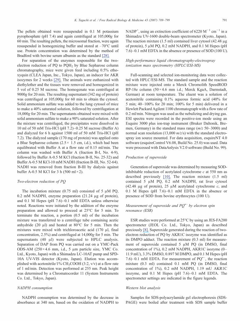
Fig. 1. Assay for two-electron reduction of PQ. (A) Enzymatic two-electron reducperformance liquid chromatograms of enzymatic reaction mixtures of PQ. Peak 1, PQsystem; b, +NADH/–NADPH; c, –cytosol; d, –NADPH; e, –acetic anhydride; f, –0.2 mMNADPH, and 0.1 M Hepes (pH 7.6)–0.1 mM EDTA at 25°C for 5 min. Determethods. (C) Positive-ion electrospray mass spectra of authentic DAP (a) and the d
taining 4.5 g/L of D-glucose, 100 U/ml penicillin, 100 μg/mlstreptomycin, 2 mM GlutaMAX-I supplement, and 10% (v/v)heat-inactivated fetal bovine serum at 37°C under 95% air plus5% CO2.
The 3-(4,5-dimethylthiazol-2-yl)-2,5-diphenyltetrazoliumbromide (MTT) assay [31] was used to estimate the cytotoxicityof PQ or DAP. Proliferating A549 cells were exposed to PQ orDAP (1–20 μM) for 12 h in 96-well microtiter plates and thentreated with 5 mg/ml MTT (1/20 (v/v)) for 4 h at 37°C. Afterremoval of the medium, DMSO (100 μl/well) was added todissolve cells. Absorbance at 540 nm was measured by anImmunoMini NJ-2300 plate reader (Nippon InterMed, Tokyo,Japan).
Determination of oxidatively modified proteins
After the treatment of A549 cells with PQ or DAP(10 μM), total cellular proteins were solubilized in SDS
tion of PQ to PQH2 which is identified as its diacetoxy derivative. (B) High-(retention time, 6.8 min); peak 2, DAP (retention time, 10.6 min). a, complete
oxygen. Rat liver cytosol (21.24 μg of protein) was incubated with 5 μM PQ,mination of DAP was performed under conditions described under Materials andiacetoxy derivative from enzymatic reducing product of PQ (b).

Fig. 2. (A) Material balance study during the reaction of PQ with rat livercytosol. (B) Concentration-dependent PQH2 formation during the reaction ofPQ with rat liver cytosol. Rat liver cytosol (21.24 μg of protein) was incubatedwith 5 μM PQ, 0.2 mM NADPH, and 0.1 M Hepes (pH 7.6)–0.1 mM EDTA at25°C for 5 min. Determination of DAP was performed under conditionsdescribed under Materials and methods. Each value is the mean ± SD of threedeterminations.
793K. Taguchi et al. / Free Radical Biology & Medicine 43 (2007) 789–799
sample buffer (50 mM Tris-HCl (pH 6.8)–10% glycerol–2%SDS). Cellular proteins were reacted with DNPH to derivatizecarbonyl groups and then subjected to SDS-PAGE (20 μg ofprotein) followed by western blotting with anti-DNP, bio-tinated anti-IgE antibodies, and streptavidin HRP conjugate[32].
Determination of LD50 value
Values obtained from MTT assay were analyzed by anonlinear regression program using PRISM version 3.0(GraphPad Software, Inc., San Diego) to calculate LD50 ± SEvalues.
Results
Identification of PQH2 as its diacetoxy derivative
Fig. 1A shows the reaction sequence in the derivatization ofthe two-electron reduction product of PQ for HPLC analysis.Incubation of PQ (peak 1; retention time, 6.8 min) with cytosolfrom rat liver in the presence of NAD(P)H resulted in aproduct indicated as peak 2 with a retention time of 10.6 min(Figs. 1B, a and b), which has a mass spectrum identical toauthentic DAP (Fig. 1C, b). No DAP was seen when eithercytosol or NADPH was omitted from the incubation mixture(Figs. 1B, c and d). When acetic anhydride was not added tothe reaction mixture, DAP was not detected (Fig. 1B, e). Underanaerobic conditions, the peak of DAP increased (1.8-fold)compared to aerobic conditions (Fig. 1B, f), suggesting thatPQH2 is highly sensitive to molecular oxygen. Addition of asuperoxide generating system (0.5 mM xanthine + 0.4 μMxanthine oxidase) to an incubation mixture of PQ and cytosoldecreased the DAP peak (43% of control), and increased thePQ peak (data not shown), indicating that PQH2 is oxidized toPQ by superoxide. The ESI mass spectra of both compoundswhich were eluted at 18.7 min under HPLC conditions with aMerck Chromolith SpeedROD RP-18e column gave amolecular ion peak at m/z of 316.8, based on a monosodiumsalt ion, [M+Na]+. These results demonstrate that PQ under-went a two-electron reduction to yield PQH2 enzymaticallyand that PQH2 was, in turn, converted to its diacetoxyderivative DAP with acetic anhydride. The standard curvegenerated from the peak area and the injected DAP quantitywas linear (r2 =0.999) from 1 to 7.5 μM and similar resultswere observed with PQ (data not shown). Although theproduction of PQH2 as determined by DAP and PQ remainingwas dependent on the enzyme preparation added, the totalamounts of PQH2 formation and PQ remaining were almostthe same (Fig. 2A), supporting that PQH2 formed from PQ wassuccessfully converted into DAP without further metabolism.In rat liver, PQH2 formation was much higher in cytosol thanin either microsomes (10.3%) or mitochondria (16.7%;percentage of PQH2 formation in liver cytosol). The PQH2
formation was the highest in cytosol from liver, followed bycytosol from brain (78%), kidney (73%), and lung (72%), andwas minimal in the cytosol from testis (28%), heart (18%), and
spleen (14%; percentage of PQH2 formation in liver cytosol).When PQ was incubated with rat liver cytosol in the presenceof NADPH, PQH2 formation reached a plateau and thendeclined at PQ concentrations of greater than 4 μM (Fig. 2B).This suggests that a disproportionation reaction of quinonesand their hydroquinone to yield the semiquinone radicals asshown by Kalyanaraman et al. [33], at least in part, occursbecause this unstable intermediate undergoes readily oxidationby molecular oxygen to produce PQ.
Generation of ROS during reduction of PQ by rat liver cytosol
When PQ (7.5 nmol) was incubated with rat liver cytosolin the presence of an excess amount of NADPH (300 nmol)at 25°C for 5 min, PQH2 (2.8 nmol) and superoxide(18.0 nmol) were produced, while excess consumption ofNADPH (50.2 nmol) was detected. In the presence of SOD(100 U), PQ added was completely converted to PQH2 andNADPH consumption was decreased by 18.3%. Unlike SOD,catalase (100 or 1000 U) and mannitol (100 or 500 mM),which are scavengers for H2O2 and ·OH, respectively, had noeffect on PQH2 formation (data not shown).
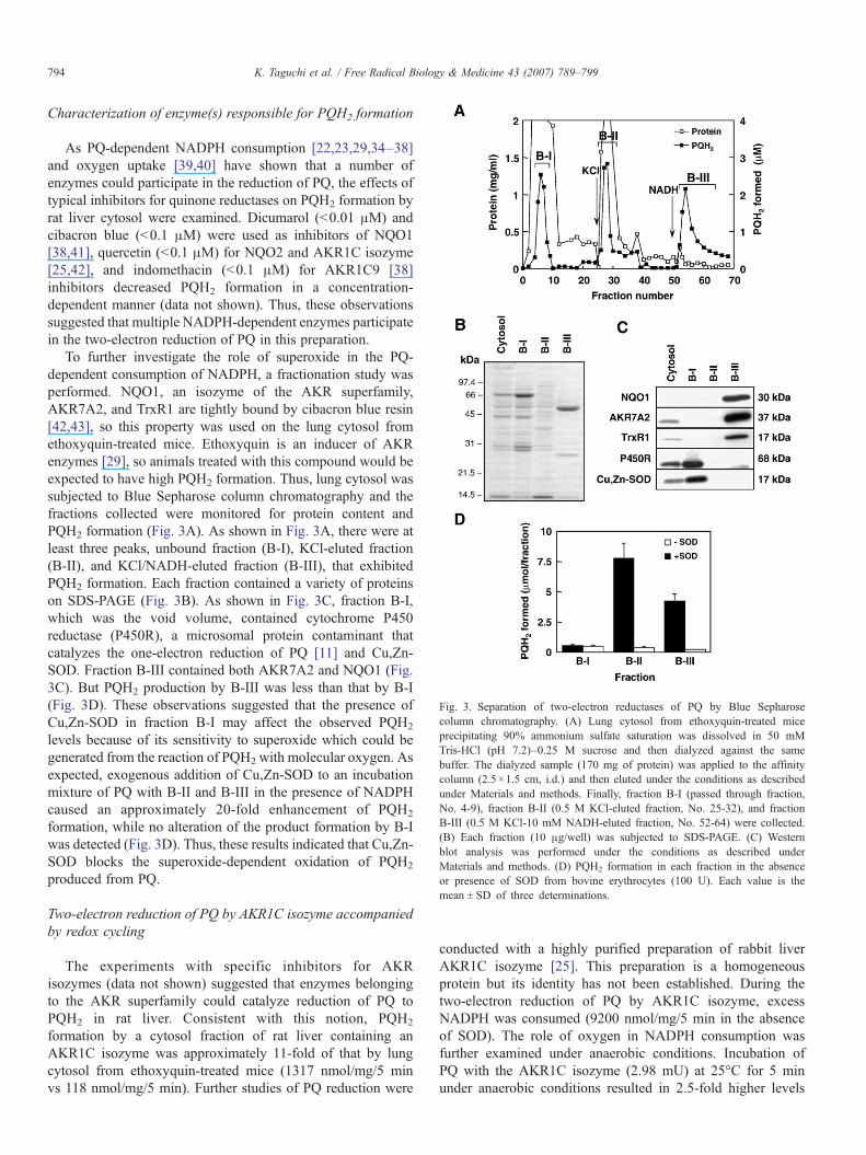
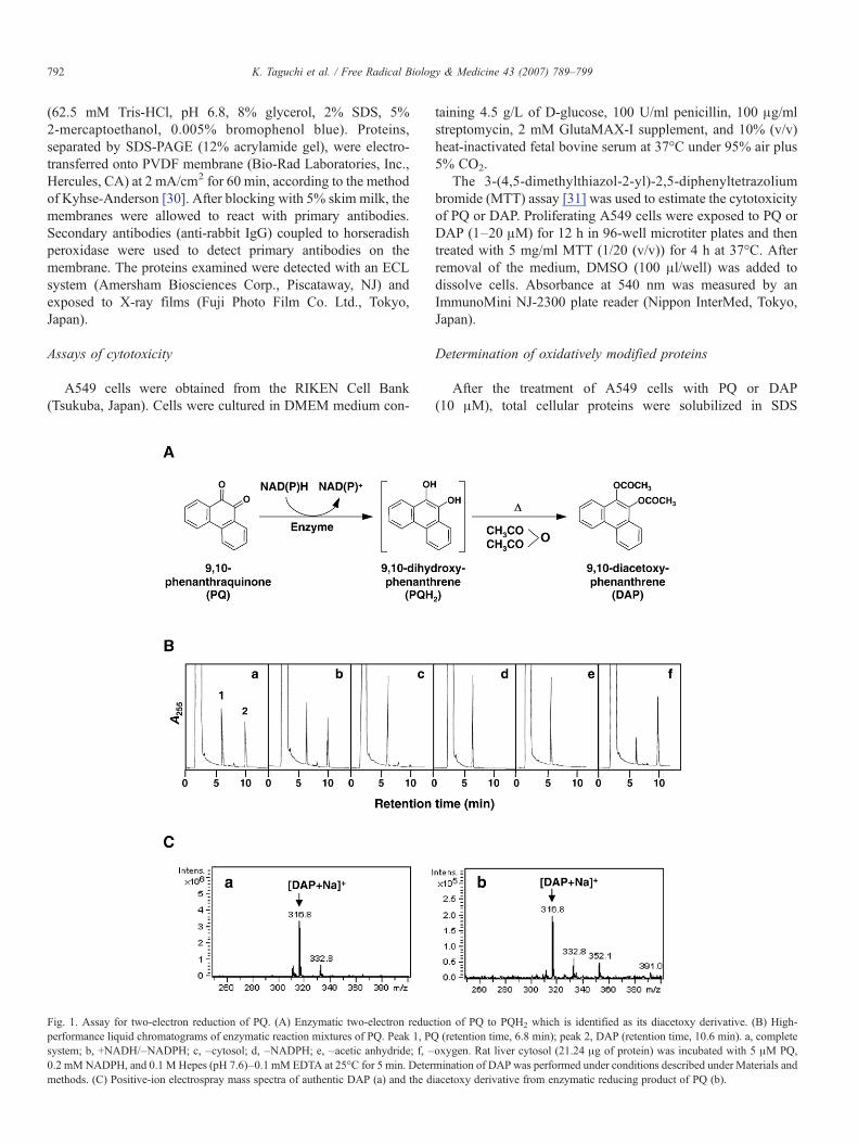
Fig. 3. Separation of two-electron reductases of PQ by Blue Sepharosecolumn chromatography. (A) Lung cytosol from ethoxyquin-treated miceprecipitating 90% ammonium sulfate saturation was dissolved in 50 mMTris-HCl (pH 7.2)–0.25 M sucrose and then dialyzed against the samebuffer. The dialyzed sample (170 mg of protein) was applied to the affinitycolumn (2.5×1.5 cm, i.d.) and then eluted under the conditions as describedunder Materials and methods. Finally, fraction B-I (passed through fraction,No. 4-9), fraction B-II (0.5 M KCl-eluted fraction, No. 25-32), and fractionB-III (0.5 M KCl-10 mM NADH-eluted fraction, No. 52-64) were collected.(B) Each fraction (10 μg/well) was subjected to SDS-PAGE. (C) Westernblot analysis was performed under the conditions as described underMaterials and methods. (D) PQH2 formation in each fraction in the absenceor presence of SOD from bovine erythrocytes (100 U). Each value is themean ± SD of three determinations.
794 K. Taguchi et al. / Free Radical Biology & Medicine 43 (2007) 789–799
Characterization of enzyme(s) responsible for PQH2 formation
As PQ-dependent NADPH consumption [22,23,29,34–38]and oxygen uptake [39,40] have shown that a number ofenzymes could participate in the reduction of PQ, the effects oftypical inhibitors for quinone reductases on PQH2 formation byrat liver cytosol were examined. Dicumarol (b0.01 μM) andcibacron blue (b0.1 μM) were used as inhibitors of NQO1[38,41], quercetin (b0.1 μM) for NQO2 and AKR1C isozyme[25,42], and indomethacin (b0.1 μM) for AKR1C9 [38]inhibitors decreased PQH2 formation in a concentration-dependent manner (data not shown). Thus, these observationssuggested that multiple NADPH-dependent enzymes participatein the two-electron reduction of PQ in this preparation.
To further investigate the role of superoxide in the PQ-dependent consumption of NADPH, a fractionation study wasperformed. NQO1, an isozyme of the AKR superfamily,AKR7A2, and TrxR1 are tightly bound by cibacron blue resin[42,43], so this property was used on the lung cytosol fromethoxyquin-treated mice. Ethoxyquin is an inducer of AKRenzymes [29], so animals treated with this compound would beexpected to have high PQH2 formation. Thus, lung cytosol wassubjected to Blue Sepharose column chromatography and thefractions collected were monitored for protein content andPQH2 formation (Fig. 3A). As shown in Fig. 3A, there were atleast three peaks, unbound fraction (B-I), KCl-eluted fraction(B-II), and KCl/NADH-eluted fraction (B-III), that exhibitedPQH2 formation. Each fraction contained a variety of proteinson SDS-PAGE (Fig. 3B). As shown in Fig. 3C, fraction B-I,which was the void volume, contained cytochrome P450reductase (P450R), a microsomal protein contaminant thatcatalyzes the one-electron reduction of PQ [11] and Cu,Zn-SOD. Fraction B-III contained both AKR7A2 and NQO1 (Fig.3C). But PQH2 production by B-III was less than that by B-I(Fig. 3D). These observations suggested that the presence ofCu,Zn-SOD in fraction B-I may affect the observed PQH2
levels because of its sensitivity to superoxide which could begenerated from the reaction of PQH2 with molecular oxygen. Asexpected, exogenous addition of Cu,Zn-SOD to an incubationmixture of PQ with B-II and B-III in the presence of NADPHcaused an approximately 20-fold enhancement of PQH2
formation, while no alteration of the product formation by B-Iwas detected (Fig. 3D). Thus, these results indicated that Cu,Zn-SOD blocks the superoxide-dependent oxidation of PQH2
produced from PQ.
Two-electron reduction of PQ by AKR1C isozyme accompaniedby redox cycling
The experiments with specific inhibitors for AKRisozymes (data not shown) suggested that enzymes belongingto the AKR superfamily could catalyze reduction of PQ toPQH2 in rat liver. Consistent with this notion, PQH2
formation by a cytosol fraction of rat liver containing anAKR1C isozyme was approximately 11-fold of that by lungcytosol from ethoxyquin-treated mice (1317 nmol/mg/5 minvs 118 nmol/mg/5 min). Further studies of PQ reduction were
conducted with a highly purified preparation of rabbit liverAKR1C isozyme [25]. This preparation is a homogeneousprotein but its identity has not been established. During thetwo-electron reduction of PQ by AKR1C isozyme, excessNADPH was consumed (9200 nmol/mg/5 min in the absenceof SOD). The role of oxygen in NADPH consumption wasfurther examined under anaerobic conditions. Incubation ofPQ with the AKR1C isozyme (2.98 mU) at 25°C for 5 minunder anaerobic conditions resulted in 2.5-fold higher levels
795K. Taguchi et al. / Free Radical Biology & Medicine 43 (2007) 789–799
of PQH2 than those obtained under aerobic conditions, sug-gesting that PQH2 rapidly undergoes autoxidation to PQ asconfirmed by HPLC (see Fig. 1B, f). This notion was furthersupported by a purified AKR1C isozyme concentration-dependent oxygen consumption as determined with anoxygen electrode during the two-electron reduction of PQ(5 μM) by AKR1C isozyme in the presence of 0.2 mMNADPH (data not shown). To demonstrate that a semiquinoneradical species (PQU−) was an intermediate in the superoxide-dependent formation of PQ and PQH2, an ESR study de-termining the presence of radicals was conducted. Generationof superoxide was determined by its adduct, DMPO-OOH,during the two-electron reduction of PQ under aerobicconditions (Fig. 4A). Addition of SOD (100 U/mL) quenchedgeneration of superoxide (data not shown). In Fig. 4B, anAKR1C isozyme concentration-dependent generation of su-peroxide was quantified. An equivalent incubation conductedin the absence of oxygen resulted in the appearance of PQU−(g-value=2.00395) as identified in Valavanidis et al. [43](Fig. 4C).
Fig. 4. Generation of superoxide (A, B) and semiquinone radical (C) by two-electron reduction of PQ. (A) ESR spectra. AKR1C isozyme (0 or 5.97 mU)was reacted with 5 μM PQ, 0.2 mM NADPH, 3.3% DMSO, 0.897 M DMPO,and 0.1 M Hepes (pH 7.6)–0.1 mM EDTA. The instrument settings were asfollows: modulation width, 100 kHz–0.1 mT; sweep time, 4 min; time constant,0.3 s; microwave power, 7.99 mW; microwave frequency, 9417 MHz; magneticfield, 335.4±5 mT. Closed circle indicates DMPO-OOH adduct. (B) Enzymeconcentration-dependent superoxide generation. The area (*) was quantified.(C) AKR1C isozyme (1.19 mU) was reacted with 0.2 mM NADPH, 0.1 mMPQ, and 0.1 M Hepes (pH 7.6)–0.1 mM EDTA. PQ was dissolved in DMSO(final concentration of 1%). The instrument settings were as follows:modulation width, 100 kHz–0.4 mT; sweep time, 10 s; time constant, 0.01 s;microwave power, 7.99 mW; microwave frequency, 9417 MHz; magnetic field,335.4±5 mT.
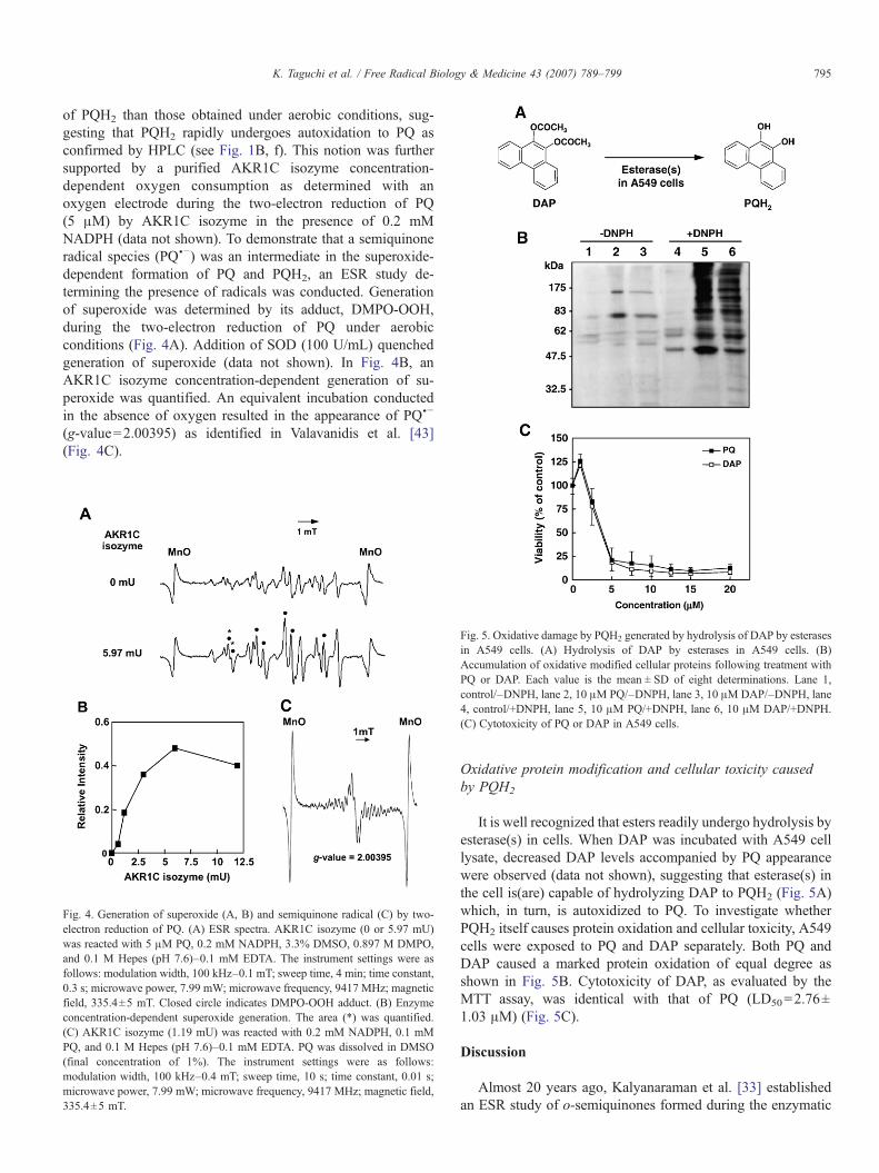
Fig. 5. Oxidative damage by PQH2 generated by hydrolysis of DAP by esterasesin A549 cells. (A) Hydrolysis of DAP by esterases in A549 cells. (B)Accumulation of oxidative modified cellular proteins following treatment withPQ or DAP. Each value is the mean ± SD of eight determinations. Lane 1,control/–DNPH, lane 2, 10 μM PQ/–DNPH, lane 3, 10 μMDAP/–DNPH, lane4, control/+DNPH, lane 5, 10 μM PQ/+DNPH, lane 6, 10 μM DAP/+DNPH.(C) Cytotoxicity of PQ or DAP in A549 cells.
Oxidative protein modification and cellular toxicity causedby PQH2
It is well recognized that esters readily undergo hydrolysis byesterase(s) in cells. When DAP was incubated with A549 celllysate, decreased DAP levels accompanied by PQ appearancewere observed (data not shown), suggesting that esterase(s) inthe cell is(are) capable of hydrolyzing DAP to PQH2 (Fig. 5A)which, in turn, is autoxidized to PQ. To investigate whetherPQH2 itself causes protein oxidation and cellular toxicity, A549cells were exposed to PQ and DAP separately. Both PQ andDAP caused a marked protein oxidation of equal degree asshown in Fig. 5B. Cytotoxicity of DAP, as evaluated by theMTT assay, was identical with that of PQ (LD50=2.76±1.03 μM) (Fig. 5C).
Discussion
Almost 20 years ago, Kalyanaraman et al. [33] establishedan ESR study of o-semiquinones formed during the enzymatic
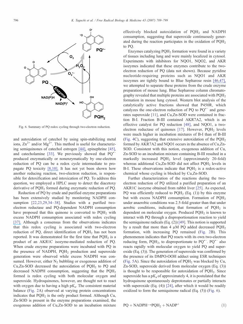
Fig. 6. Summary of PQ redox cycling through two-electron reduction.
796 K. Taguchi et al. / Free Radical Biology & Medicine 43 (2007) 789–799
and autoxidation of catechol by using spin-stabilizing metalions, Zn2+ and/or Mg2+. This method is useful for characteriz-ing semiquinones of catechol estrogen [44], epinephrine [45],and catecholamine [33]. We previously showed that PQU−produced enzymatically or nonenzymatically by one-electronreduction of PQ can be a redox cycle intermediate to pro-pagate PQ toxicity [8,10]. It has not yet been shown howanother reducing reaction, two-electron reduction, is respon-sible for detoxification and intoxication of PQ. To address thisquestion, we employed a HPLC assay to detect the diacetoxyderivative of PQH2 formed during enzymatic reduction of PQ.
Reduction of PQ by crude and purified enzyme preparationshas been extensively studied by monitoring NADPH con-sumption [22,23,29,34–38]. Studies with a purified two-electron reductase and PQ-dependent NADPH consumptionhave proposed that this quinone is converted to PQH2 withexcess NADPH consumption associated with redox cycling[37]. Although a consensus from the observations indicatesthat this redox cycling is associated with two-electronreduction of PQ, direct identification of PQH2 has not beenreported. It was demonstrated for the first time that PQH2 is aproduct of an AKR1C isozyme-mediated reduction of PQ.When crude enzyme preparations were incubated with PQ inthe presence of NADPH, PQH2 production and superoxidegeneration were observed while excess NADPH was con-sumed. However, either N2 bubbling or exogenous addition ofCu,Zn-SOD decreased the autoxidation of PQH2 to PQ anddecreased NADPH consumption, suggesting that the PQH2
formed is redox cycling with both molecular oxygen andsuperoxide. Hydroquinones, however, are thought not to reactwith oxygen due to having a high pKa. The consistent materialbalance (Fig. 2A) observed at varying protein concentrationsindicates that PQH2 is the only product formed. Although Cu,Zn-SOD is present in the enzyme preparations examined, theexogenous addition of Cu,Zn-SOD to an incubation mixture
effectively blocked autoxidation of PQH2 and NADPHconsumption, suggesting that superoxide continuously gener-ated during the reaction participates in the oxidation of PQH2
to PQ.Enzymes catalyzing PQH2 formation were found in a variety
of tissues including lung and were mainly localized in cytosol.Experiments with inhibitors for NQO1, NQO2, and AKRisozymes indicated that these enzymes contribute to the two-electron reduction of PQ (data not shown). Because pyridinenucleotide-requiring proteins such as NQO1 and AKRisozymes are tightly bound to Blue Sepharose resin [46,47],we attempted to separate these proteins from the crude enzymepreparation of mouse lung. Blue Sepharose column chromato-graphy revealed that multiple proteins are associated with PQH2
formation in mouse lung cytosol. Western blot analysis of thecatalytically active fractions showed that P450R, whichcatalyzes the one-electron reduction of PQ to PQU− and gene-rates superoxide [11], and Cu,Zn-SOD were contained in frac-tion B-I. Fraction B-III contained AKR7A2, which is aneffective catalyst for PQ reduction [48], and NQO1, a two-electron reductase of quinones [17]. However, PQH2 levelswere much higher in incubation mixtures of B-I than of B-III(Fig. 4C), suggesting that extensive autoxidation of the PQH2
formed by AKR7A2 and NQO1 occurs in the absence of Cu,Zn-SOD. Consistent with this notion, exogenous addition of Cu,Zn-SOD to an incubation mixture containing B-III resulted in amarkedly increased PQH2 level (approximately 20-fold)whereas additional Cu,Zn-SOD did not affect PQH2 levels inB-I. These observations indicate that PQH2 is a redox-activechemical whose cycling is blocked by Cu,Zn-SOD.
Further characterization of the reactions during the two-electron reduction of PQ utilized a purified preparation of anAKR1C isozyme obtained from rabbit liver [25]. As expected,PQ was efficiently reduced to PQH2 (Eq. (1)) by this enzymebut with excess NADPH consumption. Formation of PQH2
under anaerobic conditions was 2.5-fold greater than that underaerobic conditions, indicating that formation of PQH2 isdependent on molecular oxygen. Produced PQH2 is known tointeract with PQ through a disproportionation reaction to yieldthe semiquinone radicals (Eq. (2)) [49]. This notion is supportedby a result that more than 4 μM PQ added decreased PQH2
formation, with increasing PQ remained (Fig. 2B). Thisphenomenon indicates that PQ reacts with its own two-electronreducing form, PQH2, to disproportionate to PQ
U−. PQU− alsoreacts rapidly with molecular oxygen to yield PQ and super-oxide (Eq. (3)). The generation of superoxide was confirmed bythe presence of its DMPO-OOH adduct using ESR techniques(Fig. 5A). Since the autoxidation of PQH2 was blocked by Cu,Zn-SOD, superoxide derived from molecular oxygen (Eq. (3))is thought to be responsible for autoxidation of PQH2. Sincesuperoxide has a pKa of approximately 4, it is postulated that thehydroquinone spontaneously deprotonates or partially interactswith superoxide (Eq. (4)) [24], after which it would be readilyoxidized to form the semiquinone radical (Eq. (5)) (Fig. 6).
PQþ NADPHYPQH2 þ NADPþ ð1Þ

797K. Taguchi et al. / Free Radical Biology & Medicine 43 (2007) 789–799
PQH2 þ PQ±2PQU� þ 2Hþ ð2Þ
PQU� þ O2YPQþ OU�2 ð3Þ
PQH2 þ OU�2 YPQH� þ HO
U2 ð4Þ
PQH� þ HOU2 YPQ
U� þ H2O2 ð5Þ
The semiquinone radical disproportionates readily to quinoneand hydroquinone (Eq. (2)) [37]. Superoxide formed during thereduction of PQ is chemically (Eq. (6)) and/or enzymatically(Eqs. (7) and (8)) disproportionated to hydrogen peroxide. Byeliminating superoxide, Cu,Zn-SOD terminates the autoxida-tion of PQH2 (see Fig. 3D).
OU�2 þ O
U�2 2HþYH2O2 þ O2 ð6Þ
SODQCu2þ þ OU�2 YSOD� Cuþ þ O2 ð7Þ
SODQCuþ þ OU�2 þ 2HþYSODQCu2þ þ H2O2 ð8Þ
We have recently reported that exposure of A549 cells to PQcaused a marked oxidation of cellular proteins evaluated byprotein carbonyl formation, together with selective down-regulation of Cu,Zn-SOD [11]. Palackal et al. [50] reportedpreviously that AKR1C isozymes are highly expressed in A549cells, and in preliminary experiments, we found that A549 celllysate is capable of producing PQH2 from PQ (S. Fujii et al.,unpublished observation). Moreover, experiments with DAP asa precursor of PQH2 indicated strongly that PQH2 itself plays amajor role in the oxidative protein modification. Taken together,the oxidative protein damage in A549 cells caused by PQ couldbe explained by the initial two-electron reduction of PQ toPQH2 followed by oxidation of the latter to the semiquinone byoxygen to generate superoxide. The superoxide can then act topropagate the redox cycling process by overwhelming theinherent SOD.
Cadenas [19] previously classified hydroquinones, the two-electron reduction products of quinones: (i) redox-stablehydroquinones (e.g., 2-methyl-1,4-naphthoquinone) [17], (ii)redox-labile hydroquinones (e.g., 2-methyl-3-glutathionyl-1,4-naphthoquinone) [51], and (iii) alkylating hydroquinones (e.g.,AZQ) [52]. Redox-labile hydroquinones (ii) are autoxidized toundergo redox cycling, whereas redox-stable hydroquinones (i)are conjugated with sulfate and glucuronate, resulting in theirdetoxification. Alkylating hydroquinones (iii) yield alkylationproducts (e.g., DNA-quinone) by bioreductive alkylation,leading to toxicity. Autoxidation of redox-labile hydroquinones(ii) can be inhibited (ii-a) or stimulated by SOD (ii-b), bystabilizing redox unstable hydroquinones or generating hydro-gen peroxide, respectively [19]. From the present findings, weconclude that PQ is classified as (ii-a).
In conclusion, this is the first report to identify PQH2 as theproduct of a NADPH-dependent two-electron reduction of PQ.The direct assay of PQH2 allowed us to determine thestoichiometric relationship between NADPH consumption and
PQ reduction. Although previous studies based on NADPHconsumption have demonstrated that PQ is a good substratefor NQO1 [34], AKR1C9 [38,53], AKR1C12 [54], AKR1C13[54], AKR7A1 [55], AKR7A2 [48], AKR7A3 [55], andAKR7A5 [56], they were missing the mechanistic details ofthe redox cycling of PQ to PQH2 that are associated with oxi-dative stress by these enzymes. The present study suggests thatreduction of PQ to PQH2 by AKR isozymes or NQO1 can alsocause oxidative protein damage due to PQ exposure.
Acknowledgments
We thank Dr. John D. Hayes of the Biomedical ResearchCentre, Ninewells Hospital and Medical School, University ofDundee, UK, for providing antibody against AKR7A2 isozyme;Dr. Nobuyuki Koga of the Faculty of Nutritional Sciences,Nakamura Gakuen University, Japan, for donation of antibodyagainst NQO1; and Dr. Nobuhiro Shimojo of the Department ofEnvironmental Medicine, Institute of Community Medicine,University of Tsukuba, Japan, for his encouragement, and M.S.Rie Koizumi and M.S. Yuya Nishihara of the Master’s Programin Environmental Sciences, University of Tsukuba, for theirexcellent contribution to this study. We are especially grateful toMs. F. Miyamasu for grammatical correction in the preparationof the manuscript. This research was supported in part by aGrant-in-Aid Nos. 15390184, 18659167, and 18406003 (Y.K.)for scientific research from the Ministry of Education, Science,Culture, and Sports of Japan. K.T. was awarded an Ishidu ShunMemorial Scholarship. Although the research described in thisarticle has been funded in part by the United StatesEnvironmental Protection Agency through Grant R827352-01-0 to UCLA, it has not been subjected to the agency’srequired peer and policy review and therefore does notnecessarily reflect the views of the agency and no officialendorsement should be inferred.
References
[1] Sakakibara, M.; Minami, M.; Endo, T.; Hirafuji, M.; Murakami, S.;Mori, Y.; Sagai, M. Biological effects of diesel exhaust particles (DEP)on isolated cardiac muscle of guinea pigs. Res. Commun. Mol. Pathol.Pharmacol. 86:99–110; 1994.
[2] Diaz-Sanchez, D. The role of diesel exhaust particles and their associatedpolyaromatic hydrocarbons in the induction of allergic airway disease.Allergy 52:52–56; discussion 57–58; 1997.
[3] McClellan, R. O. Health effects of exposure to diesel exhaust particles.Annu. Rev. Pharmacol. Toxicol. 27:279–300; 1987.
[4] Xiao, G. G.; Wang, M.; Li, N.; Loo, J. A.; Nel, A. E. Use of proteomics todemonstrate a hierarchical oxidative stress response to diesel exhaust par-ticle chemicals in a macrophage cell line. J. Biol. Chem. 278:50781–50790;2003.
[5] Hirano, S.; Furuyama, A.; Koike, E.; Kobayashi, T. Oxidative-stresspotency of organic extracts of diesel exhaust and urban fine particles in ratheart microvessel endothelial cells. Toxicology 187:161–170; 2003.
[6] Schuetzle, D. Sampling of vehicle emissions for chemical analysis andbiological testing. Environ. Health Perspect. 47:65–80; 1983.
[7] Li, N.; Venkatesan, M. I.; Miguel, A.; Kaplan, R.; Gujuluva, C.; Alam, J.;Nel, A. Induction of heme oxygenase-1 expression in macrophages bydiesel exhaust particle chemicals and quinones via the antioxidant-responsive element. J. Immunol. 165:3393–3401; 2000.
798 K. Taguchi et al. / Free Radical Biology & Medicine 43 (2007) 789–799
[8] Kumagai, Y.; Arimoto, T.; Shinyashiki, M.; Shimojo, N.; Nakai, Y.;Yoshikawa, T.; Sagai, M. Generation of reactive oxygen species duringinteraction of diesel exhaust particle components with NADPH-cyto-chrome P450 reductase and involvement of the bioactivation in the DNAdamage. Free Radic. Biol. Med. 22:479–487; 1997.
[9] Cho, A. K.; Di Stefano, E.; You, Y.; Rodriguez, C. E.; Schmitz, D. A.;Kumagai, Y.; Miguel, A. H.; Eiguren-Fernandez, A.; Kobayashi, T.; Avol,E.; Froines, J. Determination of four quinones in diesel exhaust particles,SRM 1649a, and atmospheric PM2.5. Aerosol Sci. Tech. 38:1–14; 2004.
[10] Kumagai, Y.; Koide, S.; Taguchi, K.; Endo, A.; Nakai, Y.; Yoshikawa, T.;Shimojo, N. Oxidation of proximal protein sulfhydryls by phenanthraqui-none, a component of diesel exhaust particles. Chem. Res. Toxicol. 15:483–489; 2002.
[11] Sugimoto, R.; Kumagai, Y.; Nakai, Y.; Ishii, T. 9,10-Phenanthraquinone indiesel exhaust particles downregulates Cu,Zn-SOD and HO-1 in humanpulmonary epithelial cells: intracellular iron scavenger 1,10-phenanthro-line affords protection against apoptosis. Free Radic. Biol. Med. 38:388–395; 2005.
[12] Rodriguez, C. E.; Shinyashiki, M.; Froines, J.; Yu, R. C.; Fukuto, J. M.;Cho, A. K. An examination of quinone toxicity using the yeastSaccharomyces cerevisiae model system. Toxicology 201:185–196; 2004.
[13] Rodriguez, C. E.; Fukuto, J. M.; Taguchi, K.; Froines, J.; Cho, A. K. Theinteractions of 9,10-phenanthrenequinone with glyceraldehyde-3-phos-phate dehydrogenase (GAPDH), a potential site for toxic actions. Chem.Biol. Interact. 155:97–110; 2005.
[14] Hiyoshi, K.; Takano, H.; Inoue, K.; Ichinose, T.; Yanagisawa, R.; Tomura,S.; Cho, A. K.; Froines, J. R.; Kumagai, Y. Effects of a single intratrachealadministration of phenanthraquinone on murine lung. J. Appl. Toxicol.25:47–51; 2005.
[15] Kumagai, Y.; Hayashi, T.; Miyauchi, T.; Endo, A.; Iguchi, A.; Kiriya-Sakai, M.; Sakai, S.; Yuki, K.; Kikushima, M.; Shimojo, N. Phenan-thraquinone inhibits eNOS activity and suppresses vasorelaxation. Am. J.Physiol. Regul. Integr. Comp. Physiol. 281:R25–R30; 2001.
[16] Kumagai, Y.; Nakajima, H.; Midorikawa, K.; Homma-Takeda, S.;Shimojo, N. Inhibition of nitric oxide formation by neuronal nitric oxidesynthase by quinones: nitric oxide synthase as a quinone reductase. Chem.Res. Toxicol. 11:608–613; 1998.
[17] Lind, C.; Hochstein, P.; Ernster, L. DT-diaphorase as a quinone reductase: acellular control device against semiquinone and superoxide radicalformation. Arch. Biochem. Biophys. 216:178–185; 1982.
[18] Cadenas, E.; Hochstein, P.; Ernster, L. Pro- and antioxidant functions ofquinones and quinone reductases in mammalian cells. Adv. Enzymol.Relat. Areas Mol. Biol. 65:97–146; 1992.
[19] Cadenas, E. Antioxidant and prooxidant functions of DT-diaphorase inquinone metabolism. Biochem. Pharmacol. 49:127–140; 1995.
[20] Bucker, M.; Glatt, H. R.; Platt, K. L.; Avnir, D.; Ittah, Y.; Blum, J.; Oesch,F. Mutagenicity of phenanthrene and phenanthrene K-region derivatives.Mutat. Res. 66:337–348; 1979.
[21] Siegel, D.; Gibson, N. W.; Preusch, P. C.; Ross, D. Metabolism of diazi-quone by NAD(P)H:(quinone acceptor) oxidoreductase (DT-diaphorase):role in diaziquone-induced DNA damage and cytotoxicity in human coloncarcinoma cells. Cancer Res. 50:7293–7300; 1990.
[22] Jarabak, R.; Jarabak, J. Effect of ascorbate on the DT-diaphorase-mediatedredox cycling of 2-methyl-1,4-naphthoquinone. Arch. Biochem. Biophys.318:418–423; 1995.
[23] Klein, J.; Post, K.; Seidel, A.; Frank, H.; Oesch, F.; Platt, K. L. Quinonereduction and redox cycling catalysed by purified rat liver dihydrodiol/3alpha-hydroxysteroid dehydrogenase. Biochem. Pharmacol. 44:341–349;1992.
[24] Hiramatsu, M.; Kumagai, Y.; Unger, S. E.; Cho, A. K. Metabolism ofmethylenedioxymethamphetamine: formation of dihydroxymethampheta-mine and a quinone identified as its glutathione adduct. J. Pharmacol. Exp.Ther. 254:521–527; 1990.
[25] Yamano, S.; Ichinose, F.; Todaka, T.; Toki, S. Purification and char-acterization of two major forms of naloxone reductase from rabbit livercytosol, new members of aldo-keto reductase superfamily. Biol. Pharm.Bull. 22:1038–1046; 1999.
[26] Shimojo, N.; Kumagai, Y.; Homma-Takeda, S.; Shinyashiki, M.;
Takasawa, N.; Kushida, K. Isozyme selective induction of mouse pulmo-nary superoxide dismutase by the exposure to mercury vapor. Environ.Toxicol. Pharmacol. 2:35–37; 1996.
[27] Kumagai, Y.; Tsurutani, Y.; Shinyashiki, M.; Homma-Takeda, S.; Nakai,Y.; Yoshikawa, T.; Shimojo, N. Bioactivation of lapachol responsible forDNAS scission by NADPH-cytochrome P450 reductase. Environ. Toxicol.Pharm. 3:245–250; 1997.
[28] Bradford, M. M. A rapid and sensitive method for the quantitation ofmicrogram quantities of protein utilizing the principle of protein-dyebinding. Anal. Biochem. 72:248–254; 1976.
[29] Ellis, E. M.; Judah, D. J.; Neal, G. E.; O’Connor, T.; Hayes, J. D.Regulation of carbonyl-reducing enzymes in rat liver by chemoprotectors.Cancer Res. 56:2758–2766; 1996.
[30] Kyhse-Andersen, J. Electroblotting of multiple gels: a simple apparatuswithout buffer tank for rapid transfer of proteins from polyacrylamide tonitrocellulose. J. Biochem. Biophys. Methods 10:203–209; 1984.
[31] Denizot, F.; Lang, R. Rapid colorimetric assay for cell growth and survival.Modifications to the tetrazolium dye procedure giving improved sensitivityand reliability. J. Immunol. Methods 89:271–277; 1986.
[32] Levine, R. L.; Williams, J. A.; Stadtman, E. R.; Shacter, E. Carbonylassays for determination of oxidatively modified proteins. MethodsEnzymol. 233:346–357; 1994.
[33] Kalyanaraman, B.; Felix, C. C.; Sealy, R. C. Peroxidatic oxidation ofcatecholamines. A kinetic electron spin resonance investigation using thespin stabilization approach. J. Biol. Chem. 259:7584–7589; 1984.
[34] Chesis, P. L.; Levin, D. E.; Smith, M. T.; Ernster, L.; Ames, B. N.Mutagenicity of quinones: pathways of metabolic activation anddetoxification. Proc. Natl. Acad. Sci. USA 81:1696–1700; 1984.
[35] Wermuth, B.; Platts, K. L.; Seidel, A.; Oesch, F. Carbonyl reductaseprovides the enzymatic basis of quinone detoxication in man. Biochem.Pharmacol. 35:1277–1282; 1986.
[36] Jarabak, J.; Harvey, R. G. Studies on three reductases which havepolycyclic aromatic hydrocarbon quinones as substrates. Arch. Biochem.Biophys. 303:394–401; 1993.
[37] Jarabak, R.; Harvey, R. G.; Jarabak, J. Redox cycling of polycyclic aromatichydrocarbon o-quinones: metal ion-catalyzed oxidation of catecholsbypasses inhibition by superoxide dismutase. Chem. Biol. Interact. 115:201–213; 1998.
[38] Del Bello, B.; Maellaro, E.; Sugherini, L.; Santucci, A.; Comporti, M.;Casini, A. F. Purification of NADPH-dependent dehydroascorbatereductase from rat liver and its identification with 3 alpha-hydroxysteroiddehydrogenase. Biochem. J. 304 (Pt 2):385–390; 1994.
[39] Munday, R. Concerted action of DT-diaphorase and superoxide dismutasein preventing redox cycling of naphthoquinones: an evaluation. FreeRadic. Res. 35:145–158; 2001.
[40] Thor, H.; Smith, M. T.; Hartzell, P.; Bellomo, G.; Jewell, S. A.; Orrenius, S.The metabolism of menadione (2-methyl-1,4-naphthoquinone) by isolatedhepatocytes. A study of the implications of oxidative stress in intact cells.J. Biol. Chem. 257:12419–12425; 1982.
[41] Prochaska, H. J. Purification and crystallization of rat liver NAD(P)H:(quinone-acceptor) oxidoreductase by cibacron blue affinity chroma-tography: identification of a new and potent inhibitor. Arch. Biochem.Biophys. 267:529–538; 1988.
[42] Wu, K.; Knox, R.; Sun, X. Z.; Joseph, P.; Jaiswal, A. K.; Zhang, D.; Deng,P. S.; Chen, S. Catalytic properties of NAD(P)H:quinone oxidoreductase-2(NQO2), a dihydronicotinamide riboside dependent oxidoreductase. Arch.Biochem. Biophys. 347:221–228; 1997.
[43] Valavanidis, A.; Fiotakis, K.; Bakeas, E.; Vlahogianni, T. Electronparamagnetic resonance study of the generation of reactive oxygen speciescatalysed by transition metals and quinoid redox cycling by inhalableambient particulate matter. Redox Rep. 10:37–51; 2005.
[44] Kalyanaraman, B.; Felix, C. C.; Sealy, R. C. Semiquinone anion radicalsof catechol(amine)s, catechol estrogens, and their metal ion complexes.Environ. Health Perspect. 64:185–198; 1985.
[45] Kalyanaraman, B.; Felix, C. C.; Sealy, R. C. Electron spin resonance-spinstabilization of semiquinones produced during oxidation of epinephrineand its analogues. J. Biol. Chem. 259:354–358; 1984.
[46] Kelly, V. P.; Ireland, L. S.; Ellis, E. M.; Hayes, J. D. Purification from rat
799K. Taguchi et al. / Free Radical Biology & Medicine 43 (2007) 789–799
liver of a novel constitutively expressed member of the aldo-keto reductase7 family that is widely distributed in extrahepatic tissues. Biochem. J. 348(Pt 2):389–400; 2000.
[47] Sharkis, D. H.; Swenson, R. P. Purification by cibacron blue F3GA dyeaffinity chromatography and comparison of NAD(P)H:quinone reductase(E.C.1.6.99.2) from rat liver cytosol and microsomes. Biochem. Biophys.Res. Commun. 161:434–441; 1989.
[48] O’Connor, T.; Ireland, L. S.; Harrison, D. J.; Hayes, J. D. Major differencesexist in the function and tissue-specific expression of human aflatoxin B1aldehyde reductase and the principal human aldo-keto reductase AKR1family members. Biochem. J. 343 (Pt 2):487–504; 1999.
[49] O’Brien, P. J. Molecular mechanisms of quinone cytotoxicity. Chem. Biol.Interact. 80:1–41; 1991.
[50] Palackal, N. T.; Lee, S. H.; Harvey, R. G.; Blair, I. A.; Penning, T. M.Activation of polycyclic aromatic hydrocarbon trans-dihydrodiol proxi-mate carcinogens by human aldo-keto reductase (AKR1C) enzymes andtheir functional overexpression in human lung carcinoma (A549) cells.J. Biol. Chem. 277:24799–24808; 2002.
[51] Buffinton, G. D.; Ollinger, K.; Brunmark, A.; Cadenas, E. DT-diaphorase-catalysed reduction of 1,4-naphthoquinone derivatives and glutathionyl-
quinone conjugates. Effect of substituents on autoxidation rates. Biochem.J. 257:561–571; 1989.
[52] Gibson, N. W.; Hartley, J. A.; Butler, J.; Siegel, D.; Ross, D. Relationshipbetween DT-diaphorase-mediated metabolism of a series of aziridinylben-zoquinones and DNA damage and cytotoxicity. Mol. Pharmacol. 42:531–536; 1992.
[53] Penning, T. M.; Mukharji, I.; Barrows, S.; Talalay, P. Purification andproperties of a 3 alpha-hydroxysteroid . of rat liver cytosol and itsinhibition by anti-inflammatory drugs. Biochem. J. 222:601–611; 1984.
[54] Ikeda, S.; Okuda-Ashitaka, E.; Masu, Y.; Suzuki, T.; Watanabe, K.; Nakao,M.; Shingu, K.; Ito, S. Cloning and characterization of two novel aldo-ketoreductases (AKR1C12 and AKR1C13) from mouse stomach. FEBS Lett.459:433–437; 1999.
[55] Knight, L. P.; Primiano, T.; Groopman, J. D.; Kensler, T. W.; Sutter, T. R.cDNA cloning, expression and activity of a second human aflatoxin B1-metabolizing member of the aldo-keto reductase superfamily. AKR7A3.Carcinogenesis 20:1215–1223; 1999.
[56] Hinshelwood, A.; McGarvie, G.; Ellis, E. Characterisation of a novelmouse liver aldo-keto reductase AKR7A5. FEBS Lett. 523:213–218;2002.