Aminolevulinic Acid Dehydratase Single Nucleotide Polymorphism 2 and Peptide Transporter 2*2...
14
δ-Aminolevulinic Acid Dehydratase Single Nucleotide Polymorphism 2 and Peptide Transporter 2*2 Haplotype May Differentially Mediate Lead Exposure in Male Children Christina Sobin, Toxicology Project, Border Biomedical Research Center, University of Texas, El Paso, TX, USA Department of Public Health Sciences, College of Health Sciences, University of Texas, El Paso, TX, USA Laboratory of Neuroendocrinology, The Rockefeller University, New York, NY, USA Laboratory of Neurocognitive Genetics and Developmental Neurocognition, Department of Psychology, University of Texas, El Paso, TX, USA Natali Parisi, Chemical Analysis and Instrumentation Laboratory, College of Agricultural, Consumer and Environmental Sciences, New Mexico State University, Las Cruces, NM, USA Tanner Schaub, Chemical Analysis and Instrumentation Laboratory, College of Agricultural, Consumer and Environmental Sciences, New Mexico State University, Las Cruces, NM, USA Marisela Gutierrez, and Laboratory of Neurocognitive Genetics and Developmental Neurocognition, Department of Psychology, University of Texas, El Paso, TX, USA Alma X. Ortega Department of Public Health Sciences, College of Health Sciences, University of Texas, El Paso, TX, USA Christina Sobin: [email protected] Abstract Child low-level lead (Pb) exposure is an unresolved public health problem and an unaddressed child health disparity. Particularly in cases of low-level exposure, source removal can be impossible to accomplish, and the only practical strategy for reducing risk may be primary prevention. Genetic biomarkers of increased neurotoxic risk could help to identify small subgroups of children for early intervention. Previous studies have suggested that, by way of a distinct mechanism, δ-aminolevulinic acid dehydratase single nucleotide polymorphism 2 (ALAD 2 ) and/or peptide transporter 2*2 haplotype (hPEPT2*2) increase Pb blood burden in children. Studies have not yet examined whether sex mediates the effects of genotype on blood Pb burden. Also, previous studies have not included blood iron (Fe) level in their analyses. Blood and cheek cell samples were obtained from 306 minority children, ages 5.1 to 12.9 years. 208 Pb and 56 Fe levels were determined with inductively coupled plasma–mass spectrometry. General linear model analyses were used to examine differences in Pb blood burden by genotype and sex while controlling for blood Fe level. The sample geometric mean Pb level was 2.75 μg/dl. Pb © Springer Science+Business Media, LLC 2011 Correspondence to: Christina Sobin, [email protected]. NIH Public Access Author Manuscript Arch Environ Contam Toxicol. Author manuscript; available in PMC 2012 October 1. Published in final edited form as: Arch Environ Contam Toxicol. 2011 October ; 61(3): 521–529. doi:10.1007/s00244-011-9645-3. NIH-PA Author Manuscript NIH-PA Author Manuscript NIH-PA Author Manuscript
-
Upload
independent -
Category
Documents
-
view
1 -
download
0
Transcript of Aminolevulinic Acid Dehydratase Single Nucleotide Polymorphism 2 and Peptide Transporter 2*2...
δ-Aminolevulinic Acid Dehydratase Single NucleotidePolymorphism 2 and Peptide Transporter 2*2 Haplotype MayDifferentially Mediate Lead Exposure in Male Children
Christina Sobin,Toxicology Project, Border Biomedical Research Center, University of Texas, El Paso, TX, USA
Department of Public Health Sciences, College of Health Sciences, University of Texas, El Paso,TX, USA
Laboratory of Neuroendocrinology, The Rockefeller University, New York, NY, USA
Laboratory of Neurocognitive Genetics and Developmental Neurocognition, Department ofPsychology, University of Texas, El Paso, TX, USA
Natali Parisi,Chemical Analysis and Instrumentation Laboratory, College of Agricultural, Consumer andEnvironmental Sciences, New Mexico State University, Las Cruces, NM, USA
Tanner Schaub,Chemical Analysis and Instrumentation Laboratory, College of Agricultural, Consumer andEnvironmental Sciences, New Mexico State University, Las Cruces, NM, USA
Marisela Gutierrez, andLaboratory of Neurocognitive Genetics and Developmental Neurocognition, Department ofPsychology, University of Texas, El Paso, TX, USA
Alma X. OrtegaDepartment of Public Health Sciences, College of Health Sciences, University of Texas, El Paso,TX, USAChristina Sobin: [email protected]
AbstractChild low-level lead (Pb) exposure is an unresolved public health problem and an unaddressedchild health disparity. Particularly in cases of low-level exposure, source removal can beimpossible to accomplish, and the only practical strategy for reducing risk may be primaryprevention. Genetic biomarkers of increased neurotoxic risk could help to identify smallsubgroups of children for early intervention. Previous studies have suggested that, by way of adistinct mechanism, δ-aminolevulinic acid dehydratase single nucleotide polymorphism 2(ALAD2) and/or peptide transporter 2*2 haplotype (hPEPT2*2) increase Pb blood burden inchildren. Studies have not yet examined whether sex mediates the effects of genotype on blood Pbburden. Also, previous studies have not included blood iron (Fe) level in their analyses. Blood andcheek cell samples were obtained from 306 minority children, ages 5.1 to 12.9 years. 208Pband 56Fe levels were determined with inductively coupled plasma–mass spectrometry. Generallinear model analyses were used to examine differences in Pb blood burden by genotype and sexwhile controlling for blood Fe level. The sample geometric mean Pb level was 2.75 µg/dl. Pb
© Springer Science+Business Media, LLC 2011Correspondence to: Christina Sobin, [email protected].
NIH Public AccessAuthor ManuscriptArch Environ Contam Toxicol. Author manuscript; available in PMC 2012 October 1.
Published in final edited form as:Arch Environ Contam Toxicol. 2011 October ; 61(3): 521–529. doi:10.1007/s00244-011-9645-3.
NIH
-PA Author Manuscript
NIH
-PA Author Manuscript
NIH
-PA Author Manuscript
blood burden was differentially higher in ALAD2 heterozygous boys and hPEPT2*2 homozygousboys. These results suggest that the effect of ALAD2 and hPEPT2*2 on Pb blood burden may besexually dimorphic. ALAD2 and hPEPT2*2 may be novel biomarkers of health and mental healthrisks in male children exposed to low levels of Pb.
Child exposure to environmental lead (Pb) yielding blood levels below the threshold fortoxicity (<10 µg/dl) remains an unresolved public health dilemma as well as a child healthdisparity (Carter-Pokras and Baquet 2002). Childhood exposure to low-level Pb has beenassociated with diminished function of a specific cluster of neurocognitive abilitiesassociated with frontal and prefrontal cortical regions (Gilbert and Rice 1987; Rice andKarpinski 1988; Levin et al. 1992; Lanphear et al. 2000; Chiodo et al. 2004, 2007; Min et al.2007; Surkan et al. 2007), adult-onset renal dysfunction (Fadrowski et al. 2010), andmetabolic syndrome and cardiac abnormalities (Park et al. 2006). The Centers for DiseaseControl (CDC) and others (Gilbert and Weiss 2006) have cautioned that the toxicitythreshold of 10 µg/dl should not be taken to mean that Pb levels below this threshold are“safe” for children. For children living in lower socioeconomic conditions, multiplepotential sources increase the likelihood of exposure. More than 9,000 US industrialfacilities emit from 10 to >10,000 lb of lead/y (United States Environmental ProtectionAgency [USEPA] Emissions Inventory 2006); other common sources include (but are notlimited to) paint chips and dust in unrenovated housing, ceramic glazes, inexpensivecookware, toy paint, and inexpensive children’s jewelry (Rossi 2008; Centers for DiseaseControl and Prevention 2009). For low-level environmental Pb exposure, the only knownintervention is source removal, and this can be impossible to accomplish. Primaryprevention may be the only viable approach (Rossi 2008). Identifying genetic factors thatmediate Pb blood burden could facilitate the prevention of long-term sequelae by providinga means to identify a subgroup of highest-risk children for early targeted neurocognitiveintervention and health monitoring. Genetic findings can also suggest novel mechanisms ofaction for animal model studies.
Direct and Indirect Effects of Pb on Brain FunctionNeurodevelopmental deficits are the most widely documented childhood outcomesassociated with low-level Pb exposure (Lanphear et al. 2000; Chiodo et al. 2004; Chiodo etal. 2007; Min et al. 2007; Surkan et al. 2007). Deleterious brain effects from low-level Pbare likely to result from a dual process that includes the accumulation of Pb molecules inbrain tissue and increased brain delta-aminolevulinic acid (δ-ALA) (Kappas et al. 1995). Pbmolecules mimic calcium, easily cross the blood–brain barrier, and are preferentially storedin astrocytes (Thomas et al. 1973; Lindahl et al. 1999).
Pb also disrupts heme biosynthesis. In erythrocytes, Pb molecules are bound by delta-aminolevulinic acid dehydratase (δ-ALAD), the enzyme mediating the second step in theheme biosynthesis cascade leading to modulation of the precursor enzyme δ-ALA. Bindinginactivates δ-ALAD, resulting in higher brain levels of δ-ALA (Klaassen 2006).Extracellular concentrations of δ-ALA as low as 0.01 pM alter sodium channel activation,suggesting exquisite neuronal sensitivity to small increases in δ-ALA (Wang et al. 2005).Excess brain δ-ALA has a number of potentially deleterious effects. δ-ALA activatesGABAA autoreceptors, directly damages GABAA receptor sites (Demasi et al. 1996), andstimulates glutamate release (Brennan and Cantrill 1979). δ-ALA also alters glutamatetransporter GLT-1 and irreversibly inhibits glutamate uptake by astrocytes (Emanuelli et al.2003). When chronic, excess δ-ALA decreases NMDA receptor density (Villayandre et al.2005).
Sobin et al. Page 2
Arch Environ Contam Toxicol. Author manuscript; available in PMC 2012 October 1.
NIH
-PA Author Manuscript
NIH
-PA Author Manuscript
NIH
-PA Author Manuscript
Gene Variants May Mediate Pb Blood BurdenAt least two gene variants may exaggerate Pb blood burden in children. Located atchromosome 9q34, δ-ALAD is encoded by one of two common variants, ALAD1 or ALAD2(Wetmur et al. 1991a, b). Occurring in 15 to 20% of Anglo, European, and Asianpopulations (Secchi et al. 1974; Petrucci et al. 1982; Benkman et al. 1983), ALAD2 has ahigher affinity for Pb (Battistuzzi et al. 1981). Higher Pb blood burden has been found inPb-exposed ALAD2 adults (Ziemsen et al. 1986; Wetmur et al. 1991a, b; Schwartz et al.1995; Bergdahl et al. 1997; Fleming et al. 1998). Few studies have examined ALAD2 in Pb-exposed children. One study reported higher mean Pb blood burden in children with ALAD2(14.2 µg/dl) compared with ALAD1 (9.5 µg/dl) (N = 93, living near a Pb-contaminated areain Chile) (Pérez-Bravo et al. 2004). Another study found a similar difference among 229children in China. In this sample, geometric mean Pb levels were 11.7 versus 9.7 µg/dl forchildren with ALAD2 versus ALAD1, respectively (Shen et al. 2001).
Proton-coupled oligopeptide transporter [PEPT2 (SLC15A2)] also may mediate the effectsof Pb on brain function. PEPT2 is a protective endogenous transporter that acts on excesspeptide-bound amino acids, such as δ-ALA. In kidney, PEPT2 re-absorbs di- and tri-peptides (Shen et al. 1999). At the blood–cerebrospinal fluid barrier, PEPT2 maintainsneuropeptide homeostasis and removes potential neurotoxins (Ocheltree et al. 2005).Because PEPT2 lowers δ-ALA in cerebrospinal fluid, it has been suggested that PEPT2 maymediate neurotoxicity in cases of Pb exposure (Hu et al. 2007).
Many single nucleotide polymorphisms of unknown function have been identified in thePEPT2 gene (chromosome 3q13.3); two PEPT2 haplotypes (hPEPT2*1 and hPEPT2*2)overwhelmingly predominate. PEPT2 has been shown to have substantially lower bindingpotential in the presence of hPEPT2*2 compared with hPEPT2*1 (Ramamoorthy et al.1995; Pinsonneault et al. 2004). A first study of hPEPT2*2 (and ALAD2) in 116 childrensuggested that Pb blood burden was higher in hPEPT2*2 homozygotes (Sobin et al. 2009).
When examining the potential modifying effects on Pb blood burden of ALAD2 andhPEPT2*2, two additional factors may be important to consider. Findings have suggestedthat male children (Gochfeld 2007; Vahter et al. 2007; United States EnvironmentalProtection Agency 2010) and children with low iron (Fe) levels (Wright et al. 1999;Bradman et al. 2001; Choi and Kim 2003; Wright et al. 2003) have increased Pb bloodburden. In previous studies of ALAD2 and hPEPT2*2, the possible contribution of sex andFe level were not included as factors. Also, in the previous study of hPEPT2*2, Pb levelswere estimated using an anodic-stripping device (LeadCare System), which can lackprecision at lower limits of detection (Sobin et al. 2010).
The goal of the current study was to expand on past findings using improved methods. Forthis study, Pb and Fe levels were determined from whole blood samples using inductivelycoupled plasma–mass spectrometry (ICP-MS). Sex was included as an explanatory variable,and Fe was covaried. It was reasoned that ALAD2 and hPEPT2*2 might be expected tosynergistically amplify the male tendency toward increased Pb blood burden (see“Discussion”). Higher Pb blood burden in male ALAD2 heterozygotes compared with boyswithout ALAD2, and all subgroups of girls, was predicted (ALAD2 homozygotes are rare,and none were detected in this sample). Also, higher Pb blood burden in male hPEPT2*2homozygotes compared with boys without hPEPT2*2, male heterozygotes, and allsubgroups of girls was predicted. Possible additive effects of ALAD2 and hPEPT2*2 wereexplored in secondary analyses.
Sobin et al. Page 3
Arch Environ Contam Toxicol. Author manuscript; available in PMC 2012 October 1.
NIH
-PA Author Manuscript
NIH
-PA Author Manuscript
NIH
-PA Author Manuscript
Materials and MethodsParticipants
The project was approved by the Institutional Review Board of the University of Texas in ElPaso as well as the El Paso Independent School District Research Board. Participants werelocal elementary school students. All study forms and materials were available in Spanishand English versions, and parents were asked to complete health and household historyinformation; 249 of 306 (81.4%) parents chose to comply. Written parental informedconsent was obtained before all testing sessions. Child verbal assent was obtainedimmediately before testing.
ProceduresBlood Collection—Universal precautions, including protective barriers and retractinglancets, were used by testers during blood collection. Children washed their hands, and thefingers of the left hand were wiped clean with chelating towelettes specially formulated forindustry use to remove Pb as well as nickel, silver, cadmium, and arsenic from the skinsurface (D-Wipe; Esca-Tech, Milwaukee, WI). Saf-T-Pro 1.8 mm lancets were used on theforth finger of the left hand. Approximately, 100 µl whole blood was collected into amicrovial, refrigerated, and transferred to the chemical analysis laboratory within 72 h ofcollection.
ICP-MS Analysis of 208Pb and 56FeInstrumentation—ICP-MS analyses were performed with an Agilent 7500ce ICP/MSequipped with an octopole reaction system and a CETAC ASX-520 autosampler (Agilent,Santa Clara, CA). Detection of 208Pb was performed without the use of collision gas. Due tospectrometric interferences, the use of helium as collision gas was necessary for thedetection of 56Fe. Samples were introduced into the plasma through a MicroMist U-seriesnebulizer (Glass Expansion, West Melbourne, Australia) and a double-pass quartz spraychamber. Instrument parameters were carrier gas 0.78 l/min, makeup gas 0.15 l/min,radiofrequency power 1420 W, and spray chamber temperature 2°C.
Sample Treatment and Analysis—Certified whole blood standards were analyzed todetermine instrument reproducibility (Le Centre de Toxicologie du Quebec, Quebec,Canada). Specifically, ten solutions were prepared as described in later text for each of twostandards (4.00 and 6.59 µg/dl), and each of those were analyzed three times by ICP-MS.Standard concentrations were chosen to approximate the low-level Pb values of children.
Samples and blood standards were prepared as previously described (Agilent technical noteno. 5988-0533EN). Briefly, 5.58 ml water (18 MΩ DI; Labconco WaterPro PS Station,Kansas City, MO) was placed in a polypropylene tube into which 300 µl whole blood wasadded, followed by the addition of 60 µl aqueous internal standard solution containing 100ppb each germanium, yttrium, and terbium in 5% nitric acid (Fisher Optima; ThermoFisherScientific, Waltham, MA) and 60 µl aqueous 10 ppm gold in 3% hydrochloric acid solution(EMD Chemicals, Gibbstown, NJ). (Internal standards were added to every sample toidentify and correct instrument drift. Internal standards were also used when building thecalibration curve.) The final dilution was 20-fold; the final internal standard concentrationwas 1 ppb; and the final gold concentration was 100 ppb. A six-point external calibrationcurve was prepared from a Pb stock solution in 1% nitric acid. ICP-MS standard solutionscontaining the elements in 2% nitric acid were obtained from Inorganic Ventures(Christiansburg, VA). Samples were vortexed for a few seconds before a 1 mincentrifugation at 2,000 rcf, after which the supernatant was analyzed by ICP-MS.
Sobin et al. Page 4
Arch Environ Contam Toxicol. Author manuscript; available in PMC 2012 October 1.
NIH
-PA Author Manuscript
NIH
-PA Author Manuscript
NIH
-PA Author Manuscript
Genetic Testing—Cheek cell collection, DNA extraction, and polymorphism detectionwere completed using proprietary technology (TrimGen, Sparks, MD). Cheek cells werecollected using Easy-Swab foam collection swabs and DNA extraction was completed withBuccalQuick solution. Children rinsed their mouths with water before collection, and foursamples (two from each cheek) were collected from each child. Swabs were labeled andpacked in holders for drying. Swab heads were rinsed in extraction buffer, vortexed at highspeed (10 s), incubated at 55°C (60 s), and heated at 90°C (180 s). Polymerase chainreaction amplification consisted of one cycle 95°C (5 s); 40 cycles of 95°C (30 s), 53°C (30s) for ALAD2 or 56µC (30 s) for PEPT2, 72°C (30 s); one cycle 95µC (5 s) using thefollowing gene specific primers: PEPT2, F: 5′ AGGAAAATGGCTGTTGGTAT GATC 3′;R: 5′ CGCAACTGCAAATGCCAG 3′; ALAD, F: 5′ GACCGTTGCCTGGGAC 3′; R: 5′TCCCTTCTTAGCCCTTCC 3′. Mutation detection was accomplished with a multibaseprimer extension method (Shifted Termination Assay technology, Mutector Dual Well TestKit; TrimGen). Labeled nucleotides were examined for color-coded reactions indicating agiven genotype.
Data AnalysisSAS statistical software (version 9.1) was used for all analyses. General linear modelanalysis of covariance was used to test for Pb differences in male and female childrenwithout and with hPEPT2*2 and ALAD2 (with Fe covaried). Main and interaction effects ofALAD2 × gender and hPEPT2*2 × sex were examined. The interaction term ALAD2 ×hPEPT2*2 was examined in secondary analyses. Type III sums of squares were used todetermine the significance of main and interaction effects. Dunnett post hoc comparisons ofleast square (LS) means were used to identify the source of significant differences for eacheffect. LS means indicated the Pb value means for each group after covarying Fe. Toestimate precision, 95% confidence limits were calculated for LS means and for significantLS mean differences between groups.
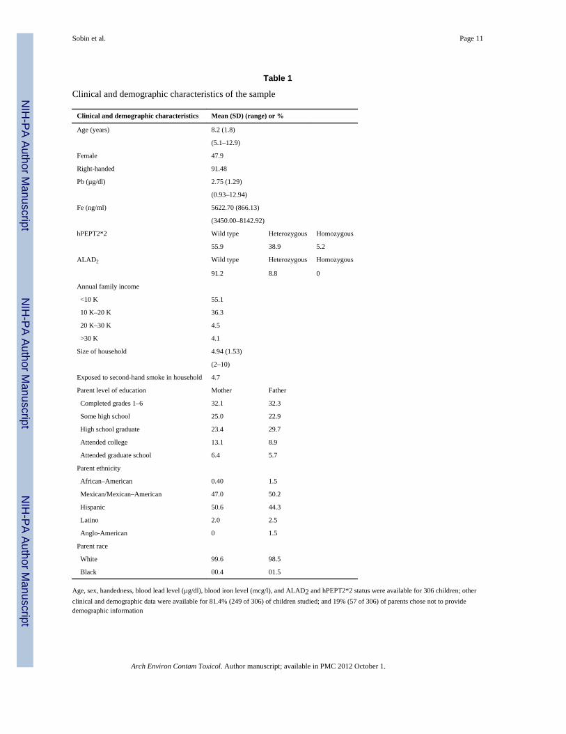
ResultsClinical and Demographic Characteristics
The sample included 306 children. Age, sex, handedness, Pb level, Fe level, ALAD2 andhPEPT2*2 were characterized for all children. The mean age of the sample was 8.2 years(±1.9), and 47.9% (145 of 306) of the children were female (Table 1). With regard toALAD2, 270 of 306 children (91.2%) did not carry the polymorphism (91.9% of boys and90.3% of girls); 27 of 306 children (8.8%) were heterozygous (8.1% of boys and 9.7% ofgirls); and no children were homozygous for ALAD2. With regard to hPEPT2*2, 171 of 306children (55.9%) did not carry the haplotype (55.95% of boys and 59.00% of girls); 119 of306 children (38.9%) were heterozygous (36.6% of boys and 41.4% of girls); and 16 of 306children (5.2%) were homozygous (4.3% of boys and 6.2% of girls).
Parents of 249 of 306 children studied (81.4%) provided demographic information (57parents declined completion of demographics forms). The annual household income was ≤$20,000 for 91.4% of reporting families; the average household size was 4.94 members; and49.9% of mothers and 44.3% of fathers had completed high school. The ethnicity of 97.3%of parents was Mexican, Mexican–American, Hispanic, or Latino, and 99.05% of familieswere white (Table 1).
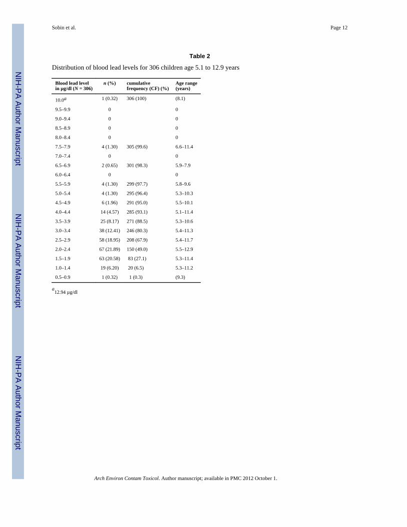
Table 2 lists the distribution of Pb levels for 306 children. Mean Pb level for the sample was2.75 µg/dl (±1.29) (geometric mean = 2.58 and median = 2.57). The inter-quartile range was1.35. The mean Fe level for the sample was 5622.70 ng/ml (±866.13). The range of Fe levels
Sobin et al. Page 5
Arch Environ Contam Toxicol. Author manuscript; available in PMC 2012 October 1.
NIH
-PA Author Manuscript
NIH
-PA Author Manuscript
NIH
-PA Author Manuscript
for all children in the sample were within previously reported normal limits (Gulson et al.2008).
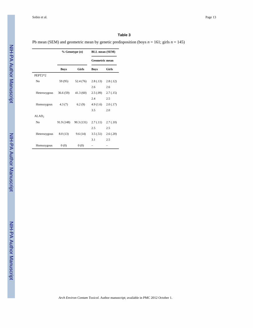
Genetic Predisposition and Pb Blood BurdenTable 3 lists mean and geometric mean blood Pb levels for boys and girls by genotype. Thegeneral linear model predicting Pb from ALAD2, hPEPT2*2, and sex, co-varying for Fe,was significant (model degrees of freedom [df] = 10, R2 = 0.120, F = 4.03, P < 0.001). Themain effects contributing to model significance, as determined from tests of type III sums ofsquares, included sex (df = 1, F = 17.88, P < 0.001) and the covariate Fe (df = 1, F = 6.65, P= 0.010). Significant interactions included ALAD2 × sex (df = 1, F = 4.15, P = 0.043) andhPEPT2*2 × sex (df = 2, F = 10.92, P < 0.001). The hPEPT2*2 × ALAD2 interaction wasnot significant (df = 2, F = 0.18, P = 0.834).
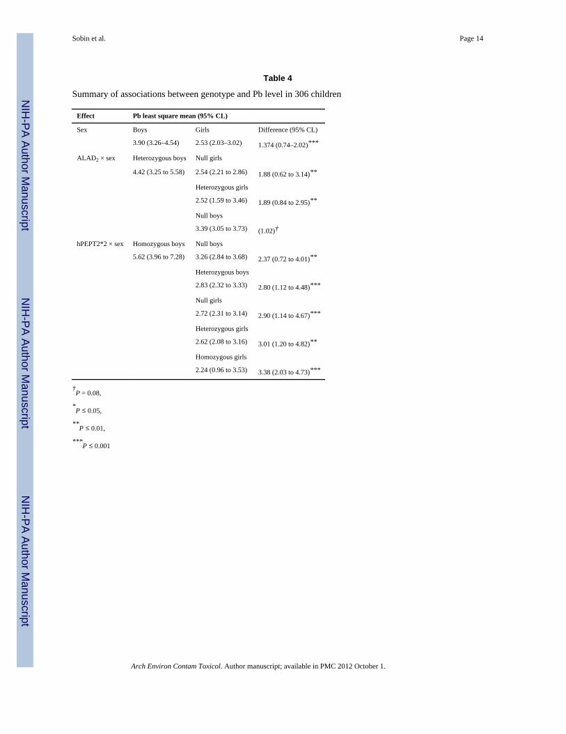
Effect statistics are listed in Table 4. With regard to the main effect of sex, Pb levels in boyswere significantly higher compared with girls. A significant interaction of ALAD2 × sex wasfound, suggesting that ALAD2 exaggerated the effect of sex on Pb level. Two differencesaccounted for the significance of this interaction. After covarying Fe, the Pb level of ALAD2hetereozygous boys was significantly greater than the Pb level of girls without ALAD2. ThePb level of heterozygous boys was also greater than the Pb level of heterozygous girls. ThePb level of heterozygous boys was also greater than the Pb level of boys without ALAD2;however, the statistical significance of the difference was marginal.
The interaction of hPEPT2*2 × sex was also significant, suggesting that hPEPT2*2exaggerated the effect of sex on Pb level. The Pb level of male homozygotes was greaterthan the other five subgroups; five paired differences accounted for the significance of theinteraction. The Pb level of homozygous boys was significantly greater than that of boyswithout hPEPT2*2 and heterozygous boys. Also, the Pb level of homozygous boys wasgreater than the Pb level of girls without hPEPT2*2, heterozygous girls, and homozygousgirls.
DiscussionALAD2 and hPEPT2*2 in Minority Children
Three previous studies suggested that ALAD2 and/or hPEPT2*2 are associated withincreased Pb blood burden in Pb-exposed children (Shen et al. 2001; Pérez-Bravo et al.2004; Sobin et al. 2009). The current study included a unique sample of 306 minoritychildren and improved on previous research by (1) increasing the sample size to allow forthe inclusion of sex as an explanatory factor; (2) including Fe as a covariate; and (3) usingICP-MS for element detection (Pb and Fe). We compared Pb levels in boys and girls withand without ALAD2 and hPEPT2*2, covarying for blood Fe. Consistent with our previousstudy of a demographically similar population (no cases overlapped) (Sobin et al. 2009),thepercentages of children heterozygous and homozygous for ALAD2 and hPEPT2*2 closelyapproximated frequencies in North American white populations (Secchi et al. 1974; Petrucciet al. 1982; Benkman et al. 1983; Pinsonneault et al. 2004), providing evidence of thefrequency of these variants in Hispanic children and also confirming that hPEPT2*2 iscommon among children of Mexican–American/Hispanic descent.
ALAD2 and hPEPT2*2 May Exaggerate Pb Blood Burden in BoysThis is the first study to examine the effects of ALAD2 and hPEPT2*2 in a single model,and it contributes three new findings to the literature. The findings are the first to suggestthat the influences of ALAD2 and hPEPT2*2 on Pb blood burden in children are
Sobin et al. Page 6
Arch Environ Contam Toxicol. Author manuscript; available in PMC 2012 October 1.
NIH
-PA Author Manuscript
NIH
-PA Author Manuscript
NIH
-PA Author Manuscript
independent (no apparent additive effects), that the associations are observable at lowestlevels of Pb exposure, and that the effects are sexually dimorphic.
By increasing Pb blood burden, ALAD2 and hPEPT2*2 might be expected to exaggerate therisks associated with low-level Pb exposure in male ALAD2 heterozygotes and malehPEPT2*2 homozygotes. Results from previous animal studies have suggested thathPEPT2*2 may be a secondary genetic modifier in cases of Pb poisoning (Hu et al.2007).The current results support the additional suggestion that the modifying effect ofhPEPT2*2 on Pb blood burden in cases of low-level Pb exposure is specific to boys.
Pb molecules enter red blood cells where they are bound by δ-ALAD. Pb binding disrupts δ-ALAD function, decreases heme biosynthesis, and causes an increase in brain levels of δ-ALA. Presumably, in children with ALAD2, Pb binding is reduced, resulting in higher Pbblood burden. Regardless of genotype, boys have higher Pb blood burden, which has beenattributed to their higher hematocrit levels (Kameneva et al. 1999). The results of this studymay suggest that in ALAD2 boys, lower Pb binding results in less disruption of hemebiosynthesis, increased hematocrit levels, and thus genotype specific increases in Pb bloodburden. This speculation requires experimental examination.
Studies exploring the possible role of hPEPT2*2 in Pb blood burden are recent. Pbmetabolism is complex and not fully understood. The current literature does not directlysuggest why and how hPEPT2*2 may differentially increase Pb blood burden in boys. Itmay be relevant that Pb exposure has been associated with increased production of at least100 different proteins (Witzmann et al. 1999). PEPT2 provides the main mechanism forproximal tubular reabsorption of peptide-bound amino acids (Rubio-Aliaga et al. 2003).Previously we suggested that the secondary effects of lowered protein reabsorption inchildren with hPEPT2*2 may partially account for their increased Pb blood burden (Sobin etal. 2009). Boys and girls differ with regard to not only hemoglobin density but also bloodviscosity and flow rate. Thus, similar to ALAD2 but by way of other mechanisms, it may belogical to suggest that hPEPT2*2 differentially lowers protein reabsorption rates in boys,thus decreasing Pb clearance. Studies are needed to examine these possibilities.
LimitationsThe primary risk associated with low-level Pb exposure is neurodevelopmental disruption asindicated by diminished neurocognitive function at the time of exposure and beyond(Lanphear et al. 2000; Chiodo et al. 2004, 2007; Gilbert and Weiss 2006; Min et al. 2007;Surkan et al. 2007); longer term effects include adult-onset renal (Fadrowski et al. 2010) andcardiovascular dysfunction (Park et al. 2006). Whether the increased Pb blood burdenobserved in children with ALAD2 and hPEPT2*2 also proves to be associated withpreviously identified neurobehavioral and physical outcomes has yet to be examined.Consistent with published population rates, the proportions of ALAD2 heterozygotes andhPEPT2*2 homozygotes were small, and no cases of children homozygous for ALAD2 wereidentified in this sample. Because of random variation, small subgroup size usually results inno significant differences. It may be noteworthy that large differences were identifieddespite small subgroup sizes. Future studies could select children by genotype to ensuremore balanced subgroups.
ConclusionALAD2 and hPEPT2*2 mediate Pb blood burden in young boys at levels of exposurepreviously associated with diminished neurocognitive function. The effects of these geneticmediators appear to be sexually dimorphic. ALAD2 and hPEPT2*2 may provide novelbiomarkers of increased health risk in young boys exposed to low-level Pb. Eventually,
Sobin et al. Page 7
Arch Environ Contam Toxicol. Author manuscript; available in PMC 2012 October 1.
NIH
-PA Author Manuscript
NIH
-PA Author Manuscript
NIH
-PA Author Manuscript
screening for these genetic variants could be used to identify a subgroup of young boys forearly neurocognitive intervention and health monitoring.
AcknowledgmentsThis research was made possible by grants from the National Institute of Child Health and Human Development,National Institutes of Health (Grant No. R21HD060120); the National Center for Research Resources, a componentof the National Institutes of Health (Grant No. 5G12RR008124); the Center for Clinical and Translational Science,The Rockefeller University, New York, NY; the Paso del Norte Health Foundation and the University ResearchInstitute, University of Texas, El Paso, TX. The funding agencies had no role in the design, implementation, dataanalysis, or manuscript preparation for this study.
ReferencesBattistuzzi G, Petrucci R, Silvagni L, Urbani FR, Caiola S. Delta-aminolevulinate dehydrase: a new
genetic polymorphism in man. Ann Hum Genet. 1981; 45(Pt 3):223–229. [PubMed: 7305279]Benkman HG, Gogdaanski P, Goedde HW. Polymorphism of delta-aminolevulinic acid dehydratase in
various populations. Hum Hered. 1983; 33:62–64. [PubMed: 6840783]Bergdahl IA, Gerhardsson L, Schutz A, Desnick RJ, Wetmur JG, Skerfving S. Delta-aminolevulinic
acid dehydratase polymorphism: influence on lead levels and kidney function in humans. ArchEnviron Health. 1997; 52(2):91–96. [PubMed: 9124881]
Bradman A, Eskenazi B, Sutton P, Athanasoulis M, Goldman L. Iron deficiency associated with higherblood lead in children living in contaminated environments—Children’s health articles. EnvironHealth Perspect. 2001; 109:1079–1084. [PubMed: 11675273]
Brennan MJW, Cantrill RC. [Delta]-aminolaevulinic acid is a potent agonist for GABA autoreceptors.Nature. 1979; 280(5722):514–515. [PubMed: 223065]
Carter-Pokras O, Baquet C. What is a health disparity? Publ Health Rep. 2002; 117:426–434.Centers for disease control and prevention. Lead. Atlanta, GA: United States Department of Health
and Human Services; 2009.Chiodo LM, Covington C, Sokol RJ, Hannigan JH, Jannise J, Ager J, Greenwald M, Delaney-Black V.
Blood lead levels and specific attention effects in young children. Neurotoxicol Teratol. 2007;29(5):538–546. [PubMed: 17553667]
Chiodo LM, Jacobson SW, Jacobson JL. Neurodevelopmental effects of postnatal lead exposure atvery low levels. Neurotoxicol Teratol. 2004; 26(3):359–371. [PubMed: 15113598]
Choi JW, Kim SK. Association between blood lead concentrations and body iron status in children.Arch Dis Child. 2003; 88(9):791–792. [PubMed: 12937100]
Demasi M, Penatti CAA, DeLucia R, Bechara EJH. The prooxidant effect of 5-aminolevulinic acid inthe brain tissue of rats: implications in neuropsychiatric manifestations in prophyries. Free RadicBiol Med. 1996; 20:291–299. [PubMed: 8720899]
Emanuelli T, Pagel FW, Prociuncula LO, Souza DO. Effects of 5-aminolevulinic acid on theglutamatergic neurotransmission. Neurochem Int. 2003; 42:115–121. [PubMed: 12421591]
Fadrowski JJ, Navas-Acien A, Tellez-Plaza M, Guallar E, Weaver VM, Furth SL. Blood lead level andkidney function in US adolescents: the third national health and nutrition examination survey.Arch Intern Med. 2010; 170(1):75–82. [PubMed: 20065202]
Fleming DE, Chettle DR, Wetmur JG, Desnick RJ, Robin JP, Boulay D, Richard NS, Gordon CL,Webber CE. Effect of the delta-aminolevulinate dehydratase polymorphism on the accumulationof lead in bone and blood in lead smelter workers. Environ Res. 1998; 77(1):49–61. [PubMed:9593628]
Gilbert SG, Rice DC. Low-level lifetime lead exposure produces behavioral toxicity (spatialdiscrimination reversal) in adult monkeys. Toxicol Appl Pharmacol. 1987; 91(3):484–490.[PubMed: 3424377]
Gilbert SG, Weiss B. A rationale for lowering the blood lead action level from 10 to 2 µg/dl.Neurotoxicology. 2006; 27(5):693–701. [PubMed: 16889836]
Gochfeld M. Framework for gender differences in human and animal toxicology. Environ Res. 2007;104(1):4–21. [PubMed: 16616135]
Sobin et al. Page 8
Arch Environ Contam Toxicol. Author manuscript; available in PMC 2012 October 1.
NIH
-PA Author Manuscript
NIH
-PA Author Manuscript
NIH
-PA Author Manuscript
Gulson B, Mizon K, Taylor A, Korsch M, Stauber J, Davis JM, Louie H, Wu M, Antin L. Longitudinalmonitoring of selected elements in blood of healthy young children. J Trace Elem Med Biol. 2008;22(3):206–214. [PubMed: 18755396]
Hu Y, Shen H, Keep RF, Smith DE. Peptide transporter 2 (PEPT2) expression in brain protects against5-aminolevulinic acid neurotoxicity. J Neurochem. 2007; 103(5):2058–2065. [PubMed:17854384]
Kameneva MV, Watach MJ, Borovetz HS. Gender differences in rheologic properties of blood andrisk of cardiovascular disease. Clin Hemorheol Microcirc. 1999; 21:357–363. [PubMed:10711771]
Kappas, A.; Sassa, S.; Galbraith, RA.; Nordmann, Y. The porphyrias. In: Scriver, CR., editor. Themetabolic basis of inherited disease. New York: McGraw Hill; 1995. p. 2103-2160.
Klaassen, CD. Heavy metals and heavy-metal antagonists. In: Brunton, LL.; Lazo, JS.; Parker, KL.,editors. Goodman & Gilman’s The phamacological basis of therapeutics. New York: McGrawHill; 2006. p. 1753-1775.
Lanphear BP, Dietrich K, Auinger P, Cox C. Cognitive deficits associated with blood leadconcentrations <10 µg/dl in US children and adolescents. Publ Health Rep. 2000; 115(6):521–529.
Levin ED, Schantz SL, Bowman RE. Use of the lesion model for examining toxicant effects oncognitive behavior. Neurotoxicol Teratol. 1992; 14(2):131–141. [PubMed: 1593987]
Lindahl L, Bird L, Legare M, Mikeska G, Bratton G, Tiffany-Castiglioni E. Differential ability ofastroglia and neuronal cells to accumulate lead: Dependence on cell type and on degree ofdifferentiation. Toxicol Sci. 1999; 50(2):236–243. [PubMed: 10478860]
Min J-Y, Min K-B, Cho S-I, Kim R, Sakong J, Paek D. Neurobehavioral function in children with lowblood lead concentrations. Neurotoxicology. 2007; 28(2):421–425. [PubMed: 16644013]
Ocheltree SM, Shen H, Hu Y, Keep RF, Smith DE. Role and relevance of peptide transporter 2(PEPT2) in the kidney and choroid plexus: In vivo studies with glycylsarcosine in wild-type andPEPT2 knockout mice. J Pharmacol Exp Ther. 2005; 315(1):240–247. [PubMed: 15987832]
Park SK, Schwartz J, Weisskopf M, Sparrow D, Vokonas PS, Wright RO, Coull B, Nie H, Hu H. Low-level lead exposure, metabolic syndrome, and heart rate variability: the VA normative aging study.Environ Health Perspect. 2006; 114(11)
Pérez-Bravo F, Ruz M, Morán-Jiménez MJ, Olivares M, Rebolledo A, Codoceo J, Sepúlveda V,Jenkin A, Santos JL, Fontanellas A. Association between aminolevulinate dehydrase genotypesand blood lead levels in children from a lead-contaminated area in Antofagasta, Chile. ArchEnviron Contam Toxicol. 2004; 47(2):276–280. [PubMed: 15386154]
Petrucci R, Leonardi A, Battistuzzi G. The genetic polymorphism of delta-aminolevulinic aciddehydratase in Italy. Hum Genet. 1982; 60:289–290. [PubMed: 7106762]
Pinsonneault J, Nielsen CU, Sadee W. Genetic variants of the human H+/dipeptide transporter PEPT2:analysis of haplotype functions. J Pharmacol Exp Ther. 2004; 311(3):1088–1096. [PubMed:15282265]
Ramamoorthy S, Liu W, Ma Y-Y, Yang-Feng TL, Ganapathy V, Leibach FH. Proton/peptidecotransporter (PEPT 2) from human kidney: functional characterization and chromosomallocalization. Biochim Biophys Acta Biomembr. 1995; 1240(1):1–4.
Rice DC, Karpinski KF. Lifetime low-level lead exposure produces deficits in delayed alternation inadult monkeys. Neurotoxicol Teratol. 1988; 10(3):207–214. [PubMed: 3211098]
Rossi E. Low level environmental lead exposure—a continuing challenge. Clin Biochem Rev. 2008;29:63–70. [PubMed: 18787644]
Rubio-Aliaga I, Frey I, Boll M, Groneberg DA, Eichinger HM, Balling R, Daniel H. Targeteddisruption of the peptide transporter Pept2 gene in mice defines its physiological role in thekidney. Mol Cell Biol. 2003; 23(9):3247–3252. [PubMed: 12697824]
Schwartz BS, Lee BK, Stewart W, Ahn KD, Springer K, Kelsey K. Associations of delta-aminolevulinic acid dehydratase genotype with plant, exposure duration, and blood lead and zincprotoporhyrin levels in Korean lead workers. Am J Epidemiol. 1995; 142:738–745. [PubMed:7572945]
Secchi GC, Erba L, Cambiaghi G. Delta-aminolevulinic acid dehydratase activity of erythrocytes andliver tissue in man. Arch Environ Health. 1974; 28:130–132. [PubMed: 4810883]
Sobin et al. Page 9
Arch Environ Contam Toxicol. Author manuscript; available in PMC 2012 October 1.
NIH
-PA Author Manuscript
NIH
-PA Author Manuscript
NIH
-PA Author Manuscript
Shen H, Smith DE, Yang T, Huang YG, Schnermann JB, Brosius FC III. Localization of PEPT1 andPEPT2 proton-coupled oligopeptide transporter mRNA and protein in rat kidney. Am J PhysiolRenal Physiol. 1999; 276(5):F658–F665.
Shen XM, Wu SH, Yan CH, Zhao W, Ao LM, Zhang YW, He JM, Ying JM, Li RQ, Wu SM, et al.Delta-aminolevulinate dehydratase polymorphism and blood lead levels in Chinese children.Environ Res. 2001; 85(3):185–190. [PubMed: 11237505]
Sobin C, Gutierrez M, Alterio H. Polymorphisms of delta-aminolevulinic acid dehydratase (ALAD)and peptide transporter 2 (PEPT2) genes in children with low-level lead exposure.Neurotoxicology. 2009; 30:881–887. [PubMed: 19723536]
Sobin C, Parisi N, Schaub T, De la Riva E. A Bland–Altman comparison of the Lead Care® Systemand inductively coupled plasma mass spectrometry for detecting low-level lead in child wholeblood samples. J Med Toxicol. 2010
Surkan PJ, Zhang A, Trachtenberg F, Daniel DB, McKinlay S, Bellinger DC. Neuropsychologicalfunction in children with blood lead levels <10 µg/dl. Neurotoxicology. 2007; 28(6):1170–1177.[PubMed: 17868887]
Thomas JA, Dallenbach FD, Thomas M. The distribution of radioactive lead in the cerebellum ofdeveloping rats. J Pathol. 1973; 109(1):45–50. [PubMed: 4719733]
United States Environmental Protection Agency. Documentation for the Final 2002 Point SourceNational Emissions Inventory. NC: Research Triangle Park; 2006.
United States Environmental Protection Agency. Blood lead level: what are the trends in exposure toenvironmental contaminants including across population subgroups and geographic regions?Online: Report on the Environment series. 2010
Vahter M, Åkesson A, Lidén C, Ceccatelli S, Berglund M. Gender differences in the disposition andtoxicity of metals. Environ Res. 2007; 104(1):85–95. [PubMed: 16996054]
Villayandre BM, Paniagua MA, Fernandez-Lopez A, Calvo P. Effect of delta-aminolevulinic acidtreatment on N-methyl-D-aspartate receptor at different ages in the rat brain. Brain Res. 2005;1061(2):80–87. [PubMed: 16229824]
Wang L, Yan D, Gu Y, Sun L-G, Ruan D-Y. Effects of extracellular-aminolaevulinic acid on sodiumcurrents in acutely isolated rat hippocampal CA1 neurons. Eur J Neurosci. 2005; 22:3122–3128.[PubMed: 16367778]
Wetmur JG, Kaya AH, Plewinska M, Desnick RJ. Molecular characterization of the human delta-aminolevulinate dehydratase 2 (ALAD2) allele: implications for molecular screening ofindividuals for genetic susceptibility to lead poisoning. Am J Hum Genet. 1991a; 49(4):757–763.[PubMed: 1716854]
Wetmur JG, Lehnert G, Desnick RJ. The delta-aminolevulinate dehydratase polymorphism: higherblood lead levels in lead workers and environmentally exposed children with the 1–2 and 2–2isozymes. Environ Res. 1991b; 56(2):109–119. [PubMed: 1769358]
Witzmann FA, Fultz CD, Grant RA, Wright LS, Kornguth SE, Siegel FL. Regional protein alterationsin rat kidneys induced by lead exposure. Electrophoresis. 1999; 20(4–5):943–951. [PubMed:10344270]
Wright R, Tsaih S, Schwartz J. Association between iron deficiency and blood lead level in alongitudinal analysis of children followed in an urban primary care clinic. J Pediatr. 2003; 142:9–14. [PubMed: 12520247]
Wright RO, Shannon MW, Wright RJ, Hu H. Association between iron deficiency and low-level leadpoisoning in an urban primary care clinic. Am J Publ Health. 1999; 89(7):1049–1053.
Ziemsen B, Angerer J, Lehnert G, Benkman HG, Goedde HW. Polymorphism of delta-aminolevulinicacid deydratase in lead exposed workers. Int Arch Occup Environ Health. 1986; 58:245–247.[PubMed: 3770964]
Sobin et al. Page 10
Arch Environ Contam Toxicol. Author manuscript; available in PMC 2012 October 1.
NIH
-PA Author Manuscript
NIH
-PA Author Manuscript
NIH
-PA Author Manuscript
NIH
-PA Author Manuscript
NIH
-PA Author Manuscript
NIH
-PA Author Manuscript
Sobin et al. Page 11
Table 1
Clinical and demographic characteristics of the sample
Clinical and demographic characteristics Mean (SD) (range) or %
Age (years) 8.2 (1.8)
(5.1–12.9)
Female 47.9
Right-handed 91.48
Pb (µg/dl) 2.75 (1.29)
(0.93–12.94)
Fe (ng/ml) 5622.70 (866.13)
(3450.00–8142.92)
hPEPT2*2 Wild type Heterozygous Homozygous
55.9 38.9 5.2
ALAD2 Wild type Heterozygous Homozygous
91.2 8.8 0
Annual family income
<10 K 55.1
10 K–20 K 36.3
20 K–30 K 4.5
>30 K 4.1
Size of household 4.94 (1.53)
(2–10)
Exposed to second-hand smoke in household 4.7
Parent level of education Mother Father
Completed grades 1–6 32.1 32.3
Some high school 25.0 22.9
High school graduate 23.4 29.7
Attended college 13.1 8.9
Attended graduate school 6.4 5.7
Parent ethnicity
African–American 0.40 1.5
Mexican/Mexican–American 47.0 50.2
Hispanic 50.6 44.3
Latino 2.0 2.5
Anglo-American 0 1.5
Parent race
White 99.6 98.5
Black 00.4 01.5
Age, sex, handedness, blood lead level (µg/dl), blood iron level (mcg/l), and ALAD2 and hPEPT2*2 status were available for 306 children; otherclinical and demographic data were available for 81.4% (249 of 306) of children studied; and 19% (57 of 306) of parents chose not to providedemographic information
Arch Environ Contam Toxicol. Author manuscript; available in PMC 2012 October 1.
NIH
-PA Author Manuscript
NIH
-PA Author Manuscript
NIH
-PA Author Manuscript
Sobin et al. Page 12
Table 2
Distribution of blood lead levels for 306 children age 5.1 to 12.9 years
Blood lead levelin µg/dl (N = 306)
n (%) cumulativefrequency (CF) (%)
Age range(years)
10.0a 1 (0.32) 306 (100) (8.1)
9.5–9.9 0 0
9.0–9.4 0 0
8.5–8.9 0 0
8.0–8.4 0 0
7.5–7.9 4 (1.30) 305 (99.6) 6.6–11.4
7.0–7.4 0 0
6.5–6.9 2 (0.65) 301 (98.3) 5.9–7.9
6.0–6.4 0 0
5.5–5.9 4 (1.30) 299 (97.7) 5.8–9.6
5.0–5.4 4 (1.30) 295 (96.4) 5.3–10.3
4.5–4.9 6 (1.96) 291 (95.0) 5.5–10.1
4.0–4.4 14 (4.57) 285 (93.1) 5.1–11.4
3.5–3.9 25 (8.17) 271 (88.5) 5.3–10.6
3.0–3.4 38 (12.41) 246 (80.3) 5.4–11.3
2.5–2.9 58 (18.95) 208 (67.9) 5.4–11.7
2.0–2.4 67 (21.89) 150 (49.0) 5.5–12.9
1.5–1.9 63 (20.58) 83 (27.1) 5.3–11.4
1.0–1.4 19 (6.20) 20 (6.5) 5.3–11.2
0.5–0.9 1 (0.32) 1 (0.3) (9.3)
a12.94 µg/dl
Arch Environ Contam Toxicol. Author manuscript; available in PMC 2012 October 1.
NIH
-PA Author Manuscript
NIH
-PA Author Manuscript
NIH
-PA Author Manuscript
Sobin et al. Page 13
Table 3
Pb mean (SEM) and geometric mean by genetic predisposition (boys n = 161; girls n = 145)
% Genotype (n) BLL mean (SEM)
Geometric mean
Boys Girls Boys Girls
PEPT2*2
No 59 (95) 52.4 (76) 2.8 (.13) 2.8 (.12)
2.6 2.6
Heterozygous 36.6 (59) 41.3 (60) 2.5 (.09) 2.7 (.15)
2.4 2.5
Homozygous 4.3 (7) 6.2 (9) 4.9 (1.6) 2.0 (.17)
3.5 2.0
ALAD2
No 91.9 (148) 90.3 (131) 2.7 (.11) 2.7 (.10)
2.5 2.5
Heterozygous 8.0 (13) 9.6 (14) 3.5 (.51) 2.6 (.20)
3.1 2.5
Homozygous 0 (0) 0 (0) – –
Arch Environ Contam Toxicol. Author manuscript; available in PMC 2012 October 1.
NIH
-PA Author Manuscript
NIH
-PA Author Manuscript
NIH
-PA Author Manuscript
Sobin et al. Page 14
Table 4
Summary of associations between genotype and Pb level in 306 children
Effect Pb least square mean (95% CL)
Sex Boys Girls Difference (95% CL)
3.90 (3.26–4.54) 2.53 (2.03–3.02) 1.374 (0.74–2.02)***
ALAD2 × sex Heterozygous boys Null girls
4.42 (3.25 to 5.58) 2.54 (2.21 to 2.86) 1.88 (0.62 to 3.14)**
Heterozygous girls
2.52 (1.59 to 3.46) 1.89 (0.84 to 2.95)**
Null boys
3.39 (3.05 to 3.73) (1.02)†
hPEPT2*2 × sex Homozygous boys Null boys
5.62 (3.96 to 7.28) 3.26 (2.84 to 3.68) 2.37 (0.72 to 4.01)**
Heterozygous boys
2.83 (2.32 to 3.33) 2.80 (1.12 to 4.48)***
Null girls
2.72 (2.31 to 3.14) 2.90 (1.14 to 4.67)***
Heterozygous girls
2.62 (2.08 to 3.16) 3.01 (1.20 to 4.82)**
Homozygous girls
2.24 (0.96 to 3.53) 3.38 (2.03 to 4.73)***
†P = 0.08,
*P ≤ 0.05,
**P ≤ 0.01,
***P ≤ 0.001
Arch Environ Contam Toxicol. Author manuscript; available in PMC 2012 October 1.