Altered Expression and Activation of the Nerve Growth Factor Receptors TrkA and p75 Provide the...
10
Breast Cancer Research and Treatment 83: 119–128, 2004. © 2004 Kluwer Academic Publishers. Printed in the Netherlands. Report Altered expression and activation of the nerve growth factor receptors TrkA and p75 provide the first evidence of tumor progression to effusion in breast carcinoma Ben Davidson 1 , Reuven Reich 2,4 , Philip Lazarovici 2,4 , Vivi Ann Flørenes 1 , Søren Nielsen 3 , and Jahn M. Nesland 1 1 Department of Pathology, The Norwegian Radium Hospital, University of Oslo, Montebello, Oslo, Norway; 2 Department of Pharmacology and Experimental Therapeutics, School of Pharmacy, Faculty of Medicine, The Hebrew University of Jerusalem, Jerusalem, Israel; 3 Department of Pathology, Aalborg University, Aalborg, Denmark; 4 David R. Bloom Center for Pharmacy, Hebrew University, Jerusalem, Israel Key words: breast cancer, neurotrophins, pleural effusion, tyrosine kinase receptors Summary The aim of this study was to characterize phenotypic alterations along the progression of breast carcinoma from primary tumor to pleural effusion through analysis of the expression of nerve growth factor (NGF) and its receptors phospho-TrkA (p-TrkA activated receptor) and p75. Sections from 42 malignant pleural effusions from breast cancer patients and 65 corresponding solid tumors (34 primary, 31 metastatic) were evaluated for protein expression of the activated p-TrkA receptor. The majority of lesions were additionally studied for NGF and p75 expression. Six effusions and four breast carcinoma cell lines were studied for expression of p-TrkA using immunoblotting (IB). Membrane expression of p-TrkA was high in carcinoma cells in effusions (39/42, 93%) and locoregional recurrences (12/13, 92%), with significantly lower expression in both primary tumors (14/34, 41%) and lymph node metastases (8/18, 44%), respectively (p< 0.001 for effusions vs. primary tumors; p = 0.001 for effusions vs. lymph nodes). In contrast, p75 expression was less frequent in effusions compared to both primary tumors and lymph node metastases, significantly so for the latter (p = 0.019). NGF expression was comparable at all sites, but its expression in tumor cells in effusions (7/21 cases) was limited to cases in which time to progression (TTP) to effusion occurred within 5 years or less from primary operation. In univariate analysis of survival, mean and median TTP were 6.3 and 6 years for NGF-negative effusions, compared to 3 and 4 years for NGF-positive cases (p = 0.013). IB confirmed expression of p-TrkA in five of six effusions, while all four breast cancer cell lines were p-TrkA-negative. Our data provide the first documented evidence of molecular events that occur along tumor progression of breast carcinoma from primary tumors to effusion. The almost universal expression of p-TrkA in cancer cells in effusions and late recurrences is in full agreement with our recent report linking this factor with poor prognosis in ovarian cancer. Furthermore, the rapid progression to effusion in cases showing NGF expression in tumor cells underscores the aggressive clinical behavior of tumors that are able to utilize this pathway in an autocrine manner. Introduction Neurotrophins are a family of growth factors, con- sisting at present of the prototype compound nerve growth factor (NGF), and additional members such as brain-derived neurotrophic factor (BDNF), and the neurotrophins NT-3, NT-4 and NT-6 [1–4]. Neuro- trophins bind to the TrkA, TrkB and TrkC receptors. NGF binds to the specific high-affinity tyrosine kinase receptor TrkA [5]. NGF ligand binding to TrkA re- ceptors activates TrkA autophosphorylation at several sites such as tyrosine 490, promoting SHC binding
Transcript of Altered Expression and Activation of the Nerve Growth Factor Receptors TrkA and p75 Provide the...

Breast Cancer Research and Treatment 83: 119–128, 2004.© 2004 Kluwer Academic Publishers. Printed in the Netherlands.
Report
Altered expression and activation of the nerve growth factorreceptors TrkA and p75 provide the first evidence of tumorprogression to effusion in breast carcinoma
Ben Davidson1, Reuven Reich2,4, Philip Lazarovici2,4, Vivi Ann Flørenes1, Søren Nielsen3, andJahn M. Nesland1
1Department of Pathology, The Norwegian Radium Hospital, University of Oslo, Montebello, Oslo, Norway;2Department of Pharmacology and Experimental Therapeutics, School of Pharmacy, Faculty of Medicine, TheHebrew University of Jerusalem, Jerusalem, Israel; 3Department of Pathology, Aalborg University, Aalborg,Denmark; 4David R. Bloom Center for Pharmacy, Hebrew University, Jerusalem, Israel
Key words: breast cancer, neurotrophins, pleural effusion, tyrosine kinase receptors
Summary
The aim of this study was to characterize phenotypic alterations along the progression of breast carcinoma fromprimary tumor to pleural effusion through analysis of the expression of nerve growth factor (NGF) and its receptorsphospho-TrkA (p-TrkA activated receptor) and p75. Sections from 42 malignant pleural effusions from breastcancer patients and 65 corresponding solid tumors (34 primary, 31 metastatic) were evaluated for protein expressionof the activated p-TrkA receptor. The majority of lesions were additionally studied for NGF and p75 expression.Six effusions and four breast carcinoma cell lines were studied for expression of p-TrkA using immunoblotting(IB). Membrane expression of p-TrkA was high in carcinoma cells in effusions (39/42, 93%) and locoregionalrecurrences (12/13, 92%), with significantly lower expression in both primary tumors (14/34, 41%) and lymphnode metastases (8/18, 44%), respectively (p < 0.001 for effusions vs. primary tumors; p= 0.001 for effusionsvs. lymph nodes). In contrast, p75 expression was less frequent in effusions compared to both primary tumors andlymph node metastases, significantly so for the latter (p= 0.019). NGF expression was comparable at all sites,but its expression in tumor cells in effusions (7/21 cases) was limited to cases in which time to progression (TTP)to effusion occurred within 5 years or less from primary operation. In univariate analysis of survival, mean andmedian TTP were 6.3 and 6 years for NGF-negative effusions, compared to 3 and 4 years for NGF-positive cases(p= 0.013). IB confirmed expression of p-TrkA in five of six effusions, while all four breast cancer cell lineswere p-TrkA-negative. Our data provide the first documented evidence of molecular events that occur along tumorprogression of breast carcinoma from primary tumors to effusion. The almost universal expression of p-TrkA incancer cells in effusions and late recurrences is in full agreement with our recent report linking this factor withpoor prognosis in ovarian cancer. Furthermore, the rapid progression to effusion in cases showing NGF expressionin tumor cells underscores the aggressive clinical behavior of tumors that are able to utilize this pathway in anautocrine manner.
Introduction
Neurotrophins are a family of growth factors, con-sisting at present of the prototype compound nervegrowth factor (NGF), and additional members suchas brain-derived neurotrophic factor (BDNF), and the
neurotrophins NT-3, NT-4 and NT-6 [1–4]. Neuro-trophins bind to the TrkA, TrkB and TrkC receptors.NGF binds to the specific high-affinity tyrosine kinasereceptor TrkA [5]. NGF ligand binding to TrkA re-ceptors activates TrkA autophosphorylation at severalsites such as tyrosine 490, promoting SHC binding

120 B Davidson et al.
and phosphorylation, coupling of GRB2-SOS com-plexes and activation of Ras [6, 7]. The net result inthe central nervous system is differentiation and sur-vival of neuronal cells [2, 7]. Ras is able to activatetwo major intracellular signal transduction pathways –the mitogen activated protein kinase (MAPK) pathwayand the phosphoinositol-3-kinase (PI3K)/AKT path-way [3, 7]. NGF signaling is able to induce survivaland proliferation, but can also induce differentiationwithout proliferation [8], through increased nuclearexpression of p53 and direct activation of the cyclin-dependent kinase inhibitor p21WAF1/CIP1 [9–11]. TrkAitself has been shown to associate with p53, and wasphosphorylated in presence of the latter even in theabsence of NGF [12–13].
Though originally discovered as a protooncogeneof the nervous system, TrkA isoforms are involvedin tumorigenesis in non-neural tumors, primarily car-cinomas, and have been generally associated with dis-ease progression and poor outcome [14–16]. Recently,it was also experimentally demonstrated in nude micethat overexpression of TrkA receptor increased thetumorogenicity of pheochromocytoma [17], as wellas that of prostatic and pancreatic adenocarcinoma[18]. Conversely, the expression of TrkA as a proto-oncogene has been associated with better prognosis inneuroblastoma [19, 20]. In addition to the alteration instructure and expression of Trk receptors, intracellularsignaling appears to be dysregulated in cancer cells[14].
p75, an additional neutrophin receptor, belongsto the tumor necrosis receptor family, has a differ-ent structure, lacks intrinsic catalytic activity and isable to bind all neurotrophins [1]. The evidence re-garding the biological role of p75 in human canceris more limited, but a large study has recently eval-uated the expression of this receptor in a variety ofhuman tumors and benign tissues [21]. Frequent ex-pression was seen in some neural and soft tissuetumors, while most carcinomas were negative [21].Work by the Djakiew group has shown that p75 ex-pression suppresses growth and metastasis of prostatecancer cells in vitro [22], and that expression ofthis receptor reduces NGF-mediated growth in thismalignancy through the induction of apoptosis [23].Furthermore, p75 expression has been found to bereduced in carcinomas compared to non-neoplastic tis-sue [24], although the latter finding was not confirmedin an additional study [25].
We have recently reported on marked differencesin the expression of the NGF receptor TrkA, its acti-
vated form p-TrkA and the low-affinity neurotrophinreceptor p75 at different anatomic sites in serousovarian carcinoma [26, 27]. Specifically, TrkA andp-TrkA expression was down-regulated in effusions,while the opposite was true for p75. Furthermore,we observed co-expression of p-TrkA with angiogenicmolecules, as well as its expression on endothelialcells in the vicinity of tumor cells [27]. Finally, p-TrkAexpression in solid tumors predicted poor outcome inour cohort [27]. Interestingly, in a recent study of ma-lignant mesotheliomas, we found opposite trends inthe comparison of effusions to solid tumors, namelyhigher p-TrkA and lower p75 expression in effusions(Davidson et al., submitted).
These and the above-mentioned studies suggest anovel emerging concept in cancer, that NGF and otherneurotrophins may be involved in the growth of certainnon-neural tumors by paracrine and/or autocrine regu-lation via humoral–tumoral–stromal Trk interaction.
Breast cancer is the most common malignancy inwomen, second in mortality only to lung cancer [28].Breast cancer metastasizes most frequently to axillarylymph nodes, but any organ may be involved. Meta-static spread to serosal surfaces involves primarily thepleural cavity, where breast cancer is the etiology ofapproximately 25% of malignant effusions [29, 30].However, breast carcinoma metastasis may be foundinfrequently in the pericardial and peritoneal cavity aswell [31]. Involvement of the pleural cavity by breastcarcinoma may occur at any phase of the clinicalcourse, and may be the sole manifestation of meta-static disease [32]. It is associated with poor prognosis,with a median survival of 5 [32] and 11 [33] monthsin two series.
Despite the magnitude of the clinical problem,studies of the biology of breast cancer have focusedexclusively on primary tumors and solid metastases.Consequently, the biological characteristics of breastcarcinoma cells in effusions have been poorly charac-terized at both the phenotypic and genotypic level, andtheir potential differences from primary and metastaticsolid tumors are entirely unknown. Identification ofthese potential differences may aid in explaining whythe appearance of pleural effusion in breast cancer isassociated with a rapidly fatal disease.
Several studies have analyzed the expression androle of NGF and its receptors in breast carcinoma.NGF has been shown to be produced by breast can-cer cells [34] and was mitogenic in breast cancer celllines, an effect mediated through TrkA and MAPKsignaling [35]. Furthermore, the pro-mitogenic role

NGF, TrkA and p75 in breast carcinoma 121
Table 1. p-TrkA expression results in the studied material (42 effusions, 34 primary tumors, 18lymph node metastases, 13 LRa)
Site Cellular Percentage of stained carcinoma cellslocation
0% 1–5% 6–25% 26–75% 76–100%
Effusion Membraneb 3 (7%) 6 (14%) 6 (14%) 14 (34%) 13 (31%)
Primary Membrane 20 (59%) 12 (35%) 1 (3%) 1 (3%) 0 (0%)
Lymph node Membrane 10 (56%) 7 (39%) 0 (0%) 1 (5%) 0 (0%)
LRa Membrane 1 (8%) 7 (54%) 2 (15%) 1 (8%) 2 (15%)
Effusion Cytoplasm 25 (59%) 4 (10%) 4 (10%) 6 (14%) 3 (7%)
Primary Cytoplasm 8 (23%) 5 (15%) 2 (6%) 6 (18%) 13 (38%)
Lymph node Cytoplasm 5 (27%) 3 (17%) 3 (17%) 3 (17%) 4 (22%)
LRa Cytoplasm 1 (8%) 1 (8%) 4 (30%) 0 (0%) 7 (54%)
Effusion Nucleus 23 (55%) 5 (12%) 10 (24%) 3 (7%) 1 (2%)
Primary Nucleus 15 (44%) 4 (12%) 1 (3%) 4 (12%) 10 (29%)
Lymph node Nucleus 12 (66%) 1 (6%) 1 (6%) 2 (11%) 2 (11%)
LRa Nucleus 11 (84%) 0 (0%) 1 (8%) 0 (0%) 1 (8%)
a Locoregional recurrences.b p < 0.001 for the difference in membrane expression between effusions and primary tumors,p= 0.001 for effusions vs. lymph nodes.
was independent of p75 activity, whereas the anti-apoptotic effect of NGF was mediated through p75and the NF-kB transcription factor [36], specificallythrough interaction with the tumor necrosis-associateddeath domain protein (TRADD) [37]. NGF effect me-diated through TrkA has been shown to be preventedby Tamoxifen in MCF-7 cells [38]. Clinical studies ofNGF receptor expression in primary breast carcinomashowed correlation with better survival and with clini-copathologic parameters that are associated with lessaggressive disease [39, 40].
The objective of the current study was to investi-gate the expression of NGF and its receptors in breastcancer cells in effusions and solid tumors, as well asto study the possible correlation between these mark-ers and the interval between primary intervention andrelapse in the form of pleural effusion.
Material and methods
Patients and clinicopathologic data. The study co-hort consisted of 39 female patients with histologicallyverified breast cancer. Patient age ranged from 35 to 85years at diagnosis. Thirty-five women were diagnosedwith infiltrating duct carcinoma, three with lobularcarcinoma and one with a tumor combining both his-tologic types. Tumor grade was available for all 35ductal carcinomas and was as follows: 1 grade onetumor, 26 grade two tumors and 8 grade three tu-
mors. The period of time from primary diagnosis tothe sampling of pleural effusion ranged from 0 (simul-taneous) to 17 years. For the purpose of this study, thisperiod was defined as time to progression (TTP).
Effusion specimens. The material consisted of 42pleural effusions from the above-mentioned 39 pa-tients, all submitted for routine diagnostic purposesto the Section of Cytology, Department of Pathology,The Norwegian Radium Hospital, and the Section ofCytology, Department of Pathology, Aalborg Hos-pital, during the period of January 1998–September2002. Submitted specimens arrived within minutesafter tapping and were processed immediately. Cell-blocks were prepared using the thrombin clot method.Additional material, available for 20 effusions, wassuspended and frozen in RPMI+DMSO at −70◦C.Smears and cellblock sections from all specimensunderwent morphological evaluation by three experi-enced cytopathologists, and were further character-ized using immunocytochemistry with broad antibodypanels against cancer and mesothelial epitopes, aspreviously detailed [41, 42].
Tumor specimens. Thirty-four corresponding pri-mary tumors, 18 lymph node metastases and 13locoregional recurrence specimens from the abovepatients were available for comparative analyses.Formalin-fixed paraffin-embedded tissue blocks wereobtained from archival material at the above-

122 B Davidson et al.
NGF, TrkA and p75 in breast carcinoma 123
mentioned departments of Pathology. All tissue spec-imens underwent microscopic confirmation of diag-nosis, tumor type and histological grade, followingestablished criteria.
Immunohistochemical analysis. Sections from all 42malignant effusions and 65 solid tumors were stainedusing a p-TrkA antibody isolated from a serum-freehybridoma culture medium [43]. The characteristicsof this antibody and the staining procedure have beenpreviously described [26, 27]. Positive immunostain-ing with this antibody indicates that the TrkA receptorwas autophosphorylated at tyrosine 490, as shown inour previous reports [26, 27].
Staining for p75 was performed in 31 effusionsand 44 solid tumors (18 primary, 13 lymph nodemetastases, 13 locoregional recurrences) using an an-tibody (clone NGFR5, diluted 1:200) purchased fromNeoMarkers (Fremont, CA).
Staining for NGF was performed in 21 effusionsand 20 solid tumors (nine primary, four lymph nodemetastases, seven locoregional recurrences) using anantibody previously described [44]. This rabbit poly-clonal antibody selectively cross-reacts with proNGFand mature NGF but lacks any cross-reactivity withother neurotrophins such as BDNF, CNTF, NT-3 orNT-4 as verified by ELISA assay.
Pretreatment using all three antibodies consistedof microwave oven antigen retrieval for 4× 5 minin citrate buffer. Staining using both antibodies wasdone using the Envision peroxidase system (Dako,
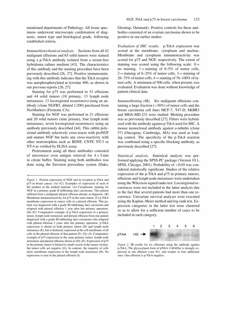
←−−−−−−−−−−−−−−−−−−−−−−−−−−−−−−−−−−−−−Figure 1. Protein expression of NGF and its receptors p-TrkA andp75 in breast cancer. (A)–(C): Examples of expression of each ofthe markers in the studied material. (A) Cytoplasmic staining forNGF in a primary grade II infiltrating duct carcinoma. This patientsuffered from a malignant pleural effusion already at diagnosis. (B)Membrane immunoreactivity for p75 in the same tumor. (C) p-TrkAmembrane expression in cancer cells in a pleural effusion. This pa-tient was diagnosed with a grade III infiltrating duct carcinoma andrelapsed with pleural effusion 1 year after her primary operation.(D)–(F): Comparative example of p-TrkA expression in a primarytumor, lymph node metastasis and pleural effusion from one patientdiagnosed with a grade III infiltrating duct carcinoma who relapsedwith pleural effusion 2 years after her primary operation. p-TrkAexpression is absent in both primary tumor (D) and lymph nodemetastasis (E), but is distinctly expressed at the cell membrane of allcells in the pleural effusion of that patient (F). (G)–(I): Comparativeexample of p75 expression in the same primary tumor, lymph nodemetastasis and pleural effusion shown in (D)–(F). Expression of p75in the primary tumor is limited to small vessels in the tumor vicinity,but tumor cells are negative (G). In contrast, the majority of cellsshow membrane expression in the lymph node metastasis (H). Noexpression is seen in the pleural effusion (I).
Glostrup, Denmark). Positive controls for these anti-bodies consisted of an ovarian carcinoma shown to bepositive in our earlier studies.
Evaluation of IHC results. p-TrkA expression wasscored at the membrane, cytoplasm and nucleus.Membrane and cytoplasm immunoreactivity wasscored for p75 and NGF, respectively. The extent ofstaining was scored using the following scale: 0=no staining, 1= staining of 0–5% of tumor cells,2= staining of 6–25% of tumor cells, 3= staining of26–75% of tumor cells, 4= staining of 76–100% of tu-mor cells. A minimum of 500 cells, when present, wasevaluated. Evaluation was done without knowledge ofpatient clinical data.
Immunoblotting (IB). Six malignant effusions con-taining a large fraction (>50%) of tumor cells and thebreast carcinoma cell lines MCF-7, T47-D, SKBR3and MDA-MD-231 were studied. Blotting procedurewas as previously described [27]. Filters were hybrid-ized with the antibody against p-TrkA used for IHC. Amouse monoclonal antibody against α-tubulin (clone57) (Oncogene, Cambridge, MA) was used as load-ing control. The specificity of the p-TrkA reactionwas confirmed using a specific blocking antibody, aspreviously described [27].
Statistical analysis. Statistical analysis was per-formed applying the SPSS-PC package (Version 10.1,SPSS, Chicago, 2001). Probability of <0.05 was con-sidered statistically significant. Studies of the relativeexpression of the p-TrkA and p75 in primary tumors,effusions and lymph node metastases were undertakenusing the Wilcoxon signed ranks test. Locoregional re-currences were not included in the latter analysis dueto the fact that several patients had more than one re-currence. Univariate survival analyses were executedusing the Kaplan–Meier method and log-rank test. Ex-pression categories in the latter test were clusteredso as to allow for a sufficient number of cases to beincluded in each category.
Figure 2. IB results for six effusions using the antibody againstp-TrkA. The glycosylated form of pTrkA (140 kDa) is strongly ex-pressed in one effusion (case #1), and weaker in four additionalones. One effusion is p-TrkA-negative.

124 B Davidson et al.
Table 2. p75 membrane expression results in the studied material (31 effusions, 18 primarytumors, 13 lymph node metastases, 13 LRa)
Site Percentage of stained carcinoma cellsb
0% 1–5% 6–25% 26–75% 76–100%
Effusion 20 (65%) 9 (29%) 2 (6%) 0 (0%) 0 (0%)
Primary 7 (39%) 10 (56%) 0 (0%) 1 (5%) 0 (0%)
Lymph node 2 (15%) 6 (46%) 1 (8%) 4 (31%) 0 (0%)
LRa 5 (38%) 7 (54%) 0 (0%) 0 (0%) 1 (8%)
a Locoregional recurrences.b Significant for the comparison between effusions and lymph nodes (p= 0.019) and forprimary tumors vs. lymph nodes (p= 0.039).
Table 3. NGF cytoplasmic expression results in the studied material (20 effusions, 9 primarytumors, 4 lymph node metastases, 7 LRa)
Site Percentage of stained carcinoma cells
0% 1–5% 6–25% 26–75% 76–100%
Effusion 14 (59%) 3 (25%) 2 (10%) 1 (6%) 1 (0%)
Primary 6 (67%) 0 (0%) 1 (11%) 1 (11%) 1 (11%)
Lymph node 1 (25%) 0 (0%) 1 (25%) 2 (50%) 0 (0%)
LRa 3 (43%) 0 (6%) 0 (0%) 1 (14%) 3 (43%)
a Locoregional recurrences.
Results
p-TrkA expression is a marker of tumor progressionin breast carcinoma. IHC results for p-TrkA expres-sion at the cell membrane, cytoplasm and nucleus areshown in Table 1. Membrane expression of p-TrkAwas frequent in carcinoma cells in effusions (39/42,93%) and locoregional recurrences (12/13, 92%), withsignificantly lower expression in both primary tumors(14/34, 41%) and lymph node metastases (8/18, 44%),respectively (p < 0.001 for effusions vs. primary tu-mors; p= 0.001 for effusions vs. lymph nodes)(Figure 1). Interestingly, both cytoplasmic (p= 0.004)and nuclear (p= 0.025) expression of p-TrkA washigher in primary tumors compared to effusions. IBconfirmed the expression of p-TrkA in effusion spec-imens (Figure 2), while all four breast carcinomaspecimens were negative (data not shown). p-TrkAexpression did not correlate with TTP.
p75 expression is down-regulated in effusions. p75expression data are shown in Table 2. Staining forthis receptor was highest in lymph nodes (11/14,79%), followed by locoregional recurrences (8/13,62%) and primary tumors (11/18, 62%). In primary
tumors, staining was frequently seen in the in situcomponent of the tumor, including in specimens fail-ing to show expression in the invasive component(not scored). Expression was distinctly lower in ef-fusions (11/31, 35%) (Figure 1). These differences
Figure 3. Kaplan–Meier survival curves showing the correlationbetween NGF expression in carcinoma cells in effusions and TTP.Patients with NGF-negative tumors (=13, solid line) had a meanand median TTP of 6.31 and 6 years, respectively, compared to 3and 4 years for patients with NGF-positive tumors (=7, dashed line)(p= 0.013).

NGF, TrkA and p75 in breast carcinoma 125
were significant for the comparison between effusionsand lymph nodes (p= 0.019) and for primary tumorsversus lymph nodes (p= 0.039). p75 expression didnot correlate with TTP.
NGF expression is seen in a subset of breast carcino-mas and predicts rapid disease progression. NGFexpression data are shown in Table 3 and Figure 1.Staining was comparable at all sites, but due to therelatively small number of solid specimens that wereanalyzed, site-related differences were not evaluatedstatistically. In univariate survival expression of NGFin carcinoma cells in effusions predicted shorter TTP(3 vs. 6.3 years, p= 0.013) (Figure 3).
Discussion
Metastatic disease is the cause of death for the ma-jority of cancer patients. This is also true for breastcarcinoma, a malignancy for which a curative surgicalapproach is attempted in the majority of cases. In viewof the distressingly high incidence of breast cancer,and of the grave prognostic implications related to thepresence of carcinoma cells in the pleural cavity inthis disease, it is nothing but astonishing that not asingle research paper exists to date which deals withthis clinical issue. This may be related to the relativedifficulty in obtaining a significant number of cases inmany centers. The cohort in our study represents a 5-year effort by two medical centers in order to collectsuch material.
Overexpression of growth factor receptors, suchas tyrosine kinase receptors, is one of the pheno-typic changes providing cancer cells with the ability tooverride normal cell growth regulatory mechanisms.Recent studies in murine models have demonstratedthe role of neurotrophin signaling in tumor growthand metastasis [18, 45]. However, the significanceof NGF signaling in epithelial malignancies is largelyunknown to date. In the present study, we report analmost universal membrane expression of p-TrkA inbreast carcinoma cells in pleural effusions and lo-coregional recurrences, with less frequent expressionin primary tumors and lymph node metastases. Thisfinding suggests that upregulation of p-TrkA expres-sion is one of the defining steps in tumor progressionin breast carcinoma. The high expression of p-TrkAin carcinoma cells in effusions is in agreement withour recent study of malignant mesotheliomas, anotheraggressive malignancy with a rapidly fatal course
(Davidson et al., submitted), and are opposite tothe findings for ovarian carcinoma [26, 27]. Ovariancarcinoma is fatal in the majority of cases, but a sig-nificant number of patients currently live 3 and even5 years from diagnosis, a phenomenon that basicallydoes not exist in malignant mesotheliomas or breastcarcinoma that has progressed to effusion. One mayhypothesize that expression of p-TrkA is a marker ofan aggressive clinical course. This would be supportedby our recent report in ovarian carcinoma, where ex-pression of this activated receptor in primary tumorspredicted poor outcome [27], as well as in malignantmelanomas (Flørenes et al., submitted). It is furthersupported by experimental models of breast cancer, inwhich TrkA had been found to activate MAPK signal-ing, resulting in proliferation, independently of p75[36].
Our results are not in agreement with the recentreport of Descamps et al., in which 363 primary breastcarcinomas were studied using real-time quantitativeRT-PCR [39]. In the cohort studied by Descampset al., TrkA mRNA expression predicted improvedoverall survival [39], whereas in our cohort p-TrkAexpression marked progression and did not correlatewith TTP. These differences may be attributed to thefact that we studied the activated receptor, ratherthan the total (pan-TrkA) expression. Another dif-ference may relate to the cohort studied. Descampset al. studied tumors from patients who presented withvariable disease stage, with highly variable clinicaloutcome, whereas our cohort was uniformly com-posed of patients who developed stage IV disease atsome point along the clinical course, and who, despitelong disease-free periods in some cases, all sufferedtumor-related death.
It is noteworthy that cytoplasmic and nuclear ex-pression of p-TrkA was by far less frequent thanmembrane expression in effusions. In fact, cytoplas-mic expression was more frequent in solid tumors atall sites. While the cytoplasmic expression probablyrepresents internalization of the receptor, we do notat present know the significance of nuclear p-TrkAexpression. Still, since it is widely accepted that thebiological function of p-TrkA is exerted mainly at thecell membrane, we regard expression at this cellularlocation as the more significant one.
IB confirmed the findings for effusions, but p-TrkAexpression was not seen in any of the four lines stud-ied. This differs from the phenotype observed by otherinvestigators [34], and may relate primarily to the factthat our lines were not stimulated by NGF, as well

126 B Davidson et al.
as to sub-clones with divergent phenotypes, a knownphenomenon with many commercial cell lines.
Recent studies suggest that the most likely mainfunction of p75 is to regulate Trk receptor activa-tion and signaling, and its presence has been shownto enhance the responsiveness of TrkA to NGF [46].Moreover, expression of p75 has been associated withapoptosis through activation of the stress kinase JNKand with cell survival through activation of nuclear-factor-kB and AKT [47]. In the present study, weobserved a markedly reduced expression of p75 in ef-fusions compared to all solid tumor sites, suggestingthat loss of expression of this receptor is charac-teristic of cells in effusions. This is also supportedby the reduction of expression from 40 to 12% ofcases observed in malignant mesotheliomas (Davidsonet al., submitted). The body cavities are a biologicallystressful environment, in which cells are exposed tosignificantly less effective flow of both oxygen andnutrients. However, we have recently shown that onlyan insignificant fraction of ovarian carcinoma cells ineffusions undergo apoptosis [27], supporting the res-istant nature of these metastatic cells. The reducedexpression of p75 in cancer cells along tumor progres-sion, and particularly in effusions, is also in agreementwith the experimental work by the group of Djakiew,in which p75 has been shown to be a suppressor ofgrowth and metastasis [22, 23]. The loss of the p75-related pro-apoptotic mechanism may provide onebiological explanation as to why the presence of ma-lignant cells in effusions is associated with a rapidlyfatal course in both breast carcinoma and malignantmesothelioma.
Calza et al. have recently demonstrated the roleof NGF in angiogenesis [48], a finding that was con-firmed by a subsequent study of HUVEC cells [49],but the issue of the angiogenic effect of NGF-TrkAsignaling in human cancer has not been investigatedpreviously. We have recently shown co-expression ofNGF and TrkA with angiogenic molecules in ovariancarcinoma, as well as unequivocal expression of ac-tivated TrkA on endothelial cell membranes, bothinside the tumor and in its vicinity [27]. p-TrkAwas only infrequently expressed in endothelial cellsin our breast carcinoma cohort. Nor did the expres-sion of this receptor in carcinoma cells in effusionsor primary tumors show any correlation with that ofangiogenic molecules, recently studied in this cohort(data not shown). These findings suggest a lesser rolefor p-TrkA as angiogenic factor in breast carcinoma,in agreement with its higher expression in effusions,
an anatomic site in which angiogenesis is of lesssignificance.
Expression of growth factors and their receptorsin an autocrine manner is one of the central mech-anisms by which cancer cells evade regulation bynormal physiological systems. Unequivocal expres-sion of NGF was seen in cancer cells in a significantnumber of cases in this study. This finding is in agree-ment with the recent report by Dolle et al., in whichactive NGF has been shown to be produced by breastcancer cells, and in which blocking of the TrkA re-ceptor led to reduced cellular growth, thus providingevidence of an autocrine loop [34]. Furthermore, al-though our study analyzed a relatively small numberof specimens, NGF expression in effusions predictedsignificantly shorter TTP. The cohort in this study is aselected one, and consists of patients with uniformlypoor outcome. Nevertheless, we now report on thefirst documented marker in the literature that is ableto predict the time interval between primary diagnosisand the detection of pleural effusion in breast cancer.
In conclusion, the expression of the activatedneurotrophin receptor p-TrkA is upregulated in effu-sions and locoregional recurrences compared to earlylesions (primary tumors and lymph node metastases)in breast carcinoma, with inverse findings for p75 ineffusions. NGF is the first molecular marker that isable to predict the time interval to progression, sug-gesting that signaling through the NGF-TrkA pathwayin an autocrine manner may be involved in more rapidtumor progression in this disease.
Acknowledgements
We gratefully acknowledge the competent technicalhelp of Mrs Inger-Liv Nordli, Mrs Mai Nguyen, MrsErika Thorbjørnsen, Mrs Ann Larsen (immunohisto-chemistry), Mrs Martina Skerede (immunoblotting),and Mrs Elisabeth Emilsen (cell lines) at the De-partment of Pathology, The Norwegian Radium Hos-pital. We thank the Norwegian Cancer Society for itssupport of this work.
References
1. Barbacid M: Neurotrophic factors and their receptors. CurrOpin Cell Biol 7: 148–155, 1995
2. Kaplan DR, Miller FD: Signal transduction by the neuro-trophin receptors. Curr Opin Cell Biol 9: 213–221, 1997

NGF, TrkA and p75 in breast carcinoma 127
3. Patapoutian A, Reichardt LF: Trk receptors: mediators ofneurotrophin action. Curr Opin Neurobiol 11: 272–280,2001
4. Barker PA, Lomen-Hoerth C, Gensch EM, Meakin SO, GlassDJ, Shooter EM: Tissue-specific alternative splicing gener-ates two isoforms of the trkA receptor. J Biol Chem 268:15150–15157, 1993
5. Weismann C, Ultsch MH, Bass SH, de Vos AM: Crystal struc-ture of the nerve growth factor in complex with the ligand-binding domain of the TrkA receptor. Nature 401: 184–188,1999
6. Greene LA, Kaplan DR: Early events in neurotrophin sig-nalling via Trk and p75 receptors. Curr Opin Neurobiol 5:579–587, 1995
7. Kaplan DR, Miller FD: Neutrophin signal transduction in thenervous system. Curr Opin Neurobiol 10: 381–391, 2000
8. Rudkin BB, Lazarovici P, Levi BZ, Abe Y, Fugita K, Guroff G:Cell cycle-specific action of nerve growth factor in PC12 cells:differentiation without proliferation. EMBO J 11: 3319–3325,1989
9. Urdiales JL, Becker E, Andrieu M, Thomas A, Jullien J, vanGrunsven LA, Menut S, Evan GI, Martin-Zanca D, RudkinBB: Cell cycle phase-specific surface expression of nervegrowth factor receptors TrkA and p75NTR. J Neurosci 17:6767–6775, 1998
10. Gollapudi L, Neet KE: Different mechanisms for inhibition ofcell proliferation via cell cycle proteins in PC12 cells by nervegrowth factor and staurosporine. J Neurosci Res 49: 461–474,1997
11. Billon N, van Grunsven LA, Rudkin BB: The CDK inhibitorp21WAF1/Cip1 is induced through a p300-dependent mechan-ism during NGF-mediated neuronal differentiation of PC12cells. Oncogene 13: 2047–2054, 1996
12. Montano X: p53 associates with trk tyrosine kinase. Oncogene15: 245–246, 1997
13. Brown A, Browes C, Mitchell M, Montano X: c-abl is involvedin the association of p53 and trk A. Oncogene 19: 3032–3040,2000
14. Nakagawara A: Trk receptor tyrosine kinases: a bridgebetween cancer and neural development. Cancer Lett 169:107–114, 2001
15. Shibayama E, Koizumi H: Cellular localization of the Trkneurotrophin receptor family in human non-neuronal tissues.Am J Pathol 148: 1807–1818, 1996
16. McGregor LM, McCune BK, Graff JR, McDowell PR,Romans KE, Yancopoulos GD, Ball DW, Baylin SB, NelkinBD: Roles of trk family neurotrophin receptors in medullarythyroid carcinoma development and progression. Proc NatlAcad Sci USA 96: 4540–4545, 1999
17. Katzir I, Shani J, Shabashov D, Dagan J, Lazarovici P: Es-tablishment and characterization of pheochromocytoma tumormodel expressing different levels of trkA receptors. CancerLett (in press)
18. Miknyoczki SJ, Wan W, Chang H, Dobrzanski P, Ruggeri BA,Dionne CA, Buchkovich K: The neurotrophin-Trk receptoraxes are critical for the growth and progression of humanprostatic carcinoma and pancreatic ductal adenocarcinoma innude mice. Clin Cancer Res 8: 1924–1931, 2002
19. Nakagawara A, Arima M, Azar CG, Scavarda NJ, BrodeurGM: Inverse relationship between trk expression and N-myc amplification in human neuroblastomas. Cancer Res 52:1364–1368, 1992
20. Suzuki E, Bogenmann H, Shimada D, Stram RC, Seeger RC:Lack of high-affinity nerve growth factor receptors in ag-
gressive neuroblastomas. J Natl Cancer Inst 85: 377–384,1993
21. Fanburg-Smith JC, Miettinen M: Low-affinity nerve growthfactor receptor (p75) in dermatofibrosarcoma protuberans andother nonneuronal tumors: a study of 1,150 tumors and fetaland adult normal tissues. Hum Pathol 32: 976–983, 2001
22. Krygier S, Djakiew D: Neurotrophin receptor p75NTR sup-presses growth and nerve growth factor-mediated metastasisof human prostate cancer cells. Int J Cancer 98: 1–7, 2002
23. Pflug B, Djakiew D: Expression of p75NTR in human prostateepithelial tumor cell line reduces nerve growth factor-inducedcell growth by activation of programmed cell death. MolCarcinog 23: 106–114, 1998
24. Pflug BR, Onoda M, Lynch JH, Djakiew D: Reduced expres-sion of the low affinity nerve growth factor receptor in benignand malignant human prostate tissue and loss of expression infour human metastatic prostate tumor cell lines. Cancer Res52: 5403–5406, 1992
25. MacGrogan D, Saint-Andre’ JP, Dicou E: Expression of nervegrowth factor and nerve growth factor receptor genes in humantissues and in prostatic adenocarcinoma cell lines. J Neuro-chem 59: 1381–1391, 1992
26. Davidson B, Lazarovici P, Ezersky A, Nesland JM, BernerA, Risberg B, Trope’ CG, Kristensen GB, Goscinski M, vande Putte G, Reich R: Expression levels of the NGF receptorsTrkA and p75 in effusions and solid tumors of serous ovariancarcinoma patients. Clin Cancer Res 7: 3457–3464, 2001
27. Davidson B, Reich R, Lazarovici P, Nesland JM, RisbergB, Trope’ CG, Flørenes VA: Expression and activation ofthe nerve growth factor receptor TrkA in serous ovariancarcinoma. Clin Cancer Res 9: 2248–2259, 2003
28. Greenlee RT, Hill-Harmon MB, Murray T, Thun M: Cancerstatistics, 2001. CA Cancer J Clin 51: 15–36, 2001
29. Hausheer FH, Yarbro JW: Diagnosis and treatment of malig-nant pleural effusion. Semin Oncol 12: 54–75, 1985
30. Martinez-Moragon E, Aparicio J, Sanchis J, Menendez R,Cruz Rogado M, Sanchis F: Malignant pleural effusion: pro-gnostic factors for survival and response to chemical pleur-odesis in a series of 120 cases. Respiration 65: 108–113,1998
31. Wilkes JD, Fidias P, Vaickus L, Perez RP: Malignancy-relatedpericardial effusion. 127 cases from the Roswell Park CenterInstitute. Cancer 76: 1377–1387, 1995
32. Dieterich M, Goodman SN, Rojas-Corona RR, Emralino AB,Jimenez-Joseph D, Sherman ME: Multivariate analysis ofprognostic features in malignant pleural effusions from breastcancer patients. Acta Cytol 38: 945–952, 1994
33. Banerjee AK, Willetts I, Robertson JF, Blamey RW: Pleuraleffusion in breast cancer: a review of the Nottingham experi-ence. Eur J Surg Oncol 20: 33–36, 1994
34. Dolle L, El Yazidi-Belkoura I, Adriaenssens E, NurcombeV, Hondermarck H: Nerve growth factor overexpressionand autocrine loop in breast cancer cells. Oncogene 22:5592–5601, 2003
35. Descamps S, Lebourhis X, Delehedde M, Boilly B,Hondermarck H: Nerve growth factor is mitogenic for cancer-ous but not normal human breast epithelial cells. J Biol Chem273: 16659–16662, 1998
36. Descamps S, Toillon RA, Adriaenssens E, Pawlowski V,Cool SM, Nurcombe V, Le Bourhis X, Boilly B, Peyrat JP,Hondermarck H: Nerve growth factor stimulates proliferationand survival of human breast cancer cells through two dis-tinct signaling pathways. J Biol Chem 276: 17864–17870,2001

128 B Davidson et al.
37. El Yazidi-Belkoura I, Adriaenssens E, Dolle L, Descamps S,Hondermarck H: Tumor necrosis factor receptor-associateddeath domain protein is involved in the neurotrophin receptor-mediated antiapoptotic activity of nerve growth factor in breastcancer cells. J Biol Chem 278: 16952–16956, 2003
38. Chiarenza A, Lazarovici P, Lempereur L, Cantarella G,Bianchi A, Bernardini R: Tamoxifen inhibits nerve growthfactor-induced proliferation of the human breast cancerousline MCF-7. Cancer Res 61: 3002–3008
39. Descamps S, Pawlowski V, Revillion F, Hornez L, HebbarM, Boilly B, Hondermarck H, Peyrat JP: Expression of nervegrowth factor receptors and prognostic value in human breastcancer. Cancer Res 61: 4337–4340, 2001
40. Aragona M, Panetta S, Silipigni AM, Romeo DL, Pastura G,Mesiti M, Cascinu S, Torre F: Nerve growth factor receptorimmunoreactivity in breast cancer patients. Cancer Invest 19:692–697, 2001
41. Davidson B, Risberg B, Kristensen G, Kvalheim G, EmilsenE, Bjåmer A, Berner A: Detection of cancer cells in effusionsfrom patients diagnosed with gynecological malignancies-evaluation of five epithelial markers. Virchows Arch 435:43–49, 1999
42. Davidson B, Nielsen S, Christensen J, Asschenfeldt P, BernerA, Risberg B, Johansen P: The role of Desmin and N-cadherinin effusion cytology. A comparative study using establishedmarkers of mesothelial and epithelial cells. Am J Surg Pathol25: 1405–1412, 2001
43. Hochman J, Park SS, Lazarovici P, Bergel M, GottesmanMM: Monoclonal antibodies to immunogenic lymphoma cellvariants displaying impaired neoplastic properties: character-ization and applications. J Natl Cancer Inst 82: 1821–1826,1990
44. Katzir I, Shani J, Regev K, Shabashov D, Lazarovici P: Aquantitative bioassay for nerve growth factor, using PC12clones expressing different levels of trkA receptors. J MolNeurosci 18: 251–264, 2002
45. Weeraratna AT, Dalrymple SL, Lamb JC, Denmeade SR,Miknyoczki S, Dionne CA, Isaacs JT: Pan-trk inhibition de-creases metastasis and enhances host survival in experimentalmodels as a result of its selective induction of apoptosisof prostate cancer cells. Clin Cancer Res 7: 2237–2245,2001
46. Segal RA, Greenberg ME: Intracellular signaling pathwaysactivated by neurotrophic factors. Annu Rev Neurosci 19:463–489, 1996
47. Roux PP, Bhakar AL, Kennedy TE, Barker PA: The p75neurotrophin receptor activates Akt (protein kinase B) througha phosphatidylinositol 3-kinase-dependent pathway. J BiolChem 276: 23097–23104, 2001
48. Calza L, Giardino L, Guiliani A, Aloe L, Levi-MontalciniR: Nerve growth factor control of neuronal expression of an-giogenic and vasoactive factors. Proc Natl Acad Sci USA 98:4160–4165, 2001
49. Cantarella G, Lempereur L, Presta M, Ribatti D,Lombardo G, Lazarovici P, Zappala G, Pafumi C, BernardiniR: Nerve growth factor-endothelial cell interaction leads toangiogenesis in vitro and in vivo. FASEB J 16: 1307–1309,2002
Address for offprints and correspondence: Dr Ben Davidson,Department of Pathology, The Norwegian Radium Hospital,Montebello, N-0310 Oslo, Norway; Tel.: 47-22934871; Fax: 47-22508554; E-mail: [email protected]