Ear ventilation tubes in children with otitis media with effusion ...
10
Romanian Journal of Rhinology, Vol. 1, No. 4, October - December 2011 Corresponding author: Vlad Postelnicu email: [email protected] Vlad Postelnicu 1 , Oana Iosif 1 , Viorel Rosu 2 , Raluca Bidiga 3 , Miruna Georgescu 3 , Constantin Giosanu 3 , Gabriela Oproiu 4 , Tatiana Serban 5 , Monica Zamfirescu 6 , Mihaela Marcu-Cristescu 3 1 MEDLIFE Baneasa, Bucharest, Romania - ENT Department 2 MEDLIFE Grivita, Bucharest, Romania - Radiology Department 3 MEDLIFE Pediatrics Hospital, Bucharest, Romania - Pediatrics Department 4 IOMC “Prof.Dr. A. Rusescu”, Bucharest, Romania - Pediatrics Clinic 5 MEDLIFE Baneasa, Bucharest, Romania - Endocrinology Department 6 MEDLIFE Unirii, Bucharest, Romania - Allergology Department Ear ventilation tubes in children with otitis media with effusion and adenoid hypertrophy ORIGINAL STUDY INTRODUCTION Acute otitis media (AOM) represents one of the most frequent diseases of children under 5 years old, and most of them require sustained medical therapy due to its possible complications and the total period of hearing impairment on the individual. Early diagnosis of otitis media in children has a crucial role in normal speech development, as the most important period to acquire it is between two and four years old 1-4 . ABSTRACT BACKGROUND. Acute otitis media is one of the most frequent diseases of children under 5 years, and most of the cases require sustained medical therapy due to possible complications and the total period of hearing impairment. Early detection of the otitis media with effusion (OME) in children is important for normal speech development. OBJECTIVE. The aim of the study is to evaluate the efficacy of adenoidectomy versus adenoidectomy with ear tubes fixation at the same time of surgery, in a selected lot of patients diagnosed with adenoid hypertrophy, OME and a history of recurrent OME. MATERIAL AND METHODS. The study was carried out on 40 selected children in the age range 2-7 years, diagnosed with adenoid hypertrophy accompanied by uni/bilateral recurrent OME. All patients underwent adenoidectomy; tympanotomy with ear tube fixation was performed in 35% cases. RESULTS. Bilateral conductive hearing loss (CHL) was found in 80% of the patients. In those patients with OME at the time of surgery, who underwent adenoidectomy as single procedure, complete remission of the CHL level was achieved up to 1 month. Immediate remission of CHL was achieved in all patients who underwent tympanotomy and ear tube fixa- tion; remission maintained also at 14 and 30 days. Recurrence of OME and CHL was present in 42.3% patients who un- derwent adenoidectomy alone; the remission of the recurrences was achieved with medical treatment up to 21 days in most patients. Recurrence of ear discharge on blocked ear tubes was noted in 21.4%; the duration of the ear discharge and CHL until remission was up to 7 days. All recurrences were secondary to acute upper respiratory tract infections (URI). CONCLUSIONS. Adenoidectomy provides a faster remission of OME and is effective in preventing its recurrences. The recurrences of OME have a much smaller healing period after adenoidectomy. Ear tubes fixation ensures the opportunity of a faster recovery of the ear, immediately reducing the CHL secondary to OME. The duration of CHL is much shorter in recurrences of ear discharge on blocked ear tubes than in patients without ear tubes. Early drain- age of the middle ear effusion ensures improvement of the quality of life, both immediately after surgery and on a long term basis, supporting the ventilation of the mastoid air cells, essential for the normal functioning of the ear. KEYWORDS: otitis media, ear tubes, adenoidectomy
-
Upload
khangminh22 -
Category
Documents
-
view
0 -
download
0
Transcript of Ear ventilation tubes in children with otitis media with effusion ...
Romanian Journal of Rhinology, Vol. 1, No. 4, October - December 2011
Corresponding author: Vlad Postelnicuemail: [email protected]
Vlad Postelnicu1, Oana Iosif1, Viorel Rosu2, Raluca Bidiga3, Miruna Georgescu3, Constantin Giosanu3, Gabriela Oproiu4, Tatiana Serban5, Monica Zamfirescu6, Mihaela Marcu-Cristescu3
1MEDLIFE Baneasa, Bucharest, Romania - ENT Department2MEDLIFE Grivita, Bucharest, Romania - Radiology Department3MEDLIFE Pediatrics Hospital, Bucharest, Romania - Pediatrics Department4IOMC “Prof.Dr. A. Rusescu”, Bucharest, Romania - Pediatrics Clinic5MEDLIFE Baneasa, Bucharest, Romania - Endocrinology Department6MEDLIFE Unirii, Bucharest, Romania - Allergology Department
Ear ventilation tubes in children with otitis media with effusion and adenoid hypertrophy
ORIGINAL STuDy
INTRODuCTION
Acute otitis media (AOM) represents one of the most frequent diseases of children under 5 years old, and most of them require sustained medical therapy
due to its possible complications and the total period of hearing impairment on the individual. Early diagnosis of otitis media in children has a crucial role in normal speech development, as the most important period to acquire it is between two and four years old1-4.
ABSTRACT
BACKGROuND. Acute otitis media is one of the most frequent diseases of children under 5 years, and most of the cases require sustained medical therapy due to possible complications and the total period of hearing impairment. Early detection of the otitis media with effusion (OME) in children is important for normal speech development.OBJECTIVE. The aim of the study is to evaluate the efficacy of adenoidectomy versus adenoidectomy with ear tubes fixation at the same time of surgery, in a selected lot of patients diagnosed with adenoid hypertrophy, OME and a history of recurrent OME.MATERIAL AND METHODS. The study was carried out on 40 selected children in the age range 2-7 years, diagnosed with adenoid hypertrophy accompanied by uni/bilateral recurrent OME. All patients underwent adenoidectomy; tympanotomy with ear tube fixation was performed in 35% cases.RESuLTS. Bilateral conductive hearing loss (CHL) was found in 80% of the patients. In those patients with OME at the time of surgery, who underwent adenoidectomy as single procedure, complete remission of the CHL level was achieved up to 1 month. Immediate remission of CHL was achieved in all patients who underwent tympanotomy and ear tube fixa-tion; remission maintained also at 14 and 30 days. Recurrence of OME and CHL was present in 42.3% patients who un-derwent adenoidectomy alone; the remission of the recurrences was achieved with medical treatment up to 21 days in most patients. Recurrence of ear discharge on blocked ear tubes was noted in 21.4%; the duration of the ear discharge and CHL until remission was up to 7 days. All recurrences were secondary to acute upper respiratory tract infections (URI).CONCLuSIONS. Adenoidectomy provides a faster remission of OME and is effective in preventing its recurrences. The recurrences of OME have a much smaller healing period after adenoidectomy. Ear tubes fixation ensures the opportunity of a faster recovery of the ear, immediately reducing the CHL secondary to OME. The duration of CHL is much shorter in recurrences of ear discharge on blocked ear tubes than in patients without ear tubes. Early drain-age of the middle ear effusion ensures improvement of the quality of life, both immediately after surgery and on a long term basis, supporting the ventilation of the mastoid air cells, essential for the normal functioning of the ear.KEywORDS: otitis media, ear tubes, adenoidectomy
201Postelnicu et al Ear ventilation tubes in children with otitis media...
Otitis media with effusion (OME) represents an ac-cumulation of non-purulent fluid of various viscosities within the middle ear cavity and is a nonspecific in-flammation2,5. The inflammation of the middle ear results in metaplasia of the middle ear mucosa, with the proliferation of mucus glands and goblet cells6. Pathophysiology of otitis media with effusion usually involves inflammatory conditions of the nasopharynx, any mechanism that causes dysfunction of the Eusta-chian tube reducing the pressure or aeration of the middle ear. This mechanism causes negative pressure in cavum tympani, leading to formation of serous effu-sion and metaplastic transformation of the mucosa in time. Mucous effusion is mostly seen in children, while in adults we mostly find the serous effusion7-10. The study performed on children forced us from the be-ginning to pay attention to the complications that the middle ear diseases may cause: chronic otitis, recur-rent acute otitis, inflammation of the mastoid tissue (acute or chronic mastoiditis), retraction pockets and adhesive complication, tympanic membrane perfora-tion, tympanosclerosis, cholesteatoma, toxic labyrin-thitis, delay in speech and language development due to the total period of hearing loss1-15.
Literature reminds about the adenoids role in pro-ducing IgA, as one of the first mechanisms of protec-tion against invasion of microorganisms and foreign molecules. Adenoid hypertrophy involves abnormal growth of adenoid tissue in the nasopharynx that can entirely block the nasal air flow; 5 is considered the maximum age when middle ear complications accom-panying adenoids can occur16-19.
MATERIAL AND METHODS
We admitted in this study 40 children diagnosed with adenoid hypertrophy and otitis media with effu-sion at the time of surgery. The patients presented in our ENT department between April 2010 and Febru-ary 2011 and were aged between 1 year 11 months and 6 years 11 months at the time of surgery. The mean interval between first visit and surgery was 71 days.
We will comment upon several aspects regarding otitis media with effusion in children with adenoid hy-pertrophy:
• the opportunity of ear tubes fixation;• the total period of the conductive hearing loss
which accompanies the otitis media with effusion;• attempt to make a correlation between the pres-
ence of adenoids and repeated otitis media with effusion and conductive hearing loss in children.
Study admission criteria:• Presence of adenoid hypertrophy;• Presence of otitis media with effusion at the time
of surgery;
• History of recurrent otitis media with effusion.Study exclusion criteria:• Absence of adenoid hypertrophy;• History of previous adenoidectomy or ear sur-
gery;Preoperatory diagnosis:• All patients included in the study benefited
from:� Nasal endoscopy, using a flexible 2.5mm fiber-
scope;� Ear endoscopy, using a 0-degree, 3 mm rigid tele-
scope;� Tympanogram and stapedian reflex;� Pure tone or behavioral audiogram - where pos-
sible, depending on patient’s age;� Subjective appreciation by parents of nasal ob-
struction, hearing level, snoring, quality of sleep, quality of life;
• Radiological examination was performed in cer-tain cases with repetitive acute otitis media, otitis media with effusion unresponsive to treatment, re-peated acute suppurative otitis media, spontaneous perforation of the tympanic membrane - Schüller’s in-cidence X-ray, CT/MRI.
Postoperatory follow-up:• Control after 3, 10 days and 1, 3 and 6 months and
every time the parents considered it necessary (upper respiratory tract infections, subjective in-crease of the hearing impairment etc.);
• The same diagnostic procedures as in preopera-tory diagnosis.
Treatment.a. Preoperatory treatment. All the children under-
went nasal lavage with isotones saline nasal sprays at least twice a day at home, from the first visit; those patients with moderate and severe nasal obstruction received topical nasal decongestants for 7-14 days; some patients, especially those who had a history of allergic rhinitis, received nasal corticosteroids once a day after toileting the nose with saline nasal sprays. In acute inflammatory diseases of the ear - acute conges-tive otitis media, acute suppurative otitis media - the patients received intra-auricular anti-inflammatory therapy for pain relief for a period of 1 to 5 days; in certain patients with acute suppurative otitis media, oral antibiotics were recommended.
b. Surgery. All patients underwent adenoidectomy with/without tympanotomy and ear ventilation tube fixation in the same conditions involving surgery; the operation was performed under general anesthesia, with resection of the adenoids in the head-extended position (“hanging position”)20 under endoscopic sur-veillance20,21,22. The resected specimens (adenoids) were sent for anatomo-pathological examination. The biopsy samples were formalin-fixed, paraffin-embed-ded and histopathologically examined; the routine

202 Romanian Journal of Rhinology, Vol. 1, No. 4, October - December 2011
stain used was Haematoxylin and Eosin for the assess-ment of the histopathological aspects23. Tympanotomy was done under optical microscopy in the same opera-tory sequence as secondary stage. Tympanotomy was followed by the suction of the middle ear discharge and lavage of the middle ear with Dexamethasone 21-phosfate in all the cases24,25,26. The secretions col-lected from middle ear mucosa were sent for bacterio-logical examination.
c. Postoperatory treatment. In the post-surgery sur-veillance period, the children continued nasal clean-ing with saline solution and decreased the use of nose drops with vasoconstriction effect only for the periods of acute upper respiratory tract infections. In those cases with recurrence of ear discharge on blocked ear tubes, we performed lavage of the ear tubes with Dex-amethasone and active drainage of the effusion trough the ear tube; these patients received local antibiotic ear solutions for up to 5 days.
RESuLTS
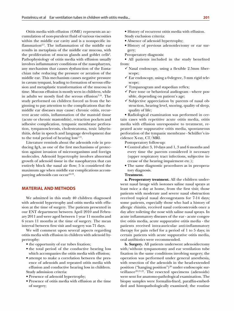
The highest incidence of adenoid hypertrophy ac-companied by otitis media with effusion was found in 3 year-old children (Figure 1). At the time of surgery, 72.5%29 of all children included in this study presented bilateral otitis media with effusion (acute, in remission or chronic otitis media with effusion), 7.5%3 otitis media with effusion and Eustachian tube dysfunction (ETD) and only 20%8 unilateral otitis media with effu-sion. Implicitly, 80% of children had bilateral conduc-
tive hearing loss of different degrees at the time of surgery (Figure 2).
The 40 patients included in this study were divided into 2 lots:
• first lot: 26 patients (65%) - treated surgical, only adenoidectomy;
• second lot: 14 patients (35%) - treated also surgi-cal, adenoidectomy and uni- or bilateral tympano-tomy with ear ventilation tube fixation.
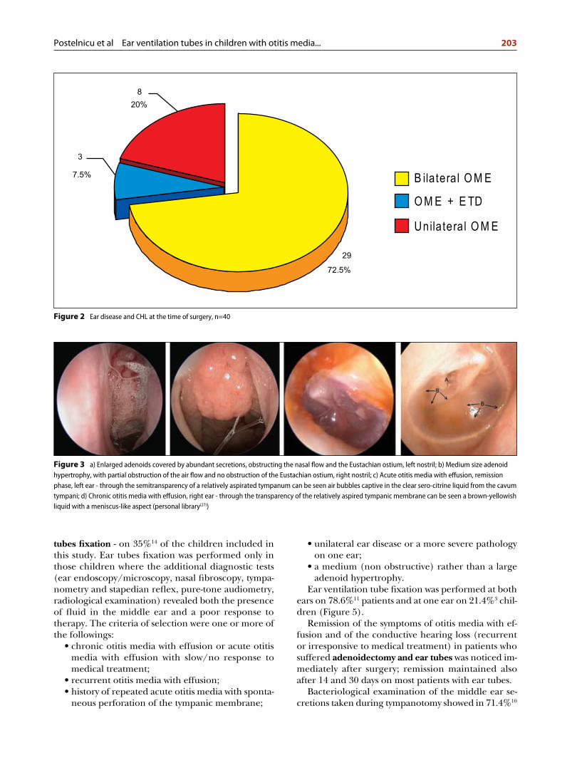
In the first lot of patients we performed adenoidec-tomy as a single procedure - on 65%26 of the children included in this study that presented at the time of surgery:
• otitis media with effusion acute or in a remission phase, and
• a large package of adenoids in the rhinopharynx (Figure 3).
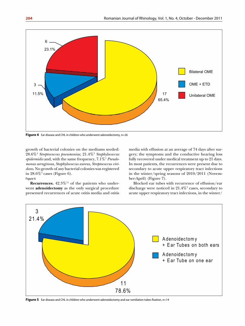
Bilateral ear disease (bilateral otitis media with effu-sion, otitis media with effusion and Eustachian tube dysfunction) and, implicitly, bilateral conductive hear-ing loss, were found in 76.9%20 of the children from the first lot. Taking into consideration the average pe-riod of 71 days between the first visit and surgery, we make a special remark on the duration of the conduc-tive hearing loss of different degrees, but in remission, present in these cases (Figure 4).
Remission of the symptoms of recurrent otitis media with effusion and of the conductive hearing loss in pa-tients who suffered adenoidectomy as single proce-dure was acquired up to 1 month after surgery.
In the second lot of patients we performed ade-noidectomy and tympanotomy with ear ventilation
Figure 1 Age distribution, n=40
203Postelnicu et al Ear ventilation tubes in children with otitis media...
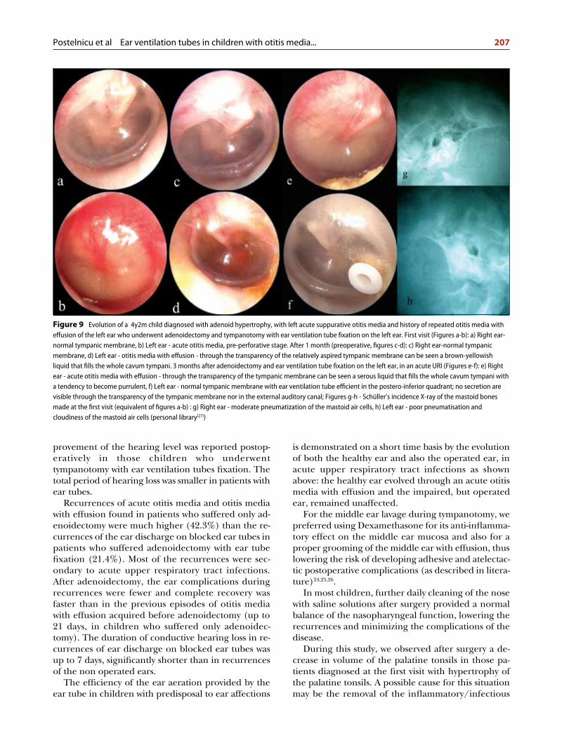
tubes fixation - on 35%14 of the children included in this study. Ear tubes fixation was performed only in those children where the additional diagnostic tests (ear endoscopy/microscopy, nasal fibroscopy, tympa-nometry and stapedian reflex, pure-tone audiometry, radiological examination) revealed both the presence of fluid in the middle ear and a poor response to thera py. The criteria of selection were one or more of the followings:
• chronic otitis media with effusion or acute otitis media with effusion with slow/no response to medical treatment;
• recurrent otitis media with effusion;• history of repeated acute otitis media with sponta-
neous perforation of the tympanic membrane;
• unilateral ear disease or a more severe pathology on one ear;
• a medium (non obstructive) rather than a large adenoid hypertrophy.
Ear ventilation tube fixation was performed at both ears on 78.6%11 patients and at one ear on 21.4%3 chil-dren (Figure 5).
Remission of the symptoms of otitis media with ef-fusion and of the conductive hearing loss (recurrent or irresponsive to medical treatment) in patients who suffered adenoidectomy and ear tubes was noticed im-mediately after surgery; remission maintained also after 14 and 30 days on most patients with ear tubes.
Bacteriological examination of the middle ear se-cretions taken during tympanotomy showed in 71.4%10
Figure 2 Ear disease and CHL at the time of surgery, n=40
Figure 3 a) Enlarged adenoids covered by abundant secretions, obstructing the nasal flow and the Eustachian ostium, left nostril; b) Medium size adenoid hypertrophy, with partial obstruction of the air flow and no obstruction of the Eustachian ostium, right nostril; c) Acute otitis media with effusion, remission phase, left ear - through the semitransparency of a relatively aspirated tympanum can be seen air bubbles captive in the clear sero-citrine liquid from the cavum tympani; d) Chronic otitis media with effusion, right ear - through the transparency of the relatively aspired tympanic membrane can be seen a brown-yellowish liquid with a meniscus-like aspect (personal library(27))
204 Romanian Journal of Rhinology, Vol. 1, No. 4, October - December 2011
growth of bacterial colonies on the mediums seeded: 28.6%4 Streptococcus pneumoniae, 21.4%3 Staphylococcus epidermidis and, with the same frequency, 7.1%1 Pseu domonas aeruginosa, Staphylococcus aureus, Streptococcus viridans. No growth of any bacterial colonies was registered in 28.6%4 cases (Figure 6).Figure 6
Recurrences. 42.3%11 of the patients who under-went adenoidectomy as the only surgical procedure presented recurrences of acute otitis media and otitis
media with effusion at an average of 74 days after sur-gery; the symptoms and the conductive hearing loss fully recovered under medical treatment up to 21 days. In most patients, the recurrences were present due to secondary to acute upper respiratory tract infections in the winter/spring seasons of 2010/2011 (Novem-ber-April) (Figure 7).
Blocked ear tubes with recurrence of effusion/ear discharge were noticed in 21.4%3 cases, secondary to acute upper respiratory tract infections, in the winter/
Figure 4 Ear disease and CHL in children who underwent adenoidectomy, n=26
Figure 5 Ear disease and CHL in children who underwent adenoidectomy and ear ventilation tubes fixation, n=14

205Postelnicu et al Ear ventilation tubes in children with otitis media...
spring seasons of 2010/2011 (November-April): 2 of them bilateral and 1 unilateral; the average period of recurrence of ear discharge was at 62 days from sur-gery (Figure 7). In these cases, the total duration of conductive hearing loss was much shorter than in pre-vious episodes with otitis media with effusion without ear tubes - the dryness of the ear was obtained up to 7
days under local treatment. In Figure 8 we present the evolution of a left ear of a 5 year-old patient with ear tubes and acute ear discharge on blocked ear tube after 60 days from surgery.
45.5%5 patients with bilateral ear tubes developed acute upper respiratory tract infections at more than 1 month from surgery but without any affection of the
Figure 6 Result of the bacteriological examination of the ear discharge taken during tympanotomy, n=14
Figure 7 Recurrence of ear effusion in patients with adenoidectomy +/- ear tube fixation (green-patients who underwent the specified procedure, red-recurrence), n=40
Streptococcus pneumoniae
Staphylococcusepidermitis
Pseudomonasaeruginosa
Staphylococcusaureus
Streptococcus viridans
No growth

206 Romanian Journal of Rhinology, Vol. 1, No. 4, October - December 2011
ears (as their major common complaint was before surgery), due to the presence of the ventilation tubes.
14.3%2 children developed otitis media with effu-sion secondary to an acute upper respiratory tract infection; after 1 month the ear tubes were sponta-neously eliminated; remission of otitis media with effusion was obtained after 1 month of medical treatment.
Two children who suffered adenoidectomy and uni-lateral ear tube fixation developed at more than 3 months after surgery acute otitis media in the healthy ear (the ear with normal pneumatization of the mas-toid air cells on Schüller’s X-ray), secondary to an acute upper respiratory tract infection. The ear with ventilation tube (the one with absence of pneumatiza-tion of the mastoid air cells on Schüller’s X-ray) re-mained unaffected in the acute inflammatory state
due to the presence of the ventilation tube, thus the hearing level of the children through the healing pe-riod remained almost normal on the operated ear. Remission of the acute otitis media with effusion, sec-ondary to acute upper respiratory tract infections of the non-operated ear, was obtained up to 14 days after medical therapy (nasal decongestants) in both pa-tients (Figure 9).
DISCuSSIONS
Significant improvement of the quality of life (nasal breathing, snoring, sleep apnea, proficiency in school) was reported by parents after surgery. Complete remis-sion of the hearing loss in patients with otitis media with effusion, who suffered only adenoidectomy, was registered up to 1 month after surgery. Significant im-
Figure 8 Evolution of a left OME on a 5 years old patient, endoscopic images: a) Enlarged adenoids covered by abundant secretions, obstructing the nasal flow and the Eustachian ostium, right nostril, at the first visit; b) OME, left ear - through the transparency of the tympanum can be seen air bubbles captive in a sero-citrine liquid, first visit; c) Ear ventilation tube, 4 days after surgery - normal tympanic membrane with no secretions in cavum tympani; d) AOM after an URI - ear ventilation tube blocked by thick, sticky secretions; through the semi-transparency of the tympanum can be seen a purulent discharge which is expanding, pushing and thinning the healthy tissues - 60 days from surgery; e) Abortive evolution of the AOM under local treatment - ear tube blocked by dried yellow gummy secretions; through the transparency of the tympanum serous secretions filling the whole cavum tympani can be seen; dried yellow gummy secretions in the external auditory canal - 66 days from surgery; f) Abortive evolution of the AOM - ear tube permeable, through which the serous secretion from the middle ear drains into the external auditory canal - 69 days from surgery (personal library(27))
207Postelnicu et al Ear ventilation tubes in children with otitis media...
Figure 9 Evolution of a 4y2m child diagnosed with adenoid hypertrophy, with left acute suppurative otitis media and history of repeated otitis media with effusion of the left ear who underwent adenoidectomy and tympanotomy with ear ventilation tube fixation on the left ear. First visit (Figures a-b): a) Right ear-normal tympanic membrane, b) Left ear - acute otitis media, pre-perforative stage. After 1 month (preoperative, figures c-d): c) Right ear-normal tympanic membrane, d) Left ear - otitis media with effusion - through the transparency of the relatively aspired tympanic membrane can be seen a brown-yellowish liquid that fills the whole cavum tympani. 3 months after adenoidectomy and ear ventilation tube fixation on the left ear, in an acute URI (Figures e-f): e) Right ear - acute otitis media with effusion - through the transparency of the tympanic membrane can be seen a serous liquid that fills the whole cavum tympani with a tendency to become purrulent, f) Left ear - normal tympanic membrane with ear ventilation tube efficient in the postero-inferior quadrant; no secretion are visible through the transparency of the tympanic membrane nor in the external auditory canal; Figures g-h - Schüller’s incidence X-ray of the mastoid bones made at the first visit (equivalent of figures a-b) : g) Right ear - moderate pneumatization of the mastoid air cells, h) Left ear - poor pneumatisation and cloudiness of the mastoid air cells (personal library(27))
provement of the hearing level was reported postop-eratively in those children who underwent tym pa no tomy with ear ventilation tubes fixation. The to tal period of hearing loss was smaller in patients with ear tubes.
Recurrences of acute otitis media and otitis media with effusion found in patients who suffered only ad-enoidectomy were much higher (42.3%) than the re-currences of the ear discharge on blocked ear tubes in patients who suffered adenoidectomy with ear tube fixation (21.4%). Most of the recurrences were sec-ondary to acute upper respiratory tract infections. After adenoidectomy, the ear complications during recurrences were fewer and complete recovery was faster than in the previous episodes of otitis media with effusion acquired before adenoidectomy (up to 21 days, in children who suffered only adenoidec-tomy). The duration of conductive hearing loss in re-currences of ear discharge on blocked ear tubes was up to 7 days, significantly shorter than in recurrences of the non operated ears.
The efficiency of the ear aeration provided by the ear tube in children with predisposal to ear affections
is demonstrated on a short time basis by the evolution of both the healthy ear and also the operated ear, in acute upper respiratory tract infections as shown above: the healthy ear evolved through an acute otitis media with effusion and the impaired, but operated ear, remained unaffected.
For the middle ear lavage during tympanotomy, we preferred using Dexamethasone for its anti-inflamma-tory effect on the middle ear mucosa and also for a proper grooming of the middle ear with effusion, thus lowering the risk of developing adhesive and atelectac-tic postoperative complications (as described in litera-ture)24,25,26.
In most children, further daily cleaning of the nose with saline solutions after surgery provided a normal balance of the nasopharyngeal function, lowering the recurrences and minimizing the complications of the disease.
During this study, we observed after surgery a de-crease in volume of the palatine tonsils in those pa-tients diagnosed at the first visit with hypertrophy of the palatine tonsils. A possible cause for this situation may be the removal of the inflammatory/infectious

208 Romanian Journal of Rhinology, Vol. 1, No. 4, October - December 2011
vicinity inflammation and improvement of the respira-tory function.
Useful diagnostic procedures in otitis media with effusion are nasal fibroscopy, ear endoscopy or mi-croscopy, tympanometry and stapedian reflex, hear-ing examination (tuning fork and pure-tone audiometry - when possible taking into consider-ation the age), microbiology of the nasal cultures and ear discharge (in otitis media with effusion - ef-fusion taken during tympanostomy), Schüller’s X-ray or CT/MRI (acute otitis media with spontane-ous perforation of the tympanum, history of re-peated acute otitis media/otitis media with effusion especially on one side, otitis media with effusion ir-responsive to treatment, chronic otitis media with effusion, mastoiditis).
Most patients with otitis media with effusion at the time of surgery or exacerbations of middle ear diseases after surgery presented a poor pneumatization with cloudiness of the projection area of the mastoid air cells or absence of pneumatization of the mastoid air cells on Schüller’s X-ray. The radiological aspect of the mastoid, found on those patients with poor re-sponse to medical therapy, guided us during the clini-cal surveillance, in taking the decision of performing tympanotomy with or without tube fixation. We con-sider the mastoid bone the key organ responsible for the functioning of the middle ear.
Also, at this time, there is no strong evidence to sup-port the surgical management of unilateral otitis media with effusion6, in specific cases where additional diagnostic tests demonstrated a significant more se-vere pathology on one ear (for example, great differ-ence of pneumatization between the two mastoids at Schüller’s incidence X-ray), we performed in the same sequence with adenoidectomy unilateral tympano-tomy with ear ventilation tube fixation on the affected ear, with good results.
We make a special remark for total periods of bi-lateral conductive hearing loss, especially in recur-rent otitis media with effusion - each episode of otitis media with effusion lasts at least 14 days until full recovery. At this age range (2 to 4 years), the plastic-ity of the brain for speech learning is maximum. Limitation of auditory information will lead to delays in language, often seen by parents as late or wrong pronunciation of letters, then words. Parents often present the child to the doctor, accusing breathing difficulties, although at the first consult most of them have hearing problems, as a consequence of otitis media with effusion due to Eustachian tube dysfunc-tion secondary to a large adenoid hypertrophy. In our study we found important to assess the children health during a correct and complete interdisciplin-ary consultation between ENT specialist, pediatrician and, when necessary, radiologist.
CONCLuSIONS
The adenoid hypertrophy plays an important role in the onset and recurrence of the otitis media with effusion in children. Adenoidectomy provides a faster remission of otitis media with effusion and is effective in preventing its recurrences. After adenoidectomy, the recurrences of otitis media with effusion have a smaller incidence, healing period and gravity. The fre-quency, gravity and poor response to medical therapy of otitis media with effusion are higher in those pa-tients who have a poor or no pneumatization of the mastoid bone.
The total period of conductive hearing loss is sig-nificantly smaller in those children with otitis media with effusion who underwent adenoidectomy with ear ventilation tubes fixation. The duration of conductive hearing loss is also shorter in recurrences of ear dis-charge on blocked ear tubes than in patients without ear tubes. Taking into consideration the total period of the hearing loss - during the remission period of otitis media with effusion after surgery, during the re-currences and the number of recurrences - the total period of hearing loss is significantly higher in those patients who underwent adenoidectomy without ear tubes. Ear tubes ensure the opportunity of a faster re-covery of the ear, immediately reducing the conduc-tive hearing loss secondary to otitis media with effusion, preventing its recurrences. Unilateral surgical man-agement of otitis media with effusion is to be consid-ered in those cases where additional diagnostic tests demonstrated a significantly more severe pathology on one ear.
Early drainage of the middle ear effusion ensures improvement of the quality of life, both immediately after surgery and on a long term basis, supporting the ventilation of the mastoid air cells, essential for the normal functioning of the ear.
REFERENCES
1. Inglis A.F., Gates G.A., Acute otitis media and otitis media with effusion. In: Cum-
mings C.W., Flint P.W., Harker L.A., et al. Cummings otolaryngology, Head and
Neck Surgery, 14th Ed, Elseviser-Mosby, 2005, p4445-68.
2. Iurato S., Martin C., Sterkers O., Arnold W., Middle ear. In: Anniko M., Bernal-
Sprekelsen M., Bonkowsky V., et al. Otorhinolaryngology, Head and Neck Sur-
gery, European Manual of Medicine, Springer-Verlag, Berlin 2010, p.55-80.
3. Norton S.J., Perkins J.A., Early detection and diagnosis of infant hearing impair-
ment. In: Cummings C.W., Flint P.W., Harker L.A., et al. Cummings otolaryngol-
ogy, Head and Neck Surgery, 14th Ed, Elseviser-Mosby, 2005, 4387-97.
4. Browning G.G., Rovers M.M., Williamson I., et al., Grommets (ventilation tubes)
for hearing loss associated with otitis media with effusion in children. Cochrane
Database System Review, 2010, 6(10):CD001801.
5. Behrbohm H., Kaschke O., Nawka T., Swift A., Ear, nose and throat diseases with
head and neck surgery, Thieme, 2009.
6. Robb P.J., Otitis media with effusion. In: Graham JM, Scadding GK, Bull PD, Edi-
tors. Pediatric ENT, Springer-Verlag, Berlin, 2007, p413-420.

209Postelnicu et al Ear ventilation tubes in children with otitis media...
7. Enache R., Sarafoleanu D., Negrila-Mezei A., The impact of nasal obstruction
upon Eustachian tube function, a correlation between rhinomanometric and
tubal manometric measurements. Romanian Journal of Rhinology, Bucharest,
2011;1:22-25.
8. Enache R., Negrila-Mezei A., Sarafoleanu D., Is adenoidectomy an effective
thera py for otitis media with effusion? Romanian Journal of Rhinology, Bucharest,
2011;2:80-84.
9. Popescu F.D., Tudose A.M., Ambrosia pollen sensitization in allergic rhinitis pa-
tients from the central part of the Romanian plain. Romanian Journal of Rhino-
logy, Bucharest, 2011;1:26-30.
10. Rosenfeld R.M., Culpepper L., Doyle K.J., et al., Clinical practice guideline: Otitis
media with effusion. Otolaryngology, Head Neck Surgery, 2004;130(Suppl 5):S95-
118.
11. Postelnicu V., Bajrami I., Radulescu M., Extra-temporal complications of chronic
otitis media in relationship to development of intra-temporal pneumatic cell sys-
tem. The 5th Balkan Congress of Otorhinolaryngology, Head and Neck Surgery,
Edirne, Turkey. Abstract in: The Turkish Journal of Ear Nose and Throat, Vol 16,
Supplement I, 2006.
12. Radulescu M., Postelnicu V., Mastoidita Bezold. Medic.ro, Bucuresti, 2005;15:40-
43.
13. Radulescu M., Planimetric radiographical conformations of mastoid air cells in
Schüller’s view. Their role in diagnosing the chronical inflammations of middle
ear. Revista Romana de ORL, Bucuresti, 2001; XXIII(3-4):118-131.
14. Bajrami I., Bajrami S., Postelnicu V., Clinical implications of the thickness of the
temporal bone cortical structure. XXXVI Conventus ORL Latina, 26-29th of April,
Bucharest, Romania.
15. Sade J., The correlation of middle ear aeration with mastoid pneumatisation. The
mastoid as a pressure buffer. European Archives of Otorhinolaryngology,
1992;49(6):301-4.
16. Erikson B.K. et al., Changes in incidence and indications of tonsillectomy and ade-
notonsillectomy. Otorhinolaryngology, Head and Neck Surgery, 2009;140:894-901.
17. Sarafoleanu C., Patologia infecto-inflamatorie rino-sinuzala. In: Sarafoleanu C.,
Editor. Rinologie, Editura Medicala, Bucuresti, 2003, p.241-294.
18. Sarafoleanu D., Fiziologia nasului si a sinusurilor paranazale. In: Sarafoleanu C.,
Editor. Rinologie, Editura Medicala, Bucuresti, 2003, p.51-82.
19. Perlea V., Perlea S., Inflamatia alergica rino-sinusala. In: Sarafoleanu C., Editor.
Rinologie, Editura Medicala, Bucuresti, 2003, p.295-372.
20. Ganzer U., Arnold A., Adenoids. In: Anniko M., Bernal-Sprekelsen M., Bonkowsky
V., et al. Otorhinolaryngology, Head and Neck Surgery, European Manual of
Medicine, Springer-Verlag, Berlin 2010, p.323-5.
21. Bonkowsky V., Nasopharynx. In: Anniko M, Bernal-Sprekelsen M, Bonkowsky V,
et al. Otorhinolaryngology, Head and Neck Surgery, European Manual of Medi-
cine, Springer-Verlag, Berlin 2010, p.457.
22. Wiatrak B.J., Wooley A.L., Pharyngitis and adenotonsillar disease. In: Cummings
C.W., Flint P.W., Harker L.A., et al. Cummings otolaryngology, Head and Neck
Surgery, 14th Ed, Elseviser-Mosby, 2005, p4135-65.
23. Iosif C., Explorarea histologica si imunohistochimica in patologia rinosinuzala. In:
Sarafoleanu C., Editor. Rinologie, Editura Medicala, Bucuresti, 2003, p.197-240.
24. Flanagan P.M., Knight L.C., Thomas A., et al., Hearing aids and glue ear. Clinical
Otolaryngology and Allied Sciences, 1996;21(4):297-300.
25. Florea A., Zwart J., Lee C., et al., Effect of topical dexamethasone versus rimexo-
lone on middle ear inflammation in experimental otitis media with effusion. Acta
Oto-Laryngologica Journal, 2006;126(9):910-915.
26. Choi J.Y., Kim S.Y., Son E.J., et al., Dexamethasone increases fluid absorption via
Na+/H+ exchanger (NHE) 3 activation in normal human middle ear epithelial
cells. European Journal of Pharmacology, 2006; 536(1-2):12-18.
27. Sarafoleanu C., Manea C., Neagu A., Postelnicu V., Atlas of Endoscopy in Otolary-
ngology. Editura Academiei Romane, Bucuresti, 2009, p.61-70, 105-143.


![[Clinical and laboratory features of patients with pericardial effusion]](https://static.fdokumen.com/doc/165x107/6325f6805547884b71068814/clinical-and-laboratory-features-of-patients-with-pericardial-effusion.jpg)