Aldosterone deficiency adversely affects pregnancy outcome in mice
13
INTEGRATIVE PHYSIOLOGY Aldosterone deficiency adversely affects pregnancy outcome in mice Abhijeet Todkar & Marianna Di Chiara & Dominique Loffing-Cueni & Carla Bettoni & Markus Mohaupt & Johannes Loffing & Carsten A. Wagner Received: 20 April 2012 / Revised: 18 August 2012 / Accepted: 19 August 2012 / Published online: 2 September 2012 # Springer-Verlag 2012 Abstract Circulating aldosterone levels are increased in human pregnancy. Inadequately low aldosterone levels as present in preeclampsia, a life-threatening disease for both mother and child, are discussed to be involved in its patho- genesis or severity. Moreover, inactivating polymorphisms in the aldosterone synthase gene have been detected in preeclamptic women. Here, we used aldosterone synthase- deficient (AS -/- ) mice to test whether the absence of aldo- sterone is sufficient to impair pregnancy or even to cause preeclampsia. AS -/- and AS +/+ females were mated with AS +/+ and AS -/- males, respectively, always generating AS +/- offspring. With maternal aldosterone deficiency in AS -/- mice, systolic blood pressure was low before and further reduced during pregnancy with no increase in pro- teinuria. Yet, AS -/- had smaller litters due to loss of fetuses as indicated by a high number of necrotic placentas with massive lymphocyte infiltrations at gestational day18. Surviving fetuses and their placentas from AS -/- females were smaller. High-salt diet before and during pregnancy increased systolic blood pressure only before pregnancy in both genotypes and abolished the difference in blood pres- sure during late pregnancy. Litter size from AS -/- was slightly improved and the differences in placental and fetal weights between AS +/+ and AS -/- mothers disappeared. Overall, an increased placental efficiency was observed in both groups paralleled by a normalization of elevated HIF1α levels in the AS -/- placentas. Our results demon- strate that aldosterone deficiency has profound adverse effects on placental function. High dietary salt intake im- proved placental function. In this animal model, aldosterone deficiency did not cause preeclampsia. Keywords Aldosterone . Placenta . Fetal growth . Systolic blood pressure . High-salt diet Introduction Pregnancy is characterized by a profound volume expansion due to high aldosterone levels meant to support fetal well- being [5]. Preeclampsia is a clinical syndrome in pregnant women defined as the new onset of hypertension and pro- teinuria after 20 weeks of gestation. It affects 3–5 % of all pregnancies and is the leading cause of maternal and fetal morbidity and mortality [40]. The etiology of preeclampsia is multifactorial. Insufficient placental implantation oxidative stress, ge- netic predisposition, natural killer cell dysfunction, or J. Loffing and C.A. Wagner contributed equally to this work and share last authorship A. Todkar : M. Di Chiara : D. Loffing-Cueni : J. Loffing Institute of Anatomy, University of Zurich, Zurich, Switzerland A. Todkar : C. Bettoni : C. A. Wagner (*) Institute of Physiology, University of Zurich, Winterthurerstrasse 190, CH-8057, Zurich, Switzerland e-mail: [email protected] A. Todkar : M. Di Chiara : D. Loffing-Cueni : C. Bettoni : J. Loffing : C. A. Wagner Zurich Center for Integrative Human Physiology (ZIHP), University of Zurich, Zurich, Switzerland M. Mohaupt Department of Nephrology/Hypertension, University Hospital Bern, Berne, Switzerland Pflugers Arch - Eur J Physiol (2012) 464:331–343 DOI 10.1007/s00424-012-1145-4
-
Upload
independent -
Category
Documents
-
view
0 -
download
0
Transcript of Aldosterone deficiency adversely affects pregnancy outcome in mice

INTEGRATIVE PHYSIOLOGY
Aldosterone deficiency adversely affects pregnancy outcomein mice
Abhijeet Todkar & Marianna Di Chiara &
Dominique Loffing-Cueni & Carla Bettoni &Markus Mohaupt & Johannes Loffing &
Carsten A. Wagner
Received: 20 April 2012 /Revised: 18 August 2012 /Accepted: 19 August 2012 /Published online: 2 September 2012# Springer-Verlag 2012
Abstract Circulating aldosterone levels are increased inhuman pregnancy. Inadequately low aldosterone levels aspresent in preeclampsia, a life-threatening disease for bothmother and child, are discussed to be involved in its patho-genesis or severity. Moreover, inactivating polymorphismsin the aldosterone synthase gene have been detected inpreeclamptic women. Here, we used aldosterone synthase-deficient (AS−/−) mice to test whether the absence of aldo-sterone is sufficient to impair pregnancy or even to causepreeclampsia. AS−/− and AS+/+ females were mated withAS+/+ and AS−/− males, respectively, always generatingAS+/− offspring. With maternal aldosterone deficiency inAS−/− mice, systolic blood pressure was low before andfurther reduced during pregnancy with no increase in pro-
teinuria. Yet, AS−/− had smaller litters due to loss of fetusesas indicated by a high number of necrotic placentas withmassive lymphocyte infiltrations at gestational day18.Surviving fetuses and their placentas from AS−/− femaleswere smaller. High-salt diet before and during pregnancyincreased systolic blood pressure only before pregnancy inboth genotypes and abolished the difference in blood pres-sure during late pregnancy. Litter size from AS−/− wasslightly improved and the differences in placental and fetalweights between AS+/+ and AS−/− mothers disappeared.Overall, an increased placental efficiency was observed inboth groups paralleled by a normalization of elevatedHIF1α levels in the AS−/− placentas. Our results demon-strate that aldosterone deficiency has profound adverseeffects on placental function. High dietary salt intake im-proved placental function. In this animal model, aldosteronedeficiency did not cause preeclampsia.
Keywords Aldosterone . Placenta . Fetal growth . Systolicblood pressure . High-salt diet
Introduction
Pregnancy is characterized by a profound volume expansiondue to high aldosterone levels meant to support fetal well-being [5]. Preeclampsia is a clinical syndrome in pregnantwomen defined as the new onset of hypertension and pro-teinuria after 20 weeks of gestation. It affects 3–5 % of allpregnancies and is the leading cause of maternal and fetalmorbidity and mortality [40]. The etiology of preeclampsiais multifactorial.
Insufficient placental implantation oxidative stress, ge-netic predisposition, natural killer cell dysfunction, or
J. Loffing and C.A. Wagner contributed equally to this work and sharelast authorship
A. Todkar :M. Di Chiara :D. Loffing-Cueni : J. LoffingInstitute of Anatomy, University of Zurich,Zurich, Switzerland
A. Todkar : C. Bettoni : C. A. Wagner (*)Institute of Physiology, University of Zurich,Winterthurerstrasse 190,CH-8057, Zurich, Switzerlande-mail: [email protected]
A. Todkar :M. Di Chiara :D. Loffing-Cueni :C. Bettoni :J. Loffing :C. A. WagnerZurich Center for Integrative Human Physiology (ZIHP),University of Zurich,Zurich, Switzerland
M. MohauptDepartment of Nephrology/Hypertension,University Hospital Bern,Berne, Switzerland
Pflugers Arch - Eur J Physiol (2012) 464:331–343DOI 10.1007/s00424-012-1145-4

preformed agonistic angiotensin II receptor subtype 1 auto-antibodies may contribute to the development of preeclamp-sia [6, 29, 40]. Histologically, preeclampsia is characterizedby a defect in the invasion of the endovascular trophoblastthat may eventually cause reduced blood supply to thefetoplacental unit [25, 28].
Placental ischemia may trigger the release of vasoactive,angiogenic, and antiangiogenic factors such as vascularendothelial growth factor (VEGF) and placental growthfactor as well as an enhanced placental expression of theangiogenic receptors fms-like tyrosine kinase 1 (Flt-1) andendoglin [22]. Likewise, their soluble forms soluble fms-like tyrosine kinase-1 (sFlt-1) and sEng are released intosystemic circulation leading to endothelial dysfunction andincreased blood pressure.
Normal pregnancy is characterized by a marked expan-sion of plasma and extracellular volume associated withchanges in renal hemodynamics as well as in the circulatinglevel of adrenal steroid hormones [17, 27], which likely helpadapting the maternal circulation to the increasing blood andsubstrate requirements of the growing placenta and fetus.Consistently, plasma aldosterone concentrations increaseduring normal pregnancy coinciding with plasma volumeexpansion [33, 38]. In preeclampsia, a reduced plasma vol-ume precedes the onset of and is consistently reported to bereduced during the disease associated with a shift of fluidfrom intravascular to interstitial space and edema [8, 12, 14,38] while plasma aldosterone concentration and renin activ-ity are suppressed [37]. This observation suggested pre-eclampsia to be caused or aggravated by an inadequatebalance of extracellular volume and the activity of therenin–angiotensin–aldosterone system. As a substantialgroup of preeclamptic women also show reduced aldoste-rone synthase enzyme (CYP11B2 gene) activity [35], aldo-sterone deficiency was implicated to play a role in thepathogenesis of preeclampsia. Aldosterone deficiency maylower extracellular volume and increase thereby the risk ofplacental hypoperfusion [35] thus indirectly causing the re-lease of vasoactive factors leading to preeclampsia. The pos-sible role of aldosterone was further evidenced by the findingthat loss and gain of function due to distinct gene variants ofCYP11B2 may predispose for or protect from preeclampsia,respectively. In fact, the V386Avariant of the CYP11B2 gene,which causes a deficiency in the rate-limiting step of aldoste-rone synthesis, the 18-hydroxycorticosterone methyl oxidaseactivity [30], was observed solely in a subgroup of preeclamp-tic women, but never in normal pregnant women [35]. Incontrast, the gain of function variants of the CYP11B2 genein the SF-1 and Int2 (C) sites, which have been shown to beassociated with hypertension in non-pregnant subjects [26],apparently reduce the risk of developing preeclampsia [9].Recently, observations by Gennari-Moser et al. [16] suggesteda role of aldosterone in fetal perfusion and also in placental
growth. Taken together, these studies propose a role of aldo-sterone availability for placental size, fetal perfusion, and alsothe development of preeclampsia. Whether these effects ofaldosterone are mediated directly via the mineralocorticoidreceptor in placental tissue or more indirectly via the regula-tion of NaCl homeostasis and blood pressure regulation hasnot been fully clarified.
Aldosterone is closely linked to salt uptake. Of interest,an old clinical study forwards the observation of a reducedincidence of preeclampsia and fetal death on a high-salt diet[32]. In addition, in a hypertensive woman homozygous forthe V386A CYP11B2 mutation and low aldosterone avail-ability salt supplementation lowered systolic blood pressure(SBP) throughout pregnancy [10]. However, it is not clearwhether aldosterone deficiency alone can lead to preeclamp-sia and whether aldosterone deficiency can be counterbal-anced by a high-salt diet.
Thus, we examined whether aldosterone deficiency wouldcompromise pregnancy and even cause preeclampsia-likesymptoms in mice. Using CYP11B2-deficient mice (AS−/−)[21], we found that the absence of maternal aldosterone didnot cause proteinuria or arterial hypertension, two majorsymptoms of preeclampsia, but reduced litter size due to lossof fetuses and severe placenta pathologies during the period ofpregnancy. Feeding a high-salt diet during pregnancy partiallyrescued this defect suggesting that reduced placental perfusionmay play an important role.
Methods
Animals
All experiments were performed with inbred 129 SvEvgenetic background [24] aldosterone synthase wild type(AS+/+) and aldosterone synthase-deficient (AS−/−) femalemice. This mouse model was generously provided by Prof.Oliver Smithies, University of North Carolina, Chapel Hill,NC, USA [21]. Unless indicated otherwise, animals werefed standard rodent chow (3430, Kliba Nafag, Kaiseraugst,Switzerland) with free access to normal tap water. Allexperiments were performed according to Swiss AnimalWelfare laws.
Study design and treatment
Age-matched female AS+/+ and AS−/− mice, between 3.5 to6.5 months old, were used for experiments. Animals weredivided into two groups, a control group and an exper-imental group. In the control group, AS−/− male weremated with AS+/+ female mice, whereas in the experi-mental group, AS+/+ male mice were mated with AS−/−
females to generate only heterozygous pups to have the
332 Pflugers Arch - Eur J Physiol (2012) 464:331–343
same genotype for all pups. Both groups were fed witheither the standard rodent chow powder, (3433 KlibaNafag) containing 0.20 % Na+ and 0.36 % Cl− (controldiet) (AS+/+ n09 and AS−/− mice n09) or with standarddiet supplemented with NaCl (high-salt diet, 5 % NaCl;AS+/+ n06–8 and AS−/− mice n08–10). To feed themice with high-salt diet, 100 g of standard rodent chowpowder was mixed with 5 g NaCl and then mixed withan equal volume of deionised water to moisten the foodfor feeding. Male and female mice were kept together inindividual cages for breeding and female mice werecontrolled daily in the early morning for the presenceof copulation plugs and the following day was consid-ered as gestational day1. SBP was measured in femalemice using the tail cuff method (see below). In anotherset of experiments, mice were fed either the control diet(AS+/+ n08, AS−/− n015) or high-salt diet (5 % NaCl)(AS+/+ n06, AS−/− n09) and sacrificed on day18 ofpregnancy by cervical dislocation. The number andweight of pups and placentas were measured, and tis-sues collected for further analysis. Placentas were col-lected and rapidly frozen in liquid nitrogen and storedat −80 °C for RNA extraction. Some placentas werefixed in 4 % paraformaldehyde for histology.
Tail cuff SBP measurements
SBP was measured by a non-invasive computerized tail cuffmethod (BP2000, Visitech Systems, Apex, NC, USA) asdescribed previously [20]. Mice were adapted for 5 daysbefore the start of the study. This period of adaption hasbeen characterized previously for this specific tail cuff meth-od and found to reduce stress-induced side effects [20].Blood pressure was measured at the same time everydayfrom 1 to 5 pm. Averages of SBP measurements havingstandard deviations less than 10 mmHg were considered forfurther analysis. Systolic blood pressure from female miceon control diet was measured on control conditions 2 to3 days before pregnancy and from day1 of pregnancy untilthe end of pregnancy up to 21 days. Mean SBP was calcu-lated from each day's average SBP measurements. SBP forhigh-salt diet experiments was measured on control diet,then mice were given high-salt and SBP was measuredagain after 4 days of high-salt diet. Thereafter, female micewere kept together with male mice for mating and SBP wasmeasured at gestational days5 to 7 and 15 to 17. Mean SBPwas calculated from 3 days of average SBP measurements.
Urinary protein concentration measurement
Spot urine was collected from female mice during pregnan-cy everyday by gentle massage of the urinary bladder. Totalurinary protein concentration was measured with a standard
protein assay kit (Bio-Rad Dc Protein Assay, Bio-Rad,Hercules, CA, USA). Urinary creatinine concentration wasmeasured by the Jaffe method [34].
RNA extraction and semi-quantitative real-time PCR
Semi-quantitative real-time polymerase chain reaction (qRT-PCR) was performed to assess the expression of VEGF,sFlt-1, the hypoxia-inducible factor 1α (HIF1α), tumornecrosis factor 1-alpha, and the monocyte chemo-attractantprotein 1 (MCP-1). Snap frozen placentas (five placentasper group) were homogenized in RLT buffer (Qiagen,Hilden, Germany) supplemented with 2-mercaptoethanolto a final concentration of 1 %. Total RNA was extractedfrom 200 μl aliquots of each homogenized sample usingRNeasy Mini Kit (Qiagen, Hilden, Germany) according tothe manufacturer's instructions. Quality and concentration ofthe isolated RNA preparations were measured on a ND-1000 spectrophotometer (Thermo Fischer Scientific Inc.,Wilmington, DE, USA). Total RNA samples were storedat −80 °C until further use. Each RNA sample was diluted to100 ng/μl and 3 μl was used as a template for reversetranscription using the TaqMan Reverse Transcription Kit(Applied Biosystems, Forster City, CA). Semi-quantitativereal-time (qRT-PCR) was performed on the ABE PRISM7500 Sequence Detection System (Applied Biosystem,Foster City, CA, USA). Primers for VEGF, sFlt-1, and theHIF1α were designed using Primer 3 online software,whereas primers for hypoxanthine guanine phosphoribosyltransferase (HPRT) and glyceraldehyde 3-phosphate dehy-drogenase (GAPDH) as housekeeping genes were designedusing Primer Express software from Applied Biosystem.Sequences of primers are as follows: GAPDH forward 5-GTCGTGGATCTGACGTGCC-3 ′ and reverse 5 ′-GATGCCTG CTTCACCACCTT-3′, VEGF forward 5′-CAGGCTGCTGTAACGATGAA-3′ and reverse 5′-GCATTCACATCTGCTGTGCT-3 ′ , sF l t f o rwa rd 5 ′ -GGGAAGACATCCTTCGGAAGA-3′ and reverse 5′-TGTGGTACAATCATTCCTCCTG-3′, HIF1α forward 5′-ATCTCGGCGAAGCAAAGAG and reve r se 5 ′ -CTGTCTAGACCACCGGCATC-3′, HPRT forward 5′-TTATCAGACTGAAGAGCTACTGTAAGATC-3 and re-verse 5′-TTACCAGTGTCAATTATATCTTCAACAATC-3′. The primers and probes used from Applied Biosystemare MCP-1 (Mm00441242) and TNFα (Mm99999068).Specificity of the primers was first tested in a standardPCR and always resulted in a single product of the expectedsize on 2 % agarose gel. Real-time PCR reactions wereperformed using the iQ SYBR Green Supermix (Bio-Rad,Hercules, CA, USA). ROX Passive reference Dye (Bio-Rad,Hercules, CA, USA) 3.3 μl/1.25 ml of iQ SYBR GreenSupermix was added. Briefly, 3 μl cDNA, 0.8 μl of eachprimer (10 μM), 5.4 μl RNase free water, and 10 μl iQ
Pflugers Arch - Eur J Physiol (2012) 464:331–343 333

SYBR Green Supermix were mixed to 20 μl final reactionvolume. Reaction conditions were: denaturation at 95 °C for10 min followed by 40 cycles of denaturation at 95 °C for15 s and annealing/elongation at 60 °C for 60 s followed bydissociation stage (95 °C for 15 s, 60 °C for 15 s followedby a slow ramp to 95 °C). All reactions were run in dupli-cate. The expression of gene of interest was calculated inrelation to HPRT. Relative expression ratios were calculatedas R ¼ 2 Ct HPRT �actin=ð Þ Ct testgeneð Þ½ �.
Histology
Placentas were fixed by immersion in 4 % paraformalde-hyde (AppliChem, Darmstadt, Germany). After tissue pro-cessing and embedding in paraffin, 5-μm sections wereprepared. Paraffin sections were stained with hematoxylin–eosin (H&E) and scanned with a Mirax Midi Slide Scannermicroscope (Zeiss, Jena, Germany).
Immunoblotting
Placentas, stored at −80 °C, were homogenized in ice-coldhomogenisation buffer [0.27 M sucrose, 2 mM EDTA,0.5 % NP40 prepared in 1× buffer A supplemented with
protease and phosphatase inhibitor cocktails (Roche,Mannheim, Germany; buffer A: 0.6 M KCl, 150 mMNaCl, 150 mM HEPES, pH7.5)]. Homogenates were over-laid on a sucrose cushion (30 %w/v sucrose, 2 mM EDTA,pH8 prepared in 1× buffer A) and centrifuged at 3,000 rpmfor 10 min at 4 °C. After centrifugation, supernatantobtained were stored as a cytoplasmic fraction, and pelletsof nuclei were resuspended in 1× nuclei extraction buffer(20 mM HEPES, pH7.5, 400 mM NaCl, 1 mM EDTA, pH8) and incubated for 15 min on ice with intermittent vortex-ing. Solubilised nucleated pellet fractions were centrifugedagain at 15,000 rpm for 5 min at 4 °C and supernatantcollected as nuclear protein fraction. Protein concentrationwas measured by protein assay kit (Bio-Rad protein assay,Munich, Germany) as per manufactures instructions.Proteins (60 to 80 μg) were solubilised in 5× Laemmlisample buffer and run on 8 % polyacrylamide gels.Briefly, protein was separated by SDS-PAGE, transferredon nitrocellulose membrane by wet transfer method andincubated overnight with following primary antibodies: rab-bit polyclonal anti-HIF1α (1:1,000, Novus Biologicals) andmouse monoclonal β-actin (1:10,000, Sigma). After wash-ing, nitrocellulose membranes were incubated with anti-rabbit secondary fluorescent antibody (goat anti-rabbit
A
100
120
140 +/+-/-
BP
(m
mH
g)
-1 1 3 5 7 9 11 13 15 17 19 21 2360
80 **** * * **** **
* * * **** * ********
Gestation (d)
Sys
tolic
B
8
B C0.3
+/+-/-
ne
0
2
4
6
**
Litte
r Siz
e at
Birt
h (n
)
0 0
0.1
0.2
Pro
tein
/cre
atin
in(m
g/m
l/mg/
dl)
+/+ -/-0
0 5 10 15 200.0
Gestation (d)
Fig. 1 SBP, proteinuria, andlitter size on control diet. a SBPmeasured during the course ofpregnancy from days0 to 21 inAS+/+ (n06–9 animals/timepoint) and AS−/− (n03–9animals/time point) dams. bUrine total proteinconcentration normalized tocreatinine concentrationmeasured from spot urineduring pregnancy in AS+/+
(n08) and AS−/− dams (n06). cNumber of pups counted on theday of delivery in AS+/+ (n09)and AS−/− (n09) dams. Valuesare means ± SEM. *P<0.05,**P<0.005, ***P<0.001 vs.AS+/+
334 Pflugers Arch - Eur J Physiol (2012) 464:331–343

IRDye 800 1:10,000 or goat anti-mouse IRDye 6801:20,000, Li-COR, Nebraska, USA). After washing themembranes three times, the protein signal was detected byscanning the membrane with the Odyssey Infrared ImagingSystem (Li-COR Biosciences). All images were analyzedusing analysis software provided with the system to calcu-late the protein of interest/actin ratio.
Statistical analysis
Results are expressed as mean ± SEM. All data were testedfor significance using paired or unpaired Student's test, one-or two-way ANOVA analysis followed by Tukey's multiple
comparison post-test where appropriate. P<0.05 was con-sidered statistically significant.
Results
Absence of a maternal preeclamptic phenotypein AS−/− mice during pregnancy
To test the hypothesis that aldosterone deficiency during preg-nancy may contribute to the pathogenesis of preeclampsia, wemeasured SBP and urinary protein excretion in AS+/+ andAS−/− dams before and during pregnancy. In order to eliminate
6
8 ***
18 (n
)
4
5
at E
18 (n
)A
+/+ -/-0
2
4
Litte
r Siz
e at
E1
+/+ -/-0
1
2
3
Nec
rotic
Pla
cent
as a
+/+
B
+/+
-/-
Normal placenta
Necrotic placenta
900
1000 +/+-/-***E
18 (m
g)
90
100+/+/t E
18 (m
g)C
1 2 3 5 6 7 8 9500
600
700
800**
Litter Size (n)
Mea
n Li
tter W
eigh
t at
1 2 3 5 6 7 8 950
60
70
80
90 -/-
**
Litt Si ( )Mea
n P
lace
nta
Wei
ght a
t
Litter Size (n) Litter Size (n)M
Fig. 2 Litter size, litter andplacental weights, and healthyand necrotic placentas in AS+/+
and AS−/− dams on day18 ofpregnancy. a Number of fetusesand necrotic placentas werecounted in the uterus fromAS+/+ (n0 total number offetuses and placentas from eightfemales) and AS−/− dams(n0 total number of fetuses andplacentas from 15 females). bPhotos of fetuses and placentasin the uterus from AS+/+ andAS−/− dams and comparison ofhealthy and necrotic placenta. cMean litter and mean placentalweight versus litter size inAS+/+ (n08 litters) and AS−/−
dams (n012 litters). Values aremeans ± SEM. **P<0.005,***P<0.001 vs. AS+/+
Pflugers Arch - Eur J Physiol (2012) 464:331–343 335

the genotype of the fetuses as confounding factor, AS+/+
females were mated to AS−/− males and AS−/− females toAS+/+ males producing only AS+/− offspring. Before andduring pregnancy, SBP was significantly higher in AS+/+ thanin AS−/− dams (Fig. 1a). While blood pressure of AS+/+
remained constant at about 110 mmHg throughout pregnancy,SBP in AS−/− dams decreased from about 105 mmHg at day1to about 90 mmHg at day21. Importantly, there was nodifference in urinary protein excretion and no increase inAS+/+ and AS−/− mice during pregnancy (Fig. 1b). Thus,
AS−/− dams did not become hypertensive and did not developproteinuria during pregnancy.
Presence of a fetoplacental phenotype in AS−/− miceon control diet
Mean litter size at birth was lowered in AS−/− dams com-pared to AS+/+ mice (3.0±0.55 vs. 6.22±0.66, respectively;p<0.005) representing a strong fetal phenotype (Fig. 1c). Toexamine whether the reduced litter size at birth was due to
A
BDBD DBJZLZ JZ ZLZL
#
* *
+/+Normal
-/-Normal
-/-Necrotic
B
* #
BDBD DBJZLZ JZ ZLZL
Fig. 3 Histology of placentas. a Cross-section and HE stain of aplacenta from an AS+/+ dam, and healthy and necrotic placentas fromAS−/− dams. Bar02,000 μM. b Higher magnification of boxed area
from a. Bar0200 μM. DB0decidua basalis, JZ0 junctional zone, andLZ0 labyrinth zone. Asterisk indicates lymphocyte infiltration andnumber sign indicates coagulative necrosis
336 Pflugers Arch - Eur J Physiol (2012) 464:331–343

intrauterine death of fetuses, dams were sacrificed on gesta-tional day18 (E18) and the number and weight of fetusesand placentas were measured. The mean litter size was againsmaller in AS−/− compared to AS+/+ dams (2.67±0.54 vs.7.13±0.48, respectively; p<0.0001; Fig. 2a). The reductionof the number of pups paralleled the presence of dark and
necrotic placentas in AS−/− mice (Fig. 2a, b). Necroticplacentas were mostly observed in series next to each otherbut sometimes also observed interspersed between healthyplacentas. Necrotic placentas were not seen in any AS+/+
dams on control diet.When the mean litter and placental weights were plotted
against litter sizes (Fig. 2c), it became evident that the datafor the AS−/− mice were not only shifted to smaller littersizes but also to reduced litter and placental weights. This ismost evident when litters of the same sizes in AS−/− and inAS+/+ are compared (i.e., litter sizes of five and seven pupsin Fig. 2c). In contrast, in very small litters (n01–3), pla-cental weight appeared to be normal. Histological examina-tion of the placentas further confirmed the reduced size ofplacentas in AS−/− dams, but did also reveal that most of theplacentas were otherwise healthy with a similar structuralorganization of decidua basalis, junctional zone, and laby-rinth as in AS+/+ dams (Fig. 3a, b, left and middle section).Only dark placentas from AS−/− dams showed severe coag-ulative necrosis with lymphocyte infiltrations in all threelayers of the placenta (Fig. 3a, b; right section).
High-salt diet does not increase intrauterine survival,but improves fetal growth in AS−/− mice and lowers SBPirrespective of the presence of aldosterone
Hypothesizing that aldosterone deficiency in AS−/− micemight compromise placental perfusion, we tested if replac-ing renal sodium losses by feeding a high-salt diet (5 %NaCl) before and during pregnancy may improve SBP, theweight of placentas and pups, and litter sizes. AS+/+ andAS−/− females were fed a high-salt diet starting 12 daysbefore until the end of pregnancy. After 4 days on a high-salt diet, SBP rose in both groups of mice, but this increaseoccurred in parallel and hence AS−/− female mice continuedto have lower SBP than AS+/+ female mice (Fig. 4a). Duringthe first days (days5–7) of pregnancy, SBP dropped signifi-cantly in both AS+/+ and AS−/− dams, but the decrease of SBPwas much more pronounced in AS+/+ mice than in AS−/−
mice. Therefore, the SBP difference between genotypes be-came very small (days5–7) and was no longer significant on
A
120
130-/-+/+
**5%NaCl
g)
90
100
110 **
**
*
Sys
tolic
BP
(m
mH
g
-17 to-15 -10 to-8 5-7 15-1780
Gestation (d)
B
120
130 -/- (cont.)-/- (5% NaCl)
g)
80
90
100
110 ###
* *Sys
tolic
BP
(m
mH
g
130 +/+ (cont.)/ (5% N Cl)
C
70-10 to -1 5-7 15-17
Gestation (d)
80
90
100
110
120 +/+ (5% NaCl)
###
*** ***
Sys
tolic
BP
(m
mH
g)
70
80
-10 to-1 5-7 15-17
###
Gestation (d)
�Fig. 4 Effect of high-salt diet on systolic blood pressure (SBP), littersize, litter and placental weights and necrotic placentas at day18 ofpregnancy. a SBP was measured at different time points (days) beforeand during pregnancy in AS+/+ (n06–8 animals/time point) and AS−/−
(n07–10 animals/time point). b In AS−/− mice, no differences in SBPwas observed upon salt loading in pregnancy. c In pregnant wild-typemice, salt loading led to a significant drop in SBP. For figure b and cSBP compared between control and high-salt (5 % NaCl) diet atdifferent time points just before pregnancy (−4 to −1 day forcontrol and −10 to −8 for high salt), at gestational days5 to 7 and 15 to17. Values are means ± SEM. *P<0.05, **P<0.005, ***P<0.001 vs.AS+/+ (a) or vs. control diet (b, c), ###P<0.005 vs. high salt (5 %NaCl) at −10 to −1 days
Pflugers Arch - Eur J Physiol (2012) 464:331–343 337

days15 to 17. This is in contrast to the measurements in damson standard chow, where a significant difference in SBP wasmaintained throughout the entire pregnancy (Fig. 1). In AS−/−,blood pressure was lower towards the end of gestation irre-spective of salt intake (Fig. 4b). In AS+/+ mice, SBP did notdrop significantly in pregnancy on a normal salt diet.However, if a high-salt diet was provided, blood pressuredropped reaching the lowest levels at the end of pregnancy(Fig. 4c). Of interest, both a low- and a high-volume state ledto a blood pressure reduction in pregnancy with the latterbeing even more consistent throughout pregnancy.
The high Na+ diet did neither increase the mean litter sizenor decrease the number of necrotic placentas in AS−/− dams(Fig. 5a). However, the mean weights of litters and placentaswere no longer different between the genotypes when littersof the same sizes were compared (Fig. 5b). This contrastswith the standard salt intake, when both mean litter andplacental weights were smaller in AS−/− than in AS+/+ dams(Fig. 2c). Thus high-salt intake does not increase the sur-vival rate of fetuses, but appears to improve intrauterinegrowth of surviving pups.
The beneficial effect of a high-salt intake was seen forboth genotypes. Although the 5 % NaCl intake did notincrease mean placental weight, it significantly improvedthe weight of the fetuses in AS+/+ and AS−/− dams (Fig. 6a,b). Consistent with these differential effects on fetal andplacental growth, placental efficiency (ratio of fetal weightover placental weight) was significantly increased in bothAS+/+ and AS−/− dams on high-salt diet compared to control
diet (Fig. 6c). Interestingly, placental efficiency was notsignificantly different between AS+/+ and AS−/− dams, nei-ther on control nor on high-salt diet (Fig. 6c).
Expression of the hypoxia-inducible factor HIF1αin placentas
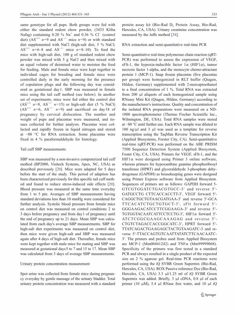
Preeclampsia is associated with altered expression of keyanti- and pro-angiogenic factors [40]. We did not detect anydifferences in the mRNA expression of HIF1α (Fig. 7a),VEGF (Fig. 7b), and sFlt (Fig. 7c) between healthy appear-ing placentas from AS+/+ and AS−/− dams kept on either acontrol or a high-salt diet. Since HIF1α is mostly regulatedat the post-translational level by stabilization and degrada-tion of the protein, we performed immunoblots. HIF1αprotein abundance was significantly increased in healthyplacentas from AS−/− dams on control diet (Fig. 8a). Thisdifference disappeared on high-salt diet (Fig. 8b). HIF1αprotein was not detected in necrotic placentas (Fig. 8a).
Expression of pro-inflammatory factors in necroticplacentas
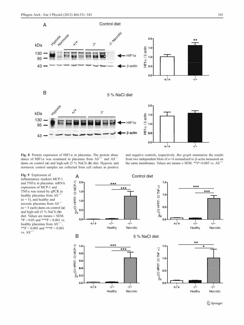
The MCP-1 and TNFα are key pro-inflammatory cytokinesinvolved in inflammatory process. Expression of MCP-1and TNFα mRNA was significantly increased in necroticplacentas from AS−/− dams on control (Fig. 9a) and high-saltdiet (Fig. 9b) but not in healthy-appearing placentas fromboth genotypes.
6
8
E18
(n)
4
6
*
at E
18 (n
)A
+/+ -/- 0
2
4
Litt
er S
ize
at
E
+/+ -/-0
2
4
Nec
rotic
Pla
cent
as
1600mg) 150
8 (m
g)
B
600
800
1000
1200
1400+/+-/-
n Li
tter
Wei
ght a
t E18
(
50
100
150
+/+-/-
Pla
cent
a W
eigh
t at E
18
1 2 3 4 5 6 7 8
Litter Size (n)
Mea
n
1 2 3 4 5 6 7 80
Litter Size (n)Mea
n P
Fig. 5 Effect of high-salt dieton litter size, litter and placentalweights and necrotic placentasat day 18 of pregnancy. aNumber of fetuses and necroticplacentas detected in AS+/+
(n06) and AS−/− dams (n09) atday 18 of pregnancy (P00.088). b Mean litter and meanplacental weights versus littersize in AS+/+ (n06) and AS−/−
dams (n09) (P00.015). Valuesare means ± SEM. *P<0.05 vs.AS+/+
338 Pflugers Arch - Eur J Physiol (2012) 464:331–343

Discussion
Plasma volume expansion in the presence of high aldoste-rone levels is considered crucial for healthy pregnancies,both of which are low in the most severe form of hyperten-sive pregnancies, preeclampsia. In order to examine the roleof aldosterone in the development of preeclampsia, we usedan aldosterone synthase knockout mouse model (AS−/−).AS−/− mice have a compensatory increase of renin andangiotensin II to supraphysiological concentrations but noaldosterone [21] allowing to independently test the role ofaldosterone in pregnancy. Additional effects of these highrenin and angiotensin II may occur but could not be testedbecause survival of AS−/− mice depends on high angiotensinII levels and pharmacological blockade of AT1 receptors islethal in AS−/− mice (unpublished observations).
New-onset hypertension and proteinuria with fetoplacen-tal developmental disturbances are considered to be cardinalfeatures of preeclampsia [40]. We found that AS−/− dams didnot develop preeclampsia as suggested by the absence ofhypertension and proteinuria during pregnancy. A previousstudy measured SBP by telemetry during pregnancy andshowed no change in SBP during the first 5 days, followedby a drop in SBP from days6 to12, and again from days15to 17 of pregnancy [4]. In our hands, using the less sensitivetail cuff method, we found reduced SBP in wild-type miceduring pregnancy in response to salt loading. Though contra-intuitive, this observation is in line with some clinical obser-vations during salt loading in pregnancy suggesting a benefi-cial systemic feedback on maternal hemodynamics uponoptimal placental perfusion [10, 32]. Thus, preeclampsia isnot caused by a lack of aldosterone alone, at least in mice.
+/+ (cont.)+/+ (5% NaCl)
mg)
-/- (cont.)-/- (5% NaCl)
**mg)
A
300
600
900
1200
1500
*** ** ***
n Li
tter W
eigh
t at E
18 (m
300
600
900
1200
1500 **
*
n Li
tter W
eigh
t at E
18 (m
2 5 6 7 8 90
Litter Size (n)
Mea
n
1 2 3 4 5 6 70
Litter Size (n)
Mea
n
120/ ( t )8
(mg)
1208 (m
g)
B
40
60
80
100
+/+ (cont.)+/+ (5% NaCl)
******
Pla
cent
a W
eigh
t at E
18
40
60
80
100
-/- (cont.)-/- (5% NaCl)
Pla
cent
a W
eigh
t at E
18
2 5 6 7 8 940
Litter Size (n)Mea
n
1 2 3 4 5 6 740
Litter Size (n)Mea
n
15
20 ## ##
nta
wei
ght
mg)
C
+/+ -/- +/+ -/- 0
5
10
15
Control diet 5%NaCl
Foe
tal w
eigh
t /pl
acen
at E
18 (m
g/m
Control 5%NaCl
Fig. 6 Comparison of litter andplacental weights, and placentalefficiency between control vs.high-salt diet in AS+/+ andAS−/− dams. Mean litterweights (a) and mean placentalweights (b) on control diet vs.high-salt diet in AS+/+ (controldiet n08 litters, high-salt dietn06 litters) and AS−/− dams(control diet n012 litters, high-salt diet n09 litters). c Placentalefficiency in AS+/+ (control dietn08 litters, high-salt diet n06litters) and AS−/− (control dietn013 litters, high-salt diet n09litters) dams on control andhigh-salt diet. Values aremeans ± SEM. *P<0.05,**P<0.005, ***P<0.001 vs.control diet, ## P<0.005 vs.AS+/+ on control diet, ϕϕϕP<0.001 vs. AS−/− on controldiet
Pflugers Arch - Eur J Physiol (2012) 464:331–343 339

However, the mouse model revealed a number of otherinteresting observations demonstrating an important role ofaldosterone in normal pregnancy. AS−/− dams were hypoten-sive before pregnancy and became even more hypotensiveduring mid to term pregnancy. The resulting fetoplacentalphenotype of a decreased litter size and smaller or necrotic
placentas without fetuses at term suggests a severe placentalhypoperfusion. The sum of intact fetuses and necrotic pla-centas in AS−/− mice was similar to the average number offetuses in AS+/+ dams, indicating that AS−/− damswere able toconceive normally but had impaired placental developmentand function, predisposing to intrauterine death. The fewnormal weight placentas and fetuses observed in AS−/− damscarrying small litters would such provide a residual blood andnutrient supply to the remaining placentas.
Histological examination of healthy placentas from AS−/−
dams demonstrated normal structural patterns but reducedsize. One important reason for lower placental weight mightbe the absence of direct effects of aldosterone on the pla-centa in AS−/− dams. In vitro experiments have revealed adirect role for aldosterone in the modulation of trophoblastgrowth and placental size [16] and in vivo studies in ewessuggested a role of aldosterone in placental growth [19].Placental size in rats and humans is positively correlatedwith plasma levels of aldosterone [16]. Thus, aldosteronemay positively modulate placental growth, similar to itsproliferative capabilities in organs such as the heart andthe kidney [36, 39].
Reduced fetal weight in AS−/− dams might thus be due toplacental insufficiency. In normal human pregnancy, plasmavolume expansion is associated with increased plasma aldo-sterone level, whereas both were decreased in pregnancieswith intrauterine growth restriction and preeclampsia [33].Preeclampsia is characterized by extracellular volume ex-pansion but low intravascular volume which is caused atleast in part by a shift of fluid from the intravascular to theinterstitial fluid space due to vasoconstriction and leakyendothelium [12, 13, 31]. Lack of aldosterone in AS−/−
dams might thus critically impair plasma volume expansionand thereby cause reduced uterine blood supply similar tofunctional aldosterone deficiency in mice chronically ex-posed to the mineralcorticoid receptor antagonist spirono-lactone during pregnancy which led to reduced fetalumbilical blood flow [16]. Moreover, modeling a reduceduterine blood supply by constriction of the uterine artery inrats from gestational day14 caused placental insufficiencyresulting in low-birth weight [2].
One common cause for the decreased litter size, reducedfetal and placental weights, and necrotic and inflammatoryplacentas in AS−/− dams could be hypoxia. In rats, hypoxiaduring the last 11 days of pregnancy results in smaller-sizedlitters, decreased placental and fetal weights, and placentalresorption [18]. Low oxygen tension affects cytotrophoblastproliferation and invasion and ultimately fetal growth [15].Consistently, the hypoxia-induced factor HIF1α, a masterregulator in the adaption to hypoxia, is essential for mam-malian placentation [1]. HIF1α protein abundance was sig-nificantly higher in AS−/− mice on control diet, but wasreduced to control levels on 5 % NaCl diet in the normal-
A
2
3
4control diet5% NaCl
HP
RT-
Ct H
IF1
)
+/+ -/-0
1
2^(C
t H
4control dietF)
B
1
2
35% NaCl
^(Ct
HP
RT
- C
t VE
GF
+/+ -/-0
2^
4control diet5% NaClt-
1)
C
1
2
35%
2^(C
t H
PR
T- C
t sFl
t
+/+ -/-0
2
Fig. 7 Expression of VEGF, sFlt, and HIF1α in placentas. mRNAexpression of HIF1α (a), VEGF (b), and sFlt (c) in healthy placentasfrom AS+/+ (n05) and AS−/− (n04–5) dams on control and high-salt(5 % NaCl) diet
340 Pflugers Arch - Eur J Physiol (2012) 464:331–343
A Control diet
0.5
1.0
1.5
2.0
******
^(Ct
HP
RT
- C
t MC
P-1
)
0.5
1.0
1.5
******
2^(C
t HP
RT-
Ct T
NF
)
+/+ -/- -/- 0.0
Healthy Necrotic
2
+/+ -/- -/- 0.0
Healthy Necrotic
2
0 4
0.6
0.8
1.0
******
PR
T-
Ct M
CP
-1)
1.0
1.5 ***
PR
T- C
t TN
F)
teidlCaN % 5B
+/+ -/- -/- 0.0
0.2
0.4
Healthy Necrotic
2^(C
t H
P
+/+ -/- -/- 0.0
0.5
Healthy Necrotic
2^(C
t HP
Fig. 9 Expression ofinflammatory markers MCP-1and TNFα in placentas. mRNAexpression of MCP-1 andTNFα was tested by qPCR inhealthy placentas from AS+/+
(n = 5), and healthy andnecrotic placentas from AS−/−
(n = 5 each) dams on control (a)and high-salt (5 % NaCl) (b)diet. Values are means ± SEM.*P < 0.05 and ***P < 0.001 vs.healthy placentas from AS−/−,**P < 0.005 and ***P < 0.001vs. AS+/+
A Control diet
1.0
1.5
2.0 **
1 /
act
in
HIF1
kDa
95130
+/+ -/-0.0
0.5Hif 1-actin
B 5 % NaCl diet
43
HIF1
-actin 0 5
1.0
1.5
2.0
Hif1
/ a
ctinkDa
95130
actin
+/+ -/-0.0
0.5H
43
Fig. 8 Protein expression of HIF1α in placentas. The protein abun-dance of HIF1α was examined in placentas from AS+/+ and AS−/−
dams on control (a) and high-salt (5 % NaCl) (b) diet. Hypoxic andnormoxic control samples are collected from cell culture as positive
and negative controls, respectively. Bar graph summarize the resultsfrom two independent blots of n06 normalized to β-actin measured onthe same membranes. Values are means ± SEM. **P<0.005 vs. AS+/+
Pflugers Arch - Eur J Physiol (2012) 464:331–343 341
appearing placentas suggesting hypoxia and rescue by highNaCl intake. Consequently, AS−/− necrotic placentasshowed coagulative necrosis and severe lymphocyte infil-tration and elevated levels of proinflammatory cytokinesMCP-1 and TNFα.
Aldosterone plays a central role in the long-term controlof extracellular volume by adapting renal and extrarenalreabsorption of sodium to blood pressure [3]. Thereby,extracellular NaCl and volume is enlarged; blood pressureand organ perfusion is increased. AS−/− mice are hypoten-sive despite highly elevated levels of renin and angiotensinII. High-salt diet reverses renin and angiotensin II abnor-malities without increasing blood pressure indicating thatthe extracellular volume status is at least partially normal-ized [23]. To rescue the possibly reduced plasma volumeand lower uterine blood flow in AS−/− dams, we treatedmice with high salt for 12 days before pregnancy untilgestational day18. Both AS+/+ and AS−/− mice respondedby increasing SBP suggesting that AS−/− dams also retainedsalt and fluid even though plasma volume was not directlymeasured.
High-salt diet had several interesting implications. Nextto the blood pressure lowering potency in pregnant wild-type mice, it partially rescued the fetal phenotype with asubstantial effect on fetal weight, placental hypoxia, andplacental efficiency in both AS+/+ and AS−/− dams.Placental efficiency can be altered experimentally by manip-ulation of uterine blood flow, oxygen availability, and by theintake and composition of maternal diet. Increased efficien-cy is also seen in naturally small relative to large placentasin pigs, sheep, goats, rats, and mice. Placental efficiency inthese polytocous species is positively related to litter size[11]. We found that smaller placentas in AS+/+ dams onhigh-salt diet and AS−/− dams were associated with largerlitter size indicating positive correlation between placen-tal efficiency and litter size. Decreased placental weightwith larger litter size in AS+/+ dams on high-salt dietalso suggests increased placental efficiency thus sup-porting an unrestricted fetal growth. While small, darkplacentas were still observed in AS−/− dams on high-saltdiet similar to control diet it remains amenable a con-sequence of altered local aldosterone availability to sup-port placental development.
Limitations of this model of aldosterone deficiency arethe high renin and angiotensin II levels which may triggerlocal dysregulatory events in preeclampsia. In the absenceof aldosterone synthase, deoxycorticosterone, corticoste-rone, and some 18-hydroxycorticosterone are still synthe-sized via 11β-hydroxylase activity. Thus, the life-longinhibition of aldosterone synthesis in these animals poten-tially allowing for alternative mineralocorticoid active ste-roid hormones might have supplemented some extrarenalaldosterone effects or even enhanced the inhibition of
proliferation of trophoblasts via the activation of glucocor-ticoid receptors.
Aldosterone availability appears to be an important prereq-uisite to support normal fetoplacental development. AS−/−
dams had a fetoplacental phenotype with decreased litter size,smaller placentas and fetuses, yet no overt preeclampsia. Inthe absence of aldosterone, the fetal phenotype might berescued in part by high-salt diet. In wild-type mice, the high-salt diet led to profound blood pressure reduction in pregnancywhich suggests an important role of plasma volume andplacental perfusion for maternal well-being. Thus, the absenceof aldosterone cannot trigger preeclampsia-like symptoms inmice, but might well contribute to the development ofpreeclampsia-related intrauterine growth restriction while pre-eclampsia needs to be triggered by additional factors. Thus,our study supports the importance of aldosterone in placentalfunction in vivo and a possible role for high-salt supplemen-tation in pregnancies specifically associated with hypoaldos-teronism or reduced plasma volume expansion to maintainfetoplacental integrity. In contrast, in preeclampsia, the bene-fits of salt supplementation are still controversial and have nomajor impact on outcome as indicated by a recent meta-analysis [7].
Acknowledgments We gratefully acknowledge the technical supportby the Zurich Integrative Rodent Physiology (ZIRP) facility. We thankCharlotte Burger for expert technical help with tissue processing andslide scanning. Imaging was performed with equipment maintained bythe Center for Microscopy and Image Analysis, University of Zurich.This study was supported by a collaborative project grant by the ZurichCenter for Integrative Human Physiology (ZIHP) to J. Loffing andC.A. Wagner. The laboratories of J. Loffing and C.A. Wagner are alsosupported by independent project grants from the Swiss NationalScience Foundation (JL: 310030-122243; CAW: 31003A_138143/1)and by funds from the Swiss National Centre of Competence inResearch “Kidney.CH”. M. Mohaupt was supported by the SwissNational Science Foundation by an independent project grant(3200030_135596/1) and also by funds from the Swiss National Cen-tre of Competence in Research “Kidney.CH”.
References
1. Adelman DM, Gertsenstein M, Nagy A, Simon MC, Maltepe E(2000) Placental cell fates are regulated in vivo by HIF-mediatedhypoxia responses. Genes Dev 14:3191–3203
2. Alexander BT (2003) Placental insufficiency leads to developmentof hypertension in growth-restricted offspring. Hypertension41:457–462
3. Booth RE, Johnson JP, Stockand JD (2002) Aldosterone. AdvPhysiol Educ 26:8–20
4. Burke SD, Barrette VF, Bianco J, Thorne JG, Yamada AT, PangSC, Adams MA, Croy BA (2010) Spiral arterial remodeling is notessential for normal blood pressure regulation in pregnant mice.Hypertension 55:729–737
5. Chapman AB, Abraham WT, Zamudio S, Coffin C, Merouani A,Young D, Johnson A, Osorio F, Goldberg C, Moore LG, Dahms T,Schrier RW (1998) Temporal relationships between hormonal and
342 Pflugers Arch - Eur J Physiol (2012) 464:331–343

hemodynamic changes in early human pregnancy. Kidney Int54:2056–2063
6. Dahlstrom B, Romundstad P, Oian P, Vatten LJ, Eskild A (2008)Placenta weight in pre-eclampsia. Acta Obstet Gynecol Scand87:608–611
7. Duley L, Henderson-Smart D, Meher S (2005) Altered dietary saltfor preventing pre-eclampsia, and its complications. CochraneDatabase Syst Rev. doi:10.1002/14651858.CD005548, Issue 4,Art. No. CD005548
8. Escher G, Mohaupt M (2007) Role of aldosterone availability inpreeclampsia. Mol Asp Med 28:245–254
9. Escher G, Cristiano M, Causevic M, Baumann M, Frey FJ, SurbekD, Mohaupt MG (2009) High aldosterone-to-renin variants ofCYP11B2 and pregnancy outcome. Nephrol Dial Transplant24:1870–1875
10. Farese S, Shojaati K, Kadereit B, Frey FJ, Mohaupt MG (2006)Blood pressure reduction in pregnancy by sodium chloride.Nephrol Dial Transplant 21:1984–1987
11. Fowden AL, Sferruzzi-Perri AN, Coan PM, Constancia M, BurtonGJ (2009) Placental efficiency and adaptation: endocrine regula-tion. J Physiol 587:3459–3472
12. Gallery ED (1982) Pregnancy-associated hypertension: interrela-tionships of volume and blood pressure changes. Clin ExpHypertens B 1:39–47
13. Gallery ED, Brown MA (1987) Control of sodium excretion inhuman pregnancy. Am J Kidney Dis 9:290–295
14. Gallery ED, Hunyor SN, Gyory AZ (1979) Plasma volume con-traction: a significant factor in both pregnancy-associated hyper-tension (pre-eclampsia) and chronic hypertension in pregnancy. QJ Med 48:593–602
15. Genbacev O, Zhou Y, Ludlow JW, Fisher SJ (1997) Regulation ofhuman placental development by oxygen tension. Science277:1669–1672
16. Gennari-Moser C, Khankin EV, Schuller S, Escher G, Frey BM,Portmann CB, Baumann MU, Lehmann AD, Surbek D,Karumanchi SA, Frey FJ, Mohaupt MG (2011) Regulation of placen-tal growth by aldosterone and cortisol. Endocrinology 152:263–271
17. Hays PM, Cruikshank DP, Dunn LJ (1985) Plasma volume deter-mination in normal and preeclamptic pregnancies. Am J ObstetGynecol 151:958–966
18. Huang ST, Vo KC, Lyell DJ, Faessen GH, Tulac S, Tibshirani R,Giaccia AJ, Giudice LC (2004) Developmental response to hyp-oxia. FASEB J 18:1348–1365
19. Jensen E, Wood CE, Keller-Wood M (2007) Reduction of maternaladrenal steroids results in increased VEGF protein without in-creased eNOS in the ovine placenta. Placenta 28:658–667
20. Krege JH, Hodgin JB, Hagaman JR, Smithies O (1995) A noninva-sive computerized tail-cuff system for measuring blood pressure inmice. Hypertension 25:1111–1115. doi:10.1161/01.HYP.25.5.1111
21. Lee G, Makhanova N, Caron K, Lopez ML, Gomez RA, SmithiesO, Kim HS (2005) Homeostatic responses in the adrenal cortex tothe absence of aldosterone in mice. Endocrinology 146:2650–2656
22. Levine RJ, Maynard SE, Qian C, Lim KH, England LJ, Yu KF,Schisterman EF, Thadhani R, Sachs BP, Epstein FH, Sibai BM,Sukhatme VP, Karumanchi SA (2004) Circulating angiogenic fac-tors and the risk of preeclampsia. N Engl J Med 350:672–683
23. Makhanova N, Lee G, Takahashi N, Sequeira Lopez ML,Gomez RA, Kim HS, Smithies O (2006) Kidney function inmice lacking aldosterone. Am J Physiol Renal Physiol 290:F61–F69
24. Makhanova N, Sequeira-Lopez ML, Gomez RA, Kim HS,Smithies O (2006) Disturbed homeostasis in sodium-restrictedmice heterozygous and homozygous for aldosterone synthase genedisruption. Hypertension 48:1151–1159
25. Moffett-King A (2002) Natural killer cells and pregnancy. Nat RevImmunol 2:656–663
26. Nicod J, Bruhin D, Auer L, Vogt B, Frey FJ, Ferrari P (2003) Abiallelic gene polymorphism of CYP11B2 predicts increased aldo-sterone to renin ratio in selected hypertensive patients. J ClinEndocrinol Metab 88:2495–2500
27. Nolten WE, Lindheimer MD, Oparil S, Ehrlich EN (1978)Desoxycorticosterone in normal pregnancy. I. Sequential studiesof the secretory patterns of desoxycorticosterone, aldosterone, andcortisol. Am J Obstet Gynecol 132:414–420
28. Noris M, Perico N, Remuzzi G (2005) Mechanisms of disease: pre-eclampsia. Nat Clin Pract Nephrol 1:98–114, quiz 120
29. Parikh SM, Karumanchi SA (2008) Putting pressure on pre-eclampsia. Nat Med 14:810–812
30. Pascoe L, Curnow KM, Slutsker L, Rosler A, White PC (1992)Mutations in the human CYP11B2 (aldosterone synthase) genecausing corticosterone methyloxidase II deficiency. Proc NatlAcad Sci U S A 89:4996–5000
31. Puschett JB (2006) The role of excessive volume expansion in thepathogenesis of preeclampsia. Med Hypotheses 67:1125–1132
32. Robinson M (1958) Salt in pregnancy. Lancet 1:178–18133. Salas SP, Marshall G, Gutierrez BL, Rosso P (2006) Time course
of maternal plasma volume and hormonal changes in women withpreeclampsia or fetal growth restriction. Hypertension 47:203–208
34. Seaton B, Ali A (1984) Simplified manual high performanceclinical chemistry methods for developing countries. Med LabSci 41:327–336
35. Shojaati K, Causevic M, Kadereit B, Dick B, Imobersteg J,Schneider H, Beinder E, Kashiwagi M, Frey BM, Frey FJ,Mohaupt MG (2004) Evidence for compromised aldosteronesynthase enzyme activity in preeclampsia. Kidney Int 66:2322–2328
36. Sohn HJ, Yoo KH, Jang GY, Lee JH, Choi BM, Bae IS, Yim HE,Son CS, Lee JW (2010) Aldosterone modulates cell proliferationand apoptosis in the neonatal rat heart. J Korean Med Sci 25:1296–1304
37. Symonds EM, Broughton Pipkin F, Craven DJ (1975) Changes inthe renin-angiotensin system in primigravidae with hypertensivedisease of pregnancy. Br J Obstet Gynaecol 82:643–650
38. Wacker J, Piel P, Lewicka S, Haack D, Vecsei P, Bastert G (1995)Increased aldosterone-18-glucuronide/tetrahydroaldosterone ratiosin pregnancy. Endocr Res 21:197–202
39. Yim HE, Yoo KH, Bae IS, Jang GY, Hong YS, Lee JW (2009)Aldosterone regulates cellular turnover and mitogen-activated pro-tein kinase family expression in the neonatal rat kidney. J CellPhysiol 219:724–733
40. Young BC, Levine RJ, Karumanchi SA (2010) Pathogenesis ofpreeclampsia. Annu Rev Pathol 5:173–192
Pflugers Arch - Eur J Physiol (2012) 464:331–343 343