14-3-3 Protein interacts with nuclear localization sequence of forkhead transcription factor FoxO4
Upload
independentCategory
view
0download
0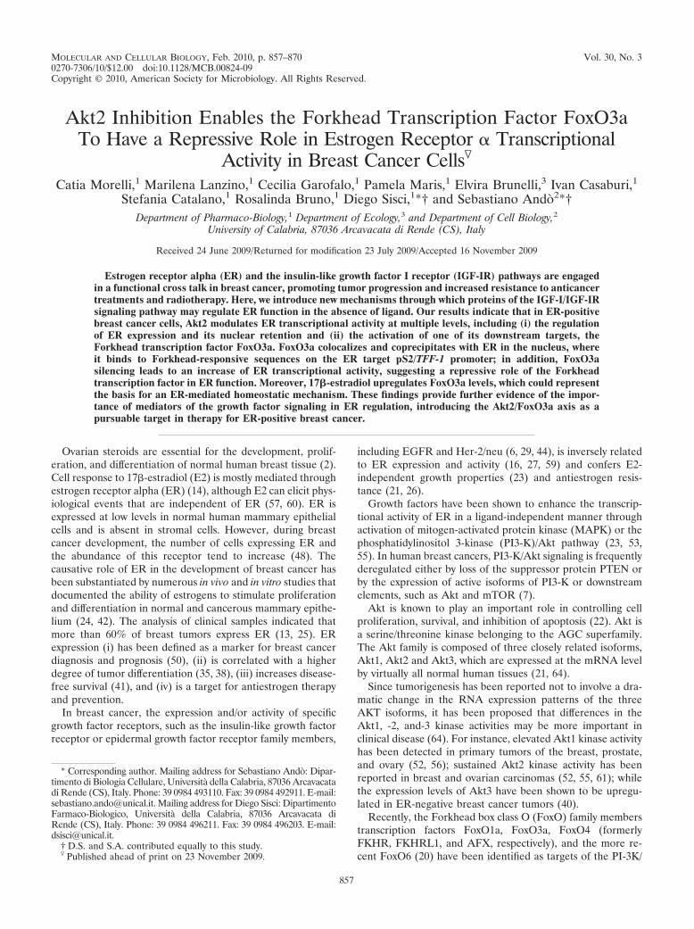
MOLECULAR AND CELLULAR BIOLOGY, Feb. 2010, p. 857–870 Vol. 30, No. 30270-7306/10/$12.00 doi:10.1128/MCB.00824-09Copyright © 2010, American Society for Microbiology. All Rights Reserved.
Akt2 Inhibition Enables the Forkhead Transcription Factor FoxO3aTo Have a Repressive Role in Estrogen Receptor � Transcriptional
Activity in Breast Cancer Cells�
Catia Morelli,1 Marilena Lanzino,1 Cecilia Garofalo,1 Pamela Maris,1 Elvira Brunelli,3 Ivan Casaburi,1Stefania Catalano,1 Rosalinda Bruno,1 Diego Sisci,1*† and Sebastiano Ando2*†
Department of Pharmaco-Biology,1 Department of Ecology,3 and Department of Cell Biology,2
University of Calabria, 87036 Arcavacata di Rende (CS), Italy
Received 24 June 2009/Returned for modification 23 July 2009/Accepted 16 November 2009
Estrogen receptor alpha (ER) and the insulin-like growth factor I receptor (IGF-IR) pathways are engagedin a functional cross talk in breast cancer, promoting tumor progression and increased resistance to anticancertreatments and radiotherapy. Here, we introduce new mechanisms through which proteins of the IGF-I/IGF-IRsignaling pathway may regulate ER function in the absence of ligand. Our results indicate that in ER-positivebreast cancer cells, Akt2 modulates ER transcriptional activity at multiple levels, including (i) the regulationof ER expression and its nuclear retention and (ii) the activation of one of its downstream targets, theForkhead transcription factor FoxO3a. FoxO3a colocalizes and coprecipitates with ER in the nucleus, whereit binds to Forkhead-responsive sequences on the ER target pS2/TFF-1 promoter; in addition, FoxO3asilencing leads to an increase of ER transcriptional activity, suggesting a repressive role of the Forkheadtranscription factor in ER function. Moreover, 17�-estradiol upregulates FoxO3a levels, which could representthe basis for an ER-mediated homeostatic mechanism. These findings provide further evidence of the impor-tance of mediators of the growth factor signaling in ER regulation, introducing the Akt2/FoxO3a axis as apursuable target in therapy for ER-positive breast cancer.
Ovarian steroids are essential for the development, prolif-eration, and differentiation of normal human breast tissue (2).Cell response to 17�-estradiol (E2) is mostly mediated throughestrogen receptor alpha (ER) (14), although E2 can elicit phys-iological events that are independent of ER (57, 60). ER isexpressed at low levels in normal human mammary epithelialcells and is absent in stromal cells. However, during breastcancer development, the number of cells expressing ER andthe abundance of this receptor tend to increase (48). Thecausative role of ER in the development of breast cancer hasbeen substantiated by numerous in vivo and in vitro studies thatdocumented the ability of estrogens to stimulate proliferationand differentiation in normal and cancerous mammary epithe-lium (24, 42). The analysis of clinical samples indicated thatmore than 60% of breast tumors express ER (13, 25). ERexpression (i) has been defined as a marker for breast cancerdiagnosis and prognosis (50), (ii) is correlated with a higherdegree of tumor differentiation (35, 38), (iii) increases disease-free survival (41), and (iv) is a target for antiestrogen therapyand prevention.
In breast cancer, the expression and/or activity of specificgrowth factor receptors, such as the insulin-like growth factorreceptor or epidermal growth factor receptor family members,
including EGFR and Her-2/neu (6, 29, 44), is inversely relatedto ER expression and activity (16, 27, 59) and confers E2-independent growth properties (23) and antiestrogen resis-tance (21, 26).
Growth factors have been shown to enhance the transcrip-tional activity of ER in a ligand-independent manner throughactivation of mitogen-activated protein kinase (MAPK) or thephosphatidylinositol 3-kinase (PI3-K)/Akt pathway (23, 53,55). In human breast cancers, PI3-K/Akt signaling is frequentlyderegulated either by loss of the suppressor protein PTEN orby the expression of active isoforms of PI3-K or downstreamelements, such as Akt and mTOR (7).
Akt is known to play an important role in controlling cellproliferation, survival, and inhibition of apoptosis (22). Akt isa serine/threonine kinase belonging to the AGC superfamily.The Akt family is composed of three closely related isoforms,Akt1, Akt2 and Akt3, which are expressed at the mRNA levelby virtually all normal human tissues (21, 64).
Since tumorigenesis has been reported not to involve a dra-matic change in the RNA expression patterns of the threeAKT isoforms, it has been proposed that differences in theAkt1, -2, and-3 kinase activities may be more important inclinical disease (64). For instance, elevated Akt1 kinase activityhas been detected in primary tumors of the breast, prostate,and ovary (52, 56); sustained Akt2 kinase activity has beenreported in breast and ovarian carcinomas (52, 55, 61); whilethe expression levels of Akt3 have been shown to be upregu-lated in ER-negative breast cancer tumors (40).
Recently, the Forkhead box class O (FoxO) family memberstranscription factors FoxO1a, FoxO3a, FoxO4 (formerlyFKHR, FKHRL1, and AFX, respectively), and the more re-cent FoxO6 (20) have been identified as targets of the PI-3K/
* Corresponding author. Mailing address for Sebastiano Ando: Dipar-timento di Biologia Cellulare, Universita della Calabria, 87036 Arcavacatadi Rende (CS), Italy. Phone: 39 0984 493110. Fax: 39 0984 492911. E-mail:[email protected]. Mailing address for Diego Sisci: DipartimentoFarmaco-Biologico, Universita della Calabria, 87036 Arcavacata diRende (CS), Italy. Phone: 39 0984 496211. Fax: 39 0984 496203. E-mail:[email protected].
† D.S. and S.A. contributed equally to this study.� Published ahead of print on 23 November 2009.
857
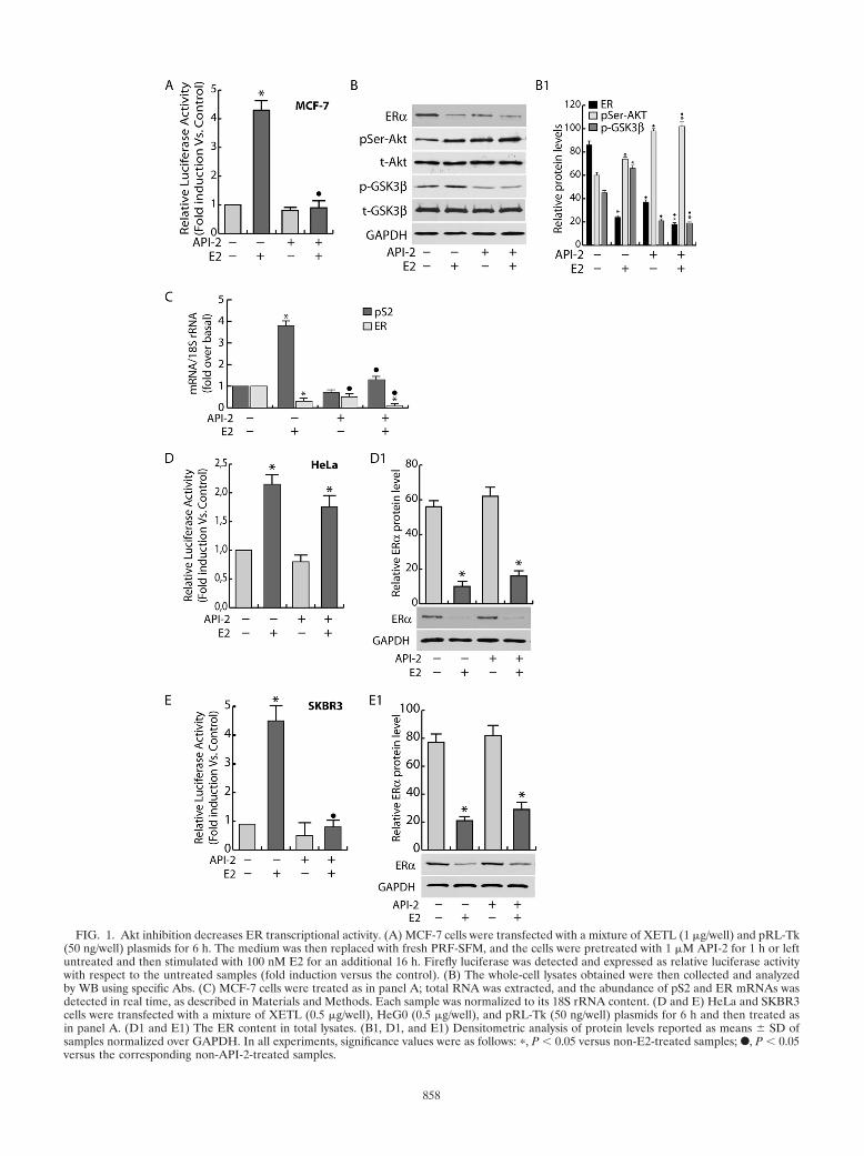
FIG. 1. Akt inhibition decreases ER transcriptional activity. (A) MCF-7 cells were transfected with a mixture of XETL (1 �g/well) and pRL-Tk(50 ng/well) plasmids for 6 h. The medium was then replaced with fresh PRF-SFM, and the cells were pretreated with 1 �M API-2 for 1 h or leftuntreated and then stimulated with 100 nM E2 for an additional 16 h. Firefly luciferase was detected and expressed as relative luciferase activitywith respect to the untreated samples (fold induction versus the control). (B) The whole-cell lysates obtained were then collected and analyzedby WB using specific Abs. (C) MCF-7 cells were treated as in panel A; total RNA was extracted, and the abundance of pS2 and ER mRNAs wasdetected in real time, as described in Materials and Methods. Each sample was normalized to its 18S rRNA content. (D and E) HeLa and SKBR3cells were transfected with a mixture of XETL (0.5 �g/well), HeG0 (0.5 �g/well), and pRL-Tk (50 ng/well) plasmids for 6 h and then treated asin panel A. (D1 and E1) The ER content in total lysates. (B1, D1, and E1) Densitometric analysis of protein levels reported as means � SD ofsamples normalized over GAPDH. In all experiments, significance values were as follows: �, P � 0.05 versus non-E2-treated samples; F, P � 0.05versus the corresponding non-API-2-treated samples.
858
Akt pathway (8). Some reports showed evidence that the over-expression of any of these Forkhead transcription factorsinduced either cell cycle arrest or apoptosis (1, 8, 39, 45).Activation of PI-3K controls cell cycle entry by inactivatingFoxO factors, which have been shown to regulate expression ofp27/Kip1 (36), cyclin D1, and cyclin E (46). FoxO transcriptionfactors have been shown to be functional in mammary cells andto be regulated by Akt (19); in fact, Akt-phosphorylated FoxObinds to 14-3-3 proteins, and the complex is translocated fromthe nucleus to the cytoplasm (8). When hypophosphorylated,Forkhead proteins are released from 14-3-3 and translocateinto the nucleus, where they transactivate specific proapoptotictarget genes (36, 47, 54). Recently, several reports have sug-gested a functional interaction between ER and FoxO mem-bers. E2 has been noted to determine ER binding to FKHR,FKHRL1, and AFX (49, 62, 65) and to induce FKHR phos-phorylation in breast cancer cells (34). Particularly, E2-depen-dent ER binding to FKHR seems to be involved in ER nucle-ocytoplasmic shuttling, since site-directed mutagenesis of theER nuclear export sequence inhibits FKHR nuclear export,the estradiol-induced cytoplasmic relocalization of receptor,and DNA synthesis (33). In transient-transfection experiments,FoxO members seemed also to regulate ER-mediated tran-scription, showing either coactivator or corepressor functionson estrogen-responsive element (ERE) sites, depending on thecellular model (49, 62, 65). Additionally, FoxO3a has recentlybeen reported to suppress cell growth and tumorigenesis inbreast cancer cells and in an orthotopic mouse model of breastcancer (65).
The aim of this study was to better elucidate the molecularmechanisms underlying the regulation exerted by the Akt/FoxO axis on ER in human breast cancer cells.
MATERIALS AND METHODS
Cell culture, conditions, and treatments. The ER-positive human breast can-cer epithelial cell line MCF-7 was maintained in monolayer culture in Dulbecco’smodified Eagle’s/Ham’s F-12 medium (1:1) (DMEM/F-12), supplemented with5% fetal bovine serum (FBS) and 1% Eagle’s nonessential amino acids. Humancervical cancer (HeLa) cells (ATCC, United Kingdom) were grown in modifiedEagle’s medium (MEM) containing 10% calf serum, and human breast cancer(SKBR3) cells (ATCC, United Kingdom) were cultured in RPMI medium plus10% FBS. Additionally, culture media were supplemented with 100 IU/ml pen-icillin, 100 �g/ml streptomycin, and 0.2 mM L-glutamine (all media and reagentswere purchased from Sigma-Aldrich, United Kingdom).
For experimental purposes, cells were synchronized in phenol red-free andserum-free DMEM/F-12 (PRF-SFM) (Sigma-Aldrich, United Kingdom) for 24 hin the absence of steroids and growth factors. Following starvation, the cells werepretreated or not with 1 �M API-2 (triciribine; Calbiochem), an inhibitor of Aktactivity, for 1 h and then treated with 100 nM E2 (Sigma) for 5 min (for Western
FIG. 2. Short-term Akt inhibition affects ER retention in the nu-cleus. (A) MCF-7 cells synchronized in PRF-SFM for 24 h were pre-treated with API-2 for 1 h and then treated with 100 nM E2 or leftuntreated for 45 min. Then, samples were processed for ChIP analysisas described in Materials and Methods. The ERE-containing pS2 pro-moter region was amplified using a specific pair of primers (dataavailable on request). A mixture of normal rabbit IgG and normalmouse IgG was used as a negative control for both primary Abs toprecipitate the E2-treated samples. (B) MCF-7 cells were serumstarved for 24 h, pretreated for 1 h with API-2, and then treated with
100 nM E2 for 5 min or left untreated. Cytoplasmic and nuclearprotein extracts were collected as described in Materials and Methodsand subjected to WB (50 �g/lane) using different Abs. (B1) The rel-ative protein levels were analyzed, and the optical density is reported.(C) MCF-7 cells were treated as in panel B, and total protein extractswere collected with RIPA buffer (see Materials and Methods) andprocessed by WB. Samples were normalized over input (A), GAPDH(for cytosolic proteins [B] and total proteins [C]), and lamin B (fornuclear proteins [B]). In all experiments, significance values were asfollows: �, P � 0.05 versus non-E2-treated samples; F, P � 0.05 versusthe corresponding non-API-2-treated samples. The error bars indicateSD.
VOL. 30, 2010 Akt2/FoxO3a SIGNALING REGULATES ER� ACTIVITY 859
860 MORELLI ET AL. MOL. CELL. BIOL.
blotting [WB]) or 45 min (for chromatin immunoprecipitation [ChIP] and DNAaffinity precipitation assays) and 16 h (gene reporter assay and reverse transcrip-tion [RT]-PCR).
Plasmids, transfections, and ERE-luciferase assay. The following plasmidswere used: the reporter plasmid XETL, driving the expression of firefly luciferaseby an ERE from the Xenopus vitellogenin promoter (9); pS2/TFF-1-ERE (pS2-ERE) containing a 1,050-bp fragment of the pS2 promoter preceding the lucif-erase reporter of pGL3 (5); pSG5-HeG0, a simian virus 40 (SV40) promoter-based pSG5 vector encoding wild-type ER (HeG0) (56a); the Renilla reniformisluciferase expression vector pRL-Tk, used to assess transfection efficiency (Pro-mega); and pECE-HA-FoxO3a (HA-FoxO3a) WT, encoding wild-type FoxO3a(Addgene plasmid 1787).
To monitor the effect of API-2 on ER transactivation, MCF-7, HeLa, andSKBR3 cells (density, 5 � 104) were plated on 24-well plates, grown in culturemedium to an approximate confluence of 70 to 80%, and then switched toPRF-SFM and cotransfected with XETL and pRL-Tk (MCF-7) or XETL, pRL-Tk, and HeG0 (HeLa and SKBR3). All the transfections were carried out usingFugene 6 (Roche) (DNA/Fugene ratio, 3:1). After 6 h, the medium was replacedwith fresh PRF-SFM, and the cells were pretreated for 1 h with 1 �M API-2(triciribine; Calbiochem) and then treated for 16 h with 100 nM E2 (Sigma-Aldrich, United Kingdom).
To evaluate the effect of FoxO3a on ER transactivation, FoxO3a was eithersilenced for 24 h with FoxO3a small interfering RNA (siRNA) (Invitrogen) oroverexpressed using hemagglutinin (HA)-FoxO3a WT and then cotransfectedwith pRL-Tk and XETL or pRL-Tk and pS2-ERE and exposed to 100 nM E2 for16 h. Luciferase activity was measured using the dual-luciferase assay system(Promega), normalized to pRL-Tk activity, and expressed as fold induction overthe control. Cell extracts were also processed by Western blot analysis and/orRT-PCR. Similar experiments were conducted to evaluate the effect of FoxO3aon ER gene transcription, using a pGL3 plasmid bearing the full ER promoter(fragment E, containing both promoters A and B), pGL3-ERprom(E), mappingfrom �4,100 to �212 bp from the first transcription start site (15).
Real-time reverse transcription-PCR. Before the experiments, cells were se-rum starved for 24 h and then treated for the indicated times. Total RNA wasisolated using TRIzol reagent (Invitrogen) according to the manufacturer’s in-structions and treated with DNase I (Ambion). Two �g of total RNA was reversetranscribed with the ImProm-II reverse transcription system kit (Promega) usingrandom primers; cDNA was diluted 1:3 in nuclease-free water, and 5 �l wasanalyzed in triplicate by real-time PCR in an iCycler iQ Detection System(Bio-Rad) using SYBR green Universal PCR Master Mix (Bio-Rad) with 0.1�mol/liter of each primer in a total volume of 30 �l of reaction mixture. Theprimers used for the amplification were based on published sequences for humanAkt 2, ER, FoxO3a, cyclin D1 (CD1), and pS2 (data available on request). ThePCR conditions were 95°C for 3 min and 40 cycles of 95°C for 30 s, Ta (dataavailable on request) for 30 s, and 72°C for 30 s; negative controls containedwater instead of first-strand cDNA. Each sample was normalized on its 18SrRNA content. The 18S quantification was done using a TaqMan rRNA reagentkit (Applied Biosystems) following the manufacturer’s instructions. The relativegene expression levels were normalized to a calibrator that was chosen to be thebasal, untreated sample. The final results were expressed as n-fold differences ingene expression relative to 18S rRNA and the calibrator, calculated using theCT method as follows: n-fold 2�(CTsample � CTcalibrator), where the CT
values of the sample and calibrator were determined by subtracting the averageCT value of the 18S rRNA reference gene from the average CT value of thedifferent genes analyzed.
Chromatin immunoprecipitation. MCF-7 cells were grown in 100-mm plates.Subconfluent cultures (70%) were shifted to PRF-SFM for 24 h, pretreated with
API-2 for 1 h, and then treated with 100 nM E2 or left untreated for 45 min.Alternatively, growing cells were switched to PRF-SFM, transfected with HA-FoxO3a using Fugene 6 (Fugene 6/plasmid ratio, 3:1), and treated the followingday with E2 for 45 min. ChIP methodology was performed as described previ-ously (37). The precleared chromatin was precipitated for 16 h with anti-ERmonoclonal antibody (MAb) (Santa Cruz) for ER, anti-FoxO3a polyclonal an-tibody (PAb) (Cell Signaling) for FoxO3a, and anti-polymerase II PAb (SantaCruz) for Pol II. Normal rabbit IgG and normal mouse IgG (Santa Cruz) wereused instead of primary Abs as negative controls. Immunoprecipitated DNA wasanalyzed in triplicate by real-time PCR using 5 �l of the diluted (1:3) templateDNA as described above, and the the pS2 promoter region (pS2-prom) wasamplified using specific primers (data available on request).
Real-time PCR data were normalized with respect to unprocessed lysates(input DNA). Input DNA quantification was performed by using 5 �l of thediluted (1/50) template DNA. The relative antibody-bound fractions were nor-malized to a calibrator that was chosen to be the basal, untreated sample. Thefinal results were expressed as fold differences with respect to the relative inputs.
Immunoprecipitation and Western blotting. Protein expression and complexformation were assessed by Western blotting (WB) or immunoprecipitation (IP),followed by WB using total protein lysates, cytoplasmic protein lysates, or frac-tionated proteins, where appropriate. MCF-7 cells were serum starved for 24 hand treated with 100 nM E2 and/or the Akt inhibitor API-2 (1 �M) for differenttimes, depending on the experiment. Cytoplasmic proteins were obtained usinglysis buffer containing 50 mmol/liter HEPES (pH 7.5), 150 mmol/liter NaCl, 1%Triton X-100, 1.5 mmol/liter MgCl2, 10 mmol/liter EGTA (pH 7.5), 10% glyc-erol, and inhibitors (0.1 mmol/liter Na3VO4, 1% phenylmethylsulfonyl fluoride,and 20 mg/ml aprotinin). After the collection of cytoplasmic proteins, the nucleiwere lysed with nuclear buffer containing 20 mmol/liter HEPES (pH 8), 0.1mmol/liter EDTA, 5 mmol/liter MgCl2, 0.5 mol/liter NaCl, 20% glycerol, 1%NP-40, and inhibitors (as described above). For total protein extracts, RIPAbuffer was used (50 mM Tris-HCl, pH 7.4, 150 mM NaCl, 1% NP-40, 0.25% Nadeoxycholate, plus inhibitors). The protein content was determined using Brad-ford dye reagent (Bio-Rad). For WB, 50 �g of lysates was separated on an 11%polyacrylamide denaturing gel (SDS-PAGE) and transferred to nitrocellulosemembranes. Proteins of interest were detected with specific Abs, recognized byperoxidase-coupled secondary Abs, and developed using the ECL Plus WesternBlotting detection system (Amersham Pharmacia Biotech, United Kingdom).For IP, 500 �g of protein lysates was precleared for 1 h with protein A/G-agarose(Santa Cruz) for either MAb or PAbs, incubated with primary Abs at 4°C for 18 hin HNTG buffer (20 mmol/liter HEPES, pH 7.5, 150 mmol/liter NaCl, 0.1%Triton X-100, 10% glycerol, and 0.1 mmol/liter Na3VO4), and then the anti-gen-Ab complexes were precipitated with protein A/G agarose for 2 h in HNTGbuffer. In control samples, the primary immunoprecipitating Abs were replacedwith normal rabbit IgG (Santa Cruz Biotechnology). The immunoprecipitatedproteins were washed three times with HNTG buffer, separated on SDS-PAGE,and processed by WB. The images were acquired by using an Epson Perfectionscanner (Epson, Japan) using Photoshop software (Adobe). The optical densitiesof the spots were analyzed by using ImageJ software (NIH; http://rsb.info.nih.gov/IJ).
Antibodies for Western blotting and immunoprecipitation. Total and phos-phorylated Akt isoforms were detected by WB with specific Abs: anti-Akt1/2(H-136) PAb, anti-Akt1 (G5) MAb, anti p-Akt1/2/3 (Ser473)-R (Santa Cruz),and anti-Akt2 (5B5) MAb (Cell Signaling). ER and FoxO3a were assessed byWB and IP with anti-ER F-10 MAb (Santa Cruz) and anti-FoxO3a PAb (CellSignaling). pS2 was probed with anti-pS2 PAb (Santa Cruz), phosphorylatedGSK-3� with anti-GSK-3� (Ser9) PAb (Cell Signaling), total GSK-3� with anti-GSK-3� MAb (Cell Signaling), CD1 with anti-cyclin D1 PAb (Santa Cruz), and
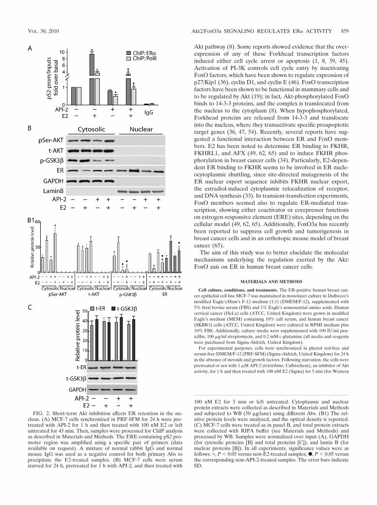
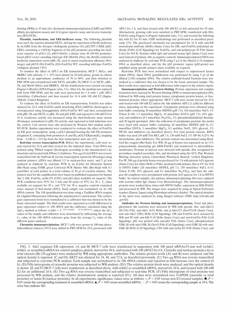
FIG. 3. Akt2 regulates ER expression. (A and B) MCF-7 cells were transfected in suspension with 100 pmol siRNAs/35-mm well (siAkt1,siAkt2, or scrambled siRNA for control samples), plated, starved for 48 h, and treated with 100 nM E2 for 6 h. Cytosolic and nuclear proteins (A) ortotal extracts (B) (50 �g/lane) were analyzed by WB using appropriate antibodies. The relative protein levels (A1 and B) were analyzed, and theoptical density is reported. (C and D) AKT2 was silenced for 24, 48, and 72 h, as described previously. (C) Two �g RNA was reverse transcribedand subjected to real-time PCR analysis. Each sample was normalized to its 18S rRNA content and reported as fold increase over the control (0h). (D) Fifty micrograms of cytosolic proteins was subjected to WB analysis. (D1) The relative protein levels were analyzed, and the optical densityis shown. (E and F) MCF-7 cells were transfected, as described above, with siAkt2 or scrambled siRNA, starved for 24 h, and treated with 100 nME2 for an additional 24 h. (E) Two �g RNA was reverse transcribed and subjected to real-time PCR. (F) Fifty micrograms of total proteins wasprocessed by WB analysis, and the relative densitometric analysis is reported (F1). All data were normalized over GAPDH (cytosolic or totalproteins) or lamin B (nuclear proteins). In all experiments, significance values were as follows: �, P � 0.05 versus non-E2-treated samples; F, P �0.05 versus the corresponding treatment in scrambled siRNA; Œ, P � 0.05 versus scrambled siRNA; � , P � 0.05 versus the corresponding sample at 24 h. Theerror bars indicate SD.
VOL. 30, 2010 Akt2/FoxO3a SIGNALING REGULATES ER� ACTIVITY 861
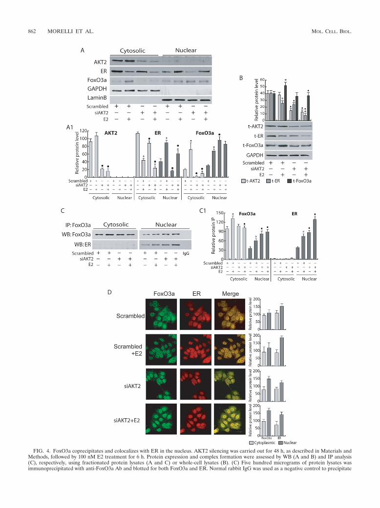
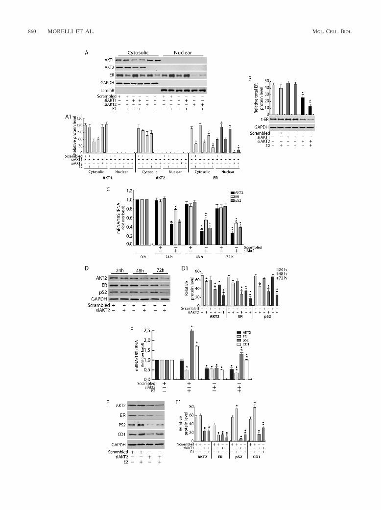
FIG. 4. FoxO3a coprecipitates and colocalizes with ER in the nucleus. AKT2 silencing was carried out for 48 h, as described in Materials andMethods, followed by 100 nM E2 treatment for 6 h. Protein expression and complex formation were assessed by WB (A and B) and IP analysis(C), respectively, using fractionated protein lysates (A and C) or whole-cell lysates (B). (C) Five hundred micrograms of protein lysates wasimmunoprecipitated with anti-FoxO3a Ab and blotted for both FoxO3a and ER. Normal rabbit IgG was used as a negative control to precipitate
862 MORELLI ET AL. MOL. CELL. BIOL.
glyceraldehyde-3-phosphate dehydrogenase (GAPDH) and lamin B were as-sessed by WB as controls for loading and purity of lysates with the anti-GAPDH(FL-335) PAb and the anti-lamin B (C-20) PAb (Santa Cruz), respectively.Normal rabbit IgG and normal mouse IgG (Santa Cruz) were used instead ofprimary Abs for IP negative controls. All Abs were used at concentrationsrecommended by the manufacturers.
siRNA-mediated RNA interference. Custom-synthesized siRNA (Invitrogen)-annealed duplexes (25-bp double-stranded RNA [dsRNA]) were used for effec-tive depletion of Akt1, Akt2, and FoxO3a genes. A scrambled siRNA (Invitro-gen) that lacked identity with known gene targets was used as a control fornon-sequence-specific effects. Cells were trypsinized; transfected in suspensionwith Lipofectamine 2000 (Invitrogen), as suggested by the manufacturer; andthen plated in six-well plates (3.5 � 105 cells per well). Briefly, the cells weretransfected with 100 pmol siRNAs (siAkt1, siAkt2, siFoxO3a, or nonspecificsiRNA) in 3 ml total growing medium. The siRNAs were diluted in 500 �l ofPRF-SFM without antibiotic; after 5 min, Lipofectamine 2000 was added to themixture and incubated at room temperature for 20 min. The siRNA-Lipo-fectamine 2000 complex was then added to the cells and incubated at 37°C for6 h, switched to PRF-SFM, and further incubated for an additional 18 h. Then,the cells were treated with E2 (100 nM) for 6 h or 24 h before analysis, dependingon the experiment. For time course purposes, cells were harvested after 24 h,48 h, or 72 h.
Confocal laser scanning microscopy (CLSM). MCF-7 cells were transfected insuspension with Akt2 siRNA as described previously, plated on coverslips, serumstarved, and then treated with 100 nM E2 for 6 h. After incubation, the cells werefixed with 3% paraformaldehyde and permeabilized with 0.2% Triton X-100, andnonspecific sites were blocked with bovine serum albumin (BSA) (3% for 30min). The blocked samples were incubated for 1 h with a mixture of primaryantibodies (2 mg/ml each) recognizing ER (MAb; Santa Cruz) and FoxO3a(PAb; Cell Signaling), washed with phosphate-buffered saline (PBS) (Gibco),and incubated with a mixture of fluorescein-conjugated goat anti-rabbit IgG andrhodamine-conjugated goat anti-mouse IgG (Santa Cruz) secondary Abs. Thecellular localization of the two proteins was examined under a Leica TCS SP2confocal laser scanning microscope at �400 magnification. The optical sectionswere taken at the central plane. The fluorophores were imaged separately toensure there was no excitation/emission wavelength overlap. The optical densi-ties of stained proteins were analyzed by ImageJ software.
DAPA. The binding of nuclear FoxO3a to Forkhead-responsive elements onthe pS2 promoter was assessed in vitro using a modified version of the DNAaffinity precipitation assay (DAPA) protocol of Zhu et al. (63). Briefly, nuclear-protein extracts were obtained from starved cells pretreated with API-2 (1 �M)for 1 h and stimulated with E2 (100 nM) for 45 min. One hundred �g of nuclearproteins was mixed with 2 �g of specific biotinylated DNA probes (see below) in400 �l of buffer D (20 mM HEPES, pH 7.9, 10% glycerol, 50 mM KCl, 0.2 mMEDTA, 1.5 mM MgCl2, 10 �M ZnCl2, 1 mM dithiothreitol, and 0.25% TritonX-100) and then incubated on ice for 45 min. After that, 20 �l of streptavidin-agarose beads (Promega) was added, and the samples were incubated underrotation for 2 h at 4°C. Next, the agarose bead-protein complexes were collectedby brief centrifugation and washed twice in buffer D. Proteins were uncoupledfrom DNA probes by the addition of 40 �l of 2� Laemmli’s sample buffer andby heating them at 96°C for 10 min. The beads were removed by centrifugation,and the supernatants were analyzed by WB for the presence of FoxO3a. TheDNA motif probes (pS2/FKH) were prepared by annealing a 5�-biotinylatedsense oligonucleotide bearing a Forkhead consensus sequence (5�-Bio-ACGCTCTTTAAGCAAACAGAGCCTGCCCTA-3�) with a nonbiotinylated antisenseoligonucleotide (5�-TAGGGCAGGCTCTGTTTGCTTAAAGAGCGT-3�). Alabeled probe with the consensus sequence [pS2/FKH(�)], underlined above,deleted was used as a negative control (forward, 5�-Bio-ACGCTCTTTAAACAGAGCCTGCCCTA-3�; reverse, 5�-TAGGGCAGGCTCTGTTTAAAGAGCGT-3�). The optical densities of the spots were analyzed by ImageJ software.
Proliferation assay. MCF-7 cells were transfected in suspension with eitherHA-FoxO3a or siFoxO3a (the empty vector or scrambled siRNA, respectively,was used as a control) in growth medium without antibiotic and plated in 12-wellplates at a concentration of 105 cells/plate. After 6 h, the cells were starved for18 h (day zero) and then treated or not with E2 (100 nM) for 1, 2, and 3 days (thehormone was refreshed every day to maintain constant levels in the medium). Ateach time point, the cells were harvested by trypsinization and counted in ahemocytometer using the trypan blue exclusion assay.
Statistical analysis. All data were expressed as the means � standard devia-tions (SD) of at least three independent experiments. Statistical significanceswere tested using Student’s t test.
RESULTS
Akt inhibition decreases ER transcriptional activity. It hasbeen well established that Akt is involved in the control of cellsurvival and that its inhibition is responsible for growth retar-dation; in MCF-7 cells, this effect is paralleled by a decrease ofER transcriptional activity (32, 53). This assumption was con-firmed through transactivation experiments using an ER-re-sponsive 1� ERE-Luc construct (XETL). In our experimentalsystem, E2 treatment, as expected, determined a 4-fold induc-tion of luciferase activity (Fig. 1A). The inhibition of Aktkinase function by API-2 pretreatment completely abrogatedER transcriptional activity in response to E2 administration(Fig. 1A). Similar results were obtained in two additional ER-positive cell lines, T47D and ZR75 (data not shown). Hormonetreatment caused an increase of Akt phosphorylation in eitheruntreated or API-2-treated samples (Fig. 1B). Interestingly,API-2 treatment resulted in pSer-Akt accumulation, while Aktkinase activity was dramatically reduced, since its downstreamtarget, GSK-3�, was no longer phosphorylated (Fig. 1B). No-tably, the inhibition of ER transactivation is consistent with areduction of the ER-mediated transcription, since, under thesame experimental conditions, a dramatic decrease in mRNAlevels of the ER target gene pS2 was observed both under basalconditions and after E2 treatment (Fig. 1C). This event wasparalleled by a significant decrease in ER protein and mRNAlevels (Fig. 1B and C). Interestingly, the inhibition of Aktkinase function did not alter either Akt or GSK-3� proteinexpression (Fig. 1B), while it did induce a 40% reduction ofER protein and mRNA levels in untreated samples, as well asfurther emphasizing the ligand-induced downregulation of thereceptor (Fig. 1B and C).
To assess cell specificity, a gene reporter assay was con-ducted in two ER-negative cell lines, HeLa and SKBR3, ec-topically expressing ER. In both cell systems, ER presence wasresponsible for E2-dependent transactivation (Fig. 1D and E).Interestingly, in HeLa cells, no significant difference in lucif-erase induction was observed in API-2-treated samples com-pared to nontreated samples, while in SKBR3 cells, the re-
E2-treated samples. (A1, B, and C1) The relative protein levels were analyzed, and the optical density is reported. Samples were normalized overGAPDH (A, cytosolic, or B, total proteins) and lamin B (A, nuclear proteins). In all experiments, significance values were as follows: �, P � 0.05versus non-E2 treated; F, P � 0.05 versus the corresponding treatment in scrambled siRNA. (D) Cytoplasmic and nuclear distribution of ER andFoxO3a in response to AKT2 knockdown was also evaluated by confocal microscopy. To avoid fluorescence overlapping, a rabbit PAb for FoxO3aand a mouse MAb for ER were used. A mixture of fluorescein-conjugated goat anti-rabbit IgG (green) and rhodamine-conjugated goat anti-mouseIgG (red) secondary Abs was used to detect primary immune complexes of FoxO3a and ER, respectively. The merged images show FoxO3a-ERcolocalization (yellow). The optical sections were taken at the central plane at �400 magnification. The fluorophores were imaged separately toensure there was no excitation/emission wavelength overlap. The histograms on the right show the corresponding means and SD of thedensitometric analysis performed in three independent experiments.
VOL. 30, 2010 Akt2/FoxO3a SIGNALING REGULATES ER� ACTIVITY 863
sponse to the same treatment paralleled that observed inMCF-7 cells (Fig. 1D and E). In both cell lines, API-2 did notaffect the ER protein content (Fig. 1, D1 and D2).
To clarify the role of Akt in ER transcriptional activity,chromatin-bound ER and Pol II were precipitated fromMCF-7 cell nuclear extracts. As shown in Fig. 2A, strong in-hibition of ER and Pol II recruitment on the estrogen-respon-sive sequence of the pS2 promoter was observed in cells pre-treated with API-2 for 1 h and exposed to E2 for 45 min. Thereduced occupancy of ER on the pS2 promoter was related toa decrease in ER nuclear content. Indeed, 1 h of exposure toAPI-2 was able to interfere with ligand-dependent retention ofER in the nucleus (Fig. 2B) without affecting total ER andGSK-3� protein amounts (Fig. 2C).
In our system, E2 induced phosphorylation of Akt and, as aconsequence, GSK-3� phosphorylation after 5 min of treat-ment (Fig. 2B). The efficacy of API-2 pretreatment was evi-denced by the clear inhibition of GSK-3� phosphorylationwhile, as expected, at this time point, API-2 did not alter Aktexpression or its phosphorylation on Ser473 in either the pres-ence or absence of E2 (Fig. 2B).
Akt2 silencing reduces ER expression and function. SinceAPI-2 is not able to discriminate between the Akt isoforms,Akt1 and Akt2 mRNA transcripts (Akt3 is not expressed inMCF-7 cells [reference 21 and data not shown]) were silencedby siRNA. A strong reduction of ER total expression wasobserved in Akt2, but not in Akt1, silenced samples (Fig. 3Aand B). Time course experiments (24 to 72 h) showed that asignificant decrease in ER expression also occurs at the tran-scriptional level following Akt2 silencing (Fig. 3C and D). Thisevent was reflected by a strong reduction in pS2 mRNA (Fig.3C) and protein (Fig. 3D) expression at all investigated timepoints. The reduction of Akt2 and ER cytosolic content ob-served after 72 h in scrambled-siRNA samples (Fig. 3D) couldsimply be ascribed to a general phenomenon in response to theprolonged starvation. As mentioned above, siAkt1 did not altereither ER (Fig. 3A and B) or pS2 (data not shown) expression,supporting the hypothesis that ER functional disruption spe-cifically depends on Akt2 inhibition. A comparable trend wasmaintained in the presence of E2 in Akt2 silenced samples. Infact, E2 treatment did not alter the inhibitory effect of silencedAkt2 on the mRNA and protein levels of ER and pS2, as wellas an additional ER-regulated gene, CD1 (Fig. 3E and F).Similar experiments were conducted in T47D and ZR75 cells(data not shown). In both cell lines, Akt2 inhibition led to adramatic reduction of ER, and consequently pS2, mRNA andprotein levels.
FoxO3a coprecipitates and colocalizes with ER in the nu-cleus. To elucidate the mechanism underlying the involvementof Akt2 on the regulation of both ER expression and function,we focused our attention on one of its downstream effectors,FoxO3a, a member of the Forkhead transcription factor fam-ily. FoxO3a is well expressed in MCF-7 cells and has beenreported to functionally interact with ER (17, 62). As shown inFig. 4A and B, an increase in total FoxO3a protein levels wasobserved in response to E2 stimulation. siAkt2 caused a drasticreduction of FoxO3a cytosolic content in MCF-7 cells in eitherthe presence or absence of E2 (Fig. 4A). The latter effect wasparalleled by a marked increase in FoxO3a nuclear content(Fig. 4A and D), even though it is worth noting that E2 treat-
ment did not significantly affect siAkt2-induced FoxO3a nu-clear translocation. Interestingly, Akt2 knockdown did notaffect total expression of FoxO3a, while it counteracted E2-induced FoxO3a upregulation, since, as previously described,total ER expression was significantly reduced (Fig. 4B).
Immunoprecipitation experiments and confocal microscopyconfirmed the existence of a physical interaction (Fig. 4C) andcolocalization (Fig. 4D) between ER and FoxO3a. In accor-dance with previously reported data in cell-free systems (49),the ER/FoxO3a interaction occurred mainly at the nuclear andperinuclear levels, where it was enhanced by E2, and becamemore evident in Akt2 silenced samples (Fig. 4C and D).
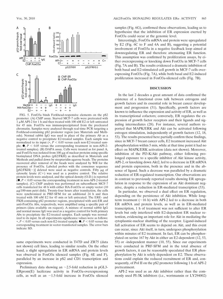
FoxO3a binds Forkhead-responsive elements on the pS2promoter. Some recently published reports evidenced an over-representation of TA-rich motifs, particularly Forkhead bind-ing sites (AAG[A]TAAA[G]C[T]A), in several ER-bound re-gions (30–31), including the pS2 promoter (4, 11). Toinvestigate if ER/FoxO3a interaction does exert a functionalrole in ER-mediated transcription, chromatin-bound FoxO3awas immunoprecipitated with a specific antibody. The pres-ence of FoxO3a on the pS2 promoter was detected targetingthe Forkhead DNA binding site close to the TATAA box (4).As shown in Fig. 5A, a constitutive association of FoxO3a withthe pS2 promoter was observed, which was increased by E2stimulation. As expected, the inhibition of Akt kinase activityby API-2 treatment increased the promoter occupancy byFoxO3a, consistent with the induced FoxO3a translocationinto the nucleus (Fig. 4A). Additionally, to investigate ifFoxO3a binding occurs on the Forkhead-responsive element ofthe pS2 promoter (pS2/FKH), a DAPA assay was conductedon nuclear extracts from MCF-7 cells. The results obtainedshowed that nuclear FoxO3a binds with high affinity to thepS2/FKH sequence but not to the same region partially deletedin the Forkhead-responsive element [pS2/FKH(�)] used as anegative control (Fig. 5B). These data support the hypothesisthat this region is involved in pS2 regulation by the Forkheadtranscription factor.
To evaluate the effects of FoxO3a recruitment to Forkhead-responsive sites on the binding of ER to ERE-containing pro-moters, we performed a ChIP assay transiently overexpressingFoxO3a. As shown in Fig. 5C, a significant decrease of bothbasal and E2-induced recruitment of ER on the pS2 promoterwas observed in FoxO3a-overexpressing samples, corroborat-ing our hypothesis of the negative role of FoxO3a in ERfunction.
FoxO3a inhibits ER expression and functional activity. Tofurther understand the role of FoxO3a in inhibiting ER tran-scription, we both overexpressed and silenced FoxO3a andperformed gene reporter experiments using either XETL (Fig.6A) or a full-length pS2 promoter construct (Fig. 6B). Ourresults showed that FoxO3a overexpression caused a signifi-cant decrease of ER-dependent transcription in response to E2stimulation, while FoxO3a knockdown led to the opposite ef-fect (Fig. 6A and B). Comparable results were obtained inT47D and ZR75 (data not shown) cell lines transfected withXETL.
Moreover FoxO3a overexpression determined strong down-regulation in ER mRNA and protein levels, both in controlsand in E2-treated samples, which most likely is responsible fordecreased pS2 and CD1 transcription (Fig. 6C and D). The
864 MORELLI ET AL. MOL. CELL. BIOL.
same experiments were conducted in T47D and ZR75 (datanot shown) cell lines, leading to similar results. On the otherhand, a slight upregulation in ER mRNA and protein levelswas observed in FoxO3a silenced samples (Fig. 6E and F),paralleled by an increase in pS2 and CD1 transcription andexpression.
Preliminary data showing an �2.5-fold reduction in pGL3-ERprom(E) luciferase activity in FoxO3a-overexpressiongcells, as well as an �1.5-fold increase in FoxO3a silenced
samples (Fig. 6G), confirmed these observations, leading us tohypothesize that the inhibition of ER expression exerted byFoxO3a could occur at the genomic level.
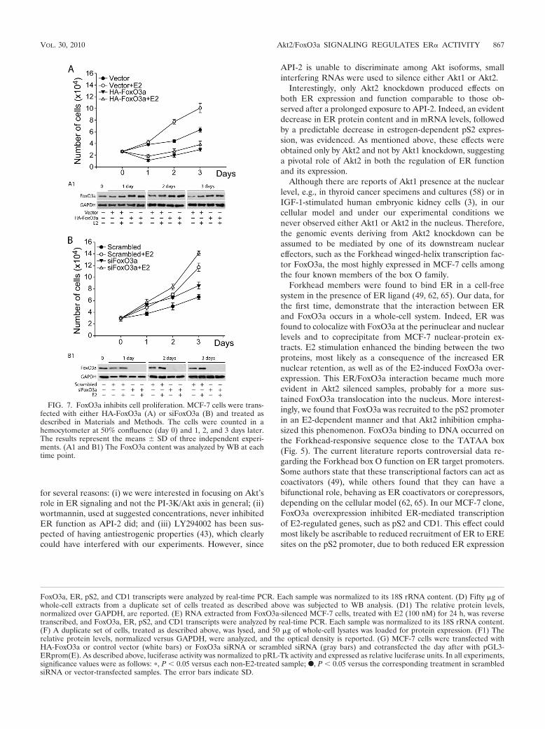
Interestingly, FoxO3a mRNA and protein were upregulatedby E2 (Fig. 6C to F and 4A and B), suggesting a potentialinvolvement of FoxO3a in a negative feedback loop aimed atdownregulating ER and therefore attenuating ER function.This assumption was confirmed by proliferation assays, by ei-ther overexpressing or knocking down FoxO3a in MCF-7 cells(Fig. 7A and B). The results evidenced a dramatic inhibition ofboth basal and E2-stimulated cell growth in MCF-7 cells over-expressing FoxO3a (Fig. 7A), while both basal and E2-inducedproliferation increased in FoxO3a-silenced cells (Fig. 7B).
DISCUSSION
In the last 2 decades a great amount of data confirmed theexistence of a functional cross talk between estrogens andgrowth factors and its essential role in breast cancer develop-ment and progression (51). Specifically, growth factors areknown to influence the expression and activity of ER, as well asits transcriptional cofactors; conversely, ER regulates the ex-pression of growth factor receptors and their ligands and sig-naling intermediates (28). For instance, several authors re-ported that MAPK/ERK and Akt can be activated followingestrogen stimulation, independently of growth factors (12, 18,53). The results presented here partially confirm these findings,since in MCF-7 breast cancer cells, E2 treatment increased Aktphosphorylation within 5 min, while at that time point it had noeffect on MAPK/ERK activation (data not shown). Moreover,inhibition of the PI3-K/Akt pathway, either following pro-longed exposure to a specific inhibitor of Akt kinase activity,API-2, or knocking down Akt2, led to a decrease in ER mRNAand protein expression, both in the presence and in the ab-sence of ligand. Such a decrease was paralleled by a dramaticreduction of ER-regulated transcription. Our observations arein contrast to previously reported results showing ER upregu-lation in response to the expression of an Akt dominant neg-ative, despite a reduction in ER-mediated transcription (53).
In particular, we observed a dual effect on ER regulation,depending on the persistence of Akt inhibition. While long-term treatment (�16 h) with API-2 led to a decrease in bothER mRNA and protein levels, as well as in ER-mediatedtranscription, 1 h of treatment was not sufficient to alter ERlevels but only interfered with E2-dependent ER nuclear re-tention, evidencing an important role for Akt in mediating thecytoplasmic-nuclear shuttling of the receptor. Ligand-inducedfull activation of ER seems to depend on Akt, and this eventcan occur, since Akt itself, in turn, undergoes phosphorylationwithin minutes of E2 treatment. In fact, ER can be phosphor-ylated on serine 167 by Akt in either an E2-dependent (12, 53,55) or -independent manner (10, 55). Since our experimentswere conducted in PRF-SFM and in the total absence ofgrowth factors, it can be reasonably speculated that ER phos-phorylation by Akt is solely dependent on E2. These observa-tions could explain the reduced recruitment of ER and, con-sequently, of Pol II on the pS2 promoter following 1 h of API-2pretreatment.
API-2 was used as an Akt inhibitor rather than the com-monly used PI-3K inhibitors (i.e., wortmannin or LY294002)
FIG. 5. FoxO3a binds Forkhead-responsive elements on the pS2promoter. (A) ChIP assay. Starved MCF-7 cells were pretreated with1 �M API-2 for 1 h and then treated with 100 nM E2 or left untreatedfor 45 min. FoxO3a was immunoprecipitated from the preclearedchromatin. Samples were analyzed through real-time PCR targeting aForkhead-containing pS2 promoter region (see Materials and Meth-ods). Normal rabbit IgG was used in place of the primary Ab as anegative control to precipitate E2-treated samples. Each sample wasnormalized to its input (�, P � 0.05 versus each non-E2-treated sam-ple; F, P � 0.05 versus the corresponding treatment in non-API-2-treated samples). (B) DAPA assay. Cells were treated as for panel A,and FoxO3a was isolated from 100 �g of nuclear proteins using specificbiotinylated DNA probes (pS2/FKH) as described in Materials andMethods and pulled down by streptavidin-agarose beads. The proteinsrecovered after removal of the beads were analyzed by WB for thepresence of FoxO3a. Labeled probes with the consensus sequence[pS2/FKH(�)] deleted were used as negative controls. Fifty �g ofcytosolic lysate (C�) was used as a positive control. The relativeprotein levels were analyzed, and the optical density (O.D.) is reported(F, P � 0.05 versus the corresponding treatment in non-API-2-treatedsamples). (C) ChIP analysis was performed on subconfluent MCF-7cells transfected for 48 h with either HA-FoxO3a or empty vector (10�g/100-mm petri dish). Twenty-four hours after transfection, the cellswere synchronized in PRF-SFM for an additional 24 h and thentreated with 100 nM E2 for 45 min or left untreated. The ERE- andFKH-containing pS2 promoter regions, precipitated with anti-ER andanti-FoxO3a Abs, respectively, were amplified using a specific pair ofprimers (data available on request). A mixture of normal rabbit IgGand normal mouse IgG was used as a negative control for both primaryAbs to precipitate the E2-treated samples. Each sample was normal-ized to its input. In all experiments significance values were as follows:�, P � 0.05 versus each non-E2-treated sample; F, P � 0.05 versus thecorresponding treatment in vector-transfected samples. The error barsindicate SD.
VOL. 30, 2010 Akt2/FoxO3a SIGNALING REGULATES ER� ACTIVITY 865
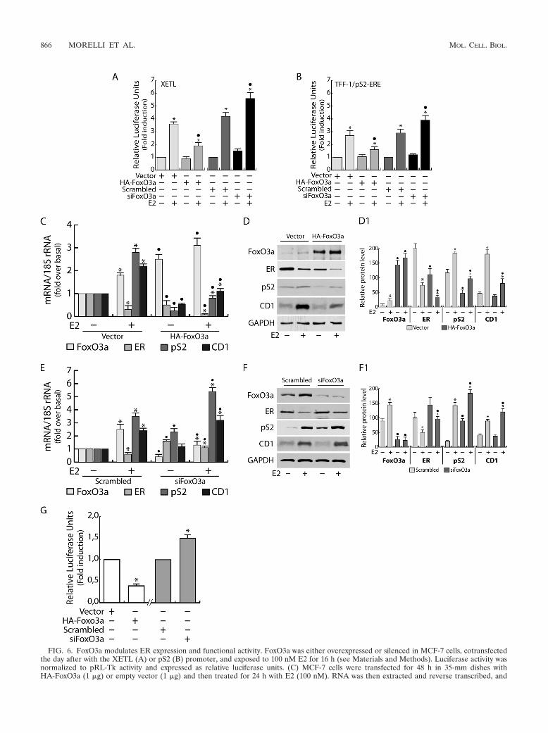
FIG. 6. FoxO3a modulates ER expression and functional activity. FoxO3a was either overexpressed or silenced in MCF-7 cells, cotransfectedthe day after with the XETL (A) or pS2 (B) promoter, and exposed to 100 nM E2 for 16 h (see Materials and Methods). Luciferase activity wasnormalized to pRL-Tk activity and expressed as relative luciferase units. (C) MCF-7 cells were transfected for 48 h in 35-mm dishes withHA-FoxO3a (1 �g) or empty vector (1 �g) and then treated for 24 h with E2 (100 nM). RNA was then extracted and reverse transcribed, and
866 MORELLI ET AL. MOL. CELL. BIOL.
for several reasons: (i) we were interested in focusing on Akt’srole in ER signaling and not the PI-3K/Akt axis in general; (ii)wortmannin, used at suggested concentrations, never inhibitedER function as API-2 did; and (iii) LY294002 has been sus-pected of having antiestrogenic properties (43), which clearlycould have interfered with our experiments. However, since
API-2 is unable to discriminate among Akt isoforms, smallinterfering RNAs were used to silence either Akt1 or Akt2.
Interestingly, only Akt2 knockdown produced effects onboth ER expression and function comparable to those ob-served after a prolonged exposure to API-2. Indeed, an evidentdecrease in ER protein content and in mRNA levels, followedby a predictable decrease in estrogen-dependent pS2 expres-sion, was evidenced. As mentioned above, these effects wereobtained only by Akt2 and not by Akt1 knockdown, suggestinga pivotal role of Akt2 in both the regulation of ER functionand its expression.
Although there are reports of Akt1 presence at the nuclearlevel, e.g., in thyroid cancer specimens and cultures (58) or inIGF-1-stimulated human embryonic kidney cells (3), in ourcellular model and under our experimental conditions wenever observed either Akt1 or Akt2 in the nucleus. Therefore,the genomic events deriving from Akt2 knockdown can beassumed to be mediated by one of its downstream nucleareffectors, such as the Forkhead winged-helix transcription fac-tor FoxO3a, the most highly expressed in MCF-7 cells amongthe four known members of the box O family.
Forkhead members were found to bind ER in a cell-freesystem in the presence of ER ligand (49, 62, 65). Our data, forthe first time, demonstrate that the interaction between ERand FoxO3a occurs in a whole-cell system. Indeed, ER wasfound to colocalize with FoxO3a at the perinuclear and nuclearlevels and to coprecipitate from MCF-7 nuclear-protein ex-tracts. E2 stimulation enhanced the binding between the twoproteins, most likely as a consequence of the increased ERnuclear retention, as well as of the E2-induced FoxO3a over-expression. This ER/FoxO3a interaction became much moreevident in Akt2 silenced samples, probably for a more sus-tained FoxO3a translocation into the nucleus. More interest-ingly, we found that FoxO3a was recruited to the pS2 promoterin an E2-dependent manner and that Akt2 inhibition empha-sized this phenomenon. FoxO3a binding to DNA occurred onthe Forkhead-responsive sequence close to the TATAA box(Fig. 5). The current literature reports controversial data re-garding the Forkhead box O function on ER target promoters.Some authors state that these transcriptional factors can act ascoactivators (49), while others found that they can have abifunctional role, behaving as ER coactivators or corepressors,depending on the cellular model (62, 65). In our MCF-7 clone,FoxO3a overexpression inhibited ER-mediated transcriptionof E2-regulated genes, such as pS2 and CD1. This effect couldmost likely be ascribable to reduced recruitment of ER to EREsites on the pS2 promoter, due to both reduced ER expression
FoxO3a, ER, pS2, and CD1 transcripts were analyzed by real-time PCR. Each sample was normalized to its 18S rRNA content. (D) Fifty �g ofwhole-cell extracts from a duplicate set of cells treated as described above was subjected to WB analysis. (D1) The relative protein levels,normalized over GAPDH, are reported. (E) RNA extracted from FoxO3a-silenced MCF-7 cells, treated with E2 (100 nM) for 24 h, was reversetranscribed, and FoxO3a, ER, pS2, and CD1 transcripts were analyzed by real-time PCR. Each sample was normalized to its 18S rRNA content.(F) A duplicate set of cells, treated as described above, was lysed, and 50 �g of whole-cell lysates was loaded for protein expression. (F1) Therelative protein levels, normalized versus GAPDH, were analyzed, and the optical density is reported. (G) MCF-7 cells were transfected withHA-FoxO3a or control vector (white bars) or FoxO3a siRNA or scrambled siRNA (gray bars) and cotransfected the day after with pGL3-ERprom(E). As described above, luciferase activity was normalized to pRL-Tk activity and expressed as relative luciferase units. In all experiments,significance values were as follows: �, P � 0.05 versus each non-E2-treated sample; F, P � 0.05 versus the corresponding treatment in scrambledsiRNA or vector-transfected samples. The error bars indicate SD.
FIG. 7. FoxO3a inhibits cell proliferation. MCF-7 cells were trans-fected with either HA-FoxO3a (A) or siFoxO3a (B) and treated asdescribed in Materials and Methods. The cells were counted in ahemocytometer at 50% confluence (day 0) and 1, 2, and 3 days later.The results represent the means � SD of three independent experi-ments. (A1 and B1) The FoxO3a content was analyzed by WB at eachtime point.
VOL. 30, 2010 Akt2/FoxO3a SIGNALING REGULATES ER� ACTIVITY 867
and the increased recruitment of the overexpressed negativemodulator FoxO3a on its own binding motif, while FoxO3asilencing led to the opposite effect. It is worthwhile to empha-size that, unlike pS2, we cannot state at the moment whetherCD1 is directly regulated by FoxO3a through its binding toForkhead-responsive elements, though they are present on theCD1 promoter, or whether this effect is due to a reducedgeneral ER content. In fact, as mentioned above, a strongdecrease in ER expression, at both the RNA and proteinlevels, was observed in FoxO3a-overexpressing samples. Al-though our preliminary results suggest possible regulation ofthe ER promoter by FoxO3a, further investigations are neededto elucidate if this effect really depends on the loss of FoxO3aas a repressive element for ER gene transcription or if it is dueto prolonged ER mRNA stability. Indeed, at least two func-tional Forkhead binding sites were identified on ER promoterB (17), but the authors demonstrateed an activating effect ofconstitutively active FoxO3a at this level in mouse mammarytumor-derived NF639 cells paralleled by an increase in ERprotein in cells transiently overexpressing FoxO3a. These datado not support our hypothesis, nor are they in agreement withrecent observations by Zou et al., who did not find significantdifferences in ER expression in FoxO3a-overexpressingMCF-7 stable clones (65). Furthermore, our findings also con-trast with the reported decrease in ER levels in MCF-7 cellsinfected with dominant-negative FoxO3a-expressing adenovi-rus (17). The incongruence of these data could be partiallyexplained by the different cell systems, culture conditions, andexperimental procedures used, but this cannot replace furtherstudy of the possible mechanism through which FoxO3a regu-lates ER expression and/or stability. However, our findingswere further corroborated by the growth inhibition observed inFoxO3a-overexpressing cells, as well as by the proliferativerate increase in FoxO3a silenced samples, evidencing the phys-iological relevance of this transcription factor in regulating theER mitogenic signal. Moreover, we reproduced the main ex-periments in two additional ER-positive cell lines, T47D and
ZR75, where Akt inhibition, as well as FoxO3a overexpression,led to ER downregulation and impaired transcriptional activ-ity, suggesting how the Akt2/FoxO3a axis has a pivotal role inmodulating ER function in ER-expressing breast cancer cells.
In fact, in two ER-negative cell lines, HeLa and SKBR3, thatectopically express ER, Akt inhibition did not lead to ER loss(protein and mRNA), supporting our hypothesis that the Aktpathway might control ER expression at a genomic level. No-tably SKBR3 showed a significant decrease of ER transcrip-tional activity in response to API-2, mimicking MCF-7 behav-ior, while in HeLa cells, API-2 did not significantly affect ERtransactivaton. The latter result could be explained by consid-ering the different expression levels of Akt2 and Foxo3a in thetwo cell lines: in fact, HeLa cells express negligible amounts ofboth proteins, while their levels in SKBR3 cells are comparableto those found in MCF-7 cells, underlining the fact that themolecular machinery introduced here might work properly incells expressing both Akt2 and FoxO3a.
Finally, as already mentioned, E2 stimulation inducedFoxO3a transcription and protein expression, which leads us tohypothesize the presence of estrogen target sequences on theFoxO3a promoter region. This observation could suggest ahomeostatic control mechanism through which E2-inducedFoxO3a upregulation is a required event to ensure ligandedER inactivation. The molecular basis on which E2 activatesFoxO3a expression is currently under investigation in our lab-oratory. In conclusion, in our model, FoxO3a, transcriptionallyactivated by Akt inhibition, might behave as a repressor forER-mediated transcription (i) directly, by binding to Fork-head-responsive elements on ER target gene promoters; (ii)indirectly, by the recruitment on ERE sequences of theFoxO3a/ER complex; and (iii) by inhibiting ER expression.
Taken together, the results presented here show that, amongPKB members, only Akt2 seems to modulate ER activity atmultiple levels, having a key role in the regulation of (i) theretention of ER in the nucleus, (ii) ER expression, and (iii)FoxO3a activation. A schematic summary of our findings is
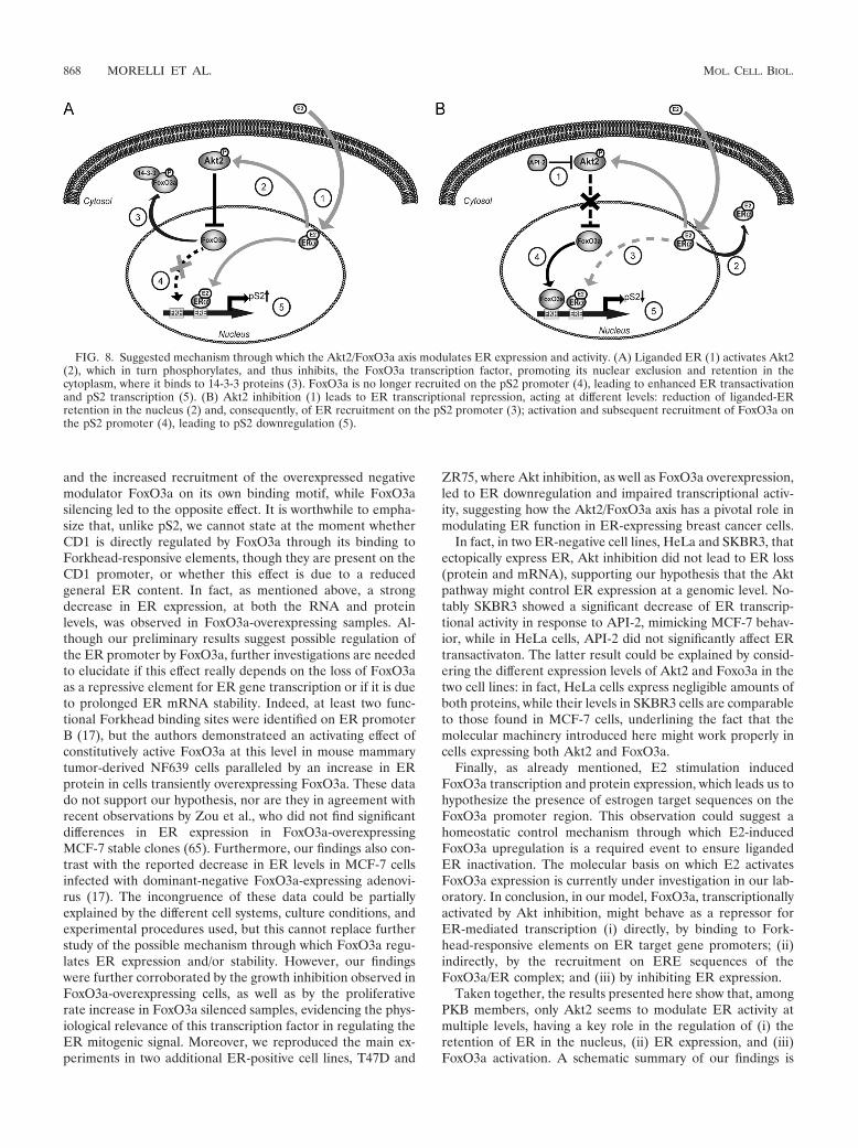
FIG. 8. Suggested mechanism through which the Akt2/FoxO3a axis modulates ER expression and activity. (A) Liganded ER (1) activates Akt2(2), which in turn phosphorylates, and thus inhibits, the FoxO3a transcription factor, promoting its nuclear exclusion and retention in thecytoplasm, where it binds to 14-3-3 proteins (3). FoxO3a is no longer recruited on the pS2 promoter (4), leading to enhanced ER transactivationand pS2 transcription (5). (B) Akt2 inhibition (1) leads to ER transcriptional repression, acting at different levels: reduction of liganded-ERretention in the nucleus (2) and, consequently, of ER recruitment on the pS2 promoter (3); activation and subsequent recruitment of FoxO3a onthe pS2 promoter (4), leading to pS2 downregulation (5).
868 MORELLI ET AL. MOL. CELL. BIOL.
shown in Fig. 8. Our data point out the importance of theAkt/FoxO axis in E2-induced ER activation and signaling, ev-idencing additional mechanisms that could represent noveltargets in ER-positive breast cancer therapy.
ACKNOWLEDGMENTS
We thank B. Van der Burg (Utrecht, the Netherlands) for providingMCF-7 cells, V. Giguere (McGill University, Quebec City, Canada) forthe pS2/TFF-1-ERE (pS2-ERE) plasmid, and S. A. W. Fuqua (BaylorCollege of Medicine, Breast Center, Houston, TX) for the pGL3-ERprom(E).
Financial support for this work was received from AIRC, MIUR Ex60%, and Regione Calabria.
We declare that there is no conflict of interest that would prejudicethe impartiality of this scientific work.
REFERENCES
1. Alvarez, B., A. C. Martinez, B. M. Burgering, and A. C. Carrera. 2001.Forkhead transcription factors contribute to execution of the mitotic pro-gramme in mammals. Nature 413:744–747.
2. Anderson, E., R. B. Clarke, and A. Howell. 1998. Estrogen responsivenessand control of normal human breast proliferation. J. Mammary Gland Biol.Neoplasia 3:23–35.
3. Andjelkovic, M., D. R. Alessi, R. Meier, A. Fernandez, N. J. Lamb, M. Frech,P. Cron, P. Cohen, J. M. Lucocq, and B. A. Hemmings. 1997. Role oftranslocation in the activation and function of protein kinase B. J. Biol.Chem. 272:31515–31524.
4. Beck, S., P. Sommer, E. dos Santos Silva, N. Blin, and P. Gott. 1999.Hepatocyte nuclear factor 3 (winged helix domain) activates trefoil factorgene TFF1 through a binding motif adjacent to the TATAA box. DNA CellBiol. 18:157–164.
5. Berry, M., A. M. Nunez, and P. Chambon. 1989. Estrogen-responsive ele-ment of the human pS2 gene is an imperfectly palindromic sequence. Proc.Natl. Acad. Sci. U. S. A. 86:1218–1222.
6. Biswas, D. K., A. P. Cruz, E. Gansberger, and A. B. Pardee. 2000. Epidermalgrowth factor-induced nuclear factor kappa B activation: a major pathway ofcell-cycle progression in estrogen-receptor negative breast cancer cells. Proc.Natl. Acad. Sci. U. S. A. 97:8542–8547.
7. Brader, S., and S. A. Eccles. 2004. Phosphoinositide 3-kinase signallingpathways in tumor progression, invasion and angiogenesis. Tumori 90:2–8.
8. Brunet, A., A. Bonni, M. J. Zigmond, M. Z. Lin, P. Juo, L. S. Hu, M. J.Anderson, K. C. Arden, J. Blenis, and M. E. Greenberg. 1999. Akt promotescell survival by phosphorylating and inhibiting a Forkhead transcriptionfactor. Cell 96:857–868.
9. Bunone, G., P. A. Briand, R. J. Miksicek, and D. Picard. 1996. Activation ofthe unliganded estrogen receptor by EGF involves the MAP kinase pathwayand direct phosphorylation. EMBO J. 15:2174–2183.
10. Campbell, R. A., P. Bhat-Nakshatri, N. M. Patel, D. Constantinidou, S. Ali,and H. Nakshatri. 2001. Phosphatidylinositol 3-kinase/AKT-mediated acti-vation of estrogen receptor alpha: a new model for anti-estrogen resistance.J. Biol. Chem. 276:9817–9824.
11. Carroll, J. S., X. S. Liu, A. S. Brodsky, W. Li, C. A. Meyer, A. J. Szary, J.Eeckhoute, W. Shao, E. V. Hestermann, T. R. Geistlinger, E. A. Fox, P. A.Silver, and M. Brown. 2005. Chromosome-wide mapping of estrogen recep-tor binding reveals long-range regulation requiring the Forkhead proteinFoxA1. Cell 122:33–43.
12. Castoria, G., A. Migliaccio, A. Bilancio, M. Di Domenico, A. de Falco, M.Lombardi, R. Fiorentino, L. Varricchio, M. V. Barone, and F. Auricchio.2001. PI3-kinase in concert with Src promotes the S-phase entry of oestra-diol-stimulated MCF-7 cells. EMBO J. 20:6050–6059.
13. Clark, G. M., C. K. Osborne, and W. L. McGuire. 1984. Correlations be-tween estrogen receptor, progesterone receptor, and patient characteristicsin human breast cancer. J. Clin. Oncol. 2:1102–1109.
14. Couse, J. F., and K. S. Korach. 1999. Estrogen receptor null mice: what havewe learned and where will they lead us? Endocr. Rev. 20:358–417.
15. deGraffenried, L. A., S. G. Hilsenbeck, and S. A. Fuqua. 2002. Sp1 is essentialfor estrogen receptor alpha gene transcription. J. Steroid Biochem. Mol.Biol. 82:7–18.
16. Dowsett, M. 2001. Overexpression of HER-2 as a resistance mechanism tohormonal therapy for breast cancer. Endocr. Relat. Cancer 8:191–195.
17. Guo, S., and G. E. Sonenshein. 2004. Forkhead box transcription factorFOXO3a regulates estrogen receptor alpha expression and is repressed bythe Her-2/neu/phosphatidylinositol 3-kinase/Akt signaling pathway. Mol.Cell. Biol. 24:8681–8690.
18. Improta-Brears, T., A. R. Whorton, F. Codazzi, J. D. York, T. Meyer, andD. P. McDonnell. 1999. Estrogen-induced activation of mitogen-activatedprotein kinase requires mobilization of intracellular calcium. Proc. Natl.Acad. Sci. U. S. A. 96:4686–4691.
19. Jackson, J. G., J. I. Kreisberg, A. P. Koterba, D. Yee, and M. G. Brattain.2000. Phosphorylation and nuclear exclusion of the forkhead transcriptionfactor FKHR after epidermal growth factor treatment in human breastcancer cells. Oncogene 19:4574–4581.
20. Jacobs, F. M., L. P. van der Heide, P. J. Wijchers, J. P. Burbach, M. F.Hoekman, and M. P. Smidt. 2003. FoxO6, a novel member of the FoxO classof transcription factors with distinct shuttling dynamics. J. Biol. Chem. 278:35959–35967.
21. Jordan, N. J., J. M. Gee, D. Barrow, A. E. Wakeling, and R. I. Nicholson.2004. Increased constitutive activity of PKB/Akt in tamoxifen resistant breastcancer MCF-7 cells. Breast Cancer Res. Treat. 87:167–180.
22. Kandel, E. S., and N. Hay. 1999. The regulation and activities of the multi-functional serine/threonine kinase Akt/PKB. Exp. Cell Res. 253:210–229.
23. Kato, S., H. Endoh, Y. Masuhiro, T. Kitamoto, S. Uchiyama, H. Sasaki, S.Masushige, Y. Gotoh, E. Nishida, H. Kawashima, D. Metzger, and P. Cham-bon. 1995. Activation of the estrogen receptor through phosphorylation bymitogen-activated protein kinase. Science 270:1491–1494.
24. Katzenellenbogen, B. S., K. L. Kendra, M. J. Norman, and Y. Berthois. 1987.Proliferation, hormonal responsiveness, and estrogen receptor content ofMCF-7 human breast cancer cells grown in the short-term and long-termabsence of estrogens. Cancer Res. 47:4355–4360.
25. Keen, J. C., and N. E. Davidson. 2003. The biology of breast carcinoma.Cancer 97:825–833.
26. Knuefermann, C., Y. Lu, B. Liu, W. Jin, K. Liang, L. Wu, M. Schmidt, G. B.Mills, J. Mendelsohn, and Z. Fan. 2003. HER2/PI-3K/Akt activation leads toa multidrug resistance in human breast adenocarcinoma cells. Oncogene22:3205–3212.
27. Konecny, G., G. Pauletti, M. Pegram, M. Untch, S. Dandekar, Z. Aguilar, C.Wilson, H. M. Rong, I. Bauerfeind, M. Felber, H. J. Wang, M. Beryt, R.Seshadri, H. Hepp, and D. J. Slamon. 2003. Quantitative association be-tween HER-2/neu and steroid hormone receptors in hormone receptor-positive primary breast cancer. J. Natl. Cancer Inst. 95:142–153.
28. Lanzino, M., C. Morelli, C. Garofalo, M. L. Panno, L. Mauro, S. Ando, andD. Sisci. 2008. Interaction between estrogen receptor alpha and insulin/IGFsignaling in breast cancer. Curr. Cancer Drug Targets 8:597–610.
29. Lee, C. S., A. deFazio, C. J. Ormandy, and R. L. Sutherland. 1996. Inverseregulation of oestrogen receptor and epidermal growth factor receptor geneexpression in MCF-7 breast cancer cells treated with phorbol ester. J. SteroidBiochem. Mol. Biol. 58:267–275.
30. Lin, C. Y., V. B. Vega, J. S. Thomsen, T. Zhang, S. L. Kong, M. Xie, K. P.Chiu, L. Lipovich, D. H. Barnett, F. Stossi, A. Yeo, J. George, V. A.Kuznetsov, Y. K. Lee, T. H. Charn, N. Palanisamy, L. D. Miller, E. Cheung,B. S. Katzenellenbogen, Y. Ruan, G. Bourque, C. L. Wei, and E. T. Liu. 2007.Whole-genome cartography of estrogen receptor alpha binding sites. PLoSGenet. 3:e87.
31. Liu, Y., H. Gao, T. T. Marstrand, A. Strom, E. Valen, A. Sandelin, J. A.Gustafsson, and K. Dahlman-Wright. 2008. The genome landscape of ER-alpha- and ERbeta-binding DNA regions. Proc. Natl. Acad. Sci. U. S. A.105:2604–2609.
32. Lobenhofer, E. K., G. Huper, J. D. Iglehart, and J. R. Marks. 2000. Inhibi-tion of mitogen-activated protein kinase and phosphatidylinositol 3-kinaseactivity in MCF-7 cells prevents estrogen-induced mitogenesis. Cell GrowthDiffer. 11:99–110.
33. Lombardi, M., G. Castoria, A. Migliaccio, M. V. Barone, R. Di Stasio, A.Ciociola, D. Bottero, H. Yamaguchi, E. Appella, and F. Auricchio. 2008.Hormone-dependent nuclear export of estradiol receptor and DNA synthe-sis in breast cancer cells. J. Cell Biol. 182:327–340.
34. Mazumdar, A., and R. Kumar. 2003. Estrogen regulation of Pak1 and FKHRpathways in breast cancer cells. FEBS Lett. 535:6–10.
35. McCarty, K. S., Jr., T. K. Barton, B. F. Fetter, B. H. Woodard, J. A. Mossler,W. Reeves, J. Daly, W. E. Wilkinson, and K. S. McCarty, Sr. 1980. Corre-lation of estrogen and progesterone receptors with histologic differentiationin mammary carcinoma. Cancer 46:2851–2858.
36. Medema, R. H., G. J. Kops, J. L. Bos, and B. M. Burgering. 2000. AFX-likeForkhead transcription factors mediate cell-cycle regulation by Ras and PKBthrough p27kip1. Nature 404:782–787.
37. Morelli, C., C. Garofalo, D. Sisci, S. del Rincon, S. Cascio, X. Tu, A.Vecchione, E. R. Sauter, W. H. Miller, Jr., and E. Surmacz. 2004. Nuclearinsulin receptor substrate 1 interacts with estrogen receptor alpha at EREpromoters. Oncogene 23:7517–7526.
38. Mossler, J. A., K. S. McCarty, Jr., and W. W. Johnston. 1981. The correla-tion of cytologic grade and steroid receptor content in effusions of metastaticbreast carcinoma. Acta Cytol. 25:653–658.
39. Nakamura, N., S. Ramaswamy, F. Vazquez, S. Signoretti, M. Loda, andW. R. Sellers. 2000. Forkhead transcription factors are critical effectors ofcell death and cell cycle arrest downstream of PTEN. Mol. Cell. Biol. 20:8969–8982.
40. Nakatani, K., D. A. Thompson, A. Barthel, H. Sakaue, W. Liu, R. J. Weigel,and R. A. Roth. 1999. Up-regulation of Akt3 in estrogen receptor-deficientbreast cancers and androgen-independent prostate cancer lines. J. Biol.Chem. 274:21528–21532.
VOL. 30, 2010 Akt2/FoxO3a SIGNALING REGULATES ER� ACTIVITY 869
41. Osborne, C. K. 1998. Steroid hormone receptors in breast cancer manage-ment. Breast Cancer Res. Treat. 51:227–238.
42. Osborne, C. K., K. Hobbs, and G. M. Clark. 1985. Effect of estrogens andantiestrogens on growth of human breast cancer cells in athymic nude mice.Cancer Res. 45:584–590.
43. Pasapera Limon, A. M., J. Herrera-Munoz, R. Gutierrez-Sagal, and A.Ulloa-Aguirre. 2003. The phosphatidylinositol 3-kinase inhibitor LY294002binds the estrogen receptor and inhibits 17beta-estradiol-induced transcrip-tional activity of an estrogen sensitive reporter gene. Mol. Cell Endocrinol.200:199–202.
44. Perou, C. M., T. Sorlie, M. B. Eisen, M. van de Rijn, S. S. Jeffrey, C. A. Rees,J. R. Pollack, D. T. Ross, H. Johnsen, L. A. Akslen, O. Fluge, A. Pergamen-schikov, C. Williams, S. X. Zhu, P. E. Lonning, A. L. Borresen-Dale, P. O.Brown, and D. Botstein. 2000. Molecular portraits of human breast tumours.Nature 406:747–752.
45. Reagan-Shaw, S., and N. Ahmad. 2006. RNA interference-mediated deple-tion of phosphoinositide 3-kinase activates forkhead box class O transcrip-tion factors and induces cell cycle arrest and apoptosis in breast carcinomacells. Cancer Res. 66:1062–1069.
46. Rena, G., S. Guo, S. C. Cichy, T. G. Unterman, and P. Cohen. 1999. Phos-phorylation of the transcription factor forkhead family member FKHR byprotein kinase B. J. Biol. Chem. 274:17179–17183.
47. Samatar, A. A., L. Wang, A. Mirza, S. Koseoglu, S. Liu, and C. C. Kumar.2002. Transforming growth factor-beta 2 is a transcriptional target for Akt/protein kinase B via forkhead transcription factor. J. Biol. Chem. 277:28118–28126.
48. Santini, D., C. Ceccarelli, M. Taffurelli, S. Pileri, and D. Marrano. 1996.Differentiation pathways in primary invasive breast carcinoma as suggestedby intermediate filament and biopathological marker expression. J. Pathol.179:386–391.
49. Schuur, E. R., A. V. Loktev, M. Sharma, Z. Sun, R. A. Roth, and R. J. Weigel.2001. Ligand-dependent interaction of estrogen receptor-alpha with mem-bers of the forkhead transcription factor family. J. Biol. Chem. 276:33554–33560.
50. Sisci, D., C. Morelli, C. Garofalo, F. Romeo, L. Morabito, F. Casaburi, E.Middea, S. Cascio, E. Brunelli, S. Ando, and E. Surmacz. 2007. Expressionof nuclear insulin receptor substrate 1 in breast cancer. J. Clin. Pathol.60:633–641.
51. Sisci, D., and E. Surmacz. 2007. Crosstalk between IGF signaling and steroidhormone receptors in breast cancer. Curr. Pharm. Des. 13:705–717.
52. Stal, O., G. Perez-Tenorio, L. Akerberg, B. Olsson, B. Nordenskjold, L.Skoog, and L. E. Rutqvist. 2003. Akt kinases in breast cancer and the resultsof adjuvant therapy. Breast Cancer Res. 5:R37–R44.
53. Stoica, G. E., T. F. Franke, M. Moroni, S. Mueller, E. Morgan, M. C. Iann,A. D. Winder, R. Reiter, A. Wellstein, M. B. Martin, and A. Stoica. 2003.Effect of estradiol on estrogen receptor-alpha gene expression and activitycan be modulated by the ErbB2/PI 3-K/Akt pathway. Oncogene 22:7998–8011.
54. Suhara, T., H. S. Kim, L. A. Kirshenbaum, and K. Walsh. 2002. Suppressionof Akt signaling induces Fas ligand expression: involvement of caspase and
Jun kinase activation in Akt-mediated Fas ligand regulation. Mol. Cell. Biol.22:680–691.
55. Sun, M., J. E. Paciga, R. I. Feldman, Z. Yuan, D. Coppola, Y. Y. Lu, S. A.Shelley, S. V. Nicosia, and J. Q. Cheng. 2001. Phosphatidylinositol-3-OHkinase (PI3K)/AKT2, activated in breast cancer, regulates and is induced byestrogen receptor alpha (ERalpha) via interaction between ERalpha andPI3K. Cancer Res. 61:5985–5991.
56. Sun, M., G. Wang, J. E. Paciga, R. I. Feldman, Z. Q. Yuan, X. L. Ma, S. A.Shelley, R. Jove, P. N. Tsichlis, S. V. Nicosia, and J. Q. Cheng. 2001.AKT1/PKBalpha kinase is frequently elevated in human cancers and itsconstitutive activation is required for oncogenic transformation in NIH3T3cells. Am. J. Pathol. 159:431–437.
56a.Tora, L., A. Mullick, D. Metzger, M. Ponglikitmongkol, I. Park, and P.Chambon. 1989. The cloned human oestrogen receptor contains a mutationwhich alters its hormone binding properties. EMBO J. 8:1981–1986.
57. Tsai, E. M., S. C. Wang, J. N. Lee, and M. C. Hung. 2001. Akt activation byestrogen in estrogen receptor-negative breast cancer cells. Cancer Res. 61:8390–8392.
58. Vasko, V., M. Saji, E. Hardy, M. Kruhlak, A. Larin, V. Savchenko, M.Miyakawa, O. Isozaki, H. Murakami, T. Tsushima, K. D. Burman, C. DeMicco, and M. D. Ringel. 2004. Akt activation and localisation correlate withtumour invasion and oncogene expression in thyroid cancer. J. Med. Genet.41:161–170.
59. Witton, C. J., J. R. Reeves, J. J. Going, T. G. Cooke, and J. M. Bartlett. 2003.Expression of the HER1-4 family of receptor tyrosine kinases in breastcancer. J. Pathol. 200:290–297.
60. Yu, X., R. V. Rajala, J. F. McGinnis, F. Li, R. E. Anderson, X. Yan, S. Li,R. V. Elias, R. R. Knapp, X. Zhou, and W. Cao. 2004. Involvement ofinsulin/phosphoinositide 3-kinase/Akt signal pathway in 17 beta-estradiol-mediated neuroprotection. J. Biol. Chem. 279:13086–13094.
61. Yuan, Z. Q., M. Sun, R. I. Feldman, G. Wang, X. Ma, C. Jiang, D. Coppola,S. V. Nicosia, and J. Q. Cheng. 2000. Frequent activation of AKT2 andinduction of apoptosis by inhibition of phosphoinositide-3-OH kinase/Aktpathway in human ovarian cancer. Oncogene 19:2324–2330.
62. Zhao, H. H., R. E. Herrera, E. Coronado-Heinsohn, M. C. Yang, J. H.Ludes-Meyers, K. J. Seybold-Tilson, Z. Nawaz, D. Yee, F. G. Barr, S. G.Diab, P. H. Brown, S. A. Fuqua, and C. K. Osborne. 2001. Forkhead homo-logue in rhabdomyosarcoma functions as a bifunctional nuclear receptor-interacting protein with both coactivator and corepressor functions. J. Biol.Chem. 276:27907–27912.
63. Zhu, Y., M. A. Saunders, H. Yeh, W. G. Deng, and K. K. Wu. 2002. Dynamicregulation of cyclooxygenase-2 promoter activity by isoforms of CCAAT/enhancer-binding proteins. J. Biol. Chem. 277:6923–6928.
64. Zinda, M. J., M. A. Johnson, J. D. Paul, C. Horn, B. W. Konicek, Z. H. Lu,G. Sandusky, J. E. Thomas, B. L. Neubauer, M. T. Lai, and J. R. Graff. 2001.AKT-1, -2, and -3 are expressed in both normal and tumor tissues of the lung,breast, prostate, and colon. Clin. Cancer Res. 7:2475–2479.
65. Zou, Y., W. B. Tsai, C. J. Cheng, C. Hsu, Y. M. Chung, P. C. Li, S. H. Lin,and M. C. Hu. 2008. Forkhead box transcription factor FOXO3a suppressesestrogen-dependent breast cancer cell proliferation and tumorigenesis.Breast Cancer Res. 10:R21.
870 MORELLI ET AL. MOL. CELL. BIOL.
Copyright © 2022 FDOKUMEN