Airway epithelial wound repair: role of carbohydrate sialyl Lewisx
36
LCMP-00120-2006.R1 Airway Epithelial Wound Repair: Role of Carbohydrate Sialyl Lewis X Sima Allahverdian 1 , Kimberly R Wojcik 2 , Delbert R Dorscheid 1 1 The James Hogg iCAPTURE Centre for Cardiovascular and Pulmonary Research, Faculty of Medicine, St. Paul's Hospital, University of British Columbia, Vancouver, CANADA 2 Section of Pulmonary and Critical Care Medicine, Department of Medicine, Division of Biological Sciences, University of Chicago, Chicago, Illinois 60637, USA Correspondence author: Delbert R Dorscheid, MD, PhD The James Hogg iCAPTURE Centre for Cardiovascular and Pulmonary Research, St. Paul's Hospital, Room 166 - 1081 Burrard Street, Vancouver, B.C. V6Z 1Y6 (604) 806-8346 (ext. 62746) (604) 806 8351 (fax) E-mail: [email protected] Running title: sialyl Lewis x in epithelial repair Page 1 of 36 Articles in PresS. Am J Physiol Lung Cell Mol Physiol (June 2, 2006). doi:10.1152/ajplung.00120.2006 Copyright © 2006 by the American Physiological Society.
-
Upload
independent -
Category
Documents
-
view
3 -
download
0
Transcript of Airway epithelial wound repair: role of carbohydrate sialyl Lewisx

LCMP-00120-2006.R1
Airway Epithelial Wound Repair: Role of Carbohydrate Sialyl Lewis X
Sima Allahverdian1, Kimberly R Wojcik2, Delbert R Dorscheid1
1The James Hogg iCAPTURE Centre for Cardiovascular and Pulmonary Research, Faculty of
Medicine, St. Paul's Hospital, University of British Columbia, Vancouver, CANADA
2Section of Pulmonary and Critical Care Medicine, Department of Medicine, Division of
Biological Sciences, University of Chicago, Chicago, Illinois 60637, USA
Correspondence author: Delbert R Dorscheid, MD, PhD
The James Hogg iCAPTURE Centre for Cardiovascular and Pulmonary Research,
St. Paul's Hospital, Room 166 - 1081 Burrard Street, Vancouver, B.C. V6Z 1Y6
(604) 806-8346 (ext. 62746)
(604) 806 8351 (fax)
E-mail: [email protected]
Running title: sialyl Lewis x in epithelial repair
Page 1 of 36Articles in PresS. Am J Physiol Lung Cell Mol Physiol (June 2, 2006). doi:10.1152/ajplung.00120.2006
Copyright © 2006 by the American Physiological Society.

LCMP-00120-2006.R12
Abstract
Background: Epithelial repair is a complex cellular and molecular process, the details of which
are still not clearly understood. Plasma membrane glycoconjugates can modulate cell function by
altering the function of protein and lipids. Sialyl-Lewis x (sLex), a fucose containing
tetrasaccharide, decorates membrane bound and secreted proteins and mediates cell-cell
interaction. In the present study we investigated the role of sLex in airway epithelial repair.
Methods and Results: Using immunohistochemistry we showed an increased expression of sLex
in areas of damaged bronchial epithelium when compared to intact regions. Confluent
monolayers of airway epithelial cells were mechanically wounded and allowed to close.
Wounded monolayers were photographed for wound closure kinetics, fixed for
immunocytochemical studies, or subjected to RNA extraction. Examining the expression of
different α1,3-fucosyltransferases (FucT), enzymes which mediate the final step in the synthesis
of sLex, we found that FucT-IV was the common gene expressed in all cell lines and primary
airway epithelial cells. We demonstrated an increased expression of sLex over time after
mechanical injury. Blocking of sLex with an inhibitory antibody completely prevented epithelial
repair.
Conclusion: Our data suggest an essential functional role for sLex in epithelial repair. Further
studies are necessary to explore the exact mechanism for sLex in mediating cell-cell interaction
in bronchial epithelial cells to facilitate epithelial migration and repair.
Word Count: 215
Key words: Fucosyltransferase, Selectin, Airway epithelium, Epidermal growth factor
Page 2 of 36
LCMP-00120-2006.R13
Introduction
As the barrier to the external environment, the bronchial epithelium is continuously
exposed to gaseous and particulate components of inhaled air and therefore is frequently injured.
Inflammation is an initial response to tissue injury, which provides immune cells dedicated to
debris removal and growth factors to promote tissue repair (7). In addition to this inflammatory
response, wound healing involves migration and spreading of epithelial cells into the damaged
region and proliferation of new epithelial cells (12, 21). The complete healing of wounds
represents an important process by which the respiratory epithelial barrier integrity is
maintained.
Several proteins essential for normal cell physiology, like membrane bound receptors for
growth factors and cytokines, are glycosylated. Several lines of evidence support the theory that
oligosaccharide moieties are crucial for the function of some of those proteins and that variation
in their glycosylation pattern often leads to changes in their function (13, 27, 38).
Oligosaccharides on cell surface proteins and lipids have functional roles in cell adhesion (36),
migration (40), proliferation (4) and growth potential (28). The molecular events that initiate,
mediate, and regulate different processes involved in epithelial repair have not been fully
elucidated, but a number of studies have suggested that glycoconjugates attached to proteins
within the plasma membrane of epithelial cells play a central role in these events. Our laboratory
has determined the pattern of cell surface glycosylation in normal human airway epithelial cells
(9). We have shown that glycosylation profiles in airway epithelium change overtime during
repair of a wound created by mechanical injury (47). Our data also suggested that cell surface N-
Page 3 of 36

LCMP-00120-2006.R14
glycosylation has a functional role in airway epithelial cell adhesion and migration and that N-
glycosylation with terminal fucosylation plays an essential role in the complex process of repair
by coordination of certain cell-cell functions (10). In asthma, detailed cellular and ultrastructural
examination of bronchial biopsies and bronchoalveolar lavage fluid have provided evidence for
epithelial damage, even in mild cases (2, 18, 26, 31). This excessive epithelial damage can arise
from an enhanced susceptibility to injury or an inadequate repair response, or a combination of
both (7). Kauffmann et al. (20) has reported an under-representation of carbohydrate structures
with terminal fucose in asthmatic patients with a correlation between this deficiency and the
severity of the disease. These data suggest that defects in epithelial repair in asthma patients may
be due, in part, to improper glycosylation of airway epithelial cells. Taken together, these studies
indicate an involvement of cell surface carbohydrates especially those with terminal fucose in
regulation of epithelial repair processes.
Lewis blood group antigens are biosynthetically and structurally related carbohydrate
structures used as markers of cell differentiation and embryonic development (34). Expression of
these antigens is not limited to erythrocytes and they can be found in different tissues and organs.
It has been shown that these oligosaccharide structures are involved in cell-cell interaction. Sialyl
Lewis x, (sLex)1, a fucose containing tetrasaccharide [NeuAcα2-3Galβ1-4(Fucα1-3)GlcNAc]
belonging to this family, has been recognized as a ligand for E-selectin and therefore has an
important role in lymphocyte trafficking (28, 36, 45). This antigen has been detected in various
tumors where it mediates binding of cancerous cells to endothelial selectins and thereby
promoting tumor metastasis (14, 30). sLex is also found at the non-reducing termini of N-linked
or O-linked oligosaccharides on glycoproteins as well as on glycosphingolipids. Final step in
synthesis of sLex is catalyzed by specific α1,3-fucosyltransferases (FucT)2. Six human α1,3-
Page 4 of 36

LCMP-00120-2006.R15
FucTs have been cloned and partially characterized: FucT-III, FucT-IV, FucT-V, FucT-VI,
FucT-VII, and FucT-IX which show different pattern of expression among tissues.
Neither the normal processes leading to complete epithelial repair nor the abnormalities
that permit chronic damage in disease states of the epithelium are fully understood. While
previous studies in epithelial repair suggested a role for glycoconjugates, none of these studies
specified an oligosaccharide structure(s) to be involved in repair. Our present study, to our
knowledge, for the first time demonstrates a critical role for the tetrasaccharide sLex in airway
epithelial repair. We found an over expression of sLex, in vivo and in vitro after injury. In a
culture model of epithelial repair, we were able to demonstrate an inhibition of repair after
blocking of sLex. These observations have important implications not only for understanding the
epithelial injury-repair cycle but also for identifying novel therapies for conditions resulting from
impaired epithelial repair, such as asthma.
Page 5 of 36

LCMP-00120-2006.R16
Materials and Methods
Collection of airway specimens from normal human subjects. Approval for the use of all
human tissue was granted by University of British Columbia and Providence Health Care Ethics
Review Board. Normal bronchial segments were collected from pathological specimens from
adults undergoing lung resection in a regional chest hospital in Großhansdorf, Germany. Samples
generally were of fourth, fifth or sixth generation central airways in transverse section, so that a
complete circumference could be examined. Bronchial specimens were fixed in 4%
paraformaldehyde (PFA) for 1 hr at room temperature. Samples were then shipped on ice to our
laboratory, washed in Dulbecco's Phosphate-Buffered Saline (DPBS) and stored at 4°C. After
paraffin-embedding airways were retrieved and tissue sections were prepared in transverse
orientation from the paraffin blocks for preparation of 5 µm thick sections. No subject had a
history of asthma.
Immunohistochemistry. Immunostaining was performed using a mouse monoclonal anti-
human sLex antibody (KM 93, Seikagaku America, Ijamsville, MD, USA) or an isotype matched
non-specific antibody.
Quantification. To study sLex immunoreactivity, the entire epithelium of one airway
section was systematically assessed in each subject. Several sections were obtained from the
same airway and more than one airway per donor sample was assessed. Several images were
taken from the entire airway section (usually 10-15 images based on the size of airway) in each
subject. In this manner the entire circumference of the airway was documented in images. If a
Page 6 of 36

LCMP-00120-2006.R17
wound was detected in one of these images this section then became the representative airway
section for that donor. Next, from the pool of images for the representative section, 3 numbers
were randomly selected and if they contained damaged area they would be further assessed for
sLex expression in the areas of damaged and intact epithelium. In this manner selection bias was
minimized. Epithelial damage was characterized morphologically by the absence of
differentiated ciliated and secretory cells (44). To study the immunoreactivity of sLex in areas of
epithelial damage and normal epithelium, percentage of positively stained basal and columnar
cells in three areas exhibiting epithelial damage and three areas of intact epithelium was
determined in each subject. A total of 6 normal airways were assessed. Therefore, together 18
areas of intact epithelium were compared with 18 areas of damaged epithelium. Previous studies
have shown that migratory epithelium presents within 40 µm from the wound edge (12).
Therefore, to evaluate sLex immunoreactivity in a damaged area, basal and columnar cells within
40 µm from either wound edge were counted. Next, epithelium farther than 40 µm from the
wound edge was considered as intact epithelium if both the pseudo-stratified layer was complete
and 40 µm of this area was evaluated for sLex staining. The ImagePro Plus image analysis
software (Media Cybernetics) was used to collate the airway images and to determine the
positive cells using a point counting grid. Positive counts were confirmed by manual inspection
by one author (S.A.).
Cell culture. 1HAEo- and 16HBE 14o- cells are SV40-transformed normal human airway
epithelial cells that have been characterized previously (6, 16) and express multiple surface
carbohydrate markers of normal primary basal airway epithelial cells (9). Primary normal human
bronchial epithelial (NHBE) cells were collected from subjects undergoing lung resection. These
Page 7 of 36

LCMP-00120-2006.R18
cells were derived from different donors. Epithelial cell purity was determined by examining the
typical morphological features of primary epithelial cells in culture and staining the cells with
anti-cytokeratin 18 antibody (USBiological, Swampscott, Massachusetts, USA). Cells were
subcultured and used between passages 3 and 5.
Monolayer wound repair assay. We have established this method previously using 6-well
culture dishes (10, 22-24). In two experiments NHBE cells grown in a monolayer were treated
with an inhibitory anti-sLex antibody (40 ng/ml) (KM 93, Seikagaku America, Ijamsville, MD,
USA) or an isotype-matched non-specific antibody (40 ng/ml) immediately after mechanical
injury. In three experiments, 1HAEo- cells were treated concurrently with both Epidermal
Growth Factor (EGF) (15ng/ml) and 4-Deoxy-fucose, a general inhibitor of fucosyltransferases
(FuTi)3 (10-3 to 10-5 M) (Calbiochem, La Jolla, CA, USA). In three experiments, 1HAEo- cells
were treated concurrently with both EGF (15ng/ml) and soluble sLex (10-4 to 10-7 M)
(Calbiochem, La Jolla, CA, USA). In two experiments, 1HAEo- cells were treated with either
FuTi3 (10-3 to 10-5 M) or soluble sLex (10-4 to 10-7 M) without the addition of EGF. In each
experiment, one well was used as a negative control with no treatment and one well was treated
with 15 ng/ml of EGF, which has previously been demonstrated to be a potent accelerant in
models of epithelial monolayer wound closure (22).
Immunocytochemistry. NHBE cells were grown on four-well chamber slides until
confluency. Three linear wounds were created using a rubber stylet. Monolayers were then fixed
at 0, 2, 6, 12, 24, and 48 h after mechanical injury using Clark’s solution (90% ethanol, 10%
Page 8 of 36

LCMP-00120-2006.R19
glacial acetic acid). Expression of sLex was detected using mouse anti-human sLex (KM 93,
Seikagaku America, Ijamsville, MD, USA).
RNA isolation and Real-Time Polymerase Chain Reaction. 1HAEo-, 16HBE 14o-, and
NHBE cells were grown to confluency and then RNA extracted. Expression of four subtypes of
α1,3-FucTs was studied by real-time RT-PCR4 using primers specific for FucT-III , -IV, -VII,
and –IX. RNA was extracted at specific time points after injury using the TRIzol reagent
(GIBCO/BRL), according to the manufacturer’s protocol. mRNA expression was quantified by
real-time PCR using LightCycler (Roche, Mannheim, Germany). The levels of target mRNAs
were normalized to the level of β-actin mRNA in the same sample.
Flow cytometry analysis. Flow cytometric analysis was performed on 1HAEo- and NHBE
cells using anti-E, -L and, -P selectin mouse monoclonal antibodies (RDI, Flanders, NJ).
Statistical Analysis. Data were entered in and analyzed by means of SPSS 7.1 for
Windows. Wound closure is expressed as a percentage of area at time 0. In previous
videomicroscopy experiments with cell monolayers (24), intra-observer variability was <2%, and
inter-observer variability was <4% for all measurements.
Page 9 of 36

LCMP-00120-2006.R110
Results
Expression of sLex is higher in areas of epithelial damage compared to intact epithelium.
We initiated investigating the role of sLex in epithelial repair by studying the expression of sLex
in damaged and intact areas of normal airway epithelium. Immunoreactivity of sLex was
analyzed in sections of bronchial specimens obtained from normal subjects (n=6). In each
subject three areas of epithelial damage and three areas of intact epithelium were identified as
described in Materials and Methods. The percentage of positively stained basal and columnar
cells was significantly higher in areas of damaged compared to intact epithelium (p<0.002,
Mann-Whitney U-test) (Figs. 1 and 2).
Mechanical injury enhances the expression of sLex in a culture model of airway
epithelium. We examined the effect of mechanical injury on sLex expression in mechanically
wounded monolayers of NHBE cells. Epithelial cells were characterized by staining the cells
with anti-cytokeratin-18 antibody and examination for typical morphological features of airway
epithelial cells in culture. Immunocytochemical staining of the wounded monolayer showed a
time-dependant increase in sLex expression coordinate with wound closure and a decrease once
the repair is complete (Fig. 3). Increased expression of sLex after mechanical injury was
associated with a change in the epithelial cell shape. The majority of the epithelial cells
expressing sLex exhibit an elongated morphology. These elongated cells appear to be migratory
epithelial cells in phenotype. Evaluation of migration and migratory phenotype of these cells was
beyond the scope of the present study.
Blocking of sLex with an anti-sLex inhibitory antibody prevents epithelial monolayer
wound repair. To determine whether sLex plays a role in bronchial epithelial repair, we studied
Page 10 of 36
LCMP-00120-2006.R111
kinetics of epithelial wound repair in the presence of a blocking anti-sLex antibody. A linear
wound was made in confluent monolayer of NHBE cells using a rubber stylet. Cells were treated
with an anti-sLex antibody (40 ng/ml) or an isotype-matched non-specific antibody (40 ng/ml)
immediately after injury. In each experiment, one well was used as a negative control with no
treatment. Corresponding wound areas were determined at 0, 2, 6, 12, and 24 h after wound
creation using time-lapse videomicroscopy (Fig. 4, Panel A). The remaining wound area 24 h
after wounding was significantly higher in monolayers treated with anti-sLex antibody compared
to non-treated monolayers and the ones treated with non-specific antibody (p<0.05) (Fig. 4,
Panel B). These data showed that blocking of sLex in our culture model of airway epithelial
wound repair inhibited wound closure.
α1,3-fucosyltransferases exhibit a diverse pattern of expression in 1HAEo-, 16HBE 14o-,
and NHBE cells. Expression of four subtypes of α1,3-FucTs, the enzyme responsible for the
fucosylation step in sLex antigen synthesis, was studied in 1HAEo-, 16HBE 14o-, and NHBE
cells using real-time RT-PCR (Table 1). FucT-IX was not expressed in any of the cell lines nor
primary cells examined. FucT-III and -VII showed a different pattern of expression among two
cell lines and primary cells. We found that FucT-IV is the only gene transcribed in both primary
cells and cell lines of airway epithelium.
A general fucosyltransferase inhibitor (FuTi) reduces epithelial repair in a culture model
of epithelial cell monolayer wound repair in the presence and absence of exogenous EGF. Our
previous data provide some evidence on the role of sLex in epithelial repair. We further
investigated the role of fucose containing oligosaccharides in epithelial repair by inhibiting
synthesis of fucose containing oligosaccharides using FuTi. Initial wound area and perimeter for
monolayers within each experimental series were equivalent and consistent. In control and EGF-
Page 11 of 36

LCMP-00120-2006.R112
only experiments pooled across experimental series, the remaining wound area after 24 h was
30±3.8% in control cultures and 3.7±1.5% in EGF-stimulated wounds (p< 0.01; n=10).
Concurrent treatment of monolayers with EGF (15 ng/ml) and the FuTi inhibited wound repair in
a dose-dependent manner when compared to EGF-alone. Monolayers treated with 10-3 compared
to monolayers treated with 10-4 M and 10-5 M FuTi and monolayers treated with 10-4 M
compared to 10-5 M FuTi had higher wound area 24 h after treatment (p<0.05). The remaining
wound area at 24 h after treatment with 10-4 M FuTi + EGF (35±1.4%) and 10-3 M FuTi + EGF
(43±2.6%) were significantly higher compared to EGF alone treated group (3.5±1.4%, p< 0.01)
(Fig. 5, Panel B). In the absence of exogenous EGF, FuTi in its highest doses (10-3 and 10-4 M)
inhibited epithelial repair compared to control monolayers. The remaining wound area at 24 h
after treatment with 10-4 M FuTi (34±1.0%) and 10-3 M FuTi (35±3.0%) were significantly
higher compared to control group with no treatment (23±1.6%, p< 0.05) (Fig. 5, Panel D).
Soluble sLex reduces epithelial repair in a culture model of epithelial cell monolayer
wound repair only in the presence of exogenous EGF. We further investigated the role of sLex in
epithelial repair by blocking the potential receptors for sLex using soluble sLex. Initial wound
area and perimeter for monolayers within each experimental series were equivalent and
consistent. In control and EGF-only experiments pooled across experimental series, the
remaining wound area after 24 h was 25±4.1% in control cultures and 1±0.3% in EGF-stimulated
wounds (p< 0.01; n=10). Concurrent treatment of monolayers with soluble sLex and EGF
inhibited wound repair in a concentration-dependent manner. Monolayers treated with 10–4 M
soluble sLex compared to monolayers treated with 10–5, 10–6, and 10–7 M soluble sLex had higher
wound area 24 h after treatment (p<0.05). The remaining wound area at 24 h after treatment with
10–4, 10–5, and 10–6 M soluble sLex + EGF was significantly higher compared to EGF alone
Page 12 of 36

LCMP-00120-2006.R113
treated group (p<0.01) (Fig. 6, Panel B). In the absence of exogenous EGF, soluble sLex had no
effect on epithelial repair (data are not shown). Altogether our data showed that co-treatment of
the injured monolayers with EGF and FuTi or soluble sLex reverse the acceleration effect of EGF
on epithelial repair. While the inhibitory effect of FuTi on epithelial repair remained in the
absence of EGF, soluble sLex showed no effect without the concomitant stimulation of EGF.
These data provide further evidence on the role of fucose containing oligosaccharides and sLex in
epithelial repair.
E-selectin is expressed by a subset of airway epithelial cells. Our previous studies
identified that fucose-containing ligands are essential for repairing airway epithelium. Over-
expression of sLex may promote closure of wounds by increasing the selectin (CD62) receptor-
ligand interaction in airway epithelial cells. Next we examined the expression of P-, E-, and L-
selectin by 1HAEo- cells using cytofluorometric analysis. We found that E- but not P- and L-
selectin was expressed by a subset of airway epithelial cells (data are not shown). We also
examined the expression of E-selectin by NHBE and 1HAEo- cells before (N=5) and 24 h after
mechanical injury (N=3) both in the presence of EGF (15ng/ml). There was no statistically
significant difference in the percent of cells expressing E-selectin receptor before and after
mechanical injury (Table 2).
Page 13 of 36

LCMP-00120-2006.R114
Discussion
In providing the physical barrier to the external environment, the bronchial epithelium is
continuously exposed to injuries. The airway epithelium is therefore routinely challenged as part
of normal function. Epithelial wound healing represents an important process by which the
respiratory epithelial barrier restores the physical barrier and tissue integrity is maintained.
Epithelial repair involves a series of ordered events including migration, spreading, proliferation
and differentiation of epithelial cells. Cell surface glycoconjugates have crucial function in a
variety of normal and disease states. It has been shown that glycans can modulate function of
proteins and lipids they are attached to and are involved in cell-cell and cell-matrix interaction
(13, 27, 36, 38, 42). There is a growing interest in exploring the role of cell surface
carbohydrates in epithelial repair. Using lectins in an in vitro model of wound repair, Adam et al.
(1) recently demonstrated that N-acetylglucosamine (GlcNAc)5 which is recognized by the lectin
wheat germ agglutinin (WGA)6 is required for epithelial repair. Our previous work demonstrated
that cell surface N-glycosylation has a functional role in airway epithelial cell adhesion and
migration. N-glycans with terminal fucosylation plays an essential role in the complex process of
repair by coordination of cell-cell functions, including migration (10). In the present study, for
the first time we examined the role of a specific fucose containing carbohydrate structure, sLex,
in bronchial epithelial repair.
Sialyl Lewis x is a member of the Lewis blood group structures which are found at the
non-reducing termini of N-linked or O-linked glycans on glycoproteins and glycolipids. sLex has
been identified as a necessary component of selectin-ligand interaction which mediate cell-cell
interaction and migration in several systems (14, 28, 30, 36, 45). To investigate the role of sLex
in epithelial repair we examined the expression of this antigen on bronchial epithelium by
Page 14 of 36

LCMP-00120-2006.R115
immunostaining. There was increased expression of sLex in areas of damaged epithelium
compared to intact regions. This finding suggests a possible contribution of sLex in epithelial
repair. Gipson (15) showed previously that cell surface carbohydrates on epithelial cells that are
spreading and/or migrating to cover a wound are different from cell surface carbohydrate
structures found on normal epithelial cells. Our laboratory has shown that glycosylation profiles
in airway epithelium change overtime during repair of a wound created by mechanical injury
(47). It has been shown that injury of the respiratory epithelium enhances bacterium
Pseudomonas aeruginosa adhesion to the epithelium and it has been speculated that changes of
cell surface glycoconjugates related to wound repair, cell migration and/or spreading may favor
P. aeruginosa adhesion (37).
The final step in synthesis of sLex is catalyzed by specific α1,3-FucT. The
fucosyltransferase gene family encodes for a group of proteins that show a complex tissue- and
cell type-specific expression pattern. FucT-IV and -VII are expressed in human leukocytes where
they modify carbohydrate motifs that can act as E- and P-selectin ligands (17, 32, 39). In contrast
FucT-III, -V and –VI are not expressed in leukocytes (5) and FucT-IX is abundantly expressed in
brain, stomach, spleen, and peripheral blood leukocytes (19). Among six α1,3-FucTs responsible
for synthesis of sLex, we examined the expression of FucT-III, -IV, -VII and -IX in two
bronchial epithelial cell lines (1HAEo- and 16HBE 14o-) and primary cells. FucT-V and -VI do
not appear to have an essential biological role and not all humans have functional forms of these
enzymes (8). We found that FucT-IV is the only gene expressed in all airway epithelial cells
examined (Table 1), therefore FucT-IV is to be considered the main FucT in the study of airway
epithelial repair. It has been shown that expression of FucT varies during development and
malignant transformation (3, 25, 35). This may explain the diversity of FucT expression between
Page 15 of 36

LCMP-00120-2006.R116
transformed bronchial epithelial cell lines and primary cells studied. Our data showed a time-
dependent increase in the expression of FucT-IV after mechanical injury coordinate with both
sLex expression and wound closure (data are not shown). Several studies have pointed to the
similarities between pathways and genes activated during development, malignant
transformation and tissue healing. It has been shown by several studies that FucT-IV expression
is significantly higher in tumors than in adjacent normal cells (25, 33, 35). Cailleau-Thomas et
al. (3) examined the expression of FucT during human development. They found that FucT-IV
and –IX are the only FucT strongly expressed during the first two months of embryogenesis.
In the present study, we demonstrated an increased expression of sLex during repair of a
wounded in vitro monolayer. To our knowledge, there is no other report indicating expression of
specific carbohydrate structure after injury. This finding confirms our in vivo observation of over
expression of sLex in the area of epithelial damage. Over expression of sLex by the epithelial
cells distant from the wound edge suggests involvement of a soluble factor(s) released by the
injured epithelium. This soluble factor would initiate the repair process including migration of
distant cells.
To confirm the role of fucose containing oligosaccharides and specifically sLex in
epithelial repair we treated human airway epithelial cells in monolayer culture with a
fucosyltransferase inhibitor or soluble sLex in the presence and absence of EGF, a potent
accelerator of epithelial repair (22). Wounded monolayers were followed for closure by use of
time-lapse videomicroscopy. Our data demonstrated that preventing the synthesis of fucosylated
glycans by FuTi inhibited epithelial repair, in the presence and absence of EGF. However,
blocking of potential receptors for sLex by soluble sLex, inhibited epithelial repair only in the
presence of EGF. There are several mechanisms by which sLex can participate in epithelial
Page 16 of 36

LCMP-00120-2006.R117
repair. First, sLex is a decorating motif for many membrane-bound and secreted proteins and can
modulate the function of certain glycoproteins. Second, sLex has been shown to act as a common
ligand for the selectin family of receptors (41, 45). Selectins are a family of three adhesion
molecules (L-, E-, and P-selectin) initially described as receptors specialized for capturing
leukocytes from the bloodstream on the blood vessel endothelium. It seems that interaction of
selectins with their ligands mediate cell adhesion and migration in several cell systems, including
leukocyte adhesion on the endothelium, and cancer cell metastasis through interaction with E-
selectin presented on vascular endothelial cells (11, 29, 32, 43, 46). Our finding that soluble sLex
only inhibited accelerated repair with no effect on a non-accelerated repair suggests that
interaction of sLex with its selectin receptor does not have a prominent role in epithelial repair
and is not the sole mechanism utilizing sLex to affect repair. It also suggests that sLex binding to
CD62E during repair requires pathways activated by EGF. FuTi on the other hand inhibited
epithelial repair in either the presence or absence of EGF. This demonstrates that fucose
containing structures have an essential role in epithelial repair. The universal fucosyltransferase
inhibitor, FuTi, prevents the synthesis of fucose-containing structures and thus sLex on the
surface of the repairing airway epithelial cells. To address the specific role of sLex in epithelial
repair we inhibited sLex motifs with an inhibitory antibody, KM 93 which demonstrated an
inhibitory effect on epithelial repair. The exact structure carrying the sLex structure needed for
repair remains to be identified.
Our data suggests that binding of sLex to its receptor, in part, contributes to epithelial
repair after mechanical injury, so we investigated the expression of E-, P- and L-selectins on
bronchial epithelial cells by flow cytometry. We showed that only E-selectin is expressed by a
subset of airway epithelial cells. We also demonstrated that expression of E-selectin by bronchial
Page 17 of 36

LCMP-00120-2006.R118
epithelial cells does not change during repair in the presence of exogenous EGF. Constant
expression of E-selectin by a subset of airway epithelial cells in response to injury suggests that
this receptor does not play an essential role in epithelial repair whereas the regulation of the
synthesis of the ligand, (sLex) rather than E-selectin expression itself is the essential link during
repair to affect closure. We have previous demonstrated the role for N-linked fucosylation but
now more specifically demonstrate the role for the fucose containing sLex (10).
In conclusion, our data demonstrates that the oligosaccharide sLex plays an essential role
in airway epithelial repair. Our data may explain the previous observation of under-
representation of fucose-containing carbohydrate structures in asthmatic patients reported by
Kauffmann et al. (20). In that report severity of asthma and thus epithelial damage was inversely
related to the amount of detected fucose-containing antigens. As such, these results and reports
suggest that defects in epithelial repair in asthma patients may be due, in part, to improper
glycosylation of airway epithelial cells. We demonstrated that FucT-IV is the main FucT
expressed in bronchial epithelial cells. The expression of FucT-IV is increased upon mechanical
injury. sLex has been identified as a tumor specific antigen which promotes tumor cell motility
through interaction with endothelial E-selectins. Another unexplored possibility is that sLex as a
carbohydrate modification of another protein structure control cell motility. Our data showed an
important role for the carbohydrate structure sLex in epithelial repair, however the interaction of
sLex with E-selectin receptor only in part plays a role in epithelial repair. Further investigation is
required to elucidate how sLex as a post-translational modification of cell protein(s) may alter
protein binding or receptor activity in bronchial epithelial cell to affect migration and repair.
Page 18 of 36

LCMP-00120-2006.R119
Footnotes
The abbreviations used are: 1sialyl Lewis x (sLex), 2Fucosyltransferase (FucT),
3Fucosyltransferase inhibitor (FucTi), 4Polymerase Chain Reaction (PCR), 5N-acetylglucosamine
(GlcNAc), 6Wheat germ agglutinin (WGA)
Page 19 of 36

LCMP-00120-2006.R120
Acknowledgements
This work was supported in part by the Canadian Institutes for Health Research
(D.R.D.), BC Lung Association (D.R.D), Parker B Francis Fellowship in Pulmonary Research
(D.R.D), Michael Smith Foundation for Health Research (Scholar) (D.R.D), and Cordula and
Gunter Paetzold Fellowship, University of British Columbia (S.A.). The authors gratefully thank
Dr. Gurpreet K. Singhera and Dr. Ryo Atsuta for their technical assistance and intellectual input
and Dr. Maziar Rahmani and Gillian Kent for their fruitful discussions and critical reading of the
manuscript.
Page 20 of 36
LCMP-00120-2006.R121
References
1. Adam EC, Holgate ST, Fildew CJ, and Lackie PM. Role of carbohydrates in repair of human respiratory epithelium using an in vitro model. Clin Exp Allergy 33: 1398-1404., 2003.
2. Beasley R, Roche WR, Roberts JA, and Holgate ST. Cellular events in the bronchi in mild asthma and after bronchial provocation. Am Rev Respir Dis 139: 806-817., 1989.
3. Cailleau-Thomas A, Coullin P, Candelier JJ, Balanzino L, Mennesson B, Oriol R, and Mollicone R. FUT4 and FUT9 genes are expressed early in human embryogenesis. Glycobiology 10: 789-802., 2000.
4. Chammas R, Jasiulionis MG, Cury PM, Travassos LR, and Brentani RR. Functional hypotheses for aberrant glycosylation in tumor cells. Braz J Med Biol Res 27: 505-507., 1994.
5. Clarke JL, and Watkins W. Alpha1,3-L-fucosyltransferase expression in developing human myeloid cells. Antigenic, enzymatic, and mRNA analyses. J Biol Chem 271: 10317-10328., 1996.
6. Cozens AL, Yezzi MJ, Kunzelmann K, Ohrui T, Chin L, Eng K, Finkbeiner WE, Widdicombe JH, and Gruenert DC. CFTR expression and chloride secretion in polarized immortal human bronchial epithelial cells. Am J Respir Cell Mol Biol 10: 38-47., 1994.
7. Davies DE. The bronchial epithelium: translating gene and environment interactions in asthma. Curr Opin Allergy Clin Immunol 1: 67-71., 2001.
8. de Vries T, Knegtel RM, Holmes EH, and Macher BA. Fucosyltransferases: structure/function studies. Glycobiology 11: 119R-128R., 2001.
9. Dorscheid DR, Conforti AE, Hamann KJ, Rabe KF, and White SR. Characterization of cell surface lectin-binding patterns of human airway epithelium. Histochem J 31: 145-151., 1999.
10. Dorscheid DR, Wojcik KR, Yule K, and White SR. Role of cell surface glycosylation in mediating repair of human airway epithelial cell monolayers. Am J Physiol Lung Cell Mol Physiol 281: L982-992., 2001.
11. Erdmann I, Scheidegger EP, Koch FK, Heinzerling L, Odermatt B, Burg G, Lowe JB, and Kundig TM. Fucosyltransferase VII-deficient mice with defective E-, P-, and L-selectin ligands show impaired CD4+ and CD8+ T cell migration into the skin, but normal extravasation into visceral organs. J Immunol 168: 2139-2146, 2002.
12. Erjefalt JS, Erjefalt I, Sundler F, and Persson CG. In vivo restitution of airway epithelium. Cell Tissue Res 281: 305-316., 1995.
13. Feige JJ, and Baird A. Glycosylation of the basic fibroblast growth factor receptor. The contribution of carbohydrate to receptor function. J Biol Chem 263: 14023-14029., 1988.
14. Fujii Y, Yoshida M, Chien LJ, Kihara K, Kageyama Y, Yasukochi Y, and Oshima H. Significance of carbohydrate antigen sialyl-Lewis X, sialyl-Lewis A, and possible unknown ligands to adhesion of human urothelial cancer cells to activated endothelium. Urol Int 64: 129-133., 2000.
15. Gipson IK, Riddle CV, Kiorpes TC, and Spurr SJ. Lectin binding to cell surfaces: comparisons between normal and migrating corneal epithelium. Dev Biol 96: 337-345., 1983.
Page 21 of 36

LCMP-00120-2006.R122
16. Gruenert DC, Finkbeiner WE, and Widdicombe JH. Culture and transformation of human airway epithelial cells. Am J Physiol 268: L347-360., 1995.
17. Hiraiwa N, Dohi T, Kawakami-Kimura N, Yumen M, Ohmori K, Maeda M, and Kannagi R. Suppression of sialyl Lewis X expression and E-selectin-mediated cell adhesion in cultured human lymphoid cells by transfection of antisense cDNA of an alpha1-->3 fucosyltransferase (Fuc-T VII). J Biol Chem 271: 31556-31561., 1996.
18. Jeffery PK, Wardlaw AJ, Nelson FC, Collins JV, and Kay AB. Bronchial biopsies in asthma. An ultrastructural, quantitative study and correlation with hyperreactivity. Am Rev Respir Dis 140: 1745-1753., 1989.
19. Kaneko M, Kudo T, Iwasaki H, Ikehara Y, Nishihara S, Nakagawa S, Sasaki K, Shiina T, Inoko H, Saitou N, and Narimatsu H. Alpha1,3-fucosyltransferase IX (Fuc-TIX) is very highly conserved between human and mouse; molecular cloning, characterization and tissue distribution of human Fuc-TIX. FEBS Lett 452: 237-242., 1999.
20. Kauffmann F, Frette C, Pham QT, Nafissi S, Bertrand JP, and Oriol R. Associations of blood group-related antigens to FEV1, wheezing, and asthma. Am J Respir Crit Care Med 153: 76-82., 1996.
21. Keenan KP, Combs JW, and McDowell EM. Regeneration of hamster tracheal epithelium after mechanical injury. I. Focal lesions: quantitative morphologic study of cell proliferation. Virchows Arch B Cell Pathol Incl Mol Pathol 41: 193-214., 1982.
22. Kim JS, McKinnis VS, Nawrocki A, and White SR. Stimulation of migration and wound repair of guinea-pig airway epithelial cells in response to epidermal growth factor. Am J Respir Cell Mol Biol 18: 66-74., 1998.
23. Kim JS, McKinnis VS, and White SR. Migration of guinea pig airway epithelial cells in response to bombesin analogues. Am J Respir Cell Mol Biol 16: 259-266., 1997.
24. Kim JS, Rabe KF, Magnussen H, Green JM, and White SR. Migration and proliferation of guinea pig and human airway epithelial cells in response to tachykinins. Am J Physiol 269: L119-126., 1995.
25. Kudo T, Ikehara Y, Togayachi A, Morozumi K, Watanabe M, Nakamura M, Nishihara S, and Narimatsu H. Up-regulation of a set of glycosyltransferase genes in human colorectal cancer. Lab Invest 78: 797-811., 1998.
26. Laitinen LA, Heino M, Laitinen A, Kava T, and Haahtela T. Damage of the airway epithelium and bronchial reactivity in patients with asthma. Am Rev Respir Dis 131: 599-606., 1985.
27. Leconte I, Auzan C, Debant A, Rossi B, and Clauser E. N-linked oligosaccharide chains of the insulin receptor beta subunit are essential for transmembrane signaling. J Biol Chem 267: 17415-17423., 1992.
28. Lowe JB, Stoolman LM, Nair RP, Larsen RD, Berhend TL, and Marks RM. ELAM-1--dependent cell adhesion to vascular endothelium determined by a transfected human fucosyltransferase cDNA. Cell 63: 475-484., 1990.
29. Maly P, Thall A, Petryniak B, Rogers CE, Smith PL, Marks RM, Kelly RJ, Gersten KM, Cheng G, Saunders TL, Camper SA, Camphausen RT, Sullivan FX, Isogai Y, Hindsgaul O, von Andrian UH, and Lowe JB. The alpha(1,3)fucosyltransferase Fuc-TVII
Page 22 of 36

LCMP-00120-2006.R123
controls leukocyte trafficking through an essential role in L-, E-, and P-selectin ligand biosynthesis. Cell 86: 643-653, 1996.
30. Matsushita Y, Kitajima S, Goto M, Tezuka Y, Sagara M, Imamura H, Tanabe G, Tanaka S, Aikou T, and Sato E. Selectins induced by interleukin-1beta on the human liver endothelial cells act as ligands for sialyl Lewis X-expressing human colon cancer cell metastasis. Cancer Lett 133: 151-160., 1998.
31. Montefort S, Roberts JA, Beasley R, Holgate ST, and Roche WR. The site of disruption of the bronchial epithelium in asthmatic and non-asthmatic subjects. Thorax 47: 499-503., 1992.
32. Niemela R, Natunen J, Majuri ML, Maaheimo H, Helin J, Lowe JB, Renkonen O, and Renkonen R. Complementary acceptor and site specificities of Fuc-TIV and Fuc-TVII allow effective biosynthesis of sialyl-TriLex and related polylactosamines present on glycoprotein counterreceptors of selectins. J Biol Chem 273: 4021-4026., 1998.
33. Ogawa J, Inoue H, and Koide S. Expression of alpha-1,3-fucosyltransferase type IV and VII genes is related to poor prognosis in lung cancer. Cancer Res 56: 325-329., 1996.
34. Pennington JE, Rastan S, Roelcke D, and Feizi T. Saccharide structures of the mouse embryo during the first eight days of development. Inferences from immunocytochemical studies using monoclonal antibodies in conjunction with glycosidases. J Embryol Exp Morphol 90: 335-361., 1985.
35. Petretti T, Schulze B, Schlag PM, and Kemmner W. Altered mRNA expression of glycosyltransferases in human gastric carcinomas. Biochim Biophys Acta 1428: 209-218., 1999.
36. Phillips ML, Nudelman E, Gaeta FC, Perez M, Singhal AK, Hakomori S, and Paulson JC. ELAM-1 mediates cell adhesion by recognition of a carbohydrate ligand, sialyl-Lex. Science 250: 1130-1132., 1990.
37. Plotkowski MC, Chevillard M, Pierrot D, Altemayer D, Zahm JM, Colliot G, and Puchelle E. Differential adhesion of Pseudomonas aeruginosa to human respiratory epithelial cells in primary culture. J Clin Invest 87: 2018-2028., 1991.
38. Rands E, Candelore MR, Cheung AH, Hill WS, Strader CD, and Dixon RA. Mutational analysis of beta-adrenergic receptor glycosylation. J Biol Chem 265: 10759-10764., 1990.
39. Sasaki K, Kurata K, Funayama K, Nagata M, Watanabe E, Ohta S, Hanai N, and Nishi T. Expression cloning of a novel alpha 1,3-fucosyltransferase that is involved in biosynthesis of the sialyl Lewis x carbohydrate determinants in leukocytes. J Biol Chem 269: 14730-14737., 1994.
40. Schnaar RL. Glycosphingolipids in cell surface recognition. Glycobiology 1: 477-485., 1991.
41. Takada A, Ohmori K, Takahashi N, Tsuyuoka K, Yago A, Zenita K, Hasegawa A, and Kannagi R. Adhesion of human cancer cells to vascular endothelium mediated by a carbohydrate antigen, sialyl Lewis A. Biochem Biophys Res Commun 179: 713-719., 1991.
42. Trinkaus-Randall V, Newton AW, Gipson IK, and Franzblau C. Carbohydrate moieties of the basal lamina: their role in attachment and spreading of basal corneal epithelial cells. Cell Tissue Res 251: 315-323., 1988.
Page 23 of 36

LCMP-00120-2006.R124
43. Vestweber D, and Blanks JE. Mechanisms that regulate the function of the selectins and their ligands. Physiol Rev 79: 181-213, 1999.
44. Vignola AM, Bonsignore G, Siena L, Melis M, Chiappara G, Gagliardo R, Bousquet J, and Merendino AM. ICAM-1 and alpha3beta1 expression by bronchial epithelial cells and their in vitro modulation by inflammatory and anti-inflammatory mediators. Allergy 55: 931-939., 2000.
45. Walz G, Aruffo A, Kolanus W, Bevilacqua M, and Seed B. Recognition by ELAM-1 of the sialyl-Lex determinant on myeloid and tumor cells. Science 250: 1132-1135., 1990.
46. Wittig BM, Thees R, Kaulen H, Gott K, Bartnik E, Schmitt C, Meyer zum Buschenfelde KH, and Dippold W. alpha(1,3)Fucosyltransferase expression in E-selectin-mediated binding of gastrointestinal tumor cells. Int J Cancer 67: 80-85, 1996.
47. Xiantang L, Dorscheid DR, Wojcik KR, and White SR. Glycosylation profiles of airway epithelium after repair of mechanical injury in guinea pigs. Histochem J 32: 207-216., 2000.
Page 24 of 36

LCMP-00120-2006.R125
Figure Legends
Figure 1. Expression of sLex on airway epithelium in normal subjects. Bronchial segments
obtained from pathological specimens of adults undergoing lung resection, were processed and
sectioned as described in Materials and Methods. New fuschin was applied for visualization and
positive sLex detection is noted by red staining. Panels A and B show immunoreactivity for sLex
in areas characterized by epithelial damage (A) and intact epithelium (B). Panels (C) and (D)
show the appearance of an isotype matched non-specific antibody staining in the areas of
damaged and intact epithelium. Scale bar = 10µm.
Figure 2. Expression of sLex is higher in areas of epithelial damage compared to intact
epithelium. Immunostaining of sLex in intact and damaged epithelial zones was assessed in each
subject. Epithelial damage was characterized morphologically by the absence of ciliated and
secretory cells. Quantification was performed with ImagePro Plus image analysis software. To
evaluate sLex immunoreactivity in damaged areas, positive staining basal and columnar cells
within 40 µm from either wound edge were counted. Next intact epithelium farther than 40 µm
from the wound edge was considered as intact epithelium, 40 µm of this area was assessed for
sLex staining. Percentage of positively stained basal and columnar cells in three areas exhibiting
epithelial damage and three areas of intact epithelium were determined in each subject. The
statistical significance was determined by Mann-Whitney U-test. The horizontal line represents
the median. Expression of sLex is significantly higher in areas of epithelial damage compared to
intact epithelium (p<0.002).
Page 25 of 36

LCMP-00120-2006.R126
Figure 3. Mechanical injury induces the expression of sLex in a culture model of epithelial
repair. A linear wound was made in confluent monolayers of primary bronchial epithelial cells
using a rubber stylet. Monolayers were fixed with Clark’s solution at 0, 2, 6, 12, 24, and 48 h
after the mechanical injury. Expression of sLex was detected using mouse α-human sLex and
Vector Red for visualization. Detection of sLex increased with repair and noted by increased red
stain. A correlation between increased expression of sLex and the cell-shape phenotype also
changed over time after mechanical injury. Cells that had higher detection of sLex demonstrated
an elongated shape characteristic of migratory cells (see enlarged inset from T12).
Figure 4. Blocking of sLex with an anti-sLex inhibitory antibody prevents epithelial
monolayer wound repair. A linear wound was made in confluent monolayers of primary
bronchial epithelial cells as described and wounds were treated with an anti-sLex antibody or
control antibody. In each experiment monolayers with no treatment considered as control group.
Corresponding wound areas determined 2, 6, 12, and 24 h after wound creation using time-lapse
videomicroscopy are presented in Panel A. Data are mean ± SEM, for 24 wounds measured in
two independent experiments. The effect of anti-sLex antibody on wound repair is demonstrated
in Panel B. Anti-sLex antibody significantly reduced wound repair compared to controls (*
p<0.05). The statistical significance of the differences between groups was determined by one-
way ANOVA.
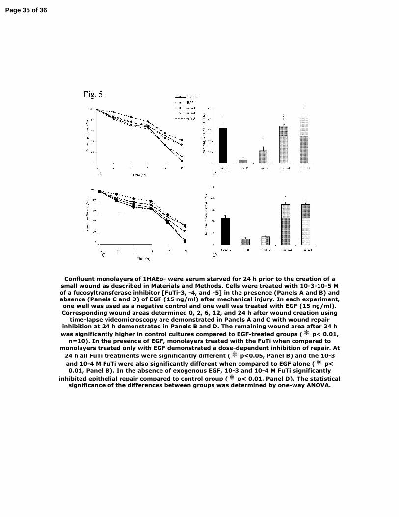
Figure 5. Wound repair of 1HAEo- cells is impaired in the presence of a fucosyltransferase
inhibitor. Confluent monolayers of 1HAEo- were serum starved for 24 h prior to the creation of
a small wound as described in Materials and Methods. Cells were treated with 10-3-10-5 M of a
Page 26 of 36

LCMP-00120-2006.R127
fucosyltransferase inhibitor [FuTi-3, -4, and -5] in the presence (Panels A and B) and absence
(Panels C and D) of EGF (15 ng/ml) after mechanical injury. In each experiment, one well was
used as a negative control and one well was treated with EGF (15 ng/ml). Corresponding wound
areas determined 0, 2, 6, 12, and 24 h after wound creation using time-lapse videomicroscopy are
demonstrated in Panels A and C with wound repair inhibition at 24 h demonstrated in Panels B
and D. The remaining wound area after 24 h was significantly higher in control cultures
compared to EGF-treated groups (* p< 0.01, n=10). In the presence of EGF, monolayers treated
with the FuTi when compared to monolayers treated only with EGF demonstrated a dose-
dependent inhibition of repair. At 24 h all FuTi treatments were significantly different (‡ p<0.05,
Panel B) and the 10-3 and 10-4 M FuTi were also significantly different when compared to EGF
alone (* p< 0.01, Panel B). In the absence of exogenous EGF, 10-3 and 10-4 M FuTi significantly
inhibited epithelial repair compared to control group (* p< 0.01, Panel D). The statistical
significance of the differences between groups was determined by one-way ANOVA.
Figure 6. Wound repair of 1HAEo- cells is by soluble sLex only in the presence of exogenous
EGF. Confluent monolayers of 1HAEo- were serum starved for 24 h prior to the creation of a
small wound as described in Materials and Methods. Cells were treated with 10-4 to 10-7 M of
soluble sLex [sLex-4, -5, -6, and -7] and EGF (15 ng/ml) (Panels A and B) after mechanical
injury. In each experiment, one well was used as a negative control and one well was treated with
EGF (15 ng/ml). Corresponding wound areas determined 0, 2, 6, 12, and 24 h after wound
creation using time-lapse videomicroscopy and wound repair inhibition at 24 h demonstrated.
The remaining wound area after 24 h was significantly higher in control cultures compared to
EGF-treated groups (* p< 0.01, n=10). The remaining wound area at 24 h after treatment with
10–4, 10–5, and 10–6M soluble sLex + EGF (15 ng/ml) was significantly higher compared to EGF
Page 27 of 36

LCMP-00120-2006.R128
alone (* p<0.01, Panel B). Monolayers treated with 10–4 M soluble sLex compared to monolayers
treated with 10–5, 10–6, and 10–7 M soluble sLex have higher wound area 24 h after treatment (‡
p<0.05, Panel B). The statistical significance of the differences between groups was determined
by one-way ANOVA.
Table 1. α1,3-fucosyltransferases show a diverse pattern of expression in 1HAEo-, 16HBE
14o-, and NHBE cells. Expression of four subtypes of α1,3-FucTs (FucT III, IV, VII and IX)
was studied in 1HAEo-, 16HBE 14o-, and NHBE cells using real-time RT-PCR. α1,3-FucTs
showed a different pattern of expression among two cell lines and primary cells but FucT-IV was
the only gene transcribed in both primary cells and cell lines of airway epithelium.
Table 2. Expression of E-selectin is not changed after mechanical injury in 1HAEo- and
NHBE cells. Expression of E-selectin by 1HAEo- and NHBE cells was examined before (N=5)
and 24 h after mechanical injury (N=3) in the presence of exogenous EGF (15ng/ml). There was
no statistically significant difference in the percent of cells expressing E-selectin receptor before
and after mechanical injury.
Page 28 of 36

Page 29 of 36

Page 30 of 36

Figure 1. Expression of sLex on airway epithelium in normal subjects. Bronchial segments obtained from pathological specimens of adults undergoing lung resection, were
processed and sectioned as described in Materials and Methods. New fuschin was applied for visualization and positive sLex detection is noted by red staining. Panels A and B show
immunoreactivity for sLex in areas characterized by epithelial damage (A) and intact epithelium (B). Panels (C) and (D) show the appearance of an isotype matched non-specific antibody staining in the areas of damaged and intact epithelium. Scale bar =
10 m.
Page 31 of 36

Figure 2. Expression of sLex is higher in areas of epithelial damage compared to intact epithelium. Immunostaining of sLex in intact and damaged epithelial zones was assessed in each subject. Epithelial damage was characterized morphologically by the absence of
ciliated and secretory cells. Quantification was performed with ImagePro Plus image analysis software. To evaluate sLex immunoreactivity in damaged areas, positive staining basal and columnar cells within 40 µm from either wound edge were counted. Next intact epithelium farther than 40 µm from the wound edge was considered as intact epithelium, 40 µm of this area was assessed for sLex staining. Percentage of positively stained basal and columnar cells in three areas exhibiting epithelial damage and three areas of intact epithelium were determined in each subject. The statistical significance was determined by Mann-Whitney U-test. The horizontal line represents the median. Expression of sLex is
significantly higher in areas of epithelial damage compared to intact epithelium (p<0.002).
Page 32 of 36

Figure 3. Mechanical injury induces the expression of sLex in a culture model of epithelial repair. A linear wound was made in confluent monolayers of primary bronchial epithelial cells using a rubber stylet. Monolayers were fixed with Clark's solution at 0, 2, 6, 12, 24,
and 48 h after the mechanical injury. Expression of sLex was detected using mouse á-human sLex and Vector Red for visualization. Detection of sLex increased with repair and noted by increased red stain. A correlation between increased expression of sLex and the
cell-shape phenotype also changed over time after mechanical injury. Cells that had higher detection of sLex demonstrated an elongated shape characteristic of migratory
cells (see enlarged inset from T12).
Page 33 of 36

Figure 4. Blocking of sLex with an anti-sLex inhibitory antibody prevents epithelial monolayer wound repair. A linear wound was made in confluent monolayers of primary
bronchial epithelial cells as described and wounds were treated with an anti-sLex antibody or control antibody. In each experiment monolayers with no treatment
considered as control group. Corresponding wound areas determined 2, 6, 12, and 24 h after wound creation using time-lapse videomicroscopy are presented in Panel A. Data
are mean SEM, for 24 wounds measured in two independent experiments. The effect of anti-sLex antibody on wound repair is demonstrated in Panel B. Anti-sLex antibody
significantly reduced wound repair compared to controls ( p<0.05). The statistical significance of the differences between groups was determined by one-way ANOVA.
Page 34 of 36
Confluent monolayers of 1HAEo- were serum starved for 24 h prior to the creation of a small wound as described in Materials and Methods. Cells were treated with 10-3-10-5 M of a fucosyltransferase inhibitor [FuTi-3, -4, and -5] in the presence (Panels A and B) and absence (Panels C and D) of EGF (15 ng/ml) after mechanical injury. In each experiment, one well was used as a negative control and one well was treated with EGF (15 ng/ml). Corresponding wound areas determined 0, 2, 6, 12, and 24 h after wound creation using
time-lapse videomicroscopy are demonstrated in Panels A and C with wound repair inhibition at 24 h demonstrated in Panels B and D. The remaining wound area after 24 h was significantly higher in control cultures compared to EGF-treated groups ( p< 0.01,
n=10). In the presence of EGF, monolayers treated with the FuTi when compared to monolayers treated only with EGF demonstrated a dose-dependent inhibition of repair. At
24 h all FuTi treatments were significantly different ( p<0.05, Panel B) and the 10-3 and 10-4 M FuTi were also significantly different when compared to EGF alone ( p< 0.01, Panel B). In the absence of exogenous EGF, 10-3 and 10-4 M FuTi significantly
inhibited epithelial repair compared to control group ( p< 0.01, Panel D). The statistical significance of the differences between groups was determined by one-way ANOVA.
Page 35 of 36

Confluent monolayers of 1HAEo- were serum starved for 24 h prior to the creation of a small wound as described in Materials and Methods. Cells were treated with 10-4 to 10-7
M of soluble sLex [sLex-4, -5, -6, and -7] and EGF (15 ng/ml) (Panels A and B) after mechanical injury. In each experiment, one well was used as a negative control and one well was treated with EGF (15 ng/ml). Corresponding wound areas determined 0, 2, 6, 12, and 24 h after wound creation using time-lapse videomicroscopy and wound repair inhibition at 24 h demonstrated. The remaining wound area after 24 h was significantly
higher in control cultures compared to EGF-treated groups ( p< 0.01, n=10). The remaining wound area at 24 h after treatment with 10�4, 10�5, and 10�6M soluble sLex+ EGF (15 ng/ml) was significantly higher compared to EGF alone ( p<0.01, Panel B).
Monolayers treated with 10�4 M soluble sLex compared to monolayers treated with 10�5, 10�6, and 10�7 M soluble sLex have higher wound area 24 h after treatment (
p<0.05, Panel B). The statistical significance of the differences between groups was determined by one-way ANOVA.
Page 36 of 36