Agrobacterium type IV secretion system and its substrates form helical arrays around the...
6
Agrobacterium type IV secretion system and its substrates form helical arrays around the circumference of virulence-induced cells Julieta Aguilar, John Zupan, Todd A. Cameron, and Patricia C. Zambryski 1 Department of Plant and Microbial Biology, University of California, Berkeley, CA 94720 Contributed by Patricia C. Zambryski, December 30, 2009 (sent for review November 25, 2009) The genetic transformation of plant cells by Agrobacterium tumefa- ciens results from the transfer of DNA and proteins via a specific viru- lence (vir) -induced type IV secretion system (T4SS). To better understand T4SS function, we analyzed the localization of its structural components and substrates by deconvolution fluorescence micro- scopy. GFP fusions to T4SS proteins with cytoplasmic tails, VirB8 and VirD4, or cytoplasmic T4SS substrate proteins, VirD2, VirE2, and VirF, localize in a helical pattern of fluorescent foci around the perimeter of the bacterial cell. All fusion proteins were expressed at native levels of vir induction. Importantly, most fusion proteins are functional and do not exhibit dominant-negative effects on DNA transfer to plant cells. Further, GFP-VirB8 complements a virB8 deletion strain. We also detect native VirB8 localization as a helical array of foci by immunofluores- cence microscopy. T4SS foci likely use an existing helical scaffold during their assembly. Indeed, the bacterial cytoskeletal component MinD colocalizes with GFP-VirB8. Helical arrays of foci are found at all times investigated between 12 and 48 h post vir induction at 19 °C. These data lead to a model with multiple T4SSs around the bacterial cell that likely facilitate host cell attachment and DNA transfer. In support, we find multiple T pili around vir-induced bacterial cells. T he soil bacterium Agrobacterium tumefaciens is a source of fundamental insights and is intensively used due to its ability to transfer any DNA of interest to plant cells (1). The transferred DNA is a single-stranded copy of the T-DNA region on its Ti plasmid. A separate region of the Ti plasmid, the virulence (vir) region, encodes proteins to produce the transferable DNA, and to form a membrane-spanning DNA transporter called the type IV secretion system (T4SS). Several Vir proteins, VirD2, VirE2, VirE3, and VirF, are also transported to plants by the vir- encoded T4SS (2). The transfer of ssDNA to plants via the T4SS is highly evo- lutionarily conserved, best exemplified by T4SS-mediated con- jugal ssDNA transfer between bacterial cells. Bacteria have evolved to use this ancient mechanism of DNA transfer between bacteria to also transfer DNA or protein toxins into eukaryotic host cells. Important human pathogens such as Helicobacter pylori, Legionella pneumophila, Bordetella pertussis, Brucella suis, Bartonella henselae, and Ricksettsai prowazekii transport disease- causing factors via T4SSs (3). T4SS components can be placed into subgroups based on function or location. VirB4, VirB11, and VirD4 have ATPase homology and ATP-binding motifs, which may energize substrate translocation (4–7) at the inner membrane. VirB3 interacts with VirB4 (5). VirB6 is an inner membrane protein and VirB8 spans the inner membrane, placing most of the protein in the periplasm (8, 9). Recent cryoelectron microscopy (10) and crystallography (11) reveal a high-resolution structure consisting of 14 copies each of the Escherichia coli pKM101 conjugation system VirB7, VirB9, and VirB10 homologs that form a double-chambered channel spanning the inner membrane, periplasm, and outer membrane forming the core of the T4SS. VirB2 is the major T4SS pilus component (12), and the minor component, VirB5 (13), localizes at its tip (14). VirB1 has two domains: the N terminus has homology to lytic transglycosylases and likely cleaves the pepti- doglycan cell-wall layer to facilitate assembly of the T4SS (15–17), and the C-terminal processed portion, VirB1*, is secreted to the cell surface (18, 19) and is required for pilus formation (20). To understand T4SS function it is critical to determine its localization in the bacterial cell. Several reports suggest that some T4SS proteins localize to one or a few predominantly polar sites (21–23); such localization is proposed to lead to polar sub- strate transport. However, the results are not entirely consistent, as some proteins localize to both poles, the midcell, and subpolar regions. Here we use deconvolution microscopy to provide high- resolution images of fluorescent fusions to T4SS components and its secretion substrates. The results reveal that the T4SS localizes to the poles and equally well as helically arranged foci around the perimeter of the bacterial cell. These data lead to a model where multiple T4SSs around the bacterial cell provide multiple sites for interaction with the host cell surface. Results We constructed GFP fusions to T4SS components and T4SS substrate proteins. As GFP loses its fluorescence when trans- ported to the periplasm, we fused GFP to components that either reside at the cytoplasmic face of the T4SS channel, VirD4, or have predicted cytoplasmic tails, VirB8 and VirB10. Further, we fused GFP to the substrate proteins VirD2, VirE2, and VirF. As the latter proteins must target the T4SS for transport, their localization pattern should reflect the localization of the T4SS. The data presented are representative of all cells in a particular optical field, and we analyzed hundreds of cells for each con- struct; we explicitly state when fewer than 100% of the cells exhibit a particular pattern. VirB8 Localizes in a Helical Pattern in vir-Induced Agrobacterium. We first assayed the localization pattern of GFP-VirB8. VirB8 has a cytoplasmic N-terminal region of ∼40 amino acids, followed by a membrane-spanning domain of ∼20 amino acids and a C-terminal domain of 177 amino acids in the periplasm (24). GFP was fused in-frame to the N terminus of VirB8, sequestering GFP in the cytoplasm, leaving the majority of VirB8 intact in the periplasm allowing for association with other proteins of the T4SS. GFP- VirB8 coding sequences were placed downstream of a vir-indu- cible promoter on the low-copy-number plasmid pDW029 (20). GFP-VirB8 was transferred into wild-type Agrobacterium or a deletion of virB8 (ΔvirB8). Following induction of vir gene expression, GFP-fusion protein localization was monitored by fluorescence deconvolution microscopy. Author contributions: J.A., J.Z., and P.C.Z. designed research; J.A., J.Z., and T.A.C. performed research; J.A., J.Z., and T.A.C. contributed new reagents/analytic tools; J.A., J.Z., T.A.C., and P.C.Z. analyzed data; and J.A., J.Z., and P.C.Z. wrote the paper. The authors declare no conflict of interest. 1 To whom correspondence should be addressed. E-mail: [email protected]. This article contains supporting information online at www.pnas.org/cgi/content/full/ 0914940107/DCSupplemental. 3758–3763 | PNAS | February 23, 2010 | vol. 107 | no. 8 www.pnas.org/cgi/doi/10.1073/pnas.0914940107
Transcript of Agrobacterium type IV secretion system and its substrates form helical arrays around the...

Agrobacterium type IV secretion system and itssubstrates form helical arrays around thecircumference of virulence-induced cellsJulieta Aguilar, John Zupan, Todd A. Cameron, and Patricia C. Zambryski1
Department of Plant and Microbial Biology, University of California, Berkeley, CA 94720
Contributed by Patricia C. Zambryski, December 30, 2009 (sent for review November 25, 2009)
The genetic transformation of plant cells by Agrobacterium tumefa-ciens results from the transfer of DNA and proteins via a specific viru-lence (vir) -induced type IV secretion system (T4SS). To betterunderstandT4SS function,weanalyzed the localizationof its structuralcomponents and substrates by deconvolution fluorescence micro-scopy. GFP fusions to T4SS proteins with cytoplasmic tails, VirB8 andVirD4, or cytoplasmic T4SS substrate proteins, VirD2, VirE2, and VirF,localize in a helical pattern of fluorescent foci around the perimeter ofthe bacterial cell. All fusion proteinswere expressed at native levels ofvir induction. Importantly, most fusion proteins are functional and donot exhibit dominant-negative effects on DNA transfer to plant cells.Further, GFP-VirB8 complements avirB8deletion strain.Wealso detectnative VirB8 localization as a helical array of foci by immunofluores-cencemicroscopy. T4SS foci likelyuseanexistinghelical scaffoldduringtheir assembly. Indeed, the bacterial cytoskeletal component MinDcolocalizes with GFP-VirB8. Helical arrays of foci are found at all timesinvestigated between 12 and 48 h post vir induction at 19 °C. Thesedata lead to amodel withmultiple T4SSs around the bacterial cell thatlikely facilitate host cell attachment and DNA transfer. In support, wefind multiple T pili around vir-induced bacterial cells.
The soil bacterium Agrobacterium tumefaciens is a source offundamental insights and is intensively used due to its ability to
transfer any DNA of interest to plant cells (1). The transferredDNA is a single-stranded copy of the T-DNA region on its Tiplasmid. A separate region of the Ti plasmid, the virulence (vir)region, encodes proteins to produce the transferable DNA, and toform a membrane-spanning DNA transporter called the type IVsecretion system (T4SS). Several Vir proteins, VirD2, VirE2,VirE3, and VirF, are also transported to plants by the vir-encoded T4SS (2).The transfer of ssDNA to plants via the T4SS is highly evo-
lutionarily conserved, best exemplified by T4SS-mediated con-jugal ssDNA transfer between bacterial cells. Bacteria haveevolved to use this ancient mechanism of DNA transfer betweenbacteria to also transfer DNA or protein toxins into eukaryotichost cells. Important human pathogens such as Helicobacterpylori, Legionella pneumophila, Bordetella pertussis, Brucella suis,Bartonella henselae, and Ricksettsai prowazekii transport disease-causing factors via T4SSs (3).T4SS components can be placed into subgroups based on
function or location. VirB4, VirB11, and VirD4 have ATPasehomology and ATP-binding motifs, which may energize substratetranslocation (4–7) at the inner membrane. VirB3 interacts withVirB4 (5). VirB6 is an inner membrane protein and VirB8 spansthe inner membrane, placing most of the protein in the periplasm(8, 9). Recent cryoelectron microscopy (10) and crystallography(11) reveal a high-resolution structure consisting of 14 copies eachof the Escherichia coli pKM101 conjugation system VirB7, VirB9,and VirB10 homologs that form a double-chambered channelspanning the inner membrane, periplasm, and outer membraneforming the core of the T4SS. VirB2 is the major T4SS piluscomponent (12), and the minor component, VirB5 (13), localizesat its tip (14). VirB1 has two domains: the N terminus hashomology to lytic transglycosylases and likely cleaves the pepti-
doglycan cell-wall layer to facilitate assembly of the T4SS (15–17),and the C-terminal processed portion, VirB1*, is secreted to thecell surface (18, 19) and is required for pilus formation (20).To understand T4SS function it is critical to determine its
localization in the bacterial cell. Several reports suggest thatsome T4SS proteins localize to one or a few predominantly polarsites (21–23); such localization is proposed to lead to polar sub-strate transport. However, the results are not entirely consistent,as some proteins localize to both poles, the midcell, and subpolarregions. Here we use deconvolution microscopy to provide high-resolution images of fluorescent fusions to T4SS components andits secretion substrates. The results reveal that the T4SS localizesto the poles and equally well as helically arranged foci around theperimeter of the bacterial cell. These data lead to a model wheremultiple T4SSs around the bacterial cell provide multiple sites forinteraction with the host cell surface.
ResultsWe constructed GFP fusions to T4SS components and T4SSsubstrate proteins. As GFP loses its fluorescence when trans-ported to the periplasm, we fused GFP to components thateither reside at the cytoplasmic face of the T4SS channel, VirD4,or have predicted cytoplasmic tails, VirB8 and VirB10. Further,we fused GFP to the substrate proteins VirD2, VirE2, and VirF.As the latter proteins must target the T4SS for transport, theirlocalization pattern should reflect the localization of the T4SS.The data presented are representative of all cells in a particularoptical field, and we analyzed hundreds of cells for each con-struct; we explicitly state when fewer than 100% of the cellsexhibit a particular pattern.
VirB8 Localizes in a Helical Pattern in vir-Induced Agrobacterium. Wefirst assayed the localization pattern of GFP-VirB8. VirB8 has acytoplasmic N-terminal region of ∼40 amino acids, followed by amembrane-spanning domain of∼20 amino acids and a C-terminaldomain of 177 amino acids in the periplasm (24). GFP was fusedin-frame to the N terminus of VirB8, sequestering GFP in thecytoplasm, leaving the majority of VirB8 intact in the periplasmallowing for association with other proteins of the T4SS. GFP-VirB8 coding sequences were placed downstream of a vir-indu-cible promoter on the low-copy-number plasmid pDW029 (20).GFP-VirB8 was transferred into wild-type Agrobacterium or adeletion of virB8 (ΔvirB8). Following induction of vir geneexpression, GFP-fusion protein localization was monitored byfluorescence deconvolution microscopy.
Author contributions: J.A., J.Z., and P.C.Z. designed research; J.A., J.Z., and T.A.C.performed research; J.A., J.Z., and T.A.C. contributed new reagents/analytic tools; J.A.,J.Z., T.A.C., and P.C.Z. analyzed data; and J.A., J.Z., and P.C.Z. wrote the paper.
The authors declare no conflict of interest.1To whom correspondence should be addressed. E-mail: [email protected].
This article contains supporting information online at www.pnas.org/cgi/content/full/0914940107/DCSupplemental.
3758–3763 | PNAS | February 23, 2010 | vol. 107 | no. 8 www.pnas.org/cgi/doi/10.1073/pnas.0914940107
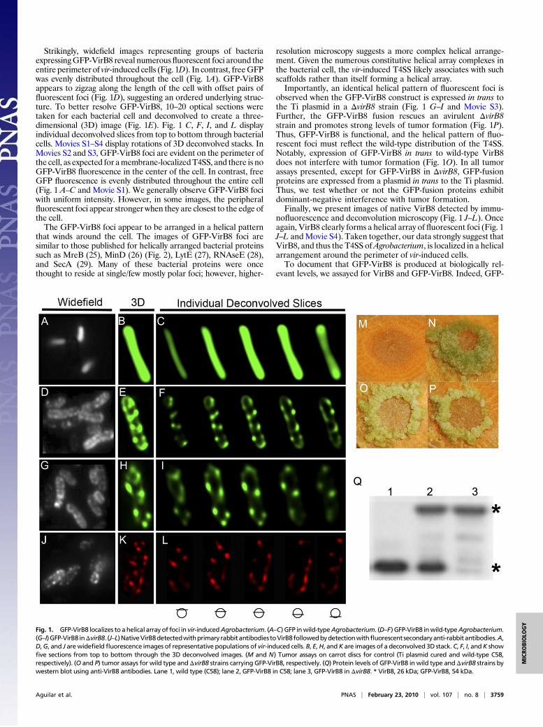
Strikingly, widefield images representing groups of bacteriaexpressingGFP-VirB8 reveal numerousfluorescent foci around theentire perimeter of vir-induced cells (Fig. 1D). In contrast, freeGFPwas evenly distributed throughout the cell (Fig. 1A). GFP-VirB8appears to zigzag along the length of the cell with offset pairs offluorescent foci (Fig. 1D), suggesting an ordered underlying struc-ture. To better resolve GFP-VirB8, 10–20 optical sections weretaken for each bacterial cell and deconvolved to create a three-dimensional (3D) image (Fig. 1E). Fig. 1 C, F, I, and L displayindividual deconvolved slices from top to bottom through bacterialcells. Movies S1–S4 display rotations of 3D deconvolved stacks. InMovies S2 and S3, GFP-VirB8 foci are evident on the perimeter ofthe cell, as expected for amembrane-localizedT4SS, and there is noGFP-VirB8 fluorescence in the center of the cell. In contrast, freeGFP fluorescence is evenly distributed throughout the entire cell(Fig. 1 A–C and Movie S1). We generally observe GFP-VirB8 fociwith uniform intensity. However, in some images, the peripheralfluorescent foci appear stronger when they are closest to the edge ofthe cell.The GFP-VirB8 foci appear to be arranged in a helical pattern
that winds around the cell. The images of GFP-VirB8 foci aresimilar to those published for helically arranged bacterial proteinssuch as MreB (25), MinD (26) (Fig. 2), LytE (27), RNAseE (28),and SecA (29). Many of these bacterial proteins were oncethought to reside at single/few mostly polar foci; however, higher-
resolution microscopy suggests a more complex helical arrange-ment. Given the numerous constitutive helical array complexes inthe bacterial cell, the vir-induced T4SS likely associates with suchscaffolds rather than itself forming a helical array.Importantly, an identical helical pattern of fluorescent foci is
observed when the GFP-VirB8 construct is expressed in trans tothe Ti plasmid in a ΔvirB8 strain (Fig. 1 G–I and Movie S3).Further, the GFP-VirB8 fusion rescues an avirulent ΔvirB8strain and promotes strong levels of tumor formation (Fig. 1P).Thus, GFP-VirB8 is functional, and the helical pattern of fluo-rescent foci must reflect the wild-type distribution of the T4SS.Notably, expression of GFP-VirB8 in trans to wild-type VirB8does not interfere with tumor formation (Fig. 1O). In all tumorassays presented, except for GFP-VirB8 in ΔvirB8, GFP-fusionproteins are expressed from a plasmid in trans to the Ti plasmid.Thus, we test whether or not the GFP-fusion proteins exhibitdominant-negative interference with tumor formation.Finally, we present images of native VirB8 detected by immu-
nofluorescence and deconvolution microscopy (Fig. 1 J–L). Onceagain, VirB8 clearly forms a helical array of fluorescent foci (Fig. 1J–L andMovie S4). Taken together, our data strongly suggest thatVirB8, and thus the T4SS ofAgrobacterium, is localized in a helicalarrangement around the perimeter of vir-induced cells.To document that GFP-VirB8 is produced at biologically rel-
evant levels, we assayed for VirB8 and GFP-VirB8. Indeed, GFP-
Fig. 1. GFP-VirB8 localizes to ahelical array of foci in vir-inducedAgrobacterium. (A–C) GFP inwild-typeAgrobacterium. (D–F) GFP-VirB8 inwild-typeAgrobacterium.(G–I)GFP-VirB8 inΔvirB8. (J–L)NativeVirB8detectedwithprimary rabbitantibodiestoVirB8followedbydetectionwithfluorescent secondaryanti-rabbitantibodies.A,D,G, and J arewidefieldfluorescence images of representative populations of vir-induced cells. B, E,H, andK are images of a deconvolved 3D stack.C, F, I, andK showfive sections from top to bottom through the 3D deconvolved images. (M and N) Tumor assays on carrot discs for control (Ti plasmid cured and wild-type C58,respectively). (O and P) tumor assays for wild type andΔvirB8 strains carrying GFP-VirB8, respectively. (Q) Protein levels of GFP-VirB8 in wild type andΔvirB8 strains bywestern blot using anti-VirB8 antibodies. Lane 1, wild type (C58); lane 2, GFP-VirB8 in C58; lane 3, GFP-VirB8 in ΔvirB8. * VirB8, 26 kDa; GFP-VirB8, 54 kDa.
Aguilar et al. PNAS | February 23, 2010 | vol. 107 | no. 8 | 3759
MICRO
BIOLO
GY
VirB8 is produced at the same level as native VirB8 in wild type(Fig. 1Q, lanes 1 and 2) and ΔvirB8 (Fig. 1Q, lane 3). GFP-VirB8is stably expressed as antibodies to GFP to detect only the fusionprotein and not lower-molecular-weight proteins (Fig. S1, lane 5).
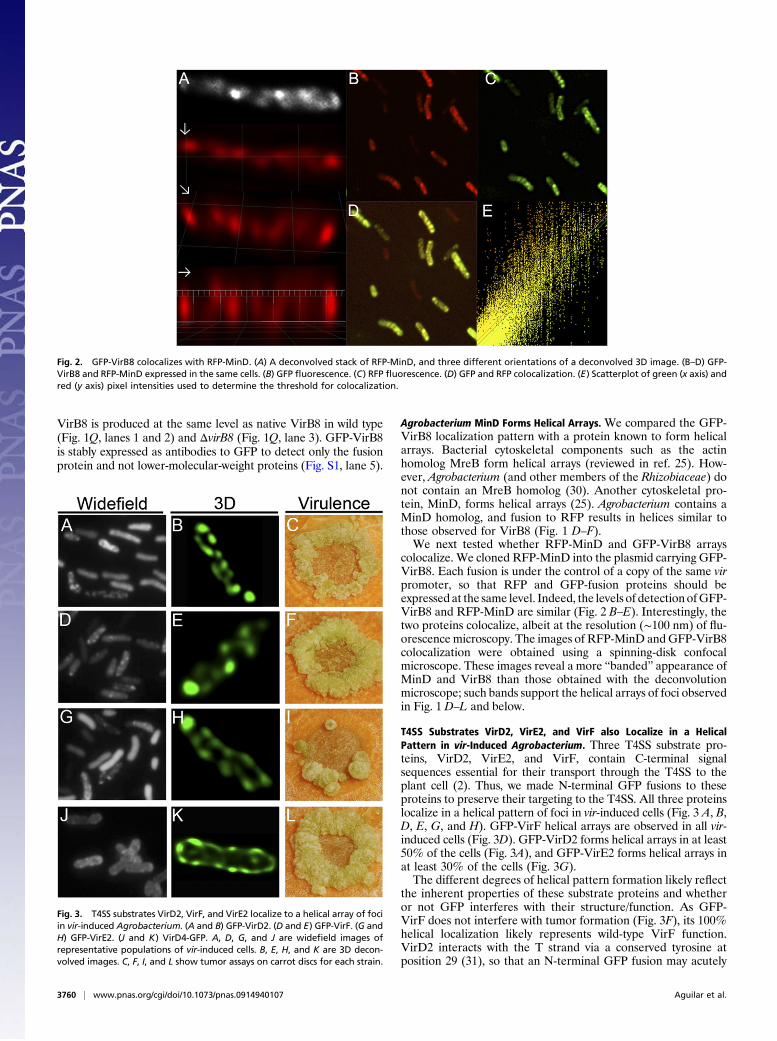
Agrobacterium MinD Forms Helical Arrays. We compared the GFP-VirB8 localization pattern with a protein known to form helicalarrays. Bacterial cytoskeletal components such as the actinhomolog MreB form helical arrays (reviewed in ref. 25). How-ever, Agrobacterium (and other members of the Rhizobiaceae) donot contain an MreB homolog (30). Another cytoskeletal pro-tein, MinD, forms helical arrays (25). Agrobacterium contains aMinD homolog, and fusion to RFP results in helices similar tothose observed for VirB8 (Fig. 1 D–F).We next tested whether RFP-MinD and GFP-VirB8 arrays
colocalize. We cloned RFP-MinD into the plasmid carrying GFP-VirB8. Each fusion is under the control of a copy of the same virpromoter, so that RFP and GFP-fusion proteins should beexpressed at the same level. Indeed, the levels of detection ofGFP-VirB8 and RFP-MinD are similar (Fig. 2 B–E). Interestingly, thetwo proteins colocalize, albeit at the resolution (∼100 nm) of flu-orescence microscopy. The images of RFP-MinD andGFP-VirB8colocalization were obtained using a spinning-disk confocalmicroscope. These images reveal a more “banded” appearance ofMinD and VirB8 than those obtained with the deconvolutionmicroscope; such bands support the helical arrays of foci observedin Fig. 1 D–L and below.
T4SS Substrates VirD2, VirE2, and VirF also Localize in a HelicalPattern in vir-Induced Agrobacterium. Three T4SS substrate pro-teins, VirD2, VirE2, and VirF, contain C-terminal signalsequences essential for their transport through the T4SS to theplant cell (2). Thus, we made N-terminal GFP fusions to theseproteins to preserve their targeting to the T4SS. All three proteinslocalize in a helical pattern of foci in vir-induced cells (Fig. 3 A, B,D, E, G, and H). GFP-VirF helical arrays are observed in all vir-induced cells (Fig. 3D). GFP-VirD2 forms helical arrays in at least50% of the cells (Fig. 3A), and GFP-VirE2 forms helical arrays inat least 30% of the cells (Fig. 3G).The different degrees of helical pattern formation likely reflect
the inherent properties of these substrate proteins and whetheror not GFP interferes with their structure/function. As GFP-VirF does not interfere with tumor formation (Fig. 3F), its 100%helical localization likely represents wild-type VirF function.VirD2 interacts with the T strand via a conserved tyrosine atposition 29 (31), so that an N-terminal GFP fusion may acutely
Fig. 3. T4SS substrates VirD2, VirF, and VirE2 localize to a helical array of fociin vir-induced Agrobacterium. (A and B) GFP-VirD2. (D and E) GFP-VirF. (G andH) GFP-VirE2. (J and K) VirD4-GFP. A, D, G, and J are widefield images ofrepresentative populations of vir-induced cells. B, E, H, and K are 3D decon-volved images. C, F, I, and L show tumor assays on carrot discs for each strain.
Fig. 2. GFP-VirB8 colocalizes with RFP-MinD. (A) A deconvolved stack of RFP-MinD, and three different orientations of a deconvolved 3D image. (B–D) GFP-VirB8 and RFP-MinD expressed in the same cells. (B) GFP fluorescence. (C) RFP fluorescence. (D) GFP and RFP colocalization. (E) Scatterplot of green (x axis) andred (y axis) pixel intensities used to determine the threshold for colocalization.
3760 | www.pnas.org/cgi/doi/10.1073/pnas.0914940107 Aguilar et al.
interfere with its localization. However, GFP-VirD2 does notinterfere with long-term T4SS function and tumor formation(Fig. 3C). VirE2 is the most abundant Vir protein produced,forms homodimers, heterodimers with VirE1, and complex sol-enoids in complex with ssDNA (32). VirE2 dimers and solenoidsare head to tail (N- to C-terminal), so an N-terminal GFP taglikely interferes with multimer formation, localization to T4SShelices, and tumor formation (Fig. 3I). Or, the abundance ofVirE2, coupled with the size of the fusion protein, 95 kDa, maylead to plugging of the T4SS and reduction of tumor formation.
The VirD4-Coupling Protein Forms Helical Arrays. An importantmember of the T4SS is the VirD4-coupling protein that escortsthe VirD2-bound T strand to the T4SS (reviewed in ref. 33).VirD4 association with VirB11 in the inner membrane is the firststep in T-strand transport (34). The E. coli TrwB homolog ofVirD4 forms a hexamer residing in the inner membrane with aC-terminal cytoplasmic tail (35). Thus, we made a C-terminalGFP fusion to VirD4. VirD4-GFP also localizes in a helicalpattern in vir-induced cells (Fig. 3 J and K), and allows tumorformation (Fig. 3L). These results support that the C terminus ofVirD4 resides in the cytoplasm and show that fusion to GFPdoes not interfere with its hexameric structure.
Do GFP Fusions to Other T4SS Components Form Helical Arrays? Asmentioned earlier, we also made a GFP fusion to the N terminusof VirB10, as it has a predicted ∼30 amino acid cytoplasmic tailfollowed by a ∼20 amino acid membrane-spanning domain and327 amino acids in the periplasm (36). However, even thoughVirB10 has the same general topology as VirB8, it did not formfoci (Fig. S2E). Instead, GFP-VirB10 forms a wide band of flu-orescence at polar and subpolar sites around approximately one-third of the bacterial cell. This localization pattern is abnormal,as GFP-VirB10 is not functional and interferes with tumor for-mation (Fig. S2F).Cryoelectron microscopy (10) and crystallographic studies (11)
reveal that E. coli pKM101 homologs of VirB7, VirB9, andVirB10 are each found in 14 copies in a complex that spans theperiplasm with its ends contacting the inner and outer mem-brane. Notably, even though VirB7, VirB8, VirB9, and VirB10were expressed together, VirB8 was not purified along with thecore complex (10). Hence, VirB8 appears not to associate tightlywith the VirB7-VirB9-VirB10 core complex.VirB8 may have more structural flexibility than VirB10, so the
addition of GFP to VirB8 does not hinder its function andlocalization. Remarkably, the independence of VirB8 from thecore complex allowed us to detect the formation of GFP-VirB8helical arrays. The most logical explanation for the lack of helicalarray formation by GFP-VirB10 is that VirB10 is part of theT4SS multimeric core complex (10), and its fusion to GFPinterferes with its ability to form the core complex.We also predicted that not all T4SS proteins would fold or
function correctly when tagged with GFP. Indeed, GFP fusions toVirB4, VirB6, VirB7, VirB9, and VirB11 do not form helical foci,and all exhibit dominant-negative effects on tumor formation(Fig. S2). The lack of foci formation for GFP fusions to VirB7 andVirB9 is expected, as these proteins are entirely periplasmic (10).VirB4 and VirB11 are hexameric ATPases that associate with theinner membrane, and GFP fusion may interfere with theirassembly and function. Whereas VirD4 is also a hexamericATPase, it has a structurally distinct C-terminal cytoplasmic tail(35), and GFP fusion did not impair its function or localization.VirB6 is highly hydrophobic with six membrane-spanning regions,and GFP may interfere with its folding and function.The lack of distinct foci formation cannot be explained by
degradation of the fusion proteins; whereas VirB4, VirB6,VirB7, VirB9, and VirB11 fusions display some breakdownproducts, the expected GFP fusions represent at least 50% of
their GFP signals (Fig. S1, lanes 2–4 and 6–7, respectively), andwhereas VirD4-GFP displays significant breakdown (Fig. S1,lane 9), it still forms distinct helical arrays of foci (Fig. 3K).
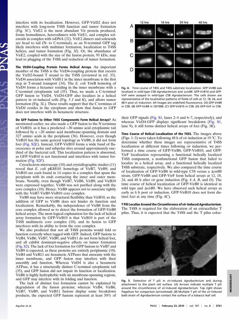
Time Course of Helical Localization of the T4SS. The images above(Figs. 1–3) were taken following 48 h of vir induction at 19 °C. Todetermine whether these images are representative of T4SSlocalization at different times following vir induction, we per-formed a time course of GFP-VirB8, GFP-VirB10, and GFP-VirF localization representing a functional helically localizedT4SS component, a nonfunctional GFP fusion that failed tolocalize in a helical array, and a functional helically localizedT4SS substrate, respectively. We also compared the time courseof localization of GFP-VirB8 in wild-type C58 versus a ΔvirB8strain. GFP-VirB8 and GFP-VirF form helical arrays at 12, 18,24, and 48 h after vir gene induction (Fig. 4 A, B, and D). Thetime course of helical localization of GFP-VirB8 is identical inwild type and ΔvirB8. We have observed such helical arrays asearly as 6 h post vir induction. GFP-VirB10 does not form dis-tinct foci at any time (Fig. 4C).
T Pili Localize Around the Circumference of vir-InducedAgrobacterium.The T4SS is required for the elaboration of an extracellular Tpilus. Thus, it is expected that the T4SS and the T pilus coloc-
Fig. 4. Time course of T4SS and T4SS substrate localization. GFP-VirB8 waslocalized in wild-type C58 Agrobacterium and ΔvirB8. GFP-VirB10 and GFP-VirF were assayed in wild-type C58 Agrobacterium. The cells shown arerepresentative of the localization patterns of fields of cells at 12, 18, 24, and48 h post vir induction. All images are widefield fluorescence. (A) GFP-VirB8in C58. (B) GFP-VirB8 in CB1008. (C) GFP-VirB10 in C58. (D) GFP-VirF in C58.
Fig. 5. Detection of T pili in vir-induced Agrobacterium and duringattachment to the plant cell surface. (A) Arrows indicate multiple T piliaround the circumference of vir-induced Agrobacterium. Top right showsflagellum for comparison (arrowhead). (B) Multiple T pili of the vir-inducedbald strain of Agrobacterium contact the surface of a tobacco leaf cell.
Aguilar et al. PNAS | February 23, 2010 | vol. 107 | no. 8 | 3761
MICRO
BIOLO
GY
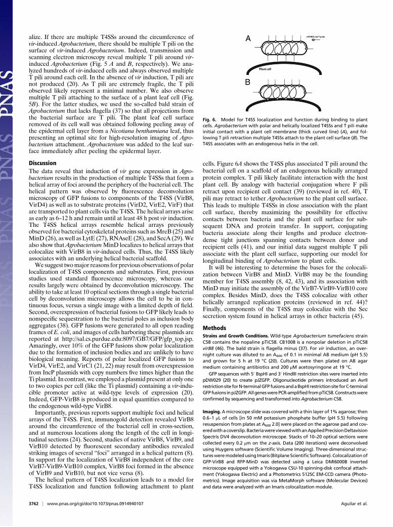
alize. If there are multiple T4SSs around the circumference ofvir-induced Agrobacterium, there should be multiple T pili on thesurface of vir-induced Agrobacterium. Indeed, transmission andscanning electron microscopy reveal multiple T pili around vir-induced Agrobacterium (Fig. 5 A and B, respectively). We ana-lyzed hundreds of vir-induced cells and always observed multipleT pili around each cell. In the absence of vir induction, T pili arenot produced (20). As T pili are extremely fragile, the T piliobserved likely represent a minimal number. We also observemultiple T pili attaching to the surface of a plant leaf cell (Fig.5B). For the latter studies, we used the so-called bald strain ofAgrobacterium that lacks flagella (37) so that all projections fromthe bacterial surface are T pili. The plant leaf cell surfaceremoved of its cell wall was obtained following peeling away ofthe epidermal cell layer from a Nicotiana benthamiana leaf, thuspresenting an optimal site for high-resolution imaging of Agro-bacterium attachment. Agrobacterium was added to the leaf sur-face immediately after peeling the epidermal layer.
DiscussionThe data reveal that induction of vir gene expression in Agro-bacterium results in the production of multiple T4SSs that form ahelical array of foci around the periphery of the bacterial cell. Thehelical pattern was observed by fluorescence deconvolutionmicroscopy of GFP fusions to components of the T4SS (VirB8,VirD4) as well as to substrate proteins (VirD2, VirE2, VirF) thatare transported to plant cells via the T4SS. The helical arrays ariseas early as 6–12 h and remain until at least 48 h post vir induction.The T4SS helical arrays resemble helical arrays previouslyobserved for bacterial cytoskeletal proteins such asMreB (25) andMinD (26), as well as LytE (27), RNAseE (28), and SecA (29).Wealso show that AgrobacteriumMinD localizes to helical arrays thatcolocalize with VirB8 in vir-induced cells. Thus, the T4SS likelyassociates with an underlying helical bacterial scaffold.We suggest twomajor reasons for previous observations of polar
localization of T4SS components and substrates. First, previousstudies used standard fluorescence microscopy, whereas ourresults largely were obtained by deconvolution microscopy. Theability to take at least 10 optical sections through a single bacterialcell by deconvolution microscopy allows the cell to be in con-tinuous focus, versus a single image with a limited depth of field.Second, overexpression of bacterial fusions to GFP likely leads tononspecific sequestration to the bacterial poles as inclusion bodyaggregates (38). GFP fusions were generated to all open readingframes of E. coli, and images of cells harboring these plasmids arereported at http://sal.cs.purdue.edu:8097/GB7/GFP/gfp_top.jsp.Amazingly, over 10% of the GFP fusions show polar localizationdue to the formation of inclusion bodies and are unlikely to havebiological meaning. Reports of polar localized GFP fusions toVirD4, VirE2, and VirC1 (21, 22) may result from overexpressionfrom IncP plasmids with copy numbers five times higher than theTi plasmid. In contrast, we employed a plasmid present at only oneto two copies per cell (like the Ti plasmid) containing a vir-indu-cible promoter active at wild-type levels of expression (20).Indeed, GFP-VirB8 is produced in equal quantities compared tothe endogenous wild-type VirB8.Importantly, previous reports support multiple foci and helical
arrays of the T4SS. First, immunogold detection revealed VirB8around the circumference of the bacterial cell in cross-section,and at numerous locations along the length of the cell in longi-tudinal sections (24). Second, studies of native VirB8, VirB9, andVirB10 detected by fluorescent secondary antibodies revealedstriking images of several “foci” arranged in a helical pattern (8).In support for the localization of VirB8 independent of the coreVirB7-VirB9-VirB10 complex, VirB8 foci formed in the absenceof VirB9 and VirB10, but not vice versa (8).The helical pattern of T4SS localization leads to a model for
T4SS localization and function following attachment to plant
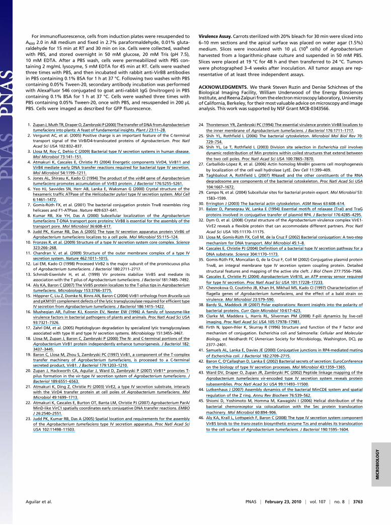
cells. Figure 6A shows the T4SS plus associated T pili around thebacterial cell on a scaffold of an endogenous helically arrangedprotein complex. T pili likely facilitate interaction with the hostplant cell. By analogy with bacterial conjugation where F piliretract upon recipient cell contact (39) (reviewed in ref. 40), Tpili may retract to tether Agrobacterium to the plant cell surface.This leads to multiple T4SSs in close association with the plantcell surface, thereby maximizing the possibility for effectivecontacts between bacteria and the plant cell surface for sub-sequent DNA and protein transfer. In support, conjugatingbacteria associate along their lengths and produce electron-dense tight junctions spanning contacts between donor andrecipient cells (41), and our initial data suggest multiple T piliassociate with the plant cell surface, supporting our model forlongitudinal binding of Agrobacterium to plant cells.It will be interesting to determine the bases for the colocali-
zation between VirB8 and MinD. VirB8 may be the foundingmember for T4SS assembly (8, 42, 43), and its association withMinD may initiate the assembly of the VirB7-VirB9-VirB10 corecomplex. Besides MinD, does the T4SS colocalize with otherhelically arranged replication proteins (reviewed in ref. 44)?Finally, components of the T4SS may colocalize with the Secsecretion system found in helical arrays in other bacteria (45).
MethodsStrains and Growth Conditions. Wild-type Agrobacterium tumefaciens strainC58 contains the nopaline pTiC58. CB1008 is a nonpolar deletion in pTiC58virB8 (46). The bald strain is flagella minus (37). For vir induction, an over-night culture was diluted to an A600 of 0.1 in minimal AB medium (pH 5.5)and grown for 5 h at 19 °C (20). Cultures were then plated on AB agarmedium containing antibiotics and 200 μM acetosyringone at 19 °C.
GFP sequences with 5′ BspHI and 3′ HindIII restriction sites were inserted intopDW029 (20) to create pJZGFP. Oligonucleotide primers introduced an AvrIIrestriction site forN-terminalGFP fusionsandaBspHI restriction site forC-terminalGFP fusions inpJZGFP.All geneswerePCR-amplifiedfrompTiC58.Constructswereconfirmed by sequencing and transformed into Agrobacterium C58.
Imaging.Amicroscope slidewas coveredwith a thin layer of 1% agarose; then0.6–1 μL of cells [in 50 mM potassium phosphate buffer (pH 5.5) followingresuspension from plates at A600 2.0] were placed on the agarose pad and cov-eredwithacoverslip.BacteriawereviewedwithanAppliedPrecisionDeltavisionSpectris DV4 deconvolution microscope. Stacks of 10–20 optical sections werecollected every 0.2 μm on the z-axis. Data (200 iterations) were deconvolvedusing Huygens software (Scientific Volume Imaging). Three-dimensional struc-turesweremodeledusing Imaris (Bitplane Scientific Software). ColocalizationofGFP-VirB8 and RFP-MinD was detected using a Leica DMI6000B invertedmicroscope equipped with a Yokogawa CSU-10 spinning-disk confocal attach-ment (Yokogawa Electric) and a Photometrics 512SC EM-CCD camera (Photo-metrics). Image acquisition was via MetaMorph software (Molecular Devices)and data were analyzed with an Imaris colocalization module.
Fig. 6. Model for T4SS localization and function during binding to plantcells. Agrobacterium with polar and helically localized T4SSs and T pili makeinitial contact with a plant cell membrane (thick curved line) (A), and fol-lowing T pili retraction multiple T4SSs attach to the plant cell surface (B). TheT4SS associates with an endogenous helix in the cell.
3762 | www.pnas.org/cgi/doi/10.1073/pnas.0914940107 Aguilar et al.
For immunofluorescence, cells from induction plates were resuspended toA600 2.0 in AB medium and fixed in 2.7% paraformaldehyde, 0.01% gluta-raldehyde for 15 min at RT and 30 min on ice. Cells were collected, washedwith PBS, and stored overnight in 50 mM glucose, 20 mM Tris (pH 7.5),10 mM EDTA. After a PBS wash, cells were permeabilized with PBS con-taining 2 mg/mL lysozyme, 5 mM EDTA for 45 min at RT. Cells were washedthree times with PBS, and then incubated with rabbit anti-VirB8 antibodiesin PBS containing 0.1% BSA for 1 h at 37 °C. Following two washes with PBScontaining 0.05% Tween-20, secondary antibody incubation was performedwith AlexaFluor 546 conjugated to goat anti-rabbit IgG (Invitrogen) in PBScontaining 0.1% BSA for 1 h at 37 °C. Cells were washed three times withPBS containing 0.05% Tween-20, once with PBS, and resuspended in 200 μLPBS. Cells were imaged as described for GFP fluorescence.
Virulence Assay. Carrots sterilized with 20% bleach for 30 min were sliced into6–10 mm sections and the apical surface was placed on water agar (1.5%)medium. Slices were inoculated with 10 μL (109 cells) of Agrobacteriumharvested from a logarithmic-phase culture and suspended in 50 mM PBS.Slices were placed at 19 °C for 48 h and then transferred to 24 °C. Tumorswere photographed 3–4 weeks after inoculation. All tumor assays are rep-resentative of at least three independent assays.
ACKNOWLEDGMENTS. We thank Steven Ruzin and Denise Schichnes of theBiological Imaging Facility, William Underwood of the Energy BiosciencesInstitute, andReenaZalpuri fromtheelectronmicroscopy laboratory,Universityof California, Berkeley, for their most valuable advice onmicroscopy and imageanalysis. This work was supported by NSF Grant MCB-0343566.
1. ZupanJ,MuthTR,DraperO,Zambryski P (2000) ThetransferofDNAfromAgrobacteriumtumefaciens into plants: A feast of fundamental insights. Plant J 23:11–28.
2. Vergunst AC, et al. (2005) Positive charge is an important feature of the C-terminaltransport signal of the VirB/D4-translocated proteins of Agrobacterium. Proc NatlAcad Sci USA 102:832–837.
3. Llosa M, Roy C, Dehio C (2009) Bacterial type IV secretion systems in human disease.Mol Microbiol 73:141–151.
4. Atmakuri K, Cascales E, Christie PJ (2004) Energetic components VirD4, VirB11 andVirB4 mediate early DNA transfer reactions required for bacterial type IV secretion.Mol Microbiol 54:1199–1211.
5. Jones AL, Shirasu K, Kado CI (1994) The product of the virB4 gene of Agrobacteriumtumefaciens promotes accumulation of VirB3 protein. J Bacteriol 176:5255–5261.
6. Yeo HJ, Savvides SN, Herr AB, Lanka E, Waksman G (2000) Crystal structure of thehexameric traffic ATPase of the Helicobacter pylori type IV secretion system. Mol Cell6:1461–1472.
7. Gomis-Rüth FX, et al. (2001) The bacterial conjugation protein TrwB resembles ringhelicases and F1-ATPase. Nature 409:637–641.
8. Kumar RB, Xie YH, Das A (2000) Subcellular localization of the Agrobacteriumtumefaciens T-DNA transport pore proteins: VirB8 is essential for the assembly of thetransport pore. Mol Microbiol 36:608–617.
9. Judd PK, Kumar RB, Das A (2005) The type IV secretion apparatus protein VirB6 ofAgrobacterium tumefaciens localizes to a cell pole. Mol Microbiol 55:115–124.
10. Fronzes R, et al. (2009) Structure of a type IV secretion system core complex. Science323:266–268.
11. Chandran V, et al. (2009) Structure of the outer membrane complex of a type IVsecretion system. Nature 462:1011–1015.
12. Lai EM, Kado CI (1998) Processed VirB2 is the major subunit of the promiscuous pilusof Agrobacterium tumefaciens. J Bacteriol 180:2711–2717.
13. Schmidt-Eisenlohr H, et al. (1999) Vir proteins stabilize VirB5 and mediate itsassociation with the T pilus of Agrobacterium tumefaciens. J Bacteriol 181:7485–7492.
14. Aly KA, Baron C (2007) The VirB5 protein localizes to the T-pilus tips in Agrobacteriumtumefaciens. Microbiology 153:3766–3775.
15. Höppner C, Liu Z, Domke N, Binns AN, Baron C (2004) VirB1 orthologs from Brucella suisandpKM101 complement defects of the lytic transglycosylase required for efficient typeIV secretion from Agrobacterium tumefaciens. J Bacteriol 186:1415–1422.
16. Mushegian AR, Fullner KJ, Koonin EV, Nester EW (1996) A family of lysozyme-likevirulence factors in bacterial pathogens of plants and animals. Proc Natl Acad Sci USA93:7321–7326.
17. Zahrl DM, et al. (2005) Peptidoglycan degradation by specialized lytic transglycosylasesassociated with type III and type IV secretion systems. Microbiology 151:3455–3467.
18. Llosa M, Zupan J, Baron C, Zambryski P (2000) The N- and C-terminal portions of theAgrobacterium VirB1 protein independently enhance tumorigenesis. J Bacteriol 182:3437–3445.
19. Baron C, Llosa M, Zhou S, Zambryski PC (1997) VirB1, a component of the T-complextransfer machinery of Agrobacterium tumefaciens, is processed to a C-terminalsecreted product, VirB1. J Bacteriol 179:1203–1210.
20. Zupan J, Hackworth CA, Aguilar J, Ward D, Zambryski P (2007) VirB1* promotes T-pilus formation in the vir-type IV secretion system of Agrobacterium tumefaciens. JBacteriol 189:6551–6563.
21. Atmakuri K, Ding Z, Christie PJ (2003) VirE2, a type IV secretion substrate, interactswith the VirD4 transfer protein at cell poles of Agrobacterium tumefaciens. MolMicrobiol 49:1699–1713.
22. Atmakuri K, Cascales E, Burton OT, Banta LM, Christie PJ (2007) Agrobacterium ParA/MinD-like VirC1 spatially coordinates early conjugative DNA transfer reactions. EMBOJ 26:2540–2551.
23. Judd PK, Kumar RB, Das A (2005) Spatial location and requirements for the assemblyof the Agrobacterium tumefaciens type IV secretion apparatus. Proc Natl Acad SciUSA 102:11498–11503.
24. Thorstenson YR, Zambryski PC (1994) The essential virulence protein VirB8 localizes to
the inner membrane of Agrobacterium tumefaciens. J Bacteriol 176:1711–1717.25. Shih YL, Rothfield L (2006) The bacterial cytoskeleton. Microbiol Mol Biol Rev 70:
729–754.26. Shih YL, Le T, Rothfield L (2003) Division site selection in Escherichia coli involves
dynamic redistribution of Min proteins within coiled structures that extend between
the two cell poles. Proc Natl Acad Sci USA 100:7865–7870.27. Carballido-López R, et al. (2006) Actin homolog MreBH governs cell morphogenesis
by localization of the cell wall hydrolase LytE. Dev Cell 11:399–409.28. Taghbalout A, Rothfield L (2007) RNaseE and the other constituents of the RNA
degradosome are components of the bacterial cytoskeleton. Proc Natl Acad Sci USA
104:1667–1672.29. Campo N, et al. (2004) Subcellular sites for bacterial protein export. Mol Microbiol 53:
1583–1599.30. Errington J (2003) The bacterial actin cytoskeleton. ASM News 63:608–614.31. Balzer D, Pansegrau W, Lanka E (1994) Essential motifs of relaxase (TraI) and TraG
proteins involved in conjugative transfer of plasmid RP4. J Bacteriol 176:4285–4295.32. Dym O, et al. (2008) Crystal structure of the Agrobacterium virulence complex VirE1-
VirE2 reveals a flexible protein that can accommodate different partners. Proc Natl
Acad Sci USA 105:11170–11175.33. Llosa M, Gomis-Rüth FX, Coll M, de la Cruz F (2002) Bacterial conjugation: A two-step
mechanism for DNA transport. Mol Microbiol 45:1–8.34. Cascales E, Christie PJ (2004) Definition of a bacterial type IV secretion pathway for a
DNA substrate. Science 304:1170–1173.35. Gomis-Rüth FX, Moncalían G, de la Cruz F, Coll M (2002) Conjugative plasmid protein
TrwB, an integral membrane type IV secretion system coupling protein. Detailed
structural features and mapping of the active site cleft. J Biol Chem 277:7556–7566.36. Cascales E, Christie PJ (2004) Agrobacterium VirB10, an ATP energy sensor required
for type IV secretion. Proc Natl Acad Sci USA 101:17228–17233.37. Chesnokova O, Coutinho JB, Khan IH, Mikhail MS, Kado CI (1997) Characterization of
flagella genes of Agrobacterium tumefaciens, and the effect of a bald strain on
virulence. Mol Microbiol 23:579–590.38. Bardy SL, Maddock JR (2007) Polar explorations: Recent insights into the polarity of
bacterial proteins. Curr Opin Microbiol 10:617–623.39. Clarke M, Maddera L, Harris RL, Silverman PM (2008) F-pili dynamics by live-cell
imaging. Proc Natl Acad Sci USA 105:17978–17981.40. Firth N, Ippen-Ihler K, Skurray R (1996) Structure and function of the F factor and
mechanism of conjugation. Escherichia coli and Salmonella: Cellular and Molecular
Biology, ed Neidhardt FC (American Society for Microbiology, Washington, DC), pp
2377–2401.41. Samuels AL, Lanka E, Davies JE (2000) Conjugative junctions in RP4-mediated mating
of Escherichia coli. J Bacteriol 182:2709–2715.42. Baron C, O’Callaghan D, Lanka E (2002) Bacterial secrets of secretion: EuroConference
on the biology of type IV secretion processes. Mol Microbiol 43:1359–1365.43. Ward DV, Draper O, Zupan JR, Zambryski PC (2002) Peptide linkage mapping of the
Agrobacterium tumefaciens vir-encoded type IV secretion system reveals protein
subassemblies. Proc Natl Acad Sci USA 99:11493–11500.44. Lutkenhaus J (2007) Assembly dynamics of the bacterial MinCDE system and spatial
regulation of the Z ring. Annu Rev Biochem 76:539–562.45. Shiomi D, Yoshimoto M, Homma M, Kawagishi I (2006) Helical distribution of the
bacterial chemoreceptor via colocalization with the Sec protein translocation
machinery. Mol Microbiol 60:894–906.46. Aly KA, Krall L, Lottspeich F, Baron C (2008) The type IV secretion system component
VirB5 binds to the trans-zeatin biosynthetic enzyme Tzs and enables its translocation
to the cell surface of Agrobacterium tumefaciens. J Bacteriol 190:1595–1604.
Aguilar et al. PNAS | February 23, 2010 | vol. 107 | no. 8 | 3763
MICRO
BIOLO
GY