Virulence and supervirulence of Agrobacterium tumefaciens in woody fruit plants
Upload
khangminh22Category
view
0download
0
ICSAFS Conference Proceedings2nd International Conference on Sustainable Agriculture and Food Security:A Comprehensive ApproachVolume 2017
Conference Paper
Agrobacterium tumefaciens-mediatedTransformation of Embryogenic Callus andSomatic Embryos of the Banana cv “AmbonLumut” (Musa acuminata)Tifa R. Kusumastuti1, Rizkita R. Esyanti1, and Fenny M. Dwivany2
1Department of Plant Science and Biotechnology, School of life science and technology,Bandung Institute of Technology, Bandung 40132, West Java, Indonesia2Department of Genetics and Molecular Biotechnology, School of life science and technology,Bandung Institute of Technology, Bandung 40132, West Java, Indonesia
AbstractBanana is one of themajor fruit crops, though its conventional breeding has limitations,such as sterility and high polyploidy levels. Biotechnological approach using genetictransformation crop for improvement offers an alternative solution. In this study aprotocol was developed for establishing genetic transformation from embryogeniccallus and somatic embryos of the banana cv Ambon Lumut. Embryogenic callus wasobtained in ID4 medium (MS-based medium) supplemented with 1 mg L−1 IAA, 4 mgL−1 2,4D, and 0.03 g L−1 active charcoal. Embryogenic callus was transferred into liquidmediu m to establish somatic embryos. Embryogenic callus and somatic embryoswere used for Agrobacterium tumefaciens-mediated transformation. A. tumefaciens
strain A GL1, containing pART-TEST7 p lasmid with gfp gene as a reporter and CaM V35Sas a promoter, was used for transformations. The embryogenic callus and somaticembryos were transformed using heat-shock method followed by centrifugation(2000 rpm) and co-cult ivation in liquid medium containing acetosyringone (100 𝜇M)for 3 days. Results of the GFP analysis showed transient expression from gfp genereporter in transformed embryogenic callus and somatic embryos. Transformationefficiency in somatic embryos (85,9%) was higher than that in embryogenic callus(32.09%). PCR analysis using CaMV primer showed bands that compatible withCaMV35S promoter at 507 bp. This is a report showing establisment of embryogeniccallus and somatic embryo culture transformation by using A. tumefaciens-mediatedtransformation protocol of the local banana cv Ambon Lumut. This study provedthe huge potential for genetic transformation of banana cv Ambon Lumut for cropimprovement, such as pest or disease resistance and abiotic factor stress tolerance.
Keywords: banana; embryogenic callus; somatic embryos.
How to cite this article: Tifa R. Kusumastuti, Rizkita R. Esyanti, and Fenny M. Dwivany, (2017), “Agrobacterium tumefaciens-mediated Transformation ofEmbryogenic Callus and Somatic Embryos of the Banana cv “Ambon Lumut” (Musa acuminata” in 2nd International Conference on Sustainable Agriculture andFood Security: A Comprehensive Approach, KnE Life Sciences, pages 599–616. DOI 10.18502/kls.v2i6.1082
Page 599
Corresponding Author:
Rizkita R. Esyanti
Received: 28 July 2017
Accepted: 14 September 2017
Published: 23 November 2017
Publishing services provided
by Knowledge E
Tifa R. Kusumastuti
et al. This article is distributed
under the terms of the
Creative Commons Attribution
License, which permits
unrestricted use and
redistribution provided that
the original author and source
are credited.
Selection and Peer-review
under the responsibility of the
ICSAFS Conference
Committee.

ICSAFS Conference Proceedings
1. Introduction
Banana is one of the most important staple foods in the world. Since 2004, Indonesiasupplied about 6% banana in international trades [1]. Data showed that banana pro-duction in Indonesia was increasing and reached 5.8 million tonnes in 2010. It wasabout 30% from national fruit production [2]. Unfortunately, Indonesia still cannotincrease its export, due to low quality product for export standard. Conventional breed-ing to improve bananas quality has limitations, such as sterility of cultivated banana,high polyploidy levels and long generation time to grow new cultivar. In this study,a new method to improve local banana quality using biotechnological approach wasdeveloped. Genetic transformation using Agrobacterium tumefaciens offers the bestsolution for crop improvement in banana. Relative success in A. tumefaciens genetictransformation had been achieved in other studies using different cultivars such asAgbagba [3, 4], Rasthali, Robusta [5], Grand Naine [6], Cavendish and Lady finger [7]by inserting genes such as Carica papaya cystatin (CpCYS), rice chitinase (RCC) [8] 𝛽-1,3-glucanase, and Human lysozym (HL) [9].
The purpose of this study was to obtain a protocol for A. tumefaciens-mediatedtransformation in embryogenic callus and somatic embryos of banana cv. AmbonLumut that had never been reported elsewhere. Embryogenic callus and somaticembryos were chosen as the subjects in this study. Embryogenic callus had beenreported for its highly merismatic cells that could increase transformation efficiency[10]. Somatic embryos have been reported for their advantages in transformation,such as potential to develop more transformed plants via secondary somatic embryo-genesis protocol (SE2). Secondary somatic embryogenesis is a process of induction ofnew somatic embryos from existing embryos, and since new embryos are continuallyformed from existing embryos, this method has the potential to produce many plants[11].Somatic embryos start from unicellular origin, which make them excellent candi-date for genetic transformation since the potential for production of chimeric plants islow [12]. Somatic embryos have the potential to regenerate a large number of plantswithout passing through cell suspension that requires long culture times from cultureinitiation to plant regeneration [11, 13].
Plasmid pART-TEST7 used in this study contained gfp reporter gene, nptII selectablemarker gene and CaMV35S promoter. CaMV35S was promoter that derived fromcauliflower mosaic virus (CaMV), a specified promoter for plants. The CaMV35S pro-moter is widely used to drive transgene expression since it has been shown to beactive in a wide range of tissue and in many crop species. The CaMV promoter ispreferred because it is a more powerful promoter than others and is not greatly
DOI 10.18502/kls.v2i6.1082 Page 600

ICSAFS Conference Proceedings
influenced by environmental conditions or tissue types. CaMV has two promoters,19S and 35S, of these two the 35S promoter is more frequently used in biotechnologybecause it is most powerful [14, 15].
The use of the green fluorescent protein (GFP) from Aequorea victoria as a vitalmarker has attracted interest and has been described in different organisms. GFP isa 27 kDa protein that fluorescent green when excited by ultraviolet or blue light. TheGFP has several advantages over other visual reporter genes such as 𝛽-glucuronidase(GUS), 𝛽-galactosidase (LacZ) and luciferase (LUC). One of important advantages isthat GFP does not require the addition of a substrate or cofactors for fluorescenceto be detected. On the contrary, other reporter genes require either the addition ofexogenous substrates or co-factors, and their applications are limited because theassays are generally destructive. This makes GFP the best choice for reporter geneif the explants or samples are limited [16, 17].
In this study, protocol was developed for transformation in liquid co-cultivationmedium of banana local cultivar Ambon Lumut, detected by using transient expressionof gfp gene reporter in both embryogenic callus and somatic embryos of banana localcultivar Ambon Lumut.
2. Materials and Method
2.1. Materials
2.1.1. pART-TEST7 plasmid
pART-TEST7 plasmid used in this study was acquired from Queensland Universityof Technology (QUT), Queensland, Australia. The plasmid has reporter gene gfp,
selectable marker gene nptII (neomycin phospotransferase) and promoter CaMV35S(Fig 1.).
2.1.2. Banana inflorescens male flowers
Banana inflorescencemale flowerswere obtained from local farmers in Bandung,WestJava Indonesia.
DOI 10.18502/kls.v2i6.1082 Page 601

ICSAFS Conference Proceedings
Figure 1: Schematic diagram of pART -T EST 7 plasmid T -DNA region.
2.1.3. A. tumefaciens strain AGL1
A. tumefaciens bacterium strain AGL1 were obtained from Genetics and Molecular Lab-oratory, School of life science and technology, Bandung Institute of Technology.
2.2. Methods
2.2.1. Plant materials and culture initiation
Male inflorescence bud was prepared by removing the bracts and the hands of themale flower. The buds disinfected with NaOCl, rinsed three times with sterile distilledwater and further reduced in size to tips that were about, 5.0 cm long × 1.0 cm wide,in a laminar flow hood. The explants, the hands of immature male flowers (IMFs),were isolated aseptically from the buds by removing the bracts under a binocularstereomicroscope. The immature flower hands were isolated from positions 15 to 3(1𝑠𝑡 being the hand closest to the meristematic dome of the male bud) each bud werecultured in callus induction medium NID4 (MS Macronutrient 1x, MS Micronutrient 1x,MS Fe-EDTA 1x, Sucrose 30 gL−1, Activated charcoal 0.03 g/ L, NAA 1 mg L−1, IAA mgL−1, 2,4-D 4 mg L−1, Agar 8 g L−1, pH 5.7-5.8). The cultures were kept in a growth roomat 27 ± 2∘C under total darkness and sub-cultured every four weeks. Non embryogeniccallus emerged from explants after three times sub-cultured using NID4 medium. Nonembryogenic callus were then transferred into ID4 medium (M S Macronutrient 1x, MSMicronutrient 1x, MS Fe -EDTA 1x, Sucrose 30 g L−1, Activated charcoal 0.03 g/ L, IAA1 mg L−1, 2,4-D 4 mg L−1, Agar 8 gL−1, pH 5.7-5.8), kept in a growth room at 27 ±2∘C under total darkness and sub-cultured every four weeks to produce embryogeniccallus.
DOI 10.18502/kls.v2i6.1082 Page 602
ICSAFS Conference Proceedings
Somatic embryos for transformation were obtained by transferring embryogeniccallus into 100 mL erlenmayer flask containing 20 ml liquid medium ME (MS Macronu-trient 1x, MS Micronutrient 1x, MS Fe-EDTA 1x, Sucrose 20 gL−1, Activated charcoal 0.05g L−1, NAA 0.1 mg L−1, BAP 1 mg L−1, pH 5.7-5.8). The cultures were agitated on an orbital shaker at 90 rpm in a growth room maintained at 27 ± 2∘C in total darkness andsub cultured every two weeks until somatic embryos dominant in globular shape wereobtained.
2.2.2. Transformation and confirmation of pART TEST7 plasmids inA. tumefaciens AGL1.
pART-TEST7 plasmid transformation was performed on competent A. tumefaciensstrain AGL1. Competent A tumefaciens AGL1 cells were made using CaCl method andA. tumefaciens plasmid was carried out using heat-shock method [18]. The existenceof pART-TEST7 plasmid in A. tumefaciens AGL1 was confirmed by crude PCR methodusing the pair of CaMV primer (primer forward 5’-CCTAACAGAACTCGCCGTAAAGA-3’andreverse 5’-CCCGTGTTCTCTCCAAATGAAATG-3’). Amplification of CaMV35S promoter wasstarted with the 3 minutes initial denaturation at 95 ∘C temperature, then performed 25cycles consisting of 30 seconds at a temperature of 95 ∘C for denaturation, 30 secondsat 60 ∘C for annealing, and twominutes at 72 ∘C for elongation. Final elongation processwas carried out for 7 minutes at a temperature of 72 ∘C. DNA fragments from PCRproducts were separated using gel electrophoresis on 1% agarose concentration inTAE buffer solution.
2.2.3. A. tumefaciens bacteria culture preparation
A. tumefaciens A GL1 culture containing plasmid pART-TEST7 was grown in YEP solidmedium (Yeast Extract 10 g/L, Pepton 10 g/ L, NaCl 5 g/ L, Agar bacto 1.5% (b/v), pH7) containing antibiotics 100 mg L−1 ampicillin, 50 mg L−1 kanamycin, and 50 mg L−1
rifampicin. The bacteria were then incubated for 3 days at a temperature of 28∘C indark conditions. One colony of the solid culture was isolated using a loop and grownin 10 ml of liquid YEP medium (Yeast Extract 10 g/ L, Pepton 10 g/L, NaCl 5 g/L, p H7) containing antibiotics (100 mg L−1 ampicillin, 50 mg L−1 kanamycin and 50 mg L−1
rifampicin). Cultures were grown at a temperature of 28∘C, with agitation of 130 rp mand incubated in the dark for 48 hours. A total of 2 mL of the culture solution wastransferred into a new liquid YEP containing antibiotics (100 mg L−1 ampicillin, 50 mgL−1 kanamycin, and 50 mg L−1 rifampicin) and made to volume of 45 ml (liquid YEP +
DOI 10.18502/kls.v2i6.1082 Page 603
ICSAFS Conference Proceedings
culture). Cultures were grown at 28∘C, for ±10 hours, with agitation of 100 rpm to reachOD600nm = 1.0.
Bacterial culture was transferred into 50 mL falcon tube, then it was centrifuged ata speed of 5000 rpm for 10 minutes. The remaining pellets were resuspended using15 mL of media BRM (MS Macronutrient 1/10x, MS Micronutrient 1/10x, MS Vitamin 10mL/ L, Sucrose 85.5 g/L, Thiamine 1125 µL/ L, Cysteine 500 mg L−1, Glucose 45 g/L, pH5.3) with the addition of 100 μM incubated acetocyringone for 2 hours at a temperatureof 25 ∘C in dark conditions with 90 rpm agitation [19].
2.2.4. Embryogenic callus culture and somatic embryosbanana Ambon Lumut preparation
Embryogenic callus was sub-cultured 3 weeks into the ID4 medium prior to transfor-mation. Embryos for transformationwere somatic embryos that had been sub-culturedone week earlier into liquid MS medium (MS Macronutrient 1x, M S Micronutrient 1x,MS Fe -EDTA 1x, Sucrose 20 g/ L, Activated charcoal 0.05 g/L, p H 5.7-5.8). Embryo-genic callus and somatic embryos were transferred into 50 mL falcon tube aseptically,liquid MS medium was added until cultures were submerged. Embryogenic callus andsomatic embryos were then given a heat-shock treatment for 5 minutes at a temper-ature of 45-48 ∘C [7, 19].
2.2.5. Transformation and co-cultivation
Embryogenic callus and somatic embryos were inoculated with A. tumefaciens culture(OD600nm = 1.0). A total of 0.02% F68 pluoronic acid was added as a surfactant.Embryogenic callus and somatic embryos were inoculated with A. tumefaciens fol-lowed by centrifugation at 2000 rpm for 10minutes, homogenized using a vortex for 20seconds, and then rested for 30 minutes [7]. Embryogenic callus and somatic embryoswere then transferred into the liquid medium for co-cultivation (MS Macronutrien 1x,MS Micronutrien 1x, MS Fe-EDTA 1x, Sucrose 20 g/ L), and incubated for 3 days at atemperature of 23 ∘C in dark conditions.
2.2.6. Decontamination and recovery
Culture was removed aseptically into the falcon tubes containing sterile distilled water,so that all parts of the culture were submerged. Falcon tube was agitated while occa-sionally homogenized by vortex for one minute, distilled water was then discarded.
DOI 10.18502/kls.v2i6.1082 Page 604

ICSAFS Conference Proceedings
The culture was added with washing liquid MS medium, which had been added with200 mg L−1 timentin. Falcon tube was agitated at a speed of 90 rpm for 15 minuteswhile occasionally homogenized by vortex. The entirewash stepwas repeated 3 times.
2.2.7. Observations of gfp reporter gene expression
Observations of gfp reporter gene expression was performed using a microscopefluorescents Nikon Eclipse E-800. Callus and embryos transformant as well as non-transformant were placed on the glass object and observed using fluorescencemicroscopy with ultraviolet light (UV) at a wavelength of 450-490 nm. Parts thatemitted green luminescence during observation indicated the presence of gfp reportergene that was expressed in transformed culture.
2.2.8. Estimation percentage of GFP expression andtransformation efficiency in transformed embryogenic callusand somatic embryos
Estimation percentage of GFP expression and transformation efficiency in transformedembryogenic callus and somatic embryos was carried using Adobe Photoshop CS2software. Transformed callus and somatic embryos pictures were viewed using AdobePhotoshop CS2, part that emitted green luminescence and non emitted green lumines-cence were counted using grid layer. Results were calculated using formula below:
Estimation of percentage ofGFP expression and trans-formation efficiency
= Total grid in transformed culture emitted green luminescenceTotal all grid in transformed culture
× 100%
2.2.9. CaMV 35S promoter molecular PCR testing
Isolation of transformed DNA was performed using Tiangen Plant Genomic DNAKit. DNA of embryogenic callus and somatic embryos were confirmed by PCR usingthe pair of primer (forward 5’-CCTAACAGAACTCGCCGTAAAGA-3’ and reverse 5’-CCCGTGTTCTCTCCAAATGAAATG-3’). Amplification of CaMV35S promoter was startedwith the 3 minutes initial denaturation at 95 ∘C temperature, then performed 25 cyclesconsisting of 30 seconds at a temperature of 95 ∘C for denaturation, 30 seconds at 60∘C for annealing, and two minutes at 72 ∘C for elongation. Final elongation process wascarried out for 7 minutes at a temperature of 72 ∘C. DNA fragment s of PCR productswere separated using gel electrophoresis on 1% agarose concentration in TAE buffersolution.
DOI 10.18502/kls.v2i6.1082 Page 605
ICSAFS Conference Proceedings
3. Result and Discussion
3.1. Embryogenic callus and somatic embryosbanana ambon lumut initiation
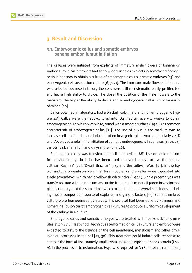
The calluses were initiated from explants of immature male flowers of banana cv.Ambon Lumut. Male flowers had been widely used as explants in somatic embryoge-nesis in bananas to obtain a culture of embryogenic callus, somatic embryos [13] andembryogenic cell suspension culture [6, 7, 21]. The immature male flowers of bananawas selected because in theory the cells were still meristematic, easily proliferatedand had a high ability to divide. The closer the position of the male flowers to themeristem, the higher the ability to divide and so embryogenic callus would be easilyobtained [20].
Callus obtained in laboratory, had a blackish color, hard and non embryogenic (Fig-ure 2.A) Callus were then sub-cultured into ID4 medium every 4 weeks to obtainembryogenic calluswhichwaswhite, roundwith a smooth surface (Fig 2.B) as commoncharacteristic of embryogenic callus [21]. The use of auxin in the medium was toincrease cell proliferation and induction of embryogenic callus. Auxin particularly 2,4-Dand IAA played a role in the initiation of somatic embryogenesis in bananas [6, 21, 23],carrots [24], alfalfa [25] and chrysanthemum [26].
Embriogenic callus was transferred into liquid medium ME. Use of liquid mediumfor somatic embryo initiation has been used in several study, such as the bananacultivar ’Rasthali’ [27], ’Dwarf Brazilian’ [13], and the cultivar ’Mas’ [21]. In the liq-uid medium, proembryos cells that form nodules on the callus were separated intosingle proembryos which had a yellowish white color (Fig 2C). Single proembryos wastransferred into a liquid medium MS. In the liquid medium not all proembryos formedglobular embryos at the same time, which might be due to several conditions, includ-ing media composition, source of explants, and genetic factors [13]. Somatic embryoculture were homogenized by stages, this protocol had been done by Fujimura andKomamine [28]on carrot embryogenic cell cultures to produce a uniform developmentof the embryo in a culture.
Embrogenic callus and somatic embryos were treated with heat-shock for 5 min-utes at 45-48∘C. Heat-shock techniques performed on callus culture and embryo wereexpected to disturb the balance of the cell membrane, metabolism and other phys-iological processes in the cell [29, 30]. This treatment could induce cells response tostress in the form of HspL namely small crystalline alpha-type heat-shock protein (Hsp-𝛼). In the process of transformation, HspL was required for VirB protein accumulation,
DOI 10.18502/kls.v2i6.1082 Page 606

ICSAFS Conference Proceedings
Figure 2: Embryogenic callus and somatic embryos of Ambon Lumut. (A) non embryogenic callus; (B)embryogenic callus on petri dish; (C) somatic embryos culture in liquid medium; (D) somatic embryosobserved under stereo microscope.
which allegedly played an important role in the transfer of T-DNA by VirB / D4 [31].Treatment of heat-shock had previously been successful in increasing the efficiency oftransformation in embryogenic cell cultures of banana cultivar ’Cavendish’ and ’LadyFinger’ [7], somatic embryos of bananas ’Cavendish’ [19] and the culture of ryegrassand rice embryogenic callus [31].
3.2. Confirmation of plasmid part-test7 inA. tumefaciens strain AGL1
Agrobacterium tumefaciens strain AGL1 were grown in YEP medium that containedantibiotics (100 mgL−1 ampicillin, 50 mgL−1 kanamycin, and 50 mgL−1 rifampicin) (Fig3).
Confirmation of the presence of plasmid pART-TEST7 was conducted through crudePCR method using a pair of CaMV primers. Visualization of electrophoresis results waspresented in Fig 4 showed colonies of A. tumefaciens strain containing plasmid positiveAGL1 pART-TEST7. The result of A. tumefaciens strain AGL1 amplification using primers
DOI 10.18502/kls.v2i6.1082 Page 607

ICSAFS Conference Proceedings
Figure 3: A. tumefaciens strain AGL1 colonies harbouring plasmid pART-TEST7 cultured in solid YEP mediumcontained antibiotics.
507 bp
1000 bp
750 bp
500 bp
250 bp
Figure 4: PCR confirmation of pART-TEST7 plasmid in A. tumefaciens AGL1. L: DNA Ladder 1 kb(FERMENTAS); (co+) positive control plasmid pART-TEST7; (1-5) A. tumefaciens strain AGL1 coloniescontained plasmid pART-TEST7.
CaMV against CaMV35S promoter in the plasmid pART-TEST7 produced bands of 507bp which is corresponded to the positive control (plasmid pART-TEST7).
DOI 10.18502/kls.v2i6.1082 Page 608

ICSAFS Conference Proceedings
Figure 5: Observation of GFP on embryogenic callus of banana cultivar Ambon Lumut;(A-B) non-transformed embryogenic callus (C-D) embryogenic callus transformed by A. tumefaciens strainAGL1/pART-TEST7.
3.3. Observations of gfp reporter gene expression in embryogeniccallus and somatic embryos banana Ambon Lumut
Observations conducted using the fluorescence microscope under UV light with awavelength of 450-490 nm indicated the success of the transformation in embryo-genic callus culture (Fig 5.D) and somatic embryos (Fig 6.D). The positive result of thetransformation was shown by green luminescens from GFP protein, which was theresult from transient expression of the gfp reporter gene expression in transformedembryogenic callus and somatic embryos, gfp gene is a gene that encodes a proteingreen fluorescence, when the gene is expressed it will produce a glowing green in partsof transformed plants or culture. GFP expression had been successfully used to test thesuccess of transformation, including the embryogenic callus of sweet orange (Citrussinensis (L.) Osbeck) [32] and somatic embryos of grapes (Vitis vinifera L. cv.ThompsonSeedless) [33].
Successful transformant part in expressing GFP protein p roved the potential ofCaMV35S promoter to be used as a promoter in transformation banana cultivars AmbonLumut. Transient expression from reporter gene could be used to estimate the pro-moter strength. Strong activity from promoter can lead to strong expression of reporter
DOI 10.18502/kls.v2i6.1082 Page 609

ICSAFS Conference Proceedings
Figure 6: Observation of GFP on somatic embryos of banana cultivar Ambon Lumut; (A-B) non-transformed somatic embryo (C-D) somatic embryo transformed by A. tumefaciens strain AGL1/pART-TEST7.
gene. Promoter activity was one of the key factors that included in gene expressionregulation. Other research of transformation in grape somatic embryos using the pro-moter double cauliflower mosaic virus 35S (CaMV35S) and double cassava vein mosaicvirus (CsVMV) produced transgenic plant expressing a transgene which last up to 5years [33]. GFP expression was stable until the plant survived and regenerated andit expressed in various tissues and organs of the plant (buds, leaves, roots, petal,stamen), even in flowers, fruits and seeds [16].
3.4. Estimation of percentage areas of GFP expression andtransformation efficiency in embryogenic callus andsomatic embryos of Ambon Lumut banana
Estimation of percentage area that expressed GFP in transformed somatic embryoswas higher than embryogenic callus. Estimate number area that emitted green lumi-nescence in somatic embryos tissuewas about 85,9% per embryos, while transformedembryogenic callus was about 32,09% per callus (Table 1). These result showed that
DOI 10.18502/kls.v2i6.1082 Page 610

ICSAFS Conference Proceedings
T 1: Percentage of area expressed GFP in transformed somatic embryos and embryogenic callus.
NameTotal transformedculture culture
tested
Average totalarea per culture
Average areaemitted greenluminescens per
culture
Percentage ofGFP expression
andtransformationefficiency
Somatic Embryos 120 1136 976 85,9%
EmbryogenicCallus 120 1296 416 32,09%
Figure 7: Result of DNA isolation from embryogenic callus and somatic embryos of banana cultivarAmbon Lumut. (L) DNA Ladder 1 kb FERMENT AS, (E+) transformed somatic embryos; (K+) transformedembryogenic callus; (E-) non transformed somatic embryos; (K-) non transformed embryogenic callus.
transformation efficiency and GFP expression in somatic embryos was higher thanembryogenic callus.
3.5. Molecular PCR testing on embryogenic callus andsomatic embryos banana Ambon Lumut
Samples of embryogenic callus and somatic embryos (transformed and non- trans-formed) were isolated using Tiangen Plant Genomic DNA Kit, specifically for the isola-tion of DNA geno mic of plants. This kit uses silica membrane technology to eliminatecomplicate stages in the process of DNA isolation. Especially, if the sample containspolyphenols, resins, or other metabolites [34, 35]. Isolated DNA (Fig 7) showed theDNA bands that were faint, but clean of impurities. This result was used for furthertesting to confirm existence of pART-TEST7 plasmid using PCR method.
DOI 10.18502/kls.v2i6.1082 Page 611

ICSAFS Conference Proceedings
1000 bp
750 bp
500 bp
250 bp
507 bp
Figure 8: PCR plasmid pART-TEST7 confirmation result in embryogenic callus and somatic embryos ofAmbon Lumut bananas. (L) DNA Ladder 1 kb FERMENTAS, (co+) positive control primer CaMV/ pART -TEST7; (K+) transformed embryogenic callus; (E+) transformed somatic embryos; (K-) non transformedembryogenic callus; (E-) non transformed somatic embryos; (co-) negative control H2O.
Confirmation presence of the plasmid pART-TEST7 in embryogenic callus and somaticembryos was done using the PCR method with CaMV primer.Amplification result usingCaMV primer against CaMV35S promotercontained in plasmid pART-TEST7 showedbands at 507 bp corresponded to the positive control (plasmid pART-TEST7). Thesebands were not found in non transformed embryogenic callus and somatic embryos(K-and E-) as well as negative control using water (H2O) (Fig 8).
The use of surfactants and the addition of acetocyringone compounds can improvethe efficiency of transformation in plants. Surfactants such as Pluronic acid F68 used inthis study were added at transformation. The use of surfactant serves to facilitate theattachment of bacteria A. tumefaciens cultures by reducing the surface tension of thesolution. The addition of surfactant can also eliminate other compounds that inhibitthe attachment of bacteria to the plant cell [10, 36].
Addition of exogenous compounds such acetosyringone, especially for mono-cotyledonous plants were proven to improve the efficiency of transformation. Ace-tosyringone can activate VirA protein located on the cell surface of membrane, andtrigger activation of other vir genes [36]. Acetosyringone compounds were commonlyp roduced in dicotyledonous plants, but only a small portion of monocotyledonousplants produce these compounds. Monocotyledonous plants, such as bananas requireadministration of exogenous acetosyringone [37]. Acetosyringone should be added inoptimum concentration, since acetosyringone compounds could effectively activatethe vir genes at the minimum concentration, but resulted in bacteriostatic when usedat high concentrations [37].
DOI 10.18502/kls.v2i6.1082 Page 612

ICSAFS Conference Proceedings
The transformation protocol had been used by Khanna et al., [8] and Apriyani [19],but it was slightly modified in this study. A liquid medium to improve the efficiency oftransformation was used in co-cultivation step. This protocol was also used in othertransformation using different types of media on the leaves of tobacco plants. Theefficiency of transformation in tobacco leaf using a liquid medium during co–cultivationwas higherwhen comparedwith tobacco leaves thatwere co-cultivated on solidmedia[39]. This protocol can be developed further as the protocol of transformation for thelocal banana cultivars Ambon Lumut to produce stable banana plant transformation,because liquidmediawas used in the process of transformation and co-cultivation. Thisstep could enhance culture contact with the bacteria in the transformation processand minimizing stress to the culture due to the media changes. The use of liquidmedia could also reduce browning and culture cell death caused by overgrowth ofA. tumefaciens that co-cultivated in solid or semi-solid medium [40].
4. Conclusion
In this present study, protocol for transformation was obtained for embryogenic callusand somatic embryos of banana local cultivar Ambon Lumut that never been reportedelsewhere. Protocol for banana cultivar Ambon Lumut plant regeneration via indi-rect somatic embryogenesis (data not shown) was also obtained. This report showedpotential of CaMV35S promoter and gfp gene reporter for transformation in local cul-tivar banana Ambon Lumut. This report also proved potential of local cultivar bananaAmbon Lumut for crop improvement using A. tumefaciens-mediated genetic transfor-mation and a step towards stable transformation in transgenic banana local cultivars.
Acknowledgements
The authors would like to thank Queensland University Technology (QUT) for providingpART-TEST7 plasmid construct.
References
[1] Smith, S., 2010. Fairtrade Bananas: A Global Assesment of Impact. Institute ofDevelopment Studies. University of Sussex. United Kingdom
[2] Direktorat Budidaya dan Pascapanen Buah., 2012. Pedoman Penanganan Pascapa-nen Pisang. Direktorat Budidaya dan Pascapanen Buah. Kementerian Pertanian.Jakarta.
DOI 10.18502/kls.v2i6.1082 Page 613

ICSAFS Conference Proceedings
[3] Tripathi, J.N., Muwonge, A., and Tripathi, L., 2012. Efficient regeneration andtransformation of plantain cv.“Gonja manjaya” (Musa spp. AAB) using embryogeniccell suspensions. In Vitro Cell.Dev.Biol. 48 (2), 216-224.
[4] Tripathi, L., Tripathi, J.N., and Hughes, J.d’A., 2012. Agrobacterium transformationof plantain (Musa spp.) cultivar Agbagba. African Journal of Biotechnology. 4 (12),1378-1383
[5] Ghosh, A., Ganapathi, T. R., Nath, P., and Bapat, V. A., 2009. Establishment ofembryogenic cell suspension cultures and Agrobacterium-mediated transformationin an important Cavendish banana cv. Robusta (AAA). Plant Cell T iss Organ Culture97, 131–139.
[6] Sutherland, R., 2006. Genetic modification of Cavendish bananas (Musa spp) inSouth Africa. Thesis. Faculty of Nature and Agricultural Science. University ofPretoria. Pretoria. South Africa.
[7] Khanna, H., Becker, D., Kleidon, J., and Dale, J., 2004. Centrifugation AssistedAgrobacterium tumefaciens mediated Transformation (CAAT) of embryogenic cellsuspensions of banana (Musa spp. Cavendish AAA and Lady Finger AAB). MolecularBreeding. 14, 239–252.
[8] Sreeramanan, S., Maziah, M., and Xavier, R., 2009. A protocol for Agrobacterium-mediated transformation of banana with a rice chitinase gene. Emirates JournalFood Agriculture. 21 (2), 18-33.
[9] Pei, X. W., Chen, S. K., Wen, R. M., Ye, S., Huang, J. Q., and Zhang, Y. Q., 2005. Creationof transgenic bananas expressing human
[10] Deo, P. C., Tyagi, A. P., T aylor, M., Harding, R., and Becker, D., 2010. Factors affectingsomatic embryogenesis and transformation in modern plant breeding. The SouthPacific Journal of Natural and Applied Sciences. 28, 27-40.
[11] Sidha, M., Suprasanna, P., Bapat, V.A., Kulkarni, U.G., and Shinde, B. N., 2006.Developing somatic embryogenic culture system and plant regeration in banana.BARC Newsletter. Founder’s day issue special Issue. 285, 153-161.
[12] Escalant, J.V., Teisson, C., and Côte, F., 1994. Amplified somatic embryogenesis frommale flower of triploid banana and plantain cultivars (Musa sp.). In Vitro Cell DevBiol 30:181–186
[13] Khalil, S. M., Cheah, K. T., Perez, E. A., and Gaskill, D. A., 2002. Regeration of banana(Musa spp. AAB cv.Dwarf Brazilian) via secondary somatic embryogenesis. PlantCell Report. 20, 1128-1134.
[14] Steinbrecher, R.A., 2002. The CaMV 35S Promoter Government and Cor-porate Scientific Incompetence: Failure to assess the safety of GM crops.http://www.econexus.info Accsessed 22 July 2015.
[15] Zannati, A., 2009. “Sang penyelenggara” itu disebut promotor. BioTrends. 4 (1), 31-34.
DOI 10.18502/kls.v2i6.1082 Page 614

ICSAFS Conference Proceedings
[16] Pérez-Clemente, R. M., Pérez-Sanjuán, A., García-Férriz, L., Beltrán, J. P., and Cañas,L. A., 2004. Transgenic peach plants (Prunus persica L.) produced by genetictransformation of embryo sections using the green fluorescent protein (GFP) asan in vivo marker. Molecular Breeding. 14, 419–427.
[17] El-Shemy, H.A., Khalafalla, M.M., and Ishimoto, M., 2008. The role of GreenFluorescent Protein (GFP) in transgenic plants to reduce gene silencing phenomena.Curr.Issues Mol. Biol. 11 (Suppl. 1), 21–28.
[18] Hofgen, R., and Millmitzer, L., 1988. Storage of competent cells for Agrobacteriumtransformation. Nucleic Acids Research. 16 (20), 9877.
[19] Apriyani, R. K., 2012. Efisiensi transformasi pada kultur embrio pisang Cavendish(Musa acuminata colla (AAA Group)) melalui Agrobacterium tumefaciens strainAGL1 dan GV3101 yang membawa plasmid pBI121. Thesis. Department of Biotech-nology.
[20] Chang, S., and Shu, H., 2013. A method to supress the browning in banana (Musa,AAA) embryogenic callus induced. Research Journal of Biotechnology. 8(4), 63-69.
[21] Wong, W.C., Jalil, M., Ong-Abdullah, M., Othman, R.Y., and Khalid, N., 2006.Enhancement of banana plant regeneration by incorporating
[22] Ikeuchi, M., Sugimoto, K., and Iwase, A., 2013. Plant callus: Mechanisms of inductionand repression. The Plant Cell. 25, 3159–3173.
[23] Khalil, S. M., and Elbanna, A. A. M., 2003. Highly efficient somatic embryogenesisand plant regeneration via suspension cultures of banana (Musa spp.). Arab JournalBiotechnology. 7(1), 99-110.
[24] Zimmerman, J.L., 1993. Somatic embryogenesis: a model for early development inhigher plants. The Plant Cell. 5, 1411-1423.
[25] Pasternak, T. P., Prinsen, E., Ayaydin, F., Miskolczi, P., Potters, G., Asard, H.,Onckelen, H. A. V., Dudits, D. and Fehér, A., 2002. The role of auxin, pH and stress inthe activationof embryogenic cell division in leaf protoplast derived cells of Alfalfa.Plant Physiology 129, 807-1809.
[26] Shinoyama, H., Nomura, Y., Tsuchiya, T. and Kozuma, T., 2004. A simple andeffecient method for somatic embryogenesis and plant regeneration from leavesof chrysanthemum [Dendranthema x grandiflorum (Ramat.) Kitamura]. PlantBiotechnology. 21, 25-33.
[27] Ganapathi, T.R., Higgs, N.S., Balint-Kurti, P.J., Arntzen, C.J., May, G.D., and VanEck, J.M., 2001. Agrobacterium-mediated transformation of embryogenic cellsuspensions of T he banana cultivar Rasthali (AAB). Plant Cell Reports. 20, 157–162.
[28] Fujimura, T., and Komamine, A., 1979. Synchronization of somatic embryogenesisin a carrot cell suspension culture. Plant Physiology. 64, 162-164.
[29] Wahid, A., Gelani, S., Ashraf, M., and Foolad, M., 2007. Heat tolerance in plants: Anoverview. Environ. Exp. Bot. 6, 199-223.
DOI 10.18502/kls.v2i6.1082 Page 615

ICSAFS Conference Proceedings
[30] Usman, M. G., Rafii, M. Y., Ismail, M. R., Malek, M. A., Latif, M. A., and Oladosu,Y., 2014. Heat shock proteins: functions and response against heat stress in plants.International Journal Of Scientific and T echnology Research. 3(11), 204-218.
[31] Patel, M., Dewey, R. E., and Qu, R., 2013. Enhancing Agrobacterium tumefaciens-mediated transformation efficiency of perennial ryegrass and rice using heat andhigh maltose treatments during bacterial infection. Plant Cell Tissue Organ Culture.114, 19–29
[32] Li, D. D., Shi, W., and Deng, X. X., 2003. Factors influencing Agrobacterium-mediatedembryogenic callus transformation of Valencia sweet orange (Citrus sinensis)containing the pTA29-barnase gene. Tree Physiology. 23, 209–1215
[33] Li, Z. T., Dhekney, S., Dutt, M., Van Aman, M., T attersall, J., Kelley K. T., and Gray D.J., 2006. Optimizing Agrobacterium-mediated transformation of grapevine. In VitroCell. Dev. Biol.—Plant. 42: 220–227.
[34] Berensmeier,S., 2006. Magnetic particles for the separationand purification ofnucleic acids. Appl. Microbiol.Biotechnol. 73, 495-504
[35] Esser, K.H., Marx,W.H., and Lisowsky, T., 2006.MaxXbond: first regeneration systemfor DNA binding silica matrices.
[36] Opabode, J. T., 2006. Agrobacterium -mediated transformation of plants: emergingfactors that influence efficiency. Biotechnology and Molecular Biology Review. 1:12-20.
[37] Suzuki, S., Supaibulwatana, K., Mii, M,. and Nakano, M., 2001. Production oftransgenic plants of the Liliaceous ornamental plant Agapanthus praecox ssp.Orientalis (Leighton) Leighton via Agrobacterium-mediated transformation ofembryogenic calli. Plant Science. 161, 89-97.
[38] Amoah, B. K., Wu, H., Sparks, C. A., and Jones, H. D., 2001. Factors influencingAgrobacterium-mediated transient expression of uidA in wheat inflorescencetissue. Journal of Experimental Botany, 52, 1135-1142.
[39] Uranbey, S., Sevimay, C.S., Kaya, M.D., Ipek, A., Sancak, C., Basalma, D., Er, C.,and Ozcan, S., 2005. Influence of different co-cultivation temperature, periods andmedia on Agrobacterium tumefaciens mediated gene transfer. Biologia Plantarum.49 (1), 53-57.
[40] Wenck, A. R., Quinn, M., Whetten, R.W., Pullman, G., and Sederoff, R., 1999. Highefficiency Agrobacterium-mediated transformation of Norway spruce (Picea abies)and loblolly pine (Pinus taeda). Plant Mol Biol. 39, 407–416.
DOI 10.18502/kls.v2i6.1082 Page 616
Copyright © 2022 FDOKUMEN